


Assist prof. of Medical Physiology. Excess GH Decreased GH Before Union of epiphysis Gigantism After...
91
Assist prof. of Medical Physiology
-
Upload
silvia-stephens -
Category
Documents
-
view
221 -
download
1
Transcript of Assist prof. of Medical Physiology. Excess GH Decreased GH Before Union of epiphysis Gigantism After...

Assist prof. of Medical Physiology


Excess GH
Decreased GH
Before Union of epiphysis
Gigantism
After Union of epiphysis
Acromegaly
In children
Dwarfism
In adults
Loss of some body
proteins

Causes:
1.Hyperplasia or
2.Tumor of the somatotrop cells (adenoma)
Manifestations: • The manifestations depends upon, if it occurs
before or after the union of the epiphyses.
A) Gigantism: ↑ed GH before the union of the epiphyses.
B) Acromegaly: ↑ed GH after the union of the epiphyses or in adults

Def:
• Condition caused by excess GH before union of epiphysis
Manifestations:
1) Marked elongation of bones but in a
relative proportion.
2) Overgrowth of soft tissues e.g. the
muscles & viscera.

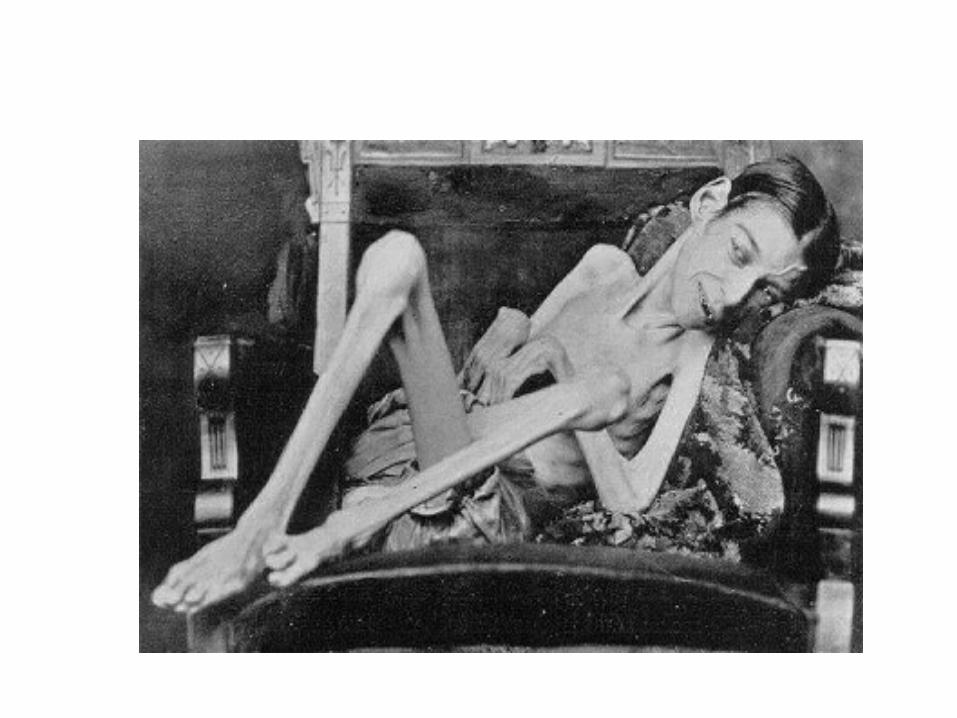
Gigantism in a 36-year-old woman. Her companions are normal.


Manifestations:
3) Hyperglycemia and increased MR.
4) Hypogonadism:
• Gonads and accessory sex organs remain
infantile due to ↓ ed gonadotropins secretion
GH secreting cells encroach upon the other
cells.

SGG
TT
CC
MM GGMM TT
SSSS
S
SS
S S
S
S
SS
S
SS
S
S
Pituitary tumor causes compression and atrophy of gonadotrophes

Manifestations:
5) Headache due to pressure on Sella Turcica
and visual disturbances (Bitemporal
hemianopia) due to pressure of the
growing tumor on the optic chiasma.
6) These patients are often mentally
subnormal

Def:
• Condition caused by excess GH after union of epiphysis in adults
Manifestations:
1.The bones become thicker and deformed; the
ms and viscera also enlarge
2.Generalized coarsening of the features due to:
a. Thick skin and SC tissues.
b. Enlargement of the head, hands and feet.
c. Prognathism: the lower jaw enlarged &
protrudes forward, and separated teeth.

Spade hand


Manifestations:
• 3) Kyphosis due to thickening of the
vertebrae.
• 4) Hyperglycemia and glucosuria.
• 5) Raised BMR.

Manifestations:
• 6) Hypogonadism.
7) Some patients have visual
fields defects due to pressure of
the tumor on optic chiasma.
Increased intracranial tension
lead to headache & vomiting.
• 8) Hirsutism (increased body
hair).
• 9) Gynaecomastia and may be
galactorrhea

Treatment:
• 1) Surgical: removal of tumor
• 2) Medical:
• Somatostatin or
• Somatostatin synthetic compounds e.g.
Octreotide and lanreotide

Def:
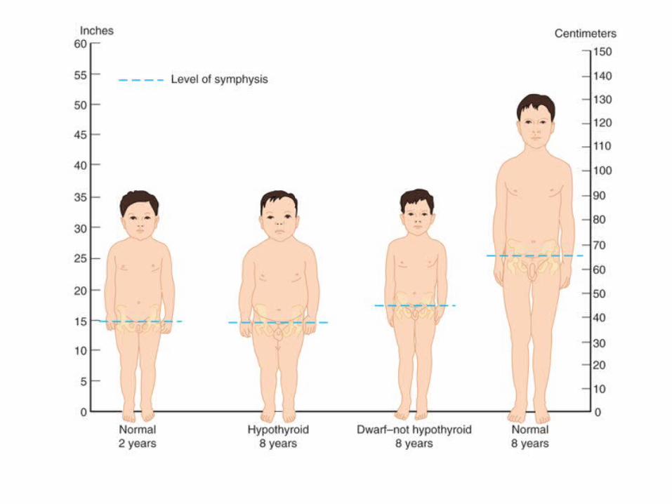
• Condition caused by deficiency of GH in children
Manifestations:
a) Short stature due to rapid closure of the
epiphyses leading to proportionate reduction of
all body sizes.
b) The growth rate of soft tissues is reduced, but
mild obesity is common.
• The patient looks much younger than his age.


Manifestations:
c) Normal Mental growth usually.
d) Low metabolic rate & episodes of
hypoglycemia due to lack of insulin
antagonism by GH.e) Normal Sexual maturation in cases of
isolated GH deficiency. • Few dwarfs show hypogonadism and the
gonads, external sexual organs and characters remain infantile Infantilism .
NB: Deficiency of GH in adults has no physical signs

• 1) Cretinism: Thyroid hypofunction in infants.
• 2) Precocious puberty:
– in cases of hypergonadism in children, which leads
to early closure of the epiphyses.
• 3) Gonadal dysgenesis e.g. Turner’s syndrome where
an XO chromosomal pattern instead of XX or XY.
• 4) Bone and metabolic diseases.
• 5) Constitutional delayed growth in many cases no
evident cause for stunted growth is found.

Turner syndromeTurner syndrome

Excess prolactin
Decreased prolactin Due to
Hypothalamic
dysfunctions
Pituitary tumors
Due to
Destruction of ant. Pituitary
G.

Manifestations:
• High prolactin inhibit GnRH and pituitary
gonadotropins resulting in;
• a) In women,
1. Loss of menses (amenorrhea),
2. Anovulation and infertility.
3. Galactorrhea: lactation unassociated with
pregnancy
4. Decreased libido.


• Diagnosis :
by a high prolactin blood level.
• Treatment:
by:
A) Surgical removal of the tumor or
B) Dopaminergic drugs to reduce
prolactin secretion

In women produces inability to lactate.
No other clinical consequences are known.

Results from destruction of the ant pituitary, leads to: – Severe deficiency of its hormones and
– Atrophy of the thyroid and adrenal glands and of the
gonads.
Manifestations:1. In children: lead to infantilism.
– failure of growth and of sexual maturity.
2. In adults, the lack of trophic hormones results in
– hypofunction of the target endocrine glands with
relative hyperinsulinism.

Manifestations:
•a) Thyroid gland (Myxoedema)
•b) Adrenal cortex (hypocorticism or
‘Addison’s disease) → leading to ms
weakness, loss of weight,
hypoglycaemia and dehydration.
• c) The gonads (hypogonadism).

Manifestations:
d) Loss of weight and severe wasting of
muscles (cachexia) due to:
– loss of appetite (anorexia) and absence of
anabolic effect of GH and androgens.
e) Premature senility:
– dry skin and wrinkled with early graying of hair
– so the patient looks older than his age.

Manifestations:
f) Hypoglycaemia due to:
– relative increase insulin level
– lack the effects of antagonistic hormones.
g) Skin colour becomes lighter due to:
– anaemia and
– deficiency of ACTH and beta-MSH.

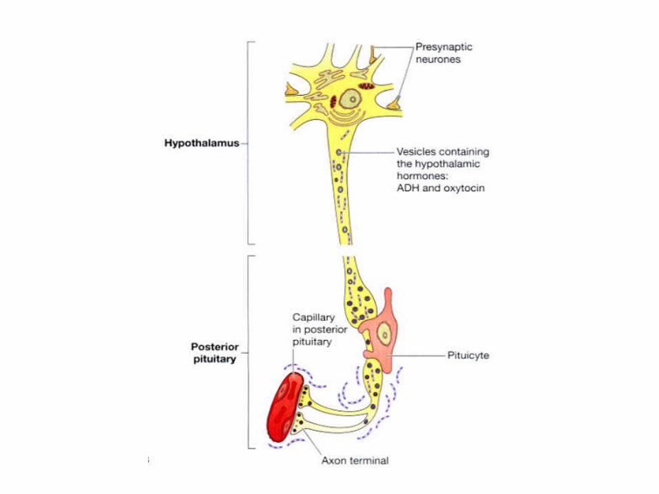

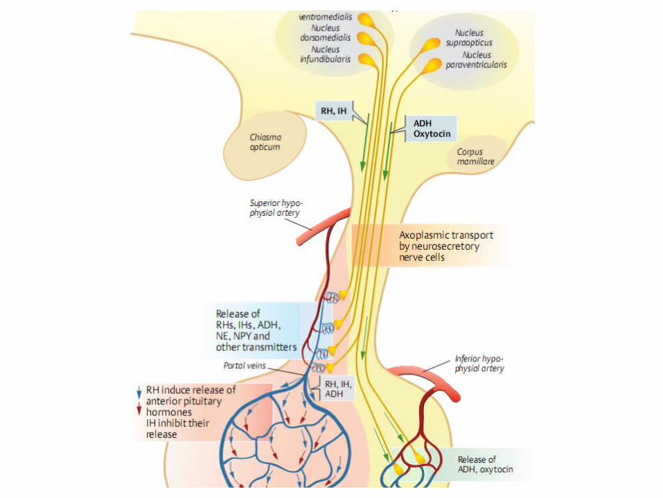
• Thousands of nerve fibres connect the
hypothalamus, (supra-optic & para-
ventricular nuclei), with the post pituitary.
• The crude extract of the post lobe is called
pituitrin that contains 2 hormones:
1). Antidiuretic hormone (ADH), also called
vasopressin or pitressin.
2). Oxytocin (or pitocin) hormone.


Synthesis and Storage:• Oxytocin & ADH, are synthesized in the
hypothalamus and stored in granules with a binding protein (neurophysin)
1. Neurophysin-1 for oxytocin2. Neurophysin-2 for antidiuretic hormone.
• Granules pass down the axons through
hypophyseal tracts to the nerve endings in post
pituitary.
• The terminal swellings of nerve endings are called
‘Herring bodies’.

• When a nerve impulse is transmitted from the cell body in hypothalamus down the axon:
1. Depolarizes the terminal Herring body.
2. Ca+ influx into the Herring body induce hormone
release by exostosis, and enters the adjacent
capillary.


Source:
• Mainly from supraoptic
hypothalamic nucleus
Chemistry:
• Peptide hormone 9 a.a.


• Is the major action of ADH
• Reabsorption of free water from the
tubular fluid.
• Target site of action:
1. Distal convoluted tubules
2. Collecting ducts


• In large doses
• ADH causes vascular smooth muscles
contraction leading to: 1. Elevation of the blood pressure2. Coronary vasoconstriction3. Intense splanchnic vasoconstriction.
• This effect used clinically in controlling,
serious GIT bleeding.

ADH Used in treatment of GIT bleeding

• Some of ADH pass to the ant pituitary via its
portal veins where it act as CRH → Increase
ACTH.

Regulation of vasopressin secretion.


• Hypothalamic supraoptic nuclei contain very sensitive osmoreceptors.
• Rise in plasma osmolarity (1%) → loss of
intracellular water from osmoreceptor neurons →
ADH secretion.
• ADH produces reabsorption of free water
(without electrolytes) from tubular fluid→ dilutes
the plasma→ return of osmolarity to its normal
value.

• Normal plasma osmolarity is 290 m osm/liter.
Causes of increase plasma osmolarity:
1) Dehydration (Water deprivation), either due
to: Decreased water intake or Excessive loss.
2) Administration of solutes: which do not rapidly
penetrate the cell membrane, such as Na+.
– Substances that enter cells rapidly, as urea do not
stimulate ADH secretion, because they do not produce
osmotic dysequilibrium between ECF and ICF.

• ADH release is stimulated by a 5% to 10% decrease in circulating blood volume, or cardiac output.
– Haemorrhage decreases blood volume,
– Standing & positive pressure breathing reduce
cardiac output.• Hypovolemia is perceived by pressure sensors :
a.Arterial baroreceptors in carotid sinus and aortic
arch
b.Stretch receptors in the walls of left atrium &
pulmonary veins.
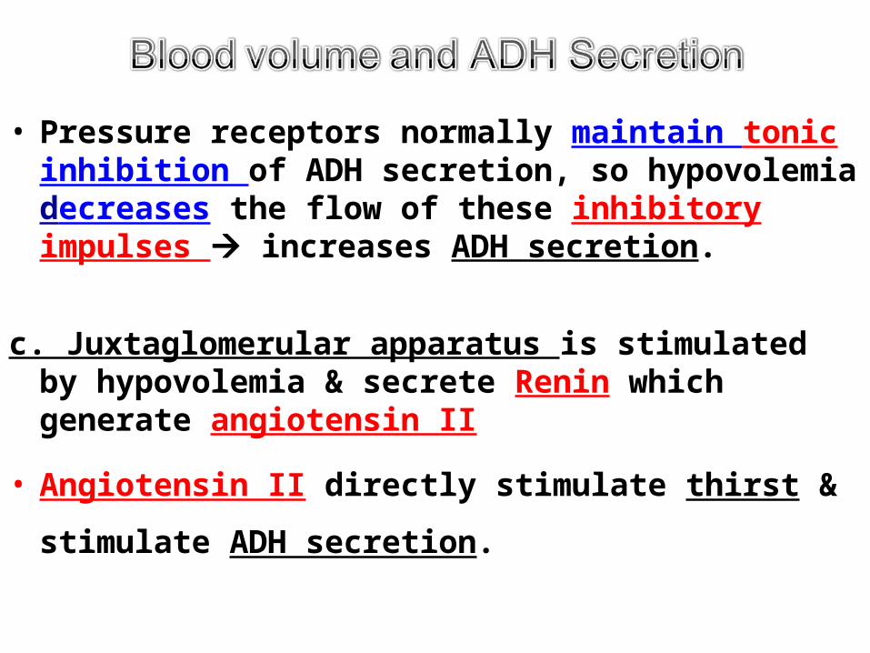
• Pressure receptors normally maintain tonic inhibition of ADH secretion, so hypovolemia decreases the flow of these inhibitory impulses increases ADH secretion.
c. Juxtaglomerular apparatus is stimulated by hypovolemia & secrete Renin which generate angiotensin II
• Angiotensin II directly stimulate thirst &
stimulate ADH secretion.

↓ Plasma Volume (Hypovolemia)
↓ Plasma Volume (Hypovolemia) Stretch receptors
and baroreceptors
Stretch receptors and
baroreceptors Juxtaglomerular
apparatus Juxtaglomerular
apparatus
ADH secretion
ADH secretion Reni
n Reni
n
Angiotensin IIAngiotensin II↑ Plasma Volume ↑ Plasma Volume


1. Inhibitorsa) Diuretics
b) Water loading
c) Prostaglandin E
d) Cortisol, and
e) K+ deficiency
f) Ca+ excess
g) -adrenergic agonists
h) Cold weather,
i) Ethanol
2. Stimulatorsa) Sulfonylureas
b) Nicotine
c) opiates
d) Hot weather
e) -adrenergic agents
f) oestrogens, & progesterone


Excess Secretion
Syndrome of inappropriate ADH secretion
(SIADH)
Decreased Secretion
Diabetes Inspidus

Causes: 1. Deficiency of ADH secretion (neurogenic
DI)2. Inability of the kidney to respond to ADH
(nephrogenic type).

Symptoms : 1) Polyuria: (urine volume reach
25 liters/day) • Due to failure of the
facultative water reabsorption by the distal tubules.
• Very low urine specific gravity (1001-1003).
2) Polydepsia: • drinking large amount of
water due to intense thirst 2ry to polyuria.
3) Anorexia and general weakness due to loss of important substances in urine as vitamins.

• Treated by:• By Administration of ADH except
Nephrogenic’ type as there is a congenital defect in the renal tubules.

Cause:
• Increased ADH than predicted by plasma volume or
tonicity
Manifestations:a) Hyponatremia (serum sodium 100 - 115 mEq/L).
• Results in headache, drowsiness, nausea and often coma.
b) High urine osmolality.
c) Slight increase in ECF volume.
d) Excess renal sodium excretion despite the low serum sodium due to;• Elevated levels of atrial natriuretic factors caused
by the expanded plasma volume.


Source:
• Mainly from
paraventricular
hypothalamic nucleus
Chemistry:
• Peptide hormone 9 a.a.

Mechanism of action:
• Bind to specific cell membrane receptors
• Exerts its effects by increasing intracellular Ca+
+ content.


• Suckling-reflex:
– Suckling stimulates touch receptors at the
nipple and areola which send afferent
impulses to the hypothalamus to release
both oxytocin and prolactin hormones.


• Lower the threshold for membrane depolarization of the myometrial ms.
• This effect is: 1. Potentiated by oestrogen2. Inhibited by progesterone.
• Oxytocin has minimal effect in initiating labour• But plays an important role in the sustained post-
partum uterine contractions that help to :
1. Maintain haemostasis after evacuation of the
placenta,
2. Involution of the uterus after delivery.


3. In females:
• Transport of the sperms into the uterus during intercourse.
– By the end of intercourse, oxytocin is secreted & induce rhythmic uterine contractions which suck, the sperms into the uterus & giving the orgasm sensation.
4. In males,
– help the discharge of sperms from the semineferous tubules and epididymis to vas deferens. During ejaculation
5. Stimulation of apocrine sweat secretion
– at the axillae, nipples, groins and perineum that produce sex attraction in animals.