ASSESSMENT ON VARIOUS ASPECTS OF …eprints.utar.edu.my/898/1/BM-2013-1003790-1.pdfASSESSMENT ON...
82
ASSESSMENT ON VARIOUS ASPECTS OF ANTIBIOTIC RESISTANCE IN ENTEROBACTERIACEAE By LEE KAH LENG A project report submitted to the Department of Biomedical Science Faculty of Science Universiti Tunku Abdul Rahman in partial fulfillment of the requirement for the degree of Bachelor of Science (Hons) Biomedical Science May 2013
Transcript of ASSESSMENT ON VARIOUS ASPECTS OF …eprints.utar.edu.my/898/1/BM-2013-1003790-1.pdfASSESSMENT ON...

ASSESSMENT ON VARIOUS ASPECTS OF ANTIBIOTIC
RESISTANCE IN ENTEROBACTERIACEAE
By
LEE KAH LENG
A project report submitted to the Department of Biomedical Science
Faculty of Science
Universiti Tunku Abdul Rahman
in partial fulfillment of the requirement for the degree of
Bachelor of Science (Hons) Biomedical Science
May 2013

ii
ABSTRACT
ASSESSMENT OF VARIOUS ASPECTS OF ANTIBIOTIC
RESISTANCE IN ENTEROBACTERIACEAE
Lee Kah Leng
Enterobacteriaceae that colonised in human intestine are highly exposed to the
antibiotic pressures. These selective pressures may induce antibiotic resistance,
as well as the transfer of resistance from the resistant bacteria to the enteric
bacteria. However, the bacteria may also lose its resistance upon the
withdrawal of antibiotic pressure. The aim of this study is to assess the losing,
induction and the transfer of antibiotic resistance on Escherichia coli and
Klebsiella pneumoniae. Firstly, the ability of losing antibiotic resistance was
studied on clinical isolate E. coli 594370 which resistant to ciprofloxacin,
moxifloxacin, gentamicin and trimethoprim. The bacteria was serially
passaged in Mueller Hinton (MH) broth in the absence of antibiotics. Secondly,
the induction of trimethoprim resistance was assessed on ATCC 25922 E. coli
and ATCC 13883 K. pneumoniae that were susceptible to trimethoprim. These
bacteria were serially passaged in MH broth in the presence of sub-lethal
trimethoprim concentration (12 µg/mL). Thirdly, the transfer of antibiotic
resistance was assessed in clinical isolate E. coli 594370 and clinical isolate
K. pneumoniae 594394. Clinical isolate E. coli 594370 acted as donor bacteria

iii
and clinical isolate K. pneumoniae 594394 acted as recipient bacteria that
showed susceptibility to ciprofloxacin, moxifloxacin and gentamicin. These
bacteria were grown and serially passaged together. There was no noticeable
losing in resistance in clinical isolate E. coli 594370 after 85 passages. Next,
the zone-of-inhibition diameter was noticeably reduced for more than 55% in
ATCC 13883 K. pneumoniae. Gradual reduction in zone-of-inhibition
diameter was observed in ATCC 25922 E. coli. However, there was no
evidence in the transfer of antibiotic resistance from clinical isolate E. coli
594370 to clinical isolate K. pneumoniae 594394 over 30 passages. Clinical
isolate K. pneumoniae 594394 remained susceptible to ciprofloxacin,
moxifloxacin and trimethoprim. As a conclusion, the losing of antibiotic
resistance did not occurred over 85 passages, and induction of trimethoprim
resistance was occurred in both of the ATCC strains. The transfer of antibiotic
resistance from donor bacteria to recipient bacteria did not happen over 30
passages.

iv
ACKNOWLEDGEMENTS
I would like to express my great appreciation and gratitude to these parties
who have assisted me along the Final Year Project. First and foremost, Mr.
Yuen Hawk Leong, my project supervisor who is always dedicated in teaching
and offered much of his advises and encouragement. A great thank to Ms. Ng
Shel Ling and Mr. Saravanan, our responsible and helpful laboratory officers.
I also appreciate the peer support from the other team members, Alicia Chew
Wen Jing, Chan Lei Mun, Cheng Jo Hao, Fong Yee Khee, Kang Chin Hooi,
Lai Kah Nyin, Law Yew Chye, Lee Ying Xian and Yew Su Ern during this
project. Thanks for giving me the precious and memorable moments that we
spent during the bench work. I would like to express my gratitude and
appreciation to my precious family members, especially my parents who make
me strong and supported along the way.

v
DECLARATION
I hereby declare that the project is based on my original work except for
quotations and citations which have been duly acknowledged. I also declare
that it has not been previously or concurrently submitted for any other degree
at Universiti Tunku Abdul Rahman or other institutions.
______________________
LEE KAH LENG

vi
APPROVAL SHEET
This project report entitled “ASSESSMENT ON VARIOUS ASPECTS OF
ANTIBIOTIC RESISTANCE IN ENTEROBACTERIACEAE” was
prepared by LEE KAH LENG and submitted as partial fulfillment
requirements for the degree of Bachelor of Science (Hons) Biomedical
Science at Universiti Tunku Abdul Rahman.
Approved by:
________________________
(Mr. YUEN HAWK LEONG) Date: ................................
Supervisor
Department of Biomedical Science
Faculty of Science
Universiti Tunku Abdul Rahman

vii
FACULTY OF SCIENCE
UNIVERSITI TUNKU ABDUL RAHMAN
Date: _________________
PERMISSION SHEET
It is hereby certified that LEE KAH LENG (ID No: 10ADB03790) has
completed this final year project entitled “ASSESSMENT ON VARIOUS
ASPECTS OF ANTIBIOTIC RESISTANCE IN
ENTEROBACTERIACEAE” supervised by Mr. Yuen Hawk Leong
(Supervisor) from the Department of Biomedical Science , Faculty of Science.
I hereby give permission to my supervisor to write and prepare manuscripts of
these research finding for publishing in any form, if I did not prepare it within
six (6) months from this date, provided that my name is included as one of the
author for this article. The arrangement of the name depends on my supervisor.
Yours truly,
_________________
(LEE KAH LENG)

viii
TABLE OF CONTENTS
Page
ABSTRACT ii
ACKNOWLEDGEMENTS iv
DECLARAION v
APPROVAL SHEET vi
PERMISSION SHEET vii
TABLE OF CONTENTS viii
LIST OF TABLES x
LIST OF FIGURES xi
LIST OF BBREVIATION xii
CHAPTER
1 INTRODUCTION
1
2 LITERATURE REVIEW 5
2.1 Enterobacteriaceae 3
2.2 Antibiotics Involved in this Project 4
2.3 Impact of Antibiotics on Normal Intestinal Composition 6
2.3.1 Losing Antibiotic Resistance 8
2.3.2 Induction of Antibiotic Resistance 10
2.3.3 Transfer of Antibiotic Resistance 12
2.4 Clinical Significance in Assessment of Antibiotic
Resistance
15
2.5 Methods Used in the Study of Antibiotic Resistance
17
3 MATERIALS AND METHODS 18
3.1 Materials 18
3.1.1 Bacteria Strains 18
3.1.2 Chemicals and Media Used 19
3.1.3 Equipment and Labwares Used 19
3.2 Methodology 20
3.2.1 Preparation of Bacteria Stock and Master Plate 20
3.2.2 Assessment of the Losing of Antibiotic Resistance 20
3.2.3 Induction of Trimethoprim Resistance in
Escherichia coli ATCC 25922 and
Klebsiella pneumoniae ATCC 13883
20
3.2.4 Assessment of the Transfer of Antibiotic 21

ix
Resistance
3.3 Antibiotic Susceptibility Testing
21
4 RESULTS 23
4.1 Losing of Antibiotic Resistance in Clinical Isolate
Escherichia coli 594370
23
4.2 Induction of Trimethoprim Resistance in Escherichia coli
ATCC 25922 and Klebsiella pneumoniae ATCC 13883
33
4.3 Transfer of Antibiotic Resistance from Clinical Isolate
Escherichia coli 594370 and Clinical Isolate
Klebsiella pneumoniae 594394
37
5 DISCUSSION 40
5.1 Losing of Antibiotic Resistance 40
5.1.1 Number of Passages 40
5.1.2 Biological Cost of Resistance 41
5.1.3 Chromosomal Drug Resistance 42
5.2 Induction of Antibiotic Resistance Escherichia coli ATCC
25922 and Klebsiella pneumoniae ATCC 13883
43
5.2.1 Traditional Selective Window 43
5.2.2 Clinical Significance in Antibiotic Dosing
Strategies
45
5.3 Transfer of Antibiotics Resistance from Clinical Isolate
Escherichia coli 594370 to Clinical Isolate
Klebsiella pneumoniae 594394
46
5.3.1 Role of Antibiotic in Transfer of Antibiotic
Resistance
46
5.3.2 Clinical Significance of the Transfer of Antibiotic
Resistance in Intestine
48
5.4 Future Study
50
6 Conclusion 51
REFERENCES
52
APPENDICES

x
LIST OF TABLES
Table Page
3.1 Reference ranges of ciprofloxacin, gentamicin,
moxifloxacin and trimethoprim
22
4.1 Diameter of zone-of-inhibition (ZI) of ciprofloxacin in
Escherichia coli 594370
25
4.2 Diameter of zone-of-inhibition (ZI) of gentamicin in
Escherichia coli 594370
27
4.3 Diameter of zone-of-inhibition (ZI) of moxifloxacin in
Escherichia coli 594370
29
4.4 Diameter of zone-of-inhibition (ZI) of trimethoprim in
Escherichia coli 594370
31
4.5 Diameter of zone-of-inhibition (ZI) of trimethorpim to
Escherichia coli ATCC 25922
34
4.6 Diameter of zone-of-inhibition (ZI) of trimethorpim to
Klebsiella pneumoniae ATCC 13883
35
4.7 Diameter of zone-of-inhibition (ZI) of ciprofloxacin to
clinical isolate Klebsiella pneumoniae 594394
38
4.8 Diameter of zone-of-inhibition (ZI) of gentamicin to
clinical isolate Klebsiella pneumoniae 594394
38
4.9 Diameter of zone-of-inhibition (ZI) of moxifloxacin to
clinical isolate Klebsiella pneumoniae 594394
39

xi
LIST OF FIGURES
Figure Page
2.1 Mechanism action of antibiotics and resistance
mechanisms on aminoglycosides, fluoroquinolone and
folate pathway inhibitor
5
2.2 Representation of the impact of antibiotic administration
on the bacteria community of the colon
6
2.3 Cluster of antibiotic resistance genes (b) with other genetic
elements
9
2.4 Predicted probability of resistance due to increase in
prescription of ciprofloxacin and trimethoprim
10
2.5 Mechanisms of horizontal gene transfer (HGT)
13
2.6 The reservoir hypothesis
14
4.1 Trends of antibiotic susceptibility of clinical isolate
Escherichia coli 594370 to ciprofloxacin
26
4.2 Diameter of zone-of-inihibition (ZI) on ciprofloxacin at
Passage 0
26
4.3 Diameter of zone-of-inihibition (ZI) on ciprofloxacin at
Passage 85
26
4.4 Trends of antibiotic susceptibility of clinical isolate
Escherichia coli 594370 to gentamicin
28
4.5 Diameter of zone-of-inihibition (ZI) on gentamicin at
Passage 0
28
4.6 Diameter of zone-of-inihibition (ZI) on gentamicin at
Passage 85
28
4.7 Trends of antibiotic susceptibility of clinical isolate
Escherichia coli 594370 to moxifloxacin
30
4.8 Diameter of zone-of-inihibition (ZI) on moxifloxacin at
Passage 0
30

xii
4.9 Diameter of zone-of-inihibition (ZI) on moxifloxacin at
Passage 85
30
4.10 Trends of antibiotic susceptibility of clinical isolate
Escherichia coli 594370 to trimethoprim
32
4.11 Absence of zone-of-inhibition
32
4.12 Trends of antibiotic susceptibility of Escherichia coli
ATCC 25922 to trimethoprim
34
4.13 Trends of antibiotic susceptibility of
Klebsiella pneumoniae to ATCC 13883 trimethoprim
35
4.14 Zone-of-inhibition (ZI) on Klebsiella pneumoniae ATCC
13883 at Passage 0 (Disk content: 5 µg)
36
4.15 Zone-of-inhibition (ZI) on Klebsiella pneumoniae ATCC
13883 at Passage 5 (Disk content: 5 µg)
36
4.16 Zone-of-inhibition (ZI) on Klebsiella pneumoniae ATCC
13883 at Passage 15 (Disk content: 5 µg)
36
4.17 Trends of antibiotic susceptibility of clinical isolate
Klebsiella pneumoniae 594394 to ciprofloxacin,
gentamicin and moxifloxacin
39
5.1 Schematic diagram of bacteria growth rate in response to
the antibiotic concentration
44

xiii
LIST OF ABBREVIATONS
CIP Ciprofloxacin
CLSI Clinical and Laboratory Standards Institute
CN Gentamicin
DHFR Dihydrofolate reductase
EMB Eosin methylene blue
ESBL Extended spectrum beta-lactamase
ENCAST European Committee on Antimicrobial Susceptibility
Testing
GI Gastrointestinal
HGT Horizontal gene transfer
IAIs Intra-abdominal infection
ICU Intensive care unit
KB Kirby-Bauer
µg/ml Microgramme per milliliter
MDR Multidrug-resistant
MHA Mueller Hinton agar
MHB Mueller Hinton broth
MIC Minimum inhibitory concentration
MICres minimal inhibitory concentration of resistant strain
MICsusc Minimal inhibitory concentration of the susceptible strain
MXF Moxifloxacin
OTC Over-the-counter
QRDR Quinolone resistance determining region

xiv
RND Resistance-nodulation-cell division
RNA Ribonucleic acid
ROAR Reservoirs of antibiotic resistance
rpm Revolutions per minute
UTI Urinary tract infection
W Trimethoprim
ZI Zoneof-inhibition

CHAPTER 1
INTRODUCTION
Emergence of antibiotic resistance in ESKAPE pathogens restricts the
antibiotic choices available for treatment (Lisboa and Nagel 2011).
Remarkably, Enterobacteriaceae that colonize human intestinal tract are highly
exposed to the antibiotic pressures exerted by the oral antibiotic therapy and
food with antibiotic residues (Jernberg et al., 2010). Moreover, reservoir
hypothesis mentioned by Salyers et al. (2004) highlighted the role of the
enteric bacteria as the reservoir of antibiotic resistant genes. Thus, the study on
dissemination and transfer of antibiotic resistance may provide a better idea on
the acquisition of antibiotic resistance among the enteric bacteria.
Aspects of antibiotic resistance, such as losing of antibiotic resistance,
induction of antibiotic resistance and transfer of antibiotic resistance were
assessed in this study. There are several community studies that evaluate the
interventions of antibiotic prescribing policies. Some of these studies showed
that the reduction in antibiotic prescription managed to bring down the
resistance rate in bacteria (Seppala et al., 1997; Enne et al., 1999). In contrast,
other studies yielded controversial findings in this aspect (Enne et al., 2001;
Sundqvist et al., 2010). On the other hand, there are increasing evidence that

2
supported the fact that antibiotic resistance can be induced using sub-lethal
antibiotic concentration (Drlisa and Zhao 2007; Andersson and Hughes 2011).
According to Schjørring and Krogfelt (2010), in vivo and in vitro model
systems are used in the study of antibiotic resistance. Assessment of antibiotic
resistance is frequently conducted in in vivo models and community settings,
but the in vitro experimental studies are relatively less established. In this
project, various aspects of antibiotic resistance were assessed using in vitro
model system which in parallel with the objectives as stated:
To assess the duration required for the losing of antibiotic resistance in
resistant bacteria.
To study the parameters (duration and concentration of antibiotics)
required for the induction of antibiotic resistance.
To analyze the duration required for the transfer of antibiotic resistance
from the resistant, donor bacteria to susceptible, recipient bacteria.

3
CHAPTER 2
LITERATURE REVIEW
2.1 Enterobacteriaceae
Escherichia coli and Klebsiella pneumoniae are gram-negative bacteria that
are classified under Enterobacteriaceae. These bacteria are commensal flora in
human gastrointestinal tract. Enterobacteriaceae are also opportunistic
pathogens that account for more than 50% of the bacteremia cases (Livermore
2012).
E. coli and K. pneumoniae are categorized in the notorious ESKAPE
pathogens (Enterococcus faecium, Staphylococcus aureus,
Klebsiella pneumoniae, Acinetobacter baumannii, Pseudomonas aeruginosa
and Enterobacter sp.) that are associated to the production of extended
spectrum beta-lactamase (ESBL) in intensive care units (ICUs) (Fraimow and
Tsigrelis, 2011; Lisboa and Nagel 2011). Common diseases that are caused by
E. coli include urinary tract infection (UTI) (Vellinga et al., 2012), intra-
abdominal infections (IAIs) (Paterson et al., 2005) and bacteremia (Wilson et
al., 2011; Livermore 2012). As mentioned by Drago et al. (2010), similar
diseases are caused by K. pneumoniae, in which nosocomial infections are
more prominent than community-acquired infection. Longer length of hospital

4
stay is another risk factor for the nosocomial infections caused by
K. pneumoniae (Lim and Webb 2005).
2.2 Antibiotics that are involved in this study
Escherichia coli that used in this study is classified as multidrug resistant
(MDR) bacteria. Hereby, multidrug resistant (MDR) is defined as resistance of
bacteria to at least 3 classes of antibiotics (Cantón and Ruiz-Garbajosa 2011;
Barie 2012). The E. coli strain used in this study is resistant to 3 classes of
antibiotics: gentamicin (aminoglycoside), ciprofloxacin and moxifloxacin
(fluoroquinolone) and trimethoprim (folate pathway inhibitor).
In a nutshell, the mechanism action of the antibiotics and resistance
mechanisms are summarized in Figure 2.1. Mechanisms confer to the
resistance to aminoglycoside include: modification of aminoglycoside by
enzymes like acetyltransferase, phosphotransferase and nucleotidyltransferase
(Alekshun and Levy 2007); elimination of aminoglycosides using efflux
pumps like AcrD, AcrA and TolC pump (Kumar and Schweizer 2005); and
decreased membrane permeability to aminoglycoside due to absence of porin
(Kumar and Schweizer 2005). In addition to efflux of fluoroquinolone by
resistance-nodulation-cell division (RND) pump (Kumar and Schweizer 2005),
mutation on the targets of fluoroquinolone, DNA gyrase and topoisomerase
also confer to the resistance to fluoroquinolone (van Hoek et al., 2011). Next,
the resistance to trimethoprim that serves as anti-folate is acquired by

5
alteration in chromosomal dihydrofolate reductase (DHFR) (van Hoek et al.,
2011), which contributes to the excess production of DHFR (Huovinen 2001).
Figure 2.1: Mechanism action of antibiotics and resistance mechanisms on
aminoglycosides, fluoroquinolone and folate pathway inhibitor (Scott 2009,
p.553).

6
2.3 Impact of antibiotics on normal intestinal microbiota composition
The ecological balance between normal flora and pathogenic microorganisms
will be affected by the administration of antibiotics. The impact of antibiotic
administration is explained by Jernberg et al. (2010, Figure 2.2). In healthy
individual, human gastrointestinal tract is colonized by normal flora and the
number of resistant, pathogenic bacteria is below the detection threshold.
Upon the initiation of antibiotic treatment, the number of antibiotic resistant
strains (represented by purple rods) increases drastically as they are able to
survive in the presence of antibiotic pressure. Most of the susceptible
enterobacteria (represented by green rods) are eliminated during the antibiotics.
As a result of the antibiotic treatment, the susceptible intestinal bacteria may
acquire resistance from horizontal gene transfer or mutational events. This
ecological imbalance among the enteric bacteria confers to both short-term
and long-term impacts on the human host.
Figure 2.2: Representation of the impact of antibiotic administration on the
bacteria community of the colon (Jernberg et al., 2010, p. 3217).

7
As explained by Sullivan et al. (2001), the short-term impacts of antibiotic
administration were manifested as diarrhea and fungal infections.
Administration of antibiotics may results in the emergence of antibiotic
resistance in other intestinal bacteria, and gives rise to long-term impacts on
the patient. As studied by Sjölund et al. (2003), antibacterial agents
(clarithromycin, metronidazole and omeprazole) used for Helicaobacter pylori
infection resulted in the emergence of clarithromycin-resistant enterococci. In
addition, Nyberg et al. (2007) also reported an increase in clindamycin-
resistant E. coli after the administration of the antibiotic. Similar study from
Lindgren et al. (2009) also mentioned that erythromycin- and clindamycin-
resistant Enterococcus sp. persisted for 9 months as a consequence of 7-day
clindamycin course. Thus, the antibiotics course may results in the undesirable
impacts on intestinal flora, eventually leading to the increase level of antibiotic
resistance in other intestinal bacteria.

8
2.3.1 Losing Antibiotic Resistance
Several studies that were conducted in community setting showed promising
findings, in which the reduction of antibiotic prescription managed to decrease
the resistance rate to the antibiotic. The resistance rate of
Streptococcus pyogenes abated from 19% to 8.6% between 1993 and 1996, as
a result of 63% reduction in the administration of macrolide (Seppala et al.,
1997). Study from Enne et al. (1999) revealed that reduction in overall
antibiotics usage over 3-year time resulted in 25% reduction of penicillin
resistance in Streptococcus pneumoniae.
Notwithstanding the successful findings, restriction in antibiotic prescription
does not always cause a decrease in the resistance rate. 97% reduction in the
prescription of sulphonamide-containing antimicrobials in UK conferred to
zero change on the resistance rate in E. coli between 1991 and 1999 (Enne et
al., 2001). A recent 24-month study conducted in Swedish concluded that 85%
cutback in trimethoprim from 2004 to 2006 did not reverse the resistance in
E. coli (Sundqvist et al., 2010).
Fitness cost and compensatory mutations are factors that influence the
persistence of antibiotic resistance, even though the antibiotic consumption is
discontinued (Levin, 2001; Andersson and Hughes 2011). In terms of
community studies, co-selection is an important factor that leads to the
persistence of resistant bacteria (Courvalin and Trieu-Cuot 2001; Martinez

9
2009; Andersson and Hughes 2010; Andersson and Hughes 2011). Resistance
gene to a particular antibiotic is often linked to genes that confer resistance to
other antibiotics and toxic metals. Thus, the use of any of the compounds will
co-select other resistance mechanisms as well (Courvalin and Trieu-Cuot 2001;
Martiniz 2009; Andersson and Hughes 2010). As shown in Figure 2.3, the
antibiotic resistant genes (r) is genetically linked to heavy metal determinants
(b) and ecologically rewarding elements (e). In pathway B, plasmid-encoded
antitioxin (A) is produced to prevent the bacterial killing by toxin (T). Thus,
co-selection may favor the expression of antibiotic resistance even in the
absence of antibiotics (Martinez 2009).
Figure 2.3: Cluster of antibiotic resistance genes (b) with other genetic
elements (Martinez 2009, p. 2526).
B

10
2.3.2 Induction of antibiotic resistance
The association of antibiotic usage and emergence of antibiotic resistance was
postulated by Gallini et al. (2010). This study relates the ciprofloxacin-
resistant E. coli in nosocomial setting to the administration of fluoroquinolone
in hospital and community. Indeed, their study was in line with the finding
from Vellinga et al. (2010). As shown in Figure 2.4, greater prescription of
ciprofloxacin and trimethoprim in general practice increases the risk of
infection caused by resistant strains of E. coli (Vellinga et al., 2010).
Figure 2.4: Predicted probability of resistance due to increase in prescription
of ciprofloxacin and trimethoprim (Vellinga et al., 2010, p. 1518).
Another study carried out by Vellinga et al. (2012) suggested that the
consumption of ciprofloxacin increases the prevalence of infections by
extended spectrum beta-lactamase (ESBL) producing E. coli. In addition,

11
bacteremia caused by fluoroquinolone-resistant E. coli is steered by the usage
of moxifloxacin and levofloxacin in community settings (Cuevas et al., 2011).
Overall, the administration of antibiotics in nosocomial and community
settings may induce the antibiotic resistance in pathogenic microorganism.

12
2.3.3 Transfer of antibiotic resistance
Antibiotic resistant genes can be transferred from the resistant, donor bacteria
to susceptible, recipient bacteria via horizontal gene transfer (HGT) (Kelly,
Vespermann and Bolton 2009a). The antibiotic resistance genes may involve
in efflux pump, alteration of target molecules, degradation of antibiotic
molecules, etc. (Andersson and Hughes 2010). As shown in Figure 2.5, the
mechanisms for horizontal gene transfer include: conjugation, transformation
and transduction. Conjugative gene transfer involves the cell-to-cell
communication between the donor bacteria and recipient bacteria, whereby the
genetic materials are transferred via direct contact and sex pili (Thomas and
Nielsen 2005; Scott 2009). Next, transformation refers to the uptake of
extracellular genetic materials when the bacteria are in normal physiological
state (Thomas and Nielsen 2005). Then, transduction is mediated by
bacteriophage which transfers the virulence gene between bacteria (Kelly,
Vespermann and Bolton 2009b). Mutations that occurred either in the
chromosome or plasmid may result in the acquisition of antibiotic resistance
(Andersson and Hughes 2010). Quinolone resistance that caused by point
mutation on the genes encode for DNA gyrase and topoisomerase is one of the
examples for chromosomal mutation (Guan et al., 2013).

13
Figure 2.5: Mechanisms of horizontal gene transfer (HGT) (Andersson and
Hughes 2010, p. 201).
Plasmids and conjugative transposons are examples of mobile genetic
elements that can be transferred from one bacterial to another (Hooper, 2001;
Bennett 2008; Scott 2009; van Hoek et al., 2011). Plasmid-carried genes play
important role in bacteria survival, and some may confer to the resistance to
cephalosporins, fluroquinolones and aminoglycosides (Bennett 2008). These
plasmids are subjected to conjugative transfer among broad range of hosts, in
which transmission among different species of bacteria is possible (Bennett
2008; van Hoek et al., 2011). Transposon, as defined by Scott (2008), refers to
the large genetic elements that carry multiple resistant genes, and encodes for
pheromones which promote the conjugative gene transfer among the bacteria.
Examples of transposons include Tn5 that encodes aminoglycoside resistance,
Tn10 that encodes for tetracycline resistance and Tn3 that encodes for
resistance to β-lactam antibiotics are prevalently found in Enterobacteriaceae
(Bennett 2008).

14
In real, human gastrointestinal (GI) tract act as the habitat for the diverse
community of enteric bacteria, which serve as reservoirs of antibiotic
resistance (ROAR) (Schjørring and Krogfelt 2010; Vespermann and Bolton
2009). Reservoir hypothesis as shown in Figure 2.6 suggested that the bacteria
in colon are capable of acquiring antibiotic resistance genes, and possibly
transfer the resistant gene to other intestinal bacteria and transient bacteria in
the gut (Salyers et al. 2004; Salyers et al. 2007; Kelly, Vespermann and Bolton
2009). The transfer of antibiotic resistance occurs mostly via conjugation
between the intestinal bacteria, whereas gene transfer by bacteriophage
transduction and transformation are most likely to occur between members of
the same species.
Figure 2.6: The reservoir hypothesis. (Salyers et al., 2007, p. 18)

15
2.4 Clinical Significance in Assessment of Antibiotics Resistance
Discovery of antimicrobial drugs provide solutions for life-threatening
bacterial infections that are critical in intensive care unit (ICU) (Lim and
Webb 2005; Fraimow and Tsigrelis 2011), surgical procedures (Carlet et al.,
2012) and also the management for the cancer patients and patients that
received organ transplantation (Livermore 2012). However, the emergence of
drug resistant bacteria have cause the antibiotic pipeline to run dry as some of
the antibiotics are no long active against these resistant bacteria (Gould 2009;
Carlet et al., 2012). Moreover, over-the-counter (OTC) prescription of
antibiotics in the third world countries even worsens the current scenario
(Morgan, Okeke and Laxminarayan 2011). The antibiotic treatment without
prescription will not give the optimal potency and thus create an optimal
antibiotic pressure that may induce the antibiotic resistance in commensal
flora (Bisht et al., 2009; Andersson and Hughes 2012).
Most of the susceptible flora are killed in the traditional antibiotic dosing
approach, leading to the selection of antibiotic resistant bacteria (Zhao and
Drlica 2008). Thus, the dosing regimen should be optimized and the selected
drug used for treatment should be prescribed accordingly (Geoff, Bauer and
Mangino 2012). Indeed, this preliminary project may provide an overview on
the induction of antibiotic resistance, particularly in E. coli and K. pneumoniae
using antibiotic with sub-lethal concentration.

16
In real, one of the major factors that lead to the emergence of antibiotic
resistance is the patient attitude on the antibiotic prescription (Bisht et al.,
2009). As mentioned by the survey conducted by McNulty et al. (2007), 11.3%
of the respondent failed to complete the entire course of antibiotic therapy, and
some even recycle the “left-over” antibiotics. The discontinued of antibiotics
consumption and delay in between the antibiotics intake will reduce the
antibiotic concentration in the body to less than the minimum inhibitory
concentration (MIC), which is insufficient to eradicate the pathogenic bacteria
in the body (Jackson et al., 2006). This amount of antibiotics will not only
favor the selection of antibiotic resistant bacteria, but also promote the
antibiotic resistance transfer from the pathogenic microorganisms to the
susceptible intestinal flora (Andersson and Hughes 2012).

17
2.5 Methods used in the Study of Antibiotic Resistance
Approaches like dissemination and reversal of antibiotic resistance are mostly
conducted in community settings. As elaborated in Section 2.3.1, this field of
research was studied by Seppala et al. (1997), Enne et al. (1999), Enne et al.
(2001), Sundqvist et al. (2010).
Schjørring and Krogfelt (2010) mentioned that both in vivo and in vitro
methods are used to study the various aspects of antibiotic resistance on the
gut flora. Some of the examples of the in vivo methods include the antibiotic-
treated mouse model (Freter 1989) and human microbiota-associated rodent
(HMA) model (Hirayama 1999). Indeed, in vitro studies which are conducted
in liquid media and on agar plates stand an advantage. This is because
parameters such as temperature, type and amount of media, incubation time
and selective pressure. that are involved in the assessment of antibiotic
resistance can be closely monitored in laboratory settings (Schjørring and
Krogfelt 2010). Moreover, antibiotic susceptibility profiles and phenotypic
characteristics of the bacteria can be evaluated using bacteria cultures
(Jernberg et al., 2010). Nevertheless, current in vitro models are still less
established in relative to in vivo models. Thus, this reflects the importance of
this study in assessing the aspects of antibiotic resistance using in vitro
methods.

18
CHAPTER 3
MATERIALS AND METHODS
3.1 Materials
3.1.1 Bacteria strains
In this study, the clinical strains of Escherichia coli (Lab Number: 594370)
and Klebsiella pneumoniae (Lab Number: 594394) were collected from
Gleneagles Medical Center, Penang. E. coli 594370 was resistant to
ciprofloxacin, gentamicin, moxifloxacin and trimethoprim. Whereas,
K. pneumoniae 594394 showed susceptibility to these antibiotics.
On the other hand, the ATCC bacteria strains were obtained from Department
of Biomedical Science, University of Tunku Abdul Rahman. The ATCC
strains were Escherichia coli (ATCC 25922) and Klebsiella pneumoniae
(ATCC 13883). These bacteria were susceptible to trimethoprim.

19
3.1.2 Chemicals and media used
The preparation of agar and media that were used in this study are listed in
Appendix A.
3.1.3 Equipment and lab wares used
The equipment and lab wares that were used in this study are listed in
Appendix B.

20
3.2 Methodology
3.2.1 Preparation of bacteria stock and master plate
Each of the bacteria strain was plated on both MacConkey agar and EMB agar.
After overnight incubation at 37 ˚C, these master cultures were kept at 4˚C.
80% glycerol stock for each bacteria sample were prepared and preserved at
-80 ˚C. This storage condition allowed long-term preservation of bacteria.
3.2.2 Assessment of the losing of antibiotic resistance
Resistant strain of E. coli 594370 was inoculated into 20 ml of MH broth and
cultured at 37 ˚C with agitation at the speed of 200 rpm. After 4.5 hour
incubation, 200 µL of the bacteria culture was serially passaged into 20 ml of
fresh MH broth and cultured continually with the previous growing condition.
The antibiotics susceptibility profile of this bacteria was monitored constantly
every 5 passages.
3.2.3 Induction of trimethoprim resistance in Escherichia coli ATCC
25922 and Klebsiella pneumoniae ATCC 13883
E. coli ATCC 25922 was serially passaged in the presence of trimethoprim at
the concentration of 12 µg/mL. The preparation of this antibiotic solution was
shown in Appendix C. The viability of the bacteria was closely monitored by
culturing the overnight bacteria on both EMB agar and MacConkey agar. In
addition, the antibiotic susceptibility of the bacteria was assessed in every 5
passages. Same procedure was carried on K. pneumoniae ATCC 13883 as well.

21
3.2.4 Assessment of the transfer of antibiotic resistance
Resistant strain of E. coli 594370 and sensitive strain of K. pneumoniae
594394 were inoculated into 20 ml MH broth. The bacteria mixture was co-
incubated at 37 ˚C with agitation at the speed of 200 rpm. After 5 hours of
incubation, 200 µL of the bacteria solution was serially passaged into 20 ml
MH broth and cultured continuously with the previous growing condition. The
antibiotics susceptibility profile of K. pneumoniae 594394 was monitored
constantly every 5 passages.
3.3 Antibiotic susceptibility testing
The susceptibility of each batch of bacteria was carried out by Kirby-Bauer
(KB) method. Antibiotics that were used in the testing were ciprofloxacin (CIP,
5 µg), gentamicin (CN, 10 µg), moxifloxacin (MXF, 5 µg) and trimethoprim
(W, 5 µg). The preparation of trimethorprim disks are mentioned in Appendix
C. 3 to 5 colonies with identical morphology were isolated and suspended into
0.87% saline. The turbidity of the inoculum was adjusted to 0.5 MacFarland.
Within 15 minutes, a sterile cotton swab was then used to streak the bacteria
suspension over the MH agar. Next, antibiotics disks were distributed evenly
on the MH agar. Within 15 minutes after placing the antibiotic disks, the
plates were incubated at 37 ˚C for 12 to 14 hours.
The reference breakpoints for the zone diameter of inhibition for ciprofloxacin,
gentamicin and trimethoprim are available in Clinical and Laboratory

22
Standards Institute (CLSI, 2007). On the other hand, the reference range for
inhibition diameter of moxifloxacin is available in the European Committee on
Antimicrobial Susceptibility Testing (EUCAST, 2013). In this study, E. coli
ATCC 25922 and K. pneumoniae ATCC 13883 were used as reference strains
and served as quality control for the susceptibility testing. The reference
ranges for ciprofloxacin, gentamicin, moxifloxacin and trimethoprim were
shown in Table 3.1.
Table 3.1: Reference ranges of ciprofloxacin, gentamicin, moxifloxacin and
trimethoprim.
Antimicrobial Agent
Zone Diameter, Nearest Whole mm
R I S
Ciprofloxacin ≤ 15 16 -20 ≥ 21
Gentamicin ≤ 12 13 - 14 ≥ 15
Trimethoprim ≤ 10 11 -15 ≥ 16
Moxifloxacin < 17 - ≥ 20

23
CHAPTER 4
RESULTS
4.1 Losing of antibiotic resistance in Escherichia coli 594370
The antibiotic susceptibility of clinical isolate of E. coli 594370 to
ciprofloxacin, gentamicin, moxifloxacin and trimethoprim was assessed in
every 5 passages. The ZI diameter measured that recorded as “0” referred to
the absence of zone-of-inhibition, as shown in Figure 4.11. The measurement
for the diameter of ZI was duplicated, and both of the readings were tabulated
accordingly in Appendix D. There was no noticeable increase in the diameter
of ZI of ciprofloxacin, gentamicin, moxifloxacin and trimethoprim, as referred
to Table 4.1, Table 4.2, Table 4.3 and Table 4.4 respectively. Based on Table
4.4, the resistant pattern to trimethoprim was relatively constant, as there was
no inhibition zone observed around the antibiotic disk throughout the 85
passages. Overall, the bacteria were resistant to ciprofloxacin, gentamicin,
moxifloxacin and trimethoprim after the 85 passages.
The trends of antibiotic susceptibility of ciprofloxacin, gentamicin,
moxifloxacin and trimethoprim throughout the 85 passages were tabulated in
line graph as shown in Figure 4.1, Figure 4.4, Figure 4.7 and Figure 4.10
respectively. Over the 85 passages, there were minor fluctuations in the
resistance pattern of ciprofloxacin, gentamicin and moxifloxacin as observed

24
in Figure 4.1, Figure 4.4 and Figure 4.7 respectively. On the other hand, the
resistance trend to trimethoprim as shown in Figure 4.10 remained constant
throughout the 85 passages in which there was no increase in the diameter of
ZI. The diameter of zone-of-inhibition (ZI) on ciprofloxacin was showed in
Figure 4.2 and Figure 4.3. Next, Figure 4.5 and Figure 4.6 showed the
diameter of zone-of-inhibition (ZI) on gentamicin. The diameter of zone-of-
inhibition (ZI) on moxifloxacin was showed in Figure 4.8 and Figure 4.9.

25
Table 4.1: Diameter of zone-of-inhibition (ZI) of ciprofloxacin in E. coli
594370.
Number of
Passages
Average reading on the diameter of
inhibition zone (mm)
Indication
0 (Initial ASP) 8.00 Resistant
5 8.50 Resistant
10 10.00 Resistant
15 8.50 Resistant
20 8.50 Resistant
25 8.50 Resistant
30 8.50 Resistant
35 7.75 Resistant
40 9.00 Resistant
45 0 Resistant
50 8.25 Resistant
55 8.50 Resistant
60 0 Resistant
65 0 Resistant
70 9.25 Resistant
75 0 Resistant
80 9.50 Resistant
85 8.50 Resistant

26
Figure 4.1: Trends of antibiotic susceptibility trends in E. coli 594370 to
ciprofloxacin.
Figure 4.2: Diameter of zone-of-inihibition (ZI) on ciprofloxacin at Passage 0.
Figure 4.3: Diameter of zone-of-inihibition (ZI) on ciprofloxacin at Passage
85.
0
2
4
6
8
10
12
0 5 10 15 20 25 30 35 40 45 50 55 60 65 70 75 80 85
Dia
met
er o
f zo
ne-
of-
inhib
itio
n (
ZI)
(mm
)
Number of passages

27
Table 4.2: Diameter of zone-of-inhibition (ZI) of gentamicin in E. coli
594370.
Number of Passages Average reading on the diameter
of inhibition zone (mm)
Indication
0 (Initial ASP) 8.50 Resistant
5 9.00 Resistant
10 9.00 Resistant
15 8.00 Resistant
20 0 Resistant
25 7.75 Resistant
30 8.25 Resistant
35 7.75 Resistant
40 8.00 Resistant
45 7.25 Resistant
50 7.25 Resistant
55 0 Resistant
60 0 Resistant
65 0 Resistant
70 10.00 Resistant
75 7.50 Resistant
80 10.00 Resistant
85 7.00 Resistant

28
Figure 4.4: Trends of antibiotic susceptibility trends of E. coli 594370 to
gentamicin.
Figure 4.5: Diameter of zone-of-inihibition (ZI) on gentamicin at Passage 0.
Figure 4.6: Diameter of zone-of-inihibition (ZI) on gentamicin at Passage 85.
0
2
4
6
8
10
12
0 5 10 15 20 25 30 35 40 45 50 55 60 65 70 75 80 85
Dia
met
er o
f zo
ne-
of-
inhib
itio
n (
ZI)
(mm
)
Number of passages

29
Table 4.3: Diameter of zone-of-inhibition (ZI) of moxifloxacin in E. coli
594370.
Number of passages Average reading on the diameter
of inhibition zone (mm)
Indication
0 (Initial ASP) 9.50 Resistant
5 10.75 Resistant
10 12.00 Resistant
15 9.75 Resistant
20 8.75 Resistant
25 9.50 Resistant
30 10.25 Resistant
35 9.75 Resistant
40 9.50 Resistant
45 8.75 Resistant
50 9.00 Resistant
55 7.75 Resistant
60 8.25 Resistant
65 8.25 Resistant
70 9.50 Resistant
75 8.50 Resistant
80 9.50 Resistant
85 9.25 Resistant

30
Figure 4.7: Trends of antibiotic susceptibility trends of E. coli 594370 to
moxifloxacin.
Figure 4.8: Diameter of zone-of-inihibition (ZI) on moxifloxacin at Passage 0.
Figure 4.9: Diameter of zone-of-inihibition (ZI) on moxifloxacin at Passage
85.
0
2
4
6
8
10
12
14
0 5 10 15 20 25 30 35 40 45 50 55 60 65 70 75 80 85
Dia
met
er o
f zo
ne-
of-
inhib
itio
n (
ZI)
(m
m)
Number of Passages

31
Table 4.4: Diameter of zone-of-inhibition (ZI) of trimethoprim in E. coli
594370.
Number of passages Average reading on the diameter
of inhibition zone (mm)
Indication
0 (Initial ASP) 0 Resistant
5 0 Resistant
10 0 Resistant
15 0 Resistant
20 0 Resistant
25 0 Resistant
30 0 Resistant
35 0 Resistant
40 0 Resistant
45 0 Resistant
50 0 Resistant
55 0 Resistant
60 0 Resistant
65 0 Resistant
70 0 Resistant
75 0 Resistant
80 0 Resistant
85 0 Resistant

32
Figure 4.10: Trends of antibiotic susceptibility trends of E. coli 594370 to
trimethoprim.
Figure 4.11: Absence of zone-of-inhibition.
0
1
2
3
4
5
6
0 5 10 15 20 25 30 35 40 45 50 55 60 65 70 75 80 85Dia
met
er o
f zo
ne-
of-
inhib
itio
n (
ZI)
(mm
)
Number of passages

33
4.2 Induction of Trimethoprim Resistance in Escherichia coli
ATCC 25922 and Klebsiella pneumoniae ATCC 13883
Antibiotic susceptibility of E. coli ATCC 25922 and K. pneumoniae ATCC
13883 to ciprofloxacin, gentamicin and moxifloxacin was assessed after
incubation in the presence of 12 µg/ml trimethoprim. The diameter of ZI of
trimethoprim on E. coli ATCC 25922 and K. pneumoniae ATCC 13883 was
tabulated in Table 4.5 and Table 4.6 respectively. Based on Table 4.5, the
diameter of ZI on E. coli ATCC 25922 reduced after 15 passages. Based on
Figure 4.12, gradual reduction in the diameter of ZI throughout 15 passages
showed that the susceptibility of E. coli ATCC 25922 to trimethoprim reduced
after serially passaged with 12 µg/ml trimethoprim.
On the other hand, the diameter of ZI on K. pneumoniae ATCC 13883 had
decreased throughout the 15 passages (Table 4.6). The bacteria became
resistant to trimethoprim at Passage 10 and there was no inhibition zone
observed in Passage 15. Based on Figure 4.13, there was a drastic reduction in
diameter of ZI between Passage 5 and Passage 10, followed by another
substantial reduction in the diameter of ZI as observed in Passage 15. The
diameter of ZI observed in Passage 0, Passage 5 and Passage 15 were clearly
shown in Figure 4.14, Figure 4.15 and Figure 4.16 respectively.

34
Table 4.5: Diameter of zone-of-inhibition (ZI) of trimethorpim to
E. coli ATCC 25922.
Number of passages Average reading on the diameter
of inhibition zone (mm)
Indication
0 35.00 Susceptible
5 29.75 Susceptible
10 26.50 Susceptible
15 25.75 Susceptible
Figure 4.12: Trends of antibiotic susceptibility of E. coli ATCC 25922 to
trimethoprim.
0
5
10
15
20
25
30
35
40
0 5 10 15
Dia
met
er o
f zo
ne-
of-
inhib
itio
n (
ZI)
(mm
)
Number of passages

35
Table 4.6: Diameter of zone-of-inhibition (ZI) of trimethorpim to
K. pneumoniae ATCC 13883.
Number of passages Average reading on the diameter
of inhibition zone (mm)
Indication
0 30.00 Susceptible
5 27.50 Susceptible
10 15.50 Resistant
15 0 Resistant
Figure 4.13: Trends of susceptibility of K. pneumoniae ATCC 13883 to
trimethoprim .
0
5
10
15
20
25
30
35
0 5 10 15
Dia
met
er o
f zo
ne-
of-
inh
ibit
ion
(Z
I)
(mm
)
Number of Passages

36
Figure 4.14: Zone-of-inhibition (ZI) on K. pneumoniae ATCC 13883 at
Passage 0 (Disk content: 5 µg).
Figure 4.15: Zone-of-inhibition (ZI) on K. pneumoniae ATCC 13883 at
Passage 5 (Disk content: 5 µg).
Figure 4.16: Zone-of-inhibition (ZI) on K. pneumoniae ATCC 13883 at
Passage 15 (Disk content: 5 µg).
Diameter of ZI: 30mm
Diameter of ZI: 27.5 mm

37
4.3 Transfer of antibiotic resistance from Escherichia coli 594370 to
Klebsiella pneumoniae 594394
Susceptible, clinical isolate K. pneumoniae 594394 that served as the recipient
bacteria was co-incubated with resistant, clinical isolate E. coli 594370 that
served as the donor bacteria. The diameters of ZI of ciprofloxacin, gentamicin
and moxifloxacin that were measured on the recipient bacteria throughout the
30 passages were tabulated in Table 4.7, Table 4.8 and Table 4.9, respectively.
Based on Table 4.7, ciprofloxacin susceptibility of the recipient bacteria
interchanged between “Intermediate” and “Susceptible” after co-incubation
with donor bacteria for 30 passages. The recipient bacteria remained
susceptible to gentamicin and intermediate to moxifloxacin (Table 4.8 and
Table 4.9). Based on Figure 4.17, there were minor fluctuations in the ZI
diameter measured and this indicated that there was little or no gain of
antibiotics resistance throughout the 30 passages of co-incubation.

38
Table 4.7: Diameter of zone-of-inhibition (ZI) of ciprofloxacin to clinical
isolate K. pneumoniae 594394.
Number of
passages
Average reading on the diameter of
inhibition zone (mm)
Indication
0 21.00 Susceptible
5 17.50 Intermediate
10 19.50 Intermediate
15 21.25 Susceptible
20 20.00 Intermediate
25 21.50 Susceptible
30 22.00 Susceptible
Table 4.8: Diameter of zone-of-inhibition (ZI) of gentamicin to clinical isolate
K. pneumoniae 594394.
Number of
passages
Average reading on the diameter of
inhibition zone (mm)
Indication
0 19.00 Susceptible
5 19.25 Susceptible
10 18.50 Susceptible
15 18.00 Susceptible
20 19.00 Susceptible
25 19.00 Susceptible
30 18.50 Susceptible

39
Table 4.9: Diameter of zone-of-inhibition (ZI) of moxifloxacin to clinical
isolate K. pneumoniae 594394.
Number of
passages
Average reading on the diameter of
inhibition zone (mm)
Indications
0 18.25 Intermediate
5 17.75 Intermediate
10 17.00 Intermediate
15 18.00 Intermediate
20 19.00 Intermediate
25 19.00 Intermediate
30 18.50 Intermediate
Figure 4.17: Trends of antibiotic susceptibility of clinical isolate
K. pneumoniae 594394 to ciprofloxacin, gentamicin and moxifloxacin.
0
5
10
15
20
25
0 5 10 15 20 25 30
Dia
met
er o
f zo
ne-
of-
inhib
itio
n (
ZI)
(m
m)
Number of Passage
Ciprofloxacin Gentamicin Moxifloxacin

40
CHAPTER 5
DISCUSSION
5.1 Losing of antibiotic resistance
As shown in Section 4.1, there was no noticeable increase in the diameter of
zone-of-inhibition (ZI) observed when ciprofloxacin, gentamicin,
moxifloxacin and trimethoprim were tested on clinical isolate E. coli 594370.
Reverse resistance of the bacteria to ciprofloxacin, gentamicin, moxifloxacin
and trimethoprim was not observed throughout the 85 passages. Various
factors may affect the losing of antibiotic resistance: number of passages,
biological cost of resistance and type of resistance mutation. These factors are
discussed in Section 5.1.1, Section 5.1.2 and Section 5.1.3.
5.1.1 Number of passages
More passages may be required for the assessment of losing antibiotic
resistance. Courvalin and Triou-Cuot (2001) mentioned that it is possible to
reverse antibiotic resistance but the reversibility occurs very slowly. This
finding is also supported by the study from De Gelder et al. (2004) which
reported that withdrawal of antibiotics pressure resulted in 6.9% increase in
the fraction of tetracycline-sensitive bacteria population after 500 generations.
Thus, clinical isolate of E. coli 594370 may possibly loss its resistance to
ciprofloxacin, gentamicin, moxifloxacin and trimethoprim by increasing the

41
number of passages. In addition to in vitro experimental settings,
epidemiological studies of antibiotic resistance in community setting also
suggested that the reverse in antibiotic resistance occurs at very slow rate,
which may take months or years even in the absence of antibiotic pressure
(Sundqvist et al. 2009). Johnsen et al. (2009) also mentioned that there is no
study that reported complete reversal of antibiotic resistance.
5.1.2 Biological cost of resistance
On the other hand, fitness cost is another factor that affects the loss of
antibiotic resistance in resistant bacteria population. In normal condition,
antibiotics that target on essential functions of bacterial cell will induce
biological fitness cost (Guo et al., 2012; Andersson and Hughes 2010; Olosson
and Cars 2007). For example, mutation in rpoB gene which encodes for β
subunit of RNA polymerase confers to disruption in the rate of gene
transcription (Reynolds 2000). Thus, resistant mutants with greater fitness cost
are expected to be outcompeted by susceptible bacteria population in the
absence of antibiotic pressure (Andersson and Hughes 2011).
As the cost of resistance increases, the time required to reduce the amount of
resistant bacteria decreases, and vice versa (Andersson and Hughes 2010).
Persistence of antibiotic resistance is attributed to the compensatory evolution
that occurs throughout the serial passages of resistant bacteria (Andersson
2006). In fact, the fitness cost caused by the resistance mutations can be

42
reduced by compensatory evolution (Andersson and Hughes 2010). Types of
compensatory mechanisms as mentioned by Maisnier-Patin and Andersson
(2004) include restoration of structure and function of altered RNA or proteins
by intragenic mutations; up regulation of gene expression, intergenic
mutations in a multi-subunit molecule and bypass mechanism in which the
mutated function is substituted by alternative pathway. Therefore, the fitness
can be restored by reducing the need for the resistance protein and substitute
the affected function of resistance protein with alternative function (Andersson
and Hughes 2010). Thus, the persistence resistance in clinical isolate E. coli
594370 may be associated to the minimal fitness cost and/or the presence of
compensatory evolution throughout the 85 passages.
5.1.3 Chromosomal drug resistance
Sander et al. (2002) mentioned that it is very difficult to remove the cost-
neutral, chromosomal resistance mutations, as these mutations have very
minimal fitness cost. Point mutation that occurs in quinolone resistance
determining region (QRDR), parC and parE are examples for the
chromosomal resistance mutations (Alekshun and Levy 2007; van Hoek et al.,
2011; Guan et al., 2013). Thus, it will be difficult to reverse quinolone
resistance caused by these chromosomal mutations. In this study, the
resistance to ciprofloxacin and moxifloxacin in clinical isolate E. coli 594370
may not be reduced easily as these antibiotics are classified as second
generation and third generation of quinolone, respectively (Oliphant and
Green 2002).

43
5.2 Induction of antibiotic resistance in E. coli ATCC 25922 and
K. pneumoniae ATCC 13883
After 15 passages in the presence of 12 µg/ml trimethoprim, there was
observable reduction in susceptibility to trimethoprim in both
K. pneumoniae ATCC 13883 and E. coli ATCC 25922. This inducible
phenotypic resistance was in line to the study from Drlica and Zhao (2007)
and Andersson and Hughes (2011). Both of these studies indicated that
resistant bacteria subpopulation will be selected in the presence of antibiotics
with sub-lethal concentration. In addition, Courvalin and Trieu-Cuot (2001)
also suggested that antibiotics exert the selective force for the dissemination of
resistant bacteria. In the presence of antibiotic pressure, the resistant
subpopulation with better survival will dominate the bacteria population
(Drlica and Zhao 2008). This selection is further discussed below.
5.2.1 Traditional Selective Window
In Figure 5.1, resistant strain outcompete the susceptible strain in the presence
of antibiotic with the concentration that falls within sub-MIC selective
window and selective window (Andersson and Hughes 2011). According to
Clinical and Laboratory Standards Institute (CLSI), minimal inhibitory
concentration of the susceptible strain (MICsusc) for trimethoprim is 8 µg/mL
and the minimal inhibitory concentration of resistant strain (MICres) is
16 µg/mL. These reference ranges are applied to Enterobacteriaceae. In this
project, both of the ATCC strains were subjected to the resistance induction
using trimethoprim at 12 µg/ml. This antibiotic concentration falls within the
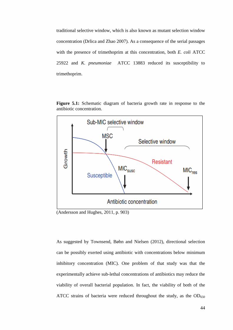
44
traditional selective window, which is also known as mutant selection window
concentration (Drlica and Zhao 2007). As a consequence of the serial passages
with the presence of trimethoprim at this concentration, both E. coli ATCC
25922 and K. pneumoniae ATCC 13883 reduced its susceptibility to
trimethoprim.
Figure 5.1: Schematic diagram of bacteria growth rate in response to the
antibiotic concentration.
(Andersson and Hughes, 2011, p. 903)
As suggested by Townsend, Bøhn and Nielsen (2012), directional selection
can be possibly exerted using antibiotic with concentrations below minimum
inhibitory concentration (MIC). One problem of that study was that the
experimentally achieve sub-lethal concentrations of antibiotics may reduce the
viability of overall bacterial population. In fact, the viability of both of the
ATCC strains of bacteria were reduced throughout the study, as the OD650

45
reading measured after each passages reduced. This problem was then
overcome by consistent assessment on the bacteria growth throughout the
study. Every 5 passages, both of the ATCC strains of bacteria were cultured
on EMB agar and MacConkey agar, followed by checking the bacteria
viability. Both of the ATCC strains of bacteria were able to survived even in
the presence of trimethoprim at 12 µg/ml. This also means that both the
bacteria were able to grow on this antibiotic concentration.
5.2.2 Clinical significance in antibiotic dosing strategies
This study confers a profound implication in antibiotic dosing strategies. In
clinical settings, traditional dosing approach using antibiotics concentration
within mutant selection window eradicates the susceptible bacteria population
and gives rise to the emergence of resistant subpopulation (Liu et al. 2005;
Drlica and Zhao 2007; Zhao and Drlica 2008). Study from Olofsson et al.
(2007) proposed that the susceptible E. coli populations were eliminated by
the approved, clinical dosing fluoroquinolone regimens, and eventually
develop resistance to the antibiotic. Somehow, the effective dose of antibiotics
prescribed by clinicians may not kill all the bacteria and exerts selective
pressure on the susceptible bacteria population. Eventually, the susceptible
bacteria may mutate and gain resistance over the antibiotics.

46
5.3 Assessment on the transfer of antibiotics resistance from clinical
isolate Escherichia coli 594370 to clinical isolate Klebsiella pneumoniae
594394
In this aspect of study, resistant clinical isolate E. coli 594370 served as the
donor bacteria while susceptible, clinical isolate K. pneumoniae 594394
served as the recipient bacteria. Based on the result, the recipient bacteria did
not gain any of the antibiotic resistance after 30 passages of co-incubation
with the donor bacteria. Thus, the transfer of antibiotic resistance from
resistant E. coli to susceptible K. pneumoniae was not observed. This may be
due to the insufficient number of passages conducted in this study. As
suggested by Townsend, Bøhn and Nielsen (2012), horizontal gene transfer
(HGT) events may not take place over a short time frame. 30 passages may
not be sufficient for transfer of antibiotic resistance from donor bacteria to
recipient bacteria. Moreover, the transfer of antibiotic resistance may be
driven by the presence of antibiotics, which is elaborated in Section 5.3.1.
5.3.1 Role of antibiotic in transfer of antibiotic resistance
The transfer of antibiotic resistance from resistant bacteria to sensitive bacteria
may be promoted by the presence of antibiotics at very low concentration. As
suggested by Courvalin and Trieu-Cuot (2001), antibiotics can act as sex
pheromones that induce the conjugative transfer of resistance gene from
resistant bacteria to susceptible bacteria.

47
However, one may argue that the resistance acquired in the recipient may be
induced by the mutation under selective pressure of antibiotic instead. As
shown in the study by Gullberg et al. (2011), ciprofloxacin with 1/10 of the
minimum inhibitory concentration (MIC) was able to induce the resistance in
susceptible strains of E. coli. In that study, the ciprofloxacin resistance in
E. coli was only induced after 600 generations of growth. As so, antibiotic
concentration less than 1/10 MIC may be essential to mediate the transfer of
antibiotic resistance from donor bacteria to recipient bacteria. In fact, it‟s not
the antibiotics that induce the mutation for the resistance in recipient bacteria
strains in this case, as this minimal amount of antibiotics will only induce the
antibiotic resistance after few hundreds of generations. Thus, this minimal
amount of antibiotics may be used to promote the antibiotic resistance transfer
from donor bacteria to recipient bacteria as the transfer of antibiotic resistance
may occur before the induction of antibiotic resistance which requires few
hundreds of passages. Thus, the transfer of antibiotic resistance may be
occurred much faster than the mutation conferring resistance in the bacteria
population.
Many of the horizontal gene transfer (HGT) events that involved integration of
genetic materials into bacteria chromosome are relatively non-beneficial and
may confer to fitness cost (Martínez, 2012; Townsend, Bøhn and Nielsen,
2012). However, the acquisition of resistant genes in the recipient bacteria by
HGT may be promoted in the presence of antibiotic pressure as the resistant
gene transferred are essential for survival of bacteria population (Townsend,
Bøhn and Nielsen, 2012).

48
5.3.2 Clinical significance of the transfer of antibiotic resistance
As described by Jernberg et al. (2010), human intestine serve as an optimal
site for the transfer of resistant genes, as the abundance in nutrients with moist
and warm environment of the intestine favor the colonization diverse
Enterobacteriaceae. Reservoir hypothesis suggested that human intestinal
bacteria may serve as the harbor for antibiotic resistant genes (Salyers, Gupta
and Wang, 2004; Marshall, Ochieng and Levy 2009).
Presence of antibiotics in food contaminants and oral antibiotic therapy may
promote the transfer of antibiotic resistance from donor bacteria to recipient
bacteria even though in minute amount (Salyers, Gupta and Wang 2004;
Schjørring and Krogfelt 2010). A survey conducted by McNulty et al. (2007)
revealed the public attitude on the consumption of antibiotics. From the survey,
some of the respondents failed to complete the full course of the antibiotic
treatment, and even recycle the left-over antibiotics. The potency of these
“left-over” antibiotics may reduce over time. Thus, the antibiotic
concentration in the body may not reach the minimum inhibitory concentration
(MIC), leading to the induction of antibiotic resistance in susceptible bacteria
population. This create an environment with sub-lethal concentration of
antibiotics, favors the transfer of antibiotic resistance from resistant
pathogenic bacteria to susceptible commensal flora, as well as the induction of
antibiotic resistance as shown in Section 5.2. Thus, public attitude is one of the
driven force that promotes the transfer and induction of antibiotic resistance
from the pathogenic bacteria to the susceptible intestinal flora.

49
5.4 Future Study
There were several methodological limitations in this project, and these
limitations are expected to be solved in future study. More number of passages
is required to assess the losing of antibiotic resistance and transfer of antibiotic
resistance. Hereby, the reversal and transfer of antibiotic resistance required
longer duration as discussed in Section 5.1 and Section 5.3 respectively. On
the other hand, in vivo models and cell culture methods can be conducted
concurrently with the in vitro experimental settings. The results obtained by
this approach may be more conclusive and provide a better picture on the
various aspects of antibiotic resistance. Furthermore, application of molecular
biology methods may provide a better understanding on the type of resistance
genes that are disseminated. In addition, bioinformatics tools and
mathematical models may be used as a guide in assessing the evolutionary
analysis of the bacteria.
50
CHAPTER 6
CONCLUSION
In conclusion, the losing of antibiotic resistance in clinical isolate
Escherichia coli 594370 did not occurred due to the limited duration in this
project. However, this reversibility is possible if the amount of passages is
increased. On the other hand, reduction of susceptibility to trimethoprim was
observed in both E. coli ATCC 25922 and K. pneumoniae ATCC 13883. Thus,
this indicates that trimethoprim resistance is inducible using trimethoprim with
sub-lethal concentration, which falls under the category of traditional selective
window. Thus, this indicates that the losing of antibiotic resistance may
require more duration as compared to the induction of the antibiotic resistance.
Last but not least, the transfer of antibiotic resistance from donor bacteria,
clinical isolate E. coli 594370 to recipient bacteria, clinical isolate
K. pneumoniae 594394 was not observed. It was discussed that the transfer of
antibiotic resistance from donor bacteria to recipient bacteria may require
longer duration and presence of antibiotics may be required.

51
REFERENCES
Alekshun, M.N. and Levy, S.B., 2007. Molecular mechanisms of
antibacterial multidrug resistance. Cell, 128(6), pp. 1037-1050.
Andersson, D.I. and Levin, B.R., 1999. The biological cost of antibiotic
resistance. Current Opinion in Microbiology, 2(5), pp. 489-493.
Andersson, D.I., 2006. The biological cost of mutational antibiotic
resistance: any practical conclusion? Current Opinion in Microbiology,
9(5), pp. 461 – 465.
Andersson, D.I. and Hughes, D., 2010. Antibiotic resistance: is it possible
to reverse resistance? Nature Reviews Microbiology, 8(4), pp. 260-271.
Andersson, D.I. and Hughes D., 2011. Persistence of antibiotic resistace in
bacterial populations. FEMS Microbiology Reviews, 35(5), pp. 901-911.
Andersson, D. I. and Hughes, D., 2012. Evolution of antibiotic resistance at
non-lethal drug concentrations.
Bean, D.C. et al., 2005. Resistance among Escherichia coli to
sulphnamides and other antimicrobials now little used in man. Journal of
Antimicrobial Chemotherapy,56(5), pp. 962-964.
Bisht, R., 2009. Antibiotic resistance – a global issue of concern. Asian
Journal of Pharmaceutical and Clinical Research, 2(2), pp. 34 – 39.
Cantón, R. and Ruiz-Garbajosa, P., 2011. Co-resistance: an opportunity for
the bacteria and resistance genes. Current Opinion in Pharmacology, 11(5),
pp. 477 – 485.
Carlet, J., 2012. The gut is the epicentre of antibiotic resistance. 1(39), doi:
10.1186/2047-2994-1-39
52
Clinical and Laboratory Standard Institute (CLSI), 2007. Performance
standards for antimicrobial susceptibility testing seventeenth informational
supplement. Available from: < http://www.microbiolab-bg.com/CLSI.pdf>.
[10 December 2012].
Courvalin, P. and Trieu-Cuot, P., 2001. Minimising potential resistance: the
molecular view. Clinical Infectious Diseases, 33(Suppl 3), pp. S138-S146.
Cuevas, O. et al., 2011. Significant ecological impact on the progression of
fluoroquinolone resistance in Escherichia coli with increased community
use of moxifloxacin, levofloxacin and amoxicillin/clavulanic acid. Journal
of Antimicrobial Chemotherapy, 66(3), pp. 664 – 669.
De Gelder, L. et al. , 2004. Combining mathematical models and statistical
methods to understand and predict the dynamics of antibiotic-sensitive
mutants in a population of resistant bacteria during experimental evolution.
Genetics, 168(3), pp. 1131-1144.
Drago, L. et al., 2010. In vitro selection of resistance in Escherichia coli
and Klebsiella spp. at vivo fluoroquinolone concentrations. BMC
Microbiology, 10(119), doi: 10.1186/1471-2180-10-119
Drlica, K. and Zhao, X., 2007. Mutant selection window hypothesis
updated. Clinical Infectious Disease, 44(5), pp. 681 – 688.
Enne, V. I., 2001. Persistence of sulphonamide resistance in Escherichia
coli in the UK despite national prescribing restriction. Lancet, 357(9265),
pp. 1325 – 1328.
Enne, V.I., 2010. Reducing antimicrobial resistance in the community by
restricting prescribing: can it be done? Journal of Antimicrobial
Chemotherapy, 65(2), pp. 179-182.
European Committee on Antimicrobial Susceptibility Testing (EUCAST),
2013. Breakpoint tables for interpretation of MICs and zone diameters.
Available from: < http://www.eucast.org/fileadmin/src/media/PDFs/EUCAST_files/Breakpoi
nt_tables/Breakpoint_table_v_3.1.pdf>. [10 December 2012].

53
Fraimow, H.S. and Tsigrelis,C., 2011. Antimicrobial resistance in the
intensive care unit: mechanisms, epidemiology, and management of
specific resistant pathogens. Critical Care Clinics, 27(1), pp. 163 – 205.
Freter, R., 1989. Control mechanisms of the large-intestinal microflora and
its influence on the host. Acta Gastroenterologica Latinoamericana, 19(4),
pp. 197 – 217.
Gallini et al., 2010. Influence of fluoroquinolone consumption in inpatients
and outpatients on ciprofloxacin-resistant Escherichia coli in a university
hospital. Journal of Antimicrobial Chemotherapy ̧65(12), pp. 2650 – 2657.
Goff, D. A., Bauer, K.A. and Mangino, J.E. (2012). Antimicrobial
stewardship management of infections: Beyond the costs of antimicrobials,
Pharmacy Practice News. Available at: < http://www.pharmacypracticenews.com/ViewArticle.aspx?d=Special%2BE
dition%2B%2F%2BEducational%2BReviews&d_id=63&i=August+2012&
i_id=872&a_id=21493>
Gould, I.M., 2009. Antibiotic resistance: the perfect storm. International
Journal of Antimicrobial Agents, 34(Suppl 3), pp. S2 – S5.
Gottesman, B.S. et al., 2009. Impact of quinolone restriction on resistance
patterns of Escherichia coli isolated from urine by culture in a community
setting. Clinical Infectious Diseases, 49(6), pp. 869-875.
Guan, X. et al., 2013. Journal of International Medical Research, 41(1), pp.
20-30.
Gullberg, E. et al., 2011. Selection of resistant bacteria at very low
antibiotic concentrations. PLoS Pathogens, 7(7). doi:
10.1371/journal.ppat.1002158.
Guo, M. et al., 2012. Predicting bacterial fitness cost associated with drug
resistance. Journal of Antimicrobial Chemotherapy, 67(4), doi:
10.1093/jac/dkr560

54
Hirayama, K., 1999. Ex-germfree mice harbouring intestinal microbiota
derived from other animal species as an experimental model for ecology
and metabolism of intestinal bacteria. Experimental Animals, 48(4), pp. 219
– 227.
Hooper, D., 2001. Minimizing potential resistance: the molecular view – a
comment on Courvalin and Trieu-Cuot. Molecular Mechanisms of
Resistance, 33(Suppl 3), pp. S157 – S160.
Jernberg, C. et al., 2010. Long-term impacts of antibiotic exposure on the
human intestinal microbiota. Microbiology, 156(Pt 11), pp. 3216-3223.
Johnsen, P.J. et al., 2009. Factors affecting the reversal of antimicrobial-
drug resistance. Latent Infectious Disease,9(6), pp. 357 – 364.
Kelly, B. G. Vespermann, A. and Bolton D. J., 2009. Gene transfer events
and their occurrence in selected environment. Food and Chemical
Toxicology, 47(5), pp. 978 – 983.
Kelly, B. G. Vespermann, A. and Bolton D. J., 2009. The role of horizontal
gene transfer in the evolution of selected foodborne bacterial pathogens.
Food and Chemical Toxicology, 47(5), pp. 951 – 968.
Kumar, A. and Schweizer, H.P., 2005. Bacterial resistance to antibiotics:
active efflux and reduced uptake. Advanced Drug Delivery Reviews, 57(10),
pp. 1486-1513.
Lambert, P.A., 2005. Bacterial resistance to antibiotics: modified target
sites. Advanced Drug Delivery Reviews, 57(10), pp. 1471-1485.
Levin, B.R., 2001. Minimizing potential resistance: a population dynamics
view. Clinical Infectious Diseases, 33(Suppl. 3), pp. S161-S169.
Lim, S.-M. and Webb, S.A.R., 2005. Nosocomial bacterial infections in
Intensive Care Units. I: Organisms and mechanisms of antibiotic resistance.
Anaesthesia, 60(9), pp. 887 – 902.

55
Lindgren, M., Lofmark, S., Edlund,C., Huovinen, P. and Jalava J., 2009.
Prolonged impact of a one-week course of clindamycin on Enterococcus
spp. in human normal microbiota. Scandinavian Journal of Infectious
Diseases, 41(3), pp. 215-219.
Lisboa, T. and Nagel, F., 2011. Infection with multi-resistant agents in the
ICUs: how to escape? Rev Bras Ter Intensiva, 23(2), pp. 120 – 124.
Livermore, D.M., 2005. Minimising antibiotic resistance. Lancet Infectious
Diseases, 5(7), pp. 450-459.
Livermore, D.M., 2012. Current epidemiology and growing resisance of
gram-negative pathogens. The Korean Journal of Internal Medicine, 27(2),
pp. 128 – 142.
Liu, Y. et al., 2005. Selection of rifampicin-resistant Staphylococcus aureus
during thuberculosis therapy: concurrent bacterial eradication and
acquisition of resistance. Journal of Antimicrobial Chemotherapy,Journal
of Antimicrobial Chemotherapy, 56(6), pp. 1172 – 1175.
Maisnier-Patin, S. and Andersson, D. I., 2004. Adaptation to the deleterious
effects of antimicrobial drug resistance mutations by compensatory
evolution. Research in Microbiology, 155(5), pp. 360-369.
Marshall, B. M., Ochieng, D. J. and Levy, S. B., 2010. Commensals:
Underappreciated reservoir of antibiotic resistance. Microbe, 4(5), pp. 231
– 238.
Martinez, J.L., 2009. The role of natural environments in the evolution of
resistance traits in a pathogenic bacteria. The Royal Society, 276(1667), pp.
2521-2530.
McNulty, C. A. M. et al., 2007. The public‟s attitudes and compliance with
antibiotics. Journal of Antimicrobial Chemotherapy, 60(Suppl 1), pp. i63 –
i68.

56
Morgan, D.J. et al., 2011. Non-prescription antimicrobial use worldwide: a
systematic review. Latent Infectious Disease. 11(9), pp. 692 – 701.
Nyberg, S. D. et al., 2007. Long-term antimicrobial resistance in
Escherichia coli from human intestinal microbiota after administration of
clindamycin. Scandinavian Journal of Infectious Disease, 39(6-7), pp. 54 –
520.
Oliphant, C.M. et al., 2002. Quinolones: a comprehensive review.
American Family Physician, 65(3), pp. 455-464.
Olofsson, S. K. et al. (2007). Dose-related selection of fluoroquinolone-
resistant Escherichia coli, 60(4), pp. 795 – 801.
Olofsson, S. K. and Cars, O., 2007. Optimizing drug exposure to minimize
selection of antibiotic resistance. Clinical Infectious Disease, 45(Suppl 2),
pp. S129 – S136.
Paterson, D. L. et al. (2005). In vitro susceptibilities of aerobic and
facultative Gram-negative bacilli isolated from patients with intra-
abdominal infections worldwide: the 2003 Study for Monitoring
Antimicrobial Resistance Trends (SMART). Journal of Antimicrobial
Chemotherapy, 55(6), pp. 965-973.
Reynolds, M. G., 2000. Compensatory evolution in rifampin-resistant
Escherichia coli. Genetics, 156(4), pp. 1471 – 1481.
Salyers, A.A., Gupta A. and Wang, Y., 2004. Human intestinal bacteria as
reservoirs for antibiotic resistance genes. in Microbiology, 12(9), pp. 412-
416.
Salyers, A.A., Moon, K. and Schlesinger, D., 2007, „The human intestinal
tract – a hotbed of resistance gene transfer? Part I‟, Clinical Microbiology
Newsletter, newsletter, viewed 1 March 2013,
http://www.cmnewsletter.com/article/S0196-4399(07)00002-5/abstract

57
Sander, P. et al., 2002. Fitness cost of chromosomal drug resistance-
conferring mutations. Antimicrobial Agents and Chemotherapy, 46(5), pp.
1204 – 1211.
Scott, G., 2009. Antibiotic resistance. Medicine, 37(10), pp. 551-556.
Sekiguchi, J. et al., 2006. Emergence of rifampicin resistance in
methicillin-resistant Staphylococcus aureus in tuberculosis wards. Journal
of Infection and Chemotherapy, 12(1), pp. 47 – 50.
Sj lund, M., Wreiber, K., Andersson D.I., Blaser, M.J. and Engstrand,L.,
2003. Long-term persistence of resistant Enterococcus species after
antibiotics to eradicate Helicobacter pylori. Annals of Internal Medicine,
139(6), pp. 483-487.
Sundqvist, M. et al., 2009. Little evidence for reversibility of trimethoprim
resistance after a drastic reduction in trimethoprim use. Journal of
Antimicrobial Chemotherapy, 65(2), pp. 350 – 360.
Sullivan, A., Edlund, C. and Nord, C.E., 2001. Effect of antimicrobial
agents in the ecological balance of human microflora. The Lancet infectious
diseases, 1(2), pp. 101-114.
Tan, C. et al., 2011. Increased rifampicin resistance in blood isolates of
methicilline-resistant Staphylococcus aureus (MRSA) amongst patients
exposed to rifampicin-containing antituberculous treatment. International
Journal of Antimicrobial Agents, 37(6), pp. 550 – 553.
Thomas, C.M. and Nielsen, K.M., 2005. Mechanisms of, and barriers to,
horizontal gene transfer between bacteria. Nature Reviews Microbiology,
3(9), pp. 711-721.
Townsend, J.P., Bøhn, T. and Nielsen, K.M., 2012. Assessing the
probability f detection of horizontal gene transfer events in bacterial
popultions. Frontiers in Microbiology, 3(27), doi:
10.3389/fmicb.2012.00027

58
van Hoek, A. H. A. M. et al., 2011. Acquired antibiotic resistance genes: an
overview. Frontiers in Microbiology.2(203), doi:
10.3389/fmicb.2011.00203
Vellinga, A. et al., 2010. A multilevel analysis of trimethoprim and
ciprofloxacin prescribing and resistance of uropathogenic Escherichia coli
in general practice. Journal of Antimicrobial Chemotherapy, 65(7), pp.
1514 – 1520.
Vellinga, A. et al., 2012. Trimethoprim and ciprofloxacin resistance and
prescribing in urinary tract infection associated with Escherichia coli: a
multilevel model. Journal of Antimicrobial Chemotherapy, 67(10), pp.
2523 – 2530.
Wilson, J. (2011). Trends among pathogens reported as causing
bacteraemia in England, 2004-2008. Clinical Microbiology and Infection,
17(3), pp. 451-458.
Wright, G.D., 2005. Bacterial resistance to antibiotics: enzymatic
degradation and modification. Advanced Drug Delivery Reviews, 57(10),
pp. 1451-1470.
Zhao, X. and Drlica, K., 2008. A unified anti-mutant dosing strategy.
Journal of Antimicrobial Chemotherapy, 62(3), pp. 434 – 436.

59
APPENDIX A
Chemicals and media used
Chemicals/Media Manufacturer
Ciprofloxacin disk Becton Dickinson and Company,
Sparks, USA
Dimethyl sulphoxide Lab-Scan Ltd., Ireland
Eosin methylene blue (EMB) agar Laboratorios Conda S.A., Spain
Ethanol (95%) Copens Scientific (M) Sdn. Bhd.,
Malaysia
Gentamicin disk Oxoid LTD. Basingstoke,
Hampshire, England
MacConkey agar Laboratorios Conda S. A., Spain
Moxifloxacin disk Benex Limited Shannon, Country
Clare, Ireland (USA)
Trimethoprim powder Nacalai Tesque, Japan

60
APPENDIX B
Equipment and lab wares used
Lab Wares/Equipment Manufacturer
Conical flask (250 ml) Schott Duran, Germany
Falcon tube (15 ml, 50 ml) BD FalconTM
, USA
Pipetter (10 µL, 100 µL, 1000 µL) Vipro, Germany
Pipette tips Axygen Scientific, CA
Schott bottle (100 ml, 500 ml, 1000
ml)
Schott Duran, Germany
Biosafety Cabinet (Level 2) TELSTAR Industrial S. L., Spain

61
APPENDIX C
Preparation of trimethoprim disks
Autoclaved paper disks with diameter of 6 mm were used for the preparation
of trimethoprim. According to Antonio-Velmote, Gonzaga and Darvin (1988),
it was assumed that 20 µl of antibiotic solution was absorbed by each paper
disk. The disk content for trimethoprim as recommended by CLSI is 5 µg, in
which the trimethoprim solution prepared must contain 250 µg of
trimethoprim in 1 ml of solution. 1 ml of working solution (250 µg/ml) was
prepared from the stock solution (10 mg/ml) with the calculation shown as
followed.

62
Preparation of 12 µg/ml trimethoprim solution
Working solution of trimethoprim with the concentration of 12 µg/ml was
used in the induction of antibiotic resistance in Section 3.2. 10 ml of working
solution (12µg/ml) was prepared from the stock solution (10 mg/ml) with the
calculation shown as followed.

63
APPENDIX D
The duplicated reading for measurement of the diameter of zone-of-inhibition
(ZI) in Section 4.1, Section 4.2 and Section 4.3 was tabulated as followed.
Zone-of-inhibition (ZI) diameter of ciprofloxacin to 594370 E. coli.
Number of passages Zone-of-inhibition Diameter (mm)*
Reading 1 Reading 2 Average reading
0 (Initial ASP) 0 0 0
5 8 9 8.5
10 10 10 10
15 8 9 8.5
20 8.5 8.5 8.5
25 8.5 8.5 8.5
30 8.5 8.5 8.5
35 7.5 8 7.75
40 9 9 9
45 0 0 0
50 8 8.5 8.25
55 9 8 8.5
60 0 0 0
65 0 0 0
70 9 9.5 9.25
75 R R R
80 10 9 9.5
85 8 9 8.5

64
Zone-of-inhibition (ZI) diameter of gentamicin to 594370 E. coli.
Number of passages
Zone-of-inhibition Diameter (mm)*
Reading 1 Reading 2 Average Reading
0 (Initial ASP) 8 9 8.5
5 9 9 9
10 9 9 9
15 8 8 8
20 0 0 0
25 8 7.5 7.75
30 8.5 8 8.25
35 7.5 8 7.75
40 8 8 8
45 7.5 7 7.25
50 7.5 7 7.25
55 0 0 0
60 0 0 0
65 0 0 0
70 10 10 10
75 7 8 7.5
80 10 10 10
85 0 0 0

65
Zone-of-inhibition (ZI) diameter of moxifloxacin to 594370 E. coli.
Number of passages Zone-of-inhibition Diameter (mm)
Reading 1 Reading 2 Average Reading
0 (Initial ASP) 10 9 9.5
5 11 10.5 10.75
10 12 12 12
15 10 9.5 9.75
20 9 8.5 8.75
25 9 10 9.5
30 10.5 10 10.25
35 9.5 10 9.75
40 9.5 9.5 9.5
45 9 8.5 8.75
50 9 9 9
55 8 7.5 7.75
60 8.5 8 8.25
65 8.5 8 8.25
70 10 9 9.5
75 9 8 8.5
80 10 9 9.5
85 9 9.5 9.25

66
Zone-of-inhibition (ZI) diameter of trimethoprim to clinical isolate E. coli
594370.
Number of passages Zone-of-inhibition Diameter (mm)*
Reading 1 Reading 2 Average Reading
0 (Initial ASP) 0 0 0
5 0 0 0
10 0 0 0
15 0 0 0
20 0 0 0
25 0 0 0
30 0 0 0
35 0 0 0
40 0 0 0
45 0 0 0
50 0 0 0
55 0 0 0
60 0 0 0
65 0 0 0
70 0 0 0
75 0 0 0
80 0 0 0
85 0 0 0

67
Zone-of-inhibition (ZI) diameter of trimethorpim to ATCC 25922 E. coli.
Number of Passages Zone-of-Inhibition (ZI) Diameter (mm)
Reading 1 Reading 2 Average Reading
0 35 35 35
5 30 29.5 29.75
10 26 27 26.5
15 25.5 26 25.75
Zone-of-inhibition (ZI) diameter of trimethorpim to ATCC 13883 K.
pneumoniae.
Number of Passages Zone-of-Inhibition (ZI) Diameter (mm)*
Reading 1 Reading 2 Average Reading
0 30 30 30
5 27 28 27.5
10 15 16 15.5
15 10 9 R
Zone-of-inhibition (ZI) diameter of ciprofloxacin to clinical isolate
K. pneumoniae 594394.
Number of Passages Zone-of-Inhibition (ZI) Diameter (mm)
Reading 1 Reading 2 Average Reading
0 21 21 21
5 17 18 17.5
10 20 19 19.5
15 21.5 21 21.25
20 20 20 20
25 21 22 21.5

68
30 22 22 22
Zone-of-inhibition (ZI) diameter of gentamicin to clinical isolate
K. pneumoniae 594394.
Number of Passages Zone-of-Inhibition (ZI) Diameter (mm)
Reading 1 Reading 2 Average Reading
0 19 19 19
5 19 19.5 19.25
10 18 19 18.5
15 18 18 18
20 19 19 19
25 19 19 19
30 19 18 18.5
Zone-of-inhibition (ZI) diameter of moxifloxacin to clinical isolate
K. pneumoniae 594394.
Number of Passages Zone-of-Inhibition (ZI) Diameter (mm)
Reading 1 Reading 2 Average Reading
0 18 18.5 18.25
5 18 17.5 17.75
10 17 17 17
15 18 18 18
20 19 19 19
25 19 19 19
30 19 18 18.5