Assessment of Reprocessed Arthroscopic Shaver...
7
Assessment of Reprocessed Arthroscopic Shaver Blades Jonathan S. King, M.D., Marilyn M. Pink, Ph.D., and Christopher M. Jobe, M.D. Purpose: The purpose of this study was to evaluate the level of contaminants on, as well as the quality of, reprocessed shaver blades. Methods: We assessed 7 new shaver blades and 27 shaver blades that bad been reprocessed with mechanical c1eaning, functional testing, and sterilization with ethylene oxide. A spectrophotometer measured the amount of nuc1eic acid and protein. The blade quality was assessed by photographing the blades with magnification and determining the percentage of damage present on each blade. A subset of shaver blades were then used to cut meniscal tissue, and the cut surface was measured for smoothness by image proeessing and automated laser scanning cytometry. In evaluation of the meniscus, for the subset of shavers, an image proeessing valne of l indicates a smooth, straight line, and values lower than 1 reflect deviations in the cut surface (the c1oser the valne is to 1, the smoother the surface). Laser scanning cytometry values indicate the percentage of irregularities in the cut surface (the lower the valne is, the smoother the surface). Results: Of the 27 reprocessed shaver blades, 13 (48%) bad detectable 1evels of protein and 17 (63%) bad detectable levels of nuc1eicacid. On the reprocessed shaver blades, protein levels ranged from 2.43 J.Lg to 60 J.Lg and nucleic !leid levels ranged from 0.40 J.Lg to 3.5 J.Lg. No new shaver blade bad contaminants. Twenty reprocessed shaver blades bad been manufactured with teeth and could be evaluated for visible damage. Ofthese, 10 bad 1% to 25% damage, 5 bad 26% to 50% damage, 3 bad 51% to 75% damage, and 2 had.76% to 100% damage. The new blades bad no visible damage. Image proeessing revealed smoothness.of the surface cut with new shaver blades, yielding valu~s of 1 :t 0.12, whereas the values for reprocessed shaver blades ranged from 0.62 :t 0.02 to 1 :t 0.07. Laser scanning cytometry values rangedfrom 3.3% to 7.1% for the new blades as compared with 5.8% to 20.0% for the reprocessed blades. Conclusions: Of the reprocessed shaver blades, 48% bad detectable levels of protein and 63% bad detectable levels of nuc1eic acid. All of the reprocessed blades visually evaluated showe~ same level of damage or wear, whereas no new blade bad such damage. In addition, menisci 90:twith reprocessed shavers showed rougher edges than didIIlenisci cut with new shavers. ClinicalRelevance: To make an informed decision regar~ingth~ lise of reprocessed shaver blades, surg~ons will want to know the level of contarnination on, andth~ quality of, reprocessed shaver blades. Key Words: Shavers-Nuc1eic acid-Laser scanning cytometry. T he modem era of managed care has brought abDul many changes in medicine. In response to eco- nomic pressure to lower costs, medical device repro- / From the Department of Orthopaedic Surgery, Loma Linda University, Loma Linda, California, U.S.A. Supported in part by equipment funds from Smith & Nephew, Andover, MA; Dyonics is a subsidiary of Smith & Nephew. The authors report no conjlict of interest. Address correspondence and reprint requests to Christopher M. Jobe, M.D., Department of Orthopaedic Surgery, Loma Linda University, East Campus, 11406 Loma Linda Dr, Suite 218, Loma Linda, CA 92354, U.S.A. E-mai/: [email protected] @ 2006 by the Arthroscopy Association of North America 0749-8063/0612210-3795$32.00/0 doi: 10.1 016/j. arthro. 2006. 07.021 cessing companies have developeda nationwide mar- ket for reprocessing and resale of single-lise surgical instruments. A wide spectrm;nofdevices are currently being reprocessed. They include orthopaedic shaver blades, burs, saw blades, anci drill bits, as well as instruments used in cardiovascular. surgery, laparos- copy, endoscopy, and oph~l1al~Qlogy. The average surgical center would savtft~%)~ach year on arthro- scopic shaver blades if eacbblade were used twice. These companies adhere t°f'pod and Drug Admin- istration (FDA) standards ofres~erilization and quality control and assert that they are able to provide shaver blades that are as safe and etftfdive as new blades. As of 2002, the reprocessing companies are considered 1046 Arthroscopy: The Journal of Arthroscopic and Related Surgery, Vol 22, No 10 (October), 2006: pp 1046-1052
Transcript of Assessment of Reprocessed Arthroscopic Shaver...

Assessment of Reprocessed Arthroscopic Shaver Blades
Jonathan S. King, M.D., Marilyn M. Pink, Ph.D., and Christopher M. Jobe, M.D.
Purpose: The purpose of this study was to evaluate the level of contaminants on, as well as thequality of, reprocessed shaver blades. Methods: We assessed 7 new shaver blades and 27 shaverblades that bad been reprocessed with mechanical c1eaning, functional testing, and sterilization withethylene oxide. A spectrophotometer measured the amount of nuc1eic acid and protein. The bladequality was assessed by photographing the blades with magnification and determining the percentageof damage present on each blade. A subset of shaver blades were then used to cut meniscal tissue,and the cut surface was measured for smoothness by image proeessing and automated laser scanningcytometry. In evaluation of the meniscus, for the subset of shavers, an image proeessing valne of lindicates a smooth, straight line, and values lower than 1 reflect deviations in the cut surface (thec1oser the valne is to 1, the smoother the surface). Laser scanning cytometry values indicate thepercentage of irregularities in the cut surface (the lower the valne is, the smoother the surface).Results: Of the 27 reprocessed shaver blades, 13 (48%) bad detectable 1evelsof protein and 17 (63%)bad detectable levels of nuc1eicacid. On the reprocessed shaver blades, protein levels ranged from2.43 J.Lgto 60 J.Lgand nucleic !leid levels ranged from 0.40 J.Lgto 3.5 J.Lg.No new shaver blade badcontaminants. Twenty reprocessed shaver blades bad been manufactured with teeth and could beevaluated for visible damage. Ofthese, 10 bad 1% to 25% damage, 5 bad 26% to 50% damage, 3 bad51% to 75% damage, and 2 had.76% to 100% damage. The new blades bad no visible damage. Imageproeessing revealed smoothness.of the surface cut with new shaver blades, yielding valu~s of 1 :t0.12, whereas the values for reprocessed shaver blades ranged from 0.62 :t 0.02 to 1 :t 0.07. Laserscanning cytometry values rangedfrom 3.3% to 7.1% for the new blades as compared with 5.8% to20.0% for the reprocessed blades. Conclusions: Of the reprocessed shaver blades, 48% baddetectable levels of protein and 63% bad detectable levels of nuc1eic acid. All of the reprocessedblades visually evaluated showe~ same level of damage or wear, whereas no new blade bad suchdamage. In addition, menisci 90:twith reprocessed shavers showed rougher edges than didIIleniscicut with new shavers. ClinicalRelevance: To make an informed decision regar~ingth~ lise ofreprocessed shaver blades, surg~ons will want to know the level of contarnination on, andth~ qualityof, reprocessed shaver blades. Key Words: Shavers-Nuc1eic acid-Laser scanning cytometry.
The modem era of managed care has brought abDulmany changes in medicine. In response to eco-
nomic pressure to lower costs, medical device repro-
/
From the Department of Orthopaedic Surgery, Loma LindaUniversity, Loma Linda, California, U.S.A.
Supported in part by equipment funds from Smith & Nephew,Andover, MA; Dyonics is a subsidiary of Smith & Nephew. Theauthors report no conjlict of interest.
Address correspondence and reprint requests to Christopher M.Jobe, M.D., Department of Orthopaedic Surgery, Loma LindaUniversity, East Campus, 11406 Loma Linda Dr, Suite 218, LomaLinda, CA 92354, U.S.A. E-mai/: [email protected]
@ 2006 by the Arthroscopy Association of North America0749-8063/0612210-3795$32.00/0doi: 10.1 016/j. arthro. 2006. 07.021
cessing companies have developeda nationwide mar-ket for reprocessing and resale of single-lise surgicalinstruments. A wide spectrm;nofdevices are currentlybeing reprocessed. They include orthopaedic shaverblades, burs, saw blades, anci drill bits, as well asinstruments used in cardiovascular. surgery, laparos-copy, endoscopy, and oph~l1al~Qlogy. The averagesurgical center would savtft~%)~ach year on arthro-scopic shaver blades if eacbblade were used twice.
These companies adhere t°f'pod and Drug Admin-istration (FDA) standards ofres~erilization and qualitycontrol and assert that they are able to provide shaverblades that are as safe and etftfdive as new blades. Asof 2002, the reprocessing companies are considered
1046 Arthroscopy: The Journal of Arthroscopic and Related Surgery, Vol 22, No 10 (October), 2006: pp 1046-1052

REPROCESSED ARTHROSCOPIC SHA VER BLADES
manufacturers and must submit their protocols forreprocessing to the FDA. The FDA has the authorityto inspect the procedures and the reprocessed equip-ment. The protocols for reprocessing are varied. Manyinclude mechanical cleaning, functional testing, andethylene oxide sterilization.1
There has been no previous study published in theorthopaedic literature evaluating the leve! of contam-ination or quality of reprocessed blades. The presenceof contaminants would certainly raise concems abåutthe transmission of microorganisms. The purpose ofthis study was to describe the level of residual con-taminating nucleic acid and protein and the quality ofreprocessed shaver blades. Further study would berequired to determine whether the presence of suchmaterial poses any clinically significant risk of infec-tion to patients.
METHODS
t!.
f
Dyonics (Smith & Nephew, Andover, MA) pro-vided 7 new shaver blades and 16 reprocessed Dyon-ics shaver blades for use in this study, which com-prised set 1. They were of different models and wereobtained from 4 different reprocessing companies. Allsterile wrapping was intact, with no obvious breachesof quality. In addition, we purchased Il reprocessedshavers from local hospitals to be used as an addi-tional set to test for contaminants and blade quality,which comprised set 2. All shaver blades were re-ported by the reprocessors to have been mechanicallycleaned, functionally tested, and then sterilized withethylene oxide. Because of the lack of a trackingsystem, there is no way to determine the type of tissueor the time period for which the shaver blades wereused. Nor could it be determined bow many timesthe blades bad been reprocessed. All shaver bladeswere as~igned a random number to prevent the datacollection from being biased toward the new blades.Each sterile blade was unwrapped in a sterile laminarflow bODd and separated into the outer and inDerblades.
I~~
f
Contaminants
For this portion of the study, we used 27 repro-cessed shaver blades, 3 new shaver blades, and 1 usedbut not reprocessed blade. The 27 reprocessed bladesand 2 of the new shaver blades (negative controis)were sequentially dipped for l hour each at roomtemperature (22°C) in a sterile tube containing 500 ILLof wash buffer (lO-mmol/L Tris, 1O0-mmol/L sodium
'li,
1047
Abbreviation: ND, none detected.*Total micrograms of nuc1eie acid detected on blade surface
(calculated from optical density measurement).tTotal micrograms of protein detected on blade surface (calcu-
lated from optical density measurement).
cWoride, and 0.1% Tween-20 in distilled water). Theoptical density at Az6o-, Az8o-, and A3zo-nm wave-lengths of the resulting solution were measured on aBeckman DU 640 spectrophotometer (BeckmanCoulter, Fullerton, CA).
The third new shaver blade was used as a positivecontrol by placement in a solution of I lLg/mL salmonsperm deoxyribonucleic acid and l mg/mL bovineserum albumin (as a source of protein) for I hour atroom temperature. Olle shaver that was used but notreprocessed was obtained from the surgical suite. Af-ter use, the shaver was rinsed in sterile saline solution,wiped dry, and placed in a plastic bag, and it was usedas an additional positive contro!. The positive controlshaver blades were allowed to dry for I hour and were
TABLE1. Residual Nucleic Acid and Protein Detectedon Shaver Blades
Reprocessed Shaver Blades New Shaver Blades
Nuc1eie Acid* Proteint Nuc1eic Acid* Proteint. (Total fJ.g) (TotalfJ.g) (TotalfJ.g) (Total fJ.g)
Sel IND ND ND NDND ND ND ND
0.40 ND0.134 ND0.19 6.8 /0.43 114ND ND
0.21 46.40.38 39.8ND NDND NDND ND
0.16 NDND ND
0.2 NDND ND
Sel 21.39 8.61.20 7.8ND ND
0.76 2.430.89 4.36Ul 10.31.38 9.81.14 6.11.36 8.9ND ND
1.19 4.75
1048
t
i
l
"Ii'J. S. KING ET AL.
A
c
then washed as described later for the experimentalblades.
The optical density values were used to calculate theamount of nucleic acid and protein washed off of theblades. The values were recorded as total micrograms ofnucleic acid. Protein (in rnilligrams per milliliter) wascalculated according to the Warburg formula as follows:3
Protein [mg/mL] = (1.55 X [A280- A320] - 0.76
X [A260- A320D/2X 10-3.
Quality
Shaver BIades
The shaver blades were photographed with anQlympus IX-7..0optical base (Scientific Instruments,Temecula, CA) equipped with lang working distanceobjectives and a C-mount analog color camera (Oly-750; Scientific Instruments). The randomized shaverblades were photographed with images of the teethfrom the ioner and Dilterblades taken at 4x, lOx, and20X magnification. Each tooth was assessed for dam-age, with any visible imperfections being recorded.The total number of teeth per blade was noted, and thepercentage of the shaver blade damaged was scored asfolIows: 0%, 1% to 25%, 26% to 50%, 51% to 75%,
!
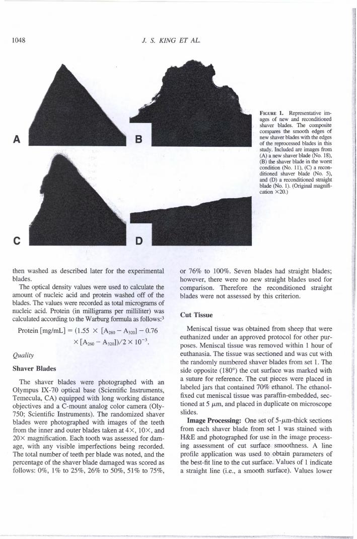
FIGURE1. Representative im-ages of new and reconditionedshaver blades. The compositecompares the smooth edges ofnew shaver blades with the edgesof the reprocessed blades in thisstudy. Inc1uded are images from(A) a new shaver blade (No. 18),(B) the shaver blade in the worstcondition (No. 11), (C) a recon-ditioned shaver blade (No. 5),and (D) a reconditioned straightblade (No. 1). (Original magnifi-cation x20.) ."
:ti
c~
or 76% to 100%. Seven blades bad straight blades;however, there were no new straight blades used forcomparison. Therefore the reconditioned straightblades were not assessed by tIDscriterion.
~!
Cut Tissue
Meniscal tissue was obtained from sheep that wereeuthanized under an approved protocol for other pur-poses. Meniscal tissue was removed within 1 hour ofeuthanasia. The tissue was sectioned and was cut with
the randomly numbered shaver blades from set 1. Theside opposite (180°) the cut surface was marked witha suture for reference. The cut'pieces were placed inlabeled jafS that contained 70% ethanol. The ethanol-fixed cut meniscal tissue was paraffin-embedded, sec-tioned at 5 JLm,and placed in duplicate on microscopeslides.
Image Proeessing: ane set of 5-JLm-thick sections'from each shaver blade from set 1 was stained with
H&E and photographed for use:,in the image process-ing assessment of cut surfaqe:.,,smoothness. A lineprofile application was used tQ;!.;pbtainparameters ofthe best-fit line to the cut surface. Values of 1 indicate
a straight line (Le., a smooth surface). Values lower
..
j...
.~~
~r.v.. "'" w'vy '_V""'~""--~#_~>"""Æ ."'~""~""'~- _."""",~,"",. ,.' 1

REPROCESSED ARTHROSCOPIC SHA VER BIADES 1049
than 1 reflect deviations (or "jaggedness") in the cutsurface relative to a straight line.
Laser Scanning Cytometry: Duplicate 5-JLm-sec-tions from each shaved meniscus w~re.fluorescentlystained by a modified Fluoro-Jade techlligu.6.3IIIbrief,the paraffin sections were fixed in 70%cold.(020°C)ethanol for 10 minutes, rinsed onc~jll' phpsphate-buffered saline solution, and transf6rr6d to Fluoro-Jade solution (l00 mg in 0.1% aceticacid;rPierceChemical) for 20 minutes at room t6mperfiture'(22°C).The labe1ed slides were rinsed 3 times inphosphate-buffered saline solution and counterstainedwith pro-pidium iodide (5 JLg/mL) (MolecularProbes, Eugene,OR) to stain the nudei red. Thefluorescentlystainedmeniscal tissue was scanned 011the laser scanningcytometer (CompuCyte, Cambridge, MA)4 for cut sur-face smoothness comparisons. The laser scanning cy-tometer hIld an Olympus BX50 base (Scientific Instru-ments) configured for epi-illumination with argon,helium-neon, and violet excitation lasers. To measurethe tissue smoothness" a scanning protocol was de-signed to acquire an x, y map of the fluorescent signal.
The cut surface smoothness was measured by use ofcomputer-generated lines that divided the bulk of thetissue from the cut surface. Values were expressed asthe percentage of tissue outside of the computer-gen-erated lines (or percentage of irregularities); thus thesmaller the valne is, the smoother the surface.
RESULTS
Contaminants
Of the 27 reprocessed shaver blades, 17 (63%) hIlddetectable 1evels of nudeic acid and 13 (48%) hIld de-tectable levels of protein (Table 1). On the reprocessedshaver blades, nucleic acid 1evelsranged from 0.4 JLgto3.5 p,g and protein levels ranged from 2.43 JLgto 60 JLg.
The 1evels of nudeic acid and total protein were 0.4JLgand 0.35 JLg,respectively, for the positive control.For the used bilt not reprocessed blades, these valueswere 4.76 JLgand 53.8 p,g, respectively. The 2 newblades hIld no detectab1e protein or nucleic acid.
The highest levels of nudeic acid and proteinwere found on the reprocessed shaver blades from lcompany, with 3 of its 4 blades being contaminated;Olle of these blades hIld 60 JLgof protein and 2.85p,g of nuc1eic acid. No company hIld a completesample of reprocessed shaver blades without con-tamination.
TABLE2. Percentage of Damage of Skaver Blades
ReprocessedShaver Blades New Shaver B1ades
Set l26%-50%
NT1%-25%
NTNT
51%-75%NT
1%-25%51%-75%
NT76%-100%51%-75%26%-50%76%-100%
1%-25%1%-25%
0%0%
.(
Set21%-25%
26%-50%0%-25%
26%-50%26%-50%
NT1%-25%1%-25%1%-25%
NT1%-25%
Abbreviation: NT, not tested.
Quality
Shaver Blades
A representative composite image of new and usedshaver blades is shown in Fig 1. Shown are the bestand worst conditions that were found when the dam-age of the reconditioned shaver blades was compared.
The data obtained for scoring the shaver blades usedin this study are compiled in Table 2. Of the 27reprocessed shaver blades, 7 were smooth, with noteeth, and were not tested. Of the 20 blades tested, 10hIld 1% to 25% damage, 5 hIld 26% to 50% damage,3 hIld 51% to 75% damage, and 2 hIld 76% to 100%damage; thus 5 shaver blades (25%) hIld greater than50% damage. The 2 new blades hIld no visible dam-age. No company hIld a complete sample of repro-cessed shaver blades without damage.
Cut Tissue
Image processing showed that tissue cut with thenew shaver blades hIld values of l :t 0.12 (where 1
1050 J. S. KING ET AL
c
indicates a straight line), whereas the values for thereprocessed shaver blades ranged from 0.62 :!::0.02 to1 :!::0.07 (Fig 2 and Table 3). Only 4 of 16 repro-cessed shaver blades bad a value of 1.
Laser scanning cytometry revealed a higher per-centage of irregularities for tissue that was cut withreprocessed blades from sel 1. This signifies a higherpercentage of tissue falling outside of the computer-generated line dividing the bulk of the tissue from thecut surface. Va1ues for tissue cut with the new blades
ranged frorp 3.3% to 7.1%. Values for the reprocessedblades ranged from 5.8% to 20.0% (Table 3). Of thereprocessed blades, only 4 scored within the range ofthe new blades. Those 4 blades were the same 4 bladesthat scored 1 on image processing.
In addition, of the 4 blades from sel 1 that ratedrelatively smooth cuts on the meniscus, 3 bad nodetectable nuc1eic acid or protein (the fourth bad0.134 JJ-gofnuc1eic acid and no protein). Because theywere straight blades, 3 of these 4 blades were not
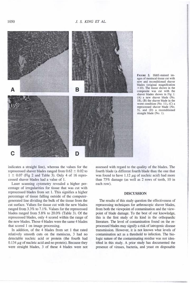
FIGURE 2. H&E-stained im-ages of meniscal tissue cut withnew and reconditioned shaverblades (original magnificationX 10). The tissue shown in thecomposite was cut with theshaver blades shown in Fig 1:(A) a new shaver blade (No.18), (B) the shaver blade in theworst condition (No. 11), (C) areprocessed shaver blade (No.5), and (D) a reconditionedstraight blade (No. I).
assessed with regard to the quality of the blades. Thefourth blade (a different fourth blade than the Ollethatwas found to have 1.12 JJ-gof nuc1eic acid) bad morethan 75% damage (as well as 2 rows of teeth, 10 ineach row).
DISCUSSION
The results of tIDsstudy question the effectiveness ofreprocessing techniques for arthroscopic shaver blades,from both the viewpoint of contamination and the view-point of blade damage. To the best of our knowledge,tIDs is the first study of its kind in the orthopaedicliterature. The level of contamination found on the re-.processed blades may signify a risk of iatrogenic diseasetransmission. However, it is not known what levels ofcontamination act as a threshold to infection. The bia-logic nature of the contaminating residue was not iden-tified in tIDsstudy. A prior study has documented thepresence of viruses, bacteria, and yeast on disposable
~-"..' -~- - ~~. ~ ,~ ........

--
REPROCESSED ARTHROSCOPIC SHA VER BLADES
TABLE3. Comparison of Cut Tissue Surface Smoothnessin Shavers From Set 1
46.63.37.13.85.97
New Shaver Blades
ImageProcessing*
LaserScanning
Cytometryt
1 ~ 0.121 ~ 0.12l ~ 0.12l ~ 0.12l ~ 0.12l ~ 0.121 ~ 0.12
*A value of 1 indicates a straight line/smooth surface. Valueslower than 1 reflect deviations in the cut surface. The doser thevalue is to I, the smoother the surface.
tPercent of irregularities. Higher numbers indicate more irreg-ularity.
:j:Theinsert was incorrectly matched to the outer blade and couldnot be used.
abdominal instruments resterilized with ethy1ene oxidegas.s Our results further support thepossib1e risk ofinfection from reuse of shaver b1adesintended for singleuse.6 Contamination of resterilized sing1e-use shaverb1ades may expose patients to an avoidab1erisk of iat-rogenic disease transmission.
The quality of reprocessed shaver b1adesis also 10werthan that of new blades, as was confirmed by the visualdamage noted on the reprocessed b1ades and the lessuniform cut edge of meniscus. This may be c1inical1yrelevant when Olleis debriding the edges of a meniscaltear during a repair, the borders of a chondral defectbefore a graft, or a simple degenerative meniscal tear.The success of meniscal repairs or chondral grafts relieson the viability of the carti1aginousborders for growthand migration of fibrob1astsand chondrocytes. This maybe compromised when Olleis using reprocessed b1adesthat 1eave behind frayed edges and possib1y a deeperzone of injury. Microscopic irregu1arities 1eft behindafter debridement of a degenerative meniscal tear maynot be intrinsical1y unstab1e bul may deteriorate morerapidly iGlo macroscopic irregu1arities that cause me-chanica1 symptoms and raiD.
There were 4 reprocessed b1adesthat 1efta smooth cut
1051
on the meniscus. Of these, only 1 bad a detectab1eleve!of nuc1eicacid. It wou1dbe interesting to know the typeand 1engthof lise to which these b1ades bad been sub~jected. For examp1e,lise of the b1adeson soft connectivetissue or for a short period of time may affect b1adecharacteristics differently than lise on bone or for anextended period of time. Currently, b1ades are nottracked for this information.
It wou1dbe of interest to exp10rethe tracking of b1adelise for another reason as well. It is possib1e,!hata b1ademay have been reprocessed more than once, thus ex-p1aining the poorer quality of the blade. A comp1extracking system, however, may increase the cost andthereby reduce the relative benefit of using recyc1edblades.
There are 1imitations to our study. A single observerperformed the assessments; however, this observer wasblinded as to the source of each specimen. In measuringthe surface roughness, we found that this was re1atedtomagnification-that is, with sufficient magnification, allcut surfaces can be iso1atedto small sections, which aretherefore smooth.
An additional limitation of this study is that the 16reprocessed shaver b1ades were not of the same model.Using b1adesof the same mode! wou1dhave facilitated amore uniform c1assificationof visib1ewear on the b1adeedges and evaluation of cut menisci. In addition, therealso may have been significant differences in the dura-tion and tissue type for which the b1ades were used.
This is bul Ollestudy among a spectrum of potentialfuture studies designed to assess the risk, if any, ofdisease transmission from reprocessed shaver blades, aswell as their quality. As amicroscopic study analyzingthe 1eve1sof protein and nuc1eic acid and evaluatingb1ade quality, these data cannot definitive1yanswer thequestion of the risk of disease transmission or the clinicalrelevanee of poor b1ade quality. Future studies will beneeded to further eva1uate the risk of disease transmis-sion and the c1inicalrelevanee of microscopic irregu1ar-ities caused by damaged shaver blades. A study of me-niscal repair in an animal mode1 comparing edgesdebrided with damaged versus new shaver b1ades mayprovide valuab1e information regarding clinical out-comes with the lise of reprocessed shaver blades. Ananimal mode1may a1sohe1p investigate whether menis-cus debrided with reprocessed b1ades deve!ops macro-scopic degeneration more quickly as a result of themicroscopic irregu1aritiesshown in our study. Olle mightalso reproduce this study with fresh menisci that arestained for living cells to evaluate whether the zone ofinjury 1eftbehind by reprocessed shaver b1adesis signif-icantly different from that of new shaver blades.
Reprocessed Shaver Blades
LaserImage Scanning
Processing* Cytometryt
0.66 0.02 16.71 0.07 6.7
0.77 0.09 12.7l 0.12 7.0
0.82 0.05 8.90.68 0.03 13.3
l 0.08 5.9
Defective:j:0.62 0.02 200.76 0.04 9.9
l 0.07 5.80.68 0.03 11.10.85 0.06 7.80.64 0.05 18.10.82 0.06 8.20.64 0.05 18.7

I1052 J. S. KING ET AL.
CONCLUSIONS
We have reached the following 3 conclusions basedon our study:
1. Of the reprocessed shaver blades, 48% bad de-tectable levels of protein and 63% bad detect-able levels of nucleic acid.
2. All of the reprocessed blades that were visuallyevaluated showed damage or wear (or both),whereas no new blade bad visible damage.
3. Menisci cut with reprocessed shavers showedrougher edges than did menisci cut with newshavers.
Acknowledgment: The authors thank Dr. LawrenceLonga for the sheep menisci, George Asberry for tissue sec-tioning, and John Christer for technical support.
f
REFERENCES
1. Updated 51O(k) sterility review guidanee K90-1; final guidaneefor industry and FDA. Rockville: Center for Devices and Ra-diological Health, US Food and Drog Administration; 2002.
2. Wilfinger WW, Mackey K, Chromcznski P. Effect of pH andionic strength on spectrophotometric assessment of nucleic acidpurity. Biotechniques 1997;22:474-481.
3. Zuch CL, Nordstroem VK, Briedrick LA, Hoemig GR, Gran-holm AC, Bickford Pc. Time course of degenerative alterationsin nigral dopaminergic neurons following a 6-hydroxydopaminelesion. J Camp Neurol 2000;427:440-454.
4. Green LM, Murray DK, Tran DT, et al. Response of thyroidfollicular cells to gamma irradiation compared to proton irradi-ation. I. Initial characterization of DNA damage, micronucleiformation, apoptosis, cell survival, and cell cycle phase redis-tribution. Radiat Res 2001;155:32-42.
5. Ulualp KM, Hamzaoglu I, UIgen SK, et al. Is it possible toresterilize disposable laparoscopy trocars in a hospital setting?Surg Laparosc Endose Percutan Tech 2000; 10:59-62.
6: Rutala WA, Weber DI. Creutzfeldt-Iakob disease: Recommen-dations for disinfection and sterilization. Clin Infect Dis 2001;32:1348-1356.
'i
i
i111


![Shaver Genealogy Descendants - arslanmb.orgarslanmb.org/shaver/Descendants-2.pdf · Shaver Family Genealogy Descendants of John Shaver [#2] & Rebecca Claxton Generations 1-4 Mark](https://static.fdocuments.us/doc/165x107/5edfaf14ad6a402d666b0343/shaver-genealogy-descendants-shaver-family-genealogy-descendants-of-john-shaver.jpg)