ASSESSMENT OF ANTIBIOTIC RESISTANT COMMENSAL BACTERIA IN … · ASSESSMENT OF ANTIBIOTIC RESISTANT...
123
ASSESSMENT OF ANTIBIOTIC RESISTANT COMMENSAL BACTERIA IN FOOD A Thesis Presented as a Requirement for the Master of Science in the Graduate School of The Ohio State University By Mark Lehman, D.V.M. * * * * * The Ohio State University 2006 Master’s Examination Committee: Dr. Hua Wang, Adviser Approved by Dr. Ahmed Yousef Dr. Randall Harris Adviser Graduate program in Food Science and Technology
Transcript of ASSESSMENT OF ANTIBIOTIC RESISTANT COMMENSAL BACTERIA IN … · ASSESSMENT OF ANTIBIOTIC RESISTANT...

ASSESSMENT OF ANTIBIOTIC RESISTANT COMMENSAL BACTERIA IN FOOD
A Thesis
Presented as a Requirement for the Master of Science in the Graduate
School of The Ohio State University
By Mark Lehman, D.V.M.
* * * * *
The Ohio State University 2006
Master’s Examination Committee:
Dr. Hua Wang, Adviser
Approved by Dr. Ahmed Yousef
Dr. Randall Harris Adviser
Graduate program in Food Science and Technology

Report Documentation Page Form ApprovedOMB No. 0704-0188
Public reporting burden for the collection of information is estimated to average 1 hour per response, including the time for reviewing instructions, searching existing data sources, gathering andmaintaining the data needed, and completing and reviewing the collection of information. Send comments regarding this burden estimate or any other aspect of this collection of information,including suggestions for reducing this burden, to Washington Headquarters Services, Directorate for Information Operations and Reports, 1215 Jefferson Davis Highway, Suite 1204, ArlingtonVA 22202-4302. Respondents should be aware that notwithstanding any other provision of law, no person shall be subject to a penalty for failing to comply with a collection of information if itdoes not display a currently valid OMB control number.
1. REPORT DATE 13 SEP 2006
2. REPORT TYPE N/A
3. DATES COVERED -
4. TITLE AND SUBTITLE Assessment Of Antibiotic Resistant Commensal Bacteria In Food
5a. CONTRACT NUMBER
5b. GRANT NUMBER
5c. PROGRAM ELEMENT NUMBER
6. AUTHOR(S) 5d. PROJECT NUMBER
5e. TASK NUMBER
5f. WORK UNIT NUMBER
7. PERFORMING ORGANIZATION NAME(S) AND ADDRESS(ES) The Ohio State University
8. PERFORMING ORGANIZATIONREPORT NUMBER
9. SPONSORING/MONITORING AGENCY NAME(S) AND ADDRESS(ES) AFIT/CIA
10. SPONSOR/MONITOR’S ACRONYM(S)
11. SPONSOR/MONITOR’S REPORT NUMBER(S)
12. DISTRIBUTION/AVAILABILITY STATEMENT Approved for public release, distribution unlimited
13. SUPPLEMENTARY NOTES The original document contains color images.
14. ABSTRACT
15. SUBJECT TERMS
16. SECURITY CLASSIFICATION OF: 17. LIMITATION OF ABSTRACT
UU
18. NUMBEROF PAGES
122
19a. NAME OFRESPONSIBLE PERSON
a. REPORT unclassified
b. ABSTRACT unclassified
c. THIS PAGE unclassified
Standard Form 298 (Rev. 8-98) Prescribed by ANSI Std Z39-18

ABSTRACT
The rapid emergence of antibiotic resistant (ART) pathogens is a major public health
concern. Although antibiotic resistance (AR) in foodborne pathogens has been studied
extensively, the contribution of foodborne commensals in disseminating the resistance
genes has been neglected in the past. Foodborne pathogens only account for a very small
portion of microbes associated with food; meanwhile, AR encoding genes can be
transferred readily from commensals to pathogens by natural gene transfer mechanisms.
Horizontal transmission of genetic material from one organism to another has been
established as a major mechanism for the expedited development of resistance. The
impacts of antibiotic applications in clinical treatments, veterinary medicine, animal
husbandry practices and animal feed on the emergence of antibiotic resistance (AR) have
been well-documented. However, knowledge on other major routes in the dissemination
of AR is limited. This information is essential to properly evaluate the contribution of
food chain in the evolvement of AR pathogen, particularly in susceptible host
populations.
ii

The objective of this study is to reveal the significance of the food chain in AR
dissemination by investigating the prevalence of ART commensal organisms in a variety
of ready-to-eat (RTE) and raw foods, Samples were evaluated for the total microbial
counts, as well as resistant population for tetracycline (Tet) and erythromycin (Em). All
food items were analyzed within the use-by dates. RTE salad mixes contained ART
population greater than 103 CFU/g, more than 80% of which were resistant to
erythromycin. Block cheeses had resistant counts greater than 102 CFU/g. Raw meat
products, both raw and cooked shrimps all have resistant population greater than 102
CFU/g.
Further studies confirmed the presence of mobile AR genes in the food isolates, by
assessing the presence of AR genes in ART commensals and their transmission to human
residential bacteria, using ermC gene as an example. Nine RTE bagged salads and nine
RTE deli-meats from local retail stores were examined for the prevalence of tetracycline
–resistant (Tetr) and erythromycin-resistant (Emr) bacteria using plate count agar (PCA)
with tetracycline (16µg-mL-1) or erythromycin (50µg-mL-1), and without antibiotics.
Cycloheximide (100µg-mL-1) was added to all plates to inhibit the growth of molds and
yeasts. All nine salad samples contained at least 103 CFU of Emr bacteria per gram of
food., but the ART bacteria were only found in RTE deli meats sporadically. A total of
108 Emr colonies were screened by conventional PCR and nearly 14% possessed the
iii

ermC gene. The ermC isolates were found to be either Pseudomonas spp. or
Staphylococcus spp. using partial 16S rRNA gene sequence analysis . The ermC gene
from a salad Staphylococcus epidermidis isolate was transferred to Streptococcus mutans
UA159 by natural transformation. The ermC gene was found associated with a
plasmid(s) of approximately 2.4 kb to 4.0 kb in both the S. mutans transformants and the
Staphylococcus epidermidis donor strain by Southern blot analysis. The minimum
inhibitory concentration (MIC) of Em for the transformants was significantly increased
compared to the parental S. mutans strain, but comparable to the donor strain. These data
suggest that ART bacteria are prevalent in the food chain and might serve as a potentially
important avenue transmitting AR genes to human microflora and possibly pathogens.
iv

This work is dedicated to Damara, Zachary and Brian for all their support and understanding.
v

ACKNOWLEDGMENTS
I would like to thank Dr. Hua H. Wang, my adviser, who served as a true mentor by
providing knowledgeable advice on all aspects of academic life and offering the
encouragement and intellectual support that made my research possible. I want to thank
her for her patience and directions to conduct the research.
I would also like to thank Dr. Ahmed Yousef for thoughtful conversations, including
providing feedback and suggestions on my research and for serving as a committee
member. I would also like to thank Dr. Randall Harris for serving as a committee
member, even though this is outside his usual discipline. People like you create a
positive learning environment both inside and outside the classroom.
I am very grateful to Hongliang Luo, Scott Hanna, Michele Manuzon, and Kai Wan,
whose help in the laboratory made my education possible.
This education program was funded through the USAF AFIT/CI program. The views
expressed in this presentation are those of the author(s) and do not reflect the official
policy or position of the United States Air Force, Department of Defense, or the U.S.
Government.
vi

VITA
1988-1993………………….……..B.S., Biology and Zoology, Colorado State University 1993-1997……………………………………..………D.V.M., Colorado State University
1997-1999……………………Associate Veterinarian, Feather and Fur Veterinary Clinic, Kailua, HI
1999-2001…………………..Hospital Director, National Pet Care Centers, Medford, OR.
2001-present……………………….……..Public Health Officer, United States Air Force 2001-2004………………………...Base Public health Officer, Kirtland AFB, NM
2004-present…………………...........….Graduate Student, AFIT/CI program, The Ohio State University
2005-2006……. ……………....Teaching Merit Award, OSU Department of Food Science and Technology 2005-2006……Outstanding Graduate Research Award, OSU Department of Food Science and Technology 2006 ……………….....Second Place, Student Poster Competition, Biotechnology Division, Institute of Food Technology annual convention
PUBLICATIONS
Wang, H., M. Manuzon, M. Lehman, K. Wan, H. Luo, T. Wittum, A. Yousef, L. Bakaletz. 2006. Food commensal microbes as a potentially important avenue in transmitting antibiotic resistance genes. FEMS Microbiol. Lett. 254, 226-231.
vii

FIELDS OF STUDY Major Field: Food Science and Technology Minor Field: Public Health
viii

TABLE OF CONTENTS
Page
Abstract…………………………………………..……………………………...………...iiDedication………………………………………..…………………………………......…vAcknowledgments……………..……………...………..…………………………….…..vi Vita…………………………………………………....………………………………....vii List of figures…………..……………..…………..……………………………….….......xi List of tables…………..……………..……………..……………………...…….…........xii
Chapters:
1. Introduction…..…………………………………………......…..………..…………….1
2. Literature review…………………………………...………..………...…………...…..6 2.1 History of antibiotics…..……...…………...…………..……..….…………….6 2.2 Classes of antibiotics............………..…………….….......….....…...…………7 2.3 Antibiotic applications..................……………........................….……........…8 2.4 Antibiotic resistance…………………………..……………...…......…….…...9
2.5 Mechanisms of resistance………………….………………….………......…11 2.6 Gene transfer mechanisms……………….…………………….…………….12
2.7 Food microbiology…………………………………………..……………….13 2.8 Antibiotic resistant bacteria and food safety…………………..…………….15
3. Objective……………………………..….…………………………….………….…..24
4. Prevalence of ART bacteria in selected retail food products...…….…………………25 4.1 Summary………………………………………….………………………….25 4.2 Introduction……………………………………………..……………………25 4.3 Materials and methods….………………………….……….…….………….27 4.4 Results…………………………………………………….…….……………30 4.5 Discussion…………….…………………………………….…….………….32 4.6 Bibliography…….………………...………………………….….………… .36 4.7 Figures……………………………………………………………………….37
5. Antibiotic resistance in ready-to-eat salads………………………………..…………46
5.1 Summary………………………..……………..…….…………...…………..47
ix

5.2 Introduction………………………………..………………...……………….48 5.3 Materials and methods….……………………….….……..…………………49 5.4 Results………………………………………….………..…………...………52 5.5 Discussion…………….…………………….……………..…………………55 5.6 Acknowledgement……………………………….…………..………………57 5.7 Bibliography…………………………………………………………………58 5.8 Figures……………………………………………………………………….61
6. Conclusions and future direction…………………..……………………..…..............66
APPENDIX A: Food Commensals as a Potential Major Avenue in Transmitting
Antibiotic Resistance Genes…….……...……...………...……..…………………..69
APPENDIX B: Supplemental research…………………….…...………………………90
Bibliography……………………………………………………………………………..98
x

LIST OF FIGURES
4.1. Prevalence of ART in raw meat……………………………...……. ……………….38
4.2. Prevalence of ART in seafood…………………………………….………..……….39
4.3. Prevalence of ART in yogurt…………………………...…………. ……………….40
4.4. Prevalence of ART in cheese.. ………………………............……. ……………….41
4.5. Prevalence of ART in deli-meats………………………………….………..……….42
4.6. Prevalence of ART in salads incubated at 20oC………...……..…...……………….43
4.7. Prevalence of ART in salads incubated at 37oC ……………...…....……………….44
4.8. PCR results comparing ermC and ermB primers…...…………….………..……….45
5.1. Prevalence of ART in salads ……………………………....….…. …………….….61
5.2. Conventional PCR using ermC primers ………………...……………...…….….….62
5.3. Results of Southern hybridization.…………………...……...……. ………….…….63
A.1. Prevalence of ART microbes in retail foods……………...…………………….…..86
B.1. Treatment of carrots using NaCl, boiling water, vinegar, Italian dressing, lemon juice or HCl…………………………………………………………..……………………..….95
B.2. Treatment of celery using NaCl, boiling water, vinegar, Italian dressing, lemon juice, or HCl………………………………………..………………………………….…….….96
B.3. Comparison of multiplexing PCR versus conventional PCR……………..…….…..97
xi

xii
LIST OF TABLES
5.1. The prevalence of ermC+ bacteria in RTE salads…………………….……..………64
5.2. Minimum inhibitory concentrations from controls, Emr salad isolates and transformants……………………………………….………………….……………..…..65 A.1. Prevalence of AR microbes in selected food samples……………….……………..87 A.2. Identification of antibiotic-resistant isolates from food based on 16S rRNA gene sequencing………….…………………………………………..…………………….......89


xiv

xv

1
CHAPTER 1
INTRODUCTION
The emergence of resistant pathogens untreatable by antibiotics is a major health
concern. Several organizations, including the World Health Organization (WHO),
Centers for Disease and Prevention (CDC), and the European Union (EU), have all
stressed the need to control the spread of resistance (1,5,18). The potential link between
the applications of antibiotics in food animal production and agriculture, and the
increased resistance in antibiotic resistance (AR) in agricultural environment and
products triggered several government-coordinated actions. In the European Union, steps
have been taken not only to track the use of antibiotics in veterinary and human medicine,
but to ban the application of certain antimicrobial agents (tylosin, spiramycin, bacitracin,
and virginiamycin) as growth promoters in food animal production; chicken and swine
farmers, as well as beef producers in Denmark voluntarily stopped using antimicrobial
agents as growth promoters since the late 1990’s (1). However, effective control
strategies are yet to be recommended and implemented in the US. In 1996 the National
Antimicrobial Resistance Monitoring System (NARMS) was established as a
collaborative effort among the CDC, United States Department of Agriculture (USDA),
Food and Drug Administration (FDA), and state and local public health departments.
The primary mission of NARMS is to monitor antibiotic resistance (AR) in foodborne
enteric pathogens (2). The increases in AR among enteric pathogens, i.e.,

2
Camplyobacter, Salmonella, and E. coli, were recognized (3). Furthermore, it was
suggested that commensal enteric bacteria of both animals and humans could be used as
indicators of potential AR development, and could serve as genetic reservoir, allowing
pathogens and opportunistic bacteria to develop AR in an accelerated manner (6,10,15).
According to Mellon et al (8), 28 million pounds of antibiotics are used annually in
agriculture in the absence of disease. The Union of Concerned Scientists estimated over
31 million pounds of antibiotics are used in animals annually (8), representing 89% of the
total annual antibiotic usage in the United States. In 2004, the Animal Health Institutes
estimated that 21.7 million pounds of antimicrobials were used in veterinary medicine in
the US (4), down from 24.4 million pounds in 1999. A landmark development is FDA
banned the use of enrofloxacin in poultry farmers in 2005 (7).
It is demonstrated that AR bacteria occur in a variety of environmental compartments; i.e.
water, sewage, and soil. Furthermore it is documented that the AR genes from these
bacteria can be transferred to other bacteria within the same compartment and between
compartments (5,9,13). Therefore reducing the use of antibiotics may not result in the
immediate slowing down of the emergence of ART bacteria. For instance, a recent study
showed that the number of ART E. coli isolates for ten antimicrobials from dairy cows
raised on organic dairies did not vary much from those from conventional dairies (12).
Further, ART bacteria are found prevalent in oral microflora of healthy adults and
children (16,17), and the gut microbial flora of normal humans have become a potential
reservoir for AR genes, likely involved in acquiring and donating AR genes to transient
intestinal bacteria (11). The food supply, specifically animal products, has been

3
implicated in the transfer of AR genes from the farms to the human intestinal tract
(11,14). However, despite the isolation and identification of ART pathogens in food
animals and retail products, which only represent a very small percentage of the
microbial flora associated with foods, the impact of the food chain on AR dissemination
is yet to be revealed.

4
BIBLIOGRAPHY
1. Anderson, A.D., J. McClellan, S. Rossiter, and F.J. Angulo. 2003. Public health consequences of use of antimicrobial agents in agriculture. In: Knobler, S.L., Lemon, S.M., Najafi, M., Burroughs, T. (Eds.), Forum on Emerging Infections: The Resistance Phenomenon in Microbes and Infectious Disease Vectors. Implications for Human Health and Strategies for Containment—Workshop Summary. Board on Global Health, Institute of Medicine, Appendix A, pp. 231–243. 2. Centers for Disease Control and Prevention. 2005. http://www.cdc.gov/narms/faq.htm#3 (viewed December 2005). 3. Centers for Disease Control and Prevention. 2001. NARMS 2000 Annual Report. 4. DVM News magazine, editorial. 2005. Survey shows spike in antibiotic sales, DVM Newsmagazine, Aug. 5. Fries, R. 2004. Conclusions and activities of previous expert groups: the Scientific Steering Committee of the EU.J Vet Med B Infect Dis Vet Public Health. 51:403-7. 6. Hummel, R., H. Tschape, and W. Witte. 1986. Spread of plasmid-mediated nourseothricin resistance due to antibiotic use in animal husbandry. J. Basic Microbiol. 26:461-466. 7. Kaufman, M. 2005. Ending Battle With FDA, Bayer Withdraws Poultry Antibiotic Washington Post, Friday, September 9, 2005; Page A03 http://www.washingtonpost.com/wpdyn/content/article/2005/09/08/AR2005090801918.html (viewed December , 2005). 8. Mellon, M., C. Benbrock, and K. Benbrock. 2001. Hogging it: estimates of antimicrobial abuse in livestock. Cambridge: Union of Concerned Scientists Publications. 9. Molbak, K. 2004. Spread of resistant bacteria and resistance genes from animals to humans--the public health consequences. J. Vet. Med. B. Infect. Dis. Vet. Public Health. 51:364-369. 10. Murray, B. 1992. Problems and dilemmas of antimicrobial resistance. Pharmacotherapy 12: 865-935. 11. Salyers, A.A., A. Gupta, and Y. Wang. 2004. Human intestinal bacteria as reservoirs for antibiotic resistance genes. Trends Microbiol. 12:412–416.

5
12. Sato, K., P. Bartlett P, and M. Saeed. 2005. Antimicrobial susceptibility of Eschericia coli isolates from dairy farms using organic versus conventional production methods, J. Am. Vet. Med. Assoc. 226:589-94. 13. Summers, A.O. 2002. Generally overlooked fundamentals of bacterial genetics and ecology. Clin Infect Disease 34 Suppl 3:S85-92. 14. Sullivan A., A. Johansson, B. Svenungsson, and C.E. Nord. 2004. Effect of Lactobacillus F19 on the emergence of antibiotic-resistant microorganisms in the intestinal microflora. J Antimicrob Chemother. 54:791-7. Epub 2004 Aug 25. 15. van Den Boggard, A., and E. Stobbering. 1999. Antibiotic usage in animals, impact on bacterial resistance and public health. Drugs 58:598-607. 16. Villedieu, A., M.L. Diaz-Torres, N. Hunt, R. McNab, D.A. Spratt, M. Wilson, and P. Mullany. 2003. Prevlanece of tetracycline resistance genes in oral bacteria. J. Antimicrob. Chemother. 47:878-882. 17. Villedieu, A., M.L. Diaz-Torres, A.P. Roberts, N. Hunt, R. McNab, D.A. Spratt, M. Wilson and P. Mullany. 2004. Genetic basis of erythromycin resistance in oral bacteria. Antimicrob Agents Chemother 48:2298–2301. 18. WHO. 2000. WHO Global Principless for the Containment of Antimicrobial Resistance in Animals Treated for Food: Report of a WHO Consultation, Geneva, Switzerland, June 5-9, 2000.

6
CHAPTER 2
LITERATURE REVIEW
2.1 History of antibiotics
As the germ theory grew in acceptance in the 19th century, so did the search for
antibiotics. In 1887, Rudolf Emmerich demonstrated that animals artificially infected
with streptococci were protected from developing cholera. In 1896, a French medical
student, Ernest Duchesne, first discovered that the soil mold Penicillium was able to
inhibit the growth of some bacteria. (37). In 1928, a Brittish physician Alexander
Fleming observed the similar phenomenon. One of his bacterial plate cultures was
contaminated with the blue-green mold Penicillium, and the bacterial colonies in close
approximation to the mold showed disrupted growth. Subsequently, Dr. Fleming grew
cultures of the mold and was able to demonstrate its ability to kill bacteria (26).
Antibiotics were first used in mass by the military in World War II. Their success in
reducing mortality due to wound infections didn’t go un-noticed. Soon after, the
application of antibiotics in therapeutics was expanded to the general population and the
production of antibiotics kept growing during the postwar years. Antibiotics quickly

7
earned the nickname "magic bullets", in part because they were much safer than previous
remedies which often involved toxic compounds such as arsenic (46).
2.2 Classes of antibiotics
Antibiotics can be classified in several different ways. These classifications can be based
on the mechanism of action (bactericidal versus bacteriostatic), chemical structure,
spectrum of activity (broad versus narrow), or route of administration. Describing
antibiotics by their common chemical structure is often the most useful way to classify
antibiotics because those with similar structure often have similar functionality, mode of
action, substrate spectrum as well as toxicities. Penicillins are perhaps the best known of
all the antibiotics and are similar in structure to cephalasporins. Both penicillins and
cephalasporins contain a beta-lactam structure and are referred to as beta-lactam
antibiotics. Beta-lactam antibiotics are considered bactericidal against Gram-positive and
Gram-negative bacteria. Beta-lactams inhibit the synthesis of the cell walls via the
disruption of metabolic functions. Macrolides, another class of antibiotics, can be
differentiated from other antibiotics by their macrocyclic lactone chemical structure and
are derived from Streptomyces spp. Erythromycin, azithromycin and clarithromycin are
all common antibiotics in this class. This class is used most commonly to treat Gram-
positive cocci, but can also be used against gram-negative anaerobes. However, reduced
susceptibility to erythromycin is commonly observed in Staphylococcus aureus due to
acquiring resistance genes thus it is recommended not to routinely use erythromycin

8
when treating Staphylococcus aureus. Clarithromycin is commonly used to treat
Helicobacter pylori infections. Macrolides are primarily bacteriostatic, inhibiting protein
synthesis by binding to the 50S subunit of the ribosome (6). Tetracyclines are named
because of their four ring structure. Although they are similar to macrolides in that they
are derived from Streptomyces spp., these bacteriostatic antibiotics differ slightly from
macrolides in their mode of action by binding to the 30S subunit of the ribosome.
Fluroquinolones are synthetic antibacterial agents. They are considered to be broad-
spectrum, bactericidial antibiotics, inhibiting DNA gyrase activity (31)
2.3 Antibiotic applications
Originally used to treat bacterial infections in both animals and humans, the applications
of antibiotics have expanded beyond their obvious use against bacteria. For instance,
antibiotics are also used extensively as growth promoters in food animal production (53).
The animals can gain approximately 5% more body weight when antibiotics are included
in their feed (14). In agriculture, antibiotics are sprayed on crops to combat plant
pathogens, but are classified as pesticides when used in this manner. Even in human
medicine, antibiotics are sometimes used therapeutically, but not to combat bacteria. For
example, macrolides have been used in cystic fibrosis patients as an inflammatory
mediator suppressor (16). Tetracycline and some tetracycline derivatives are also used to
treat the syndrome of inappropriate antidiuretic hormone (SIADH) secretion, and
protozoan infections such as malaria (31).

9
2.4 Antibiotic Resistance
Since its discovery, the scientific community has recognized the development of
antibiotic resistance. In 1943, four years after drug companies began mass-producing
antibiotics, the first reports of penicillin resistant Staphylococcus aureus were
documented (26).
Dr. Fleming recognized the potential seriousness of this development and in an interview
with The New York Times, Fleming warned the public of the implications of resistant
bacteria. Through a series of experiments using various doses of antibiotics, Flemming
postulated two theories on how resistance developed: bacterial proteins which could
degrade the drug(s) or strengthening of the cell wall (24). Unfortunately the threat of
resistance was considered unwarranted until the 1970s, when resistant strains of bacteria
causing meningitis and gonorrhea proved fatal (7). Because of the similarity in antibiotic
use between animals and humans, including but not limiting to tetracyclines,
sulfonamides, penicillins, macrolides, fluoroquinolones, cephalosporins,
aminoglycosides, chloramphenicols, and streptogramins, a serious concern is that once
resistance develops in animals it will soon affect humans (5).
Resistance to any environmental pressure is an adaptive process. It is generally believed
that resistance to antibiotics develops from new mutations which proves to be beneficial
or through the acquisition of resistant genes (53). On the other hand, if the selective
pressure is removed, the organism may lose the necessity to carry the gene over time, but
10
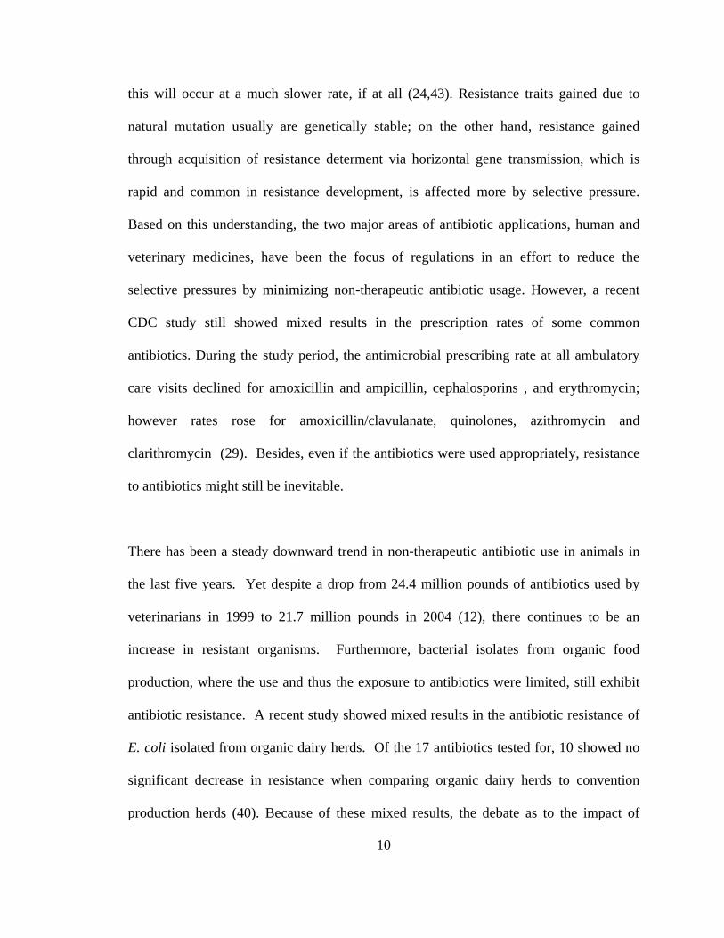
this will occur at a much slower rate, if at all (24,43). Resistance traits gained due to
natural mutation usually are genetically stable; on the other hand, resistance gained
through acquisition of resistance determent via horizontal gene transmission, which is
rapid and common in resistance development, is affected more by selective pressure.
Based on this understanding, the two major areas of antibiotic applications, human and
veterinary medicines, have been the focus of regulations in an effort to reduce the
selective pressures by minimizing non-therapeutic antibiotic usage. However, a recent
CDC study still showed mixed results in the prescription rates of some common
antibiotics. During the study period, the antimicrobial prescribing rate at all ambulatory
care visits declined for amoxicillin and ampicillin, cephalosporins , and erythromycin;
however rates rose for amoxicillin/clavulanate, quinolones, azithromycin and
clarithromycin (29). Besides, even if the antibiotics were used appropriately, resistance
to antibiotics might still be inevitable.
There has been a steady downward trend in non-therapeutic antibiotic use in animals in
the last five years. Yet despite a drop from 24.4 million pounds of antibiotics used by
veterinarians in 1999 to 21.7 million pounds in 2004 (12), there continues to be an
increase in resistant organisms. Furthermore, bacterial isolates from organic food
production, where the use and thus the exposure to antibiotics were limited, still exhibit
antibiotic resistance. A recent study showed mixed results in the antibiotic resistance of
E. coli isolated from organic dairy herds. Of the 17 antibiotics tested for, 10 showed no
significant decrease in resistance when comparing organic dairy herds to convention
production herds (40). Because of these mixed results, the debate as to the impact of

11
veterinary use of antibiotics continues on. Some argue that veterinary medicine
contributes very little risk to humans when it comes to the development and transmission
of antibiotic resistant infections (48). The more popular opinion is that the use of
antibiotics, especially in food animals, will lead to the development of antibiotic
resistance which in turn can be disseminated through the environment and led to resistant
infections in humans (1). Several studies support this idea. E. coli, for example, has
been shown to develop and disseminate antibiotic resistance in correlation with antibiotic
usage in animal feed (18,25,43,52).
2.5 Mechanisms of resistance
Since most of the antibiotics in use have a natural origin, produced either by
bacteria or by fungi, development of resistance is a natural process. The extent of
resistance can be limited to one drug within a class (8), an entire class such as
sulfanomides (31), or multiple drugs (51). Resistance development can be due to natural
mutation in the bacterium. In this case the resistance trait is stable and independent from
environmental selective pressure, and the trait can be disseminated vertically through
multiplication. Resistance can also be due to the acquisition of resistance-encoding
genetic elements via horizontal gene transmission. In this case the resistance trait can be
disseminated rapidly in the microbial population, but its distribution and maintenance is
greatly affected by the selective pressure. Several types of genetic determinants can lead
to antibiotic resistance (38). Efflux pumps are energy dependent mechanisms which can
be either acquired or located on the chromosome, and are of concern because they can
extrude not only antibiotics but other biocides, often lead to multidrug resistance (23).

12
These pumps have been further classified into two categories, ATP-binding cassette
(ABC) and secondary transporters (33). These pumps can be found in a multitude of
bacteria important in food fermentation and food safety (35,36). Another common
mechanism is enzyme modification. For instance, rRNA methylases can alter the rRNA
and thus inhibit antibiotics from attaching to a specific region of rRNA. In the case of
macrolide antibiotics there are 30 known genes which encode for rRNA methylases. All
of these methylases inhibit antibiotics from binding to the 50S ribosome subunit (38).
This is significant because other classes of antibiotics, such as lincosamides and
streptogramins, can also be blocked (49). Similar in action to methylases are ribosomal
protection proteins (RPP) which block specific binding sites of antibiotics such as
tetracyclines (7). A third mechanism includes a variety of inactivating enzymes. These
enzymes are specific for specific antibiotics, but are common in Gram-positive and
Gram-negative bacteria (28,32). The immunity genes from the antibiotic producing strain
could be an import source of resistance as well.
2.6 Gene transfer mechanisms
There are several basic types of resistance: intrinsic, mutation and acquired. Intrinsic
resistance refers to the bacterial reduced susceptibility to certain anti-microbial(s) due to
a specific property of a given organism, such as the molecular features of the cell wall or
membrane, or the lack of a particular enzyme targeted by the antibiotic. For instance,
Gram-negative bacteria are less susceptible to erythromycin. Mutation happens naturally
at the frequency of somewhere around 10-8 to 10-10. Antibiotics can accelerate the
domination of resistant bacteria in the population by acting as selective agents and cause

13
vertical dissemination of the mutated bacteria. Genetically such mutants are stable. But
if the mutants have additional auxotroph, once the antibiotics are removed, the mutated
organisms may be out-competed by the wild-type organisms. Acquired resistance refers
to bacterium exhibiting reduced sensitivity to a given antibiotic due to acquisition of a
genetic determinant encoding specific resistance mechanism. Acquired resistance is often
due to horizontal transmission of mobile elements, most commonly transposons and
plasmids, carrying genes encoding resistance. Transformation, transduction, and
conjugation are three common mechanisms of gene transmission. Transformation occurs
when DNA or RNA from the environment is taken up and expressed by a bacterium.
Transduction involves a bacteriophage mediating the transmission of bacterial DNA from
one bacterium to another. Conjugation requires direct cell-to-cell contact, allowing
genetic material to be passed from one bacterium to another.
2.7 Food microbiology
The interactions between microorganisms and foods, both beneficial and harmful to
humans, can be traced down since recorded history. Spoilage of processed foods was
first documented around 6000B.C. (42). Beer making dated back as far as 7000BC (34).
Egyptians used milk, cheese and butter as early as 300 B.C. (20). Louis Pasteur
demonstrated the souring of milk by microorganisms in 1837. Seventeen years later, he
successfully used heat to destroy unwanted microbes in beer and wine. This technique of
Pasteurization was then adapted to process milk in 1880 (11).

14
In most cases, microorganisms are abundant in raw food materials and considerable
amount of them can survive various processing conditions. Very few foods are
considered sterile, free of any microorganisms. Some of the microbes can cause diseases
and are referred to as foodborne pathogens. Despite pathogens only account for a very
small percentage in foodborne microbes, they have received the most attention because of
their significance in public health. According to the CDC, foodborne pathogens account
for over 76 million illnesses, 325,000 hospitalizations and 5000 deaths annually (30).
Most of these diseases are self-limiting, and thus the number of illnesses is thought to be
grossly underestimated. Of all the known pathogens, 90% of all food related illnesses
and deaths are caused by five pathogens, Salmonella, Listeria, Toxoplasma, Norwalk-like
viruses, Campylobacter, and E. coli O157:H7.
Most microorganisms associated with foods do not cause sickness in humans and are
referred to as commensals. An important concern of foodborne commensals is that some
of them can lead to food spoilage. Despite of all the advances in food processing and
preservation, microbial spoilage continues to be a problem today. A study by the USDA
Economic Research Service reported that 5,449 million pounds of edible food, which
represented approximately 2% of the total edible food supply in the United States, were
lost at the retail stage in 1995. Almost half of the losses came from perishable items such
as dairy products and fresh fruits and vegetables (22). A number of spoilage bacteria
responsible are also considered as opportunistic pathogens in humans, such as
Pseudomonas, Enterococcus, and Staphylococcus, in immuno-compromised patients.

15
One of the beneficial applications of microbes is food fermentation, during which
microorganisms can convert food materials such as carbohydrates through metabolic
activities into desirable end products such as lactic acid, ethanol, and other flavor
compounds. The rapid growth of fermentation cultures can inhibit the outgrowth of other
spoilage and pathogenic microbes through various means such as producing antagonistic
compounds, decreasing the pH, changing the redox potential, and minimizing the
availability of essential nutrients, etc. Therefore a primary function of fermentation is
preserving perishable raw food materials. As a consequence, many fermentation products
distinctive from the original raw materials, such as cheese, bread, and beer, were added to
our list of favorite foods. Although fermentation is usually desired, in the wrong food
category a beneficial fermentative bacterium could be considered as a spoilage bacterium.
For example, Pediococcus cerevisiae is considered desirable in the final stage of
sauerkraut fermentation, but in beer P. cerevisiae is a spoilage organism. The most
common fermentative bacteria are lactic acid bacteria (LAB). This group consists of 12
genera of Gram-positive bacteria (20) and has been extensively studied (39).
2.8 Antibiotic resistant bacteria and food safety
A number of antibiotics, such as tetracyclines and macrolides are commonly used in both
animals and humans, and bacteria resistant to these antibiotics have been found in
humans, animals and the food supply consisting of pathogens, fermentation bacteria and
commensal bacteria (3,27,44,45,47). While there are several reasons antibiotic resistant
bacteria pose a particular problem to food safety, the CDC emphasizes the following
three major concerns (4).

16
First, the emergence of ART foodborne pathogens diminishes the efficiency of
therapeutic treatment of the corresponding foodborne diseases. In the United States alone,
several million people are infected with foodborne pathogens annually. Antibiotics are
sometimes needed to successfully treat these diseases and can be life-saving especially in
infants, immuno-compromised, pregnant and elderly individuals. The most tracked and
studied foodborne pathogens are those associated with animals, such as E. coli,
Salmonella, Campylobacter, and Listeria, in both the United States and worldwide
(3,21,41,50). However, various resistance genes have been identified in many of these
pathogenic bacteria. Unfortunately, the research emphasis has so far been limited to
examine the prevalence of ART pathogens in food animals and its potential transfer to
humans, and the ecological complexity of antibiotic resistance has not been fully
explored (41). A few studies have shown that human sewage instead of animal waste
might be the source of resistant bacteria into the environment (17,19). Obviously, just
illustrating the prevalence of antibiotic resistant microbes in the food supply is not
enough. Appropriate regulation and monitoring can only be accomplished when the
origins and entire ecology of antibiotic resistance is understood (9,21).
Second, the normal microflora of animals and humans can be disrupted when exposed to
sublethal doses of antibiotics, which can increase the susceptibility of the subjects to
disease causing agents. Normally healthy people can tolerant the infection of a small
amount of pathogenic bacteria without getting the disease because the normal intestinal
microflora of a healthy individual can out-compete the pathogens. When antibiotics are
given the subsequent disruption of normal flora can make healthy individuals more

17
susceptible to foodborne pathogens, such as Salmonella, by reducing the number of
bacteria needed to cause disease, and leading to a more severe disease because the
resistant bacteria has a selective advantage over the normal microflora (5). It is also
important to note that the intestinal track of both humans and animals has been speculated
to be the location of gene transfer for Listeria (10) thus further promoting the pathology
of the disease.
Related to this is the third major concern of antibiotic resistance relating to food safety.
Although pathogens are of major public health concern the number of commensal
bacteria far outnumber that of pathogens. Thus, if antibiotic resistance develops in
commensal bacteria this genetic material could be transferred to pathogens. While the
CDC states this phenomenon would be difficult to quantify, several studies have
demonstrated this phenomenon. Genes encoding for resistance against tetracycline,
erythromycin and vancomycin have been detected in several commensal lactic acid
bacteria from fermented meats and dairy products (27,47). Several countries in the
European community have tracked resistant zoonotic species using non-pathogenic E.
coli as an indicator organism (3,15). DeFrancesco et al (2004) demonstrated that
commensal E. coli had higher rates of resistance than pathogenic multi-drug resistant
Salmonella from the same herds, suggesting that commensal bacteria could be used to
survey the prevalence of antibiotic resistant bacteria. Another study demonstrated
similarities in virulence factors between human isolates and food isolates examining
Enterococcus isolates, concluding that food commensals may be an overlooked aspect of
antibiotic resistance (13). Boehme et al (2004) examined antibiotic resistant enterococci

18
in agricultural foodstuffs and found that vegetable foods, although not as well studied as
animal products, could serve as a common gene pool for resistant genes.
Therefore, the overall impact of the food chain in AR transmission has not been fully
understood, and studies on the prevalence of AR in the foodborne microbes, particularly
in the dominant commensal bacteria, and the potential transmission to host ecosystems,
need to be evaluated.

19
BIBLIOGRAPHY
1. Anderson, A.D., J. McClellan, S. Rossiter, and F.J. Angulo. 2003. Public health consequences of use of antimicrobial agents in agriculture. In: Knobler, S.L., Lemon, S.M., Najafi, M., Burroughs, T. (Eds.), Forum on Emerging Infections: The Resistance Phenomenon in Microbes and Infectious Disease Vectors. Implications for Human 2. Boehme S, G. Werner, I. Klare, R. Reissbrodt, and W. Witte. 2004. Occurrence of antibiotic-resistant enterobacteria in agricultural foodstuffs. Mol. Nutr. Food Res. 7:522-531. 3. Bywater, R. H. Deluyker, E. Deroover, A. de Jong, H. Marion, M. McConville, T. Rowan, T. Shryock, D. Shuster, V. Thomas, M. Valle, and J. Walters. 2004. A European survey of antimicrobial susceptibility among zoonotic and commensal bacteria isolated from food-producing animals. J Antimicrob Chemother. 54:744-54. Epub 2004 Sep 16. 4. Centers for Disease Control and Prevention. 2005. http://www.cdc.gov/narms/ (viewed Nov. 2005). 5. Centers for Disease Control and Prevention. 2005. http://www.cdc.gov/narms/faq.htm#3 (viewed December 2005). 6. Chopra, I., and M.C. Roberts. 2001. Tetracycline antibiotics: mode of action, applications, molecular biology, and epidemiology of bacterial resistance. Microbiol Mol Biol Rev. 65:232–260. 7. Connell, S., D. Tracz , K. Nierhaus , and D. Taylor. 2003. Ribosomal protection proteins and their mechanism of tetracycline resistance. Antimicrob. Agents Chemother. 47:3675-3681. 8. Cuzzolin, L., and V. Fanos. 2002. Use of macrolides in children:areview of the literature, Infct Med: 19:279-285. 9. DeFrancesco, K.A., R.N. Cobbold, D.H. Rice, T.E. Besser, and D.D. Hancock. 2004. Antimicrobial resistance of commensal Escherichia coli from dairy cattle associated with recent multi-resistant salmonellosis outbreaks.Vet Microbiol. 98:55-61. 10. Doucet-Populaire, F., P. Trieu-Cuot , I. Dsbaa , A. Andremont , and P. Courvalin. 1991. Inducible transfer of conjugative transposon Tn1545 from Entercoccus faecalis to Listeria monocytogenes in the digestive trats of gnotobiotic mice. Antimicrob. Agents Chemother. 35:185-187.

20
11. Doyle, M., L. Beuchat L, and T. Montvill. 1997. Food microbiology, Fundamentals and Frontiers, Washington DC, ASM press: 3-12. 12. DVM News magazine, editorial. 2005. Survey shows spike in antibiotic sales, DVM Newsmagazine, Aug. 13. Eaton, T., and M. Gasson. 2001. Molecular screening of Enterococcus virulane determinanats and potential for genetic exchange between food and medical isolates. Appl. Environ. Microbio. 67:1628-1635. 14. European Commission. 1998. Commission regulation of amending council directive 70/524/EEC concerning additives in feedinstuffs as regards withdrawl of the authorization of certain antibiotics. Document No. VI/7767/98, Brussels, Belgium. 15. Franklin, A., J. Acar, and F. Anthony. 2000. Antimicrobial resistance: harmonization of national resistance monitoring and surveillance programmes inanimals and in animal-dervied food. Scientific and Technical Revue, Office International des Epizootiques (O.I.E.) 20:859-870. 16. Gaylor, A., S. Allyson, and J. Reilly. 2002. Therapy with macrolides in patients with cystic fibrosis. Pharmacotherapy 22:227-239. 17. Hardwood, V., M. Brownell , and W. Perusek. 2001. Vancomycin-resistant Enterococcus spp. isolated from wastewater and chicken feces in the United States. Appl. Environ. Microb. 24:122-130. 18. Hummel, R., H. Tschape, and W. Witte. 1986. Spread of plasmid-mediated nourseothricin resistance due to antibiotic use in animal husbandry. J. Basic Microbiol. 26:461-466. 19. Huys, G., D. Gevers, and R. Temmermen. 2001. Comparison of the antimicrobial tolerance of oxytetracycline-ressitant heterotrophic bacteria isolated from hospital sewage and freshwater fishfarm water in Belgium. Syst. Appl. Microb. 24:122-130. 20. Jay, J. Modern Food Microbiology, 6th ed, Kluwer Academic/Plenum Pulishers, New York. 21. Johnston, L.M., and L. A. Jaykus. 2004. Antimicrobial resistance of Enterococcus species isolated from produce. Appl. Environ. Microbiol. 70:3133-3137 22. Kantor, L., K. Lipton, A. Manchester, and V. Oliveira. 1997. Estimating and addressing America's food losses. Food Review 20:Jan-Apr. 23. Levy, S.B. 2002. Active efflux, a common mechanism for biocide and antibiotic resistance. Sym Ser Soc Appl Microbiol. 31:65-71.

21
24. Levy, S.B. 1998. Antimicrobial resistance-a sign of the times, New England Journal of Medicine 338:1376-1378. 25. Levy, S.B., G.B. Fitzgerald, and A.B. Macone. 1976. Changes in intestinal flora of farm personnel after introduction of tetracycline-supplemented feed on a farm. New Eng. Journal of Medicine 295:583-8.
26. Lewis, R. 1995. The rise of antibiotic-resistant infections. FDA Consumer magazine September 1995. 27. Mathur, S., and R. Singh. 2005. Antibiotic resistance in food lactic acid bacteria--a review. Int J Food Microbiol. 105:281-95. Epub 2005 Nov 8. Review. 28. Matsuoka, M., K. Endou, H. Kobayashi, M. Inoue, and Y. Nakajima. 1998. A plasmid that encodes three genes for resistance to macrolide antibiotics in Staphylococcus aureus. FEMS Microbiol Lett. 167:221-227. 29. McCaig, L., R. Besser, and J. Hughes. 2001. Antimicrobial drug prescription in ambulatory care settings, United States, 1992–2000. National Center for Infectious Diseases, CDC, Atlanta, Georgia, USA. 30. Mead, P.S., L. Slutsker, V. Dietz, L.F. McCaig, J.S. Bresse, and C. Shapiro. 1999. Food-related illness and death in the United States. Emerg. Infect. Dis. 7:5607-5625. 31. The Merck Manual of Medical Information 1999. - Home Edition, Robert Berkow (Ed.), ISBN 0-671-02727-1. 32. 60. Noguchi, N., J. Katayama, and K. O’Hara. 1998. Cloning and nucleotide sequence of the mphB gene from macrolide 2’-phosphotransferase II in Escherichia coli. FEMS Microbiol Lett. 144:197-202. 33. Paulsen, I., M. Brown, and R. Skurray. 1996. Proton-dependent multidrug efflux systems. Microbiol Rev 60:575-608. 34. Pederson C.S. 1971. Microbiology of Food Fermentations. Westport, CT: AVI. 35. Perreten, V., F. Schwartz, M. Teuber, and S.B. Levy. 2001. Mdt(A), a new efflux protein conferring multiple antibiotic resistant Lactococcus lactis and Escherichia coli. Antimicrob Agents Chemother. 45:1109-1114. 36. Perreten, V., F. Schwarz, L. Cresta, M. Boeglin, G. Dasen, and M. Teuber. 1997. Antibiotic resistance spread in food. Nature 389:801–802. 37. Princeton. 2005. http://www.molbio.princeton.edu/courses/mb427/2001/projects /02/antibiotics.htm#Introduction (viewed Nov. 2005).

22
38. Roberts, M.C. 2004. Resistance to macrolide, lincosamide, streptogramin, ketolide, and oxazolidinone antibiotics. Mol Biotechnol. 28:47–62. 39. Sandine, W.E., P.C. Randich, and P.R. Elliker. 1972. Ecology of the lactic streptococci: A review. J. Milk Food Tech. 35:176-185. 40. Sato, K., P. Bartlett P, and M. Saeed. 2005. Antimicrobial susceptibility of Eschericia coli isolates from dairy farms using organic versus conventional production methods, J. Am. Vet. Med. Assoc. 226:589-94. 41. Scott, H.M., L.D. Campbell, B.B. Harvey, K.M. Bischoff, W.Q. Alali, K.S. Barling, and R.C. Anderson. 2005. Patterns of antimicrobial resistance among commensal Escherichia coli isolated from integrated multi-site housing and worker cohorts of humans and swine. Foodborne Pathog Dis. 2:24-37. 42. Tanner, F., and L. Tanner. 1953. Food-borne infections and intoxications. 2 ed. Champaign IL. 43. Teuber, M. 2001. Veterinary use and antibiotic resistance. Curr. Opin. Microbiol. 4:493-499. 44. Villedieu, A., M.L. Diaz-Torres, N. Hunt, R. McNab, D.A. Spratt, M. Wilson, and P. Mullany. 2003. Prevlanece of tetracycline resistance genes in oral bacteria. J. Antimicrob. Chemother. 47:878-882. 45. Villedieu, A., M.L. Diaz-Torres, A.P. Roberts, N. Hunt, R. McNab, D.A. Spratt, M. Wilson and P. Mullany. 2004. Genetic basis of erythromycin resistance in oral bacteria. Antimicrob Agents Chemother 48:2298–2301. 46. Wainwright, W. 1990. Miracle Cure: The Story of Penicillin and the Golden Age of Antibiotics. (Oxford: Basil Blackwell, 1990). 47. Wang, H.H., M. Manuzon, M. Lehman, K. Wan, H. Luo, T. E.Wittum, A. Yousef, L. O. Bakaletz. 2006. Food commensal microbes as a potentially important avenue in transmitting antibiotic resistance genes. FEMS Microbiol. Lett. 254:226-231. 48. Wassenaar, T. 2005. Use of antimicrobial agents in veterinary medicine and implications for human health. Critical Reviews in Microbiology 31:155-169. 49. Weisblum, B. 1995. Erythromycin resistance by ribosome modification. Antimicrob. Agents Chemother 39:577-585. 50. White D.G., S. Zhao, S. Simjee, D. Wagner , and W. McDermott. 2002. Antimicrobial resistance of foodborne pathogens, Microbes and Infection 5:405-412.

23
51. World Health Organization. 2005. http://www.who.int/mediacentre/factsheets/fs194/en/index.html (viewed Nov. 2005). 52. WHO. 2000. WHO Global Principless for the Containment of Antimicrobial Resistance in Animals Treated for Food: Report of a WHO Consultation, Geneva, Switzerland, June 5-9, 2000. 53. Witte, W. 1998. Medical consequences of antibiotic use in agriculture. Science 279:996-997.

24
CHAPTER 3
OBJECTIVE
The objective of this study was to assess antibiotic resistance in various foods.
This would be accomplished by 1) determining the prevalence of ART bacteria targeting
two commonly used broad-spectrum antibiotics, erythromycin and tetracycline, using
both conventional microbiology as well as molecular biology techniques; 2) conducting a
more focused study to determine the genetic characteristics of the ARTs based on
information from the prevalence study as well as to demonstrate the transfer of the
genetic material from food commensal bacteria to human residential bacterium in
laboratory settings. While several studies have been done to assess antibiotic resistance
in both foodborne pathogens and clinical pathogens, only a few have attempted to
demonstrate the role of commensal bacteria from food in disseminating antibiotic
resistance to humans. This study is in an attempt to fill this knowledge gap and to help
better understand the ecology involved in the development of antibiotic resistance.

25
CHAPTER 4
PREVALENCE OF ART BACTERIA IN SELECTED RETAIL FOOD
PRODUCTS
4.1 Summary
The rapid emergence of antibiotic-resistant (ART) pathogens is a major threat to public
health. While the surfacing of ART food-borne pathogens is alarming, the magnitude of
the antibiotic resistance (AR) gene pool in food-borne commensal microbes is yet to be
revealed. Incidence of ART commensals in selected retail food products was examined in
this study. The presence of 102–107 CFU of ART bacteria per gram of foods in many
samples, particularly in ready-to-eat, ‘healthy’ food items, indicates that ART bacteria are
abundant in the food chain. AR-encoding genes were detected in ART isolates,
suggesting that food could be an important avenue for ART bacteria evolution and
dissemination.

26
4.2 Introduction Resistant pathogens to various antibiotics are emerging rapidly. Surfacing of these
resistant pathogens, untreatable by antibiotics, constitutes a real threat to public health.
To effectively combat this problem, a comprehensive understanding of the major
pathways in antibiotic resistance (AR) gene dissemination, as well as the key
mechanisms in the evolution of antibiotic-resistant (ART) bacteria is essential. Horizontal
gene transfer among pathogens in the hospital environment has been recognized as an
important avenue for the rapid spread of AR genes among pathogens. It is also believed
that horizontal transfer of AR genes between commensal and pathogenic microorganisms
in ecosystems are much more likely events than direct AR gene dissemination from one
pathogen to another (1). The presence of AR gene reservoirs in commensal microbes in
various environmental and host ecosystems (6,9,12,13,14,15, 18,19), and the illustration
of commensals as facilitators for human microbiota (11) suggest the importance of
commensals in mediating the dissemination of AR genes. The isolation of AR genes in
food-borne pathogens from retail products exemplified the potential contribution of the
food chain in transmitting ART pathogens to humans (2,7,11,20). These studies on
commensal bacteria, however, are limited and primarily focused on the opportunistic
pathogen enterococci (4,8).
A standard laboratory enrichment procedure (http://www.fda.gov/cvm/
Documents/AppendicesA-6.pdf) is often used to detect the presence of the ART bacteria,
masking the real magnitude of the AR problem associated with the food chain. To

27
examine the AR risks associated with the food chain, this study aimed at revealing the
distribution spectrum and magnitude of ART commensal bacteria and AR gene pool in
retail foods, by targeting total food microbiota instead of a particular group of
microorganisms or pathogens. Therefore, food samples were analyzed without any
laboratory enrichment procedures. Prevalence of microbial resistance to tetracycline (Tet)
and erythromycin (Em), still commonly used in animal production and human therapy,
was investigated (3,17). The presence of representative resistance gene markers in
selected food isolates was examined and main AR gene(s) and hosts were identified.
Minimum inhibitory concentrations (MIC) were used to determine the susceptibility
levels to further distinguish between intrinsic and extrinsic resistance.
4.3 Materials and methods
Food sample preparation and enumeration of total and ART populations. Seventy-one various food samples, were purchased from five local grocery stores and
analyzed within the products sell-by dates. Among the 11 RTE bagged or boxed salad
samples, representing different mixtures of greens and other vegetables RTE, two were
from two different fast-food chain restaurants. Five grams of each sample were
aseptically removed from the product packaging and placed in disposable plastic bags
containing 10mL of sterile 0.1% peptone water. Bagged samples were hand massaged for
10 min. Homogenized samples or rinsing liquids were serially diluted and plated on
nonselective plate count agar (PCA, Becton Dickinson and Company, Sparks, MD) for
nonselective total microbial counting, and on PCA plates containing 16 µg-mL-1 of Tet or

28
50 µg-mL-1 of Em (Fisher Biotech, Fair Lawn, NJ). Plates were incubated aerobically at
20oC and/or 37oC for up to 48 h for assessing Tet- and Em-resistant population of non-
fermented food products. Homogenized fermented food samples were serially diluted and
plated on Difco Lactobacilli MRS Agar (MRS, Becton Dickinson and Company)
incubated anaerobically at 30oC for 48 hours. The levels of antibiotics used in selective
agar plates were based federal standards (http://www.fda.gov/cvm/Documents/
AppendicesA-6.pdf). The ART bacteria counts for each of the duplicated samples were
converted to CFU/g and the means and standard deviations for both the total plate count
on PCA and Emr count on Em plate were calculated. The peptone water rinse from an
unused sandwich bag was included as the negative control for each examination.
Antibiotic resistance gene detection and host isolates identification.
Conventional PCR was conducted to detect AR genes in the ART isolates. Bacterial cells
from single colonies were re-suspended in 300 µL sterile dH2O containing 100 mg of 1:1
mixture of 0.5 mm diameter and 0.1 mm diameter glass beads (Biospec Products Inc.,
Bartlesville, OK). The sample mixtures were homogenized using the Mini-Bead-Beater-8
(Biospec Products Inc.) for 3 min. at maximum speed. The resulting cell extracts were
placed in a boiling water bath for 10–15 min and 5 µL of the supernatant were used as
PCR templates. The primer pair ermC FP 5’-GCTAATATTGTTTAAATCGTCAAT-3’
and ermC RP 5’-TCAAAACATAATATAGATAAA- 3’ were used to amplify the 640 bp
ermC fragment (Chung et al.,1999). PCR was conducted using reagents and conditions as
described previously (Luo et al., 2004). PCR products with expected sizes were purified
using the QIAquicks kit (Qiagen, Valencia, CA) following manufacturer’s instruction.

29
ART isolates containing resistance genes were identified by PCR amplification of the
16S rRNA gene fragment and sequence analysis following procedures as described
previously (5). DNA sequences of the 16S rRNA gene and the ermC gene fragments
were determined using a DNA analyzer (ABI PRISMs 3700, Applied Biosystems, Foster
City, CA) at the Plant Genome Sequence Facility, The Ohio State University. The DNA
sequences were compared with published Em resistance gene sequences deposited in the
NCBI database.
Minimum inhibition concentration (MIC) profiles of ART isolates.
The MIC profiles of selected ART isolates were determined using a commercial kit
(Sensititres 18–24 h MIC and Breakpoint Susceptibility Plates; TREK Diagnostic
Systems, Cleveland, OH) following the manufacturer’s instructions, with modifications.
Tryptic soy broth (TSB) broth instead of the standard Mueller–Hinton broth was used to
culture fastidious organisms. The MIC panels were incubated at either 30 or 370C for 24–
48 h. Additional 96- well microtiter plates with wells containing up to 256µg-mL-1 of Tet
and Em were used to determine the MIC of Emr or Tetr isolates which exhibited positive
growth in wells containing 16 µg-mL-1 of Tet or 8 µg-mL-1 of Em, the maximum
concentrations on the Sensititres plates. The MICs were reported as the minimum
concentration of the antibiotic that inhibited visible growth, as indicated by a lack of
turbidity of broth or deposition of cells at the bottom of the wells. Control strains used in
the study include Staphylococcus aureus ATCC 292136 (American Type Culture
Collection (ATCC), Manassas, VA), Pseudomonas aeruginosa ATCC 27853 (ATCC),
30
and E. coli DH5α .
(http://www.ncbi.nlm.nih.gov/entrez/query.fcgi?db=genomeprj&cmd=Retrieve&dopt=O
verview&list_uids=13773).
4.4 Results
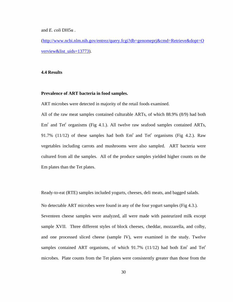
Prevalence of ART bacteria in food samples.
ART microbes were detected in majority of the retail foods examined.
All of the raw meat samples contained culturable ARTs, of which 88.9% (8/9) had both
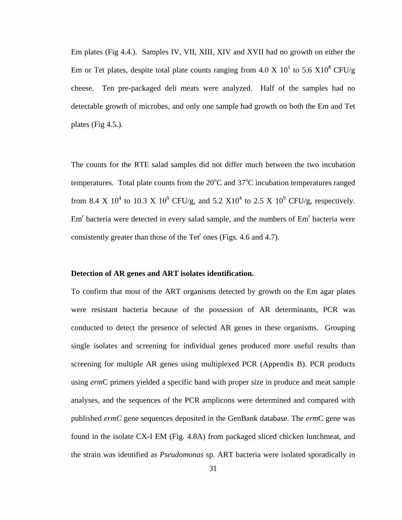
Emr and Tetr organisms (Fig 4.1.). All twelve raw seafood samples contained ARTs,
91.7% (11/12) of these samples had both Emr and Tetr organisms (Fig 4.2.). Raw
vegetables including carrots and mushrooms were also sampled. ART bacteria were
cultured from all the samples. All of the produce samples yielded higher counts on the
Em plates than the Tet plates.
Ready-to-eat (RTE) samples included yogurts, cheeses, deli meats, and bagged salads.
No detectable ART microbes were found in any of the four yogurt samples (Fig 4.3.).
Seventeen cheese samples were analyzed, all were made with pasteurized milk except
sample XVII. Three different styles of block cheeses, cheddar, mozzarella, and colby,
and one processed sliced cheese (sample IV), were examined in the study. Twelve
samples contained ART organisms, of which 91.7% (11/12) had both Emr and Tetr
microbes. Plate counts from the Tet plates were consistently greater than those from the
31
Em plates (Fig 4.4.). Samples IV, VII, XIII, XIV and XVII had no growth on either the
Em or Tet plates, despite total plate counts ranging from 4.0 X 101 to 5.6 X10⁸ CFU/g
cheese. Ten pre-packaged deli meats were analyzed. Half of the samples had no
detectable growth of microbes, and only one sample had growth on both the Em and Tet
plates (Fig 4.5.).
The counts for the RTE salad samples did not differ much between the two incubation
temperatures. Total plate counts from the 20oC and 37oC incubation temperatures ranged
from 8.4 X 10⁴ to 10.3 X 10⁹ CFU/g, and 5.2 X10⁴ to 2.5 X 10⁹ CFU/g, respectively.
Emr bacteria were detected in every salad sample, and the numbers of Emr bacteria were
consistently greater than those of the Tetr ones (Figs. 4.6 and 4.7).
Detection of AR genes and ART isolates identification.
To confirm that most of the ART organisms detected by growth on the Em agar plates
were resistant bacteria because of the possession of AR determinants, PCR was
conducted to detect the presence of selected AR genes in these organisms. Grouping
single isolates and screening for individual genes produced more useful results than
screening for multiple AR genes using multiplexed PCR (Appendix B). PCR products
using ermC primers yielded a specific band with proper size in produce and meat sample
analyses, and the sequences of the PCR amplicons were determined and compared with
published ermC gene sequences deposited in the GenBank database. The ermC gene was
found in the isolate CX-I EM (Fig. 4.8A) from packaged sliced chicken lunchmeat, and
the strain was identified as Pseudomonas sp. ART bacteria were isolated sporadically in

32
lunchmeat, which is probably because of occasional contamination after food processing.
Other members in our group examined the presence of other representative Emr and Tetr
genes in dairy and raw meat samples. Their data showed that although the ermB-primers
were problematic in screening for the target gene in produce samples (Fig. 4.8B), the
primers functioned well in identifying the Emr bacteria in cheeses, perhaps due to the
difference in food matrices or different bacteria. They have found that among the Emr
isolates from cheese, more than 50% contained the ermB gene, and the carrier organisms
identified so far include Staphylococcus sp. (five out of 28) and S. thermophilus (23 out
of 28). Related results were summarized in our recent publication (Appendix A).
Minimum inhibition concentration analysis.
Minimum inhibition concentration (MIC) analysis showed that the ermC+ Staphylococcus
sp. CX-I EM from packaged sliced chicken lunchmeat was resistant to both Tet (<64 µg-
mL-1) and Em (>256 µg-mL-1) as compared to the control strain S. aureus ATCC 292136
(Tet <2 µg-mL-1, Em <1 µg-mL-1). The MIC analysis of the Pseudomonas sp. Pork Tet
24 from ground raw pork was resistant to Tet (>32 µg mL-1) and susceptible to Em (<0.5
µg mL-1). Control strains of P. aeruginosa ATCC 27853 and E. coli DH5Aα were both
susceptible to Tet at less than 4 µg mL-1. The MICs of other isolates were run by other
members of the lab and can be found in Appendix A.

33
4.5 Discussion
It is worth noting that the study was conducted using limited incubation conditions, and
the antibiotic concentrations used to screen for resistant organisms might not be optimal
for all bacteria. Therefore the numbers reported here only represent a portion of the total
ART bacterial load in these foods. Despite the fact that this current study only screened
for a limited number of resistance markers, it illustrated the prevalence of ART
commensals and AR genes in retail foods.
Many ART bacteria-containing RTE products are consumed without further cooking or
processing. Consequently, human are routinely inoculated with ART bacteria
through daily food intake, including opportunistic pathogens and commensals such as
Pseudomonas sp., Streptococcus sp. and Staphylococcus sp. The detection of high
numbers (i.e. up to 108 CFU per serving of food) in several products is suggestive of the
potential role food could have in transmitting ART bacteria. This finding is in agreement
with a previous report showing that consuming sterile foods can significantly decrease
the presence of ART bacteria in the gastrointestinal system (10). While further research is
needed to establish the direct correlation between the ART microbes from foods and the
ART population in the host ecosystems, it is evident that a constant supply of ART
bacteria, partnered with occasional colonization and horizontal gene transfer, are at least
partially responsible for the increased AR profiles seen in human. Such an intrinsic AR
gene pool could have significant impact on pathogen resistance in susceptible population,
particularly those receiving antibiotic treatment. Oral cavity could be an important area

34
where many initial interactions between food microbes and human microbiota, including
horizontal gene transfer events such as conjugation and transformation, took place during
the retention of food residues in the oral cavity. Furthermore, some acid resistant bacteria,
such as Helicobacter, can survive the acidity of the stomach and are known to be
antibiotic resistant. The AR genes from commensal food bacteria may complicate such a
situation and aid in the development of such resistance. In fact, the tetS/M (Appendix A)
and ermB genes were found to be abundant in bacteria isolated from foods, which is in
agreement with the prevalence of these Tet- and Em-resistance genes in human oral
microflora (18).
Interestingly, no ART bacteria were detected on neither of the selective plates containing
antibiotics from yogurt, another fermented dairy food with high total plate counts. This
suggests that the variabilities in starter strain selection or processing conditions may have
a role in the emergence of ART bacteria in the fermented dairy products. This may point
to the future direction of using altered processing conditions to control the spread of
ART. Also, the produce, which normally is not associated with antibiotic exposure,
contained some of the highest levels of ARTs. This is especially concerning because of
these foods are often consumed without further processing by the consumers. However,
the ART bacteria in produce may reflect the AR profiles of the associated environments,
such as water, soil, fertilizers, and processing environments. Therefore agricultural food
items such as produce may further be used as an indicator for the AR status of the related
ecological environments.

35
Future streamlining of a surveillance methodology is needed and should include
molecular techniques such as real-time PCR to better evaluate the total resistance gene
pools in foods. In doing so we could better estimate the true risk by including AR genes
form fastidious organisms as well as non-viable cells.

36
BIBLIOGRAPHY
1. Anderson, A.D., J. McClellan, S. Rossiter, and F.J. Angulo. 2003. Public health consequences of use of antimicrobial agents in agriculture. In: Knobler, S.L., Lemon, S.M., Najafi, M., Burroughs, T. (Eds.), Forum on Emerging Infections: The Resistance Phenomenon in Microbes and Infectious Disease Vectors. Implications for Human Health and Strategies for Containment—Workshop Summary. Board on Global Health, Institute of Medicine, Appendix A, pp. 231–243. 2. Charpentier, E., and P. Courvalin. 1999. Antibiotic resistance in Listeria spp. Antimicrob Agents Chemother 43:2103–2108. 3. Chopra, I., and M.C. Roberts. 2001. Tetracycline antibiotics: mode of action, applications, molecular biology, and epidemiology of bacterial resistance. Microbiol Mol Biol Rev. 65:232–260. 4. Cocconcelli, P.S., D. Cattiveli , and S. Gazzola. 2003. Gene transfer of vancomycin and tetracycline resistances among Enterococcus faecalis during cheese and sausage fermentation. Int J Food Microbiol 88:315–323. 5. Connor, C.J., H. Luo, B.B. McSpadden-Gardener, and H.H. Wang. 2005. Development of a real-time PCR-based system targeting the 16S rRNA gene sequence for rapid detection of Alicyclobacillus spp. in juice products. Int. J. Food Microbiol. 99:229-235. 6. Gilliver, M.A., M. Bennett, M. Begon, S.M. Hazel, and C.A. Hart. 1999. Antibiotic resistance found in wild rodents. Nature 401:233–234. 7. Johnson, J.R., P. Delavari, T.T. O'Bryan, K.E.Smith, and S. Tatini. 2005. Contamination of retail foods, particularly turkey, from community markets (Minnesota, 1999-2000) with antimicrobial-resistant and extraintestinal pathogenic Escherichia coli. Foodborne Pathog Dis. 2:38-49. 8. Johnston, L.M., and L. A. Jaykus. 2004. Antimicrobial resistance of Enterococcus species isolated from produce. Appl. Environ. Microbiol. 70:3133-3137 9. Lancaster, H., D. Ready, P. Mullany, D. Spratt, R. Bedi, and M. Wilson. 2003. Prevalence and identification of tetracycline-resistant oral bacteria in children not receiving antibiotic therapy. FEMS Microbiol. Lett. 228:99–104. 10. Levy, S.B. 1998. The challenge of antibiotic resistance. Scientific American. 278:46-53.

37
11. Luo, N., S. Pereira, O. Shin, J. Lin, S. Huang, L. Michel, and Q. Zhang. 2005a. Enhanced in vivo fitness of fluoroquinolone-resistant Campylobacter jejuni in the absence of antibiotic selection pressure. Proc Natl Acad Sci USA 102: 541–546. 12. Nandi, S., J.J. Maurer, C. Hofacre, and A.O. Summers. 2004. Grampositive bacteria are a major reservoir of Class 1 antibiotic resistance integrons in poultry litter. Proc. Natl. Acad. Sci. USA 101:7118–7122. 13. O¨sterblad, M., K. Norrdahl, E. Korpimaki, and P. Huovinen. 2001. How wild are wild mammals? Nature 409:37–38. 14. O¨sterblad, M., O. Pensala, M. Peterzens, H. Heleniusc, and P. Huovinen. 1999. Antimicrobial susceptibility of Enterobacteriaceae isolated from vegetables. J. Antimicrob. Chemother. 43:503-509. 15. Ready, D., R. Bedi, D.A. Spratt, P. Mullany, and M. Wilson. 2003. Prevalence, proportions, and identities of antibiotic-resistant bacteria in the oral microflora of healthy children. Microb. Drug Resist. 9:367–372. 16. Roberts, M.C. 1998. Antibiotic resistance mechanisms in bacteria of oral and upper respiratory origin. Int J Antimicrob Agents 9:255–267. 17. Roberts, M.C. 2004. Resistance to macrolide, lincosamide, streptogramin, ketolide, and oxazolidinone antibiotics. Mol Biotechnol. 28:47–62. 18. Salyers, A.A., A. Gupta, and Y. Wang. 2004. Human intestinal bacteria as reservoirs for antibiotic resistance genes. Trends Microbiol. 12:412–416. 19. Smith, M.S., R.K.Yang, C.W. Knapp, Y. Niu, N. Peak, M.Hanfelt., J.C. Galland, and D.W. Graham. 2004. Quantification of tetracycline resistance genes in feedlot lagoons by real-time PCR. Appl Environ Microbiol 70:7372–7372. 20. Zhao, C., B. Ge , J. De Villena , R. Sudler , E. Yeh, S. Zhao, D.G. White, D. Wagner and J. Meng. 2001. Prevalence of Campylobacter spp., Escherichia coli, and Salmonella serovars in retail chicken, turkey, pork, and beef from the Greater Washington, D.C.area. Appl Environ Microbiol 67:5431–5436.

01234567
III
III
IVV
VI
VII
VII
IIX
PCA
EM TET
Figu
re 4
.1. P
reva
lenc
e of
AR
T m
icro
bes i
n ra
w m
eat s
ampl
es:
I. G
roun
d B
eef;
II. G
roun
d Tu
rkey
; III
. Gro
und
Bee
f; IV
. Bre
akfa
st sa
usag
e pa
tties
V. G
roun
d Po
rk;
VI.
Gro
und
Bee
f;
VII.
Gro
und
Bee
f; V
III.
Gro
und
Bee
f; IX
. Por
k C
hop.
Mea
t Sam
ples
Log 10CFU/g Sample
38

012345678910
III
III
IVV
VI
VII
VII
IIX
XX
IX
II
PCA
EM TET
Seaf
ood
Sam
ples
Figu
re 4
.2. P
reva
lenc
e of
AR
T m
icro
bes i
n se
afoo
d sa
mpl
es g
row
n at
sele
cted
incu
batio
n te
mpe
ratu
res.
I. C
ooke
d sh
rimp,
37o C
; II.
Raw
cat
fish
nugg
ets,
37o C
; II
I. W
hole
fres
h sh
rimp,
hea
d an
d th
orax
onl
y, 2
0o C;
IV. F
resh
who
le sh
rimp,
tail
only
, 20o C
; V
. Fro
zen
raw
shrim
p, 2
0o C; V
I.
Froz
en ra
w sh
rimp,
37o C
; V
II. F
roze
n co
oked
shrim
p, 2
0o C;
VII
I. Fr
ozen
coo
ked
shrim
p, 3
7o C;
IX. F
resh
raw
shrim
p, 2
0o C;
X. F
resh
raw
shrim
p, 3
7o C;
XI.
Froz
en c
ooke
d sh
rimp,
20o C
; X
II.
Froz
en c
ooke
d sh
rimp,
37o C
.
Log 10CFU/g Sample
39

0123456789
III
IIIIV
MRS
EM TET
Yog
urt S
ampl
es
Figu
re 4
.3. P
reva
lenc
e of
AR
T m
icro
bes i
n yo
gurt.
Sam
ples
: I.
Bra
nd A
low
-fat
;
II. B
rand
B lo
w-
fat;
III.
Bra
nd A
full-
fat;
IV. B
rand
C lo
w-f
at.
Log 10CFU/g Sample
0123456789
III
IIIIV
MRS
EM TET
Yog
urt S
ampl
es
Figu
re 4
.3. P
reva
lenc
e of
AR
T m
icro
bes i
n yo
gurt.
Sam
ples
: I.
Bra
nd A
low
-fat
;
II. B
rand
B lo
w-
fat;
III.
Bra
nd A
full-
fat;
IV. B
rand
C lo
w-f
at.
Log 10CFU/g Sample
40

0123456789
III
IIIIV
VV
IV
IIV
IIIIX
XX
IX
IIX
IIIX
IVX
VX
VI
XV
II
MRS
EM TET
Che
ese
Sam
ples
Figu
re 4
.4. P
reva
lenc
e of
AR
T m
icro
bes i
n ch
eese
. Sa
mpl
es:
I. C
hedd
ar A
; II.
Che
ddar
B;
III.
Mild
che
ddar
A
; IV
. Mild
che
ddar
B;
V. M
ild c
hedd
ar C
; V
I. M
ild c
hedd
ar D
;VII.
Med
ium
che
ddar
A;
VII
I. M
ediu
m
ched
dar B
; IX
. Sha
rp c
hedd
ar;
X.
Extra
-sha
rp c
hedd
ar;
XI.
Org
anic
che
ddar
; X
II. C
olby
A;
XII
I. C
olby
B
; X
IV. M
ozza
rella
A;
XV
. M
ozza
rella
B;
XV
I. M
ozza
rella
B;
XV
II. S
liced
Am
eric
an.
Log 10CFU/g Sample
41

012345678
III
IIIIV
VV
IV
IIV
IIIIX
X
PCA
EM TET
Del
i-mea
t Sam
ples
Figu
re 4
.5. P
reva
lenc
e of
AR
T m
icro
bes i
n de
li m
eats
. Sam
ples
: I.
Bra
nd A
. Ham
; II.
Bra
nd B
. Ham
; II
I. B
rand
C.
Ham
; IV
. Bra
nd B
. Bee
f; V
. Bra
nd A
. Tur
key;
VI.
Bra
nd B
. Tur
key;
VII.
Bra
nd C
. Tu
rkey
; V
III.
Bra
nd B
. Chi
cken
; IX
. Bra
nd B
. Chi
cken
; X
. Bra
nd C
. Chi
cken
.
Log 10CFU/g Sample 012345678
III
IIIIV
VV
IV
IIV
IIIIX
X
PCA
EM TET
Del
i-mea
t Sam
ples
Figu
re 4
.5. P
reva
lenc
e of
AR
T m
icro
bes i
n de
li m
eats
. Sam
ples
: I.
Bra
nd A
. Ham
; II.
Bra
nd B
. Ham
; II
I. B
rand
C.
Ham
; IV
. Bra
nd B
. Bee
f; V
. Bra
nd A
. Tur
key;
VI.
Bra
nd B
. Tur
key;
VII.
Bra
nd C
. Tu
rkey
; V
III.
Bra
nd B
. Chi
cken
; IX
. Bra
nd B
. Chi
cken
; X
. Bra
nd C
. Chi
cken
.
Log 10CFU/g Sample
42

012345678910
III
IIIIV
VV
IV
IIV
IIIIX
XX
I
PCA
EM TET
Sala
d Sa
mpl
es
Figu
re 4
.6. P
reva
lenc
e of
AR
T m
icro
bes i
n sa
lad.
Sam
ples
wer
e in
cuba
ted
aero
bica
lly a
t 20o C
for 4
8 ho
urs.
Sam
ples
: I.
Bra
nd A
lettu
ce m
ix;
II. B
rand
B Ic
eber
g m
ix;
III.
Iceb
erg
head
; IV
. Bra
nd C
sa
lad
mix
; V
. Bra
nd A
mix
; V
I. B
rand
E to
ssed
gre
en; V
II. B
rand
F;
VII
I. B
rand
B ic
eber
g m
ix;
IX.
Bra
nd F
; X
. B
rand
G fi
eld
mix
; X
I. B
rand
A ic
eber
g m
ix.
Log 10CFU/g Sample
43

012345678910
III
IIIIV
VV
IV
IIV
IIIIX
XX
I
PCA
EM TET
Sala
d Sa
mpl
es
Figu
re 4
.7. P
reva
lenc
e of
AR
T in
sala
d. S
ampl
es w
ere
incu
bate
d ae
robi
cally
at 3
7o C fo
r 48
hour
s. Sa
mpl
es:
I. B
rand
A le
ttuce
mix
; II.
Bra
nd B
Iceb
erg
mix
; II
I. Ic
eber
g he
ad;
IV. B
rand
C sa
lad
mix
; V
. Bra
nd A
mix
; V
I. B
rand
E to
ssed
gre
en; V
II. B
rand
F;
VII
I. B
rand
B ic
eber
g m
ix;
IX. B
rand
F;
X.
Bra
nd G
fiel
d m
ix;
XI.
Bra
nd A
iceb
erg
mix
.
Log 10CFU/g Sample
44

45 Figu
re 4
.8.
A. P
CR
scre
enin
g of
the
erm
C g
ene
from
food
isol
ates
. Lan
es 2
-5; s
alad
isol
ates
: lan
es 6
-9; c
hees
e is
olat
es: l
ane
10; C
X I
isol
ated
from
Em
pla
te: l
ane
11; C
X I
isol
ated
from
Tet
pla
te: l
anes
12-
14 sh
rimp
isol
ates
. The
arr
ow in
dica
tes a
pos
itive
ban
d (6
40bp
).B
. PC
R sc
reen
ing
of th
e er
mB
(389
bp)
gen
e fr
om fo
od is
olat
es.
Lane
s 2-3
; sal
ad is
olat
es: l
anes
4-5
; che
ese
isol
ates
: lan
e 6
beef
isol
ate:
la
ne 7
; sal
ad is
olat
e. T
he a
rrow
indi
cate
s a p
ossi
ble
posi
tive
band
(380
bp)
.
A.
B.
1.
2.
3.
4.
5.
6.
7.
8.
9.
10.
11. 1
2. 1
3. 1
4.
1.
2 .
3
.
4.
5
.
6.
7.
8.
Figu
re 4
.8.
A. P
CR
scre
enin
g of
the
erm
C g
ene
from
food
isol
ates
. Lan
es 2
-5; s
alad
isol
ates
: lan
es 6
-9; c
hees
e is
olat
es: l
ane
10; C
X I
isol
ated
from
Em
pla
te: l
ane
11; C
X I
isol
ated
from
Tet
pla
te: l
anes
12-
14 sh
rimp
isol
ates
. The
arr
ow in
dica
tes a
pos
itive
ban
d (6
40bp
).B
. PC
R sc
reen
ing
of th
e er
mB
(389
bp)
gen
e fr
om fo
od is
olat
es.
Lane
s 2-3
; sal
ad is
olat
es: l
anes
4-5
; che
ese
isol
ates
: lan
e 6
beef
isol
ate:
la
ne 7
; sal
ad is
olat
e. T
he a
rrow
indi
cate
s a p
ossi
ble
posi
tive
band
(380
bp)
.
A.
B.
1.
2.
3.
4.
5.
6.
7.
8.
9.
10.
11. 1
2. 1
3. 1
4.
1.
2 .
3
.
4.
5
.
6.
7.
8.

CHAPTER 5
ANTIBIOTIC RESISTANCE IN READY-TO-EAT SALADS
RUNNING TITLE: Antibiotic resistance in RTE salad
Mark Lehman1ξ and H. H. Wang1,2* (submitted)
1Department of Food Science and Technology, 2Department of Microbiology
The Ohio State University Columbus, OH 43210
* Author for correspondence. Mailing address: Department of Food Science and
Technology, The Ohio State University, Parker FST Building, 2015 Fyffe Ct., Columbus,
OH 43210-1007, USA. Phone: +1-614-292-0579. Fax: +1-614-292-0218. E-mail:
Key Words: antibiotic resistance, ready-to-eat, salad, ermC
46
ξ Current address: USAF School of Aerospace Medicine, 2601 Louis Bauer Drive,
Brooks City-Base, Texas 78235-5130

47
5.1 Summary
The rapid emergence of antibiotic resistant pathogens is a major public health concern
and the contribution of the food chain in the dissemination of antibiotic resistance is yet
to be evaluated. In this study, the prevalence of resistant commensal organisms in ready-
to-eat salads was examined and the potential impact on the resistance profiles of human
microflora was discussed. At least 103 CFU of erythromycin-resistant bacteria per gram
of food were isolated from all nine ready-to-eat bagged salads purchased from local retail
stores. A total of 108 Emr colonies were screened by conventional PCR and nearly 14%
possessed the ermC gene. Identified ermC carriers included Pseudomonas spp.,
Staphylococcus epidermidis and Staphylococcus spp., by partial 16S rRNA gene
sequence analysis. The ermC gene from a salad isolate Staphy. epidermidis S102-35 was
transferred to a human oral residential bacterium Streptococcus mutans UA159 by natural
transformation in the laboratory setting. The ermC gene was found associated with a
plasmid of approximately 2.4 kb in both the S. mutans transformants and the donor strain
Staphy. epidermidis S102-35 by Southern blotting analysis. The S. mutans transformants
acquired resistance to erythromycin, comparable to the donor staphylococcal strain but
more than 100 times higher than the recipient UA159. Thus the resistance gene from the
foodborne bacterium is transmissible to and functional in human residential bacterium.
Our data suggest that a large antibiotic resistance gene pool already exists in the food
chain, which may serve as a potentially important avenue in resistance gene
dissemination.

48
5.2 Introduction
The rapid emergence of antibiotic resistant (ART) pathogens is a main threat to public
health. Horizontal gene transfer among pathogens in the hospital environment has been
recognized as the major avenue for the rapid spread of antibiotic resistance (AR) genes
among pathogens. The potential contributions of other pathways in the dissemination of
AR are also under investigation. In the past couple of decades, many AR genes have been
identified in foodborne pathogens isolated from retail foods, particularly meat products
(4, 21, 25), and it is well-documented that the increased use of antibiotics in veterinary
and agricultural practices could have contributed to the AR in foodborne pathogens (22).
Recently, a much larger AR gene pool in commensal microbes was found, not only in
various environmental and host ecosystems (3, 10, 15, 18, 20), but in many ready-to-eat
(RTE) food items such as shrimp, block cheeses and even sporadically in RTE lunch
meats (7, 24). Commensal bacteria may not just serve as reservoirs for AR genes, but also
as enhancers for the dissemination of genetic determinants beneficial to microbial
survival in biotic and abiotic environments (13). Because the amount of bacteria
introduced into human through routine food intake is quite large, it is important to
identify the prevalence of ART bacteria in various foods comprising a typical diet, which
would urge implementing proper processing conditions at both the industry and consumer
levels to interrupt this AR gene transmission pathway.
So far, AR studies on RTE vegetable products other than sprouts are very limited (2, 8, 9,
17). These food items are important components in the western diet and are often

49
consumed on daily basis without further processing. Thus the AR status in these items
directly reflects the magnitude of resistant bacteria introduced into the human microbiota
via food intake. The ermC gene is one of the common erythromycin (Em) resistance
determinants found in human staphylococcal and oral commensal isolates (16, 19), but its
status in the food chain has not been examined much. Therefore, the AR profiles,
particularly the prevalence of the ermC gene in RTE bagged salads were investigated in
this study.
5.3 Materials and methods
Food sample preparation and enumeration of total and ART populations.
Nine RTE bagged salads (3 types of products, included 5 different brands) were
randomly purchased from 4 different local and chain grocery stores and analyzed within
the products’ sell-by dates using procedures previously established in our laboratory (24),
with minor modification. Basically, five grams of each sample were aseptically removed
from the product package and placed in disposable plastic bag containing 10 ml of sterile
0.1% peptone water. Bagged samples were hand-messaged for 5 min and the rinsing
liquids were serially diluted and plated on nonselective plate count agar (PCA, Becton
Dickinson and Company, Sparks, MD) for counting total microbes, on PCA plates
containing 16 µg/ml of tetracycline (Tet, Fisher Biotech, Fair Lawn, NJ) for Tetr
microbes, and on PCA containing 100 µg/ml of Em (Fisher Biotech) for Emr microbes.
All plates were incubated aerobically at 370C for 48 hours. All agar plates contained 100
µg/ml cyclohexamide (Fisher Biotech) to inhibit the growth of molds and yeasts.

50
Reported data were means of duplicated results with the standard deviations less than
10% of the mean values.
Antibiotic resistance gene detection and host isolates identification.
Conventional PCR was used to detect the presence of AR genes in ART isolates
following procedures described previously (Wang et al., 2006). The primers ermC FP 5’-
GCTAATATTGTTTAAATCGTCAAT-3’ and ermC RP 5’-TCAAAACATAATATAGA
TAAA-3’ were used to amplify the 640 bp ermC fragment (5). PCR products with
expected sizes were purified using the QIAquick® kit (Qiagen Inc., Valencia, CA)
following manufacturer’s instruction and subjected to DNA sequence analysis using a
DNA analyzer (ABI PRISMs 3700, Applied Biosystems, Foster City, CA) at the Plant
Genome Sequence Facility, The Ohio State University. The DNA sequences were
compared with published ermC gene sequences deposited in the GenBank database.
ART isolates containing the ermC gene were identified by PCR amplification of the
partial 16S rRNA gene fragment and sequence analysis following procedures as
described previously (6).
DNA extraction.
Total DNA of representative ART isolates used for Southern blotting analysis and natural
gene transformation was isolated following procedures described by Yu and Morrison
(26) with modification. Basically, each bacterial isolate was inoculated into 25 ml of
tryptic soy broth (TSB, Becton Dickinson and Company) containing 50 µg/ml Em and

51
incubated aerobically in a C24 incubator shaker at 125 r.p.m (New Brunswick Scientific
Co. Inc., Edison, NJ), 350C for 24 hr. The cells were harvested by centrifugation and the
cell pellet was resuspended in 1 ml lysis buffer, and transferred into 2 ml screw-cap tubes
along with 0.4 g sterile glass beads (0.3g of 0.1 mm and 0.1g of 0.5 mm, Biospec
Products Inc., Bartlesville, Okla). The sample was homogenized for 3 min on the Mini-
Beadbeater™ (BioSpec Products), centrifuged for 5 min, and the supernatant was
transferred to a microcentrifuge tube. The extraction was repeated one more time by
adding 300 µl of fresh lysis buffer to the lysis tube followed by homogenization and
centrifugation. The supernatant aliquots from two extracts were combined (total of 1.3
ml) and the total DNA was precipitated with 3M ammonium acetate and 1 vol. of
isopropanol at –200C for 18 hr. The pellet was dried and resuspended in TE buffer. RNA
and protein contaminants were removed by incubating with 20 µl RNase (1mg/ml) at
37oC for 15 min, and 15 µl protienaseK at 70oC for 10 min. DNA was purified using the
DNeasy® tissue kit (Qiagen Inc.).
Southern blotting analysis.
The purified 640 bp ermC PCR amplicon was DIG-labeled using the DIG DNA labeling
and detection kit (Roche Diagnostics Co., Indianapolis, IN) as the hybridization probe.
Hybridization and color detection were conducted following procedures described by the
manufacturer (Genius System User’s Guide, version3), and the hybridization buffer
contained standard buffer plus 50% formamide.

52
Natural gene transformation.
DNA extract from salad isolate Staphy. epidermidis S 102-35 was used to transform S.
mutans UA159 following procedures described previously (12). Emr transformants were
selected on BHI agar containing 5µg/ml Em and incubated anaerobically for 48 hours at
370C. Transformation efficiency was calculated based on the ratio of Emr transformants
to the total number of viable cells.
Minimum inhibition concentration (MIC) profiles of ART isolates.
MIC profiles of the ermC+ salad isolates, S. mutans UA 159, and S. mutans UA 159
transformants (ZB35-A and ZB35-B) were determined using commercial kit (Sensititres
18–24 h MIC and Breakpoint Susceptibility Plates, TREK Diagnostic Systems,
Cleveland, OH) with modification, following procedures described previously (24).
5.4 Results
Prevalence of ART in RTE salad samples.
Nine RTE salad samples were examined for the presence of ART bacteria. Total
bacterial counts for the salad samples ranged from 1.2X10⁵ to 4.3X10⁷ CFU per gram of
food. The Emr bacteria ranged from 1.1X10³ CFU to 4.3X10⁶ CFU per gram of food,
corresponding to less than 1% to more than 84% of the total bacterial population. The
Tetr bacteria ranged from non-detectable to 5.3X10³ CFU per gram of food. In all cases,
the Emr bacterial counts for the salad samples were much higher than the Tetr counts
(Fig. 1).

53
Detection of ermC gene and ART isolates identification.
A total of 108 isolates from the 9 salad samples (12 isolates from each) were examined
for the presence of the ermC gene by conventional PCR (Fig. 5.2). About 14 % (15
isolates) contained the resistance gene. The overall prevalence for each salad sample
ranged from non-detectable to around 42% (Table 1).
The ermC positive isolates identified by 16S rRNA gene sequence analysis included
Staphylococcus epidermidis, Staphylococcus spp., and Pseudomonas spp. (Table 1).
Southern blotting analysis.
Southern hybridization was conducted to analyze the location of the ermC gene from six
of the staphylococcal isolates and two of the pseudomonas isolates, from different salad
samples. The ermC gene was found to be associated with two plasmid bands
corresponding to 2.4 kb and 3.9 kb (Fig. 5.3) for all three staphylococcal isolates from
salads, in agreement with those found in staphylococcal clinical isolates (16), and with
the genomic DNA for the Pseudomonas spp. strains. The 3.9 kb band in staphylococcal
strains most likely represents the open or relaxed circular form of the 2.4 kb plasmid,
although the presence of a second plasmid carrying the ermC gene is still possible.
Natural Transformation.
DNA extracted from salad isolate Staphy. epidermidis S102-35 was successfully used to
transform the human oral residential bacterium Streptococcus mutans UA-159.
Transformation efficiency ranged from 1.8X10⁻⁸ to 2.3X10⁻⁸ transformants per recipient

54
cell with saturated DNA. The presence of the ermC gene in the transformants was first
confirmed by conventional PCR, followed by Southern blotting analysis with the ermC
probe hybridized to two bands in the S. mutans transformant ZB35-A, with the sizes
identical to those found in the donor strain Staphy. epidermidis S102-35 (data not
shown).
Minimum Inhibitory Concentration (MIC).
The MIC profiles of the ermC+ salad isolates and the S. mutans transformants were
examined and all ermC+ salad isolates exhibited reduced sensitivity to Em up to the
maximum concentration tested (256 µg/ml). The natural transformation recipient strain
S. mutans UA159 was sensitive to Em (MIC less than 2 µg/ml). However, both of the S.
mutans transformants, ZB35-A and ZB35-B, exhibited reduced sensitivity to Em with the
level similar to the ermC gene donor strain Staphy. epidermidis S102-35. Both MIC and
Southern hybridization data suggest that increased resistance to Em and antibiotics with
overlapping mode of actions (such as clarithromycin and clindamycin) can be
disseminated from the salad isolate to the human residential bacterium S. mutans UA159
through horizontal transmission of the resistance-encoding element, at least in the
laboratory settings. It is worth noting that several Emr isolates exhibited reduced
sensitivity to other types of antibiotics, for instance up to 256 µg/ml Tet, indicating the
possible possession of multi-drug resistance determinant(s) in these ART bacteria.
Characterization of additional resistance determinants in these isolates is currently
underway.

55
5.5 Discussion and Conclusion
In this study, the prevalence of AR in RTE salads and the potential transmission of the
AR gene from food isolate to human residential bacterium were investigated. While
samples reported in this study were limited to those purchased from retail stores, several
boxed salads purchased randomly from fast food chain restaurants were also examined in
our laboratory, and the AR assessment results were comparable with the findings from
retail foods (data not shown). The results suggest that this popular “healthy” food item
could be an important vehicle transmitting ART bacteria to human on daily basis. The
AR status in RTE produce could be affected by microflora in soil and water, which are
prevalent in ART bacteria. So far, the size of the food samples analyzed was relatively
small and only the profiles of a few AR genes were examined by genetic means in this
study and our previous work. But the 100% prevalence of the ART bacteria in sampled
salads from multiple brands and sources suggests that this is not a rare occurrence, but
rather a widespread phenomenon. Nevertheless, a nationwide large scale prevalence
study for ART bacteria and more resistance genes may be necessary in the future for a
thorough assessment of the AR risks in the food chain.
It is worth noting that limited cultural conditions and genetic screening are applied in the
study, thus the data only reflect part of the AR profiles in these foods. In addition, a
noticeable difference between AR profiles from the salad (this study) and cheese samples
(24) is that the numbers of Emr bacteria are much higher than those of Tetr ones in all
salad samples, while the trend is opposite in cheeses. Because neither antibiotic is used
in vegetable production or cheese fermentation, the difference could be due to the

56
survival advantage by other genes associated with the mobile element(s) encoding the
corresponding AR genes, or the difference in antibiotic residues (serving as the selective
pressure) found in these environments.
While the prevalence studies on foodborne ART bacteria, particularly pathogens and
opportunistic pathogens in the past decade suggested the potential involvement of the
food chain in transmitting such resistant organisms to human, there are still knowledge
gaps to be closed in assessing the impact of the food chain on public health, especially on
the emergence of AR in host microflora through horizontal gene transmission. The recent
demonstrations of the large AR gene pools in many popular RTE items and the
identification of the potential AR gene donors with diversified genetic background (7, 24)
confirmed the constant inoculation of ART bacteria to human microflora through food
intake. Thus horizontal transmission of AR genes from foodborne bacteria to human
pathogens becomes a likely event. The prevalence of ART bacteria in oral microflora
even from healthy children and adults without antibiotic treatments makes the food chain
a logical source for the AR genes (23). The facts that applications of antibiotics in food
animals affected the AR profiles of microbiotia in farmers, consuming sterile foods
reduced the numbers of ART bacteria in gut microflora, and the AR patterns of clinical
isolates from persons with Salmonella infections showed more resistance to antibiotics
used in agriculture than to those used for the treatment of Salmonella infections in
humans (1, 11, 14) also support the notion that food likely played an important role
affecting the AR status of host microbiota, besides the clinical route. Horizontal transfer
of the AR genes among food commensals, oral commensals and pathogens by

57
conjugation and transformation in the laboratory settings illustrated the functionality and
mobility of the resistance genes (19, 24). However, the direct connection between AR in
foods and in human microflora is yet to be established by well-designed human studies.
Revealing potential AR transmission pathway(s) is important for scientific understanding.
But the ultimate goal of this line of applied research is to develop counter strategies to
effectively interrupt AR gene dissemination to humans. Our preliminary results showed
that simple processing modification such as applying brief heat treatment and good
manufacturing practices can significantly reduce the amount of ART bacteria in targeted
foods, suggesting focusing our effort on interrupting the AR gene transmission through
the food chain is likely practical and affordable.
5.6 Acknowledgements
This project was sponsored by OSU start-up fund and OARDC project OHOA1265 for
H. H. Wang. M. Lehman is supported by US Air Force Institute of Technology.
The views expressed in this article are those of the author(s) and do not reflect the official
policy or position of the United States Air Force, Department of Defense, or the U.S.
Government.

58
BIBLIOGRAPHY
1. Angulo, F.J., K.J. Johnson, R.T. Tauxe, and M.L. Cohen. 2000. Origins and consequences of antimicrobial-resistant nontyphoidal Salmonella: implications for the use of fluoroquinolones in food animals. Microb. Drug Resist. 6:77–83. 2. Boehme S, G. Werner, I. Klare, R. Reissbrodt, and W. Witte. 2004. Occurrence of antibiotic-resistant enterobacteria in agricultural foodstuffs. Mol. Nutr. Food Res. 7:522-531. 3. Chee- Sanford, J. C., R. J. Aminov, I. J. Krapac, N. Garrigues-Jeanjean, and R. I. Mackie. 2001. Occurrence and diversity of tetracycline resistance genes in lagoons and groundwater underlying two swine production facilities. Appl. Environ. Microbiol. 67:1494-1502. 4. Chen, S., S. Zhao, P.F. McDermott, C.M. Schroeder, D.G. White, J. Meng. 2004. Characterization of multiple-antimicrobial-resistant salmonella serovars isolated from retail meats. Appl. Environ. Microbiol. 70:1-7. 5. Chung, W.O., C. Werckenthin, S. Schwarz, and M.C. Roberts. 1999. Host range of the ermF rRNA methylase gene in bacteria of human and animal origin. J. Antimicrob. Chemother. 43:5–14. 6. Connor, C.J., H. Luo, B.B. McSpadden-Gardener, and H.H. Wang. 2005. Development of a real-time PCR-based system targeting the 16S rRNA gene sequence for rapid detection of Alicyclobacillus spp. in juice products. Int. J. Food Microbiol. 99:229-235. 7. Duran, G.M., and D.L. Marshall. 2005. Ready-to-eat shrimp as an international vehicle of antibiotic-resistant bacteria. J. Food Prot. 68:2395-2401. 8. Hamilton-Miller, J.M., and S. Shah. 2001. Identity and antibiotic susceptibility of enterobacterial flora of salad vegetables. Int. J. Antimicrob. Agents. 18:81-83. 9. Johnston, L.M., and L. A. Jaykus. 2004. Antimicrobial resistance of Enterococcus species isolated from produce. Appl. Environ. Microbiol. 70:3133-3137. 10. Lancaster, H., D. Ready, P. Mullany, D. Spratt, R. Bedi, and M. Wilson. 2003. Prevalence and identification of tetracycline-resistant oral bacteria in children not receiving antibiotic therapy. FEMS Microbiol. Lett. 228:99–104.

59
11. Levy, S.B. 1998. The challenge of antibiotic resistance. Scientific American. 278:46-53. 12. Li, Y.H., P.C.Y. Lau, J. H. Lee, R. P. Ellen, and D. G. Cvitkovitch. 2001. Natural genetic transformation of Streptococcus mutans growing in biofilms. J. Bacteriol. 183, 897-908. 13. Luo, H., K. Wan, and H. H. Wang. 2005. High-frequency conjugation system facilitates biofilm formation and pAMβ1 transmission by Lactococcus lactis. Appl. Environ. Microbiol. 71:2970-2978. 14. Molbak K. 2004. Spread of resistant bacteria and resistance genes from animals to humans--the public health consequences. J. Vet. Med. B. Infect. Dis. Vet. Public Health. 51:364-369. 15. Nandi, S., J.J. Maurer, C. Hofacre, and A.O. Summers. 2004. Grampositive bacteria are a major reservoir of Class 1 antibiotic resistance integrons in poultry litter. Proc. Natl. Acad. Sci. USA 101:7118–7122. 16. Nawaz, M.S., S. A. Khan, A.A. Khan, F.M. Khambaty, and C. E. Cerniglia. 2000. Comparative molecular analysis of erythromycin-resistance determinants in staphylococcal isolates of poultry and human origin. Mol. Cell. Probes. 14:311-319. 17. O¨sterblad, M., O. Pensala, M. Peterzens, H. Heleniusc, and P. Huovinen. 1999. Antimicrobial susceptibility of Enterobacteriaceae isolated from vegetables. J. Antimicrob. Chemother. 43:503-509. 18. Ready, D., R. Bedi, D.A. Spratt, P. Mullany, and M. Wilson. 2003. Prevalence, proportions, and identities of antibiotic-resistant bacteria in the oral microflora of healthy children. Microb. Drug Resist. 9:367–372. 19. Roberts, M.C., W.O. Chung, D. Roe, M. Xia, C. Marquez, G. Borthagaray, W.L. Whittington, and K.K. Holmes. 1999. Erythromycin-resistant Neisseria gonorrhoeae and oral commensal Neisseria spp. carry known rRNA methylase genes. Antimicrob. Agents Chemother. 43:1367-1372. 20. Salyers, A.A., A. Gupta, and Y. Wang. 2004. Human intestinal bacteria as reservoirs for antibiotic resistance genes. Trends Microbiol. 12:412–416.

60
21. Simjee, S., D.G. White, J. Meng, D.D. Wagner, S. Qaiyumi, S. Zhao, J.R. Hayes, and P.F. McDermott. 2002. Prevalence of streptogramin resistance genes among Enterococcus isolates recovered from retail meats in the Greater Washington DC area. J. Antimicrob. Chemother. 50:877-882. 22. Teuber, M. 2001. Veterinary use and antibiotic resistance. Curr. Opin. Microbiol. 4:493-499. 23. Villedieu, A., M.L. Diaz-Torres, N. Hunt, R. McNab, D.A. Spratt, M. Wilson, and P. Mullany. 2003. Prevlanece of tetracycline resistance genes in oral bacteria. J. Antimicrob. Chemother. 47:878-882. 24. Wang, H.H., M. Manuzon, M. Lehman, K. Wan, H. Luo, T. E.Wittum, A. Yousef, L. O. Bakaletz. 2006. Food commensal microbes as a potentially important avenue in transmitting antibiotic resistance genes. FEMS Microbiol. Lett. 254:226-231. 25. White, D.G., S. Zhao, R. Singh, and P.F. McDermott. 2004. Antimicrobial resistance among gram-negative foodborne bacterial pathogens associated with foods of animal origin. Foodborne Pathog Dis. 1:137-152. 26. Yu, Z., and M. Morrison. 2004. Improved extraction of PCR-quality community DNA from digesta and fecal samples. BioTechniques 36:808-812.

012345678
12
34
56
78
9
PCA
EM TET
Sala
d Sa
mpl
es
Figu
re 5
.1. P
reva
lenc
e of
ant
ibio
tic-r
esis
tant
mic
robe
s in
sala
ds. S
ampl
es; 1
. Bra
nd A
I; 2
. Bra
nd A
II;
3. B
rand
A
III;
4. B
rand
BI;
5. B
rand
BII;
6.
Bra
nd C
I; 7
. Bra
nd D
I; 8
. Bra
nd E
I; 9
. Bra
nd E
II.
Log 10CFU/g Sample
61

1
2
3
4
5
6
7
8
9
10
11
1
2 1
3
14
15
16
17
640
BP
600
BP
Figu
re 5
.2. C
onve
ntio
nal P
CR
scr
eeni
ng f
or e
rmC
gen
e fr
om s
elec
ted
sala
d sa
mpl
es.
1. 1
00 b
p la
dder
; 2.
S1
01-2
9; 3
. S10
1-30
; 4.
S10
2-29
; 5.
S10
2-35
; 6.
S10
5-7;
7. S
107-
2; 8
. S10
7-3;
9. S
107-
5; 1
0. S
107-
7;
11. S
107-
8; 1
2. S
108-
4; 1
3. S
108-
5; 1
4. S
108-
7; 1
5. S
108-
12;
16. Z
B35
-A;
17. P
ositi
ve c
ontro
l
1
2
3
4
5
6
7
8
9
10
11
1
2 1
3
14
15
16
17
640
BP
600
BP
Figu
re 5
.2. C
onve
ntio
nal P
CR
scr
eeni
ng f
or e
rmC
gen
e fr
om s
elec
ted
sala
d sa
mpl
es.
1. 1
00 b
p la
dder
; 2.
S1
01-2
9; 3
. S10
1-30
; 4.
S10
2-29
; 5.
S10
2-35
; 6.
S10
5-7;
7. S
107-
2; 8
. S10
7-3;
9. S
107-
5; 1
0. S
107-
7;
11. S
107-
8; 1
2. S
108-
4; 1
3. S
108-
5; 1
4. S
108-
7; 1
5. S
108-
12;
16. Z
B35
-A;
17. P
ositi
ve c
ontro
l
62

63
1 2
3
4
5
6
7
8
9
1
0
1
1
12
Figu
re 5
.3. R
esul
ts o
f Sou
ther
n hy
brid
izat
ion.
Bla
ck a
rrow
s in
dica
te g
enom
ic D
NA
; R
ed a
rrow
s in
dica
te s
uper
coile
d pl
asm
id (2
.4
kb);
Ope
n ar
row
s in
dica
te o
pen
circ
ular
pla
smid
(3.9
kb)
. La
nes:
1. e
rmC
PC
R p
rodu
ct (b
lue
arro
w 6
40 b
p);
2. S
105-
7; 3
. S10
1-29
; 4.
S10
1-30
; 5.
S10
2-35
; 6.
CX
-I; 7
. S10
5-7;
8. S
102-
29;
9. S
107-
7; 1
0. S
108-
4; 1
1. S
108-
5; 1
2. Z
B35
-A.

64
Sala
d #1
2
/12
Stap
hylo
cocc
ussp
. (1)
Plas
mid
Stap
hylo
cocc
us e
pide
rmid
is (1
)
Plas
mid
Sala
d #2
2
/12
Stap
hylo
cocc
us e
pide
rmid
is (1
)
Plas
mid
Pseu
dom
onas
sp.(1
)
G
enom
ic
Sala
d #3
0
/12
Sala
d #4
0
/12
Sala
d #5
1
/12
Pseu
dom
onas
sp.(
1)
Gen
omic
Sala
d #6
0
/12
Sala
d #7
5
/12
Stap
hylo
cocc
us e
pide
rmid
is (4
)
Plas
mid
Ps
eudo
mon
as sp
.(1)
ND
Sala
d #8
4
/12
Stap
hylo
cocc
us e
pide
rmid
is (1
)
Plas
mid
Stap
hylo
cocc
us sp
.(1)
Plas
mid
Pseu
dom
onas
sp. (
2)N
DSa
lad
#9
1/1
2
N
D
Ove
rall
1
5/10
8
erm
C g
ene
loca
tion
Stra
in Id
entif
icat
ion
(num
ber o
f iso
late
s w
ithin
the
cate
gory
)N
o. o
f erm
C g
ene
carr
iers
/ No.
is
olat
es sc
reen
edSa
mpl
e
Tabl
e 5.
1. T
he p
reva
lenc
e of
erm
C+
bact
eria
in R
TE sa
lads
.
ND
=Not
det
erm
ined
Sala
d #1
2
/12
Stap
hylo
cocc
ussp
. (1)
Plas
mid
Stap
hylo
cocc
us e
pide
rmid
is (1
)
Plas
mid
Sala
d #2
2
/12
Stap
hylo
cocc
us e
pide
rmid
is (1
)
Plas
mid
Pseu
dom
onas
sp.(1
)
G
enom
ic
Sala
d #3
0
/12
Sala
d #4
0
/12
Sala
d #5
1
/12
Pseu
dom
onas
sp.(
1)
Gen
omic
Sala
d #6
0
/12
Sala
d #7
5
/12
Stap
hylo
cocc
us e
pide
rmid
is (4
)
Plas
mid
Ps
eudo
mon
as sp
.(1)
ND
Sala
d #8
4
/12
Stap
hylo
cocc
us e
pide
rmid
is (1
)
Plas
mid
Stap
hylo
cocc
us sp
.(1)
Plas
mid
Pseu
dom
onas
sp. (
2)N
DSa
lad
#9
1/1
2
N
D
Ove
rall
1
5/10
8
erm
C g
ene
loca
tion
Stra
in Id
entif
icat
ion
(num
ber o
f iso
late
s w
ithin
the
cate
gory
)N
o. o
f erm
C g
ene
carr
iers
/ No.
is
olat
es sc
reen
edSa
mpl
e
Tabl
e 5.
1. T
he p
reva
lenc
e of
erm
C+
bact
eria
in R
TE sa
lads
.
ND
=Not
det
erm
ined

65
<0.2
5<0
.25
>8>8
>8<0
.20.
50.
5<0
.2>8
20.
25<0
.25
>8O
xaci
llin
<0.5
/9.5
<0.5
/9.5
>4/7
6>4
/76
>4/7
6>4
/76
>4/7
6>4
/76
>4/7
6>4
/76
>4/7
6>4
/76
<0.5
/9.5
>4/7
6TM
S<0
.5<0
.5<0
.5<0
.5<0
.5<0
.5<0
.5<0
.5<0
.54
2<1
<0.5
>4R
ifam
pin
<0.0
6<0
.06
0.25
0.25
0.5
>80.
06<0
.06
0.5
>8>8
>8<0
.06
>8Pe
nici
llin
22
<2<2
<2<2
<2<2
<2<2
<22
42
Gen
tam
icin
<0.1
2<0
.12
0.5
0.5
.05
>16
<0.1
<0.1
0.25
>16
>16
1<0
.12
>16
Am
pici
llin
<2<2
<2<2
<2>1
6<2
<2<2
>16
>16
<2<2
>16
Cef
azol
in<1
<12
22
>32
2<1
2>3
2>3
21
<1>3
2V
anco
myc
in>4
>40.
52
0.5
>4<0
.5>4
>4>4
>4<0
.5<0
.5>4
Clin
dam
ycin
<2<2
>256
128
128
<212
832
>256
<22
<2<2
8Te
tracy
clin
e>8
>8>8
>8>8
>8>8
>8>8
>8>8
<1<1
>8C
larit
hrom
ycin
>256
>256
>256
>256
>256
>256
>256
>256
>256
>256
>256
1<0
.25
>256
Eryt
hrom
ycin
ZB35
-B
ZB35
-A
84#
72#
85*
77*
73*
35*
30*
57@
29@
S. a
ureu
sA
TCC
29
2136
S.
mut
ans
UA
159
P. a
erug
inos
aA
TCC
278
53
Tran
sfor
man
tSa
lad
Isol
ate
Con
trol
Ant
ibio
tic *S. e
pide
rmid
is@
Pseu
dom
onas
spp.
# St
aphy
loco
ccus
spp.
Tabl
e 5.
2. M
inim
um in
hibi
tory
con
cent
ratio
ns fr
om c
ontro
ls, E
mr sa
lad
isol
ates
and
tran
sfor
man
ts.

66
CHAPTER 6
CONCLUSION AND FUTURE DEVELOPMENT Despite the limitations in experimental conditions and the number of AR genes screened,
data from this study clearly illustrate that the commensal bacteria in the food chain could
serve as a significant source of AR genes introduced into human. The total plate counts
yielded anticipated results for total microbial counts of all food items examined; however
the prevalence and magnitude of the ART bacteria in almost all of the food commodities
examined, except yogurt, was a major surprise. In addition, results from examination of a
few organic foods tested were mixed. These data indicate that merely limiting the use of
antibiotics likely is not enough to slow down the future development of antibiotic
resistance, at least in the near future. Additional strategies need to be in place to interrupt
the gene transmission pathways. And a practical and economical way is through
modifying food processing at both industrial and consumer levels to minimize the
transmission of live ART bacteria to human through the food chain. In addition,
screening for resistance profiles of all the environmental microflora possibly involved in
food production seems inefficient. Developing a proactive surveillance system to
monitor changes in the commensal bacteria of agricultural products such as foods seems
much more realistic especially because food serves as an intermediate between the

67
environment and humans regardless of where you live. In order to do this a more
streamline approach would need to be developed. Replacing the conventional PCR
techniques described here with a multiplex real time PCR or microarrays would seem to
be the next logical step. This would be beneficial because genetic materials from
fastidious and non-culturable organisms could also be assessed.
The plamid(s) yielding the ermC gene should be sequenced to determine if it is a
previously recognized plasmid or a different one. Also, the isolates demonstrating
possible multi-drug resistant (MDR) patterns, based on MIC results, should be further
evaluated to determine if their resistance is from multiple plasmids or perhaps a single
MDR plasmid. Resistance patterns from hospital isolates need to be more thoroughly
evaluated to determine if there is a commonality between pathogens, healthcare-acquired
infections (HCAI) and these commensal bacteria. This is especially concerning since
organisms such as MRSA and Pseudomonas arugeniosa are of the same Genera as those
ART isolated from the foods sampled in this study.
While pathogen monitoring is important commensal and opportunistic bacteria should not
be overlooked. Recent studies have shown that better healthcare in this country and most
developed countries is leading towards an older population as well was a population
growing in the number of immuno-compromised individuals. These are the people most
at risk for acquiring an antibiotic resistant infection. The lack of monitoring of antibiotic
usage in this country, coupled with, at best, a reactive surveillance system creates an
environment void of any safety net for those individuals at greatest risk. We need to take

68
a more proactive approach, including monitoring of the emergence of ART bacteria. A
better understanding of the genomics of bacteria needs to be developed, rather than
waiting for selective pressure in pathogens to create “superbugs” especially when found
in organisms closely related so gene transfer would be more likely.

69
APPENDIX A
FOOD COMMENSAL MICROBES AS A POTENTIALLY
IMPORTANT AVENUE IN TRANSMITTING ANTIBIOTIC
RESISTANCE GENES
Hua H. Wang1, 2*, Michele Manuzon1†, Mark Lehman1†ζ, KaiWan1†, Hongliang
Luo1†, Thomas E. Wittum3, Ahmed Yousef1, 2, Lauren O. Bakaletz4
1Department of Food Science and Technology, 2Department of Microbiology,
3Department of Veterinary Preventive Medicine, 4Department of Pediatrics and
Children’s Hospital, The Ohio State University, Columbus, OH 43210
RUNNING HEAD: Food commensals in AR transmission
Key Words: Foods; commensals; antibiotic resistance; horizontal gene transfer;
Streptococcus thermophilus
*Author for correspondence. Tel: 614-292-0579; Fax: 614-292-0218; E-mail: [email protected]
† M. Manuzon, M. Lehman, K. Wan and H. Luo contributed equally to the project.

70
A.1 Summary
The rapid emergence of antibiotic resistant (ART) pathogens is a major threat to public
health. While the surfacing of ART foodborne pathogens is alarming, the magnitude of
the antibiotic resistance (AR) gene pool in foodborne commensal microbes is yet to be
revealed. Incidence of ART commensals in selected retail food products was examined in
this study. The presence of 102 to 107 CFU of ART bacteria per gram of foods in many
samples, particularly in ready-to-eat, "healthy" food items, indicates that the ART
bacteria are abundant in the food chain. AR-encoding genes were detected in ART
isolates, and Streptococcus thermophilus was found to be a major host for AR genes in
cheese microbiota. Lactococcus lactis and Leuconostoc sp. isolates were also found
carrying AR genes. The data indicate that food could be an important avenue for ART
bacteria evolution and dissemination. AR-encoding plasmids from several foodborne
commensals were transmitted to S. mutans via natural gene transformation under
laboratory conditions, suggesting the possible transfer of AR genes from food
commensals to human residential bacteria via horizontal gene transfer.

71
A.2 Introduction
Resistant pathogens to various antibiotics are emerging rapidly. Surfacing of these
resistant pathogens, untreatable by antibiotics, constitutes a real threat to public health.
To effectively combat this problem, a comprehensive understanding of the major
pathways in antibiotic resistance (AR) gene dissemination as well as the key mechanisms
in the evolution of antibiotic resistant (ART) bacteria is essential.
Horizontal gene transfer among pathogens in the hospital environment has been
recognized as an important avenue for the rapid spread of AR genes among pathogens. It
is believed also that horizontal transmissions of AR genes between commensal and
pathogenic microorganisms in ecosystems are much more likely events than direct AR
gene dissemination from one pathogen to another (1). The presence of AR gene
reservoirs in commensal microbes in various environmental and host ecosystems (2-8),
the illustration of commensals as facilitators for AR gene dissemination (9), and the
correlation of antibiotics usage in animals with increased AR in human microbiota (10,
11) suggest the importance of commensals in mediating the dissemination of AR genes.
The isolation of AR genes in foodborne pathogens from retail products exemplified the
potential contribution of the food chain in transmitting ART pathogens to humans (12-
14). The studies on commensal bacteria, however, are limited and primarily focused on

72
the opportunistic pathogen enterococci (15, 16). A standard laboratory enrichment
procedure (http://www.fda.gov/cvm/Documents/AppendicesA-6.pdf) is often used to
detect the presence of the ART bacteria, masking the real magnitude of the AR problem
associated with the food chain.
To examine the AR risks associated with the food chain, this study aimed at revealing the
distribution spectrum and magnitude of ART commensal bacteria and AR gene pool in
retail foods, by targeting total food microbiota instead of a particular group of
microorganisms or pathogens. Therefore, food samples were analyzed without any
laboratory enrichment procedures. Microbial resistance to tetracycline (Tet) and
erythromycin (Em), which are still used in animal production and human therapy, was
investigated (17, 18). The presence of several AR markers including ermB, ermC, tetS/M
and tetA, encoding ribosomal modification and Tet efflux mechanisms, in selected food
isolates was examined and main AR gene hosts were identified.
A.3 Materials and Methods
Food sample preparation and enumeration of total and ART populations.
Food samples (Fig. 1) were purchased from local grocery stores and analyzed within the
products sell-by dates. Fresh raw milk was obtained from the dairy pilot plant at OSU and
analyzed the same day as shipped. Five grams of each sample were aseptically removed
from the product packaging and placed in disposable plastic bags containing 10 ml of

73
sterile 0.1% peptone water. Bagged samples were hand-massaged for 10 minutes.
Homogenized samples or rinsing liquids were serially diluted and plated on non-selective
plate count agar (PCA, Becton Dickinson and Company, Sparks, MD) for non-selective
total microbial counting, and on PCA plates containing 16 µg ml-1 of Tet or 8 µg ml-1 of
Em (Fisher Biotech, Fair Lawn, NJ). Plates were incubated at conditions as indicated
(Fig.1) for up to 48 h for assessing Tet and Em resistant population. The levels of
antibiotics used in selective agar plates were based on that used to screen for ART
enterococci (http://www.fda.gov/cvm/Documents/AppendicesA-6.pdf). Serially diluted
samples were also plated on Difco Lactobacilli MRS Agar (MRS, Becton Dickinson and
Company) and Pseudomonas isolation agar (PIA, EMD Chemicals Inc., Gibbstown, NJ)
plates with proper antibiotics to recover ART lactic acid bacteria and Pseudomonas
species, respectively. The cell numbers reported (CFU g-1 of food, Fig. 1A and 1B) were
the mean values from duplicates.
AR gene detection and host isolates identification.
Conventional PCR was conducted to detect AR genes in the ART isolates. Bacterial cells
from single colonies were re-suspended in 300 µl sterile dH2O containing 100 µg of 1:1
mixture of 0.5 µm diameter and 0.1 µm diameter glass beads (Biospec Products, Inc,
Bartlesville, OK). The sample mixtures were homogenized using the Mini-Bead-Beater-8
(Biospec Products, Inc, Bartlesville, OK) for 2 min at maximum speed. The resulting cell
extracts were placed in a boiling water bath for 10-15 min and 5 µl of the supernatant
were used as PCR templates. The PCR primers tetA-FP 5’GCTACATCCTGCTTGCC
TTC3’ and tetA-RP 5’CATAGATCGCCGTGAAGAGG3’ were used to amplify the 220

74
bp tetA fragment (19), tetS-FP 5’CATAGACAAGCCGTTGACC3’ and tetS-RP 5’AT
GTTTTTGGAACGCCAGAG3’ for the 667 bp tetS/M fragment (19), ermB-FP 5’GGAA
CAGGTAAAGGGC3’ and ermB-RP 5’GGTTTAGGATGAAAGC3’ for the 389 bp
ermB fragment (this study), and ermC FP 5'GCTAATATTGTTTAAATCGTCAAT3’
and ermC RP 5’TCAAAACATAATATAGATAAA3’ for the 640 bp ermC fragment
(20). PCR was conducted using reagents and conditions as described previously (21).
PCR products with expected sizes were purified using the QIAquick® kit (Qiagen,
Valencia, CA) following manufacturer’s instruction. DNA sequences of the 16S rRNA,
ermC, tetA gene fragments and approximately 50% of the ermB and tetS/M gene
fragments were determined using a DNA analyzer (ABI PRISM® 3700, Applied
Biosystems, Foster City, CA) at the Plant Genome Sequence Facility, The Ohio State
University. The DNA sequences were compared with published Tet or Em resistance
gene sequences deposited in the NCBI database. ART isolates containing the resistance
genes were identified by PCR amplification of the 16S rRNA gene fragment and
sequence analysis following procedures as described previously (22).
The minimum inhibition concentration (MIC) profiles of ART isolates.
The MIC profiles of selected ART isolates were determined using a commercial kit
(Sensititre ® 18-24 Hour MIC and Breakpoint Susceptibility Plates; TREK Diagnostic
Systems, Cleveland, OH) following the manufacturer’s instructions, with modifications.
MRS or brain heart infusion (BHI) broth instead of the standard Mueller-Hinton broth

75
was used to culture fastidious organisms. The MIC panels were incubated at either 300C
or 370C for 24-48 h. Additional 96-well microtiter plates with wells containing up to 256
µg ml-1 of Tet and Em were used to determine the MIC of Emr or Tetr isolates which
exhibited positive growth in wells containing 16 µg ml-1 of Tet or 8 µg ml-1 of Em on the
Sensititre® plates. The MICs were reported as the minimum concentration of the
antibiotic that inhibited visible growth, as indicated by increased turbidity or by
deposition of cells at the bottom of the wells. Control strains used in the study include
Staphylococcus aureus ATCC 29213 (American Type Culture Collection (ATCC),
Manassas, VA), Pseudomonas aeruginosa ATCC27853 (ATCC), L. lactis ML3 (22),
and S. thermophilus LMD-9 (http://www.ncbi.nlm.nih.gov/entrez/query.fcgi?db=geno
meprj&cmd =Retrieve&dopt=Overview&list_uids=13773).
Plasmid isolation and natural gene transformation.
Lactococcus sp. CZ-T4 (Tetr) and CZ-T8 (Tetr) were isolated from commercial cheddar
cheese, while strain RMK-T14 (Tetr) was obtained from raw milk (this study). The multi-
drug resistant L. lactis K214 was isolated from soft cheese made from raw milk (24). The
strains were grown in MRS broth or M17 broth with 0.5% glucose, supplemented with 5
µg ml-1 Tet, and the mixture were incubated at 30°C for 24 h. Plasmids were isolated
from these strains following the method of Anderson and McKay (25) and were used in
the natural transformation experiments, following procedures as described by Li et al
(26). For the selection of Tetr transformants, BHI plates were supplemented with 5 µg ml-

76
1 Tet. Plates were incubated in a 5% CO2 incubator at 37°C for 48 h. Transformation
efficiency was calculated based on the ratio of Tetr transformants to the total number of
viable cells.
A.4 Results
Prevalence of ART bacteria in food samples.
ART microbes were detected in majority of the retail foods examined, from raw food
materials such as meat and shrimp to ready-to-eat items such as cheeses and salad. No
detectable ART microbes were found in processed cheese (heat treated during
manufacture) and yogurt samples, with representative data illustrated in Fig. 1. Twenty
out of the 23 cheese samples analyzed contained Tetr and/or Emr microbes ranging from
102 to 107 CFU g-1 of food, which are equivalent to 103 to 108 CFU ART microbes per
slice of cheese (about 20 g). In general, the number of Tetr microbes was greater in
cheeses than that of Emr bacteria. It is worth of noting that the study was conducted using
limited incubation conditions, and the antibiotic concentrations used to screen for
resistant organisms might not be optimal for all bacteria. Therefore the numbers reported
here only represent a portion of the total ART bacterial load in these foods.
Detection of AR genes and ART isolates identification.
To confirm that most of the ART organisms detected by growth on the selective agar
plates were resistant bacteria due to the possession of AR determinants, conventional
PCR was conducted to detect the presence of selected AR genes in these organisms and

77
the results were summarized in Table 1. Among the Tetr isolates recovered from cheese,
about 10% contained the tetS/M gene. Seven out of 11 tetS/M+ cheese isolates identified
were S. thermophilus; 2 tetS/M+ isolates were found to be L. lactis. Two additional
cheese isolates CZ-T4 and CZ-T8 had 97% 16S rRNA gene sequence identity to
unidentified Lactococcus sp., and particularly had 93-94% identity to L. garvieae and L.
lactis, similar with that of the raw milk ART isolate RMK-T14, suggesting this might be
a common organism from milk. Therefore, it is possible that the Lactococcus sp. tetS/M+
isolates from cheese were originated from milk (pasteurized but not sterile) or dairy
processing environment during cheese fermentation. A tetS/M+ isolate from raw milk
was identified as Leuconostoc sp. In addition, the tetA gene was found in two cheese
isolates CZ-T3, CZ-T7 and several isolates from raw pork meat. These isolates were all
identified as Pseudomonas sp.
Among the Emr isolates from cheese, more than 50% contained the ermB gene, and the
carrier organisms identified so far include Staphylococcus sp. (5 out of 28) and S.
thermophilus (23 out of 28). Both tetS/M and ermC genes were found in the isolate CX-I
EM from packaged sliced chicken lunchmeat, suggesting a multi-drug resistance
phenotype of the strain. CX-I EM was identified as Pseudomonas sp. ART bacteria were
isolated sporadically in lunchmeat (data not shown), which is probably due to occasional
contamination during the processing of the meat.

78
MIC analysis.
MIC tests of selected cheese isolates showed that Lactococcus sp. CZ-T4 and RMK-T14
(tetS/M+) were resistant to at least 128 µg ml-1 Tet, and S. thermophilus E4 (ermB+) was
resistant to Em (>256 µg ml -1), clarithromycin (> 8 µg ml-1), and clindamycin (>4 µg ml-
1). S. thermophilus BOR-COCZ-T19 (tetS/M+) was resistant to Tet (>128 µg ml-1).
Staphylococcus sp. C202 was resistant to both Em (>256 µg m-1) and Tet (>32 µg ml-1),
suggesting the possible possession of both resistance determinants in this isolate. The
control strains L. lactis ML3, S. thermophilus LMD-9, two other commercial S.
thermophilus starters and S. aureus ATCC 29213 were sensitive (< 2 µg ml –1) to the
above antibiotics.
Lactococcus sp. RMK-T14 (tetS/M+) from raw milk was resistant to Tet (>128 µg ml-1),
Em (>64 µg ml-1), clarithromycin (>8 µg ml-1), and clindamycin (>4 µg ml-1). Therefore
this isolate likely carried multi-drug resistant determinants or multi-drug resistant
mechanism(s). The raw milk isolate Streptococcus uberis RMK-T22W exhibited
resistance to Tet (>128 µg ml-1).
All of the Pseudomonas tetA+ isolates recovered from pork and cheese exhibited
resistance to Tet (>128 µg m-1) and vancomycin (>32 µg ml-1); The Pseudomonas sp.
CX-I EM (ermC+tetS/M+) from packaged sliced chicken lunchmeat was resistant to Tet
(>64 µg ml-1) and its tolerance to Em (>256 µg ml-1) was much higher than the control
strain P. aeruginosa ATCC 27853 (Tet <16 µg ml-1, Em <32 µg ml-1).

79
Horizontal transfer of the AR gene from food isolates to oral residential bacterium.
The tetS/M-containing lactococcal isolates CZ-T4 and CZ-T8, recovered from cheese,
and RMK-T14, isolated from raw milk, contained a plasmid with an approximate size of
20-25 kb. To assess the potential risk of the foodborne ART bacteria in disseminating
AR genes to human microbiota, plasmids isolated from the above strains were used for
natural transformation of the oral cariogenic pathogen, S. mutans in laboratory media.
The tetS/M gene was successfully transferred to S. mutans UA159 at frequencies ranging
from 1.9 x10⁻⁷ to 2.8 x 10⁻⁵, 4.7 x10⁻⁷ to 2.3 x 10⁻⁶, and 3.8 x 10⁻⁷ to 2.1 x 10⁻⁶
transformants per recipient cell using CZ-T4, CZ-T8 and RMK-T14 plasmid extracts,
respectively. In addition, the multi-drug resistant plasmid pK214 from the cheese isolate
L. lactis K214 was also successfully transformed into S. mutans UA159 at frequencies of
1.1 x 10⁻⁶ to 1.2 x 10⁻⁵ transformants per recipient cell. PCR amplification confirmed the
presence of the tetS/M gene in the streptococcal transformants. MIC test showed that the
transformants had significantly increased resistance to Tet (>128 µg ml-1) compared to
the parental strain UA159 (<4 µg ml-1). These results illustrated that the tetS/M gene
from food isolates can lead to resistance in residential host bacteria or pathogens, if
acquired by horizontal gene transfer.
A.5 Discussion and Conclusion
Despite the fact that this current study only screened for a limited number of resistance
markers, it illustrated the prevalence of ART commensals and AR genes in retail foods.
Many ART bacteria-containing ready-to-eat products are consumed without further
cooking or processing. Consequently, human are routinely inoculated with ART bacteria

80
through daily food intake, including opportunistic pathogens and commensals such as
Pseudomonas sp., Streptococcus sp. and Staphylococcus sp. The detection of high
numbers (i.e., up to 108 CFU per serving of food) in several products is alarming,
suggesting that food can be a potentially important avenue transmitting ART bacteria.
This finding is in agreement with a previous report showing that consuming sterile foods
can significantly decrease the presence of ART bacteria in the gastrointestinal system
(27). While further research is needed to establish the direct correlation between the
ART microbes from foods and the ART population in the host ecosystems, it is evident
that a constant supply of ART bacteria, partnered with occasional colonization and
horizontal gene transfer, are at least partially responsible for the increased AR profiles
seen in human. Such an intrinsic AR gene pool could have significant impact on
pathogen resistance in susceptible population, particularly those receiving antibiotic
treatment.
Oral cavity could be an important area where many initial interactions between food
microbes and human microbiota, including horizontal gene transfer events such as
conjugation and transformation, took place during the retention of food residues in the
oral cavity. Our data are consistent with results from recent studies showing that the
microbiota in children and adults is becoming increasingly resistant to antibiotics, even in
the absence of antibiotic treatment (7, 8, 28). In fact, the tetS/M and ermB genes were
found to be abundant in bacteria isolated from foods, which is in agreement with the
prevalence of these Tet- and Em-resistance genes in human oral microflora (29).
Successful transmission of the resistance genes from the food isolates to the oral
residential bacterium S. mutans, by natural gene transformation, further confirmed the

81
functionality of the mobile resistance-encoding elements from food isolates, if acquired
by horizontal gene transfer.
Identification of the key pathways in AR gene transfer is critical, but developing a
strategy to combat this problem is even more important. The identification of ART
bacteria in cheeses often associated with raw milk, such as Lactococcus sp.,
Streptococcus sp. and Staphylococcus sp., suggests that cheese fermentation is a
susceptible process during which ART bacteria could evolve and proliferate. Improving
sanitation and milk heat treatment are thereby essential steps in reducing ART bacteria.
While it is a major challenge to track the direct and indirect gene transfer events among
microbes in complicated ecosystems (1), identifying key AR gene host organisms in
foods, and likely in other ecosystems, not only reveals the ultimate consequence of these
events in the food chain and the organisms involved in horizontal gene transfer, but opens
the door for further characterization of conditions in these ecosystems that might
facilitate horizontal gene transfer and features of the organisms that might grant their
fitness in such ecological niches (13). Such understanding would be critical for effective
counteractive strategies to interfere with the detrimental gene swapping in both natural
and host ecosystems. An industrially important lactic acid bacterium, S. thermophilus,
was found a dominant host for both Tet and Em genes. ART L. lactis was also isolated
from cheese. Genetic screening and MIC tests of three commercial S. thermophilus starter
cultures as well as the control L. lactis strain showed that they are free of the above AR
genes, suggesting the susceptibility of these starter cultures to horizontal gene transfer
during at least certain cheese fermentation processes. The potential health impact of these
organisms thus needs to be carefully evaluated. Although it would be a tedious and likely

82
long-term effort to clean up the AR gene pool in the environment, interrupting the
transmission of ART bacteria into human by focusing our efforts on the food chain could
be an effective strategy to combat the AR challenge in human.
A.6 Acknowledgements
The authors thank Drs. John Reeve, Steve Schwartz (OSU), Peter Greenberg (UW), Paul
Kolenbrander (NIH/NIDCR), Stuart Levy (Tufts) and David White (FDA) for helpful
discussions. This project was sponsored by OSU start-up fund for H. H. Wang. M.
Lehman is supported by US Air Force Institute of Technology.
The views expressed in this article are those of the author(s) and do not reflect the official
policy or position of the United States Air Force, Department of Defense, or the U.S.
Government.

83
BIBLIOGRAPHY
1. Andremont, A. (2003). Commensal flora may play key role in spreading
antibiotic resistance. ASM News 69, 601-607.
2. Gilliver, M. A., Bennett, M., Begon, M., Hazel, S. M. and Hart, C. A. (1999) Antibiotic resistance found in wild rodents. Nature 401, 233-234.
3. Österblad, M., Norrdahl, K., Korpimäki, E. and Huovinen, P. (2001) How
wild are wild mammals? Nature 409, 37-38. 4. Nandi, S., Maurer, J. J., Hofacre, C. and Summers, A.O. (2004) Gram-positive
bacteria are a major reservoir of Class 1 antibiotic resistance integrons in poultry litter. Proc. Natl. Acad. Sci. USA 101, 7118-7122.
5. Smith, M.S., Yang R.K, Knapp C.W, Niu Y., Peak N., Hanfelt M.M, Galland
J.C. and Graham D.W. (2004) Quantification of tetracycline resistance genes in feedlot lagoons by real-time PCR. Appl. Environ. Microbiol. 70, 7372-7372.
6. Salyers, A. A., Gupta, A. and Wang,Y. (2004) Human intestinal bacteria as
reservoirs for antibiotic resistance genes. Trends Microbiol. 12, 412-416.
7. Lancaster, H., Ready, D., Mullany, P., Spratt, D., Bedi, R. and Wilson, M.
(2003) Prevalence and identification of tetracycline-resistant oral bacteria in children not receiving antibiotic therapy. FEMS Microbiol Lett. 228, 99-104.
8. Ready, D., Bedi, R., Spratt, D.A., Mullany, P. and Wilson, M. (2003)
Prevalence, proportions, and identities of antibiotic-resistant bacteria in the oral microflora of healthy children. Microb Drug Resist. 9, 367-372.
9. Luo, H., Wan, K. and Wang, H.H. (2005) A high frequency conjugation system
facilitated biofilm formation and pAMβ1 transmission in Lactococcus lactis. Appl. Environ. Microbiol. 71, 2970-2978.
10. Levy, S. B., FitzGerald, G. B. and Macone, A. B. (1976) Changes in intestinal
flora of farm personnel after introduction of a tetracycline-supplemented feed on a farm. N. Engl. J. Med. 295, 583-588.

84
11. Smith, D. L., Harris, A. D., Johnson, J.A., Silbergeld, E. K. and Morris, J. G. (2002) Animal antibiotic use has an early but important impact on the emergence of antibiotic resistance in human commensal bacteria. Proc. Natl. Acad. Sci. USA. 99, 6434-6439.
12. Zhao, C. Ge, B., De Villena, J., Sudler, R., Yeh, E., Zhao, S., White D.G.,
Wagner, D. and Meng, J. (2001) Prevalence of Campylobacter spp., Escherichia coli, and Salmonella serovars in retail chicken, turkey, pork, and beef from the Greater Washington, D.C., area. Appl. Environ. Microbiol. 67, 5431-5436.
13. Luo, N. Pereira, S., Shin, O., Lin, J., Huang, S., Michel, L., and Zhang, Q.
(2005) Enhanced in vivo fitness of fluoroquinolone-resistant Campylobacter jejuni in the absence of antibiotic selection pressure. Proc. Natl. Acad. Sci. USA 102, 541-546.
14. Charpentier, E. and Courvalin, P. (1999) Antibiotic resistance in Listeria spp.
Antimicrob. Agents Chemother. 43, 2103-2108.
15. Cocconcelli, P. S., Cattiveli, D. and Gazzola, S. (2003) Gene transfer of
vancomycin and tetracycline resistances among Enterococcus faecalis during cheese and sausage fermentation. Int. J. Food. Microbiol. 88, 315-323.
16. Johnston, L. M. and Jaykus, L. A. (2004) Antimicrobial resistance of
Enterococcus species isolated from produce. Appl. Environ. Microbiol. 70, 3133-3137.
17. Chopra, I. and Roberts, M. (2001). Tetracycline antibiotics: mode of action,
applications, molecular biology, and epidemiology of bacterial resistance. Microbiol Mol Biol Rev. 65, 232-260.
18. Roberts, M.C. (2004). Resistance to macrolide, lincosamide, streptogramin,
ketolide, and oxazolidinone antibiotics. Mol Biotechnol. 28:47-62.
19. Ng, L. K., Martin, I., Alfa, M. and Mulvey, M. (2001) Multiplex PCR for the
detection of tetracycline resistant genes. Mol. Cell. Probes. 15, 209-215. 20. Chung, W. O., Werckenthin, C., Schwarz, S. and Roberts, M.C. (1999) Host
range of the ermF rRNA methylase gene in bacteria of human and animal origin. J. Antimicrob. Chemother. 43, 5-14.

85
21. Luo, H., Yousef, A., and Wang, H.H. (2004) Rapid detection of Alicyclobacillus species in pure culture and apple juice using real-time PCR. Lett. Appl. Microbiol. 39:376-382.
22. Connor, C., Luo, H., McSpadden-Gardener, B.B. and Wang, H.H. (2005)
Development of a second rapid detection system for Alicyclobacillus spp. using real-time PCR. Int. J. Food Microbiol. 99, 229-235.
23. Kuhl, S. A., Larsen, L.D. & Mckay, L.L. (1979) Plasmid profiles of lactose-
negative and proteinase-deficient mutants of Streptococcus lactis C10, ML3, and M18. Appl. Environ. Microbiol. 37, 1193-1195.
24. Perreten, V. Schwarz, F., Cresta, L., Boeglin, M., Dasen, G. and Teuber, M.
(1997) Antibiotic resistance spread in food. Nature 389, 801-802.
25. Anderson, D. G. and McKay, L.L. (1984) Genetic and physical characterization
of recombinant plasmids associated with cell aggregation and high-frequency conjugal transfer in Streptococcus lactis ML3. J. Bacteriol. 158, 954-962.
26. Li, Y.H., Lau, P.C.Y., Lee, J.H., Ellen, R.P. and Cvitkovitch, D.G. (2001)
Natural genetic transformation of Streptococcus mutans growing in biofilms. J. Bacteriol. 183, 897-908.
27. Levy, S.B. (1998) The challenge of antibiotic resistance. Scientific American. 278, 46-53.
28. Villedieu, A., Diaz-Torres, M.L., Roberts, A.P., Hunt, N., McNab, R., Spratt,
D.A., Wilson, M. and Mullany, P. (2004) Genetic basis of erythromycin resistance in oral bacteria. Antimicrob Agents Chemother. 48,2298-2301.
29. Roberts, M.C. (1998) Antibiotic resistance mechanisms in bacteria of oral and upper respiratory origin. Int J Antimicrob Agents. 9, 255-267.

Fig. A.1. Prevalence of ART microbes in retail foods. A) Representative cheese samples. B) aMicroorganisms were recovered from cheese samples by plating on MRS agar, incubated anaerobically at 30ºC. bMicroorganisms were recovered by plating on PCA agar, incubated aerobically at 20ºC. cMicroorganisms were recovered by plating on PCA agar, incubated aerobically at 37ºC. dMicroorganisms were recovered by plating on MRS agar, incubated anaerobically at 20ºC. eMicroorganisms were recovered by plating on PCA agar, incubated aerobically at 30ºC.
86

87
Food Item Sample Source Total Plate Count
(CFU/g food)
Tet-resistant Count*
(CFU/g food)
Em-resistant Count§
(CFU/g food) Cheddar Cheese #1a
Store I (Brand A) 3.2x10⁷+1.4x10⁵ 1.1x10²+1.4x101 9.0x101+1.4x101
Cheddar Cheese #2 a
Store II (Brand B) 2.9x10⁶+9.1x10⁵ 1.2x10³+6.7x10² 9.5x10²+4.2x101
Cheddar Cheese #3 a
Store II (Brand C) 2.5x10⁸+3.1x10⁷ 4.1x10⁷+7.0x10⁵ 4.3x10³+1.3x10³
Cheddar Cheese #4 a
Store II (Brand D) 5.5x10⁶+3.5x10⁶ 1.1x10²+1.0x101 0
Cheddar Cheese #5 a
Store I (Brand E) 1.9x10⁸+4.0x10⁷ 8.0x10³+3.2x10³ 4.0x101+4.0x101
Cheddar Cheese #6 a
Store I Brand (B) 7.0x10⁷+2.8x10⁷ 1.8x10²+2.0x101 2.0x101+2.0x101
Cheddar Cheese #7 a
Store III Brand (B)
3.9x10⁷+9.1x10⁶ 3.7x10⁴+1.5x10⁴ 3.7x10²+7.0x101
Cheddar Cheese #8 a
Store I (Brand A) 2.3x10⁸+3.6x10⁶ 1.6x10³+2.0x10² <1
Cheddar Cheese #9 a
Store III (Brand C)
5.2x10⁸+5.2x10⁶ <1 1.0x101+1.0x101
Colby Cheese #1 a
Store I (Brand C) 5.6x10⁸+4.4x10⁷ 2.3x10³+6.1x10² 6.0x101+0
Colby Cheese#2 a
Store II (Brand B) 2.2x10⁸+8.5x10⁶ 2.8x10⁶+8.6x10⁵ 2.2x10³+1.4x10²
Swiss Cheese #1 a
Store I (Brand E) 2.4x10⁸+1.1x10⁷ 4.7x10⁵+4.4x10⁴ 2.7x10²+1.0x101
Mozzarella Cheese #1
Store I (Brand B) 3.6x10⁶+7.6x10⁵ 5.4x10²+2.0x101 <1
Mozzarella Cheese #2
Store I (Brand A) 1.3x10⁸+1.7x10⁷ <1 <1
Mozzarella Cheese #3
Store I (Brand B) 4.2x10⁵+4.9x10⁴ <1 <1
Continued
Table A.1 Prevalence of AR microbes in selected food samples

88
Table A.1 Continued
Baby Carrots #1c
Store I (Brand F) 1.2x10⁶+2.5x10⁴ - 6.7x10⁵+1.0x10⁴
Baby Carrots #2c
Store II (Brand J) 4.5x10⁷+1.4x10⁶ 1.6x10⁴+7.1x10² 1.2x10⁷+1.4x10⁵
Baby Carrots #3c
Store III (Brand K) 9.0x10⁷+6.6x10⁷ 4.1x10⁵+3.5x10⁵ 4.0x10⁷+1.9x10⁷
Mushroom #1b
Store I (Brand G) 9.3x10⁶+1.4x10⁵ 2.4x10⁵+4.2x10³ 8.1x10⁶+1.4x10⁵
Mushroom #1c
Store I (Brand G) 1.5x10⁵+1.1x10⁴ 5.0x101+1.4x101 1.0x10⁵+5.7x10³
Salad #1b Store II (Brand H) 2.4x10⁶+8.5x10⁴ 1.9x10⁴+7.1x10² 2.2x10⁶+1.4x10⁵
Salad #1c Store II (Brand H) 6.5x10⁵+1.0x10⁵ 2.8x10³+4.5x10² 5.4x10⁵+2.8x10⁴
Salad #2b Store I (Brand I) 3.0x10⁷+2.8x10⁵ 4.0x10⁵+1.7x10⁴ 1.5x10⁷+5.7x10⁵
Salad #2c Store I (Brand I) 1.7x10⁶+7.1x10⁴ 6.0x10²+2.8x101 1.4x10⁶+1.4x10⁴
Salad #3b Chain Restaurant A 4.8x10⁷+5.7x10⁵ 5.6x10⁵+2.8x10⁵ 3.7x10⁷+2.3x10⁶
Salad #3c Chain Restaurant A 2.0x10⁷+7.1x10⁵ 4.8x10³+3.4x10² 5.2x10⁶+3.1x10⁶
Shrimp #1b Store III (Brand C) 1.3x10⁴+1.4x10² 6.9x10²+1.4x101 1.5x10³+2.8x10²
Shrimp #1c Store III (Brand C) 9.3x10²+9.9x101 3.4x10²+2.8x101 2.1x10²+1.4x101
Shrimp #2b Store IV 4.3x10³+1.6x10² 1.2x10²+2.8x101 3.1x10²+4.2x101
Shrimp #2c Store IV 1.3x10⁴+1.8x10² 6.0x101++2.8x101 1.4x10²+5.7x101
Pork Chopb Store II 3.2x10⁴+1.6x10³ 5.7x10³+1.4x10² 1.8x10⁴+2.8x10²
Pork Chopc Store II 4.6x10²+2.8x101 1.0x101+1.4x101 6.0x101+2.8x101
Raw Milkd Pilot Plant 4.5x10³+2.5x10³ 3.4x10²+3.5x101 3.4x10²+8.0x101
Raw Milka Pilot Plant 8.5x10³+1.5x10³ 4.8x10²+2.0x101 7.6x10²+2.0x10²
Raw Milkb Pilot Plant 7.6x10²+3.5x101 7.0x10⁴+1.2x10⁴ - *Screened on agar plates containing 16µg/ml tetracycline. §Screened on agar plates containing 8µg/ml erythromycin. aMicroorganisms were recovered from cheese samples by plating on MRS agar plate and incubated at 30ºC. bMicroorganisms were recovered by plating on PCA agar plates and incubated at 20ºC. cMicroorganisms were recovered by plating on PCA agar plates and incubated at 37ºC. dMicroorganisms were recovered by plating on MRS agar plates and incubated at 20ºC.

89
Food ART trait Resistance gene
(# carriers /# isolates screened)
16S rRNA gene identity
(#organisms/ #identified)
TetS/M (8/33) Lactococcus sp. (2/8)
Streptococcus thermophilus (5/8) Tet
tetA (2/33) Pseudomonas sp. (2/2)
Cheese
Em ermB (32/56)
Staphylococcus sp. (5/28)
Streptococcus thermophilus. (23/28)
Raw
milk
Tet tetS/M (8/108) Lactococcus sp. (1/8)
Streptococcus sp. (1/8) ++
Salad Em ErmB (7/20)
Enterobacter sp. (3/4)
Pseudomonas sp. (1/4)+3
Table A.2. Identification of antibiotic-resistant isolates from food based on 16S rRNA gene sequencing.

90
APPENDIX B
SUPPLEMENTAL RESEARCH
B.1 Introduction.
This appendix presents additional preliminary results obtained during the study. Due to
the limitation on time and budget, I am unable to follow up with great details thus the
data are not publishable yet. However they do offer further insights as to the breadth of
this research as well as the directions where future research will be continued.
B.2 Methods and materials for multiplex PCR using various Em resistant primers.
In order to determine the most efficient technique for screening using PCR, multiplex
PCR was compared to conventional PCR techniques using ereA, ereB, mphA, mef AE,
ermB, ermC, ermF, and ermQ primers. Single colonies from pork, cheese, salad, and
shrimp Tet and Em plates were streaked for isolation. Bacterial cells from the subsequent
isolates were re-suspended in 300 mL sterile dH2O and placed in 1.5 mL microcentrifuge
tubes and placed in boiling water for 10 minutes. Multiplex PCR was performed using 5
µL of the supernatant from each individual sample as the templates in an attempt to
screen individual colonies for the presence of the Em resistance-encoding genes. The
multiplex primers I used in the study included 8 oligonucleotides to detect 4 sets of Emr

91
genes (Sutcliffe et al., 1996): ereA-FP 5’-AACACCCTGAA CCCAAGGG AC-3’, ereA-
RP 5’-CTTCACATCCGGATTCGCTCG-3’, ereB-FP 5’-AGAAATGGAGGTTCATA
CTTA-3’, ereB-RP 5’-CATATAATCATCACCAATGGCA-3’, mphA-FP 5’-AACTG
TACGCACTTGC-3’, mphA-RP 5’-GGTACTCTTCGTTACC-3’, mefAE-FP 5’-AGTA
TCATTAATCACTAGTGC-3’, and mefAE-RP 5’-TTCTTCTGGTACTAAAAGTGG-
3’. The anticipated PCR products were the 420bp ereA, 546bp ereB, 837bp mphA, and
348 bp mefAE fragments. PCR reagents and conditions were followed as outlined by
Sutcliffe, et al. (1996).
Results of multiplex PCR.
In these limited experiments, multiple non-specific amplification products were detected
by the multiplex PCR. Even when bands appeared at the appropriate size(s), the results
were not reproducible when single colonies were screened (Fig. B.3.). Based on these
limited experiments, subsequent screening relied on the pooling of DNA from multiple
colonies using a single primer pair. Presumptive positives were then individually
screened using the same primers, followed by genetic sequencing to validate all positive
results. Our results suggest that screening multiple colonies for a single gene was more
efficient than multiplexing the resistance gene primers for a single isolate.
Discussion
If multiplexing could be successfully accomplished the time necessary to screen for
specific genes could be greatly reduced. The key would be to find the proper
combination of primers that are different enough in size and specific enough for the target

92
genes that they would appear as distinct bands. Combining primers for different
mechanisms of antibiotic resistance as well as different antibiotics would be helpful to
help determine that prevalence of mechanisms.
B.3 Methods and materials for examination of the efficacy of processing treatments
on reducing live ART bacteria in selected produces.
RTE baby carrots and celery sticks were purchased from local retail stores. All samples
were tested before the sell-by dates and three samples were examined for each food type.
Five treatments were evaluated for their efficacy at different time periods in reducing the
number of bacterial colonies on both PCA, and PCA plates with 50 µg-mL-1
erythromycin. The five treatments consisted of boiling water, distilled white vinegar
(pH2.2), Italian dressing (pH3.0), fresh squeezed lemon juice (pH 2.0), and hydrochloric
acid (pH1.5). The pH was measured using a pH meter for all samples except the salad
dressing in which case litmus paper was used. Five grams of food sample were
aseptically measured and soaked in 10 mL of a treatment solution for designated period
of time and then removed from solution and placed in an aseptic bag containing 10 mL of
0.85% NaCl and homogenized for 5 seconds. This obtained saline solution was then
serially diluted and plated in duplicates on PCA and PCA-Em plates, incubated for 48 h,
aerobically at 300C. For the boiling water treatment, the food samples were placed in a
clean pot of boiling water for 5 sec, 10 sec or 30 sec, respectively. For the rest of the
experiments, the food samples were treated for three time intervals, 5 sec, 30 sec, and 60
sec, respectively, using a new food sample each time. The dilutions and plating
techniques were the same for all the treatments. The non-treatment base-line counts for

93
each trial were conducted by soaking the samples in 0.85% sodium chloride in lieu of a
treatment. Blank reagent controls without food items were also evaluated by plating on
both PCA and PCA-Em plates. The total microbial and ART bacterial counts were
converted to CFU/g for each food sample and treatment. Mean CFU/g and standard
deviation for each PCA and Em sample were calculated using Excel (Microsoft 2000).
Efficacy of processing treatments on reducing total and ART bacteria in selected
foods.
The carrots had less variability in counts than the celery. Overall the acid treatments
decreased the number of colonies for both food types (Figs. B.1. and B.2). The decreases
in counts were most dramatic in the Em plates. Boiling also reduced the counts but not as
much as the acid treatments. There was growth from all treatments when grown in broth
indicating that the microorganisms were only damaged by the treatment and not
eliminated. Only the dressing and lemon juice had growth on the control plates. In both
cases the growth was very low and comprised of eukaryotes as confirmed by microscopic
evaluation. Celery had higher eukaryotic colonies when representative colonies were
characterized microscopically (approximately 40% vs. <10% for carrots). This could be
due to the packaging, since the celery was packed in water and the carrots were not.
Discussion
Treatment with acids reduced the colony counts suggesting that acid treatments could be
used as part of a hurdle method of food processing to help reduce the number of
antibiotic resistant organisms entering the food chain. Also, because the HCl treatment

94
reduced the counts as well, it is promising that the stomach acid of healthy individuals
will further protect individuals from antibiotic resistant organisms. However that doesn’t
hold true for acid resistant organisms such as LAB or Helicobacter. It should be noted
that while the acid treatments would help reduce the number of viable organisms it might
not be affective in reducing the amount of DNA available for transformation. This would
further warrant the necessity to develop a screening test, such as Real time PCR, which
could detect the amount of DNA in a sample rather than merely the DNA from viable
cells.

95
A. Log10mean CFU/g
C. Log10mean CFU/g
B.
Log10mean CFU/g
01234567
A1
A2
A3
B1
B2
B3
C1
C2
C3
D1
D2
D3
Figu
re B
.1. P
reva
lenc
e of
AR
T in
car
rots
afte
r var
ious
trea
tmen
ts.
A) A
1-A
3, n
o tre
atm
ent;
B1-
B3
boile
d 5
sec.
; C1-
C3
boile
d 10
sec.
; D1-
D3
boile
d 30
sec.
B) A
1-A
3, n
o tre
atm
ent;
B1-
B3
HC
l5 se
c.; C
1-C
3H
Cl1
0 se
c.;
D1-
D3
HC
l30s
ec. C
) A1-
A3,
no
treat
men
t; B
1-B
3 vi
nega
r 5 se
c.; C
1-C
3 vi
nega
r 10
sec.
; D1-
D3
vine
gar 3
0sec
. D
) A1-
A3,
no
treat
men
t; B
1-B
3 dr
essi
ng 5
sec.
; C1-
C3
dres
sing
10
sec.
; D1-
D3
dres
sing
30s
ec. E
) A1-
A3,
no
treat
men
t; B
1-B
3 le
mon
5 se
c.; C
1-C
3 le
mon
10
sec.
; D1-
D3
lem
on 3
0sec
.
D.
E.
01234567
A1
A2
A3
B1
B2
B3
C1
C2
C3
D1
D2
D3
01234567
A1
A2
A3
B1
B2
B3
C1
C2
C3
D1
D2
D3
PC
AE
m
01234567
A1
A2
A3
B1
B2
B3
C1
C2
C3
D1
D2
D3
01234567
A1
A2
A3
B1
B2
B3
C1
C2
C3
D1
D2
D3
PC
AE
m
A. Log10mean CFU/gA. Log10mean CFU/g
C. Log10mean CFU/g
B.
Log10mean CFU/g
01234567
A1
A2
A3
B1
B2
B3
C1
C2
C3
D1
D2
D3
Figu
re B
.1. P
reva
lenc
e of
AR
T in
car
rots
afte
r var
ious
trea
tmen
ts.
A) A
1-A
3, n
o tre
atm
ent;
B1-
B3
boile
d 5
sec.
; C1-
C3
boile
d 10
sec.
; D1-
D3
boile
d 30
sec.
B) A
1-A
3, n
o tre
atm
ent;
B1-
B3
HC
l5 se
c.; C
1-C
3H
Cl1
0 se
c.;
D1-
D3
HC
l30s
ec. C
) A1-
A3,
no
treat
men
t; B
1-B
3 vi
nega
r 5 se
c.; C
1-C
3 vi
nega
r 10
sec.
; D1-
D3
vine
gar 3
0sec
. D
) A1-
A3,
no
treat
men
t; B
1-B
3 dr
essi
ng 5
sec.
; C1-
C3
dres
sing
10
sec.
; D1-
D3
dres
sing
30s
ec. E
) A1-
A3,
no
treat
men
t; B
1-B
3 le
mon
5 se
c.; C
1-C
3 le
mon
10
sec.
; D1-
D3
lem
on 3
0sec
.
D.
E.
01234567
A1
A2
A3
B1
B2
B3
C1
C2
C3
D1
D2
D3
01234567
A1
A2
A3
B1
B2
B3
C1
C2
C3
D1
D2
D3
PC
AE
m
01234567
A1
A2
A3
B1
B2
B3
C1
C2
C3
D1
D2
D3
01234567
A1
A2
A3
B1
B2
B3
C1
C2
C3
D1
D2
D3
PC
AE
m

96
Figu
re B
.2. P
reva
lenc
e of
AR
T in
cel
ery
afte
r var
ious
trea
tmen
ts.
A) A
1-A
3, n
o tre
atm
ent;
B1-
B3
boile
d 5
sec.
; C
1-C
3 bo
iled
10 se
c.; D
1-D
3 bo
iled
30se
c. B
) A1-
A3,
no
treat
men
t; B
1-B
3H
Cl5
sec.
; C1-
C3
HC
l10
sec.
; D1-
D3
HC
l30s
ec. C
) A1-
A3,
no
treat
men
t; B
1-B
3 vi
nega
r 5 se
c.; C
1-C
3 vi
nega
r 10
sec.
; D1-
D3
vine
gar 3
0sec
. D)
A1-
A3,
no
treat
men
t; B
1-B
3 dr
essi
ng 5
sec.
; C1-
C3
dres
sing
10
sec.
; D1-
D3
dres
sing
30s
ec. E
) A1-
A3,
no
treat
men
t; B
1-B
3 le
mon
5 se
c.; C
1-C
3 le
mon
10
sec.
; D1-
D3
lem
on 3
0sec
.
0123456
A1
A2
A3
B1
B2
B3
C1
C2
C3
D1
D2
D3
D.
0123456
A1
A2
A3
B1
B2
B3
C1
C2
C3
D1
D2
D3
PC
AE
m
E.
0123456
A1
A2
A3
B1B2
B3C
1C
2C
3D
1D
2D
3
A. Log10mean CFU/g
0123456
A1
A2
A3
B1
B2
B3
C1
C2
C3
D1
D2
D3
C. Log10mean CFU/g
0123456
A1
A2
A3
B1
B2
B3
C1
C2
C3
D1
D2
D3
PC
AE
m
B.
Log10mean CFU/g
Figu
re B
.2. P
reva
lenc
e of
AR
T in
cel
ery
afte
r var
ious
trea
tmen
ts.
A) A
1-A
3, n
o tre
atm
ent;
B1-
B3
boile
d 5
sec.
; C
1-C
3 bo
iled
10 se
c.; D
1-D
3 bo
iled
30se
c. B
) A1-
A3,
no
treat
men
t; B
1-B
3H
Cl5
sec.
; C1-
C3
HC
l10
sec.
; D1-
D3
HC
l30s
ec. C
) A1-
A3,
no
treat
men
t; B
1-B
3 vi
nega
r 5 se
c.; C
1-C
3 vi
nega
r 10
sec.
; D1-
D3
vine
gar 3
0sec
. D)
A1-
A3,
no
treat
men
t; B
1-B
3 dr
essi
ng 5
sec.
; C1-
C3
dres
sing
10
sec.
; D1-
D3
dres
sing
30s
ec. E
) A1-
A3,
no
treat
men
t; B
1-B
3 le
mon
5 se
c.; C
1-C
3 le
mon
10
sec.
; D1-
D3
lem
on 3
0sec
.
0123456
A1
A2
A3
B1
B2
B3
C1
C2
C3
D1
D2
D3
D. 0123456
A1
A2
A3
B1
B2
B3
C1
C2
C3
D1
D2
D3
D.
0123456
A1
A2
A3
B1
B2
B3
C1
C2
C3
D1
D2
D3
PC
AE
m
E.
0123456
A1
A2
A3
B1
B2
B3
C1
C2
C3
D1
D2
D3
PC
AE
m
E.
0123456
A1
A2
A3
B1B2
B3C
1C
2C
3D
1D
2D
3
A. Log10mean CFU/g 0123456
A1
A2
A3
B1B2
B3C
1C
2C
3D
1D
2D
3
A. Log10mean CFU/g
0123456
A1
A2
A3
B1
B2
B3
C1
C2
C3
D1
D2
D3
C. Log10mean CFU/g 0123456
A1
A2
A3
B1
B2
B3
C1
C2
C3
D1
D2
D3
C. Log10mean CFU/g
0123456
A1
A2
A3
B1
B2
B3
C1
C2
C3
D1
D2
D3
PC
AE
m
B.
Log10mean CFU/g 0123456
A1
A2
A3
B1
B2
B3
C1
C2
C3
D1
D2
D3
PC
AE
m
B. 0123456
A1
A2
A3
B1
B2
B3
C1
C2
C3
D1
D2
D3
PC
AE
m
B.
Log10mean CFU/g

A. M
ultip
lex;
usi
ng g
roup
of c
olon
ies.
C. G
roup
ing
colo
nies
usi
ng si
ngle
erm
C p
rimer
.D
. Sin
gle
colo
nies
Sin
gle
prim
er e
rmC
640
bp
B. M
ultip
lex
usin
g si
ngle
col
onie
s.
Figu
re B
.3.
Com
paris
on o
f mul
tiple
PC
R v
ersu
s con
vent
iona
l PC
R.
All
sam
ples
are
from
pr
oduc
e.
A. M
ultip
lex;
usi
ng g
roup
of c
olon
ies.
C. G
roup
ing
colo
nies
usi
ng si
ngle
erm
C p
rimer
.D
. Sin
gle
colo
nies
Sin
gle
prim
er e
rmC
640
bp
B. M
ultip
lex
usin
g si
ngle
col
onie
s.A
. Mul
tiple
x; u
sing
gro
up o
f col
onie
s.A
. Mul
tiple
x; u
sing
gro
up o
f col
onie
s.
C. G
roup
ing
colo
nies
usi
ng si
ngle
erm
C p
rimer
.C
. Gro
upin
g co
loni
es u
sing
sing
le e
rmC
prim
er.
D. S
ingl
e co
loni
es S
ingl
e pr
imer
erm
C 6
40bp
D. S
ingl
e co
loni
es S
ingl
e pr
imer
erm
C 6
40bp
B. M
ultip
lex
usin
g si
ngle
col
onie
s.B
. Mul
tiple
x us
ing
sing
le c
olon
ies.
Figu
re B
.3.
Com
paris
on o
f mul
tiple
PC
R v
ersu
s con
vent
iona
l PC
R.
All
sam
ples
are
from
pr
oduc
e.
97

98
BIBLIOGRAPHY
1. Anderson, A.D., J. McClellan, S. Rossiter, and F.J. Angulo. 2003. Public health consequences of use of antimicrobial agents in agriculture. In: Knobler, S.L., Lemon, S.M., Najafi, M., Burroughs, T. (Eds.), Forum on Emerging Infections: The Resistance Phenomenon in Microbes and Infectious Disease Vectors. Implications for Human Health and Strategies for Containment—Workshop Summary. Board on Global Health, Institute of Medicine, Appendix A, pp. 231–243. 2. Anderson, D. G. and McKay, L.L. 1984. Genetic and physical characterization of recombinant plasmids associated with cell aggregation and high-frequency conjugal transfer in Streptococcus lactis ML3. J. Bacteriol. 158, 954-962. 3. Andremont, A. 2003. Commensal flora may play key role in spreading antibiotic resistance. ASM News 69, 601-607. 4. Angulo, F.J., K.J. Johnson, R.T. Tauxe, and M.L. Cohen. 2000. Origins and consequences of antimicrobial-resistant nontyphoidal Salmonella: implications for the use of fluoroquinolones in food animals. Microb. Drug Resist. 6:77–83. 5. Boehme S, G. Werner, I. Klare, R. Reissbrodt, and W. Witte. 2004. Occurrence of antibiotic-resistant enterobacteria in agricultural foodstuffs. Mol. Nutr. Food Res. 7:522-531. 6. Bywater, R. H. Deluyker, E. Deroover, A. de Jong, H. Marion, M. McConville, T. Rowan, T. Shryock, D. Shuster, V. Thomas, M. Valle, and J. Walters. 2004. A European survey of antimicrobial susceptibility among zoonotic and commensal bacteria isolated from food-producing animals. J Antimicrob Chemother. 54:744-54. Epub 2004 Sep 16. 7. Centers for Disease Control and Prevention. 2005. http://www.cdc.gov/narms/ (viewed Nov. 2005). 8. Centers for Disease Control and Prevention. 2005. http://www.cdc.gov/narms/faq.htm#3 (viewed December 2005). 9. Centers for Disease Control and Prevention. 2001. NARMS 2000 Annual Report. 10. Charpentier, E., and P. Courvalin. 1999. Antibiotic resistance in Listeria spp. Antimicrob Agents Chemother 43:2103–2108.

99
11. Chee- Sanford, J. C., R. J. Aminov, I. J. Krapac, N. Garrigues-Jeanjean, and R. I. Mackie. 2001. Occurrence and diversity of tetracycline resistance genes in lagoons and groundwater underlying two swine production facilities. Appl. Environ. Microbiol. 67:1494-1502. 12. Chen, S., S. Zhao, P.F. McDermott, C.M. Schroeder, D.G. White, J. Meng. 2004. Characterization of multiple-antimicrobial-resistant salmonella serovars isolated from retail meats. Appl. Environ. Microbiol. 70:1-7. 13. Chopra, I., and M.C. Roberts. 2001. Tetracycline antibiotics: mode of action, applications, molecular biology, and epidemiology of bacterial resistance. Microbiol Mol Biol Rev. 65:232–260. 14. Chung, W.O., C. Werckenthin, S. Schwarz, and M.C. Roberts. 1999. Host range of the ermF rRNA methylase gene in bacteria of human and animal origin. J. Antimicrob. Chemother. 43:5–14. 15. Cocconcelli, P.S., D. Cattiveli , and S. Gazzola. 2003. Gene transfer of vancomycin and tetracycline resistances among Enterococcus faecalis during cheese and sausage fermentation. Int J Food Microbiol 88:315–323. 16. Connell, S., D. Tracz , K. Nierhaus , and D. Taylor. 2003. Ribosomal protection proteins and their mechanism of tetracycline resistance. Antimicrob. Agents Chemother. 47:3675-3681. 17. Connor, C.J., H. Luo, B.B. McSpadden-Gardener, and H.H. Wang. 2005. Development of a real-time PCR-based system targeting the 16S rRNA gene sequence for rapid detection of Alicyclobacillus spp. in juice products. Int. J. Food Microbiol. 99:229-235. 18. Cuzzolin, L., and V. Fanos. 2002. Use of macrolides in children:areview of the literature, Infct Med: 19:279-285. 19. DeFrancesco, K.A., R.N. Cobbold, D.H. Rice, T.E. Besser, and D.D. Hancock. 2004. Antimicrobial resistance of commensal Escherichia coli from dairy cattle associated with recent multi-resistant salmonellosis outbreaks.Vet Microbiol. 98:55-61. 20. Doucet-Populaire, F., P. Trieu-Cuot , I. Dsbaa , A. Andremont , and P. Courvalin. 1991. Inducible transfer of conjugative transposon Tn1545 from Entercoccus faecalis to Listeria monocytogenes in the digestive trats of gnotobiotic mice. Antimicrob. Agents Chemother. 35:185-187. 21. Doyle, M., L. Beuchat L, and T. Montvill. 1997. Food microbiology, Fundamentals and Frontiers, Washington DC, ASM press: 3-12.

100
22. Duran, G.M., and D.L. Marshall. 2005. Ready-to-eat shrimp as an international vehicle of antibiotic-resistant bacteria. J. Food Prot. 68:2395-2401. 23. DVM News magazine, editorial. 2005. Survey shows spike in antibiotic sales, DVM Newsmagazine, Aug. 24. Eaton, T., and M. Gasson. 2001. Molecular screening of Enterococcus virulane determinanats and potential for genetic exchange between food and medical isolates. Appl. Environ. Microbio. 67:1628-1635. 25. European Commission. 1998. Commission regulation of amending council directive 70/524/EEC concerning additives in feedinstuffs as regards withdrawl of the authorization of certain antibiotics. Document No. VI/7767/98, Brussels, Belgium. 26. Franklin, A., J. Acar, and F. Anthony. 2000. Antimicrobial resistance: harmonization of national resistance monitoring and surveillance programmes inanimals and in animal-dervied food. Scientific and Technical Revue, Office International des Epizootiques (O.I.E.) 20:859-870. 27. Fries, R. 2004. Conclusions and activities of previous expert groups: the Scientific Steering Committee of the EU.J Vet Med B Infect Dis Vet Public Health. 51:403-7. 28. Gaylor, A., S. Allyson, and J. Reilly. 2002. Therapy with macrolides in patients with cystic fibrosis. Pharmacotherapy 22:227-239. 29. Gilliver, M.A., M. Bennett, M. Begon, S.M. Hazel, and C.A. Hart. 1999. Antibiotic resistance found in wild rodents. Nature 401:233–234. 30. Hamilton-Miller, J.M., and S. Shah. 2001. Identity and antibiotic susceptibility of enterobacterial flora of salad vegetables. Int. J. Antimicrob. Agents. 18:81-83. 31. Hardwood, V., M. Brownell , and W. Perusek. 2001. Vancomycin-resistant Enterococcus spp. isolated from wastewater and chicken feces in the United States. Appl. Environ. Microb. 24:122-130. 32. Hummel, R., H. Tschape, and W. Witte. 1986. Spread of plasmid-mediated nourseothricin resistance due to antibiotic use in animal husbandry. J. Basic Microbiol. 26:461-466. 33. Huys, G., D. Gevers, and R. Temmermen. 2001. Comparison of the antimicrobial tolerance of oxytetracycline-ressitant heterotrophic bacteria isolated from hospital sewage and freshwater fishfarm water in Belgium. Syst. Appl. Microb. 24:122-130. 34. Jay, J. Modern Food Microbiology, 6th ed, Kluwer Academic/Plenum Pulishers, New York.

101
35. Johnson, J.R., P. Delavari, T.T. O'Bryan, K.E.Smith, and S. Tatini. 2005. Contamination of retail foods, particularly turkey, from community markets (Minnesota, 1999-2000) with antimicrobial-resistant and extraintestinal pathogenic Escherichia coli. Foodborne Pathog Dis. 2:38-49. 36. Johnston, L.M., and L. A. Jaykus. 2004. Antimicrobial resistance of Enterococcus species isolated from produce. Appl. Environ. Microbiol. 70:3133-3137 37. Kantor, L., K. Lipton, A. Manchester, and V. Oliveira. 1997. Estimating and addressing America's food losses. Food Review 20:Jan-Apr. 38. Kaufman, M. 2005. Ending Battle With FDA, Bayer Withdraws Poultry Antibiotic Washington Post, Friday, September 9, 2005; Page A03 http://www.washingtonpost.com/wpdyn/content/article/2005/09/08/AR2005090801918.html (viewed December , 2005). 39. Kuhl, S. A., Larsen, L.D. & Mckay, L.L. 1979. Plasmid profiles of lactose-negative and proteinase-deficient mutants of Streptococcus lactis C10, ML3, and M18. Appl. Environ. Microbiol. 37, 1193-1195. 40. Lancaster, H., D. Ready, P. Mullany, D. Spratt, R. Bedi, and M. Wilson. 2003. Prevalence and identification of tetracycline-resistant oral bacteria in children not receiving antibiotic therapy. FEMS Microbiol. Lett. 228:99–104. 41. Levy, S.B. 1998. The challenge of antibiotic resistance. Scientific American. 278:46-53. 42. Levy, S.B. 2002. Active efflux, a common mechanism for biocide and antibiotic resistance. Sym Ser Soc Appl Microbiol. 31:65-71. 43. Levy, S.B. 1998. Antimicrobial resistance-a sign of the times, New England Journal of Medicine 338:1376-1378. 44. Levy, S.B., G.B. Fitzgerald, and A.B. Macone. 1976. Changes in intestinal flora of farm personnel after introduction of tetracycline-supplemented feed on a farm. New Eng. Journal of Medicine 295:583-8.
45. Lewis, R. 1995. The rise of antibiotic-resistant infections. FDA Consumer magazine September 1995.
46. Li, Y.H., P.C.Y. Lau, J. H. Lee, R. P. Ellen, and D. G. Cvitkovitch. 2001. Natural genetic transformation of Streptococcus mutans growing in biofilms. J. Bacteriol. 183, 897-908.

102
47. Luo, H., K. Wan, and H. H. Wang. 2005. High-frequency conjugation system facilitates biofilm formation and pAMβ1 transmission by Lactococcus lactis. Appl. Environ. Microbiol. 71:2970-2978.
48. Luo, N., S. Pereira, O. Shin, J. Lin, S. Huang, L. Michel, and Q. Zhang. 2005a. Enhanced in vivo fitness of fluoroquinolone-resistant Campylobacter jejuni in the absence of antibiotic selection pressure. Proc Natl Acad Sci USA 102: 541–546. 49. Mathur, S., and R. Singh. 2005. Antibiotic resistance in food lactic acid bacteria--a review. Int J Food Microbiol. 105:281-95. Epub 2005 Nov 8. Review. 50. Matsuoka, M., K. Endou, H. Kobayashi, M. Inoue, and Y. Nakajima. 1998. A plasmid that encodes three genes for resistance to macrolide antibiotics in Staphylococcus aureus. FEMS Microbiol Lett. 167:221-227. 51. McCaig, L., R. Besser, and J. Hughes. 2001. Antimicrobial drug prescription in ambulatory care settings, United States, 1992–2000. National Center for Infectious Diseases, CDC, Atlanta, Georgia, USA. 52. Mead, P.S., L. Slutsker, V. Dietz, L.F. McCaig, J.S. Bresse, and C. Shapiro. 1999. Food-related illness and death in the United States. Emerg. Infect. Dis. 7:5607-5625. 53. The Merck Manual of Medical Information 1999. - Home Edition, Robert Berkow (Ed.), ISBN 0-671-02727-1. 54. Mellon, M., C. Benbrock, and K. Benbrock. 2001. Hogging it: estimates of antimicrobial abuse in livestock. Cambridge: Union of Concerned Scientists Publications. 55. Molbak, K. 2004. Spread of resistant bacteria and resistance genes from animals to humans--the public health consequences. J. Vet. Med. B. Infect. Dis. Vet. Public Health. 51:364-369. 56. Murray, B. 1992. Problems and dilemmas of antimicrobial resistance. Pharmacotherapy 12: 865-935. 57. Nandi, S., J.J. Maurer, C. Hofacre, and A.O. Summers. 2004. Grampositive bacteria are a major reservoir of Class 1 antibiotic resistance integrons in poultry litter. Proc. Natl. Acad. Sci. USA 101:7118–7122. 58. Nawaz, M.S., S. A. Khan, A.A. Khan, F.M. Khambaty, and C. E. Cerniglia. 2000. Comparative molecular analysis of erythromycin-resistance determinants in staphylococcal isolates of poultry and human origin. Mol. Cell. Probes. 14:311-319.

103
59. Ng, L. K., Martin, I., Alfa, M. and Mulvey, M. 2001. Multiplex PCR for the detection of tetracycline resistant genes. Mol. Cell. Probes. 15, 209-215. 60. Noguchi, N., J. Katayama, and K. O’Hara. 1998. Cloning and nucleotide sequence of the mphB gene from macrolide 2’-phosphotransferase II in Escherichia coli. FEMS Microbiol Lett. 144:197-202. 61. O¨sterblad, M., K. Norrdahl, E. Korpimaki, and P. Huovinen. 2001. How wild are wild mammals? Nature 409:37–38. 62. O¨sterblad, M., O. Pensala, M. Peterzens, H. Heleniusc, and P. Huovinen. 1999. Antimicrobial susceptibility of Enterobacteriaceae isolated from vegetables. J. Antimicrob. Chemother. 43:503-509. 63. Paulsen, I., M. Brown, and R. Skurray. 1996. Proton-dependent multidrug efflux systems. Microbiol Rev 60:575-608. 64. Pederson C.S. 1971. Microbiology of Food Fermentations. Westport, CT: AVI. 65. Perreten, V., F. Schwartz, M. Teuber, and S.B. Levy. 2001. Mdt(A), a new efflux protein conferring multiple antibiotic resistant Lactococcus lactis and Escherichia coli. Antimicrob Agents Chemother. 45:1109-1114. 66. Perreten, V., F. Schwarz, L. Cresta, M. Boeglin, G. Dasen, and M. Teuber. 1997. Antibiotic resistance spread in food. Nature 389:801–802. 67. Princeton. 2005. http://www.molbio.princeton.edu/courses/mb427/2001/projects /02/antibiotics.htm#Introduction (viewed Nov. 2005). 68. Ready, D., R. Bedi, D.A. Spratt, P. Mullany, and M. Wilson. 2003. Prevalence, proportions, and identities of antibiotic-resistant bacteria in the oral microflora of healthy children. Microb. Drug Resist. 9:367–372. 69. Roberts, M.C. 1998. Antibiotic resistance mechanisms in bacteria of oral and upper respiratory origin. Int J Antimicrob Agents 9:255–267. 70. Roberts, M.C., W.O. Chung, D. Roe, M. Xia, C. Marquez, G. Borthagaray, W.L. Whittington, and K.K. Holmes. 1999. Erythromycin-resistant Neisseria gonorrhoeae and oral commensal Neisseria spp. carry known rRNA methylase genes. Antimicrob. Agents Chemother. 43:1367-1372. 71. Roberts, M.C. 2004. Resistance to macrolide, lincosamide, streptogramin, ketolide, and oxazolidinone antibiotics. Mol Biotechnol. 28:47–62.

104
72. Salyers, A.A., A. Gupta, and Y. Wang. 2004. Human intestinal bacteria as reservoirs for antibiotic resistance genes. Trends Microbiol. 12:412–416. 73. Sandine, W.E., P.C. Randich, and P.R. Elliker. 1972. Ecology of the lactic streptococci: A review. J. Milk Food Tech. 35:176-185. 74. Sato, K., P. Bartlett P, and M. Saeed. 2005. Antimicrobial susceptibility of Eschericia coli isolates from dairy farms using organic versus conventional production methods, J. Am. Vet. Med. Assoc. 226:589-94. 75. Scott, H.M., L.D. Campbell, B.B. Harvey, K.M. Bischoff, W.Q. Alali, K.S. Barling, and R.C. Anderson. 2005. Patterns of antimicrobial resistance among commensal Escherichia coli isolated from integrated multi-site housing and worker cohorts of humans and swine. Foodborne Pathog Dis. 2:24-37. 76. Simjee, S., D.G. White, J. Meng, D.D. Wagner, S. Qaiyumi, S. Zhao, J.R. Hayes, and P.F. McDermott. 2002. Prevalence of streptogramin resistance genes among Enterococcus isolates recovered from retail meats in the Greater Washington DC area. J. Antimicrob. Chemother. 50:877-882. 77. Smith, M.S., R.K.Yang, C.W. Knapp, Y. Niu, N. Peak, M.Hanfelt., J.C. Galland, and D.W. Graham. 2004. Quantification of tetracycline resistance genes in feedlot lagoons by real-time PCR. Appl Environ Microbiol 70:7372–7372. 78. Smith, D. L., Harris, A. D., Johnson, J.A., Silbergeld, E. K. and Morris, J. G. 2002. Animal antibiotic use has an early but important impact on the emergence of antibiotic resistance in human commensal bacteria. Proc. Natl. Acad. Sci. USA. 99, 6434-6439. 79. Summers, A.O. 2002. Generally overlooked fundamentals of bacterial genetics and ecology. Clin Infect Disease 34 Suppl 3:S85-92. 80. Sullivan A., A. Johansson, B. Svenungsson, and C.E. Nord. 2004. Effect of Lactobacillus F19 on the emergence of antibiotic-resistant microorganisms in the intestinal microflora. J Antimicrob Chemother. 54:791-7. Epub 2004 Aug 25. 81. Sutcliffe, J., T. Grebe, A. Tait-Kamradt, and L. Wondrack. 1996. Detection of erythromycin-resistant determinants by PCR. Antimicrob Agents Chemother 40:2562-2566. 82. Tanner, F., and L. Tanner. 1953. Food-borne infections and intoxications. 2 ed. Champaign IL. 83. Teuber, M. 2001. Veterinary use and antibiotic resistance. Curr. Opin. Microbiol. 4:493-499.

105
84. van Den Boggard, A., and E. Stobbering. 1999. Antibiotic usage in animals, impact on bacterial resistance and public health. Drugs 58:598-607. 85. Villedieu, A., M.L. Diaz-Torres, N. Hunt, R. McNab, D.A. Spratt, M. Wilson, and P. Mullany. 2003. Prevlanece of tetracycline resistance genes in oral bacteria. J. Antimicrob. Chemother. 47:878-882. 86. Villedieu, A., M.L. Diaz-Torres, A.P. Roberts, N. Hunt, R. McNab, D.A. Spratt, M. Wilson and P. Mullany. 2004. Genetic basis of erythromycin resistance in oral bacteria. Antimicrob Agents Chemother 48:2298–2301. 87. Wainwright, W. 1990. Miracle Cure: The Story of Penicillin and the Golden Age of Antibiotics. (Oxford: Basil Blackwell, 1990). 88. Wang, H.H., M. Manuzon, M. Lehman, K. Wan, H. Luo, T. E.Wittum, A. Yousef, L. O. Bakaletz. 2006. Food commensal microbes as a potentially important avenue in transmitting antibiotic resistance genes. FEMS Microbiol. Lett. 254:226-231. 89. Wassenaar, T. 2005. Use of antimicrobial agents in veterinary medicine and implications for human health. Critical Reviews in Microbiology 31:155-169. 90. Weisblum, B. 1995. Erythromycin resistance by ribosome modification. Antimicrob. Agents Chemother 39:577-585. 91. White D.G., S. Zhao, S. Simjee, D. Wagner , and W. McDermott. 2002. Antimicrobial resistance of foodborne pathogens, Microbes and Infection 5:405-412. 92. White, D.G., S. Zhao, R. Singh, and P.F. McDermott. 2004. Antimicrobial resistance among gram-negative foodborne bacterial pathogens associated with foods of animal origin. Foodborne Pathog Dis. 1:137-152. 93. Yu, Z., and M. Morrison. 2004. Improved extraction of PCR-quality community DNA from digesta and fecal samples. BioTechniques 36:808-812. 94. World Health Organization. 2005. http://www.who.int/mediacentre/factsheets/fs194/en/index.html (viewed Nov. 2005). 95. WHO. 2000. WHO Global Principless for the Containment of Antimicrobial Resistance in Animals Treated for Food: Report of a WHO Consultation, Geneva, Switzerland, June 5-9, 2000. 96. Witte, W. 1998. Medical consequences of antibiotic use in agriculture. Science 279:996-997.

106
97. Zhao, C., B. Ge , J. De Villena , R. Sudler , E. Yeh, S. Zhao, D.G. White, D. Wagner and J. Meng. 2001. Prevalence of Campylobacter spp., Escherichia coli, and Salmonella serovars in retail chicken, turkey, pork, and beef from the Greater Washington, D.C.area. Appl Environ Microbiol 67:5431–5436.

107