Assembling and Unraveling Carbohydrates Structures957006/FULLTEXT02.pdf · Assembling and...
97
Assembling and Unraveling Carbohydrates Structures Conformational analysis of synthesized branched oligosaccharides Thibault Angles d’Ortoli
Transcript of Assembling and Unraveling Carbohydrates Structures957006/FULLTEXT02.pdf · Assembling and...
Assembling and Unraveling Carbohydrates Structures Conformational analysis of synthesized branched oligosaccharides
Thibault Angles d’Ortoli

©Thibault Angles d’Ortoli, Stockholm University 2016 Cover picture: Sugar Tetris ISBN 978-91-7649-473-8 Printed by Holmbergs, Malmö 2016 Distributor: Department of Organic Chemistry

I am finetuning my soul To the universal wavelength No one is a lover alone I propose an atom dance. Björk & Anohni – Atom Dance

- Till Rasmus
v
Abstract
Advances in the elaboration of vaccines and enzyme inhibitors rely on acquir-ing more knowledge about protein-carbohydrate binding events. The relation-ships between biological function and the three-dimensional properties of large glycans can be studied by focusing on the structural components they contain, namely, by scaling down the system under analysis. Chemical meth-ods are useful assets as they allow the determination and isolation of epitopes; these small and recognizable fragments that lead to very specific interactions. In this thesis, biologically relevant saccharides were obtained using recently developed concepts in carbohydrate synthesis and NMR spectroscopy was used to unravel their conformational preferences.
In paper I, the convergent synthesis of the tetrasaccharide found in the nat-ural product solaradixine is described. Reactivity enhanced disaccharide gly-cosyl donors were coupled to a disaccharide acceptor in a 2 + 2 fashion. The computer program CASPER was subsequently used to verify the synthesized structure.
The conformation arming concept employed in paper I was further investi-gated in paper II. An NMR-based methodology enabled the determination of the ring conformations of a set of donors. Subsequenttly, glycosylation reac-tions were performed and yields were correlated to donors ring shapes. Per-turbations in the rings shape caused by bulky silyl ether protective groups were sufficient to boost the potency of several donors. As a matter of fact, complex branched oligosaccharides could be obtained in good to excellent yields.
In paper III, NMR spectroscopy observables were measured to elucidate the ring shape, the mutual orientation of the rings across the glycosidic bond and the positions of the side chains of 5 trisaccharides found in larger struc-tures. With the aid of molecular dynamics simulations, their overall confor-mational propensities were revealed.
Finally, the prediction skills of the software CASPER were improved by adding, inter alia, NMR information of synthesized mono- and disaccharides to its database. Unassigned chemical shifts from a branched heptasaccharide repeating unit served as input to challenge its ability to solve large carbohy-drate structures.

vi
vii
List of Publications
This thesis is based on the following papers, which will be referred to by Roman numerals I – IV. Reprints were made with the kind permission of the pusblishers.
I. Synthesis of the Tetrasaccharide Glycoside Moiety of Solaradix-ine and Rapid NMR-based Structure Verification Using the Pro-gram CASPER Angles d’Ortoli, T.; Widmalm, G. Tetrahedron. 2016, 72, 912 − 927. Supporting information available online
II. Structure-Reactivity Relationships of Conformationally Armed Disaccharide Donors and Their Use in Synthesis of a Hexasaccha-ride Related to the Capsular Polysaccharide from Streptococcus pneumoniae type 37 Angles d’Ortoli, T.; Hamark, C.; Widmalm, G. In manuscript.
III. Delineating the Conformational Flexibility of Trisaccharides from NMR Spectroscopy Experiments and Computer Simula-tions Yang, M.;⸸ Angles d’Ortoli, T.;⸸ Säwén, E.; Jana, M.; Widmalm, G.; MacKerell, Jr., A. D. Phys. Chem. Chem. Phys.. 2016, 18, 18776 − 18794. Supporting information available online
IV. Complete 1H and 13C NMR Chemical Shift Assignments of Mono-
to Tetrasaccharides as Basis for NMR Chemical Shift Predictions of Oligo- and Polysaccharides Using the Computer Program CASPER Angles d’Ortoli, T.; Mobarak, H.; Ståhle, J.; Hamark, C.; Fontana, C.; Engström, O.; Apostolica, P.; Widmalm, G. In manuscript.
viii
Also published: Complete 1H and 13C NMR Chemical Shift Assignments of Mono- to Tetrasaccharides as Basis for NMR Chemical Shift Predictions of Oligosaccharides Using the Computer Program CASPER Rönnols, J.; Pendrill, P.; Fontana, C.; Hamark, C.; Angles d’Or-toli, T.; Engström, O.; Ståhle, J.; Zaccheus, M. V.; Säwén, E.; Hahn, L. E.; Iqbal, S.; Widmalm, G. Carb. Res. 2013, 380, 156 − 166. Temperature Dependence of Hydroxymethyl Group Rota-mer Populations in Cellooligomers Angles d’Ortoli, T.; Sjoberg, N. A.; Vasiljeva, P.; Lindman, J.; Widmalm, G.; Bergenstrahle-Wohlert, M.; Wohlert, J. J. Phys. Chem. B. 2015, 119, 9559 − 9570.
On the Anomalous Temperature Dependence of Cellulose Aqueous Solubility Bergenstrahle-Wohlert, M.; Angles d’Ortoli, T.; Sjoberg, N. A.; Widmalm, G.; Wohlert, J. Cellulose. 2016, 23, 2375 − 2387. A Molecular Dynamics Study of the Effect of Glycosidic Linkage Type in the Hemicellulose Backbone on the Molec-ular Chain Flexibility Berglund, J.; Angles d'Ortoli, T.; Vilaplana, F.; Widmalm, G.; Bergenstråhle-Wohlert, M.; Lawoko, M.; Henriksson, G.; Lindström, M.; Wohlert, J. Accepted in: The Plant Journal.
ix
Contents
1 ........................................................................................................... 1-iv
Abstract ......................................................................................................... v
List of Publications .................................................................................... vii
Abbreviations ............................................................................................... xi
1 Introduction ......................................................................................... 1 1.1 Introduction to carbohydrates ...................................................................... 2 1.2 Structure of carbohydrates ........................................................................... 4 1.3 Conformation of carbohydrates .................................................................... 6
2 Synthesis of Carbohydrates ........................................................... 10 2.1 Enzymatic synthesis ..................................................................................... 10 2.2 Chemical synthesis ....................................................................................... 12
3 Analytical Methods ........................................................................... 17 3.1 NMR spectroscopy ........................................................................................ 17
3.1.1 Chemical shifts ................................................................................... 18 3.1.2 Coupling constants............................................................................. 19 3.1.3 The nuclear Overhauser effect ........................................................ 23
3.2 Molecular dynamics simulations in conformational analysis ................. 25
4 Synthesis of the Tetrasaccharide Glycoside Moiety of Solaradixine and Rapid NMR-Based Structure Verification Using the Program CASPER (Paper I)...................................................................... 26
4.1 Synthesis ........................................................................................................ 28 4.1.1 Synthesis of monosaccharide acceptor 8 ...................................... 28 4.1.2 Synthesis of disaccharide 14 ........................................................... 29 4.1.3 Synthesis of trisaccharide 20 .......................................................... 30 4.1.4 Synthesis of tetrasaccharide 1 ........................................................ 32
4.2 Characterization of tetrasaccharide 1 by NMR spectroscopy ................ 35 4.3 The program CASPER as a tool for fast structure verification .............. 36 4.4 Conclusion ...................................................................................................... 37
5 Structure-Reactivity Relationships of Conformationally Armed Disaccharide Donors and Their Use in Synthesis of a Hexasaccharide
x
Related to the Capsular Polysaccharide from Streptococcus pneumoniae type 37 (Paper II) .............................................................. 39
5.1 Glycosyl donors ............................................................................................. 40 5.2 Trisaccharides and tetrasaccharides ......................................................... 42 5.3 Hexasaccharide ............................................................................................. 47 5.4 Conclusion ...................................................................................................... 51
6 Delineating the Conformational Flexibility of Trisaccharides from NMR Spectroscopy Experiments and Computer Simulations (Paper III) ................................................................................................... 52
6.1 Synthesis ........................................................................................................ 54 6.2 NMR spectroscopy ........................................................................................ 55
6.2.1 1H,1H internuclear distances ............................................................ 55 6.2.2 Spin-spin coupling constants ........................................................... 57 6.2.3 Comparison between MD and NMR spectroscopy results ........... 58 6.2.4 Conformational preferences of the trisaccharides ........................ 60
6.3 Conclusion ...................................................................................................... 62
7 Complete 1H and 13C NMR Chemical Shift Assignments of Mono- to Tetrasaccharides as Basis for NMR Chemical shift Predictions of Oligo- and Polysaccharides Using the Computer Program CASPER (Paper IV).................................................................................................... 63
7.1 Synthesis ........................................................................................................ 64 7.2 NMR assignments ......................................................................................... 66 7.3 NMR chemical shift predictions by CASPER ............................................. 67 7.4 Conclusion ...................................................................................................... 69
8 Conclusion and outlook ................................................................... 70
9 Populärvetenskaplig sammanfattning på svenska .................... 72
10 Appendix ............................................................................................ 74
11 Acknowledgements .......................................................................... 75
12 References ......................................................................................... 77
xi
Abbreviations
1DLR 1 Dimensional Long Range BAIB (Diacetoxyiodo)benzene BOC tert-ButylOxyCarbonyl BSP 1-BenzeneSulfinyl Piperidine CASPER Computer Assisted SPectrum Evaluation of Regular
polysaccharides CDMT 2-Chloro-4,6-DiMethoxy-1,3,5-Triazine CFG Consortium for Functional Glycomics CHARMM CHemistry At Harvard Macromolecular Mechanics CPS Capsular Polysaccharide CSA CamphorSulfonic Acid DCM DiChloroMethane DQF Double-Quantum Filtered DDQ 2,3-Dichloro-5,6-Dicyano-1,4-benzoQuinone DMAP 4-DimethylAminoPyridine DMDS DiMethyl DiSulfide DMF DiMethylFormamide DPS/DPSO DiPhenyl Sulphoxide DTBMP 2,6-Di-Tert-Butyl-4-MethylPyridine ECODAB Escherichia Coli O-antigen DAtaBase Fmoc FluorenylMethylOxyCarbonyl GAG GlycosAminoGlycan Gal Galactose GalA Galacturonic Acid Glc Glucose GlcNac 2-Acetamido-2-deoxy-D-glucose GT GlycosylTransferase H2BC Heteronuclear 2-Bond Correlation HETCOR HETeronuclear CORrelation HIV Human Immunodeficiency Virus HMBC Heteronuclear Mutliple-Bond Correlation HSQC Heteronuclear Single-Quantum Correlation INADEQUATE Incredible Natural Abondance Double Quantum Transfer
Experiment ISPA Isolated Spin-Pair Approximation LG Leaving Group
xii
Man Mannose MD Molecular Dynamics NAP 2-NAPhthylmethyl NIS N-IodoSuccinimide NMM N-MethylMorpholine NMR Nuclear Magnetic Resonance NOE(SY) Nuclear Overhauser Effect (SpectroscopY) NUC NUCleophile p Pyranose PG Protective Group PANIC Peak Amplitude Normalization for Improved Cross-
relaxation Rha Rhamnose RMSD Root-Mean-Square Deviation SN Substitution Nucleophile STEP Selective TOCSY-Edited Preparation T-ROE(SY) Transverse Rotating frame Overhauser Effect
(SpectroscopY) TBAF Tetra-n-ButylAmmonium Fluoride TBS/TBDMS Tert-Butyl-DiMethylSilyl TEMPO (2,2,6,6-TEtraMethylPiperidin-1-yl)Oxyl Tf Trifluoromethanesulfonyl THF TetraHydroFuran TMS TriMethylSilyl TOCSY TOtal Correlation SpectroscopY TSP 3-(TrimethylSilyl)-2,2,3,3-tetradeuteroPropionic acid TTBP 2,4,6-Tri-Tert-ButylPyrimidine UDP Uridine DiPhosphate
1
1 Introduction
Chemistry has been fascinating mankind, from Ancient Egyptians to modern day man. The etymology of the word “chemistry” is still debatable. It is as-sumed that it derives from the Greek word “khemeoia” via the Arabic “al-chimya”. Alchemy is often defined as a fusion of philosophical concepts and experimental methods that emerged prior to the development of modern la-boratory techniques. The belief that lead could become gold by simple “trans-mutation” or “chrysopeia” forced alchemists to set up rules and tools for the work on the transformation of matter. The basic theory and terminology de-veloped at that time was modernized through the centuries by great scientists; most notably Antoine-Laurent de Lavoisier (and his wife Marie-Anne Pierrette Paulze) who is considered as the father of modern chemistry. Indeed, he realized that during a chemical transformation the concept of conservation of mass could still apply despite the fact that matter can change its state. His observations dated of 1774 played an important role for the establishment of quantitative chemistry:
“rien ne se crée, rien ne se perd, tout se transforme”.1
Emil Fischer is considered to be the founder of modern carbohydrate chem-istry. He received the Nobel Prize in chemistry in 1902 for, among other ac-complishments, his work on structure elucidation of monosaccharides. One of his invention was a way to draw carbohydrate molecules that could take their three-dimensional structure into account. He also performed the first chemical experiment to couple sugars together. Those are now called the Fischer pro-jection and the Fischer glycosylation.2 Nevertheless, due to the lack of modern techniques, he had to contend with the complex nature of carbohydrates.
Formidable progress has been made in chemical and enzymatic synthesis of glycans during the last 30 years and today highly elaborate carbohydrate molecules can be obtained from these methods. Furthermore, two major mod-ern techniques now enable characterization of the structures of glycans: Nu-clear Magnetic Resonance spectroscopy (NMR) and X-ray crystallography. NMR spectroscopy can provide detailed three-dimensional information about oligosaccharides and polysaccharides at the atomic level.
2
In this thesis, state-of-the-art chemical carbohydrate synthesis methods to-gether with modern NMR spectroscopy protocols were employed as tools to explore oligosaccharide three-dimensional structure as in effort to understand its connection to reactivity and biological function.
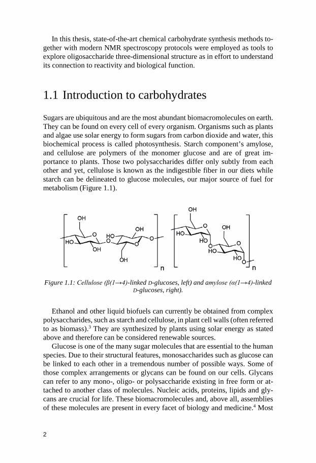
1.1 Introduction to carbohydrates Sugars are ubiquitous and are the most abundant biomacromolecules on earth. They can be found on every cell of every organism. Organisms such as plants and algae use solar energy to form sugars from carbon dioxide and water, this biochemical process is called photosynthesis. Starch component’s amylose, and cellulose are polymers of the monomer glucose and are of great im-portance to plants. Those two polysaccharides differ only subtly from each other and yet, cellulose is known as the indigestible fiber in our diets while starch can be delineated to glucose molecules, our major source of fuel for metabolism (Figure 1.1).
Figure 1.1: Cellulose (β(1→4)-linked D-glucoses, left) and amylose (α(1→4)-linked
D-glucoses, right).
Ethanol and other liquid biofuels can currently be obtained from complex
polysaccharides, such as starch and cellulose, in plant cell walls (often referred to as biomass).3 They are synthesized by plants using solar energy as stated above and therefore can be considered renewable sources.
Glucose is one of the many sugar molecules that are essential to the human species. Due to their structural features, monosaccharides such as glucose can be linked to each other in a tremendous number of possible ways. Some of those complex arrangements or glycans can be found on our cells. Glycans can refer to any mono-, oligo- or polysaccharide existing in free form or at-tached to another class of molecules. Nucleic acids, proteins, lipids and gly-cans are crucial for life. These biomacromolecules and, above all, assemblies of these molecules are present in every facet of biology and medicine.4 Most
3
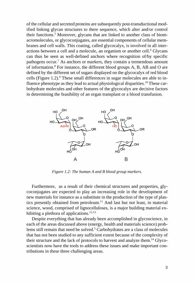
of the cellular and secreted proteins are subsequently post-transductional mod-ified linking glycan structures to there sequence, which alter and/or control their functions.5 Moreover, glycans that are linked to another class of biom-acromolecules, or glycoconjugates, are essential components of cellular mem-branes and cell walls. This coating, called glycocalyx, is involved in all inter-actions between a cell and a molecule, an organism or another cell.6 Glycans can thus be seen as well-defined anchors where recognition of/by specific pathogens occur.7 As anchors or markers, they contain a tremendous amount of information.8 For instance, the different blood groups A, B, AB and O are defined by the different set of sugars displayed on the glycocalyx of red blood cells (Figure 1.2).9 These small differences in sugar molecules are able to in-fluence phenotype as they lead to actual physiological disparities.10 These car-bohydrate molecules and other features of the glycocalyx are decisive factors in determining the feasibility of an organ transplant or a blood transfusion.
Figure 1.2: The human A and B blood group markers.
Furthermore, as a result of their chemical structures and properties, gly-coconjugates are expected to play an increasing role in the development of new materials for instance as a substitute in the production of the type of plas-tics presently obtained from petroleum.11 And last but not least, in material science, wood, comprised of lignocelluloses, is a major building material ex-hibiting a plethora of applications.12,13
Despite everything that has already been accomplished in glycoscience, in each of the areas discussed above (energy, health and materials science) prob-lems still remain that need be solved.5 Carbohydrates are a class of molecules that has not been studied to any sufficient extent because of the complexity of their structure and the lack of protocols to harvest and analyze them.14 Glyco-scientists now have the tools to address these issues and make important con-tributions in these three challenging areas.
4
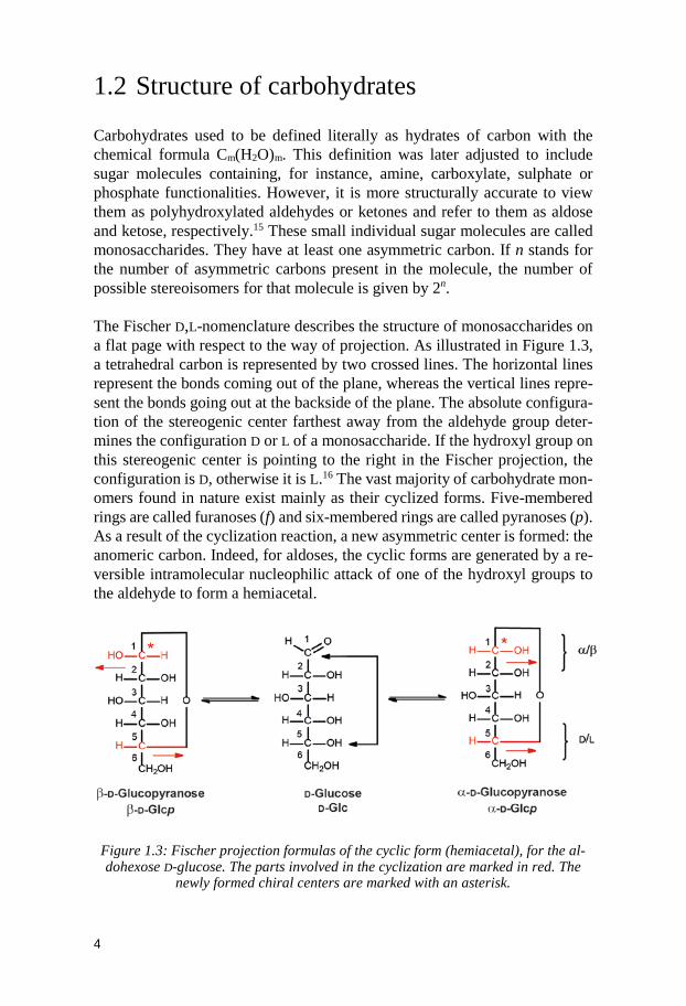
1.2 Structure of carbohydrates Carbohydrates used to be defined literally as hydrates of carbon with the chemical formula Cm(H2O)m. This definition was later adjusted to include sugar molecules containing, for instance, amine, carboxylate, sulphate or phosphate functionalities. However, it is more structurally accurate to view them as polyhydroxylated aldehydes or ketones and refer to them as aldose and ketose, respectively.15 These small individual sugar molecules are called monosaccharides. They have at least one asymmetric carbon. If n stands for the number of asymmetric carbons present in the molecule, the number of possible stereoisomers for that molecule is given by 2n. The Fischer D,L-nomenclature describes the structure of monosaccharides on a flat page with respect to the way of projection. As illustrated in Figure 1.3, a tetrahedral carbon is represented by two crossed lines. The horizontal lines represent the bonds coming out of the plane, whereas the vertical lines repre-sent the bonds going out at the backside of the plane. The absolute configura-tion of the stereogenic center farthest away from the aldehyde group deter-mines the configuration D or L of a monosaccharide. If the hydroxyl group on this stereogenic center is pointing to the right in the Fischer projection, the configuration is D, otherwise it is L.16 The vast majority of carbohydrate mon-omers found in nature exist mainly as their cyclized forms. Five-membered rings are called furanoses (f) and six-membered rings are called pyranoses (p). As a result of the cyclization reaction, a new asymmetric center is formed: the anomeric carbon. Indeed, for aldoses, the cyclic forms are generated by a re-versible intramolecular nucleophilic attack of one of the hydroxyl groups to the aldehyde to form a hemiacetal.
Figure 1.3: Fischer projection formulas of the cyclic form (hemiacetal), for the al-dohexose D-glucose. The parts involved in the cyclization are marked in red. The
newly formed chiral centers are marked with an asterisk.
5
If in the Fisher projection, the hydroxyl groups from the anomeric position and the linked configurational carbon project in the same direction the mono-saccharide is defined as the α-anomer, if in opposite directions it is defined as the β-anomer (Figure 1.3).
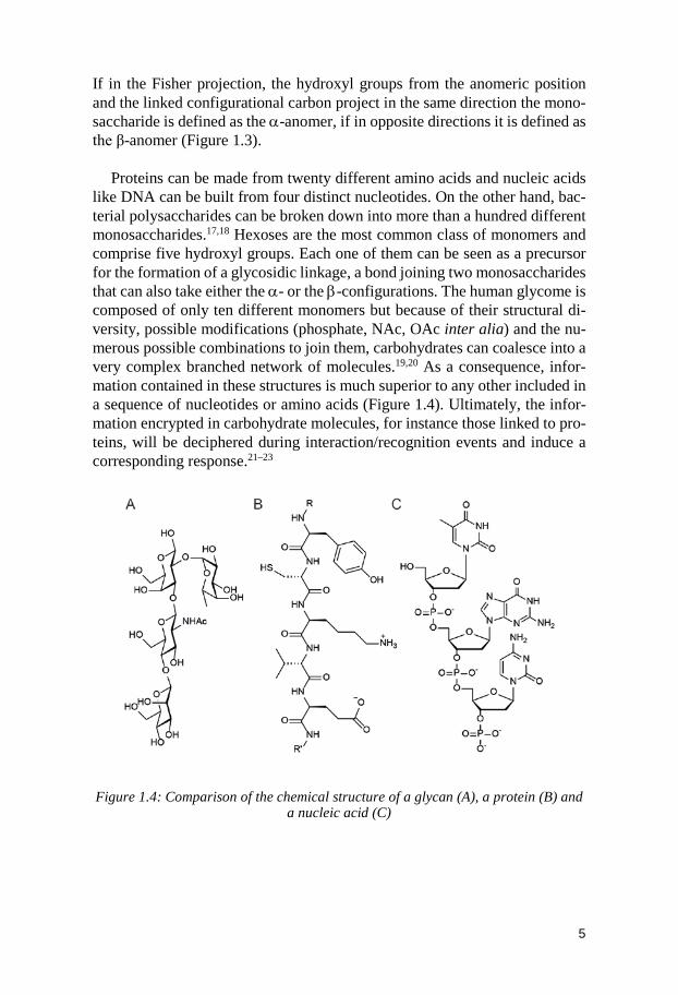
Proteins can be made from twenty different amino acids and nucleic acids like DNA can be built from four distinct nucleotides. On the other hand, bac-terial polysaccharides can be broken down into more than a hundred different monosaccharides.17,18 Hexoses are the most common class of monomers and comprise five hydroxyl groups. Each one of them can be seen as a precursor for the formation of a glycosidic linkage, a bond joining two monosaccharides that can also take either the α- or the β-configurations. The human glycome is composed of only ten different monomers but because of their structural di-versity, possible modifications (phosphate, NAc, OAc inter alia) and the nu-merous possible combinations to join them, carbohydrates can coalesce into a very complex branched network of molecules.19,20 As a consequence, infor-mation contained in these structures is much superior to any other included in a sequence of nucleotides or amino acids (Figure 1.4). Ultimately, the infor-mation encrypted in carbohydrate molecules, for instance those linked to pro-teins, will be deciphered during interaction/recognition events and induce a corresponding response.21–23
Figure 1.4: Comparison of the chemical structure of a glycan (A), a protein (B) and
a nucleic acid (C)
6
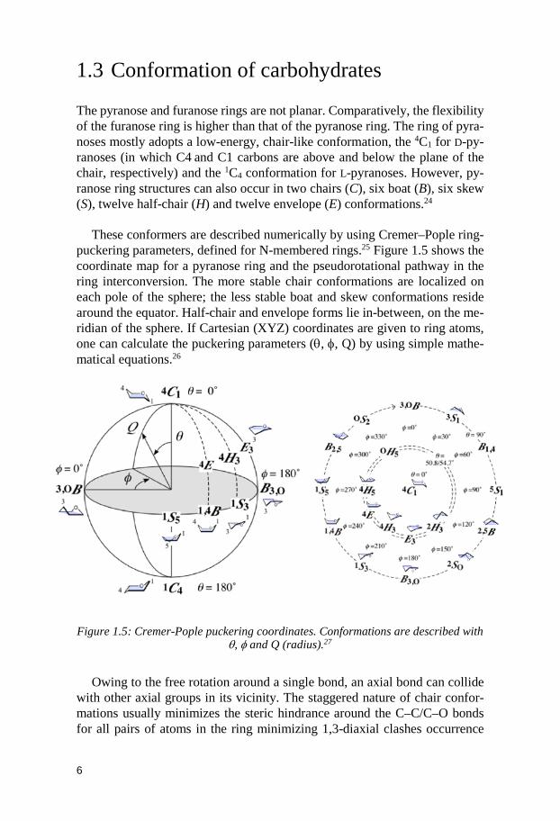
1.3 Conformation of carbohydrates The pyranose and furanose rings are not planar. Comparatively, the flexibility of the furanose ring is higher than that of the pyranose ring. The ring of pyra-noses mostly adopts a low-energy, chair-like conformation, the 4C1 for D-py-ranoses (in which C4 and C1 carbons are above and below the plane of the chair, respectively) and the 1C4 conformation for L-pyranoses. However, py-ranose ring structures can also occur in two chairs (C), six boat (B), six skew (S), twelve half-chair (H) and twelve envelope (E) conformations.24
These conformers are described numerically by using Cremer–Pople ring-puckering parameters, defined for N-membered rings.25 Figure 1.5 shows the coordinate map for a pyranose ring and the pseudorotational pathway in the ring interconversion. The more stable chair conformations are localized on each pole of the sphere; the less stable boat and skew conformations reside around the equator. Half-chair and envelope forms lie in-between, on the me-ridian of the sphere. If Cartesian (XYZ) coordinates are given to ring atoms, one can calculate the puckering parameters (θ, φ, Q) by using simple mathe-matical equations.26
Figure 1.5: Cremer-Pople puckering coordinates. Conformations are described with θ, φ and Q (radius).27
Owing to the free rotation around a single bond, an axial bond can collide
with other axial groups in its vicinity. The staggered nature of chair confor-mations usually minimizes the steric hindrance around the C–C/C–O bonds for all pairs of atoms in the ring minimizing 1,3-diaxial clashes occurrence
7
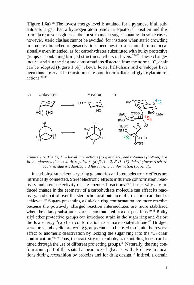
(Figure 1.6a).28 The lowest energy level is attained for a pyranose if all sub-stituents larger than a hydrogen atom reside in equatorial position and this formula represents glucose, the most abundant sugar in nature. In some cases, however, steric clashes cannot be avoided, for instance when steric crowding in complex branched oligosaccharides becomes too substantial, or are occa-sionally even intended, as for carbohydrates substituted with bulky protective groups or containing bridged structures, tethers or levers.29–35 These changes induce strain in the ring and conformations distorted from the normal 4C1 chair can be adopted (Figure 1.6b). Skews, boats, half-chairs and envelopes have been thus observed in transition states and intermediates of glycosylation re-actions.36,37
Figure 1.6: The (a) 1,3-diaxal interactions (top) and eclipsed rotamers (bottom) are both unfavored due to steric repulsion. (b) β-(1→2),β-(1→3)-linked glucoses where
each residue is adopting a different ring conformation (paper II).
In carbohydrate chemistry, ring geometries and stereoelectronic effects are intrinsically connected. Stereoelectronic effects influence conformation, reac-tivity and stereoselectivity during chemical reactions.38 That is why any in-duced change in the geometry of a carbohydrate molecule can affect its reac-tivity, and control over the stereochemical outcome of a reaction can thus be achieved.39 Sugars presenting axial-rich ring conformation are more reactive because the positively charged reaction intermediates are more stabilized when the alkoxy substituents are accommodated in axial positions.40,41 Bulky silyl ether protective groups can introduce strain in the sugar ring and distort the low energy 4C1 chair conformation to a more axial-rich one.42 Bridged structures and cyclic protecting groups can also be used to obtain the reverse effect or anomeric deactivation by locking the sugar ring into the 4C1 chair conformation.43,44 Thus, the reactivity of a carbohydrate building block can be tuned through the use of different protecting groups.45 Naturally, the ring con-formation, part of the spatial appearance of glycans, will also have implica-tions during recognition by proteins and for drug design.46 Indeed, a certain
8
ring conformation may present the best structural requirements for nucleo-philic displacement (including bond elongation/shrinking, leaving-group ori-entation and charge distribution) during glycosylation or enzymatic glycosyl hydrolysis reactions.47
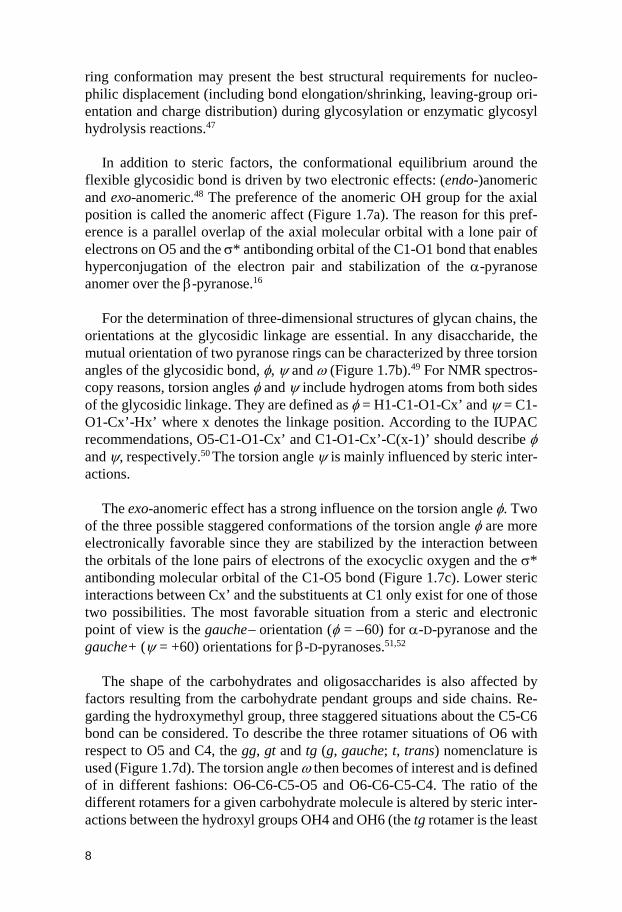
In addition to steric factors, the conformational equilibrium around the flexible glycosidic bond is driven by two electronic effects: (endo-)anomeric and exo-anomeric.48 The preference of the anomeric OH group for the axial position is called the anomeric affect (Figure 1.7a). The reason for this pref-erence is a parallel overlap of the axial molecular orbital with a lone pair of electrons on O5 and the σ* antibonding orbital of the C1-O1 bond that enables hyperconjugation of the electron pair and stabilization of the α-pyranose anomer over the β-pyranose.16
For the determination of three-dimensional structures of glycan chains, the
orientations at the glycosidic linkage are essential. In any disaccharide, the mutual orientation of two pyranose rings can be characterized by three torsion angles of the glycosidic bond, φ, ψ and ω (Figure 1.7b).49 For NMR spectros-copy reasons, torsion angles φ and ψ include hydrogen atoms from both sides of the glycosidic linkage. They are defined as φ = H1-C1-O1-Cx’ and ψ = C1-O1-Cx’-Hx’ where x denotes the linkage position. According to the IUPAC recommendations, O5-C1-O1-Cx’ and C1-O1-Cx’-C(x-1)’ should describe φ and ψ, respectively.50 The torsion angle ψ is mainly influenced by steric inter-actions.
The exo-anomeric effect has a strong influence on the torsion angle φ. Two
of the three possible staggered conformations of the torsion angle φ are more electronically favorable since they are stabilized by the interaction between the orbitals of the lone pairs of electrons of the exocyclic oxygen and the σ* antibonding molecular orbital of the C1-O5 bond (Figure 1.7c). Lower steric interactions between Cx’ and the substituents at C1 only exist for one of those two possibilities. The most favorable situation from a steric and electronic point of view is the gauche− orientation (φ = −60) for α-D-pyranose and the gauche+ (ψ = +60) orientations for β-D-pyranoses.51,52
The shape of the carbohydrates and oligosaccharides is also affected by
factors resulting from the carbohydrate pendant groups and side chains. Re-garding the hydroxymethyl group, three staggered situations about the C5-C6 bond can be considered. To describe the three rotamer situations of O6 with respect to O5 and C4, the gg, gt and tg (g, gauche; t, trans) nomenclature is used (Figure 1.7d). The torsion angle ω then becomes of interest and is defined of in different fashions: O6-C6-C5-O5 and O6-C6-C5-C4. The ratio of the different rotamers for a given carbohydrate molecule is altered by steric inter-actions between the hydroxyl groups OH4 and OH6 (the tg rotamer is the least
9
populated in glucose while it is the gg in galactose) and by the gauche effect which favors a gauche relationship between O5 and O6 (gg and gt confor-mations over tg).53
OHO OH
OHO
HO OOH
OH
OH
HOOHO
HO
OH
HOO
HOHO
OH
HO O
OH
..
..
n
σ∗
σ∗
n
a b c
ω
φ ψ
Figure 1.7: (a) Rationalization for the endo-anomeric effect. (b) β-cellobiose with the torsion angles φ, ψ and ω. (c) Rationalization for the exo-anomeric effect. (d)
The three rotamers of the ω torsion angle.
The ring conformation, the mutual orientation around a glycosidic bond and the positions of the side chains also influence the formation of intra- and inter-residual hydrogen bonds, solvent accessibility and bonding ability to bi-omacromolecules.54 Clearly, understanding the conformational behavior of ol-igosaccharides is essential to identify the relationship between the glycans’ shape and their related biochemical and physiological functions.
10
2 Synthesis of Carbohydrates
In contrast to proteins, polysaccharides and glycoconjugates are not encoded directly by genes. Instead, the biosynthesis of glycans depends on the expres-sion of genome-encoded enzymes. Then, as the cell undergoes developmental, physiological, and pathological changes, the composition of cell-surface car-bohydrates alters and becomes thereupon specific to the cell type.55 As a re-sult, the development of effective methods for glycan analysis and synthesis is required to perform the complex task of determining their biological func-tions. Unlike oligonucleotide and peptide synthesis, and despite the progress in solid-phase automated synthesis,56 there are no general protocols or scalable methods for the preparation of glycans, and glycoconjugates in biological samples are often found in low concentrations and in microheterogeneous forms.5 Furthermore, it is not only just a simple sequence of building blocks that needs to be elongated, but the glycosidic linkages created must link the correct connection points and have the right stereochemistry. Consequently, the synthesis of a carbohydrate target is often a research project in itself, which may take many months and in some cases years to complete.
Synthetic oligosaccharides have been used in the development of vaccines directed against pathogens such as HIV, Vibrio cholerae, Streptococcus pneu-moniae, Shigella dysenteriae, Neisseria meningitides, Bacillus anthraces and Haemophilus influenzae type b (Hib).57,58 The use of organic synthesis is of paramount importance in the development of glycoconjugate vaccines as it permits the isolation of pure potential antigenic epitopes, and it has recently been highlighted by the marketing of a prophylactic treatment towards Hib infections in Cuba.59,60
2.1 Enzymatic synthesis Glycosidic linkages can be constructed enzymatically or chemoenzymatically using for instance glycosyltransferases (GT), glycosidases (GH) or glycosyn-thases (GS). Theoretically, there is a unique glycosyltransferase for every type of glycosidic linkage. GTs link carbohydrate molecules together in an assem-
11
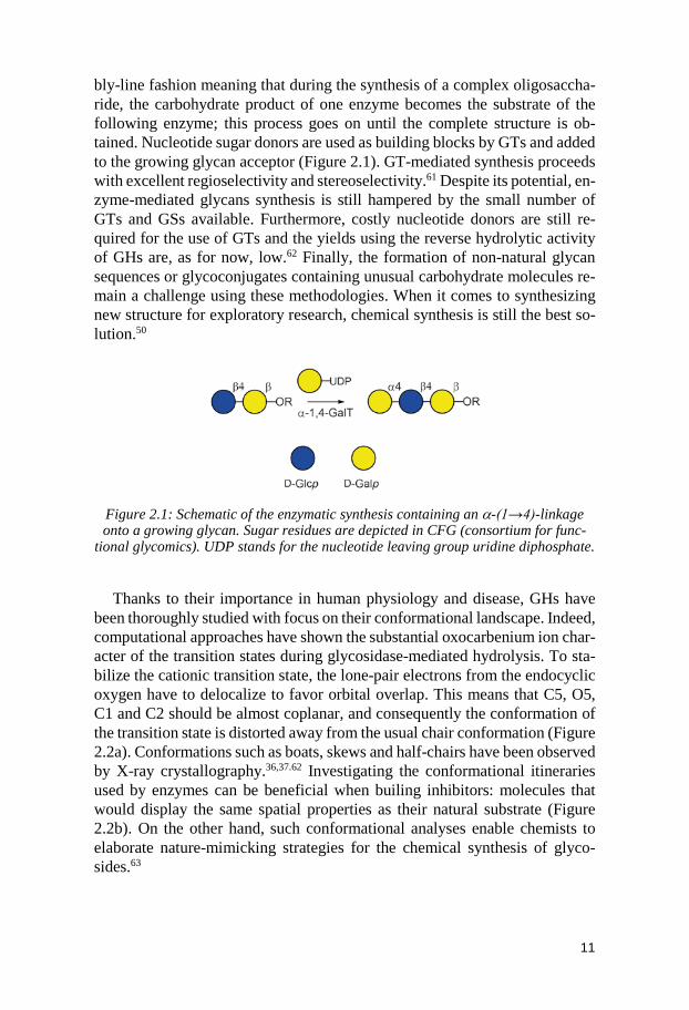
bly-line fashion meaning that during the synthesis of a complex oligosaccha-ride, the carbohydrate product of one enzyme becomes the substrate of the following enzyme; this process goes on until the complete structure is ob-tained. Nucleotide sugar donors are used as building blocks by GTs and added to the growing glycan acceptor (Figure 2.1). GT-mediated synthesis proceeds with excellent regioselectivity and stereoselectivity.61 Despite its potential, en-zyme-mediated glycans synthesis is still hampered by the small number of GTs and GSs available. Furthermore, costly nucleotide donors are still re-quired for the use of GTs and the yields using the reverse hydrolytic activity of GHs are, as for now, low.62 Finally, the formation of non-natural glycan sequences or glycoconjugates containing unusual carbohydrate molecules re-main a challenge using these methodologies. When it comes to synthesizing new structure for exploratory research, chemical synthesis is still the best so-lution.50
Figure 2.1: Schematic of the enzymatic synthesis containing an α-(1→4)-linkage onto a growing glycan. Sugar residues are depicted in CFG (consortium for func-
tional glycomics). UDP stands for the nucleotide leaving group uridine diphosphate.
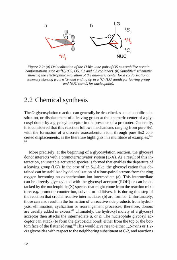
Thanks to their importance in human physiology and disease, GHs have been thoroughly studied with focus on their conformational landscape. Indeed, computational approaches have shown the substantial oxocarbenium ion char-acter of the transition states during glycosidase-mediated hydrolysis. To sta-bilize the cationic transition state, the lone-pair electrons from the endocyclic oxygen have to delocalize to favor orbital overlap. This means that C5, O5, C1 and C2 should be almost coplanar, and consequently the conformation of the transition state is distorted away from the usual chair conformation (Figure 2.2a). Conformations such as boats, skews and half-chairs have been observed by X-ray crystallography.36,37.62 Investigating the conformational itineraries used by enzymes can be beneficial when builing inhibitors: molecules that would display the same spatial properties as their natural substrate (Figure 2.2b). On the other hand, such conformational analyses enable chemists to elaborate nature-mimicking strategies for the chemical synthesis of glyco-sides.63
12
Figure 2.2: (a) Delocalization of the Π-like lone-pair of O5 can stabilize certain
conformations such as 4H3 (C5, O5, C1 and C2 coplanar). (b) Simplified schematic showing the electrophilic migration of the anomeric center for a conformational itinerary starting from a 1S3 and ending up in a 4C1 (LG stands for leaving group
and NUC stands for nucleophile).
2.2 Chemical synthesis The O-glycosylation reaction can generally be described as a nucleophilic sub-stitution, or displacement of a leaving group at the anomeric center of a gly-cosyl donor by a glycosyl acceptor in the presence of a promoter. Generally, it is considered that this reaction follows mechanisms ranging from pure SN1 with the formation of a discrete oxocarbenium ion, through pure SN2 con-certed displacements, as the literature highlights in a multitude of examples.64–
66
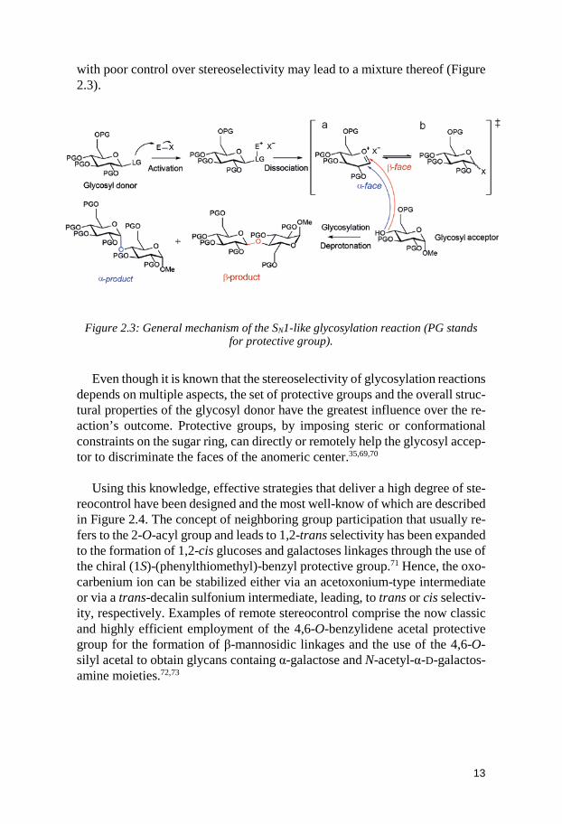
More precisely, at the beginning of a glycosylation reaction, the glycosyl donor interacts with a promoter/activator system (E-X). As a result of this in-teraction, an unstable activated species is formed that enables the departure of a leaving group (LG). In the case of an SN1-like, the glycosyl cation thus ob-tained can be stabilized by delocalization of a lone-pair electrons from the ring oxygen becoming an oxocarbenium ion intermediate (a). This intermediate can be directly glycosylated with the glycosyl acceptor (ROH) or can be at-tacked by the nucleophilic (X) species that might come from the reaction mix-ture: e.g. promoter counter-ion, solvent or additives. It is during this step of the reaction that crucial reactive intermediates (b) are formed. Unfortunately, those can also result in the formation of unreactive side products from hydrol-ysis, elimination, cyclization or rearrangement processes; therefore, donors are usually added in excess.67 Ultimately, the hydroxyl moiety of a glycosyl acceptor then attacks the intermediate a, or b. The nucleophile glycosyl ac-ceptor can attack (to form the glycosidic bond) either from the top or the bot-tom face of the flattened ring.68 This would give rise to either 1,2-trans or 1,2-cis glycosides with respect to the neighboring substituent at C-2, and reactions
13
with poor control over stereoselectivity may lead to a mixture thereof (Figure 2.3).
Figure 2.3: General mechanism of the SN1-like glycosylation reaction (PG stands for protective group).
Even though it is known that the stereoselectivity of glycosylation reactions
depends on multiple aspects, the set of protective groups and the overall struc-tural properties of the glycosyl donor have the greatest influence over the re-action’s outcome. Protective groups, by imposing steric or conformational constraints on the sugar ring, can directly or remotely help the glycosyl accep-tor to discriminate the faces of the anomeric center.35,69,70
Using this knowledge, effective strategies that deliver a high degree of ste-
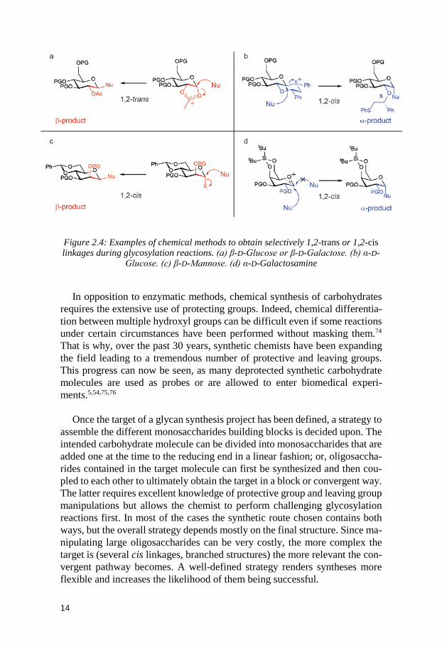
reocontrol have been designed and the most well-know of which are described in Figure 2.4. The concept of neighboring group participation that usually re-fers to the 2-O-acyl group and leads to 1,2-trans selectivity has been expanded to the formation of 1,2-cis glucoses and galactoses linkages through the use of the chiral (1S)-(phenylthiomethyl)-benzyl protective group.71 Hence, the oxo-carbenium ion can be stabilized either via an acetoxonium-type intermediate or via a trans-decalin sulfonium intermediate, leading, to trans or cis selectiv-ity, respectively. Examples of remote stereocontrol comprise the now classic and highly efficient employment of the 4,6-O-benzylidene acetal protective group for the formation of β-mannosidic linkages and the use of the 4,6-O-silyl acetal to obtain glycans containg α-galactose and N-acetyl-α-D-galactos-amine moieties.72,73
14
Figure 2.4: Examples of chemical methods to obtain selectively 1,2-trans or 1,2-cis linkages during glycosylation reactions. (a) β-D-Glucose or β-D-Galactose. (b) α-D-
Glucose. (c) β-D-Mannose. (d) α-D-Galactosamine
In opposition to enzymatic methods, chemical synthesis of carbohydrates requires the extensive use of protecting groups. Indeed, chemical differentia-tion between multiple hydroxyl groups can be difficult even if some reactions under certain circumstances have been performed without masking them.74 That is why, over the past 30 years, synthetic chemists have been expanding the field leading to a tremendous number of protective and leaving groups. This progress can now be seen, as many deprotected synthetic carbohydrate molecules are used as probes or are allowed to enter biomedical experi-ments.5,54,75,76
Once the target of a glycan synthesis project has been defined, a strategy to
assemble the different monosaccharides building blocks is decided upon. The intended carbohydrate molecule can be divided into monosaccharides that are added one at the time to the reducing end in a linear fashion; or, oligosaccha-rides contained in the target molecule can first be synthesized and then cou-pled to each other to ultimately obtain the target in a block or convergent way. The latter requires excellent knowledge of protective group and leaving group manipulations but allows the chemist to perform challenging glycosylation reactions first. In most of the cases the synthetic route chosen contains both ways, but the overall strategy depends mostly on the final structure. Since ma-nipulating large oligosaccharides can be very costly, the more complex the target is (several cis linkages, branched structures) the more relevant the con-vergent pathway becomes. A well-defined strategy renders syntheses more flexible and increases the likelihood of them being successful.
15
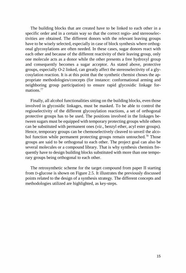
The building blocks that are created have to be linked to each other in a
specific order and in a certain way so that the correct regio- and stereoselec-tivities are obtained. The different donors with the relevant leaving groups have to be wisely selected, especially in case of block synthesis where orthog-onal glycosylations are often needed. In these cases, sugar donors react with each other and because of the different reactivity of their leaving group, only one molecule acts as a donor while the other presents a free hydroxyl group and consequently becomes a sugar acceptor. As stated above, protective groups, especially O-2 linked, can greatly affect the stereoselectivity of a gly-cosylation reaction. It is at this point that the synthetic chemist choses the ap-propriate methodologies/concepts (for instance: conformational arming and neighboring group participation) to ensure rapid glycosidic linkage for-mations.77
Finally, all alcohol functionalities sitting on the building blocks, even those
involved in glycosidic linkages, must be masked. To be able to control the regioselectivity of the different glycosylation reactions, a set of orthogonal protective groups has to be used. The positions involved in the linkages be-tween sugars must be equipped with temporary protecting groups while others can be substituted with permanent ones (viz., benzyl ether, acyl ester groups). Hence, temporary groups can be chemoselectively cleaved to unveil the alco-hol function while permanent protecting groups remain untouched.78 Those groups are said to be orthogonal to each other. The project goal can also be several molecules or a compound library. That is why synthesis chemists fre-quently have to design building blocks substituted with more than one tempo-rary groups being orthogonal to each other.
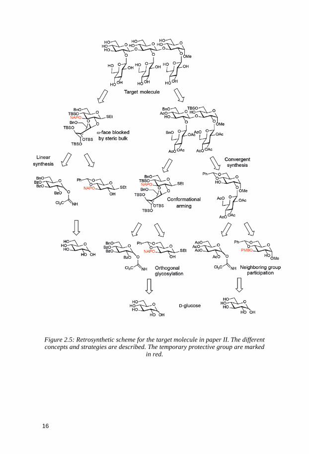
The retrosynthetic scheme for the target compound from paper II starting
from D-glucose is shown on Figure 2.5. It illustrates the previously discussed points related to the design of a synthesis strategy. The different concepts and methodologies utilized are highlighted, as key-steps.
16
Figure 2.5: Retrosynthetic scheme for the target molecule in paper II. The different concepts and strategies are described. The temporary protective group are marked
in red.
17
3 Analytical Methods
3.1 NMR spectroscopy In organic chemistry, NMR spectroscopy is a powerful non-destructive ana-lytic method as it permits swiftly to certify a structure or the purity of a com-pound and allows its total characterization at the atomic-level. Because of its ability to provide detailed information about the three-dimensional features of molecules, NMR spectroscopy is extensively used in carbohydrate chemis-try.79,80
In an NMR spectrometer, a strong magnetic field as well as external pulsed
electromagnetic radiations are applied to nuclei with a nonzero magnetic spin quantum number m (for example 1H and 13C). At equilibrium, there is a slight difference in population between the quantized spin states that also differ in energy. Electromagnetic radiations corresponding to this difference in energy perturb the system by altering the populations of the spin states. The system then relaxes, and the transitions that allows the populations to go back to their equilibrium level are detected as resonance frequencies. After applying a Fou-rier transform to the signal, each resonance frequency gives rise to a peak in the NMR spectrum called resonance chemical shift, that is relative to a refer-ence nucleus (e.g. TMS in CDCl3, TSP in H2O). The resonance frequency of a given nuclei depends on its chemical environment, its gyromagnetic ratio and the strength of the magnetic field. The sensitivity of a nucleus to the local electron distribution makes it possible to differentiate almost all nuclei of the same kind in a molecule, as it results in a difference in chemical shifts.
Total characterization of a carbohydrate molecule can be acquired through
the use of various NMR experiments. Pulse sequences have been developed to extract through-bond or through-space correlations between nuclei of the same kind (homonuclear, e.g. 1H, 1H-COSY, 1H, 1H-TOCSY, 1H, 1H-NOESY) or between two different sorts of nuclei (heteronuclear, e.g. 1H, 13C-HSQC, 1H, 13C-HMBC, 1H, 13C-HETCOR). Not only structural information can be obtained through solution NMR spectroscopy but conformations and internal motions of oligo- and polysaccharides can also be investigated. Experiments have been designed to measure scalar coupling constants, relaxation parame-ters, residual dipolar coupling and nuclear Overhauser effect correlations; all
18
related to the three-dimensional structure of carbohydrates. Furthermore, NMR samples can be prepared and experimental conditions modified. This allows, for instance, the conformation of glycan moieties linked to proteins or protein-carbohydrate interactions to be studied, in the most relevant fashion, namely, close to physiological conditions. The work in this thesis focuses on the most studied nuclei: the 1H, which has a high gyromagnetic ratio and the less sensitive 13C. Scalar coupling constants and nuclear Overhauser effect correlations are additionally used to analyze the conformation of carbohy-drates in solution.
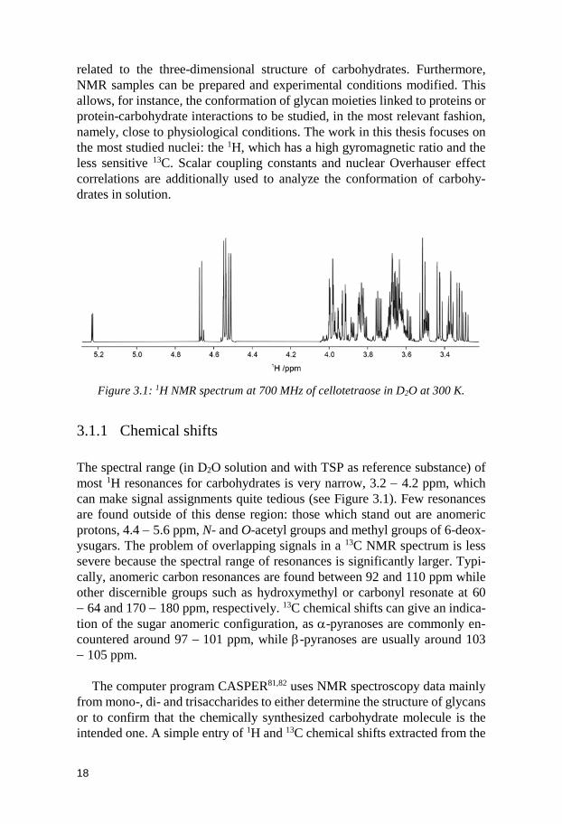
Figure 3.1: 1H NMR spectrum at 700 MHz of cellotetraose in D2O at 300 K.
3.1.1 Chemical shifts The spectral range (in D2O solution and with TSP as reference substance) of most 1H resonances for carbohydrates is very narrow, 3.2 − 4.2 ppm, which can make signal assignments quite tedious (see Figure 3.1). Few resonances are found outside of this dense region: those which stand out are anomeric protons, 4.4 − 5.6 ppm, N- and O-acetyl groups and methyl groups of 6-deox-ysugars. The problem of overlapping signals in a 13C NMR spectrum is less severe because the spectral range of resonances is significantly larger. Typi-cally, anomeric carbon resonances are found between 92 and 110 ppm while other discernible groups such as hydroxymethyl or carbonyl resonate at 60 − 64 and 170 − 180 ppm, respectively. 13C chemical shifts can give an indica-tion of the sugar anomeric configuration, as α-pyranoses are commonly en-countered around 97 − 101 ppm, while β-pyranoses are usually around 103 − 105 ppm.
The computer program CASPER81,82 uses NMR spectroscopy data mainly
from mono-, di- and trisaccharides to either determine the structure of glycans or to confirm that the chemically synthesized carbohydrate molecule is the intended one. A simple entry of 1H and 13C chemical shifts extracted from the
19
1D NMR spectra is enough for the program to list the ten-top ranked se-quences corresponding to the most probable carbohydrate structures. Besides, the software predictions ability has been enhanced by including more biolog-ically relevant carbohydrate moieties to its database (cf. paper IV). If the pro-gram is used for fast structure verification, monosaccharides and glycosidic linkages can be selected; once the data is computed, a list of chemical shifts then appears.83 The software can also take as input coupling constants 3JH1,H2
and 1JC1,H1 as well as unassigned through-bond correlations obtained from 2D NMR experiments such as TOCSY, HSQC/HETCOR and HMBC. Giving more data to the system allows refinement of the prediction from the Deter-mine Structure application and the desired structure will eventually become the top-ranked one. Thus, as stated in paper I, the synthesized oligosaccharides could be visualized as the first on the list given by CASPER with the help of 2D NMR data.
3.1.2 Coupling constants The form of a resonance can be altered when other magnetic nuclei are nearby. The influence of neighboring spins on the multiplicity of peaks is called spin-spin splitting or indirect coupling. In an NMR spectrum, the separation be-tween two peaks for the resonance of one nucleus split by another is a measure of how strongly the nuclear spins influence each other, and is called the scalar coupling constant J, measured in Hertz (Hz). The source of scalar coupling in molecules is an indirect interaction between NMR active nuclei mediated by the electrons involved in chemical bonding. Because J normally represents an interaction through covalent bonds between spins separated by 1 − 4 bonds, it is a useful parameter in drawing conclusions about molecular bonding, such as bond strengths and steric arrangements.84 Furthermore, the magnitude of both homonuclear (3JHH and 3JCC) and heteronuclear (3JCH) coupling constants is related to molecule geometry and thus, to torsion angles.85 This relationship is of great interest when one studies the conformation of oligosaccharides and can be expressed as a Karplus-type equation of the form:
J(θ) = A cos2θ + B cosθ + C
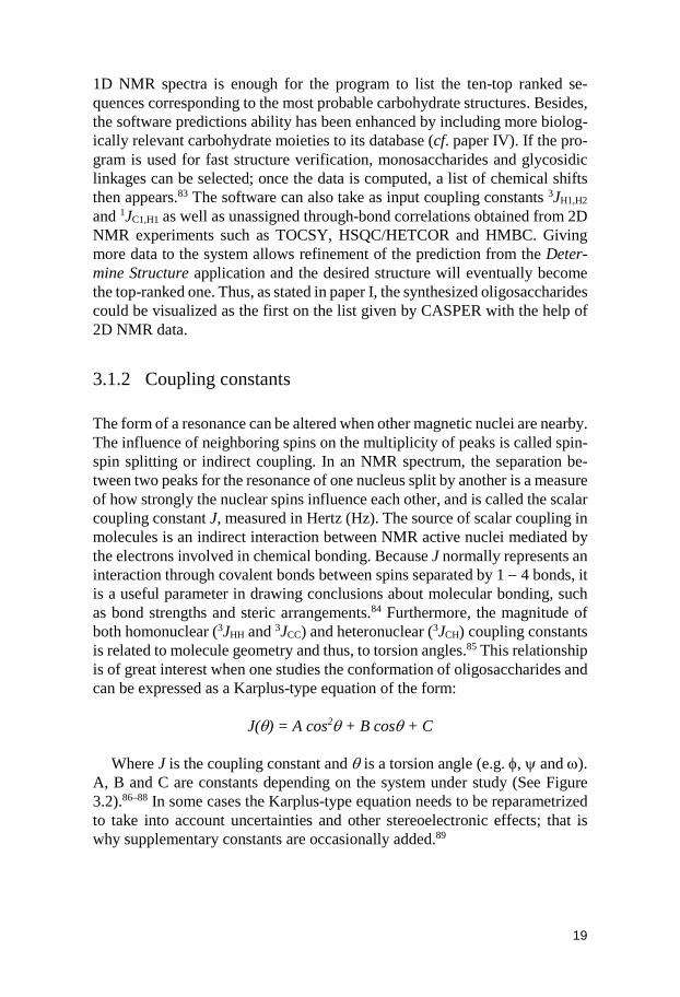
Where J is the coupling constant and θ is a torsion angle (e.g. φ, ψ and ω). A, B and C are constants depending on the system under study (See Figure 3.2).86–88 In some cases the Karplus-type equation needs to be reparametrized to take into account uncertainties and other stereoelectronic effects; that is why supplementary constants are occasionally added.89
20
Figure 3.2: Plot of the Karplus type relationship J(θ) = 3.70·cos2 (θ) + 0.18·cos (θ) + 0.11 for 3JCOCC couplings (left).89 Plot of the coupling constants 3JH5,H6 as function
of the torsion angle ω (right).86
3JHH coupling constants can often be extracted from a splitting in a 1D 1H
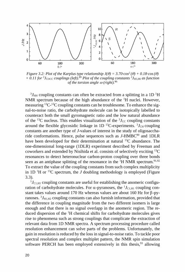
NMR spectrum because of the high abundance of the 1H nuclei. However, measuring 13C-13C coupling constants can be troublesome. To enhance the sig-nal-to-noise ratio, the carbohydrate molecule can be isotopically labelled to counteract both the small gyromagnetic ratio and the low natural abundance of the 13C nucleus. This enables visualization of the 3JCC coupling constants around the flexible glycosidic linkage in 1D 13C-experiments. 3JCH coupling constants are another type of J-values of interest in the study of oligosaccha-ride conformations. Hence, pulse sequences such as J-HMBC90 and 1DLR have been developed for their determination at natural 13C abundance. The one-dimensional long-range (1DLR) experiment described by Freeman and coworkers and extended by Nisihida et al. consists of selectively exciting 13C resonances to detect heteronuclear carbon-proton coupling over three bonds seen as an antiphase splitting of the resonance in the 1H NMR spectrum.91,92 To extract the value of the coupling constants from such complex multiplicity in 1D 1H or 13C spectrum, the J doubling methodology is employed (Figure 3.3).
1JC1,H1 coupling constants are useful for establishing the anomeric configu-ration of carbohydrate molecules. For α-pyranoses, the 1JC1,H1 coupling con-stant takes values around 170 Hz whereas values are about 160 Hz for β-py-ranoses. 3JH1,H2 coupling constants can also furnish information, provided that the difference in coupling magnitude from the two different isomers is large enough and that there is no signal overlapp in the anomeric region. The re-duced dispersion of the 1H chemical shifts for carbohydrate molecules gives rise to phenomena such as strong couplings that complicate the extraction of relevant data from 1D NMR spectra. A spectrum processing procedure called resolution enhancement can solve parts of the problems. Unfortunately, the gain in resolution is reduced by the loss in signal-to-noise ratio. To tackle poor spectral resolution and complex multiplet pattern, the NMR spin simulation software PERCH has been employed extensively in this thesis,93 allowing
21
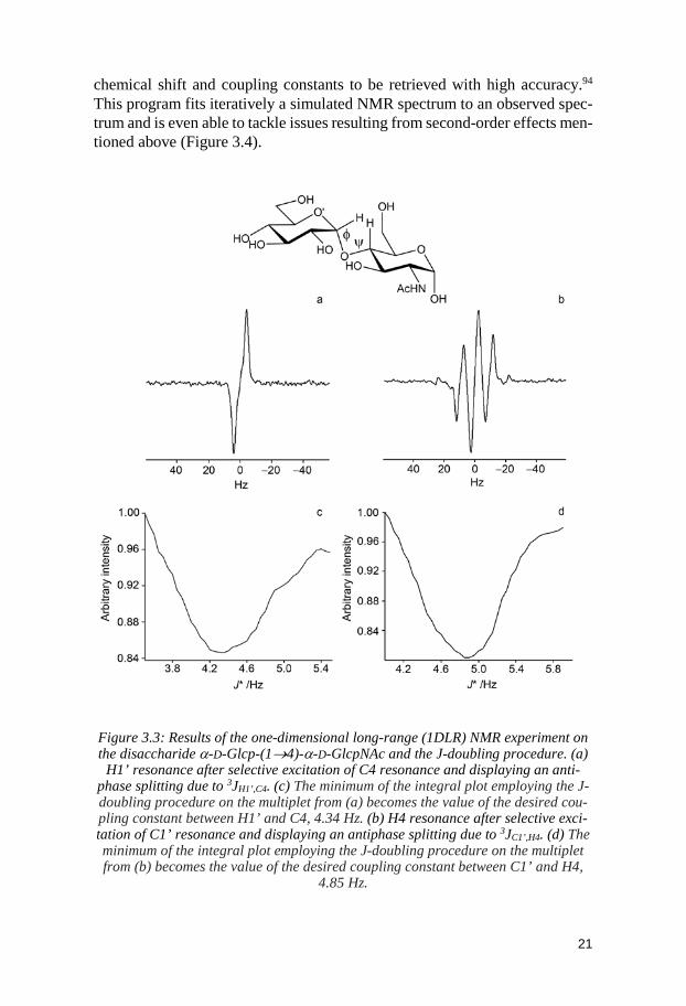
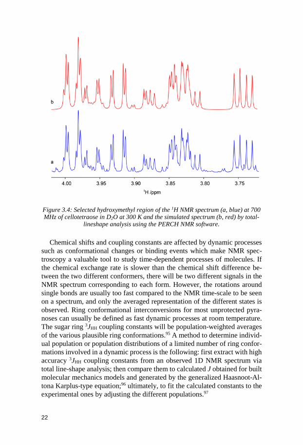
chemical shift and coupling constants to be retrieved with high accuracy.94 This program fits iteratively a simulated NMR spectrum to an observed spec-trum and is even able to tackle issues resulting from second-order effects men-tioned above (Figure 3.4).
Figure 3.3: Results of the one-dimensional long-range (1DLR) NMR experiment on the disaccharide α-D-Glcp-(1→4)-α-D-GlcpNAc and the J-doubling procedure. (a)
H1’ resonance after selective excitation of C4 resonance and displaying an anti-phase splitting due to 3JH1’,C4. (c) The minimum of the integral plot employing the J-doubling procedure on the multiplet from (a) becomes the value of the desired cou-pling constant between H1’ and C4, 4.34 Hz. (b) H4 resonance after selective exci-tation of C1’ resonance and displaying an antiphase splitting due to 3JC1’,H4. (d) The minimum of the integral plot employing the J-doubling procedure on the multiplet from (b) becomes the value of the desired coupling constant between C1’ and H4,
4.85 Hz.
22
Figure 3.4: Selected hydroxymethyl region of the 1H NMR spectrum (a, blue) at 700 MHz of cellotetraose in D2O at 300 K and the simulated spectrum (b, red) by total-
lineshape analysis using the PERCH NMR software.
Chemical shifts and coupling constants are affected by dynamic processes such as conformational changes or binding events which make NMR spec-troscopy a valuable tool to study time-dependent processes of molecules. If the chemical exchange rate is slower than the chemical shift difference be-tween the two different conformers, there will be two different signals in the NMR spectrum corresponding to each form. However, the rotations around single bonds are usually too fast compared to the NMR time-scale to be seen on a spectrum, and only the averaged representation of the different states is observed. Ring conformational interconversions for most unprotected pyra-noses can usually be defined as fast dynamic processes at room temperature. The sugar ring 3JHH coupling constants will be population-weighted averages of the various plausible ring conformations.95 A method to determine individ-ual population or population distributions of a limited number of ring confor-mations involved in a dynamic process is the following: first extract with high accuracy 3JHH coupling constants from an observed 1D NMR spectrum via total line-shape analysis; then compare them to calculated J obtained for built molecular mechanics models and generated by the generalized Haasnoot-Al-tona Karplus-type equation;96 ultimately, to fit the calculated constants to the experimental ones by adjusting the different populations.97
23
3.1.3 The nuclear Overhauser effect After being perturbed by pulsed electromagnetic radiations, the nuclei mag-netization has to return to an equilibrium state. This process is called spin re-laxation and arises from different mechanisms. One of them, which is also the dominating cause for relaxation of the nuclei studied in this thesis, is dipolar coupling. The nuclear Overhauser effect (NOE) describes the effect of a spin relaxing after selective perturbation to its neighbors involved in the dipolar interaction. The intensity of the NOE depends on the strength of the magnetic field and the correlation time τc, itself related to the global tumbling of the molecule in solution. From experiments such as 1D NOESY and STEP-NO-ESY as well as 1D T-ROESY, dipole-dipole cross relaxation rates (σ) can be measured for individual proton-proton interactions. Using the isolated spin pair approximation (ISPA)98, σij is said to be proportional to r-6 with r being the distance between the two dipolar coupled spins i and j. Thus, the internu-clear distance rij can be calculated with the aid of a reference spins pair and the associated known distance separating them.
rij = rref (σref/σij)1/6
Quantitative measurements of 1H-1H distances are undoubtedly useful for structure elucidation of molecules in organic chemistry but, they are of even greater interest for carbohydrate chemistry as they can give information about the three-dimensional structure of oligo- and polysaccharides. Interatomic dis-tances around the glycosidic linkage partly enables the determination of the preferential conformation of oligosaccharides in solution.99–101 The effective correlation time of a molecule can be estimated if cross-relaxation rates have been measured both from NOESY and T-ROESY experiments; the value can be extracted from the ratio of the two rates.102
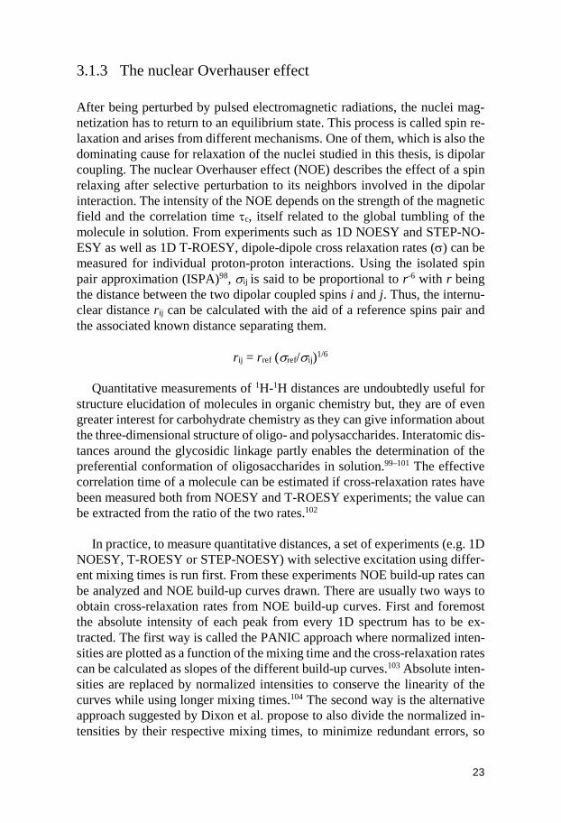
In practice, to measure quantitative distances, a set of experiments (e.g. 1D NOESY, T-ROESY or STEP-NOESY) with selective excitation using differ-ent mixing times is run first. From these experiments NOE build-up rates can be analyzed and NOE build-up curves drawn. There are usually two ways to obtain cross-relaxation rates from NOE build-up curves. First and foremost the absolute intensity of each peak from every 1D spectrum has to be ex-tracted. The first way is called the PANIC approach where normalized inten-sities are plotted as a function of the mixing time and the cross-relaxation rates can be calculated as slopes of the different build-up curves.103 Absolute inten-sities are replaced by normalized intensities to conserve the linearity of the curves while using longer mixing times.104 The second way is the alternative approach suggested by Dixon et al. propose to also divide the normalized in-tensities by their respective mixing times, to minimize redundant errors, so
24
that cross-relaxation rates can be obtained as the interception between the curves and the ordinate axis.105
Figure 3.5: Schematic of the STEP-NOESY experiment for measuring the cross-re-
laxation rate between H4 and H1' in α-cellobiose; selective excitation of H1 (1), fol-lowed by isotropic mixing transfers magnetization to H4, which then can be selec-
tively inverted (2), for subsequent cross-relaxation to H1' (bottom). 1H,1H-NOE buildup curves for α-cellobiose obtained at a 600 MHz spectrometer frequency em-ploying the PANIC approach in which –Ij/Ii vs. τmix are plotted. The cross-relaxation
rates are obtained from the slopes of the fitted data; reference distance H1-H2 (filled squares) and H4-H1' (filled triangles) (top).
25
3.2 Molecular dynamics simulations in conformational analysis
Molecular simulations play an important role in the analysis of conformations and dynamics of oligosaccharides. Unlike X-ray crystallography, which gives a static image of a crystalline state, or NMR spectroscopy, where a time aver-age of motions is seen; molecular dynamics (MD) simulations can provide detailed insights at an atomic level about motions occurring on a short time scale.106 For instance, the conformational flexibility of an oligosaccharide may be investigated using MD simulations. These computer simulations use a highly specific molecular force field relevant for the studied system and is based on Newton’s second law of motion.107–109 One of the great advantages of the MD technique is that explicit water molecules can be readily included in the simulations, which is important for extremely diluted carbohydrate set-ups or to simulate possible water mediated protein-ligand interactions. The MD simulation technique gives, in addition to conformational averaging and transitions to different conformational states, explicit information on the time-scales for these processes. A time course of a variable, such as atom-atom distances or a torsion angle, can be analyzed.110 These variables are to be com-pared with NMR spectroscopy data; for example NOE-derived internuclear distances and J couplings.
Even though calculations can be performed in a highly sophisticated man-ner, they still need experimental validation to ensure their reliability and their relevance. The combination of experimental and simulated data is very pow-erful and can give much information about a system.111 Ultimately, the results from an MD simulations may be visualized in the form of Ramachandran-like maps where the conformational space of a molecule is depicted. The global energy minimum (in vacuo), as well as low energy local minima, can then be identified from the φ/ψ grid obtained.
26
4 Synthesis of the Tetrasaccharide Glycoside Moiety of Solaradixine and Rapid NMR-Based Structure Verification Using the Program CASPER (Paper I)
Saponins are a structurally diverse class of natural products that are character-ized by a non-polar sapogenin moiety glycosidically linked to one or more polar carbohydrate moieties.112 They are biologically active compounds that can be used in pharmacological applications owing to their cytotoxic effects, immunostimulatory, anti-inflammatory, antiviral and hypoglycemic activities among others.113 Nitrogen containing steroid type skeleton glycosidically linked to carbohydrate moieties are classified as steroidal glycoalkaloids. In contrast to other saponins, the occurrences of steroidal glycoalkaloids are, thus far, limited to the members of the plant families Solanaceae and Liliaceae;114 Solaradixine being the main stereoidal glycoalkaloid type of saponins found in the roots of Solanum Laciniatum.115 Unfortunately, like many natural prod-ucts found in plants, saponins occur only in trace quantities. As a result, the isolation and purification of these molecules is a difficult task. The synthesis of saponins and notably of their covalently bound oligosaccharides facilitates their harvest. If sufficient amounts of these compounds are collected, in vitro and in vivo studies can be conducted and their numerous interesting properties can be thoroughly investigated.116,117 Furthermore, their full characterization would permit to draw parallels between the diversity of functional groups they display and their specific activity.
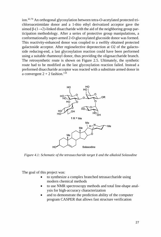
Solaradixine comprises a tetrasaccharide having the sequence β-D-Glcp-(1→2)-β-D-Glcp-(1→3)[α-L-Rhap-(1→2)]-β-D-Galp- and linked to the alka-loid moiety called Solasodine (Figure 4.1). The effect of the aglycon on the glycoside chemical shifts being quite limited, the bulky steroidal alkaloid was replaced by a methyl group and the synthesis work focused on the part of in-terest: the carbohydrate molecule 1. To obtain such a branched oligosaccha-ride in an efficient and concised way, building blocks were first designed. Early on, in the elaboration of the synthetic route, it was decided to use a β-(1→2)-linked glucosyl disaccharide donor to form the linear sequence β-D-Glcp-(1→2)-β-D-Glcp-(1→3)-β-D-Galp-OMe in a stereoselective fash-
27
ion.42,70 An orthogonal glycosylation between tetra-O-acetylated protected tri-chloroacetimidate donor and a 1-thio ethyl derivatized acceptor gave the aimed β-(1→2)-linked disaccharide with the aid of the neighboring group par-ticipation methodology. After a series of protective group manipulations, a conformationally super-armed 2-O-glucosylated glucoside donor was formed. This reactivity-enhanced donor was coupled to a swiftly obtained protected galactoside acceptor. After regioselective deprotection at O2 of the galacto-side reducing-end, a last glycosylation reaction could have been performed using a suitable rhamnosyl donor, thus providing the oligosaccharide branch. The retrosynthetic route is shown on Figure 2.5. Ultimately, the synthetic route had to be modified as the last glycosylation reaction failed. Instead a preformed disaccharide acceptor was reacted with a substitute armed donor in a convergent 2 + 2 fashion.118
Figure 4.1: Schematic of the tetrasaccharide target 1 and the alkaloid Solasodine
The goal of this project was:
• to synthesize a complex branched tetrasaccharide using modern chemical methods
• to use NMR spectroscopy methods and total line-shape anal-ysis for high-accuracy characterization
• and to demonstrate the prediction ability of the computer program CASPER that allows fast structure verification
28
4.1 Synthesis
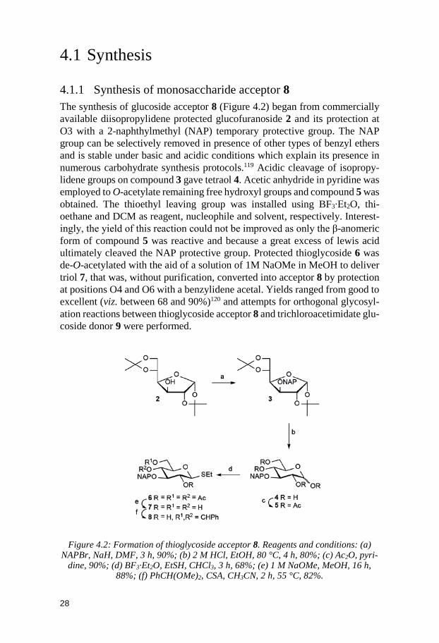
4.1.1 Synthesis of monosaccharide acceptor 8 The synthesis of glucoside acceptor 8 (Figure 4.2) began from commercially available diisopropylidene protected glucofuranoside 2 and its protection at O3 with a 2-naphthylmethyl (NAP) temporary protective group. The NAP group can be selectively removed in presence of other types of benzyl ethers and is stable under basic and acidic conditions which explain its presence in numerous carbohydrate synthesis protocols.119 Acidic cleavage of isopropy-lidene groups on compound 3 gave tetraol 4. Acetic anhydride in pyridine was employed to O-acetylate remaining free hydroxyl groups and compound 5 was obtained. The thioethyl leaving group was installed using BF3∙Et2O, thi-oethane and DCM as reagent, nucleophile and solvent, respectively. Interest-ingly, the yield of this reaction could not be improved as only the β-anomeric form of compound 5 was reactive and because a great excess of lewis acid ultimately cleaved the NAP protective group. Protected thioglycoside 6 was de-O-acetylated with the aid of a solution of 1M NaOMe in MeOH to deliver triol 7, that was, without purification, converted into acceptor 8 by protection at positions O4 and O6 with a benzylidene acetal. Yields ranged from good to excellent (viz. between 68 and 90%)120 and attempts for orthogonal glycosyl-ation reactions between thioglycoside acceptor 8 and trichloroacetimidate glu-coside donor 9 were performed.
Figure 4.2: Formation of thioglycoside acceptor 8. Reagents and conditions: (a)
NAPBr, NaH, DMF, 3 h, 90%; (b) 2 M HCl, EtOH, 80 °C, 4 h, 80%; (c) Ac2O, pyri-dine, 90%; (d) BF3·Et2O, EtSH, CHCl3, 3 h, 68%; (e) 1 M NaOMe, MeOH, 16 h,
88%; (f) PhCH(OMe)2, CSA, CH3CN, 2 h, 55 °C, 82%.
29
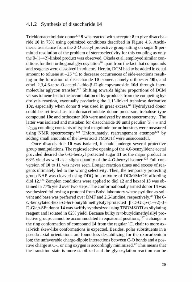
4.1.2 Synthesis of disaccharide 14 Trichloroacetimidate donor121 9 was reacted with acceptor 8 to give disaccha-ride 10 in 75% using optimized conditions described in Figure 4.3. Anchi-meric assistance from the 2-O-acetyl protective group sitting on sugar 9 per-mitted resolution of the problem of stereoselectivity for this coupling as only the β-(1→2)-linked product was observed. Okada et al. employed similar con-ditions for their orthogonal glycosylation70 apart from the fact that compounds and reagents were dissolved in toluene. Herein, DCM had to be added in equal amount to toluene at −25 °C to decrease occurrences of side-reactions result-ing in the formation of disaccharide 10 isomer, namely orthoester 10b, and ethyl 2,3,4,6-tetra-O-acetyl-1-thio-β-D-glucopyranoside 10d through inter-molecular aglycon transfer.122 Shifting towards higher proportions of DCM versus toluene led to the accumulation of by-products from the competing hy-drolysis reaction, eventually producing the 1,1’-linked trehalose derivative 10c, especially when donor 9 was used in great excess.67 Hydrolyzed donor could be retrieved as trichloroacetimidate donor precursor, trehalose type compound 10c and orthoester 10b were analyzed by mass spectrometry. The latter was isolated and mistaken for disaccharide 10 until peculiar 3JH1,H2 and 1JC1,H1 coupling constants of typical magnitude for orthoesters were measured using NMR spectroscopy.123 Unfortunately, rearrangement attempts124 by adding small amounts of the lewis acid TMSOTf were unsuccessful.
Once disaccharide 10 was isolated, it could undergo several protective group manipulations. The regioselective opening of the 4,6-benzylidene acetal provided desired the 6-O-benzyl protected sugar 11 as the major product in 68% yield as well as a slight quantity of the 4-O-benzyl isomer.125 Full con-version of 10 to 11 was never seen. Longer reaction times and excess of rea-gents ultimately led to the wrong selectivity. Then, the temporary protecting group NAP was cleaved using DDQ in a mixture of DCM/MeOH affording diol 12.126 Zemplen conditions were applied to diol 12 and hexaol 13 was ob-tained in 77% yield over two steps. The conformationally armed donor 14 was synthesized following a protocol from Bols’ laboratory where pyridine as sol-vent and base was preferred over DMF and 2,6-lutidine, respectively.42 The 6-O-benzylated-hexa-O-tert-butyldimethylsilyl-protected β-D-Glcp-(1→2)-β-D-Glcp-SEt donor 14 was swiftly synthesized using TBDMSOTf as silylating reagent and isolated in 82% yield. Because bulky tert-butyldimethylsilyl pro-tective groups cannot be accommodated in equatorial positions,127 a change in the ring conformation of compound 14 from the regular 4C1 chair to more ax-ial-rich skew-like conformations is expected. Besides, polar substituents in a pseudo-axial orientations are found less destabilizing for the oxocarbenium ion; the unfavorable charge-dipole interactions between C-O bonds and a pos-itive charge at C-1 or ring oxygen is accordingly minimized.42 This means that the transition state is more stabilized and the glycosylation reaction can be
30
enhanced. Consequently, conformationally super-armed donor 14 gained in reactivity compared to donors trapped in low-in-energy chair conformations. Satisfyingly, 3JH1,H2 and 3JH2,H3 coupling constants values for both sugar units were in good agreements with NMR data found in the literature corresponding to 3S1 skew-type of conformations (3JH1,H2 ≈ 5-6 Hz and 3JH2,H3 < 2 Hz).29
Figure 4.3: Formation of armed donor 14. Reagents and conditions: (a) TMSOTf, CH2Cl2/Tol 1:1, −25 °C → r.t., 2 h, 75%; (b) BH3·NMe3, AlCl3, THF, 6 h, 68%; (c)
DDQ, CH2Cl2/MeOH 4:1, 4 h, 85%; (d) 1 M NaOMe, MeOH, 16 h, 91%; (e) TBDMSOTf, DMAP, pyridine, 80 °C, 24 h, 82%.
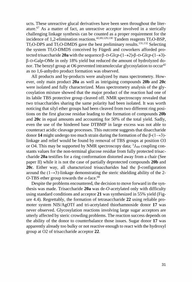
4.1.3 Synthesis of trisaccharide 20 The use of suitably protected methyl 4,6-O-benzylidene-2-O-acetyl-β-D-ga-lactopyranoside acceptor 16 was advantageous because of its facile and rapid synthesis from commercially available methyl β-D-galactopyranoside or known methyl 4,6-O-benzylidene-β-D-galactopyranoside.128 The latter was converted to acceptor 16 by regioselectively protecting position O2 with an acetyl protective group (Figure 4.5). This small acyl group was chosen for orthogonality reasons and to limit steric hindrance around free hydroxyl group OH3 to be glycosylated in the β-(1→3)-linkage formation. Several promoters were tested to build trisaccharide 20. Activator systems comprising N-iodosuccinimide (NIS) and TfOH, AgOTf, TMSOTf as well as MeOTf gave rise to the major formation of 1,2-elimination reaction by-prod-
31
ucts. These unreactive glucal derivatives have been seen throughout the liter-ature.67 As a matter of fact, an unreactive acceptor involved in a sterically challenging linkage synthesis can be counted as a proper requirement for the incidence of 1,2-elimination reactions.42,69,129,130 Tandem reagents Tf2O-BSP, Tf2O-DPS and Tf2O-DMDS gave the best preliminary results.131,132 Selecting the system Tf2O-DMDS conceived by Fügedi and coworkers afforded pro-tected trisaccharide 20a with the sequence β-D-Glcp-(1→2)-β-D-Glcp-(1→3)-β-D-Galp-OMe in only 18% yield but reduced the amount of hydrolyzed do-nor. The benzyl group at O6 prevented intramolecular glycosylation to occur42 as no 1,6-anhydro product formation was observed.
All products and by-products were analyzed by mass spectrometry. How-ever, only main product 20a as well as intriguing compounds 20b and 20c were isolated and fully characterized. Mass spectrometry analysis of the gly-cosylation mixture showed that the major product of the reaction had one of its labile TBS protective group cleaved off. NMR spectroscopy revealed that two trisaccharides sharing the same polarity had been isolated. It was worth noticing that silyl ether groups had been cleaved from two different ring posi-tions on the first glucose residue leading to the formation of compounds 20b and 20c in equal amounts and accounting for 50% of the total yield. Sadly, even the use of the hindered base DTBMP in large excess was not able to counteract acidic cleavage processes. This outcome suggests that disaccharide donor 14 might undergo too much strain during the formation of the β-(1→3)-linkage and relief would be found by removal of TBS groups at position O3 or O4. This may be supported by NMR spectroscopy data; 3JHH coupling con-stants values for the non-terminal glucose residue from fully protected trisac-charide 20a testifies for a ring conformation distorted away from a chair (See paper II) while it is not the case of partially deprotected compounds 20b and 20c. Either way, all characterized trisaccharides had the β-configuration around the (1→3)-linkage demonstrating the steric shielding ability of the 2-O-TBS ether group towards the α-face.68
Despite the problems encountered, the decision to move forward in the syn-thesis was made. Trisaccharide 20a was de-O-acetylated only with difficulty using standard conditions and acceptor 21 was synthesized in 55% yield (Fig-ure 4.4). Regrettably, the formation of tetrasaccharide 22 using reliable pro-moter system NIS/AgOTf and tri-acetylated thiorhamnoside donor 17 was never observed. Glycosylation reactions involving large sugar acceptors are utterly affected by steric crowding problems. The reaction success depends on the ability of the donor to counterbalance those issues. Sugar donor 17 was apparently already too bulky or not reactive enough to react with the hydroxyl group at O2 of trisaccharide acceptor 22.
32
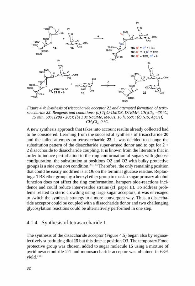
Figure 4.4: Synthesis of trisaccharide acceptor 21 and attempted formation of tetra-saccharide 22. Reagents and conditions: (a) Tf2O-DMDS, DTBMP, CH2Cl2, −78 °C,
15 min, 68% (20a - 20c); (b) 1 M NaOMe, MeOH, 16 h, 55%; (c) NIS, AgOTf, CH2Cl2, 0 °C.
A new synthesis approach that takes into account results already collected had to be considered. Learning from the successful synthesis of trisaccharide 20 and the failed attempts on tetrasaccharide 22, it was decided to change the substitution pattern of the disaccharide super-armed donor and to opt for 2 + 2 disaccharide to disaccharide coupling. It is known from the literature that in order to induce perturbation in the ring conformation of sugars with glucose configuration, the substitution at positions O2 and O3 with bulky protective groups is a sine qua non condition.29,133 Therefore, the only remaining position that could be easily modified is at O6 on the terminal glucose residue. Replac-ing a TBS ether group by a benzyl ether group to mask a sugar primary alcohol function does not affect the ring conformation, hampers side-reactions inci-dence and could reduce inter-residue strains (cf. paper II). To address prob-lems related to steric crowding using large sugar acceptors, it was envisaged to switch the synthesis strategy to a more convergent way. Thus, a disaccha-ride acceptor could be coupled with a disaccharide donor and two challenging glycosylation reactions could be alternatively performed in one step.
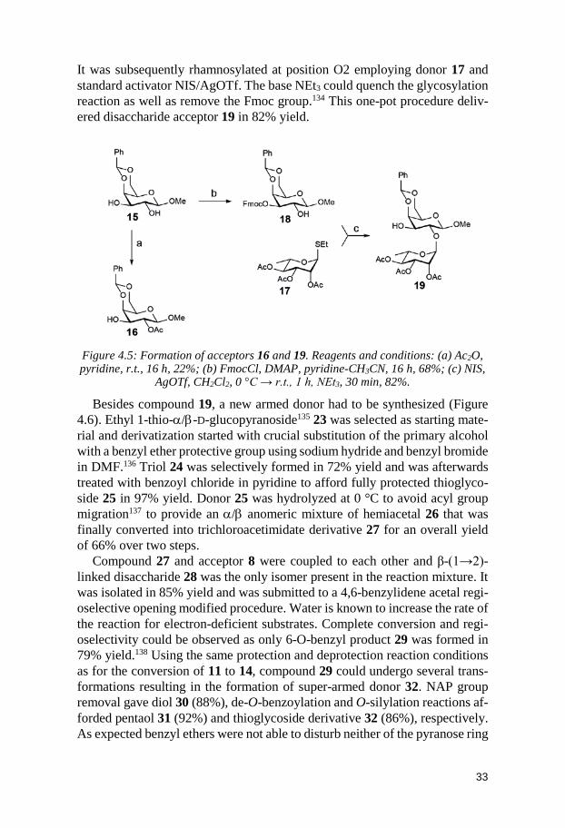
4.1.4 Synthesis of tetrasaccharide 1 The synthesis of the disaccharide acceptor (Figure 4.5) began also by regiose-lectively substituting diol 15 but this time at position O3. The temporary Fmoc protective group was chosen, added to sugar molecule 15 using a mixture of pyridine/acetonitrile 2:1 and monosaccharide acceptor was obtained in 68% yield.116
33
It was subsequently rhamnosylated at position O2 employing donor 17 and standard activator NIS/AgOTf. The base NEt3 could quench the glycosylation reaction as well as remove the Fmoc group.134 This one-pot procedure deliv-ered disaccharide acceptor 19 in 82% yield.
Figure 4.5: Formation of acceptors 16 and 19. Reagents and conditions: (a) Ac2O, pyridine, r.t., 16 h, 22%; (b) FmocCl, DMAP, pyridine-CH3CN, 16 h, 68%; (c) NIS,
AgOTf, CH2Cl2, 0 °C → r.t., 1 h, NEt3, 30 min, 82%.
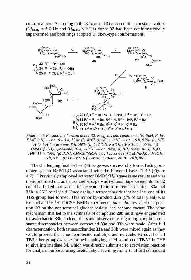
Besides compound 19, a new armed donor had to be synthesized (Figure 4.6). Ethyl 1-thio-α/β-D-glucopyranoside135 23 was selected as starting mate-rial and derivatization started with crucial substitution of the primary alcohol with a benzyl ether protective group using sodium hydride and benzyl bromide in DMF.136 Triol 24 was selectively formed in 72% yield and was afterwards treated with benzoyl chloride in pyridine to afford fully protected thioglyco-side 25 in 97% yield. Donor 25 was hydrolyzed at 0 °C to avoid acyl group migration137 to provide an α/β anomeric mixture of hemiacetal 26 that was finally converted into trichloroacetimidate derivative 27 for an overall yield of 66% over two steps.
Compound 27 and acceptor 8 were coupled to each other and β-(1→2)-linked disaccharide 28 was the only isomer present in the reaction mixture. It was isolated in 85% yield and was submitted to a 4,6-benzylidene acetal regi-oselective opening modified procedure. Water is known to increase the rate of the reaction for electron-deficient substrates. Complete conversion and regi-oselectivity could be observed as only 6-O-benzyl product 29 was formed in 79% yield.138 Using the same protection and deprotection reaction conditions as for the conversion of 11 to 14, compound 29 could undergo several trans-formations resulting in the formation of super-armed donor 32. NAP group removal gave diol 30 (88%), de-O-benzoylation and O-silylation reactions af-forded pentaol 31 (92%) and thioglycoside derivative 32 (86%), respectively. As expected benzyl ethers were not able to disturb neither of the pyranose ring
34
conformations. According to the 3JH1,H2 and 3JH2,H3 coupling constants values (3JH1,H2 ≈ 5-6 Hz and 3JH2,H3 < 2 Hz) donor 32 had been conformationally super-armed and both rings adopted 3S1 skew-type conformations.
Figure 4.6: Formation of armed donor 32. Reagents and conditions: (a) NaH, BnBr, DMF, 0 °C → r.t., 6 – 8 h, 72%; (b) BzCl, pyridine, 0 °C → r.t., 16 h, 97%; (c) NIS,
H2O, CH2Cl2-acetone, 8 h, 78%; (d) Cl3CCN, K2CO3, CH2Cl2, 4 h, 85%; (e) TMSOTf, CH2Cl2-toluene, 16 h, −10 °C → r.t., 84%; (f) BH3·NMe3, AlCl3, H2O,
THF, 16 h, 79%; (g) DDQ, CH2Cl2/MeOH 4:1, 4 h, 88%; (h) 1 M NaOMe, MeOH, 16 h, 93%; (i) TBDMSOTf, DMAP, pyridine, 80 °C, 24 h, 86%.
The challenging final β-(1→3)-linkage was successfully formed using pro-moter system BSP/Tf2O associated with the hindered base TTBP (Figure 4.7).139 Previously employed activator DMDS/Tf2O gave same results and was therefore ruled out as its use and storage was tedious. Super-armed donor 32 could be linked to disaccharide acceptor 19 to form tetrasaccharides 33a and 33b in 55% total yield. Once again, a tetrasaccharide that had lost one of its TBS group had formed. This minor by-product 33b (5% of total yield) was isolated and 1H,1H-TOCSY NMR experiments, inter alia, revealed that posi-tion O3 on the non-terminal glucose residue had become vacant. The same mechanism that led to the synthesis of compound 20b must have engendered tetrasaccharide 33b. Indeed, the same observations regarding coupling con-stants discrepancies between compound 33a and 33b were made. After full characterization, both tetrasaccharides 33a and 33b were mixed again as they would provide the same deprotected carbohydrate molecule. Removal of all TBS ether groups was performed employing a 1M solution of TBAF in THF to give intermediate 34, which was directly submitted to acetylation reaction for analysis purposes using acetic anhydride in pyridine to afford compound
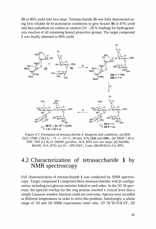
35
35 in 80% yield over two steps. Tetrasaccharide 35 was fully deprotected us-ing first reliable de-O-acetylation conditions to give hexaol 36 in 87% yield and then palladium on carbon as catalyst (10 – 20 % loading) for hydrogenol-ysis reaction of all remaining benzyl protective groups. The target compound 1 was finally obtained in 89% yield.
Figure 4.7: Formation of tetrasaccharide 1. Reagents and conditions: (a) BSP,
Tf2O, TTBP, CH2Cl2, −78 → −10 °C, 30 min, 55% (33a and 33b) ; (b) TBAF 1 M in THF, THF (c) Ac2O, DMAP, pyridine, 16 h, 80% over two steps; (d) NaOMe,
MeOH, 16 h, 87%; (e) 10 – 20% Pd/C, 3 atm, MeOH/H2O, 6 h, 89%.
4.2 Characterization of tetrasaccharide 1 by NMR spectroscopy
Full characterization of tetrasaccharide 1 was conducted by NMR spectros-copy. Target compound 1 comprised three monosaccharides with β-configu-ration, including two glucose moieties linked to each other. In the 1D 1H spec-trum, the spectral overlap for the ring protons reached a critical level that a simple Gaussian window function could not overcome. Spectra were recorded at different temperatures in order to solve this problem. Satisfyingly, a whole range of 1D and 2D NMR experiments (inter alia, 1D 1H,1H-TOCSY, 2D
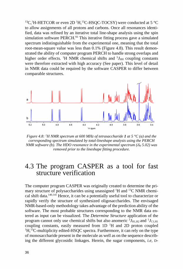
36
13C,1H-HETCOR or even 2D 1H,13C-HSQC-TOCSY) were conducted at 5 °C to allow assignments of all protons and carbons. Once all resonances identi-fied, data was refined by an iterative total line-shape analysis using the spin simulation software PERCH.93 This iterative fitting process gave a simulated spectrum indistinguishable from the experimental one, meaning that the total root-mean-square value was less than 0.1% (Figure 4.8). This result demon-strated the ability of computer program PERCH to handle strong overlaps and higher order effects. 1H NMR chemical shifts and 3JHH coupling constants were therefore extracted with high accuracy (See paper). This level of detail in NMR data could be required by the software CASPER to differ between comparable structures.
Figure 4.8: 1H NMR spectrum at 600 MHz of tetrasaccharide 1 at 5 °C (a) and the corresponding spectrum simulated by total-lineshape analysis using the PERCH
NMR software (b). The HDO resonance in the experimental spectrum (δH 5.02) was removed prior to the lineshape fitting procedure.
4.3 The program CASPER as a tool for fast structure verification
The computer program CASPER was originally created to determine the pri-mary structure of polysaccharides using unassigned 1H and 13C NMR chemi-cal shift data.140,141 Hence, it can be a potentially useful tool to characterize or rapidly verify the structure of synthesized oligosaccharides. The envisaged NMR-based-only methodology takes advantage of the prediction ability of the software. The most probable structures corresponding to the NMR data en-tered as input can be visualized. The Determine Structure application of the program cannot only use chemical shifts but also anomeric 3JH1,H2 and 1JC1,H1
coupling constants, easily measured from 1D 1H and 2D proton coupled 1H,13C-multiplicity edited-HSQC spectra. Furthermore, it can rely on the type of monosaccharide present in the molecule as well as on the sequence describ-ing the different glycosidic linkages. Herein, the sugar components, i.e, D-
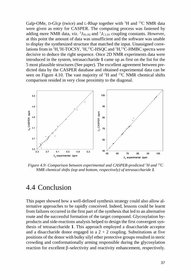
37
Galp-OMe, D-Glcp (twice) and L-Rhap together with 1H and 13C NMR data were given as entry for CASPER. The computing process was fastened by adding more NMR data, viz. 3JH1,H2 and 1JC1,H1 coupling constants. However, at this point the amount of data was unsufficient and the software was unable to display the synthesized structure that matched the input. Unassigned corre-lations from in 1H,1H-TOCSY, 1H,13C-HSQC and 1H,13C-HMBC spectra were decisive to deduce the right sequence. Once 2D NMR experiments data were introduced in the system, tetrasaccharide 1 came up as first on the list for the 5 most plausible structures (See paper). The excellent agreement between pre-dicted data by the CASPER database and obtained experimental data can be seen on Figure 4.10. The vast majority of 1H and 13C NMR chemical shifts comparison resided in very close proximity to the diagonal.
Figure 4.9: Comparison between experimental and CASPER-predicted 1H and 13C
NMR chemical shifts (top and bottom, respectively) of tetrasaccharide 1.
4.4 Conclusion This paper showed how a well-defined synthesis strategy could also allow al-ternative approaches to be rapidly conceived. Indeed, lessons could be learnt from failures occurred in the first part of the synthesis that led to an alternative route and the successful formation of the target compound. Glycosylation by-products and side-reactions analysis helped to design the first convergent syn-thesis of tetrasaccharide 1. This approach employed a disaccharide acceptor and a disaccharide donor engaged in a 2 + 2 coupling. Substitutions at five positions of the donor with bulky silyl ether protective groups resulted in steric crowding and conformationally arming responsible during the glycosylation reaction for excellent β-selectivity and reactivity enhancement, respectively.
38
In fact, we described for the first time the use of a disaccharide super-armed donor to glycosylate a hydroxyl group at a secondary carbon of an acceptor. Moreover, a methodology for fast structure verification was implemented as the computer program CASPER was able to predict the correct structure using refined unassigned 1H and 13C NMR data. Thus, the software can be used as an aid to synthesis carbohydrate chemists for NMR resonance assignments because of the very small chemical shifts deviations between experimental data and predicted ones.
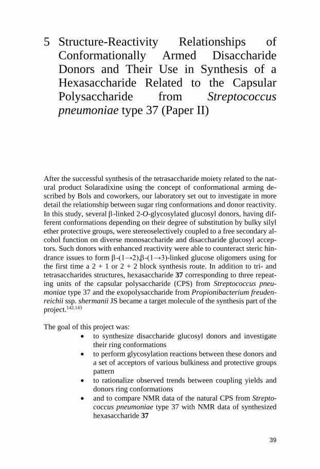
39
5 Structure-Reactivity Relationships of Conformationally Armed Disaccharide Donors and Their Use in Synthesis of a Hexasaccharide Related to the Capsular Polysaccharide from Streptococcus pneumoniae type 37 (Paper II)
After the successful synthesis of the tetrasaccharide moiety related to the nat-ural product Solaradixine using the concept of conformational arming de-scribed by Bols and coworkers, our laboratory set out to investigate in more detail the relationship between sugar ring conformations and donor reactivity. In this study, several β-linked 2-O-glycosylated glucosyl donors, having dif-ferent conformations depending on their degree of substitution by bulky silyl ether protective groups, were stereoselectively coupled to a free secondary al-cohol function on diverse monosaccharide and disaccharide glucosyl accep-tors. Such donors with enhanced reactivity were able to counteract steric hin-drance issues to form β-(1→2),β-(1→3)-linked glucose oligomers using for the first time a 2 + 1 or 2 + 2 block synthesis route. In addition to tri- and tetrasaccharides structures, hexasaccharide 37 corresponding to three repeat-ing units of the capsular polysaccharide (CPS) from Streptococcus pneu-moniae type 37 and the exopolysaccharide from Propionibacterium freuden-reichii ssp. shermanii JS became a target molecule of the synthesis part of the project.142,143
The goal of this project was:
• to synthesize disaccharide glucosyl donors and investigate their ring conformations
• to perform glycosylation reactions between these donors and a set of acceptors of various bulkiness and protective groups pattern
• to rationalize observed trends between coupling yields and donors ring conformations
• and to compare NMR data of the natural CPS from Strepto-coccus pneumoniae type 37 with NMR data of synthesized hexasaccharide 37
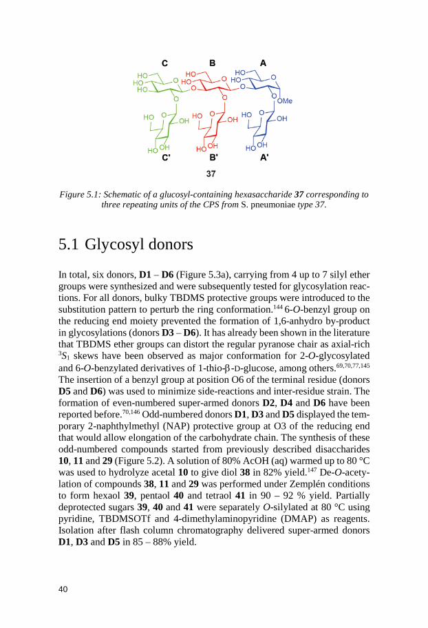
40
Figure 5.1: Schematic of a glucosyl-containing hexasaccharide 37 corresponding to
three repeating units of the CPS from S. pneumoniae type 37.
5.1 Glycosyl donors In total, six donors, D1 – D6 (Figure 5.3a), carrying from 4 up to 7 silyl ether groups were synthesized and were subsequently tested for glycosylation reac-tions. For all donors, bulky TBDMS protective groups were introduced to the substitution pattern to perturb the ring conformation.144 6-O-benzyl group on the reducing end moiety prevented the formation of 1,6-anhydro by-product in glycosylations (donors D3 – D6). It has already been shown in the literature that TBDMS ether groups can distort the regular pyranose chair as axial-rich 3S1 skews have been observed as major conformation for 2-O-glycosylated and 6-O-benzylated derivatives of 1-thio-β-D-glucose, among others.69,70,77,145 The insertion of a benzyl group at position O6 of the terminal residue (donors D5 and D6) was used to minimize side-reactions and inter-residue strain. The formation of even-numbered super-armed donors D2, D4 and D6 have been reported before.70,146 Odd-numbered donors D1, D3 and D5 displayed the tem-porary 2-naphthylmethyl (NAP) protective group at O3 of the reducing end that would allow elongation of the carbohydrate chain. The synthesis of these odd-numbered compounds started from previously described disaccharides 10, 11 and 29 (Figure 5.2). A solution of 80% AcOH (aq) warmed up to 80 °C was used to hydrolyze acetal 10 to give diol 38 in 82% yield.147 De-O-acety-lation of compounds 38, 11 and 29 was performed under Zemplén conditions to form hexaol 39, pentaol 40 and tetraol 41 in 90 – 92 % yield. Partially deprotected sugars 39, 40 and 41 were separately O-silylated at 80 °C using pyridine, TBDMSOTf and 4-dimethylaminopyridine (DMAP) as reagents. Isolation after flash column chromatography delivered super-armed donors D1, D3 and D5 in 85 – 88% yield.
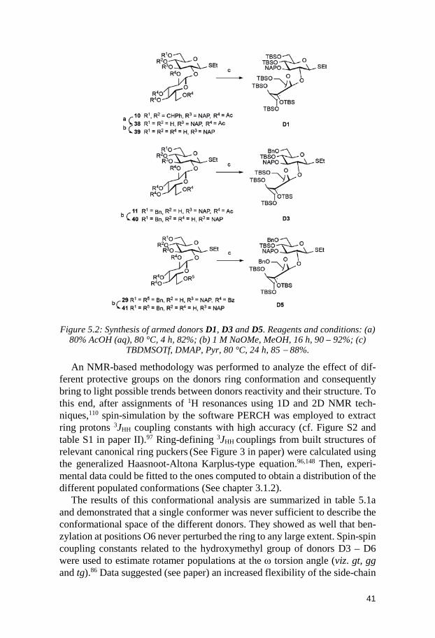
41
Figure 5.2: Synthesis of armed donors D1, D3 and D5. Reagents and conditions: (a)
80% AcOH (aq), 80 °C, 4 h, 82%; (b) 1 M NaOMe, MeOH, 16 h, 90 – 92%; (c) TBDMSOTf, DMAP, Pyr, 80 °C, 24 h, 85 − 88%.
An NMR-based methodology was performed to analyze the effect of dif-ferent protective groups on the donors ring conformation and consequently bring to light possible trends between donors reactivity and their structure. To this end, after assignments of 1H resonances using 1D and 2D NMR tech-niques,110 spin-simulation by the software PERCH was employed to extract ring protons 3JHH coupling constants with high accuracy (cf. Figure S2 and table S1 in paper II).97 Ring-defining 3JHH couplings from built structures of relevant canonical ring puckers (See Figure 3 in paper) were calculated using the generalized Haasnoot-Altona Karplus-type equation.96,148 Then, experi-mental data could be fitted to the ones computed to obtain a distribution of the different populated conformations (See chapter 3.1.2).
The results of this conformational analysis are summarized in table 5.1a and demonstrated that a single conformer was never sufficient to describe the conformational space of the different donors. They showed as well that ben-zylation at positions O6 never perturbed the ring to any large extent. Spin-spin coupling constants related to the hydroxymethyl group of donors D3 – D6 were used to estimate rotamer populations at the ω torsion angle (viz. gt, gg and tg).86 Data suggested (see paper) an increased flexibility of the side-chain
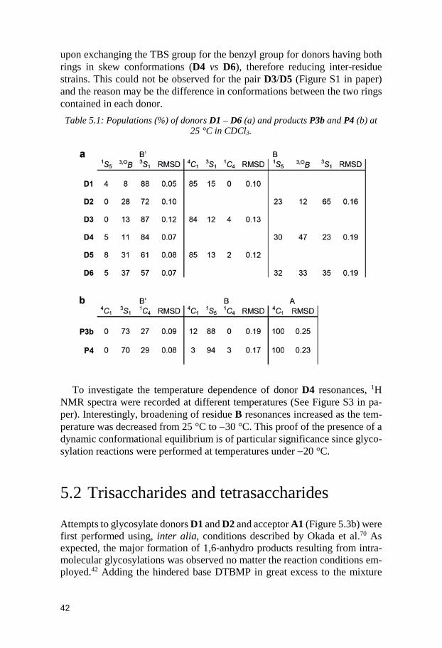
42
upon exchanging the TBS group for the benzyl group for donors having both rings in skew conformations (D4 vs D6), therefore reducing inter-residue strains. This could not be observed for the pair D3/D5 (Figure S1 in paper) and the reason may be the difference in conformations between the two rings contained in each donor. Table 5.1: Populations (%) of donors D1 – D6 (a) and products P3b and P4 (b) at
25 °C in CDCl3.
To investigate the temperature dependence of donor D4 resonances, 1H NMR spectra were recorded at different temperatures (See Figure S3 in pa-per). Interestingly, broadening of residue B resonances increased as the tem-perature was decreased from 25 °C to −30 °C. This proof of the presence of a dynamic conformational equilibrium is of particular significance since glyco-sylation reactions were performed at temperatures under −20 °C.
5.2 Trisaccharides and tetrasaccharides Attempts to glycosylate donors D1 and D2 and acceptor A1 (Figure 5.3b) were first performed using, inter alia, conditions described by Okada et al.70 As expected, the major formation of 1,6-anhydro products resulting from intra-molecular glycosylations was observed no matter the reaction conditions em-ployed.42 Adding the hindered base DTBMP in great excess to the mixture
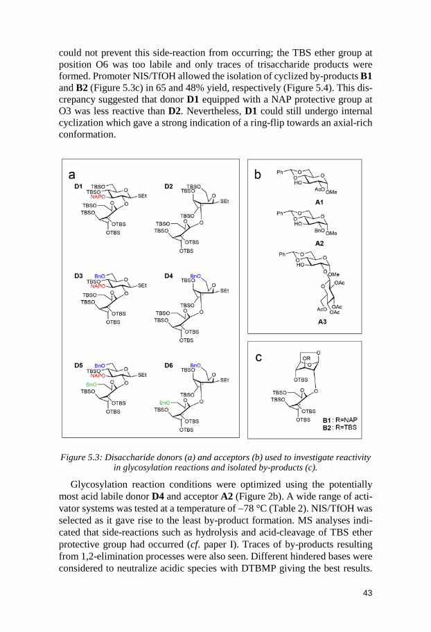
43
could not prevent this side-reaction from occurring; the TBS ether group at position O6 was too labile and only traces of trisaccharide products were formed. Promoter NIS/TfOH allowed the isolation of cyclized by-products B1 and B2 (Figure 5.3c) in 65 and 48% yield, respectively (Figure 5.4). This dis-crepancy suggested that donor D1 equipped with a NAP protective group at O3 was less reactive than D2. Nevertheless, D1 could still undergo internal cyclization which gave a strong indication of a ring-flip towards an axial-rich conformation.
Figure 5.3: Disaccharide donors (a) and acceptors (b) used to investigate reactivity
in glycosylation reactions and isolated by-products (c).
Glycosylation reaction conditions were optimized using the potentially most acid labile donor D4 and acceptor A2 (Figure 2b). A wide range of acti-vator systems was tested at a temperature of −78 °C (Table 2). NIS/TfOH was selected as it gave rise to the least by-product formation. MS analyses indi-cated that side-reactions such as hydrolysis and acid-cleavage of TBS ether protective group had occurred (cf. paper I). Traces of by-products resulting from 1,2-elimination processes were also seen. Different hindered bases were considered to neutralize acidic species with DTBMP giving the best results.
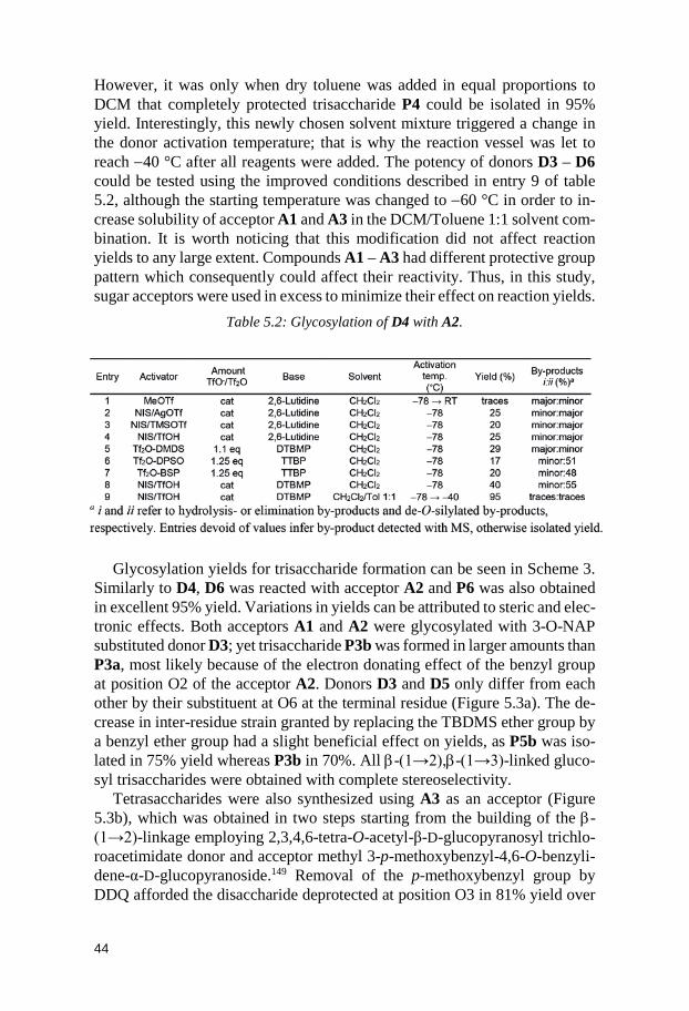
44
However, it was only when dry toluene was added in equal proportions to DCM that completely protected trisaccharide P4 could be isolated in 95% yield. Interestingly, this newly chosen solvent mixture triggered a change in the donor activation temperature; that is why the reaction vessel was let to reach −40 °C after all reagents were added. The potency of donors D3 – D6 could be tested using the improved conditions described in entry 9 of table 5.2, although the starting temperature was changed to −60 °C in order to in-crease solubility of acceptor A1 and A3 in the DCM/Toluene 1:1 solvent com-bination. It is worth noticing that this modification did not affect reaction yields to any large extent. Compounds A1 – A3 had different protective group pattern which consequently could affect their reactivity. Thus, in this study, sugar acceptors were used in excess to minimize their effect on reaction yields.
Table 5.2: Glycosylation of D4 with A2.
Glycosylation yields for trisaccharide formation can be seen in Scheme 3. Similarly to D4, D6 was reacted with acceptor A2 and P6 was also obtained in excellent 95% yield. Variations in yields can be attributed to steric and elec-tronic effects. Both acceptors A1 and A2 were glycosylated with 3-O-NAP substituted donor D3; yet trisaccharide P3b was formed in larger amounts than P3a, most likely because of the electron donating effect of the benzyl group at position O2 of the acceptor A2. Donors D3 and D5 only differ from each other by their substituent at O6 at the terminal residue (Figure 5.3a). The de-crease in inter-residue strain granted by replacing the TBDMS ether group by a benzyl ether group had a slight beneficial effect on yields, as P5b was iso-lated in 75% yield whereas P3b in 70%. All β-(1→2),β-(1→3)-linked gluco-syl trisaccharides were obtained with complete stereoselectivity.
Tetrasaccharides were also synthesized using A3 as an acceptor (Figure 5.3b), which was obtained in two steps starting from the building of the β-(1→2)-linkage employing 2,3,4,6-tetra-O-acetyl-β-D-glucopyranosyl trichlo-roacetimidate donor and acceptor methyl 3-p-methoxybenzyl-4,6-O-benzyli-dene-α-D-glucopyranoside.149 Removal of the p-methoxybenzyl group by DDQ afforded the disaccharide deprotected at position O3 in 81% yield over
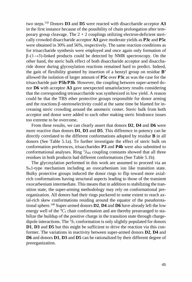
45
two steps.150 Donors D3 and D5 were reacted with disaccharide acceptor A3 in the first instance because of the possibility of chain prolongation after tem-porary group cleavage. The 2 + 2 couplings utilizing electron-deficient steri-cally crowded disaccharide acceptor A3 gave moderate yields as P3c and P5c were obtained in 30% and 56%, respectively. The same reaction conditions as for trisaccharide synthesis were employed and once again only formation of β-(1→3)-linked products could be detected by NMR spectroscopy. On the other hand, the steric bulk effect of both disaccharide acceptor and disaccha-ride donor during glycosylation reactions remained hard to predict. Indeed, the gain of flexibility granted by insertion of a benzyl group on residue B’ allowed the isolation of larger amount of P5c over P3c as was the case for the trisaccharide pair P5b/P3b. However, the coupling between super-armed do-nor D6 with acceptor A3 gave unexpected unsatisfactory results considering that the corresponding tetrasaccharide was synthesized in low yield. A reason could be that the TBS ether protective groups responsible for donor arming and the reactions β-stereoselectivity could at the same time be blamed for in-creasing steric crowding around the anomeric center. Steric bulk from both acceptor and donor were added to each other making steric hindrance issues too extreme to be overcome.
From these results, we can clearly assert that donors D2, D4 and D6 were more reactive than donors D1, D3 and D5. This difference in potency can be directly correlated to the different conformations adopted by residue B in all donors (See Table 5.1a). To further investigate the effect of steric bulk on conformation preferences, trisaccharides P3 and P4b were also submitted to conformational analyses. Ring 3JHH coupling constants showed that all three residues in both products had different conformations (See Table 5.1b).
The glycosylation performed in this work are assumed to proceed via an SN1-type mechanism including an oxocarbenium ion like transition state. Bulky protective groups induced the donor rings to flip toward more axial-rich conformations having structural aspects leading to those of the transient oxocarbenium intermediate. This means that in addition to stabilizing the tran-sition state, the super-arming methodology may rely on conformational pre-organization. All donors had their rings puckered to some extent to reach ax-ial-rich skew conformations residing around the equator of the pseudorota-tional sphere.148 Super-armed donors D2, D4 and D6 have already left the low energy well of the 4C1 chair conformation and are thereby prearranged to sta-bilize the buildup of the positive charge in the transition state through charge-dipole interactions. The 3S1 conformation is only slightly populated for donors D1, D3 and D5 but this might be sufficient to drive the reaction via this con-former. The variations in reactivity between super-armed donors D2, D4 and D6 and donors D1, D3 and D5 can be rationalized by their different degree of preorganization.
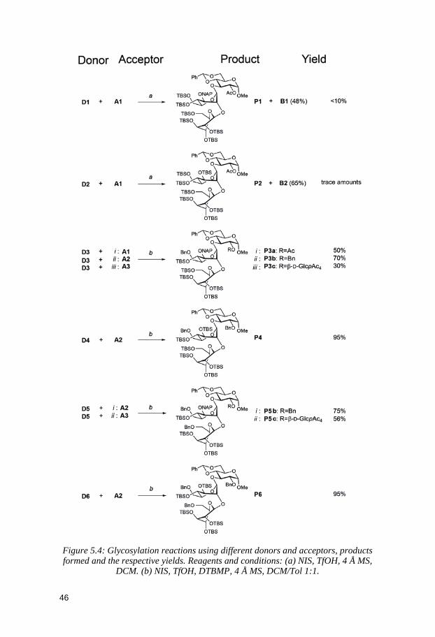
46
Figure 5.4: Glycosylation reactions using different donors and acceptors, products formed and the respective yields. Reagents and conditions: (a) NIS, TfOH, 4 Å MS,
DCM. (b) NIS, TfOH, DTBMP, 4 Å MS, DCM/Tol 1:1.
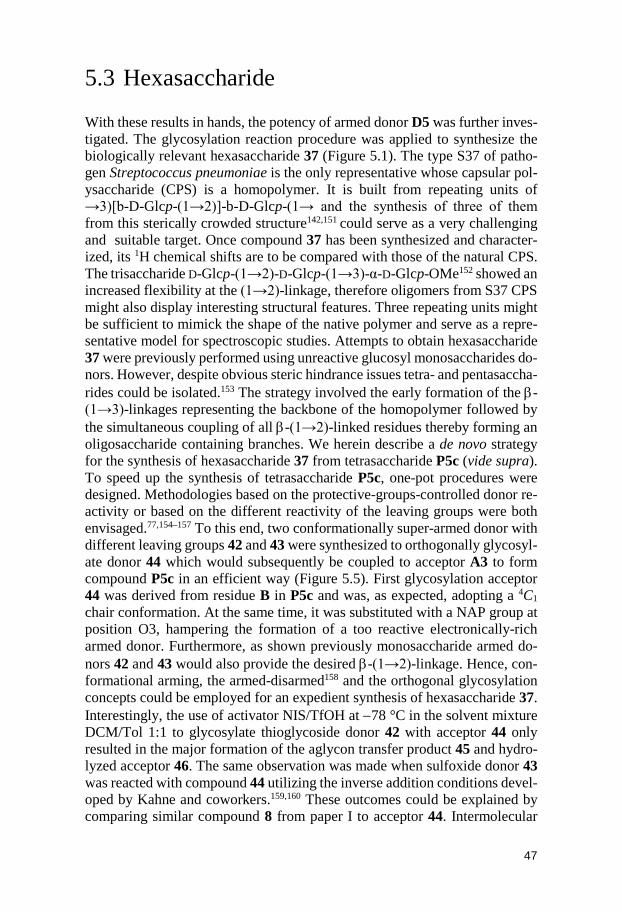
47
5.3 Hexasaccharide With these results in hands, the potency of armed donor D5 was further inves-tigated. The glycosylation reaction procedure was applied to synthesize the biologically relevant hexasaccharide 37 (Figure 5.1). The type S37 of patho-gen Streptococcus pneumoniae is the only representative whose capsular pol-ysaccharide (CPS) is a homopolymer. It is built from repeating units of →3)[b-D-Glcp-(1→2)]-b-D-Glcp-(1→ and the synthesis of three of them from this sterically crowded structure142,151 could serve as a very challenging and suitable target. Once compound 37 has been synthesized and character-ized, its 1H chemical shifts are to be compared with those of the natural CPS. The trisaccharide D-Glcp-(1→2)-D-Glcp-(1→3)-α-D-Glcp-OMe152 showed an increased flexibility at the (1→2)-linkage, therefore oligomers from S37 CPS might also display interesting structural features. Three repeating units might be sufficient to mimick the shape of the native polymer and serve as a repre-sentative model for spectroscopic studies. Attempts to obtain hexasaccharide 37 were previously performed using unreactive glucosyl monosaccharides do-nors. However, despite obvious steric hindrance issues tetra- and pentasaccha-rides could be isolated.153 The strategy involved the early formation of the β-(1→3)-linkages representing the backbone of the homopolymer followed by the simultaneous coupling of all β-(1→2)-linked residues thereby forming an oligosaccharide containing branches. We herein describe a de novo strategy for the synthesis of hexasaccharide 37 from tetrasaccharide P5c (vide supra). To speed up the synthesis of tetrasaccharide P5c, one-pot procedures were designed. Methodologies based on the protective-groups-controlled donor re-activity or based on the different reactivity of the leaving groups were both envisaged.77,154–157 To this end, two conformationally super-armed donor with different leaving groups 42 and 43 were synthesized to orthogonally glycosyl-ate donor 44 which would subsequently be coupled to acceptor A3 to form compound P5c in an efficient way (Figure 5.5). First glycosylation acceptor 44 was derived from residue B in P5c and was, as expected, adopting a 4C1 chair conformation. At the same time, it was substituted with a NAP group at position O3, hampering the formation of a too reactive electronically-rich armed donor. Furthermore, as shown previously monosaccharide armed do-nors 42 and 43 would also provide the desired β-(1→2)-linkage. Hence, con-formational arming, the armed-disarmed158 and the orthogonal glycosylation concepts could be employed for an expedient synthesis of hexasaccharide 37. Interestingly, the use of activator NIS/TfOH at −78 °C in the solvent mixture DCM/Tol 1:1 to glycosylate thioglycoside donor 42 with acceptor 44 only resulted in the major formation of the aglycon transfer product 45 and hydro-lyzed acceptor 46. The same observation was made when sulfoxide donor 43 was reacted with compound 44 utilizing the inverse addition conditions devel-oped by Kahne and coworkers.159,160 These outcomes could be explained by comparing similar compound 8 from paper I to acceptor 44. Intermolecular
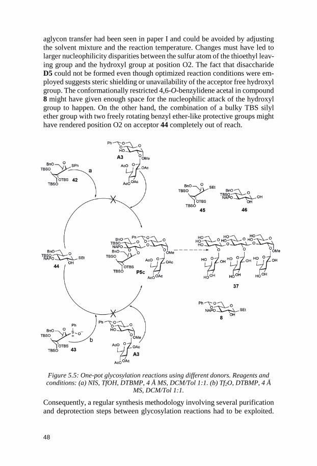
48
aglycon transfer had been seen in paper I and could be avoided by adjusting the solvent mixture and the reaction temperature. Changes must have led to larger nucleophilicity disparities between the sulfur atom of the thioethyl leav-ing group and the hydroxyl group at position O2. The fact that disaccharide D5 could not be formed even though optimized reaction conditions were em-ployed suggests steric shielding or unavailability of the acceptor free hydroxyl group. The conformationally restricted 4,6-O-benzylidene acetal in compound 8 might have given enough space for the nucleophilic attack of the hydroxyl group to happen. On the other hand, the combination of a bulky TBS silyl ether group with two freely rotating benzyl ether-like protective groups might have rendered position O2 on acceptor 44 completely out of reach.
Figure 5.5: One-pot glycosylation reactions using different donors. Reagents and
conditions: (a) NIS, TfOH, DTBMP, 4 Å MS, DCM/Tol 1:1. (b) Tf2O, DTBMP, 4 Å MS, DCM/Tol 1:1.
Consequently, a regular synthesis methodology involving several purification and deprotection steps between glycosylation reactions had to be exploited.
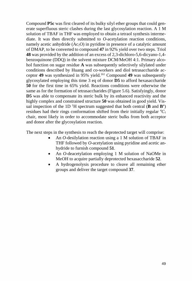
49
Compound P5c was first cleared of its bulky silyl ether groups that could gen-erate superfluous steric clashes during the last glycosylation reaction. A 1 M solution of TBAF in THF was employed to obtain a tetraol synthesis interme-diate. It was then directly submitted to O-acetylation reaction conditions, namely acetic anhydride (Ac2O) in pyridine in presence of a catalytic amount of DMAP, to be converted to compound 47 in 92% yield over two steps. Triol 48 was provided by the addition of an excess of 2,3-dichloro-5,6-dicyano-1,4-benzoquinone (DDQ) in the solvent mixture DCM/MeOH 4:1. Primary alco-hol function on sugar residue A was subsequently selectively silylated under conditions described by Huang and co-workers and diol tetrasaccharide ac-ceptor 49 was synthesized in 95% yield.161 Compound 49 was subsequently glycosylated employing this time 3 eq of donor D5 to afford hexasaccharide 50 for the first time in 65% yield. Reactions conditions were otherwise the same as for the formation of tetrasaccharides (Figure 5.6). Satisfyingly, donor D5 was able to compensate its steric bulk by its enhanced reactivity and the highly complex and constrained structure 50 was obtained in good yield. Vis-ual inspection of the 1D 1H spectrum suggested that both central (B and B’) residues had their rings conformation shifted from their initially regular 4C1 chair, most likely in order to accommodate steric bulks from both acceptor and donor after the glycosylation reaction. The next steps in the synthesis to reach the deprotected target will comprise:
• An O-desilylation reaction using a 1 M solution of TBAF in THF followed by O-acetylation using pyridine and acetic an-hydride to furnish compound 51.
• An O-deacetylation employing 1 M solution of NaOMe in MeOH to acquire partially deprotected hexasaccharide 52.
• A hydrogenolysis procedure to cleave all remaining ether groups and deliver the target compound 37.
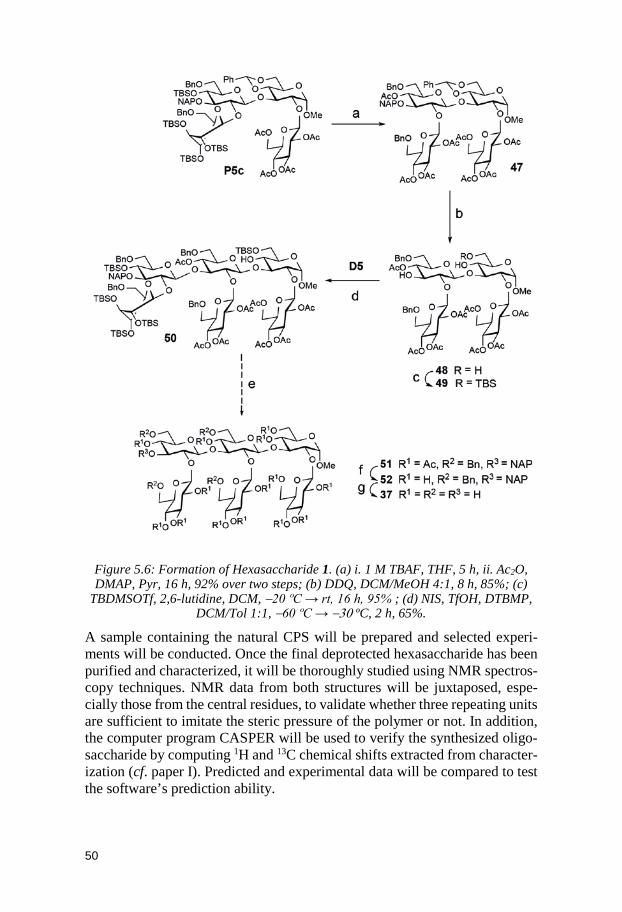
50
Figure 5.6: Formation of Hexasaccharide 1. (a) i. 1 M TBAF, THF, 5 h, ii. Ac2O, DMAP, Pyr, 16 h, 92% over two steps; (b) DDQ, DCM/MeOH 4:1, 8 h, 85%; (c)
TBDMSOTf, 2,6-lutidine, DCM, −20 ºC → rt, 16 h, 95% ; (d) NIS, TfOH, DTBMP, DCM/Tol 1:1, −60 ºC → −30 ºC, 2 h, 65%.
A sample containing the natural CPS will be prepared and selected experi-ments will be conducted. Once the final deprotected hexasaccharide has been purified and characterized, it will be thoroughly studied using NMR spectros-copy techniques. NMR data from both structures will be juxtaposed, espe-cially those from the central residues, to validate whether three repeating units are sufficient to imitate the steric pressure of the polymer or not. In addition, the computer program CASPER will be used to verify the synthesized oligo-saccharide by computing 1H and 13C chemical shifts extracted from character-ization (cf. paper I). Predicted and experimental data will be compared to test the software’s prediction ability.
51
5.4 Conclusion In conclusion, a number of conformationally armed and orthogonally pro-tected glycan building blocks have been used in the convergent syntheses of tri- and tetra- and hexasaccharides. To the best of our knowledge, the excellent yields of those sterically challenging (1→2),(1→3)-linked β-D-glucose oligo-saccharides using this approach and the super-armed donor methodology has not been reported previously. A conformational and structure-reactivity anal-ysis was performed on all six donors and on two of the trisaccharide products using NMR-based methods. All compounds were found to exist as at least two conformers in equilibrium with each other, for some of them in contrast to the single state described in the literature.70,77,145 Low-temperature NMR studies should be able to address problems causing this discrepancy. The suggested dynamic equilibrium behind the different ring conformations observed of for instance donor D4 could be elucidated.162 On the other hand, donors D2, D4 and D6 adopting more axial rich conformations were distinctly more reactive that donors D1, D3 and D5. From this observation, we could deduce that do-nors in axial-rich conformations had their potency in glycosylation reactions further enhanced due to structural preorganization towards boats and skews comparable to the transition state. In addition to those results, the glycosyla-tion procedure designed in this work allowed the formation of highly demand-ing structures with complete stereoselectivity and could be compatible with the use of automated systems for the synthesis of oligosaccharides. Finally, three repeating units from the CPS of Streptococcus pneumoniae type S37 might be sufficient to mimic the conformational behavior of the native poly-mer. That is why, hexasaccharide 37 will be submitted to deprotection proce-dures and its structural NMR spectroscopy information will be compared to the polysaccharides’.
52
6 Delineating the Conformational Flexibility of Trisaccharides from NMR Spectroscopy Experiments and Computer Simulations (Paper III)
Complex carbohydrates play important roles in biology and their diverse bio-logical activity is a function of their surface properties which are governed by their structure and conformation. Most deprotected hexopyranose monosac-charides have a defined 4C1 ring shape leaving rotations around the glycosidic linkages accountable for their flexibility.163 However, we showed in paper II that the ring conformation can be perturbed by introducing steric constraints and consequently that a single state was not enough to describe the molecules conformational space. The same applies to the overall three-dimensional as-pect of saccharides in solution. The sites of substitution, the anomeric and the absolute configurations, the functional groups as well as the degree of branch-ing will affect the three-dimensional structure and the dynamic behavior of the sugar residues in proximity to the linkage giving rise to an ensemble of conformations for the whole saccharide system.164,165,166 The conformational dynamics of oligo- and polysaccharides can be better understood by studying the smaller structural elements they contain.167 For instance, results collected on the trisaccharide α-D-Glcp-(1→2)-α-D-Glcp-(1→3)-α-D-Glcp-OMe might be extrapolated in order to learn more about the relationship between structure and function of oligosaccharide Glc3Man9GlcNAc2 present in glyco-protein precursors.168 In this paper, five trisaccharides differing by the type of monosaccharides and linkages they enclose had their conformational prefer-ences revealed by the cooperative use of NMR spectroscopy and molecular dynamics simulations. This work focuses on the use of the former method. The goal of this project was:
• To synthesize site-specific 13C-labelled isotopologues and isotopomers of one of the trisaccharides
• To measure conformationally sensitive observables using several NMR spectroscopy experiments and to compare ex-perimental measurements with computational data
• Finally, to expose the conformational propensity of all trisac-charides
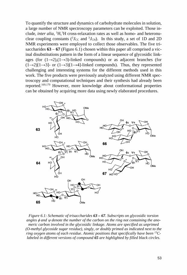
53
To quantify the structure and dynamics of carbohydrate molecules in solution, a large number of NMR spectroscopy parameters can be exploited. Those in-clude, inter alia, 1H,1H cross-relaxation rates as well as homo- and heteronu-clear coupling constants (3JCC and 3JCH). In this study, a set of 1D and 2D NMR experiments were employed to collect those observables. The five tri-saccharides 63 − 67 (Figure 6.1) chosen within this paper all comprised a vic-inal disubstitutions pattern in the form of a linear sequence of glycosidic link-ages (for (1→2),(1→3)-linked compounds) or as adjacent branches (for (1→2)[1→3]- or (1→3)[1→4]-linked compounds). Thus, they represented challenging and interesting systems for the different methods used in this work. The five products were previously analyzed using different NMR spec-troscopy and computational techniques and their synthesis had already been reported.169,170 However, more knowledge about conformational properties can be obtained by acquiring more data using newly elaborated procedures.
Figure 6.1: Schematic of trisaccharides 63 – 67. Subscripts on glycosidic torsion angles φ and ψ denote the number of the carbon on the ring not containing the ano-meric carbon involved in the glycosidic linkage. Atoms are specified as unprimed
(O-methyl glycoside sugar residue), singly, or doubly primed as indicated next to the ring oxygen atoms of each residue. Atomic positions that specifically have been 13C-labeled in different versions of compound 65 are highlighted by filled black circles.
54
6.1 Synthesis Three isotopomers and isotopologues of 65 were synthesized, viz., β-D-[1’-13C]Glcp-(1→2)[β-D-Glcp-(1→3)]-α-D-Manp-OMe (65-c1’), β-D-Glcp-(1→2)[β-D-[1’’-13C]Glcp-(1→3)]-α-D-Manp-OMe (65-c1’’), and β-D-[2’-13C]Glcp-(1→2)[β-D-[2’’-13C]Glcp-(1→3)]-α-D-Manp-OMe (65-c2’/c2’’) to facilitate the extraction of 3JCC coupling constants across the different glyco-sidic linkages. A common synthetic route was designed to form, in the most efficient way possible, those three trisaccharides from previously reported 4,6-O-benzylidene-α-D-mannopyranoside as well as isotopically labeled D-[1-13C]- and D-[2-13C]-derivatized glucosyl donors. Several variants of 13C la-belled 2,3,4,6-tetra-O-benzoyl glucopyranoside derivatives were tested to gly-cosylate 4,6-O-benzylidene-α-D-mannopyranoside 53 at position O2 and O3 (Figure 6.2). Glycosylation reactions protocols utilizing glucopyranosyl bro-mide171 54 or trichloroacetimidate172 donors 55 to simultaneously substitute both positions led to the major formation of orthoesters 56 and 57 and only small amount of the disaccharides methyl 2,3,4,6-tetra-O-benzoyl-β-D-gluco-pyranosyl-(1→3)-4,6-O-benzylidene-α-D-mannopyranoside 59 was isolated. Unfortunately, by-products were not rearranged by adding more of the pro-moter systems, respectively, AgOTf and TMSOTf.
Instead, a two-steps glycosylation strategy was therefore considered. Hence, ethyl 2,3,4,6-tetra-O-benzoyl-1-thio-β-D-glucopyranoside173 donor 58 could react with acceptor 53 to afford in a regio- and stereoselective fashion the β-(1→3)-linked disaccharide 59 in 82% yield. Neighboring group partici-pation of 2-O-benzoyl group was able to provide the desired β-configuration. The use, in excess, of donors ethyl 2,3-di-O-benzoyl-4,6-O-benzylidene-1-thio-β-D-glucopyranoside174,175 60 was required to build the last β-(1→2)-linkage. These thioglucoside donors had for particularity an O-benzylidene acetal protective group joining both O4 and O6 positions together and pulling them away from the anomeric region. This might have thereby facilitated the nucleophilic attack of OH2 from disaccharide 59. A final glycosylation reac-tion was performed between the disaccharide intermediate methyl 2,3,4,6-tetra-O-benzoyl-β-D-glucopyranosyl-(1→3)-4,6-O-benzylidene-α-D-manno-pyranoside169 59 and donors 60. Trisaccharide 61 was furnished in 75% aver-age yield. Trisaccharide was then submitted to deprotection procedures. A so-lution of 80% AcOH (aq) was employed for acidic cleavage of acetal protec-tive groups. THF was added to increase solubility of the sugars in the mixture. Tetraol intermediate 62 was obtained in 68% average yield. Finally, the last de-O-benzoylation step was performed by utilizing a solution of 1M NaOMe in MeOH and fully deprotected target trisaccharides 65-c1’, 65-c1’’ and 65-c2’/c2’’ were isolated in 91% average yield.
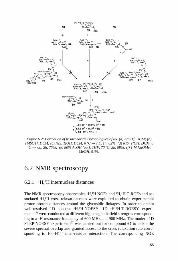
55
Figure 6.2: Formation of trisaccharide isotopologues of 65. (a) AgOTf, DCM; (b)
TMSOTf, DCM; (c) NIS, TfOH, DCM, 0 ºC → r.t., 1h, 82%; (d) NIS, TfOH, DCM, 0 ºC → r.t., 2h, 75%; (e) 80% AcOH (aq.), THF, 70 ºC, 2h, 68%; (f) 1 M NaOMe,
MeOH, 91%.
6.2 NMR spectroscopy
6.2.1 1H,1H internuclear distances The NMR spectroscopy observables 1H,1H NOEs and 1H,1H T-ROEs and as-sociated 1H,1H cross relaxation rates were exploited to obtain experimental proton-proton distances around the glycosidic linkages. In order to obtain well-resolved 1D spectra, 1H,1H-NOESY, 1D 1H,1H-T-ROESY experi-ments176 were conducted at different high magnetic field strengths correspond-ing to a 1H resonance frequency of 600 MHz and 900 MHz. The modern 1D STEP-NOESY experiment177 was carried out for compound 67 to tackle the severe spectral overlap and granted access to the cross-relaxation rate corre-sponding to H4–H1’’ inter-residue interaction. The corresponding NOE
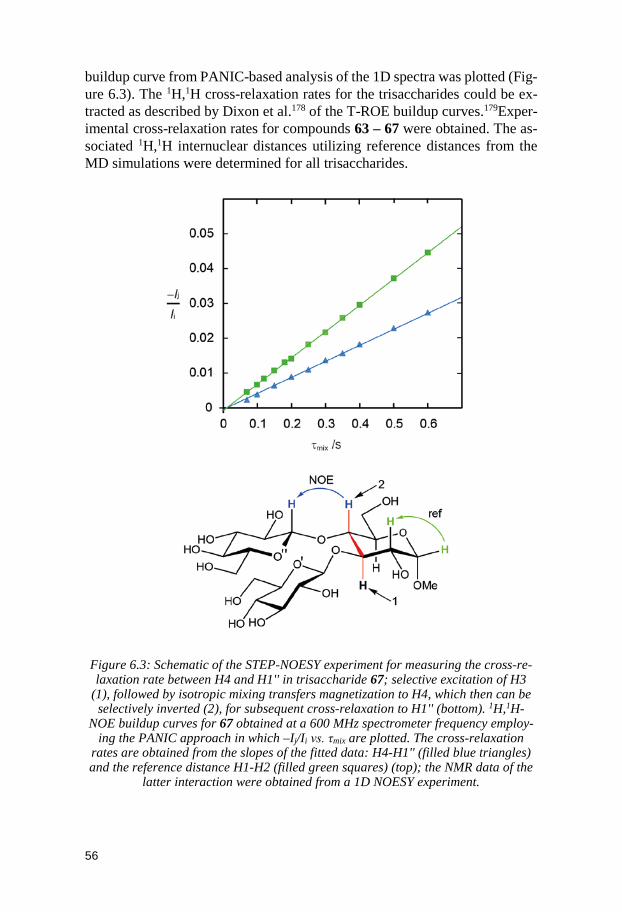
56
buildup curve from PANIC-based analysis of the 1D spectra was plotted (Fig-ure 6.3). The 1H,1H cross-relaxation rates for the trisaccharides could be ex-tracted as described by Dixon et al.178 of the T-ROE buildup curves.179Exper-imental cross-relaxation rates for compounds 63 – 67 were obtained. The as-sociated 1H,1H internuclear distances utilizing reference distances from the MD simulations were determined for all trisaccharides.
Figure 6.3: Schematic of the STEP-NOESY experiment for measuring the cross-re-laxation rate between H4 and H1'' in trisaccharide 67; selective excitation of H3
(1), followed by isotropic mixing transfers magnetization to H4, which then can be selectively inverted (2), for subsequent cross-relaxation to H1'' (bottom). 1H,1H-
NOE buildup curves for 67 obtained at a 600 MHz spectrometer frequency employ-ing the PANIC approach in which –Ij/Ii vs. τmix are plotted. The cross-relaxation
rates are obtained from the slopes of the fitted data: H4-H1'' (filled blue triangles) and the reference distance H1-H2 (filled green squares) (top); the NMR data of the
latter interaction were obtained from a 1D NOESY experiment.
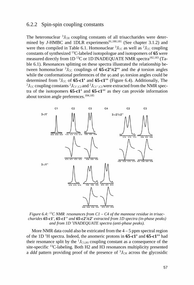
57
6.2.2 Spin-spin coupling constants The heteronuclear 3JCH coupling constants of all trisaccharides were deter-mined by J-HMBC and 1DLR experiments91,180,181 (See chapter 3.1.2) and were then compiled in Table 6.1. Homonuclear 3JCC as well as 2JCC coupling constants of synthesized 13C-labeled isotopologue and isotopomers of 65 were measured directly from 1D 13C or 1D INADEQUATE NMR spectra182,183 (Ta-ble 6.1). Resonances splitting on these spectra illustrated the relationship be-tween homonuclear 3JCC couplings of 65-c2’/c2’’ and the φ torsion angles while the conformational preferences of the ψ2 and ψ3 torsion angles could be determined from 3JCC of 65-c1’ and 65-c1’’ (Figure 6.4). Additionally, The 2JCC coupling constants 2JC1’,C2 and 2JC1’’,C3 were extracted from the NMR spec-tra of the isotopomers 65-c1’ and 65-c1’’ as they can provide information about torsion angle preferences.184,185
Figure 6.4: 13C NMR resonances from C1 – C4 of the mannose residue in trisac-
charides 65-c1', 65-c1'' and 65-c2'/c2' extracted from 1D spectra (in-phase peaks) and from 1D 1INADEQUATE spectra (anti-phase peaks).
More NMR data could also be extricated from the 4 – 5 ppm spectral region of the 1D 1H spectra. Indeed, the anomeric protons in 65-c1’ and 65-c1’’ had their resonance split by the 1JC1,H1 coupling constant as a consequence of the site-specific 13C-labeling. Both H2 and H3 resonances multiplicity presented a ddd pattern providing proof of the presence of 3JCH across the glycosidic
58
linkages. The J doubling methodology186 was used to acquire the value of these 3JCH coupling constants. They were in excellent agreement with those measured from J-HMBC or 1DLR experiments. As expected, these observa-tions could not be made from the 1D 1H spectrum of doubly labeled compound 65-c2’/c2’’ as it is known that the 2JC2,H1 coupling constant in β-D-glucose is small (See paper).187
6.2.3 Comparison between MD and NMR spectroscopy results In this study, NMR spectroscopy was used to experimentally provide inter-proton distances and J coupling constants along the flexible glycosidic link-ages of the five trisaccharides. The latter NMR observable, with the aid of Karplus equations, was used to determine the conformations being populated by the trisaccharides. However, because NMR spectroscopy only yields re-sults of a representative ensemble of states it is often used in synergy with MD simulations. In a suitable force field, MD simulations allow for investigations of conformational states at an atomic level of detail. From the conducted sim-ulations, a detailed atomic picture of the conformational ensembles being pop-ulated by the trisaccharides was extracted. It was subsequently used to calcu-late J coupling constants utilizing the reparametrized Karplus equation JCX/SU0987,167,188. Conformational information from both techniques could then be compared.
The values for 3JCH coupling constants obtained from both approaches are shown in Table 6.1. Good agreement was observed for all five trisaccharides. The largest deviation was 1.1 Hz for the φ4 dihedral in trisaccharide 67 (4.3 vs. 3.2 Hz) and the smallest deviation was 0 Hz for ψ2 in trisaccharide 63 (3.8 vs. 3.8 Hz). The computed J couplings have a root-mean-square deviation (RMSD) of 0.54 Hz with respect to the corresponding experimental values along the torsional angle ψ and the largest deviation was of 1.0 Hz for ψ3 in trisaccharide 64 (4.7 vs. 5.7 Hz). All the calculated J couplings along the di-hedral torsion φ were overestimated if the α-anomer was the non-reducing end monosaccharide while they were underestimated for residues displaying a β-anomeric configuration leading to a RMSD of 0.65 Hz. To reduce this devia-tion, an additional term was added to the Karplus equation relating 3JCH cou-pling constants and the linkage dihedral φ. The RMSD was reduced to 0.35 Hz along this torsional angle. With this correction, the largest and the smallest deviations became 0.7 Hz and zero for φ4 in trisaccharide 67 and φ3 in trisac-charide 65 (table 6.1). The origin of the discrepancy between experimental and simulated coupling constants was investigated and the results obtained led to the conclusion that the development of more accurate force fields than CHARMM36 is required to solve this issue.
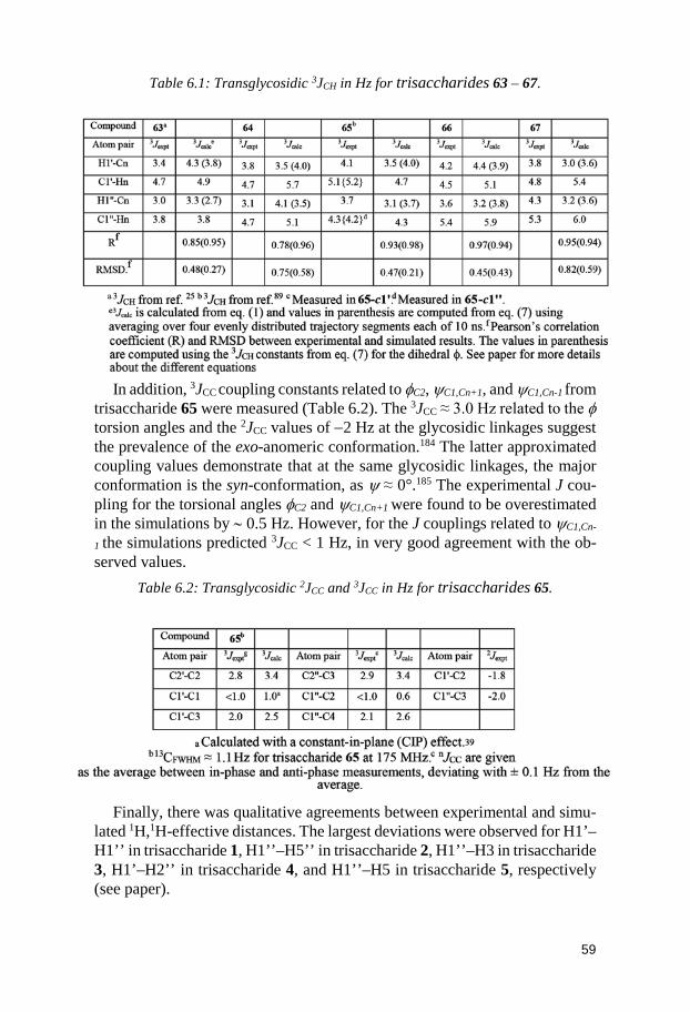
59
Table 6.1: Transglycosidic 3JCH in Hz for trisaccharides 63 – 67.
In addition, 3JCC coupling constants related to φC2, ψC1,Cn+1, and ψC1,Cn-1 from
trisaccharide 65 were measured (Table 6.2). The 3JCC ≈ 3.0 Hz related to the φ torsion angles and the 2JCC values of −2 Hz at the glycosidic linkages suggest the prevalence of the exo-anomeric conformation.184 The latter approximated coupling values demonstrate that at the same glycosidic linkages, the major conformation is the syn-conformation, as ψ ≈ 0°.185 The experimental J cou-pling for the torsional angles φC2 and ψC1,Cn+1 were found to be overestimated in the simulations by ∼ 0.5 Hz. However, for the J couplings related to ψC1,Cn-
1 the simulations predicted 3JCC < 1 Hz, in very good agreement with the ob-served values.
Table 6.2: Transglycosidic 2JCC and 3JCC in Hz for trisaccharides 65.
Finally, there was qualitative agreements between experimental and simu-
lated 1H,1H-effective distances. The largest deviations were observed for H1’–H1’’ in trisaccharide 1, H1’’–H5’’ in trisaccharide 2, H1’’–H3 in trisaccharide 3, H1’–H2’’ in trisaccharide 4, and H1’’–H5 in trisaccharide 5, respectively (see paper).
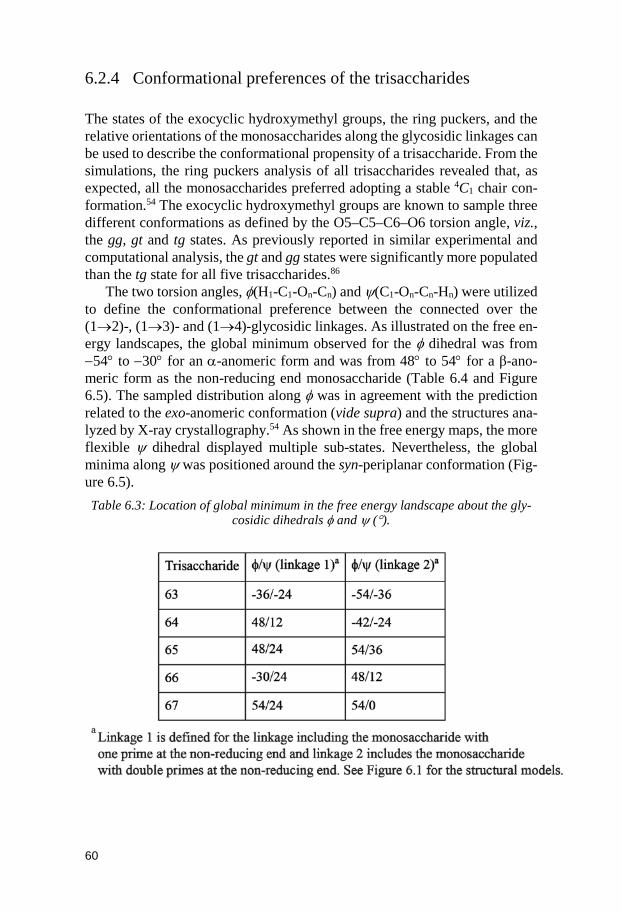
60
6.2.4 Conformational preferences of the trisaccharides The states of the exocyclic hydroxymethyl groups, the ring puckers, and the relative orientations of the monosaccharides along the glycosidic linkages can be used to describe the conformational propensity of a trisaccharide. From the simulations, the ring puckers analysis of all trisaccharides revealed that, as expected, all the monosaccharides preferred adopting a stable 4C1 chair con-formation.54 The exocyclic hydroxymethyl groups are known to sample three different conformations as defined by the O5–C5–C6–O6 torsion angle, viz., the gg, gt and tg states. As previously reported in similar experimental and computational analysis, the gt and gg states were significantly more populated than the tg state for all five trisaccharides.86
The two torsion angles, φ(H1-C1-On-Cn) and ψ(C1-On-Cn-Hn) were utilized to define the conformational preference between the connected over the (1→2)-, (1→3)- and (1→4)-glycosidic linkages. As illustrated on the free en-ergy landscapes, the global minimum observed for the φ dihedral was from −54° to −30° for an α-anomeric form and was from 48° to 54° for a β-ano-meric form as the non-reducing end monosaccharide (Table 6.4 and Figure 6.5). The sampled distribution along φ was in agreement with the prediction related to the exo-anomeric conformation (vide supra) and the structures ana-lyzed by X-ray crystallography.54 As shown in the free energy maps, the more flexible ψ dihedral displayed multiple sub-states. Nevertheless, the global minima along ψ was positioned around the syn-periplanar conformation (Fig-ure 6.5). Table 6.3: Location of global minimum in the free energy landscape about the gly-
cosidic dihedrals φ and ψ (°).
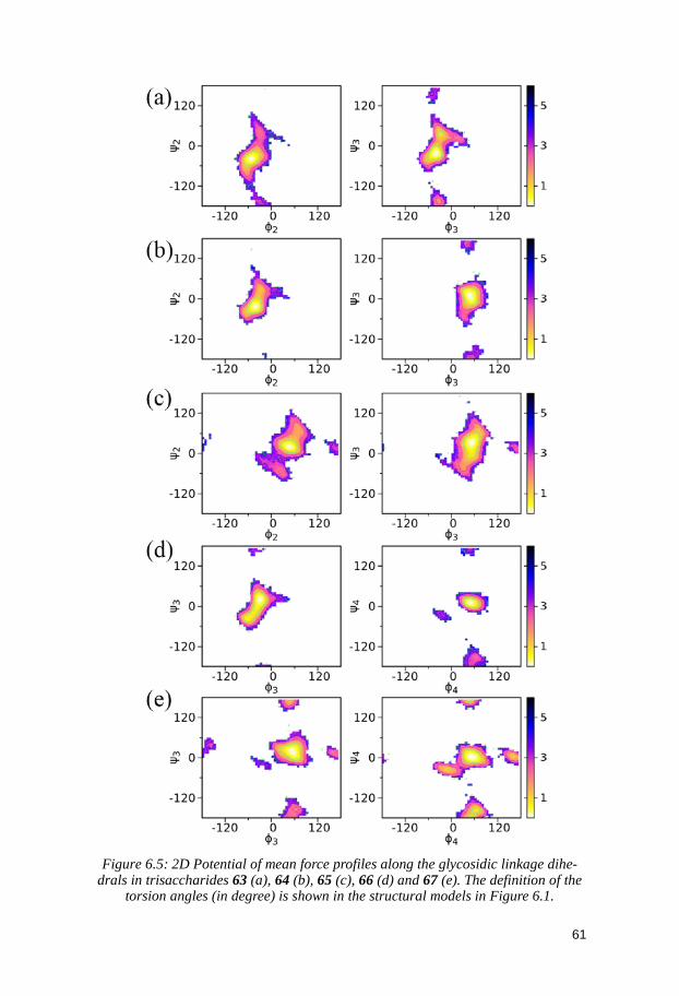
61
Figure 6.5: 2D Potential of mean force profiles along the glycosidic linkage dihe-
drals in trisaccharides 63 (a), 64 (b), 65 (c), 66 (d) and 67 (e). The definition of the torsion angles (in degree) is shown in the structural models in Figure 6.1.
62
The impact of monosaccharide compositions and linkage types onto the con-formational heterogeneity of the trisaccharides could be evaluated by thor-oughly interpreting the simulation results obtained at the atomic level of de-tail. Indeed, trisaccharides 66 and 67 only differed by the anomeric configu-ration of their (1→3)-linkages; α-D-glucose in trisaccharide 66 and β-D-glu-cose in trisaccharide 67. However, the enhanced conformational heterogeneity seen from the free energy landscape of the glycosidic linkages for trisaccha-rides 67 compared to trisaccharide 66 might indicate the possible presence of conformational epitopes in more complex oligosaccharide structures (Figure 6.5). It is worth mentioning that trisaccharide 67 represents a structural ele-ment of the lipopolysaccharide from Moraxella catarrhalis.189,190
6.3 Conclusion For the five studied trisaccharides, simulated and experimental J coupling constants as well as effective 1H,1H distances were in good agreement. The use of NOE-based experiments in the determination of an oligosaccharide 3D-structure can be limited by their inherent < r-6 > -averaging making them less sensitive to subtle conformational differences. On the other hand, the J-based experiments are excellent in detecting small variations in structure such as in the five trisaccharides investigated herein. Consequently, the conformational space sampled by the molecules can be elucidated by conducting the two com-plementary techniques. For interpretation of the experimental results, MD simulations can be a very valuable tool in gaining insight into the conforma-tional properties of compounds. However, accounting for the full range of sampled conformations is required to reproduce the experimental data ob-tained by NMR spectroscopy techniques and a force field suitable for the sys-tem studied has to be chosen. Nevertheless, because of the high-resolution description they provide, the MD simulations can help rationalizing the con-formational heterogeneity of oligosaccharides. Indeed, results show that a dif-ference in anomeric configuration can induce changes in the conformational preferences of the trisaccharides. Clearly, structural components, such as the five compounds studied, can contribute to the conformational heterogeneity of larger glycans. This observation suggests that the determination of struc-ture-function relationships can be investigated by analyzing the three-dimen-sional properties of key-fragments or epitopes. The development of vaccines relies on the rational design of saccharides containing epitopes that are able to trigger an immunogenic response.
63
7 Complete 1H and 13C NMR Chemical Shift Assignments of Mono- to Tetrasaccharides as Basis for NMR Chemical shift Predictions of Oligo- and Polysaccharides Using the Computer Program CASPER (Paper IV)
As shown throughout this thesis, carbohydrate molecules can be found in na-ture as polysaccharides and they can be linked to proteins or steroidal deriva-tives. Bacterial polysaccharides can be built from around 100 different mono-saccharides and 10 monomers are usually used to describe the human gly-come. The resulting large structural diversity reflects the numerous roles gly-cans play in biological systems. Consequently, knowledge of the primary and three-dimensional structures of sugar molecules is required to uncover rela-tionships between shape and function.81 Mass spectrometry and NMR spec-troscopy are the two prevailing techniques employed to determine the primary structure of carbohydrate molecules. The latter is preferred since details at the atomic level can be attained.82 The NMR chemical shift contains structural information, however, because of the relatively limited spectral dispersion of carbohydrates’ 1H resonances, their assignments can become a difficult task. To address this issue and to gain access to this information, computerized sys-tems have been developed.191–193
The module Determine Glycan Structure of the computer program CAS-PER is able to solve the primary structure of these molecules using only un-assigned NMR data automatically or manually extracted from spectra.194,195 The software can also be used to directly predict 1H and 13C NMR chemical shifts of oligo- and polysaccharides via its Calculate Chemical Shifts mod-ule.196 Chemical shifts of an unknown compound are calculated by computing NMR data of saccharides already entered in the database and by using empir-ical estimations of glycosylation shifts (Δδ, i.e. the chemical shift difference between the glycosylated saccharides and their free monosaccharide constitu-ents). In the present version of the web-based interface (http://www.casper.or-gan.su.se/casper/) of CASPER information on GT functions, currently stored in the database ECODAB, can additionally be given to determine the structure of a polysaccharide.197–199 Tools for extracting 3D models of glycans have also been implemented.200
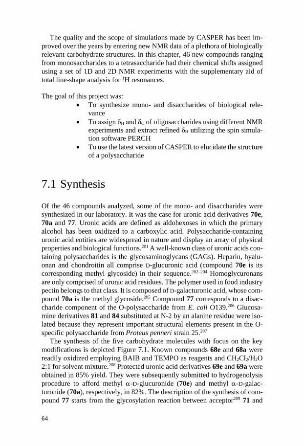
64
The quality and the scope of simulations made by CASPER has been im-proved over the years by entering new NMR data of a plethora of biologically relevant carbohydrate structures. In this chapter, 46 new compounds ranging from monosaccharides to a tetrasaccharide had their chemical shifts assigned using a set of 1D and 2D NMR experiments with the supplementary aid of total line-shape analysis for 1H resonances. The goal of this project was:
• To synthesize mono- and disaccharides of biological rele-vance
• To assign δH and δC of oligosaccharides using different NMR experiments and extract refined δH utilizing the spin simula-tion software PERCH
• To use the latest version of CASPER to elucidate the structure of a polysaccharide
7.1 Synthesis Of the 46 compounds analyzed, some of the mono- and disaccharides were synthesized in our laboratory. It was the case for uronic acid derivatives 70e, 70a and 77. Uronic acids are defined as aldohexoses in which the primary alcohol has been oxidized to a carboxylic acid. Polysaccharide-containing uronic acid entities are widespread in nature and display an array of physical properties and biological functions.201 A well-known class of uronic acids con-taining polysaccharides is the glycosaminoglycans (GAGs). Heparin, hyalu-onan and chondroitin all comprise D-glucuronic acid (compound 70e is its corresponding methyl glycoside) in their sequence.202–204 Homoglycuronans are only comprised of uronic acid residues. The polymer used in food industry pectin belongs to that class. It is composed of D-galacturonic acid, whose com-pound 70a is the methyl glycoside.205 Compound 77 corresponds to a disac-charide component of the O-polysaccharide from E. coli O139.206 Glucosa-mine derivatives 81 and 84 substituted at N-2 by an alanine residue were iso-lated because they represent important structural elements present in the O-specific polysaccharide from Proteus penneri strain 25.207
The synthesis of the five carbohydrate molecules with focus on the key modifications is depicted Figure 7.1. Known compounds 68e and 68a were readily oxidized employing BAIB and TEMPO as reagents and CH2Cl2/H2O 2:1 for solvent mixture.208 Protected uronic acid derivatives 69e and 69a were obtained in 85% yield. They were subsequently submitted to hydrogenolysis procedure to afford methyl α-D-glucuronide (70e) and methyl α-D-galac-turonide (70a), respectively, in 82%. The description of the synthesis of com-pound 77 starts from the glycosylation reaction between acceptor209 71 and
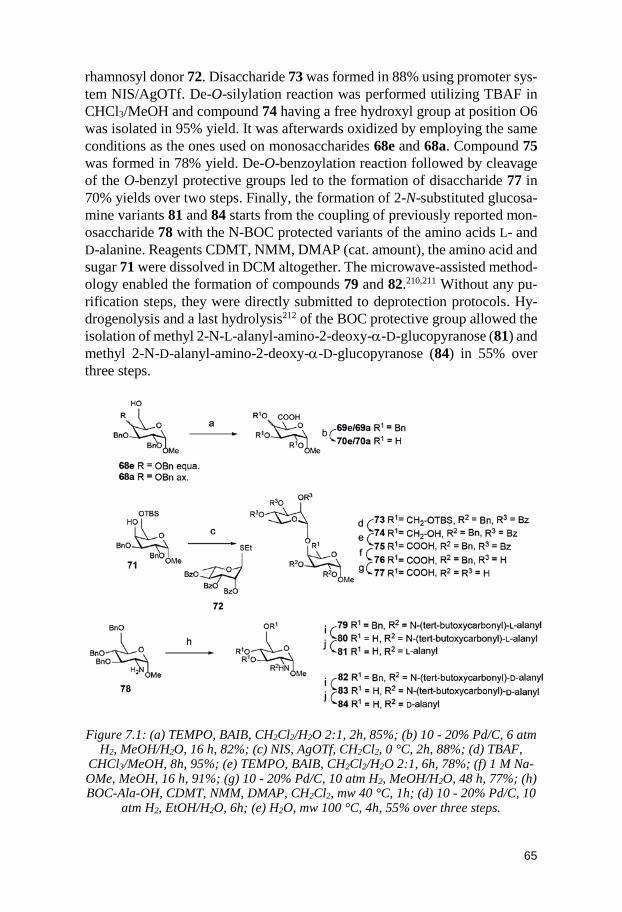
65
rhamnosyl donor 72. Disaccharide 73 was formed in 88% using promoter sys-tem NIS/AgOTf. De-O-silylation reaction was performed utilizing TBAF in CHCl3/MeOH and compound 74 having a free hydroxyl group at position O6 was isolated in 95% yield. It was afterwards oxidized by employing the same conditions as the ones used on monosaccharides 68e and 68a. Compound 75 was formed in 78% yield. De-O-benzoylation reaction followed by cleavage of the O-benzyl protective groups led to the formation of disaccharide 77 in 70% yields over two steps. Finally, the formation of 2-N-substituted glucosa-mine variants 81 and 84 starts from the coupling of previously reported mon-osaccharide 78 with the N-BOC protected variants of the amino acids L- and D-alanine. Reagents CDMT, NMM, DMAP (cat. amount), the amino acid and sugar 71 were dissolved in DCM altogether. The microwave-assisted method-ology enabled the formation of compounds 79 and 82.210,211 Without any pu-rification steps, they were directly submitted to deprotection protocols. Hy-drogenolysis and a last hydrolysis212 of the BOC protective group allowed the isolation of methyl 2-N-L-alanyl-amino-2-deoxy-α-D-glucopyranose (81) and methyl 2-N-D-alanyl-amino-2-deoxy-α-D-glucopyranose (84) in 55% over three steps.
Figure 7.1: (a) TEMPO, BAIB, CH2Cl2/H2O 2:1, 2h, 85%; (b) 10 - 20% Pd/C, 6 atm
H2, MeOH/H2O, 16 h, 82%; (c) NIS, AgOTf, CH2Cl2, 0 °C, 2h, 88%; (d) TBAF, CHCl3/MeOH, 8h, 95%; (e) TEMPO, BAIB, CH2Cl2/H2O 2:1, 6h, 78%; (f) 1 M Na-OMe, MeOH, 16 h, 91%; (g) 10 - 20% Pd/C, 10 atm H2, MeOH/H2O, 48 h, 77%; (h) BOC-Ala-OH, CDMT, NMM, DMAP, CH2Cl2, mw 40 °C, 1h; (d) 10 - 20% Pd/C, 10
atm H2, EtOH/H2O, 6h; (e) H2O, mw 100 °C, 4h, 55% over three steps.
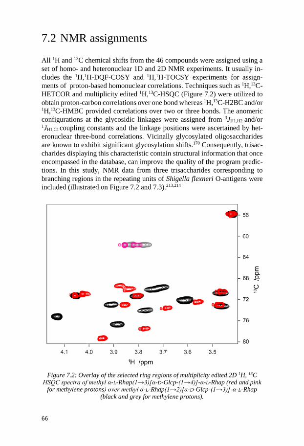
66
7.2 NMR assignments All 1H and 13C chemical shifts from the 46 compounds were assigned using a set of homo- and heteronuclear 1D and 2D NMR experiments. It usually in-cludes the 1H,1H-DQF-COSY and 1H,1H-TOCSY experiments for assign-ments of proton-based homonuclear correlations. Techniques such as 1H,13C-HETCOR and multiplicity edited 1H,13C-HSQC (Figure 7.2) were utilized to obtain proton-carbon correlations over one bond whereas 1H,13C-H2BC and/or 1H,13C-HMBC provided correlations over two or three bonds. The anomeric configurations at the glycosidic linkages were assigned from 3JH1,H2 and/or 1JH1,C1 coupling constants and the linkage positions were ascertained by het-eronuclear three-bond correlations. Vicinally glycosylated oligosaccharides are known to exhibit significant glycosylation shifts.170 Consequently, trisac-charides displaying this characteristic contain structural information that once encompassed in the database, can improve the quality of the program predic-tions. In this study, NMR data from three trisaccharides corresponding to branching regions in the repeating units of Shigella flexneri O-antigens were included (illustrated on Figure 7.2 and 7.3).213,214
Figure 7.2: Overlay of the selected ring regions of multiplicity edited 2D 1H, 13C
HSQC spectra of methyl α-L-Rhap(1→3)[α-D-Glcp-(1→4)]-α-L-Rhap (red and pink for methylene protons) over methyl α-L-Rhap(1→2)[α-D-Glcp-(1→3)]-α-L-Rhap
(black and grey for methylene protons).
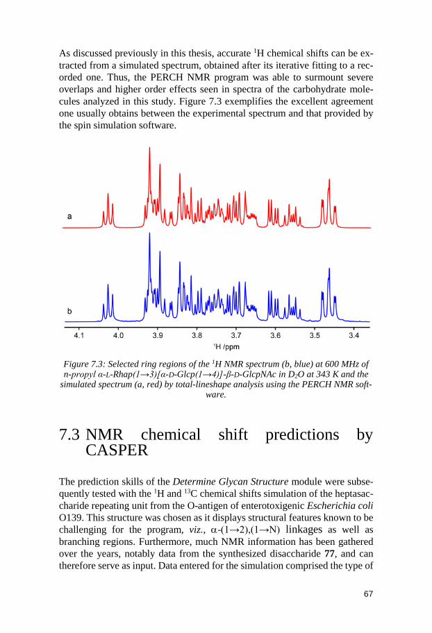
67
As discussed previously in this thesis, accurate 1H chemical shifts can be ex-tracted from a simulated spectrum, obtained after its iterative fitting to a rec-orded one. Thus, the PERCH NMR program was able to surmount severe overlaps and higher order effects seen in spectra of the carbohydrate mole-cules analyzed in this study. Figure 7.3 exemplifies the excellent agreement one usually obtains between the experimental spectrum and that provided by the spin simulation software.
Figure 7.3: Selected ring regions of the 1H NMR spectrum (b, blue) at 600 MHz of n-propyl α-L-Rhap(1→3)[α-D-Glcp(1→4)]-β-D-GlcpNAc in D2O at 343 K and the
simulated spectrum (a, red) by total-lineshape analysis using the PERCH NMR soft-ware.
7.3 NMR chemical shift predictions by CASPER
The prediction skills of the Determine Glycan Structure module were subse-quently tested with the 1H and 13C chemical shifts simulation of the heptasac-charide repeating unit from the O-antigen of enterotoxigenic Escherichia coli O139. This structure was chosen as it displays structural features known to be challenging for the program, viz., α-(1→2),(1→N) linkages as well as branching regions. Furthermore, much NMR information has been gathered over the years, notably data from the synthesized disaccharide 77, and can therefore serve as input. Data entered for the simulation comprised the type of
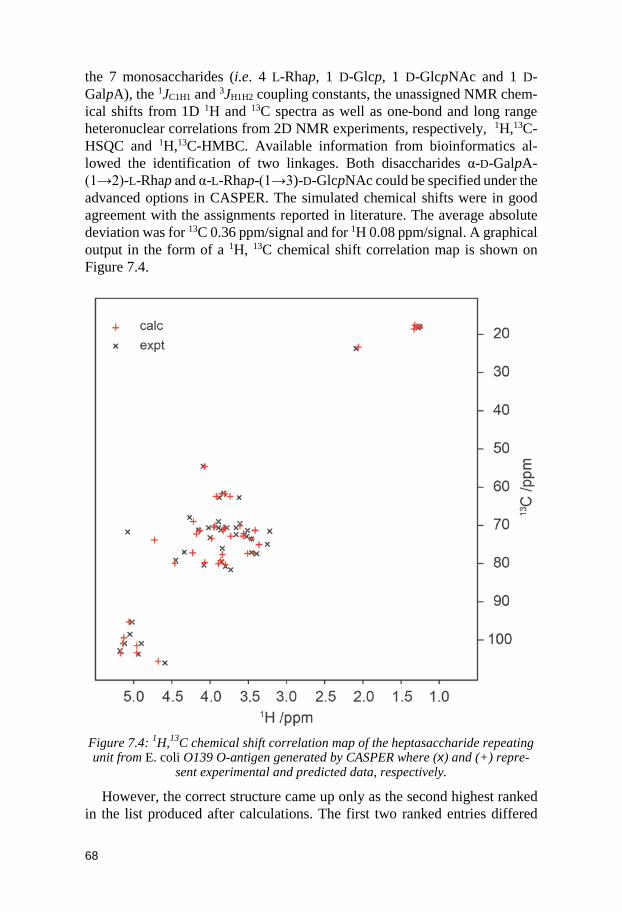
68
the 7 monosaccharides (i.e. 4 L-Rhap, 1 D-Glcp, 1 D-GlcpNAc and 1 D-GalpA), the 1JC1H1 and 3JH1H2 coupling constants, the unassigned NMR chem-ical shifts from 1D 1H and 13C spectra as well as one-bond and long range heteronuclear correlations from 2D NMR experiments, respectively, 1H,13C-HSQC and 1H,13C-HMBC. Available information from bioinformatics al-lowed the identification of two linkages. Both disaccharides α-D-GalpA-(1→2)-L-Rhap and α-L-Rhap-(1→3)-D-GlcpNAc could be specified under the advanced options in CASPER. The simulated chemical shifts were in good agreement with the assignments reported in literature. The average absolute deviation was for 13C 0.36 ppm/signal and for 1H 0.08 ppm/signal. A graphical output in the form of a 1H, 13C chemical shift correlation map is shown on Figure 7.4.
Figure 7.4: 1H,13C chemical shift correlation map of the heptasaccharide repeating unit from E. coli O139 O-antigen generated by CASPER where (x) and (+) repre-
sent experimental and predicted data, respectively.
However, the correct structure came up only as the second highest ranked in the list produced after calculations. The first two ranked entries differed
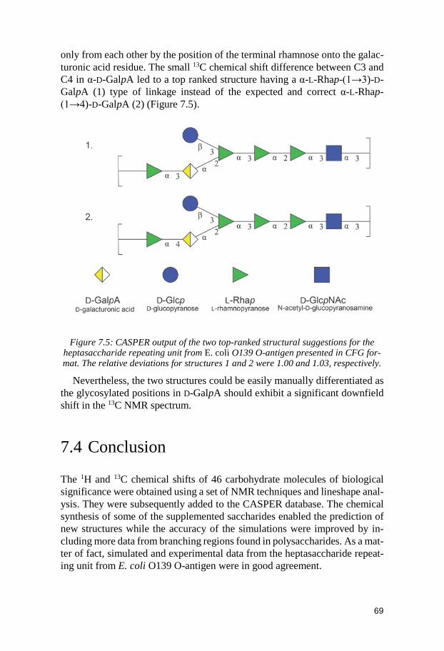
69
only from each other by the position of the terminal rhamnose onto the galac-turonic acid residue. The small 13C chemical shift difference between C3 and C4 in α-D-GalpA led to a top ranked structure having a α-L-Rhap-(1→3)-D-GalpA (1) type of linkage instead of the expected and correct α-L-Rhap-(1→4)-D-GalpA (2) (Figure 7.5).
Figure 7.5: CASPER output of the two top-ranked structural suggestions for the heptasaccharide repeating unit from E. coli O139 O-antigen presented in CFG for-mat. The relative deviations for structures 1 and 2 were 1.00 and 1.03, respectively.
Nevertheless, the two structures could be easily manually differentiated as the glycosylated positions in D-GalpA should exhibit a significant downfield shift in the 13C NMR spectrum.
7.4 Conclusion The 1H and 13C chemical shifts of 46 carbohydrate molecules of biological significance were obtained using a set of NMR techniques and lineshape anal-ysis. They were subsequently added to the CASPER database. The chemical synthesis of some of the supplemented saccharides enabled the prediction of new structures while the accuracy of the simulations were improved by in-cluding more data from branching regions found in polysaccharides. As a mat-ter of fact, simulated and experimental data from the heptasaccharide repeat-ing unit from E. coli O139 O-antigen were in good agreement.
70
8 Conclusion and outlook
The objective of this thesis was to acquire structural information of small car-bohydrate molecules and, by using this knowledge, to be able to extrapolate and get a better understanding of the conformational behavior in solution of larger glycans containing these sugar components. The same conformational behavior that has a great influence on sugars interaction ability to proteins and other biomacromolecules. Herein, modern chemical carbohydrate synthesis procedures were employed to obtain the systems under study, namely, mono- to hexasaccharides contained in biologically relevant structures. NMR spec-troscopy enabled the measurement of conformationally sensitive variables that were utilized to elucidate their overall three-dimensional aspect of these molecules in solution. Results demonstrated that the shape of saccharides can-not be described by a single conformer.
Despite progress made in enzymatic carbohydrate synthesis, the chemical fashion remains the most rapid way to obtain new structures for exploratory research. In chapter 4 and 5, an efficient methodology was designed to obtain branched oligosaccharides in a convergent manner. Bulky silyl ether protec-tive groups forced the rings of disaccharide donors to flip to more axial-rich conformations enhancing their reactivity and yielding complete stereoselec-tivity during glycosylation reactions. The robust designed strategy can be pre-sented as compatible with one-pot synthesis protocols and automated oligo-saccharide synthesis systems.
In chapter 4, the target molecule was a tetrasaccharide enclosed in the nat-ural compound solaradixine. It was isolated after a successful sterically chal-lenging 2 + 2 coupling reaction. The motive 2-O-glycosylated glucose is often found in nature and especially in saponin structures. This methodology could be employed to form more of this class of molecules, their conformational heterogeneity could be exposed by NMR spectroscopy and the relationship between their structural diversity and the various roles they play in biochem-istry may be elucidated. The program CASPER can also be continuously ex-ploited to verify the different synthesized structures.
In chapter 5, three repeating units of the CPS from Streptococcus pneu-moniae S37 were synthesized for the first time. The slightly populated 3S1 conformation in donors was sufficient to drive the glycosylation reactions overcoming steric bulks. It was proposed that donors in axial-rich skews or boats have their reactivity additionally enhanced because they are already pre-organized. They have already left the low-in-energy chair well and resemble
71
oxocarbenium ion transition state conformations. However, more information about reaction mechanism and the ring interconversion pathways involved is required. Quantum mechanics methods and dynamic NMR experiments would help address these issues. The hexasaccharide formed remains to be submitted to deprotection procedures and fully characterized. Its NMR spec-troscopy data are to be compared with the ones collected from the natural pol-ymer to determine whether three repeating units are enough to mimic the be-havior of the polysaccharide in solution.
In chapter 6, the conformational preferences of 5 trisaccharides were ex-posed. The combination of synthetic methods, NMR spectroscopy techniques and molecular dynamics simulations allowed the acquisition of a great amount of structural data. Subtle changes in the carbohydrate sequence had a strong impact on the glycosidic linkage flexibility. These results suggest that even a small system such as a trisaccharide contains enough information to contribute to the conformational heterogeneity exhibited by larger glycans. Conse-quently, these fragments might be seen as parts of conformational epitopes and can help rationalize the design of vaccines and inhibitors.
Finally in chapter 7, NMR chemical shifts of, inter alia, synthesized oligo-saccharides were added to the database of the computer program CASPER. In total, data of 46 compounds were added to improve the prediction skills of the software and allowed the structural identifications of new types of carbohy-drates. As a matter of fact, the quality of the simulations was very good as unassigned NMR data from a complex heptasaccharide could serve as input for the Determine Structure module. Data obtained from the simulations were in good agreement with the assignments reported in the literature for the hep-tasaccharide repeating unit from E. coli O139 O-antigen.
72
9 Populärvetenskaplig sammanfattning på svenska
Kolhydrater, tillsammans med nukleinsyror, proteiner och lipider, är de vikti-gaste beståndsdelarna i celler. Bland dessa fyra huvudklasser av biomolekyler, är kolhydrater den klass som vi förstår minst med avseende på deras biolo-giska funktion, främst för att vi saknar effektiva verktyg och metoder för att samla in och analysera dem. Sackarider är ofta kopplade till lipider och pro-teiner som uttrycks på cellytan och är därför involverade i kommunikation mellan celler, igenkännings processer och interaktioner med patogener. Sockerenheternas bidningar kallas glykosidbindningar och bildas i glyko-syleringsreaktioner. Identifieringen av kolhydratstrukturer för biologisk analys bygger fortfarande på att utarbeta nya kemiska syntesmetoder, framför allt de som gäller glykosidbindning. Den fortsatta utvecklingen i analysen av strukturen av glykaner gör det möjligt för forskare att finna relationer mellan tredimensionell struktur och deras biologiska funktion. NMR-spektroskopi är en icke-förstörande analytisk teknik som kan ge detaljerad strukturell infor-mation om oligosackarider och polysackarider på atomär nivå.
I denna avhandling har kemiska metoder för syntes av kolhydrater utveck-lats och NMR-spektroskopiexperiment genomfördes för att analysera deras konformations beteende i lösning. Denna studie fokuserar på små kolhy-dratmönster som är delar av större strukturer som finns i naturen. Syftet med att skala ner systemet under studien var att upptäcka epitoper; dessa fragment bär en stor mängd information och kan leda till utformning av enzyminhibi-torer och vacciner.
Saponiner är en amfifil typ av föreningar i vilka en kolhydratmolekyl kan sammanfogas, exempelvis, till en steroidal struktur. Den stora strukturella mångfalden avspeglar de många roller de spelar i biokemiska processer i väx-ter och deras biologiska aktivitet kan användas i läkemedelsindustrin. I kapitel I, finslipas en syntetisk metod som bygger på att öka reaktiviteten hos socker-molekyler under glykosyleringsreaktioner och används för att bygga en tetra-sackarid som ingår i en klass av saponiner. Målföreningen erhållas genom att först bilda disackaridens byggblock följt av en 2 + 2 koppling av ingående strukturelement. Detta förfarande kan nu användas för att syntetisera en hel rad av kolhydratstrukturer som finns i saponiner. Datorprogrammet CASPER kan hjälpa till med att karakterisera de nya föreningarna. I detta fall, uppvisade struktukförslaget utmärkt överensstämmelse med de experimentella data som samlats in för tetrasackariden i detta projekt.
73
Förfarandet som utvecklades i kapitel I bygger på ett koncept som ytterligare undersöktes i kapitel II. Sockermolekylen, som oftas består av en ring, kan ta olika former. Ändring av substituentema på ringen kan aktivera sockeren-heterna till ett mer reaktivt tillstånd. Ett NMR-baserat protokoll följdes för att bestämma ringens form för olika sackarider som användes under glykosyler-ingsreaktionerna. En korrelation mellan molekyler i mer reaktiva konfor-mationer och större mängd isolerade produkter var tydlig. Denna strategi tillämpades framgångsrikt tillämpats för att syntetisera en hexasackarid motsvarande tre repeterande enheter av en polysackarid som finns på ytan av Streptococcus pneumoniae typ S37.
Även om studien i kapitel II fokuserat på ringens form hos socker, var det den tredimensionella strukturen hos fem trisackarider som studerades hos glykaner i kapitel III. Kombinationen av syntetiska metoder, NMR tekniker och molekyldynamiksimuleringar visade att den övergripande formen av kol-hydratmolekyler inte kan beskrivas med ett enda läge och att subtila förän-dringar på atomär nivå påtagligt kan påverka hur de olika tillstånden är populerade. Dessa observationer ledde till slutsatsen att en del av de motiv och/eller tillstånd som analyseras kan fungera som igenkänningsbara fragment eller epitoper vid bindning mellan proteiner och glykaner.
Slutligen, eftersom kolhydratmolekyler kan forma mycket komplexa strukturer och den strukturella information de innehåller kan vara relaterad till deras biologiska funktion, har datoriserade system skapats för att extrahera och förutsäga sådana strukturer. I kapitel IV, har förutsägelsen av NMR-kem-iska skift med hjälp av datorprogrammet CASPER förbättras genom analys av NMR information hos 46 biologiskt relevanta strukturer.
74
10 Appendix
The author’s contribution to the publications:
I. Performed all experiments. Wrote the major part of the manu-script.
II. Performed the synthesis and conducted some NMR experiments. Wrote the major part of the manuscript.
III. Performed the synthesis and the NMR experiments. Contributed to the writing.
IV. Performed parts of the synthesis and the NMR experiments. Con-tributed to the writing.
75
11 Acknowledgements
I would like to thank the following: My supervisor professor Göran Widmalm for the support, enthusiasm and guidance over the years. It was an honor and a pleasure working with you for the past 6 years. Professor Pher Andersson for showing interest in this thesis. Jonas Ståhle, Sara Moa, Christoffer Hamark, Carolina Fontana, Olof Eng-ström, Joan Patrick, Linda Stenman, Audrey Forticaux and Rasmus Sandsten who helped me improve this thesis. Thank you for all your comments and the nice discussions. Collaborators on the trisaccharides project, in particular Dr. Mingjun Yang
and Prof. Alexander D. MacKerell Jr. All collaborators in my group: Dr. Christoffer Hamark, Jonas Ståhle, Dr. Olof Engström, Hani Mobarak, Dr. Pilar Blasco Morales, Dr. Sanchali Mukherjee, Dr. Carolina Fontana, Dr. Robert Pendrill, Dr. Jerk Rönnols, Dr. Mona Zac-cheus, Dr. Elin Säwén and Dr Magnus Lundborg. The KTH team for nice collaborations: Dr. Jakob Wohlert, Jennie Berglund, Dr Malin Bergenstråhle-Wolhert, Nils Sjöberg, Dr. Francesco Vilaplana, Prof. Martin Lawoko, Prof. Gunnar Henriksson and Prof. Mikael Lindström. My numerous diploma workers, who I have had the pleasure of sharing pro-jects with (in chronological order): Elis, Loïc, Lorna, Chris, Patricia, Azad, Mike, Ulf, Paul, Guillaume, Cyril, Janina and Alessandro. Carin, Kristina, Martin and Ola for always being available and for making my life much easier at the department. Sigrid, Christina and Louise for your pa-tience when I needed to understand the Swedish administration system. People from Biophysics, especially Torbjörn and Matthew.
76
Vetenskaprådet, Knut och Alice Wallenbergs stiftelsen, Ångpanneföreningens förskningsstiftelse, Sture Erikssons fond för Cancer och hjärtforskning och Liljevalchs resestipendien för generösa donationer. Colleagues and friends from the department: Sara, Jonas, Killian, Matt, Rich-ard, Teresa, Christoffer, Olle, Tanja, Tuo, Henrik, Tove och Tove, Johanna, Karl, Gabriella, Anuja, Joel, Elis, Arnar, Ylva, Tony, Maria, Greco, Ana, Elisa and all the Spanish crew, Angela, and Sybrand. The board and video games team: Killian, Matt, Jonas, Angela, Tony, Sybrand, Miguel, Arnar and Tanja. The tennis crew for serious work outs: Killian, Matt, Tony and Miguel. The gym crew for actual serious work outs: Sara, Micke, Greco, Tanja, Elisa, Jorge, Tove and Johanna. Mes amis français pour leur confiance en moi: Audy, Anaïs, Marine, Robin, Rémi, Yann, Margaux, Alice, Benoît, Benjamin et Yoann. Mina svenska vänner: Linda, Medea, Ana, Johanna H., Johanna M., Jenny, Prava, Tanja, Joen, Hanna, Charlotte, Martin och Cecilia som gjorde att jag har nästan blivit svennebanan. The people from South Africa. Neil, Gammon, Michelle and especially Taigh for nice conversations in and outside the lab as well as in India. The Galan group members for nice chats and wanderings in New Orleans. Sara, Lorna, Fabian and Joan for the best parties, fika, dinner, lunch, and hangouts….Glad I met you guys! Sara, one day we will stomp grapes in Borås and create our own wine brand! Maman, papa, et ma soeur Audrey qui m’ont toujours soutenus.
Christina, S.O., Therese, Sebastian, Jonas, Marie, Joel, Tyva, Molly och Stella som gör att jag känner mig alltid välkommen i familjen. Rasmus, l’amour de ma vie. La personne la plus intelligente et la plus drôle que j’ai jamais rencontré. Tack för att du förstår mig, tack för allt, tack för att du finns. Hanna, den roligaste och sötaste flickan!
77
12 References
(1) Petrucci R.H.; Harwood W.S.; Herring F.G. Gen. Chem. 2002, 32. (2) Hudson, C. S. J. Chem. Educ. 1941, 18, 353. (3) Bozell, J. J.; Petersen, G. R. Green Chem. 2010, 12 (4). (4) Varki, A.; Cummings, R.D.; Esko, J.D.; Freeze, P.; Stanley, C.;
Bertozzi, C.R.; Hart, G.W.; Etzler, M. E. Essentials Glycobiol. 2009. (5) National Research Council. Transforming Glycoscience: A Roadmap
for the Future; The National Academies Press, 2012. (6) Springer, S. A.; Gagneux, P. J. Biol. Chem. 2013, 288 (10), 6904. (7) Bardoel, B. W.; Van Strijp, J. A. G. J. Mol. Recognit. 2011, 24 (6),
1077. (8) Gabius, H. J. The Sugar Code, Wiley-VCH.; Weinheim, 2009. (9) Morgan W. T. J.; W. M. Watkins. Br. Med. Bull. 1969, 25 (1), 30. (10) Olsson, M. L.; Clausen, H. Br. J. Haematol. 140 (1), 3. (11) Habibi, Y.; Lucia, L. A.; Rojas, O. J. Chem. Rev. 2010, 110 (6), 3479. (12) Klemm, D.; Heublein, B.; Fink, H. P.; Bohn, A. Angew. Chemie - Int.
Ed. 2005, 44 (22), 3358. (13) Klemm, D.; Kramer, F.; Moritz, S.; Lindstrom, T.; Ankerfors, M.;
Gray, D.; Dorris, A. Angew. Chemie - Int. Ed. 2011, 50 (24), 5438. (14) Jones, C. J.; Larive, C. K. Nat. Chem. Biol. 2011, 7 (11), 758. (15) Weintraub, A.; Widmalm, G.; Jansson, P.E.; Jansson, M; Hultenby, K;
Albert, M. J. Microb. Pathog. 1994, No. 16, 235. (16) Kamerling, J.P.; Boons, G.J; Lee, Y.C.; Suzuki, A.;Taniguchi, N.;
Voragen, A. G. Compr. Glycosience 2007, 1, 1. (17) Stenutz, R.; Weintraub, A. FEMS Microbiol. Rev. 2006, 30, 382. (18) Widmalm, G. Carbohydr. Res. 2013, 378, 123. (19) Werz, D.B.; Ranzinger, R.; Herget, S.; Adibekian, A.; Lieth, C.W.v.D.;
Seeberger, P. H. ACS Chem. Biol. 2007, 2 (10), 685. (20) Aich, U; Yarema, K. J. Carbohydrate-Based Vaccines Immunother.
2008, 1. (21) Crocker, P. R.; Paulson, J. C.; Varki, A. Nat. Rev. Immunol. 2007, 7
(April), 255. (22) Imberty, A.; Chabre, Y. M. European J. Org. Chem. 2008, No. 14,
7490. (23) Rillahan, C. D.; Paulson, J. C. Annu. Rev. Biochem. 2011, No. 80, 797. (24) IUPAC-IUB Joint Commission on Biochemical Nomenclature. Eur. J.
Biochem. 1980, No. 111, 295. (25) Cremer, D.; Pople, J. A. J. Am. Chem. Soc. 1975, 1354 (19), 1354.
78
(26) Yates, JH; Jeffrey, G. Carbohydr. Res. 1979, 74, 319. (27) Fushinobu, S. No Title. (28) Sattelle, B. M.; Almond, A. Phys. Chem. Chem. Phys. 2012, No. 14,
5843. (29) Yamada, H.; Tanigakiuchi, K.; Nagao, K.; Okajima, K.; Mukae, T.
Tetrahedron Lett. 2004, 45 (50), 9207. (30) Guo, T.; Zhao, C.; Sun, J.; Li, W.; Control, D. Helv. Chim. Acta 2014,
97, 361. (31) Gu, G.; Du, Y.; Linhardt, R. J. 2004, No. 11, 5497. (32) Siegbahn, A.; Aili, U.; Ochocinska, A.; Olofsson, M.; Rönnols, J.;
Mani, K.; Widmalm, G.; Ellervik, U. Bioorg. Med. Chem. 2011, 19 (13), 4114.
(33) Pedersen, C. M.; Marinescu, L. G.; Bols, M. Comptes Rendus Chim. 2011, 14 (1), 17.
(34) Heuckendorff, M.; Pedersen, C. M.; Bols, M. J. Org. Chem. 2013, 78 (14), 7234.
(35) Olsen, J. I.; Kowalska, K.; Pedersen, C. M.; Bols, M. Tetrahedron Lett. 2016, 57 (1), 35.
(36) Lairson, L. L.; Henrissat, B.; Davies, G. J.; Withers, S. G. Annu. Rev. Biochem. 2008, No. 77, 521.
(37) Vocadlo, D. J.; Davies, G. J. Curr. Opin. Chem. Biol. 2008, No. 12, 539.
(38) Zhu, X.; Schmidt, R. R. Angew. Chemie - Int. Ed. 2009, 48, 1900. (39) Satoh H.; Manabe S. Chem. Soc. Rev. 2013, 42, 4297. (40) Bols, M. Acc. Chem. Res. 2006, 39 (4), 259. (41) Pedersen, C. M.; Marinescu, L. G.; Bols, M. Chem. Commun. (Camb).
2008, No. 21, 2465. (42) Pedersen, C. M.; Nordstrøm, L. U.; Bols, M. J. Am. Chem. Soc. 2007,
No. 9, 9222. (43) Jensen, H. H.; Nordstrøm, L. U.; Bols, M. J. Am. Chem. Soc. 2004, 8
(Figure 1), 9205. (44) Andrews, C.W.; Rodebaugh, R.; Fraser-Reid, B. J. Org. Chem. 1996,
No. 61, 5280. (45) Gomez, A.M.; Fraser-reid, B.; Cristobal Lopez, J. Topics in Current
Chemistry Topics in Current Chemistry, Springer.; New-York, 2010. (46) Kozar, T; André, S.; Ulicny, J.; Gabius, H. J. In Three-Dimensional
Aspects of the Sugar Code; 2009; pp 15–29. (47) Walvoort, M.T.C.; Marel, G.A.V.D.; Overkleeft, H.S.; Codee, J. D. C.
Chem. Sci. 2013, No. 4, 897. (48) Kirby, A. J. No Title, Springer.; Berlin, 1983. (49) Grindley, T. B. Glycoscience: Chemistry and Chemical Biology,
Springer.; Berlin, 2008. (50) IUPAC. Eur. J. Biochem. 1983, No. 131, 5. (51) Lemieux, R. U.; Koto, S. Tetrahedron 1974, 30, 1933. (52) Batchelor, R. J.; Green, D. F.; Johnston, B. D.; Patrick, O. B.; Pinto,
B. M. Carbohydr. Res. 2001, 330, 421. (53) Bock, K.; Duus, J. Ø. J. Carbohydr. Chem. 2006, 13 (4), 513.
79
(54) Rao, V. S. R. . et al. Conform. Carbohydrates 1998. (55) Krasnova, L.; Wong, C. Annu. Rev. Biochem. 2016, No. 85, 599. (56) Seeberger, P. H. Acc. Chem. Res. 2015, 48 (5), 1450. (57) Costantino, P.; Rappuoli, R.; Berti, F. Expert. Opin. Drug. Discov.
2011, 6, 1045. (58) Morelli, L.; Poletti, L.; Lay, L. European J. Org. Chem. 2011, No.
2011, 5723. (59) Anish, C.; Schumann, B.; Pereira, C. L.; Seeberger, P. H. Chem. Biol.
2014, 21 (1), 38. (60) Verez-Bencomo, V.; Fernandez-Santana, V.; Hardy, E.; Toledo, M.E.;
Rodrıguez, M.C.; Heynngnezz, L.; Rodriguez, A.; Baly, A.; Herrera, L. . I.; Al., M. . et. Science (80-. ). 305, 522.
(61) Hsu, C.; Hung, S.; Wu, C.; Wong, C. Angew. Chemie - Int. Ed. 2011, No. 50, 11872.
(62) Schmaltz, R. M.; Hanson, S. R.; Wong, C. Chem. Rev. 2011, No. 111, 4259.
(63) Davies, G. J.; Planas, A. Acc. Chem. Res. 2012, 45 (2), 308. (64) Crich, D.; Bohe, L. Comptes Rendus Chim. 2011, 14, 3. (65) Pratt, D. A.; Crich, D.; Huang, M.; Garrett, G. E.; Birlirakis, N.; Bohe,
L. Nat. Chem. 2012, 4 (July), 663. (66) Huang, M.; Retailleau, P.; Bohe, L.; Crich, D. J. Am. Chem. Soc. 2012,
No. 64, 14746. (67) Christensen, H. M.; Oscarson, S.; Jensen, H. H. Carbohydr. Res. 2015,
408, 51. (68) Demchenko, A.V.; Ranade, S. C. J. Carbohydr. Chem. 2013, No. 32,
1. (69) Okada, Y.; Mukae, T.; Okajima, K.; Taira, M.; Fujita, M.; Yamada, H.
Org. Lett. 2007, 9, 1573. (70) Okada, Y.; Nagata, O.; Taira, M.; Yamada, H. Org. Lett. 2007, 9, 2755. (71) Nigudkar, S. S.; Demchenko, A. V. Chem. Sci. 2015, 6, 2687. (72) Crich, D. J. Org. Chem. 2011, No. 76, 9193. (73) Imamura, A.; Ando, H.; Korogi, S.; Tanabe, G.; Muraoka, O.
Tetrahedron Lett. 2003, 44, 6725. (74) Lim, D.; Brimble, M. A.; Kowalczyk, R.; Watson, A. J. A.; Fairbanks,
A. J. Angew. Chemie - Int. Ed. 2014, No. 53, 11907. (75) Hudak, JE; Canham, SM; Bertozzi, C. Nat. Chem. Biol. 2014, 10, 69. (76) Pulsipher, A; Griffin, ME; Stone, SE; Brown, JM; Hsieh-Wilson, L. J.
Am. Chem. Soc. 2014, 136, 6794. (77) Jensen, H.H.; Pedersen, C.M.; Bols, M. European J. Org. Chem. 2007,
No. 13, 7576. (78) Ágoston, K.; Streicher, H.; Fügedi, P. Tetrahedron Asymmetry 2016,
27 (16), 707. (79) Vliegenhardt, J. F. G. NMR Spectroscopy and Computer Modeling of
Carbohydrates: Recent Advances, American C.; Washington, 2006; Vol. 930.
(80) Jiménez-Barbero, J.; Peters, T. NMR Spectroscopy of Glycoconjugates, Wiley.; Weinheim, 2003.
80
(81) Rönnols, J.; Pendrill, R.; Fontana, C.; Hamark, C.; d’Ortoli, T. A.; Engström, O.; Ståhle, J.; Zaccheus, M. V; Säwén, E.; Hahn, L. E.; Iqbal, S.; Widmalm, G. Carbohydr. Res. 2013, 380, 156.
(82) Roslund, M. U.; Säwén, E.; Landström, J.; Rönnols, J.; Jonsson, K. H. M.; Lundborg, M.; Svensson, M. V; Widmalm, G. Carbohydr. Res. 2011, 346 (11), 1311.
(83) Angles d’Ortoli T.; Widmalm, G. Tetrahedron 2016, 72, 912. (84) Lambert, J.B; Mazzola, E. . Nuclear Magnetic Resonance
Spectroscopy, Pearson Ed.; Upper Saddle River, 2003. (85) Karplus, M. J. Am. Chem. Soc. 1963, No. 85, 2870. (86) Stenutz, R.; Carmichael, I.; Widmalm, G.; Serianni, A. S. J. Org.
Chem. 2002, No. 4, 949. (87) Säwén, E.; Massad, T.; Landersjö, C.; Damberg, P.; Widmalm, G. Org.
Biomol. Chem. 2010, 8 (16), 3684. (88) Thibaudeau, C.; Stenutz, R.; Hertz, B.; Klepach, T.; Zhao, S.; Wu, Q.;
Carmichael, I.; Serianni, A. S. J. Am. Chem. Soc. 2004, No. 126, 15668.
(89) Bose, B.; Zhao, S.; Stenutz, R.; Cloran, F.; Bondo, P. B.; Bondo, G.; Hertz, B.; Carmichael, I.; Serianni, A. S. J. Am. Chem. Soc. 1998, No. 9, 11158.
(90) Meissner, A.; Sørensen, O. W. Magn. Reson. Chem. 2001, No. 39, 49. (91) Nishida, T.; Widmalm, G.; Sándor, P. Magn. Reson. Chem. 1996, 34
(5), 377. (92) del Río-Portilla, F; Blechta, V.; Freeman, R. J. Magn. Reson. 1994,
No. 111, 132. (93) Laatikainen, R.; Miemitz, M.; Weber, U.; Sundelin, J.; Hassinen, T.;
Vepsalainen, J. M. J. Magn. Reson. Ser. A 1996, No. 120, 1. (94) Pauli, G. F.; Chen, S.-N.; Lankin, D. C.; Bisson, J.; Case, R. J.;
Chadwick, L. R.; Gödecke, T.; Inui, T.; Krunic, A.; Jaki, B. U.; McAlpine, J. B.; Mo, S.; Napolitano, J. G.; Orjala, J.; Lehtivarjo, J.; Korhonen, S.-P.; Niemitz, M. J. Nat. Prod. 2014, 77, 1473.
(95) Bryant, R. G. J. Chem. Educ. 1983, 60, 933. (96) Haasnoot, C.A.G.; De Leeuw, F.A.A.M.; Altona, C. Tetrahedron
1980, 36, 2783. (97) Rönnols, J.; Burkhardt, A.; Cumpstey, I.; Widmalm, G. European J.
Org. Chem. 2012, 74. (98) Thomas, P.D; Basus, V.J.; James, T. L. Proc. Natl. Acad. Sci. U.S.A
1991, 246, 144. (99) Sayers, E.W.; Prestegard, J. H. Biophys. J. 2000, 79, 3313. (100) Woods, R.J.; Pathiaseril, A.; Wormald, M.R.; Edge, C.J.; Dwek, R. A.
. Eur. J. Biochem. 1998, 258, 372. (101) Widmalm, G; Byrd, R.A.; Egan, W. Carbohydr. Res. 1992, 229, 195. (102) Davis, D. . J. Am. Chem. Soc. 1987, 109, 3471. (103) Macura, S; Farmer II, B.T.; Brown, L. R. J. Magn. Reson. 1986, No.
70, 493. (104) Hu, H.; Krishnamurthy, K. J. Magn. Reson. 2006, 182, 173. (105) Dixon, A.M.; Widmalm, G.; Bull, T. E. J. Magn. Reson. 2000, No.
81
147, 266. (106) Karplus, M.; McCammon, J. A. Nat. Struct. Biol. 2002, 9, 646. (107) Kirschner, KN; Yongye, AB; Tschampel, SM; González-Outeirino, J;
Daniels, CR; Foley, BL; Woods, R. J. Comput. Chem. 2008, 29, 622. (108) Guvench, O; Mallajosyula, SS; Raman, EP; Hatcher, E;
Vanommeslaeghe, K; Foster, TJ; Jamison, FW; Mackerell, J. A. Chem. Theory Comput. 2011, 7, 3162.
(109) Eklund, R; Widmalm, G. Carbohydr. Res. 2003, 338, 393. (110) Widmalm, G. General NMR Spectroscopy of Carbohydrates amd
Conformational Analysis in Solution, Elsevier L.; Oxford, 2007. (111) Hinchliffe, A. Molecular Modelling for Beginners, Wiley.; Chichester,
2003. (112) Schwarz, S.; Xavier, N. M.; Csuk, R.; Rauter, A. M. Carbohydr. Chem.
2012, 37, 326. (113) Sidoryk, K.; Korda, A.; Rárová, L.; Okleštková, J.; Strnad, M.; Cmoch,
P.; Pakulski, Z.; Gwardiak, K.; Karczewski, R.; Luboradzki, R. Tetrahedron 2015, 71, 2004.
(114) Munafo, J. P.; Gianfagna, T. J. Nat. Prod. Rep. 2015, 32 (3), 454. (115) Neszmelyi, A. .; Machytka, D. .; Shabana, M. M. Phytochemistry
1988, 27 (2), 603. (116) Gao, J.; Li, X.; Gu, G.; Sun, B.; Cui, M.; Ji, M.; Lou, H.-X. Bioorg.
Med. Chem. Lett. 2011, 21 (2), 622. (117) Verma, P.; Kabra, V. K.; Mukhopadhyay, B. Carbohydr. Res. 2011,
346 (15), 2342. (118) Kong, L.; Vijayakrishnan, B.; Kowarik, M.; Park, J.; Zakharova, A.
N.; Neiwert, L.; Faridmoayer, A.; Davis, B. G. Nat. Chem. 2016, 8 (3), 242.
(119) Xia, J.; Alderfer, J. L.; Piskorz, C. F.; Matta, K. L. Chem. Eur. J. 2001, 7 (2), 356.
(120) Jamois, F.; Ferrières, V.; Guégan, J. P.; Yvin, J. C.; Plusquellec, D.; Vetvicka, V. Glycobiology 2005, 15 (4), 393.
(121) Schmidt, R. R.; Michel, J.; Roos, M. Liebigs Ann. Chem. 1984, 1343. (122) Zhu, T.; Boons, G. J. Carbohydr. Res. 2000, 329 (4), 709. (123) Häullgren, C. J. Carbohydr. Chem. 1992, 11, 37. (124) Kong, F. Carbohydr. Res. 2007, 342 (3–4), 345. (125) Ohlin, M.; Johnsson, R.; Ellervik, U. Carbohydr. Res. 2011, 346 (12),
1358. (126) Xia, J.; Abbas, S. a; Locke, R. D.; Piskorz, C. F.; Alderfer, J. L.; Matta,
K. L. Tetrahedron Lett. 2000, 41, 169. (127) Satoh, H.; Manabe, S. Chem. Soc. Rev. 2013, 42 (10), 4297. (128) Rich, J. R.; McGavin, R. S.; Gardner, R.; Reimer, K. B. Tetrahedron
Asymmetry 1999, 10, 17. (129) Classon, B.; Garegg, P.J.; Helland, A. C. J. Carbohydr. Chem. 1989,
8, 543. (130) Zhang, Z.; Yu, B. J. Org. Chem. 2003, 68, 6309. (131) Zhong, W; Boons, G. J. In Handbook of chemical glycosylation;
Demchenko AV, Ed.; Wiley-VCH Verlag GmbH & Co.: Weinheim,
82
2008; pp 261–303. (132) Tatai, J.; Fügedi, P. Org. Lett. 2007, 9 (6), 4647. (133) Yamada, H.; Tanigakiuchi, K.; Nagao, K.; Okajima, K.; Mukae, T.
Tetrahedron Lett. 2004, 45 (29), 5615. (134) Roussel, F.; Knerr, L.; Grathwohl, M.; Schmidt, R. R. Org. Lett. 2000,
2 (6), 3043. (135) Olsson, U.; Serianni, A. S.; Stenutz, R. J. Phys. Chem. B 2008, 112
(14), 4447. (136) Sawada, Y.; Nanboku, N.; Yanase, E.; Nakatsuka, S. I. Heterocycl.
Commun. 2010, 16, 21. (137) Yu, H.; Ensley, H. E. Tetrahedron Lett. 2003, 44 (52), 9363. (138) Sherman, A. a.; Mironov, Y. V.; Yudina, O. N.; Nifantiev, N. E.
Carbohydr. Res. 2003, 338, 697. (139) Picard, S.; Crich, D. Tetrahedron 2013, 69, 5501. (140) Stenutz, R.; Jansson, P.-E.; Widmalm, G. Carbohydr. Res. 306, 11. (141) Jansson, P.-E.; Kenne, L.; Widmalm, G. Anal. Biochem. 1991, 199, 11. (142) Adeyeye, A.; Jansson, P.-E.; Lindberg, B.; Henrichsen, J. Carbohydr.
Res. 1988, 180, 295. (143) Nordmark, E.-L.; Yang, Z.; Huttunen, E.; Widmalm, G.
Biomacromolecules 2005, 6, 521. (144) Yamada, H.; Nakatani, M.; Ikeda, T.; Marumoto, Y. Tetrahedron Lett.
1999, 40 (30), 5573. (145) Broddefalk, J.; Bergquist, K. E.; Kihlberg, J. Tetrahedron 1998, 54
(98), 12047. (146) Angles d’Ortoli, T.; Widmalm, G. Tetrahedron 2016, 72 (7), 912. (147) Padungros, P.; Fan, R.; Casselman, M. D.; Cheng, G.; Khatri, H. R.;
Wei, A. J. Org. Chem. 2014, 79, 4878. (148) Mayes, H. B.; Broadbelt, L. J.; Beckham, G. T. J. Am. Chem. Soc.
2014, 136 (3), 1008. (149) Cleator, E.; McCusker, C. F.; Steltzer, F.; Ley, S. V. Tetrahedron Lett.
2004, 45 (15), 3077. (150) Aito, S. S.; Himazawa, R. S.; Hirai, R. S. Chem. Pharm. Bull. 2004, 52
(6), 52. (151) Llull, D.; Garcia, E.; Lopez, R. J. Biol. Chem. 2001, 276, 21053. (152) Landersjö, C.; Stenutz, R.; Widmalm, G. J. Am. Chem. Soc. 1997, 119,
8695. (153) Larsson, E. A.; Sjöberg, M.; Widmalm, G. Carbohydr. Res. 2005, 340
(1), 7. (154) Raghavan, S.; Kahne, D. J. Am. Chem. Soc. 1993, 115, 1580. (155) Douglas, N.L.; Ley, S.V.; Lucking, U.; Warriner, S. L. J. Chem. Soc.
Perkin Trans. 1 1998, 51. (156) Yamada, H.; Harada, T.; Miyazaki, H.; Takahashi, T. Tetrahedron
Lett. 1994, 35, 3979. (157) Wang, C.C.; Lee, J.C.; Luo, S.Y.; Kulkarni, S.S.; Huang, Y. W. Nature
2007, 446, 896. (158) Mootoo, D.R.; Konradsson, P.; Udodong, U.; Fraser-Reid, B. J. Am.
Chem. Soc. 1988, No. 110, 5583.
83
(159) Gildersleve, J.; Pascal, Jr. R.A.; Kahne, D. J. Am. Chem. Soc. 1998, 120 (24), 5961.
(160) Yan, L.; Kahne, D. J. Am. Chem. Soc. 1996, 118 (39), 9239. (161) Huang, L.; Huang, X. Chem. - A Eur. J. 2007, 13 (2), 529. (162) Rönnols, J.; Manner, S.; Ellervik, U.; Widmalm, G. Org. Biomol.
Chem. 2014, 12, 8031. (163) Söderman, P.; Jansson, P.E.; Widmalm, G. J. Chem. Soc. Perkin Trans.
2 1998, 639. (164) Dixon, A. M.; Venable, R.; Widmalm, G.; Bull, T. E.; Pastor, R. W.
Biopolymers 2003, 69 (4), 448. (165) Kjellberg, A.; Widmalm, G. Biopolymers 1999, 50, 391. (166) Kjellberg, A.; Rundlöf, T.; Kowalewski, J.; Widmalm, G. J. Phys.
Chem. B 1998, 102 (6), 1013. (167) Hatcher, E.; Säwén, E.; Widmalm, G.; Mackerell, A. D. J. J. Phys.
Chem. B 2011, 115, 597. (168) Höög, C.; Widmalm, G. Arch. Biochem. Biophys. 2000, 377 (1), 163. (169) Jansson, P.; Kjellberg, A.; Rundlöf, T.; Widmalm, G. J. Chem. Soc.
Perkin Trans. 2 1995, 33. (170) Adeyeye, A.; Jansson, P.-E.; Kenne, L.; Widmalm, G. J. Chem. Soc.
Perkin Trans. 2 1991, 963. (171) Mach, M.; Schlueter, U.; Mathew, F.; Fraser-reid, B.; Hazen, K. C.
Tetrahedron 2002, 58, 7345. (172) Brimble, M. A.; Kowalczyk, R.; Harris, P. W. R.; Rod Dunbar, P.;
Muir, V. J. Org. Biomol. Chem. 2007, 6, 112. (173) Sail, D.; Kovác, P. Carbohydr. Res. 2012, 357, 47. (174) Li, J.; Dai, Y.; Li, W.; Laval, S.; Xu, P.; Yu, B. Asian J. Org. Chem.
2015, 4, 756. (175) van Steijn, A.M.P.; Kamerling, J.P.; Vliegenthart, J. F. . Carbohydr.
Res. 1992, 225, 229. (176) Stott, K.; Keeler, J.; Van, Q. N.; Shaka, A. J. J. Magn. Reson. 1997,
125 (2), 302. (177) Hu, H.; Bradley, S. A.; Krishnamurthy, K. J. Magn. Reson. 2004, 171
(2), 201. (178) Dixon, A. M.; Widmalm, G.; Bull, T. E. J. Magn. Reson. 2000, 147
(2), 266. (179) Hu, H.; Krishnamurthy, K. J. Magn. Reson. 2006, 182 (1), 173. (180) Meissner, A.; Sørensen, O. W. Magn. Reson. Chem. 2001, 39 (1), 49. (181) Nishida, T.; Sandor, P. Magn. Reson. Chem. 1996, 34, 377. (182) Berger, S. Angew. Chemie - Int. Ed. 1988, 27, 1196. (183) Bax, A.; Freeman, R.; Kempsell, S. P. J. Am. Chem. Soc. 1980, 102
(14), 4849. (184) Church, T.; Carmichael, I.; Serianni, A. S. Carbohydr. Res. 1996, 280
(2), 177. (185) Cloran, F.; Carmichael, I.; Serianni, A. S. J. Am. Chem. Soc. 2000, 122
(2), 396. (186) del Rio-Portilla, F.; Blechta, V.; Freeman, R. Journal of Magnetic
Resonance, Series A. 1994, pp 132–135.
84
(187) Oikawa, M.; Adachi, S.; Kusumoto, S. Org. Lett. 2005, 7 (4), 661. (188) Patel, D. S.; Pendrill, R.; Mallajosyula, S. S.; Widmalm, G. J. Phys.
Chem. B 2014, 118, 2851. (189) Frank, M.; Collins, P. M.; Peak, I. R.; Grice, I. D.; Wilson, J. C.
molecules 2015, 20, 14234. (190) Massoud, H.; Perry, M.B.; Brisson, J.R.; Uhrin, D.; Richards, J. C.
Can. J. Chem. 1994, 72, 1466. (191) Loß, A.; Stenutz, R.; Schwarzer, E.; von der Lieth, C. W. Nucleic Acids
Res. 2006, 34, 733. (192) Kapaev, R.R.; Toukach, P. V. Anal. Chem. 2015, 87, 7006. (193) Jansson, P.-E.; Kenne, L.; Widmalm, G. Carbohydr. Res. 1989, 188,
169. (194) Fontana, C.; Weintraub, A.; Widmalm, G. Chempluschem 2013, 78,
1327. (195) Lundborg, M.; Fontana, C.; Widmalm, G. Biomacromolecules 2011,
12, 3851. (196) Lundborg, M.; Widmalm, G. Anal. Chem. 2011, 83, 1514. (197) Rojas-Macias, M.A.; Ståhle, J.; Lütteke, T.; Widmalm, G.
Glycobiology 2014, 25, 341. (198) Lundborg, M.; Modhukur, V.; Widmalm, G. Glycobiology 2009, 20,
366. (199) Fontana, C.; Lundborg, M.; Weintraub, A.; Widmalm, G.
Glycobiology 2014, 24, 450. (200) Kuttel, M.M.; Ståhle, J.; Widmalm, G. J. Comput. Chem. 2016, 37,
2098. (201) Bos, L. J. Van Den; Codée, J. D. C.; Litjens, R. E. J. N.; Dinkelaar, J.;
Overkleeft, H. S.; Marel, G. A. Van Der. European J. Org. Chem. 2007, 3963.
(202) Capila, I.; Linhardt, R. J. Angew. Chemie - Int. Ed. 2002, 41, 391. (203) Laurent, T.C.; Fraser, J. R. E. FASEB J. 1992, 6, 2397. (204) Gama, C.I.; Tully, S.E.; Sotogaku, N.; Clark, P.M.; Rawat, M.;
Vaidehi, N.; Goddard, W.A.; Nishi, A.; Hsieh-Wilson, L. C. Nat. Chem. Biol. 2006, 2, 467.
(205) Mohnen, D. Comprehensive Natural Products Chemistry, Elsevier S.; Barton, D.; Nakanishi, K.; Meth-Cohn, O., Ed.; Amsterdam, 1999.
(206) Christèle, M.; Weintraub, A.; Widmalm, G. Eur. J. Biochem. 1998, 254, 378.
(207) Arbatsky, N. P.; Shashkov, A. S.; Widmalm, G. S.; Knirel, Y. A.; Zych, K.; Sidorczyk, Z. Carbohydr. Res. 1997, 298, 229.
(208) Vesely, J.; Rydner, L.; Oscarson, S. Carbohydr. Res. 2008, 343, 2200. (209) Dias, D. F.; Auxiliadôra, M.; Prado, F.; Daniel, G.; José, R. Quim. Nov.
2006, 29 (3), 444. (210) Kunishima, M.; Yoshimura, K.; Morigaki, H. J. Am. Chem. Soc. 2001,
123, 10760. (211) Kunishima, M.; Kitamura, M.; Tanaka, H.; Nakakura, I.; Moriya, T.;
Hioki, K. Chem. Pharm. Bull. 2013, 61 (8), 882. (212) Cheraiet, Z.; Hessainia, S.; Ouarna, S.; Berredjem, M.; Aouf, N. Green
85
Chem. Lett. Rev. 2013, 6, 211. (213) Cle, M.; Imberty, A.; Phalipon, A.; Pe, S.; Simenel, C.; Mulard, L. A.;
Delepierre, M. J. Biol. Chem. 2003, 278 (48), 47928. (214) Hargreaves, J. M.; Guen, Y. Le; Guerreiro, C.; Descroix, K.; Mulard,
L. A. Org. Biomol. Chem. 2014, 12, 7728.