Aspartame: should individuals with Type II Diabetes be ...
26
1 Aspartame: should individuals with Type II Diabetes be taking it? Running title: Aspartame: with Type II Diabetes Arbind Kumar Choudhary,* Department of Physiology, School of Medicine, Faculty of Health Sciences, University of Pretoria, South Africa. * Corresponding Author Dr, Arbind Kumar Choudhary Post-Doctoral Researcher University of Pretoria Faculty of Health Sciences Department of Physiology Private Bag x323, Arcadia, 0007, South Africa Tel: +27 12 420 2535 Fax: +27 12 420 4482 Email: [email protected] Funding: I would like to thank the funder, the UP (University of Pretoria) Funding Post- Doctoral Fellowship. Disclosure The author does not have any conflict of interest to declare.
Transcript of Aspartame: should individuals with Type II Diabetes be ...

1
Aspartame: should individuals with Type II Diabetes be taking it?
Running title: Aspartame: with Type II Diabetes
Arbind Kumar Choudhary,*
Department of Physiology, School of Medicine, Faculty of Health Sciences, University of
Pretoria, South Africa.
* Corresponding Author
Dr, Arbind Kumar Choudhary Post-Doctoral Researcher University of Pretoria Faculty of Health Sciences Department of Physiology Private Bag x323, Arcadia, 0007, South Africa Tel: +27 12 420 2535 Fax: +27 12 420 4482 Email: [email protected]
Funding: I would like to thank the funder, the UP (University of Pretoria) Funding Post-
Doctoral Fellowship.
Disclosure
The author does not have any conflict of interest to declare.

2
ABSTRACT
Background: Individuals with type II diabetes (T2D) have to manage blood glucose levels to
sustain health and longevity. Artificial sweeteners (including aspartame) are suggested
sugar alternatives for these individuals. The safety of aspartame in particular, has long been
the centre of debate. Although it is such a controversial product, many clinicians
recommend its use to T2D patients, during a controlled diet and as part of an intervention
strategy. Aspartame is 200 times sweeter than sugar and has a negligible effect on blood
glucose levels, and it is suggested for use so that T2D can control carbohydrate intake and
blood glucose levels. However, research suggests that aspartame intake may lead to an
increased risk of weight gain rather than weight loss, and cause impaired blood glucose
tolerance in T2D. Objective: This review consolidates knowledge gained from studies that
link aspartame consumption to the various mechanisms associated with T2D. Method: We
review literature that provides evidence that raise concerns that aspartame may exacerbate
T2D and add to the global burden of disease. Result: Aspartame may act as a chemical
stressor by increasing cortisol levels, and may induce systemic oxidative stress by producing
excess free radicals, and it may also alter gut microbial activity and interfere with the N-
methyl D-aspartate (NMDA) receptor, resulting in insulin deficiency or resistance.
Conclusion: Aspartame and its metabolites are safe for T2D is still debatable due to a lack
of consistent data. More research is required that provides evidence and raise concerns that
aspartame may exacerbate prevalence of pathological physiology in the already stressed
physiology of T2D.
Key words: Aspartame, type II diabetes, glucose, insulin, weight gain

3
INTRODUCTION
Artificial sweeteners are low-calorie substitutes for sugar used to sweeten a wide variety of
foods, has health controversy over perceived benefits[1]. The use of artificial sweeteners
has increased concomitantly with a rising incidence of diabetes and allows type-II diabetics
Individuals (T2D) to control carbohydrate intake and maintain blood glucose level, however
artificial sweeteners have been linked to an increased risk of extreme weight gain,
metabolic syndrome and cardiovascular complication [2-5].
“Lite or diet” carbonated soft drinks contain 150-200 mg of aspartame per serving (12 oz or
360 ml) and noncarbonated beverages usually contain 140 mg per serving (8 oz or 240
ml)[6, 7]. The European Food Safety Authority established acceptable daily intake (ADI) of
aspartame by humans at 40 mg/kg.bw/day[8].The U.S. Food and Drug Administration (FDA)
established an ADI of 50 mg/kg.bw/day[9]. Some authorities suggest that it is particularly
useful for persons with T2D to use aspartame (up to ADI levels), as it has no significant
effect on plasma glucose levels or blood lipids[10]. Aspartame may not influence on food
intake, satiety levels or postprandial glucose levels, it may not have an effect on postprandial
insulin levels compared to natural sweeteners such as sucrose[11]. Whilst aspartame
consumption may assist with weight management by reducing caloric intake compared to
sucrose[12], but there is evidence that rats may compensate for the reduction in calories by
over eating, resulting in increased body weight and adiposity[13]. It is well-known that there
is a concerning relationship between T2D and obesity[14] and that the increases in T2D
prevalence are on the rise, even with governments and private sectors spending and
increasing percentage of their funds on treating and caring for these individuals. Although
the side-effects of aspartame are well-known such as neuroendocrine imbalances[15, 16],
neurophysiological symptom[17], gut dysbiosis along with impaired blood glucose level[18,
19], altered liver function[20-22] and metabolic consequences[23]. However, people insist on
its usefulness in particularly T2D. Therefore, in this paper we literature regarding the effect
of aspartame [24, 25] and critically evaluate the mechanism of aspartame consumption in
obese T2D (Figure 1) and raise important concerns regarding the safety of aspartame
usage.

4
Figure 1: Aspartame consumption may lead to alteration of (a) neuroendocrine balances (b)
N-methyl D-aspartate (NMDA) receptor (c) liver function, (d) gut microbes; this may result in
impairment of blood glucose level in diabetic patients.
Aspartame and blood glucose levels
Aspartame is rapidly metabolized upon ingestion by gut enzymes (esterase and peptidase)
into its metabolic components: phenylalanine (50%), aspartate (40%) and methanol
(10%)[26]. Aspartame and its metabolites may cause the deregulation of blood glucose
levels (see (Figure 1) by:
(1) Interruption of neuroendocrine balances [15, 16],
(2) Alteration of N-methyl D-aspartate (NMDA) receptor[27].

5
Table 1: Aspartame and studies looking at the maintenance of normal blood glucose levels.
Species Study design Observation References
Mice Aspartame (4g/kg.bw) dissolved in water and given
to C57Bl/6 mice
At week 11, induced dysbiosis and glucose intolerance [19]
Rat Aspartame (5–7 mg/kg/d in drinking water) for 8
week.
Aspartame elevated fasting glucose levels [18]
Human Aspartame (290 kcal), preload 20 min before the
lunch and dinner meal in healthy and obese
individuals.
Postprandial glucose and insulin levels at 20 min after
consumption were significantly lower compared to the
sucrose condition
[11]
Mice Aspartame alone (50 mg/Kgbw/day) and as well as
with combination of Monosodium
Glutamate (120 mg/Kgbw/day) were given to
C57BL/6 J mice.
Significant increase in fasting blood glucose together with
reduced insulin sensitivity during an Insulin Tolerance Test
(ITT)
[30]
zebra fish Aspartame (3 mM) were fed in the diet of
hyperlipidemia, zebra fish nutritional model
Remarkable increase in serum glucose level after 12 days [32]
Human Aspartame-sweetened beverage (8 oz) was
randomly assigned to drink in Sixty-four fasted
participants.
No significant differences were observed in blood glucose
level at 5, 10, and 15 min post-consumption.
[29]
Human Ten healthy volunteers consumed one of three
isovolumetric drinks (aspartame, 1 MJ simple
carbohydrate, and 1 MJ high-fat; randomized
order)
Aspartame ingestion was followed by blood glucose
declines (40 % of subjects), increases (20 %), or stability
(40 %). This varied blood glucose responses after
aspartame support the controversy over its effects
[28]

6
(3) Impairment of liver function [20]
(4) Alteration of gut microbes [18].
The role of aspartame to maintain normal blood glucose level is controversial, in human [11,
28, 29] and animal studies[30-32] (Table 1). As, no significant differences were observed in
blood glucose level [11, 29] , but however it’s also fail to maintain normal level and raised
blood glucose level[19, 30, 32] . Aspartame has also been linked to weight gain and
hyperglycemia in common zebra fish nutritional model [32]. The chronic exposure of
aspartame (50 mg/kg.bw), for first five months (mature adulthood) of life, deteriorates insulin
sensitivity[30], and produces changes in blood glucose parameters and adversely impacts
spatial learning and memory in mice [31]. It has also been found that aspartame exposure
may cause behavioural differences and learning impairment in rodents[33-36]. Literature
states that, learning impairments suggested to be linked to glucose homeostasis and insulin
sensitivity, which affects neuronal survival and synaptic plasticity [37].
Aspartame and the neuro-endocrine balance
The disruptive effect of aspartame has been observed in the brains of aspartame treated
mice[38]. The neuro-endocrine system maintains glucose homeostasis[39]. Glucose
receptors (GLUTS) are mainly present in the liver, pancreas and brain[40]. The
hypothalamic–pituitary–adrenal (HPA) axis maintains glucose homeostasis by augmenting
liver glycogenolysis and gluconeogenesis [39]. Aspartame is a chemical stressor to the HPA
axis and produces excess corticosterone (cortisol) [16]. Disrupted glucose homeostasis may
cause hyperglycemia leading to insulin resistance[41]. A mild condition of unchecked
hyperglycemia may be indicative of pre-diabetes; defined as having an impaired fasting
glucose (IFG) (glucose level ≥ 100 mg/dL but ≤ 125 mg/dL) or impaired glucose tolerance
[42] .
Aspartame may further affect glucose homeostasis by increasing muscarinic receptor
density by 80% in the brain, including the hypothalamus [35]. The activation of muscarinic
and ACh-receptive neurons (mAChRs) in the hypothalamus triggered an elevation in rodent
plasma glucose levels, and reduced by the mAChRs antagonist, atropine, suggesting a role
for hypothalamic mAChRs in glucose homeostasis[43].
Aspartame and the N-methyl D-aspartate (NMDA) receptor
N-methyl D-aspartate (NMDA) receptors are distributed throughout the central nervous
system including the hypothalamus, amygdala and hippocampus, regulating vital metabolic
and autonomic functions including energy homeostasis[44], glucose sensing [45] and non-

7
insulin mediated hepatic glucose uptake[46]. Aspartate, a component of aspartame, may
activate the NMDA receptor and occupy binding sites for glutamate[26]. During
hypoglycemia, central excitatory amino acids through activation of NMDA receptors,
resulting in stimulation of the sympathoadrenal as well as hypothalamic–pituitary adrenal
axis and appears to play an important role in the sustained elevation in hepatic glucose
production[47]. Hence drinking aspartame sweetened drinks whilst in a hypoglycemic state
may interfere with the glucoregulatory response.
Aspartame and the liver function
The liver maintains normal glucose concentrations during fasting and after eating, and it is a
major site of insulin clearance[48]. Hepatic glucose production and glycogenolysis may
result in hyperglycemia when insulin is absent or when the liver is insulin resistant[49].
Aspartame consumption at the safety dosage (40mg/kg.bw/day) may cause abnormal
hepatocellular function[20-22, 32, 50]. The alteration of hepatic function is associated with a
decline in hepatic insulin sensitivity and impairment of blood glucose level[51].
Aspartame and changes in appetite and weight
It was previously noted that aspartame may actually stimulate appetite, increase
carbohydrate cravings, stimulate fat storage and increase weight gain [5].
Whilst aspartame is recommended to assist with weight management by reducing food
intake and controlling calories [12]. The observed weight gains in aspartame fed rats, that
consumed the same amount of calories as water fed rats, could be due to a decrease in
energy expenditure or increases in fluid retention [13]. Higher BMI was observed in human
with consumption of diet carbonated beverages containing aspartame[52, 53]
The body may use sweet taste to predict the caloric contents of food[54]. The sweet taste,
regardless of caloric content, enhances our appetite[55]. The calories contained in natural
sweeteners trigger biological responses to keep overall energy consumption constant. Non-
caloric sweeteners may promote excessive intake and body weight gain by corrupting the
predictive relationship between sweet taste and the caloric consequences of eating[5]. The
unbalanced predictive relationship may lead to a positive energy balance through increased
food intake and/or diminished energy[56]. Defective appetite control mechanisms may
trigger food cravings[56] . weight gain has been linked to the increasingly widespread use of
non-caloric artificial sweeteners, such as aspartame (e.g., Diet Coke) in food products [5].
The effects of aspartame on weight gain are summarized in (Table 2).
8
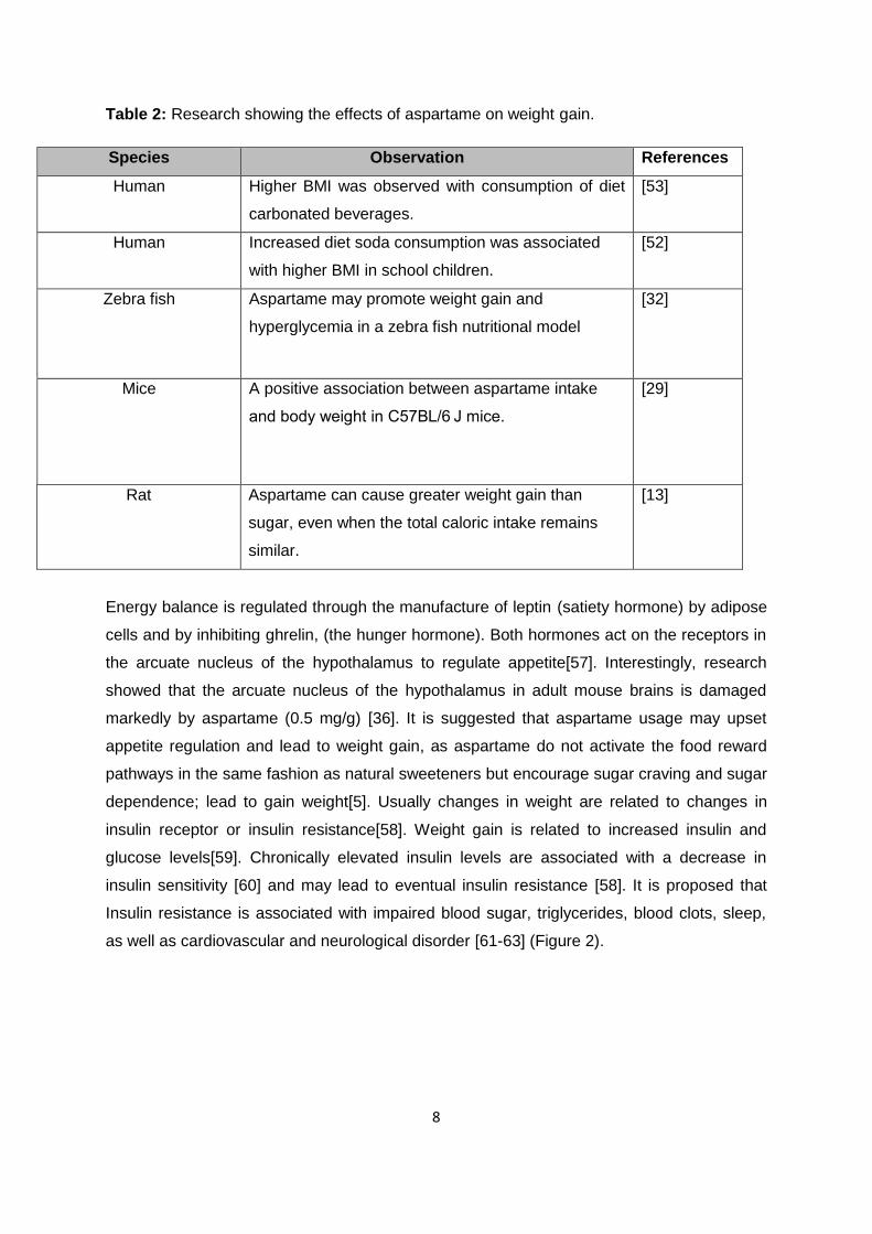
Table 2: Research showing the effects of aspartame on weight gain.
Species Observation References
Human Higher BMI was observed with consumption of diet
carbonated beverages.
[53]
Human Increased diet soda consumption was associated
with higher BMI in school children.
[52]
Zebra fish Aspartame may promote weight gain and
hyperglycemia in a zebra fish nutritional model
[32]
Mice A positive association between aspartame intake
and body weight in C57BL/6 J mice.
[29]
Rat Aspartame can cause greater weight gain than
sugar, even when the total caloric intake remains
similar.
[13]
Energy balance is regulated through the manufacture of leptin (satiety hormone) by adipose
cells and by inhibiting ghrelin, (the hunger hormone). Both hormones act on the receptors in
the arcuate nucleus of the hypothalamus to regulate appetite[57]. Interestingly, research
showed that the arcuate nucleus of the hypothalamus in adult mouse brains is damaged
markedly by aspartame (0.5 mg/g) [36]. It is suggested that aspartame usage may upset
appetite regulation and lead to weight gain, as aspartame do not activate the food reward
pathways in the same fashion as natural sweeteners but encourage sugar craving and sugar
dependence; lead to gain weight[5]. Usually changes in weight are related to changes in
insulin receptor or insulin resistance[58]. Weight gain is related to increased insulin and
glucose levels[59]. Chronically elevated insulin levels are associated with a decrease in
insulin sensitivity [60] and may lead to eventual insulin resistance [58]. It is proposed that
Insulin resistance is associated with impaired blood sugar, triglycerides, blood clots, sleep,
as well as cardiovascular and neurological disorder [61-63] (Figure 2).

9
Figure 2: Aspartame consumption may alter food habits (satiety signal), lead to weight gain,
increase free fatty acid, which may inhibit glucose utilization and promote glucose production
and result in for insulin resistance or deficiency. Insulin resistance is associated with
impaired blood sugar, triglycerides, blood clots, sleep, as well as cardiovascular and
neurological disorder.
Aspartame and Gut dysbiosis with insulin resistance or deficiency
Gut microbes modulate main host biological systems that control energy homoeostasis and
glucose metabolism in T2D [64] and plays a significant role in the development of insulin
resistance[65]. The intestinal bacterial population unique to T2D may produce toxins causing
systemic inflammation, affecting overall metabolism and insulin sensitivity[66]. Low dose
aspartame (5–7 mg/kg/day) consumption in drinking water over eight weeks resulted in
elevated fasting glucose levels and impaired insulin tolerance in diet-induced obese rats and
the fecal analysis of gut microbiota showed aspartame to increase the abundance of
Enterobacteriaceae [18]. Mice that drank water with 4% aspartame and consumed a high fat

10
diet for eleven weeks had higher glucose excursions after a glucose load, these changes
were associated with a metabolic phenotype change caused by alteration of the gut
microbiota [19] and dysregulated microbiota-gut-brain axis may explain aspartame metabolic
and other side-effects [67].
Generally it is well-known that glucose intolerance is a precursor to T2D[68]. T2D is a
heterogeneous disease with large variation in the relative contributions of insulin resistance
and beta cell dysfunction [69]. Insulin is synthesized and released from pancreatic β- cells in
response to increases in plasma glucose concentrations[70]. Increases in amino acids can
influence insulin biosynthesis and secretion[71]. The amino acid; phenylalanine, may
stimulate insulin secretion and glucagon concentration[72]. The insulin response can be
substantially increased by phenylalanine, and has high insulinotropic potential in
T2D[73].The amino acid; phenylalanine (50%), is a major aspartame component, may lead
to insulin resistance or deficiency.
Cortisol pathway to insulin resistance
The cortisol pathway plays an important role in the development of insulin resistance, and
literature suggests that aspartame (75mg/kg.bw/day), may act as a chemical stressor and
result in the production of excess corticosterone (cortisol) after 90-days of oral administration
in rats [16]. In general cortisol has been linked to insulin resistance through the following
mechanism:
1. Cortisol decreases the translocation of GLUT-4 transports and associated glucose
uptake[74, 75].
2. Cortisol inhibits the release of insulin from the beta cells of pancreas in mice[76].
3. Cortisol facilitates insulin resistance by increasing the production of glucose and
accumulation of lipids in the cell [74, 75].
Rats given the safety dosage of aspartame (40mg/kg.bw/day) for 90-days, showed a
significant increase in coticosterone level but no significant changes in blood glucose level
[50]. But however, prolonged exposure to excess levels of cortisol may affect blood glucose
levels in T2D[77]. Excess levels of cortisol may induce insulin resistance or decrease insulin
action[78] (Figure 3A and B) which decreases both hepatic and extra hepatic (peripheral)
sensitivity to insulin and increase blood glucose levels [74] (Figure 4A). Cortisol also
increases the rate of gluconeogenesis and glycogenolysis in liver, and decreases the activity
of the GLUT-4 transporter and related glucose uptake in skeletal muscle. Furthermore, it
increases lipolysis and decreases the activity of lipoprotein lipase; both of which increase
free fatty acid levels in the cell and compete with glucose for oxidative metabolism (Figure
4A) [74, 75]. Cortisol action therefore directly opposes insulin action and can be described

11
as a diabetogenic hormone, that fundamentally and possibly directly contributes to insulin
resistance (Figure 3A and B) [74, 75].
Figure 3: The link between aspartame and cortisol. A) Aspartame may act as a chemical
stressor and its intake may lead to hormonal imbalance and produce excess cortisol; this
may induce insulin resistance or deficiency, and result in increased blood sugar, increase
blood pressure, decrease immune response, decrease serotonin and pain sensation and
impaired memory and mental attention.
A

12
Figure 3: The link between aspartame and cortisol B) Increased cortisol or aspartame
component, methanol, may lead to hormonal imbalance (increase appetite and food intake,
decrease energy expenditure and increase fatigue) and increase sympathetic activity (result
in sleep loss) may lead to weight gain, insulin resistance and increase cardiovascular risk. In
addition, increased cortisol or aspartame component: phenylalanine and aspartic acid, may
affect platelet function, result into prothrobmbic state, vascular dysfunction and increase
cardiovascular risk.
Insulin resistance is a state of impaired biological response to normal or elevated serum
insulin concentrations[79] and occurs when the body does not respond properly to insulin.
Insulin resistance may also be the cause of abnormally high blood glucose levels in T2D[80]
due to (a) reduced early insulin secretory response to oral glucose, (b) decreased glucose-
sensing ability of the cell, (c) reduced the ability of the cell to compensate for the degree of
insulin resistance.
B

13
Hypercortisolism and its other complications
Augmented cortisol may also increase sympathetic activity, result in sleep loss; a risk factor
for weight gain, insulin resistance, [81] and cardiovascular risk [82] (see Figure 3A and B).
Aspartame ingestion result in sympathetic dominance with loss of vagal tone and impaired
cardiac function in rats[83, 84]. Mostly Hypercortisolism is associated with central obesity,
insulin resistance, dyslipidemia, and alterations in clotting and platelet function[85] (see
Figure 3A and B). The duration of cortisol excess correlates with increases the synthesis of
several coagulation factors, stimulating endothelial production of von Willebrand factor and
concomitantly increasing factor VIII [86] and may also enhance platelet and reduce plasma
fibrinolytic capacity[87, 88]. Increased cortisol or aspartame component phenylalanine after
aspartame usage may affect platelet function and both fibrin formation and platelet activation
in an animal model were found to be to changed fibrin packaging by aspartame
administration[89]. Pathological functioning of both platelets and fibrin, closely associated
with hypercoagulability, is known to be a hallmark of T2D, and therefore aspartame usage
would add to this pathological hypercoaguability in T2D and also in all other inflammatory
conditions.
Cortisol may influence the insulin receptor by (a) decreasing binding affinity and receptor
number[90], (b) decreasing binding affinity without decreasing numbers[91], (c) increasing
receptor number without affecting affinity [92] or (d) having no effect on receptor affinity or
number[93]. Insulin receptors are made up of 2 α and 2 β glycoprotein subunits connected
by disulphide bonds and are situated in the cell membrane[94] (Figure 4B). Insulin binds to
the extracellular α subunit, causing a conformational change, allowing ATP to bind to the
intracellular component of the β subunit [79]. ATP binding in turn activates phosphorylation
of the β subunit convening tyrosine kinase activity. This enables tyrosine phosphorylation of
intracellular substrate proteins known as insulin responsive substrates (IRS) (Figure 4B).
The phosphorylated IRS proteins bind with enzymes such as phosphatidylinositol 3-kinase
(PI 3-kinase). The PI 3-kinase acts via serine and threonine kinases such as Akt/protein
kinase B (PKB), protein kinase C (PKC) and PI dependent protein kinases 1& 2 (PIPD 1 and
2). The PI 3-kinase mediate insulin’s metabolic effects [94] by translocation of glucose
transporter proteins (GLUT), synthesis of glycogen, lipid and protein, anti-lipolysis and
hepatic gluconeogenesis [79] (Figure 4B). Cortisol may cause insulin resistance by
decreasing transcription of insulin IRS-1/ IRS-2 in skeletal muscle[95], adipose tissue [96]
and liver[97]. Excess cortisol may act as an insulin antagonist in the insulin resistant
condition. Hence excess cortisol after chronic aspartame consumption may promote to

insulin resistance, however the particular mechanism has to be explored further with more
scientific studies.
Figure 4A: Insulin and cortisol on peripheral and central glucose uptake. The GLUT 4 is expressed principally in skeletal muscle and lipoprotein lipase principally in adipose tissue. Actions of cortisol (brown color) and insulin (green color) are shown either as stimulate (+) or inhibit (×).The major effects of cortisol may be to reduce translocation of GLUT 4 to the cell surface and enhance lipolysis, thereby increasing free fatty acid competition with pyruvate for mitochondrial oxidative metabolism. In liver, insulin and glucocorticoids oppose each other’s actions, particularly on gluconeogenesis and oxidative glycolysis.
14

15
Figure 4B: Insulin and cortisol on insulin receptor (adapted from [79]). Insulin receptors are
made up of 2α and 2β glycoprotein. Insulin binds to the extracellular α subunit, subsequent
conformational change, allowing ATP to bind to the intracellular β subunit, phosphorylation
of the β subunit convening tyrosine kinase activity, and this phosphorylation of insulin
responsive substrates (IRS), The Phosphorylated IRS proteins bind with enzymes
phosphatidylinositol 3-kinase (PI 3-kinase). The PI 3-kinase acts via Akt/protein kinase B
(PKB), protein kinase C (PKC) and PI dependent protein kinases 1& 2 (PIPD 1&2). The PI
3-kinase mediate further insulin’s metabolic effects by translocation of glucose transporter
proteins (GLUT), synthesis of glycogen, lipid and protein, anti-lipolysis Actions of cortisol
(brown color) and insulin (green color) are shown either as stimulate (+) or inhibit (×) and
hepatic gluconeogenesis.

16
Cortisol also exerts bi-phasic regulation of inflammation in humans and either suppresses or
stimulates the inflammatory response in a concentration and time-dependent manner[98]. It
is unclear if aspartame consumption causing cortisol production has a pro-inflammatory or
anti-inflammatory action.
Neurotransmitter alterations within brain to insulin resistance
The CNS regulates the peripheral metabolism, including energy expenditure, glucose and
lipid metabolism, through changes in autonomic sympathetic, parasympathetic, and
hormonal outputs[99]. Any aspartame dose consumed by humans will elevate brain
phenylalanine much more than it elevates tyrosine, since the human liver converts
phenylalanine to tyrosine relatively slowly than in rats[100]. Phenylalanine, rather than
tyrosine is the amino acid that is known to be associated with suppression of brain
catecholamine synthesis[101] . Aspartame (0.625-45mg/kg) consumption may exert a dose-
dependent inhibition of brain serotonin, noradrenaline, and dopamine[38] that may result in a
changed neurological function.
Phenylalanine, an aspartame component, competes with tryptophan, the serotonin
precursor, for the same channel (NAAT) through the blood-brain barrier[26]. Phenylalanine,
penetrates the brain and suppresses serotonin levels [26]. Large doses of phenylalanine can
block important neurotransmitters including serotonin, which helps to control sensations of
satiety. Serotonin regulates the appetite mechanism and converts into melatonin to induce
sleep and serotonin deficiencies can cause depression, upset the appetite mechanism and
lead to weight gain [101]. Serotonin may also play a role in glucose homeostasis[102]. The
central serotonin 2C receptors in the pro-opiomelanocortin (POMC) neurons in the arcuate
nucleus of hypothalamus regulate energy and glucose homeostasis[103-105]. The arcuate
POMC neurons respond to circulating glucose, and if the KATP channels present in POMC
neurons are blocked by a compound such as phenylalanine, may result in impaired glucose
tolerance[99, 106]. POMC neurons are also involved in the control of lipid metabolism[107].
People with low levels of serotonin are often compelled to consume more sugar in a bid to
increase serotonin production and this often results in a sugar addiction[108], which in turn
can lead to insulin resistance (high levels of insulin cause receptors for insulin to shut down
by means of ‘down-regulation)[109]. Aspartame consumption in both higher doses [16] and
safety doses [110, 111] were shown to induce oxidative stress in the hypothalamus, leading
to neuronal death (apoptosis). The glucose regulatory role of the hypothalamus would thus
be impaired. Recent research has targeted the serotonin 2C receptors for the treatment of

17
Diabetes /obesity[112]. The activation of this receptor reduces elevated insulin levels and
improves glucose tolerance and insulin sensitivity in both genetically obese mice and in mice
with diet-induced obesity[113].
Oxidative stress to insulin resistance
An imbalance between pro-oxidants and anti-oxidants determine oxidative stress and cause
cellular disruption and damage[114]. Aspartame induces excess free radical production, in
particular, reactive oxygen species (ROS) and reactive nitrogen species (RNS). These free
radicals result in systemic oxidative stress[23] such as in blood cells[50, 89, 115-117], brain
cells[16, 110, 111, 118, 119], liver and kidney cells[20, 22, 120], heart cells [83, 84] and
immune organs[15, 121-123] (Figure 5).
Figure 5: Aspartame usage may result in systemic inflammation and lead to insulin
resistance. Aspartame consumption produces excess free radicals (ROS/RNS) production;
result in oxidative stress and can trigger pro-inflammatory factor (TNF-α, NF-Κb, IL-6, CRP,
FFA), or subsiding adiponectin, lead to systemic inflammation which may result in insulin
resistance or impaired glucose transport. [Tumor necrosis factor (TNF-α), nuclear factor
kappa B (NF-kβ), C-reactive protein (CRP), Free fatty acid (FFA)].
Systemic oxidative stress is associated with insulin resistance[124]. Oxidative activity among
diabetes patients [125] contributes to both the onset and the progression of diabetes as well

18
as its late complications[126]. Oxidative stress increases with fat accumulation[127].
Oxidative stress may also lead to insulin resistance by stimulating the expression of several
pro-inflammatory cytokines[128]. The particular link between oxidative stress and impaired
insulin signaling is not completely understood, but several mechanisms have been
proposed. ROS/RNS may impair insulin signaling[129-131] by (a) inducing IRS
serine/threonine phosphorylation, (b) upsetting cellular redistribution of insulin signaling
components, (c) declining GLUT4 gene transcription or (d) altering mitochondrial activity.
ROS can trigger signal transduction pathways, primarily through nuclear factor κB (NFκB),
promoting the production of tumor necrosis factor α (TNFα) [132]and increasing the
production of pro-inflammatory cytokines, IL-6 [133] and C-reactive protein [128].
Inflammation is recognized as a manifestation of oxidative stress and is important in the
development and progression of diabetic complications [134, 135]Increased oxidative stress
may cause insulin resistance by inhibiting insulin signals and deregulating adiponectin [127,
136] and other adipocyte-derived factors such as TNF-α[137], leptin [138] and free fatty
acids (FFAs) [139](Figure 5). Hence, systemic oxidative stress induced by aspartame usage
may exacerbate insulin resistance and impaired glucose tolerance and may increase
complications in T2D (Figure 5).
In summary, aspartame usage by T2D may lead to insulin deficiency or resistance by (a)
alteration of food habit may lead to weight gain, (b) acting as chemical stressor by increasing
plasma cortisol level,(c) inducing excess free radical production, result in systemic oxidative
stress, (d) alteration of gut microbes, (e) disrupt neurotransmitter or NMDA receptor [N
methyl D-aspartate (NMDA)], finally result in impaired glucose tolerance and may increase
complications in diabetic patients; see Figure 6.

19
Figure 6: Aspartame usage to insulin resistance. Aspartame usage by type-II diabetic individuals may lead to insulin deficiency or resistance by (a)alteration of food habit (or weight gain), (b) increase cortisol, (c) systemic oxidative stress, (d)alteration of gut
microbes, (e) disrupt neurotransmitter or NMDA receptor [N-methyl D-aspartate (NMDA)], and finally result in impaired glucose tolerance.
Limitation and Conclusion
The benefit of aspartame usage as part of regarding weight management and blood glucose
regulation in T2D has not been confirmed. To the contrary, many studies link adverse
outcomes to aspartame consumption and various systems that are important to diabetic
individuals. There are limitations to this review. In particular, data from human studies are
limited especially the lack of good quality study design and small sample sizes. In addition,
self-reported consumption has not been validated as an accurate measure of aspartame
consumption. Unfortunately, results from animal data may not be directly transferable or
applicable to human.
We conclude that aspartame use in T2D, may lead to weight gain, rather than weight loss.
Aspartame consumption may furthermore act as a chemical stressor, increasing cortisol
levels, which interfere with insulin pathways. Moreover, aspartame consumption may induce

20
systemic oxidative stress by producing excess free radicals, leading to inflammation that
may exacerbate T2D complications.
More research is required that provides evidence and raise concerns that use of aspartame
in T2D is a challenge, and we suggest that aspartame consumption regulations should be
revisited and international guidelines reviewed to add further to the already challenged
health burden of T2D.
Disclosure
The author does not have any conflict of interest to declare.
References
1. Tandel, K.R., Sugar substitutes: Health controversy over perceived benefits. J PharmacolPharmacother, 2011. 2(4): p. 236.
2. Duffey, K.J., et al., Dietary patterns matter: diet beverages and cardiometabolic risks in thelongitudinal Coronary Artery Risk Development in Young Adults (CARDIA) Study. Am J ClinNutr 2012. 95(4): p. 909-15.
3. Malik, V.S., M.B. Schulze, and F.B. Hu, Intake of sugar-sweetened beverages and weight gain:a systematic review. Am J Clin Nutr, 2006. 84(2): p. 274-88.
4. Swithers, S.E., Artificial sweeteners produce the counterintuitive effect of inducing metabolicderangements. Trends Endocrinol Metab, 2013. 24(9): p. 431-41.
5. Yang, Q., Gain weight by going diet? Artificial sweeteners and the neurobiology of sugarcravings: Neuroscience 2010. Yale J Biol Med,, 2010. 83(2): p. 101.
6. Bakal, A.I., Mixed sweetener functionality. Food Sci Technol, 2001: p. 463-80.7. Filer, L. and L.D. Stegink, Aspartame metabolism in normal adults, phenylketonuric
heterozygotes, and diabetic subjects. Diabetes Care, 1989. 12(1): p. 67-74.8. Renwick, A. and H. Nordmann, First European conference on aspartame: Putting safety and
benefits into perspective. Synopsis of presentations and conclusions. Food Chem Toxicol,2007. 45(7): p. 1308-13.
9. Di Pasquale, M., Use Of Nutritive And Nonnutritive Sweeteners. J Am Diet Assoc, 1998. 98: p.580-587.
10. Gougeon, R., et al., Canadian diabetes association national nutrition committee technicalreview: non-nutritive intense sweeteners in diabetes management. Can J Diabetes, 2004.28(4): p. 385-99.
11. Anton, S.D., et al., Effects of stevia, aspartame, and sucrose on food intake, satiety, andpostprandial glucose and insulin levels. Appetite, 2010. 55(1): p. 37-43.
12. De la Hunty, A., S. Gibson, and M. Ashwell, A review of the effectiveness of aspartame inhelping with weight control. Nutr Bull, 2006. 31(2): p. 115-28.
13. de Matos Feijó, F., et al., Saccharin and aspartame, compared with sucrose, induce greaterweight gain in adult Wistar rats, at similar total caloric intake levels. Appetite, 2013. 60: p.203-7.
14. Swinburn, B.A., et al., Diet, nutrition and the prevention of excess weight gain and obesity.Public Health Nutr 2004. 7(1a): p. 123-46.

21
15. Choudhary, A.K. and R.S. Devi, Imbalance of the oxidant-antioxidant status by aspartame inthe organs of immune system of Wistar albino rats. Afr J Pharm Pharmacol 2014. 8(8): p.220-30.
16. Iyyaswamy, A. and S. Rathinasamy, Effect of chronic exposure to aspartame on oxidativestress in brain discrete regions of albino rats. J Biosci, 2012. 37(4): p. 679-88.
17. Choudhary, A. and Y. Lee, Neuro-physiological Symptoms and Aspartame: What is theconnection? Nutr Neurosci., 2017: p. 1-11.
18. Palmnäs, M.S., et al., Low-dose aspartame consumption differentially affects gut microbiota-host metabolic interactions in the diet-induced obese rat. PLoS One, 2014. 9(10): p. e109841.
19. Suez, J., et al., Artificial sweeteners induce glucose intolerance by altering the gut microbiota.Nature, 2014. 514(7521): p. 181-6.
20. Ashok, I. and R. Sheeladevi, Oxidant stress evoked damage in rat hepatocyte leading totriggered nitric oxide synthase (NOS) levels on long term consumption of aspartame. J FoodDrug Anal, 2015. 23(4): p. 679-91.
21. Iman, M.M., Effect of aspartame on some oxidative stress parameters in liver and kidney ofrats. Afr J Pharm Pharmacol, 2011. 5(6): p. 678-82.
22. Kumar Choudhary, A., S. Selvaraj, and R. Sheela Devi, Aspartame Induce Modification inMembrane Bound and Antioxidant Enzymes in Liver and Kidney of Wistar Albino Rats. CurrNutr Food Sci, 2014. 10(4): p. 275-87.
23. Choudhary, A. and E. Pretorius, Revisiting the Safety of Aspartame. . Nutr Rev (Accepted). ,Forthcoming 2017.
24. Colagiuri, S., J.J. Miller, and R.A. Edwards, Metabolic effects of adding sucrose andaspartame to the diet of subjects with noninsulin-dependent diabetes mellitus. Am J Clin Nutr
1989. 50(3): p. 474-8. 25. Horwitz, D.L., M. McLane, and P. Kobe, Response to single dose of aspartame or saccharin by
NIDDM patients. Diabetes Care, 1988. 11(3): p. 230-4.26. Humphries, P., E. Pretorius, and H. Naude, Direct and indirect cellular effects of aspartame
on the brain. Eur J Clin Nutr, 2008. 62(4): p. 451-62.27. Pan-Hou, H., et al., Effect of aspartame on N-methyl-D-aspartate-sensitive L-[3 H] glutamate
binding sites in rat brain synaptic membranes. Brain research, 1990. 520(1): p. 351-3.28. Melanson, K.J., et al., Blood glucose and meal patterns in time-blinded males, after
aspartame, carbohydrate, and fat consumption, in relation to sweetness perception. Br JNutr, 1999. 82(06): p. 437-46.
29. Wilson, F. and K. Howes, Blood glucose changes following the ingestion of sucrose-andaspartame-sweetened beverages. Appetite, 2008. 51(2): p. 410.
30. Collison, K.S., et al., Interactive effects of neonatal exposure to monosodium glutamate andaspartame on glucose homeostasis. Nutr Metab, 2012. 9(1): p. 1.
31. Collison, K.S., et al., Gender dimorphism in aspartame-induced impairment of spatialcognition and insulin sensitivity. PLoS One, 2012. 7(4): p. e31570.
32. Kim, J.-Y., J. Seo, and K.-H. Cho, Aspartame-fed zebrafish exhibit acute deaths with swimmingdefects and saccharin-fed zebrafish have elevation of cholesteryl ester transfer proteinactivity in hypercholesterolemia. Food Chem Toxicol, 2011. 49(11): p. 2899-905.
33. Abu-Taweel, G.M., et al., Cognitive and biochemical effects of monosodium glutamate andaspartame, administered individually and in combination in male albino mice. NeurotoxicolTeratol 2014. 42: p. 60-7.
34. Ashok, I., R. Sheeladevi, and D. Wankhar, Effect of long-term aspartame (artificial sweetener)on anxiety, locomotor activity and emotionality behavior in Wistar Albino rats. Biomed AgingPathol 2014. 4(1): p. 39-43.
35. Christian, B., et al., Chronic aspartame affects T-maze performance, brain cholinergicreceptors and Na+, K+-ATPase in rats. Pharmacol Biochem Behav, 2004. 78(1): p. 121-7.

22
36. Park, C.H., et al., Glutamate and aspartate impair memory retention and damagehypothalamic neurons in adult mice. Toxicol Lett, 2000. 115(2): p. 117-25.
37. van der Heide, L.P., G.M. Ramakers, and M.P. Smidt, Insulin signaling in the central nervoussystem: learning to survive. Prog Neurobiol, 2006. 79(4): p. 205-21.
38. Abdel-Salam, O.M., N.A. Salem, and J.S. Hussein, Effect of aspartame on oxidative stress andmonoamine neurotransmitter levels in lipopolysaccharide-treated mice. Neurotox Res, 2012.21(3): p. 245-55.
39. Nadal, A., et al., The pancreatic β-cell as a target of estrogens and xenoestrogens:Implications for blood glucose homeostasis and diabetes. Mol Cell Endocrinol, 2009. 304(1):p. 63-8.
40. Nishiumi, S., et al., Green and black tea suppress hyperglycemia and insulin resistance byretaining the expression of glucose transporter 4 in muscle of high-fat diet-fed C57BL/6Jmice. J Agric Food Chem., 2010. 58(24): p. 12916-23.
41. Jung, H.S., et al., Loss of autophagy diminishes pancreatic β cell mass and function withresultant hyperglycemia. Cell Metab, 2008. 8(4): p. 318-24.
42. Rodbard, H.W., Diabetes screening, diagnosis, and therapy in pediatric patients with type 2diabetes. Medscape J Med, 2008. 10(8): p. 184.
43. Takahashi, A., et al., Stimulation of rat hypothalamus by microdialysis with K+: increase ofACh release elevates plasma glucose. Am J Physiol Regul Integr Comp Physiol 1998. 275(5):p. R1647-53.
44. Sahu, A., Minireview: a hypothalamic role in energy balance with special emphasis on leptin.Endocrinology, 2004. 145(6): p. 2613-20.
45. Lam, C.K., et al., Hypothalamic nutrient sensing activates a forebrain-hindbrain neuronalcircuit to regulate glucose production in vivo. Diabetes, 2011. 60(1): p. 107-13.
46. Molina, P.E., et al., Central NMDA enhances hepatic glucose output and non-insulin-mediated glucose uptake by a nonadrenergic mechanism. Brain Research, 1994. 634(1): p.41-8.
47. Molina, P.E. and N.N. Abumrad, Contribution of excitatory amino acids to hypoglycemiccounter-regulation. Brain Research, 2001. 899(1): p. 201-8.
48. Kalsbeek, A., S. la Fleur, and E. Fliers, Circadian control of glucose metabolism. Mol Metab,2014. 3(4): p. 372-83.
49. Rojas, J. and M. Schwartz, Control of hepatic glucose metabolism by islet and brain. DiabetesObes Metab, 2014. 16(S1): p. 33-40.
50. Choudhary, A.K. and R.S. Devi, Serum biochemical responses under oxidative stress ofaspartame in wistar albino rats. Asian Pac J Trop Dis., 2014. 4: p. S403-10.
51. Nguyen, M.A., et al., A subpopulation of macrophages infiltrates hypertrophic adipose tissueand is activated by free fatty acids via Toll-like receptors 2 and 4 and JNK-dependentpathways. J Biol Chem., 2007. 282(48): p. 35279-92.
52. Blum, J.W., D.J. Jacobsen, and J.E. Donnelly, Beverage consumption patterns in elementaryschool aged children across a two-year period. J Am Coll Nutr, 2005. 24(2): p. 93-8.
53. Forshee, R.A. and M.L. Storey, Total beverage consumption and beverage choices amongchildren and adolescents. Int J Food Sci Nutr, 2003. 54(4): p. 297-307.
54. Sørensen, L.B., et al., Effect of sensory perception of foods on appetite and food intake: areview of studies on humans. Int J Obes, 2003. 27(10): p. 1152-66.
55. Mattes, R.D. and B.M. Popkin, Nonnutritive sweetener consumption in humans: effects onappetite and food intake and their putative mechanisms. Am J Clin Nutr, 2009. 89(1): p. 1-14.
56. Swithers, S.E. and T.L. Davidson, A role for sweet taste: calorie predictive relations in energyregulation by rats. Behav Neurosci, 2008. 122(1): p. 161.
57. Brennan, A.M. and C.S. Mantzoros, Drug insight: the role of leptin in human physiology andpathophysiology—emerging clinical applications. Nat Rev Endocrinol, 2006. 2(6): p. 318-27.

23
58. Elliott, S.S., et al., Fructose, weight gain, and the insulin resistance syndrome. Am J Clin Nutr2002. 76(5): p. 911-22.
59. Lazzer, S., et al., Changes in adipocyte hormones and lipid oxidation associated with weightloss and regain in severely obese adolescents. Int J Obes, 2005. 29(10): p. 1184-91.
60. Reinehr, T., et al., Insulin sensitivity among obese children and adolescents, according todegree of weight loss. Pediatrics, 2004. 114(6): p. 1569-73.
61. Basoglu, O.K., et al., Metabolic syndrome, insulin resistance, fibrinogen, homocysteine,leptin, and C-reactive protein in obese patients with obstructive sleep apnea syndrome. AnnThorac Med 2011. 6(3): p. 120.
62. Bonora, E., et al., Insulin resistance as estimated by homeostasis model assessment predictsincident symptomatic cardiovascular disease in Caucasian subjects from the generalpopulation the bruneck study. Diabetes Care, 2007. 30(2): p. 318-24.
63. Kim, B. and E.L. Feldman, Insulin resistance in the nervous system. Trends Endocrinol.Metab., 2012. 23(3): p. 133-41.
64. Cani, P.D., et al., Glucose metabolism: Focus on gut microbiota, the endocannabinoid systemand beyond. Diabetes Metab. , 2014. 40(4): p. 246-57.
65. Shen, J., M.S. Obin, and L. Zhao, The gut microbiota, obesity and insulin resistance. MolAspects Med, 2013. 34(1): p. 39-58.
66. Giovanni, M., R. Gambino, and M. Cassader, Obesity, diabetes and gut microbiota. DiabetesCare, 2010. 33(10): p. 2277-84.
67. Choudhary, A. and Y. Lee, Dysregulated microbiota-gut-brain axis: does it explain aspartamemetabolic and other side-effects? Food and Nutrition (Accepted), Forthcoming 2017.
68. Bastard, J.-P., et al., Recent advances in the relationship between obesity, inflammation, andinsulin resistance. Eur Cytokine Netw, 2006. 17(1): p. 4-12.
69. Færch, K., A. Hulmán, and T. PJ Solomon, Heterogeneity of pre-diabetes and type 2 diabetes:implications for prediction, prevention and treatment responsiveness. Curr Diabetes Rev,2016. 12(1): p. 30-41.
70. Aronoff, S.L., et al., Glucose metabolism and regulation: beyond insulin and glucagon.Diabetes Spectr, 2004. 17(3): p. 183-90.
71. Bratanova-Tochkova, T.K., et al., Triggering and augmentation mechanisms, granule pools,and biphasic insulin secretion. Diabetes, 2002. 51(suppl 1): p. S83-90.
72. Nuttall, F., K. Schweim, and M. Gannon, Effect of orally administered phenylalanine with andwithout glucose on insulin, glucagon and glucose concentrations. Horm Metab Res, 2006.38(8): p. 518-23.
73. Schwanstecher, C., et al., Interaction of N‐benzoyl‐D‐phenylalanine and related compoundswith the sulphonylurea receptor of β‐cells. Br J Pharmacol, 1998. 123(6): p. 1023-30.
74. Andrews, R.C. and B.R. Walker, Glucocorticoids and insulin resistance: old hormones, newtargets. Clin Sci 1999. 96(5): p. 513-23.
75. Reynolds, R.M. and B.R. Walker, Human insulin resistance: the role of glucocorticoids.Diabetes Obes Metab, 2003. 5(1): p. 5-12.
76. Delaunay, F., et al., Pancreatic beta cells are important targets for the diabetogenic effects ofglucocorticoids. J Clin Invest, 1997. 100(8): p. 2094.
77. Chiodini, I., et al., Cortisol secretion in patients with type 2 diabetes. Diabetes Care, 2007.30(1): p. 83-8.
78. Phillips, D., et al., Elevated Plasma Cortisol Concentrations: A Link between Low Birth Weightand the Insulin Resistance Syndrome? 1. J Clin Endocrinol Metab, 1998. 83(3): p. 757-60.
79. Wilcox, G., Insulin and insulin resistance. Clin Biochem Rev, 2005. 26(2): p. 19.80. Polonsky, K.S., J. Sturis, and G.I. Bell, Non-insulin-dependent diabetes mellitus—a genetically
programmed failure of the beta cell to compensate for insulin resistance. N Engl J Med, 1996.334(12): p. 777-83.
24
81. Ayas, N.T., et al., A prospective study of sleep duration and coronary heart disease in women.Arch Intern Med, 2003. 163(2): p. 205-9.
82. Reynolds, R.M., et al., Altered Control of Cortisol Secretion in Adult Men with Low BirthWeight and Cardiovascular Risk Factors 1. J Clin Endocrinol Metab, 2001. 86(1): p. 245-50.
83. Choudhary, A.K., L. Sundareswaran, and R.S. Devi, Effects of aspartame on the evaluation ofelectrophysiological responses in Wistar albino rats. J Taibah Univ Sci 2016. 10: p. 505-12.
84. Choudhary, A.K., L. Sundareswaran, and R.S. Devi, Aspartame induced cardiac oxidativestress in Wistar albino rats. Nutr Clin Metabol, 2016. 30(1): p. 29-37.
85. Faggiano, A., et al., Cardiovascular risk factors and common carotid artery caliber andstiffness in patients with Cushing’s disease during active disease and 1 year after diseaseremission. J Clin Endocrinol Metab, 2003. 88(6): p. 2527-33.
86. Ambrosi, B., et al., Evaluation of haemostatic and fibrinolytic markers in patients withCushing's syndrome and in patients with adrenal incidentaloma. Exp Clin EndocrinolDiabetes, 1999. 108(4): p. 294-8.
87. Arnaldi, G., et al., Cardiovascular risk in Cushing's syndrome. Pituitary, 2004. 7(4): p. 253-6.88. Boscaro, M., et al., Anticoagulant prophylaxis markedly reduces thromboembolic
complications in Cushing’s syndrome. J Clin Endocrinol Metab, 2002. 87(8): p. 3662-6.89. Pretorius, E. and P. Humphries, Ultrastructural changes to rabbit fibrin and platelets due to
aspartame. Ultrastruct Pathol 2007. 31(2): p. 77-83.90. Beck-Nielsen, H., R. De Pirro, and O. Pedersen, Prednisone increases the number of insulin
receptors on monocytes from normal subjects. J Clin Endocrinol Metab, 1980. 50(1): p. 1-4.91. De Pirro, R., et al., Effect of Dexamethasone and Cortisone on Insulin Receptors in Normal
Human Male. J Clin Endocrinol Metab, 1980. 51(3): p. 503-7.92. Rizza, R.A., L.J. Mandarino, and J.E. Gerich, Cortisol-Induced Insulin Resistance in Man:
Impaired Suppression of Glucose Production and Stimulation of Glucose Utilization due to aPostreceptor Defect of Insulin Action. J Clin Endocrinol Metab, 1982. 54(1): p. 131-8.
93. Pagano, G., et al., An in vivo and in vitro study of the mechanism of prednisone-inducedinsulin resistance in healthy subjects. J Clin Invest 1983. 72(5): p. 1814.
94. Kido, Y., J. Nakae, and D. Accili, The Insulin Receptor and Its Cellular Targets 1. J ClinEndocrinol Metab, 2001. 86(3): p. 972-9.
95. Almon, R.R., et al., Temporal profiling of the transcriptional basis for the development ofcorticosteroid-induced insulin resistance in rat muscle. J Endocrinol 2005. 184(1): p. 219-32.
96. Burén, J., et al., Insulin action and signalling in fat and muscle from dexamethasone-treatedrats. Arch Biochem Biophys, 2008. 474(1): p. 91-101.
97. Saad, M., et al., Modulation of insulin receptor, insulin receptor substrate-1, andphosphatidylinositol 3-kinase in liver and muscle of dexamethasone-treated rats. J ClinInvest, 1993. 92(4): p. 2065.
98. Yeager, M.P., P.A. Pioli, and P.M. Guyre, Cortisol exerts bi-phasic regulation of inflammationin humans. Dose-Response, 2011. 9(3): p. 10-13.
99. Pissios, P. and E. Maratos-Flier, More than satiety: central serotonin signaling and glucosehomeostasis. Cell Metab, 2007. 6(5): p. 345-7.
100. Wurtman, R. and T. Maher, Effects of oral aspartame on plasma phenylalanine in humans and experimental rodents. J Neural Transm 1987. 70(1-2): p. 169-73.
101. Singh, M., Mood, food, and obesity. Frontiers in Psychology, 2014. 5: p. 925. 102. Xu, Y., et al., 5-HT 2C Rs expressed by pro-opiomelanocortin neurons regulate energy
homeostasis. Neuron, 2008. 60(4): p. 582-9. 103. Berglund, E.D., et al., Serotonin 2C receptors in pro-opiomelanocortin neurons regulate
energy and glucose homeostasis. J Clin Invest, 2013. 123(12): p. 5061-70.

25
104. Garfield, A.S., et al., Role of central melanocortin pathways in energy homeostasis. Trends Endocrinol Metab, 2009. 20(5): p. 203-15.
105. Lam, D.D. and L.K. Heisler, Serotonin and energy balance: molecular mechanisms and implications for type 2 diabetes. Expert Rev Mol Med., 2007. 9(05): p. 1-24.
106. Parton, L.E., et al., Glucose sensing by POMC neurons regulates glucose homeostasis and is impaired in obesity. Nature, 2007. 449(7159): p. 228-32.
107. Nogueiras, R., et al., The central melanocortin system directly controls peripheral lipid metabolism. J Clin Invest 2007. 117(11): p. 3475-88.
108. Volkow, N.D., G.-J. Wang, and R.D. Baler, Reward, dopamine and the control of food intake: implications for obesity. Trends Cogn Sci, 2011. 15(1): p. 37-46.
109. Luo, S., J. Luo, and A.H. Cincotta, Chronic ventromedial hypothalamic infusion of norepinephrine and serotonin promotes insulin resistance and glucose intolerance. Neuroendocrinology, 1999. 70(6): p. 460-5.
110. Ashok, I. and R. Sheeladevi, Biochemical responses and mitochondrial mediated activation of apoptosis on long-term effect of aspartame in rat brain. Redox Biol, 2014. 2: p. 820-31.
111. Ashok, I. and R. Sheeladevi, Neurobehavioral changes and activation of neurodegenerative apoptosis on long-term consumption of aspartame in the rat brain. J Nutr Intermed Metab, 2015. 2(3): p. 76-85.
112. Marston, O.J. and L.K. Heisler, Targeting the serotonin 2C receptor for the treatment of obesity and type 2 diabetes. Neuropsychopharmacology, 2009. 34(1): p. 252-3.
113. Zhou, L., et al., Serotonin 2C receptor agonists improve type 2 diabetes via melanocortin-4 receptor signaling pathways. Cell Metab, 2007. 6(5): p. 398-405.
114. Pisoschi, A.M. and A. Pop, The role of antioxidants in the chemistry of oxidative stress: a review. Eur J Med Chem, 2015. 97: p. 55-74.
115. Agamy, N., Effects of the natural sweetener (stevia) and the artificial sweetener (aspartame) on some biochemical parameters in normal and alloxan-induced diabetic rats. N Biotechnol
2009. 25: p. S12. 116. Arbind, K., R. Sheela Devi, and L. Sundareswaran, Role of antioxidant enzymes in oxidative
stress and immune response evaluation of aspartame in blood cells of wistar albino rats. Int Food Res J, 2014. 21(6): p. 2263-72.
117. Tsakiris, S., et al., The effect of aspartame metabolites on human erythrocyte membrane acetylcholinesterase activity. Pharmacol. Res, 2006. 53(1): p. 1-5.
118. Abhilash, M., et al., Long-term consumption of aspartame and brain antioxidant defense status. Drug Chem Toxicol 2013. 36(2): p. 135-40.
119. El-Samad, A.A., Light and electron microscopic study on the effect of aspartame on the cerebellar cortex of male albino rat. Egypt J Histol, 2010. 33(3): p. 419-30.
120. Abhilash, M., et al., Effect of long term intake of aspartame on antioxidant defense status in liver. Food Chem Toxicol, 2011. 49(6): p. 1203-7.
121. Choudhary, A.K. and R.S. Devi, Longer period of oral administration of aspartame on cytokine response in Wistar albino rats. Endocrinol Nutr., 2015. 62(3): p. 114-22.
122. Choudhary, A.K. and S.D. Rathinasamy, Aspartame induces alteration in electrolytes homeostasis of immune organs in wistar albino rats. Biomed Prev Nutr, 2014. 4(2): p. 181-87.
123. Choudhary, A.K. and S.D. Rathinasamy, Effect of long intake of aspartame on ionic imbalance in immune organs of immunized wistar albino rats. Biomed Aging Pathol 2014. 4(3): p. 243-9.
124. Ramachandran, S.V., Association of Oxidative Stress, Insulin Resistance, and Diabetes Risk Phenotypes. Diabetes Care, 2007. 30(10): p. 2529-35.
125. Azeem, E., et al., Oxidative stress correlates (OSC) in diabetes mellitus patients. Curr Diabetes Rev, 2016. 12(3): p. 279-84.

26
126. Rösen, P., et al., The role of oxidative stress in the onset and progression of diabetes and its complications: asummary of a Congress Series sponsored byUNESCO‐MCBN, the American Diabetes Association and the German Diabetes Society. Diabetes Metab Res Rev 2001. 17(3): p. 189-212.
127. Furukawa, S., et al., Increased oxidative stress in obesity and its impact on metabolic syndrome. J Clin Invest, 2004. 114(12): p. 1752-61.
128. Styskal, J., et al., Oxidative stress and diabetes: what can we learn about insulin resistance from antioxidant mutant mouse models? Free Radic Biol Med, 2012. 52(1): p. 46-58.
129. Bloch-Damti, A. and N. Bashan, Proposed mechanisms for the induction of insulin resistance by oxidative stress. Antioxid Redox Signal 2005. 7(11-12): p. 1553-67.
130. Morino, K., K.F. Petersen, and G.I. Shulman, Molecular mechanisms of insulin resistance in humans and their potential links with mitochondrial dysfunction. Diabetes, 2006. 55(2): p. S9-15.
131. Rains, J.L. and S.K. Jain, Oxidative stress, insulin signaling, and diabetes. Free Radic Biol Med, 2011. 50(5): p. 567-75.
132. Rahman, I., et al., Oxidative stress and TNF-a induce histone Acetylation and NF-кB/AP-1 activation in Alveolar epithelial cells: Potential mechanism In gene transcription in lung inflammation, in Oxygen/Nitrogen Radicals: Cell Injury and Disease. 2002. p. 239-48.
133. Park, J., et al., Increase in glucose-6-phosphate dehydrogenase in adipocytes stimulates oxidative stress and inflammatory signals. Diabetes, 2006. 55(11): p. 2939-49.
134. Kaul, K., et al., Is inflammation a common retinal-renal-nerve pathogenic link in diabetes? Curr Diabetes Rev, 2010. 6(5): p. 294-303.
135. Tangvarasittichai, S., Oxidative stress, insulin resistance, dyslipidemia and type 2 diabetes mellitus. World J Diabetes, 2015. 6(3): p. 456.
136. Houstis, N., E.D. Rosen, and E.S. Lander, Reactive oxygen species have a causal role in multiple forms of insulin resistance. Nature, 2006. 440(7086): p. 944-8.
137. Hotamisligil, G.S. and B.M. Spiegelman, Tumor necrosis factor α: a key component of the obesity-diabetes link. Diabetes, 1994. 43(11): p. 1271-8.
138. Cohen, B., D. Novick, and M. Rubinstein, Modulation of insulin activities by leptin. Science, 1996. 274(5290): p. 1185.
139. McGarry, J.D., Banting lecture 2001 Dysregulation of fatty acid metabolism in the etiology of type 2 diabetes. Diabetes, 2002. 51(1): p. 7-18.