Ascitic Fluid and Use of Immunocytochemistry - patologi.com immun.pdf · Cytomorphologic Useful...
160
Ascitic Fluid and Use of Immunocytochemistry Mercè Jordà, University of Miami
Transcript of Ascitic Fluid and Use of Immunocytochemistry - patologi.com immun.pdf · Cytomorphologic Useful...

Ascitic Fluid and Use of
Immunocytochemistry
Mercè Jordà, University of Miami

Is It Malignant ?
Yes ? No

Cytomorphologic Useful Findings
• Tight clusters with smooth borders
• Cellular and nuclear molding
• Large papillary groups
• Two-cell types
• Signet ring cells in groups
• Abnormal cell morphology
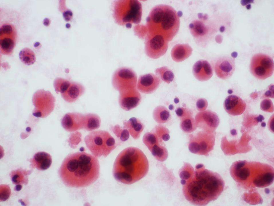
Ascitic Fluid


Ascitic Fluid
Cytomorphologic Useless Findings
Cytoplasmic Vacuoles
“Signet Ring Cells” individual
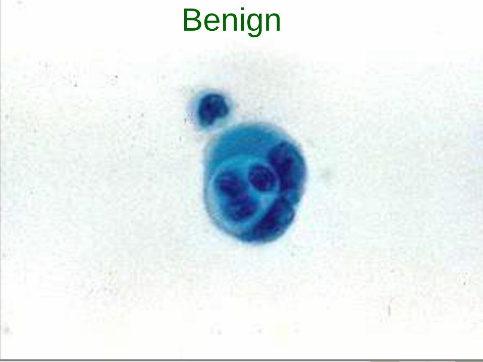
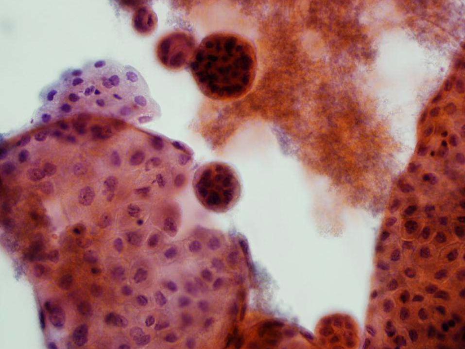
Psammoma Bodies
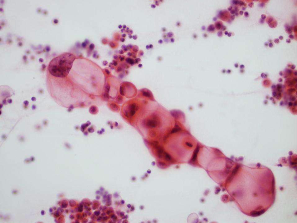
“Cell within Cell”
Prominent Nucleoli
Mitosis
Multinucleation

Reactive Mesothelium

“Signet Ring Cells”
Look for them in
Groups!


Malignant
Benign

Cellular Pattern
Cellular Morphology
By cytomorphology

Malignant Ascitic Fluid
Cellular Pattern
Cells in
Clusters Isolated
Cells

Cells
Tight and compact
Smooth borders
Background
No cells
Reactive mesothelial
cells
Malignant Ascitic Fluid: Cells in Clusters
Diagnosis : Cytomorphology


Malignant Effusions: Cells in clusters
Differential Diagnosis
Diagnosis : Cytomorphology & Immunocytochemistry
• Carcinoma
• Malignant Mesothelioma

• Abnormal single cells
• May be overlooked in low power
• Look for small clusters
Malignant Effusions: Isolated Cells

3
Abnormal Cell Morphology
• Pleomorphism
• High N/C ratio
• Hyperchromasia
• Abnormal nucleoli
• Clumped, irregular chromatin
• Intraluminal mucin
Malignant Effusions: Isolated Cells
Diagnosis : Cytomorphology & Immunocytochemistry
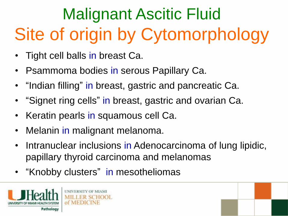
• Tight cell balls in breast Ca.
• Psammoma bodies in serous Papillary Ca.
• “Indian filling” in breast, gastric and pancreatic Ca.
• “Signet ring cells” in breast, gastric and ovarian Ca.
• Keratin pearls in squamous cell Ca.
• Melanin in malignant melanoma.
• Intranuclear inclusions in Adenocarcinoma of lung lipidic,
papillary thyroid carcinoma and melanomas
• “Knobby clusters” in mesotheliomas
Malignant Ascitic Fluid
Site of origin by Cytomorphology

Cell balls in Breast Ca.
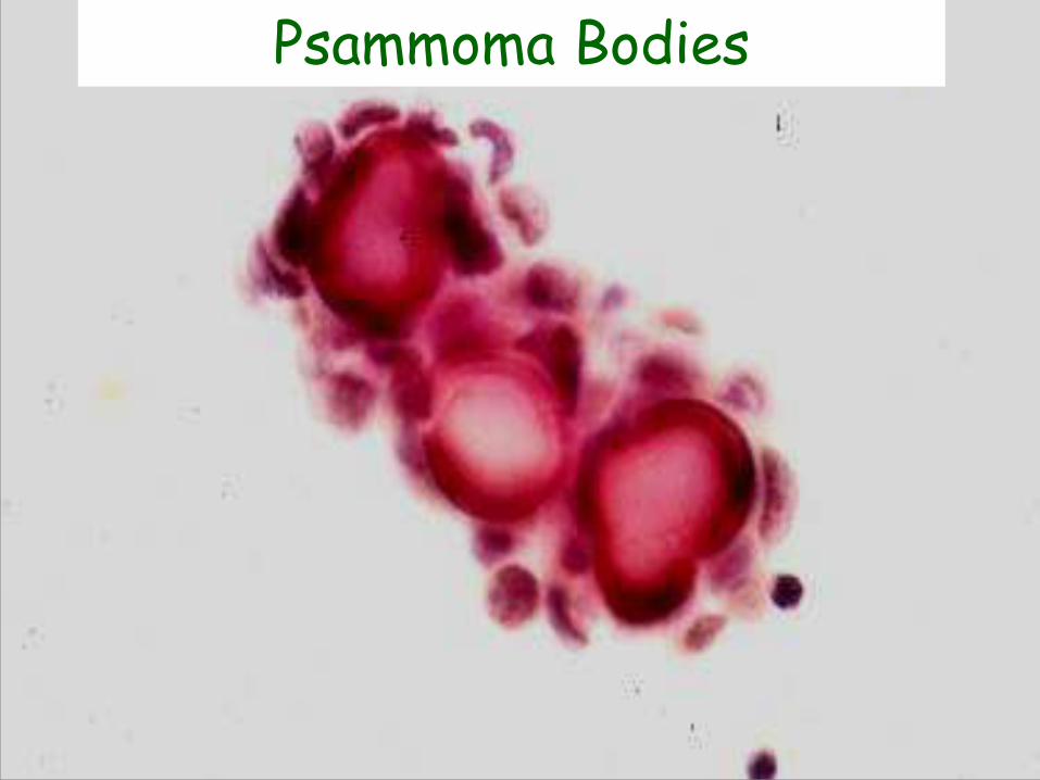
Psammoma Bodies

Cellular Chain

Keratin Pearl

Knobby cluster in Mesothelioma

Malignant Effusions
Specific Type and Site of Origin
Diagnosis : Cytomorphology & Immunocytochemistry

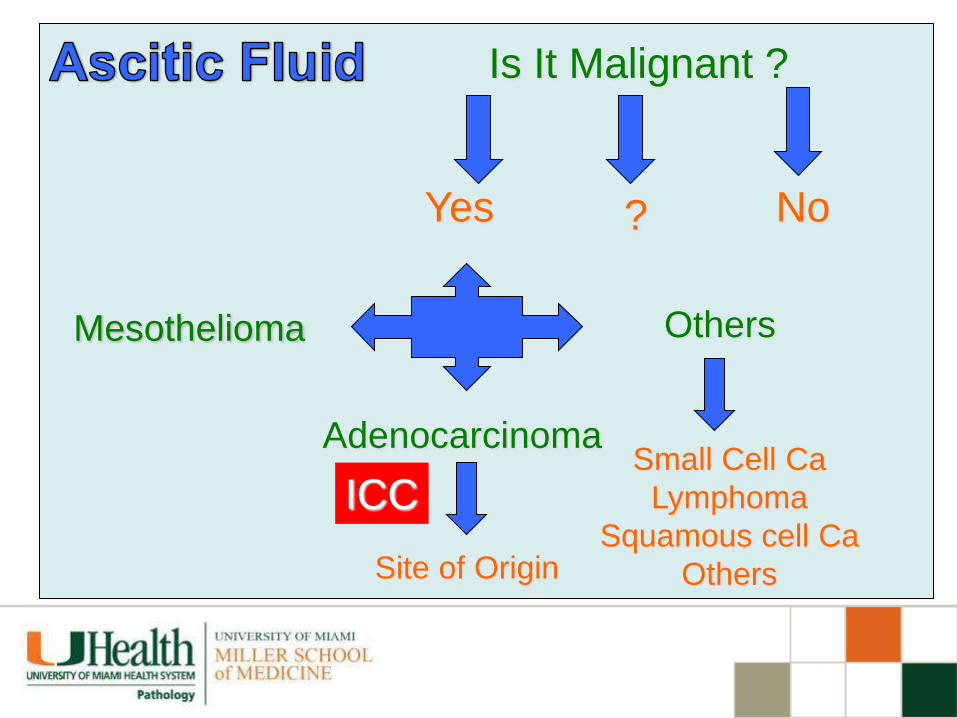
Is It Malignant ?
Yes
Adenocarcinoma
Site of Origin
Mesothelioma
Others
No ?
Small Cell Ca
Lymphoma
Squamous cell Ca
Others

When to Use
Immunocytochemistry in
Ascitic fluid Cytology

Alamo 1917

Acta Cytol 1980; 24: 442-447
Immunocytochemistry in
Cytology
University of Miami Experience

IHC Applications University of Miami
•Diagnosis/Classification 65%
•Prognosticators/Predictors 18%
•Target therapy, Others 17%

How Often? University of Miami
“Percent of our Total Cases”
•Surgical Pathology 5.9%
•Cytopathology 4.9%
•Autopsy Pathology 18%

Type of Specimen ICC in Cytology
•FNA 55%
•Effusion 41%
•Others 4%
• IHC is an important diagnostic tool in
tumor pathology
• Traditionally used on histologic
material and cytologic cell blocks
• The technique is not widely used in
diagnostic cytology
Diagnostic IHC Facts

Why IHC is Not Widely Used in
Cytology ?
• Limited cytologic material
• Problems in interpretation
• Lack of specific markers to
differentiate benign from
malignant cells

Technical Considerations
• Use cell block if possible (Cellular)
• Use alcohol fixation (95% isopropyl)
• Alcohol- fixed, Pap- stained archival
slides can be used
• No de-staining is necessary
• Most cytology samples can be used

• Air-dried slides
• Diff-Quick-stained slides
• De-stained slides (cellular
antigens maybe removed)
• Slides with plastic coverslip
Immunocytochemistry
Not good in:
Immunocytochemistry
Not good in: • Filter preparation
• Serous fluid specimens with excess blood and proteins
Wash specimen or use Saccomanno solution

Immunocytochemistry
Fixation
• 95% isopropyl alcohol
• Buffered formalin
• Formol-acetone
• Mixture of ethanol &
formalin

• Prolonged fixation (wks/months)
in formalin may result in antigenic
loss
• Prolonged fixation in alcohol-
based fixatives is not a major
problem
Immunocytochemistry
Fixation

Easy 3-Step Procedure
1. Use a diamond pen to mark
the cells on the back of the
slide
2. Remove the coverslip
3. Start your routine IHC/ICC
procedure

Immunocytochemistry Using Archival Slides
• Removal of coverslip may be difficult
• When diagnostic slides are limited,
ICC can be performed on a previously
negative slide
IHC=ICC
Technique
No technical alterations needed for cytologic
specimens

True Positive
EMA Calretinin

You Should Know your Antibody
ICC in Diagnostic Cytology
Applications
• Tumor Diagnosis/Classification
• Prognostic/Predictor Markers
• Target Therapy

ICC in Diagnostic Cytology
Applications
• Tumor Diagnosis/Classification
• Prognostic/Predictor Markers
• Target Therapy

Selection of Markers
Cytomorphology
Clinical Information
Working Diagnosis
Differential Diagnosis
Selection of ICC Markers
Final Interpretation

ICC in Diagnostic Cytology
Selection of Markers “tailor-made” Approach
• When the differential diagnosis is
narrowed down, usually not more than 2-
3 markers are needed (“tailor-made”)
• In many occasions only one marker is
used to confirm the working diagnosis
Diagnosis/Classification
Our 3-Step Approach
1. Define a specific differential Dx
2. Select a small panel of ICC markers
3. Combine cytomorphology and ICC

Is It Malignant ?
Yes
Adenocarcinoma
Site of Origin
Mesothelioma
Others
No ?
ICC
ICC
ICC
ICC
Small Cell Ca
Lymphoma
Squamous cell Ca
Others

Is It Malignant ?
Yes
Adenocarcinoma
Site of Origin
Mesothelioma
Others
No ?
ICC
Small Cell Ca
Lymphoma
Squamous cell Ca
Others

First Step…..
Reactive Mesothelial cells
versus
Malignant Process

• The reactive mesothelial cells
may group.
• If so, the grouping usually
presents as loose clusters,
without nuclear overlapping.


Reactive Mesothelial
Cells
Malignant Mesothelioma
Adenocarcinoma
Differential Diagnosis of Atypical
cells in Ascitic Fluid
Malignant
Morphology
NO
YES
YES
Resemble
Mesothelial
Cells
YES
YES
NO

EMA: Malignant: adenocarcinoma,
malignant mesothelioma
CEA: Malignant: adenocarcinoma
Ber-EP4: Malignant: adenocarcinoma
LeuM1: Malignant: adenocarcinoma
Desmin: Benign: reactive mesothelium
Commonly Used Markers In Effusions

In our laboratory, EMA
(clone E29) is the most
frequently used antibody
in defining “atypical cells”
in effusions.

Reactive Mesothelium vs.
Adenocarcinoma and Mesothelioma
Positive (Cytoplasm) Adenocarcinoma
Negative Reactive
EMA
Positive (membrane) Mesothelioma
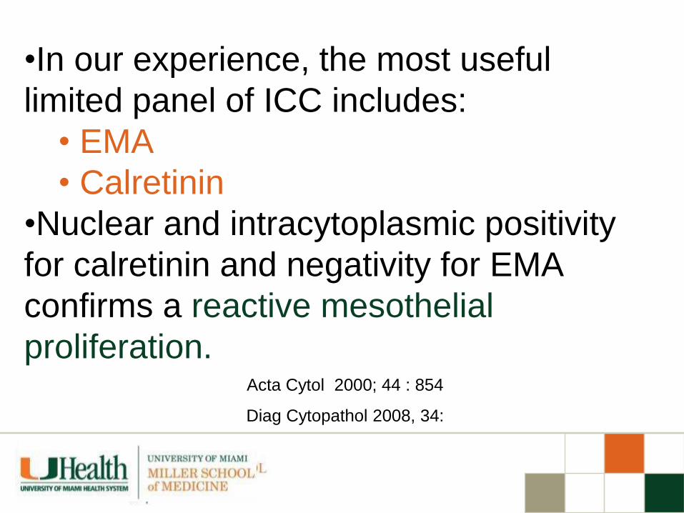
•In our experience, the most useful
limited panel of ICC includes:
• EMA
• Calretinin
•Nuclear and intracytoplasmic positivity
for calretinin and negativity for EMA
confirms a reactive mesothelial
proliferation. Acta Cytol 2000; 44 : 854
Diag Cytopathol 2008, 34:

Reactive Mesothelium

EMA

Calretinin


EMA
EMA Positivity Strong, Intracytoplasmic & Easily seen on Low Power
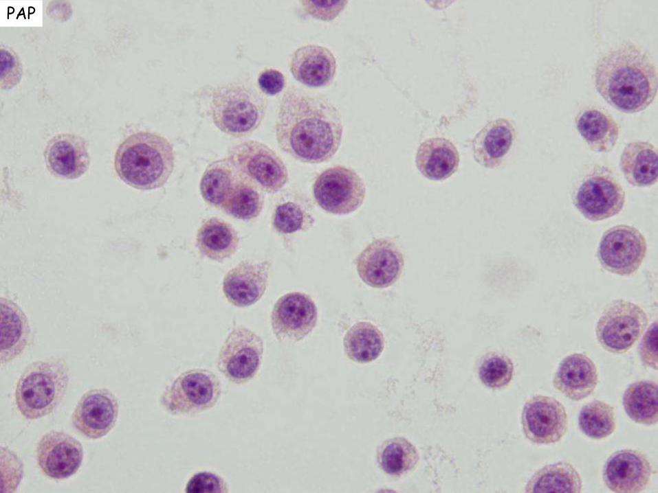
PAP

EMA

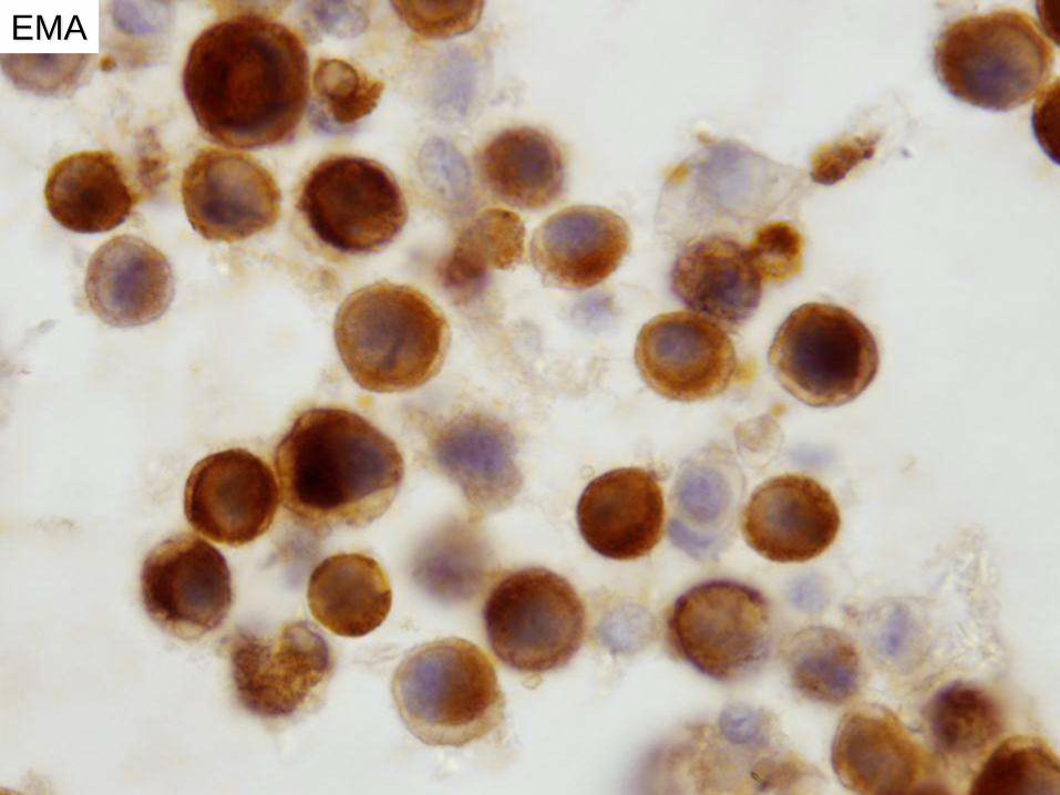
EMA
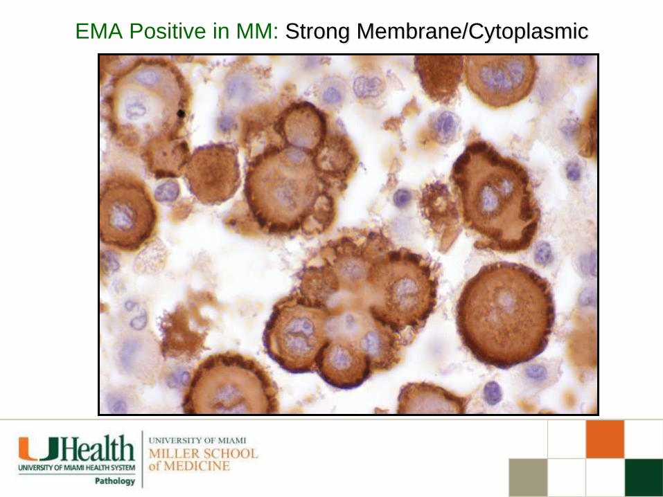
EMA Positive in MM: Strong Membrane/Cytoplasmic

EMA Positive in MM
Strong
Membrane/Cytoplasmic
Strong
Cytoplasmic

Positive EMA in
Serous Effusions
Represents adenocarcinoma, if:
–Easily seen on low power
–Is strong and intracytoplasmic

EMA

EMA

Is It Malignant ?
Yes
Adenocarcinoma
Site of Origin
Mesothelioma
Others
No ?
ICC Small Cell Ca
Lymphoma
Squamous cell Ca
Others

Malignant Mesotheliomas in
Effusions
Low Power
• Small or large 3D groups
• “Knobby clusters”
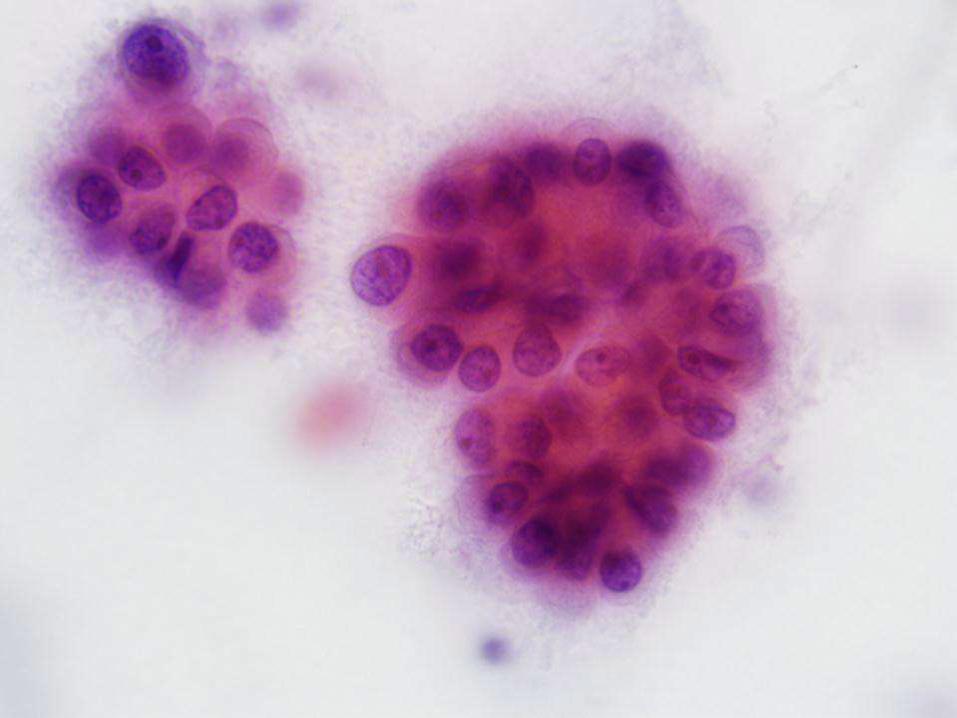
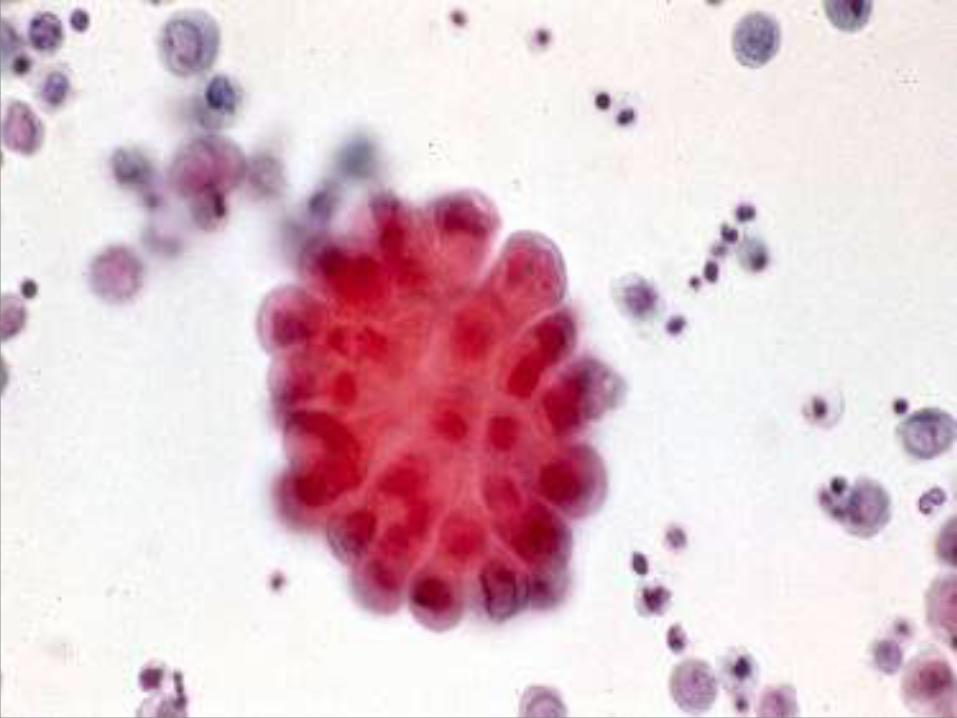


Resemblance to Mesothelial Cells

Differential Diagnosis of Mesothelioma
• Cytomorphology
• Electron microscopy
• Cytochemistry
• Immunocytochemistry (ICC)
Malignant Mesothelioma
in Effusions

When
Malignant Mesothelioma Mimics Adenocarcinomas
Use ICC
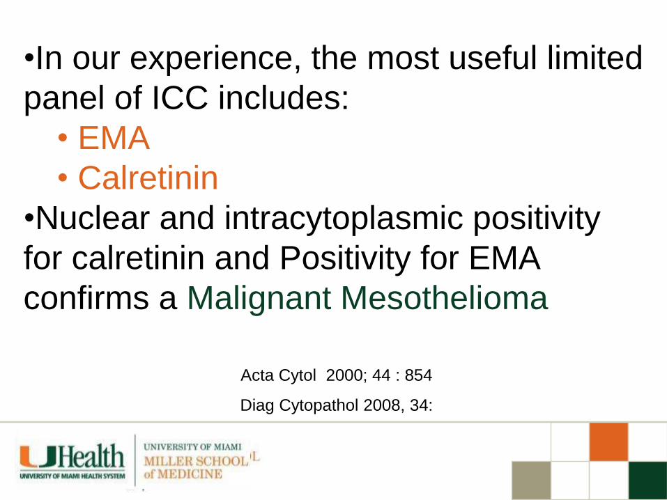
•In our experience, the most useful limited
panel of ICC includes:
• EMA
• Calretinin
•Nuclear and intracytoplasmic positivity
for calretinin and Positivity for EMA
confirms a Malignant Mesothelioma
Acta Cytol 2000; 44 : 854
Diag Cytopathol 2008, 34:

Malignant Mesothelioma Calretinin

Ascitic Fluid
Lung
Adenocarcinoma
Malignant
Mesothelioma Vs
Calret TTF-1 CEA D2-40
MM Pos Neg Neg Pos
LA Neg Pos Pos Neg
Is It Malignant ?
Yes
Adenocarcinoma
Site of Origin
Mesothelioma
Others
No ?
ICC Small Cell Ca
Lymphoma
Squamous cell Ca
Others

Adenocarcinoma in Ascitic Fluid
Primary Sites in Adult Male
• Adenocarcinoma
–GI tract-
Pancreas
–GU
–Lung
3
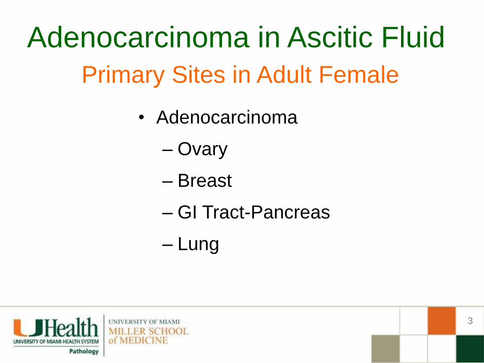
Adenocarcinoma in Ascitic Fluid
Primary Sites in Adult Female
• Adenocarcinoma
– Ovary
– Breast
– GI Tract-Pancreas
– Lung
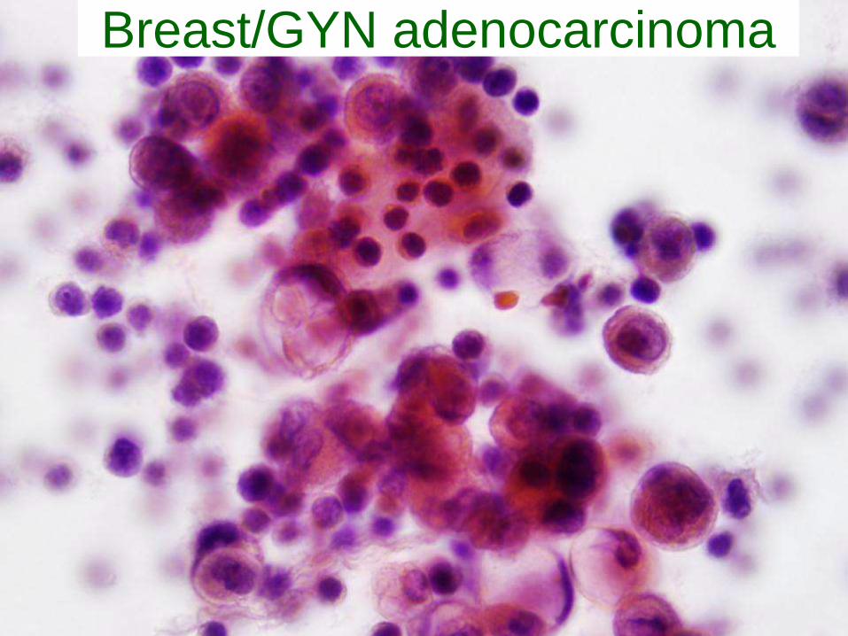
Breast/GYN adenocarcinoma
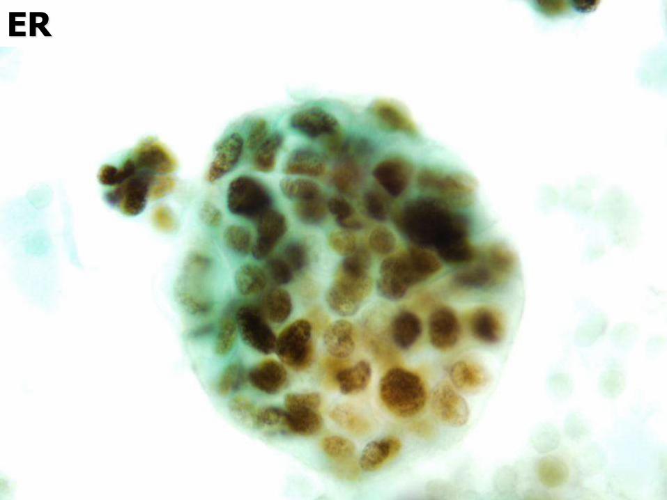
ER

ER-1D5
In Fluids

Remember ! • Be careful with the use of ER in
peritoneal effusions of female
patients
• Benign epithelial inclusions may
cause false positive results
• First establish the malignant nature
of the cells by cytomorphology
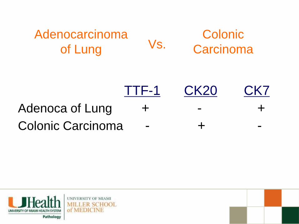
Colonic
Carcinoma
Adenocarcinoma
of Lung Vs.
TTF-1 CK20 CK7
Adenoca of Lung + - +
Colonic Carcinoma - + -

ICC Markers for Colon Cancer
• CK 7 Negative
• CK 20 Positive
• CDX-2 Positive
• CEA Positive
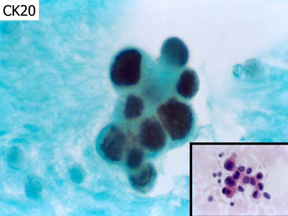
CK20

Metastatic
Adenocarcinoma Hepatocellular
Carcinoma vs
CK7 HCA
Hepatocellular Ca - +
Adenocarcinoma + -

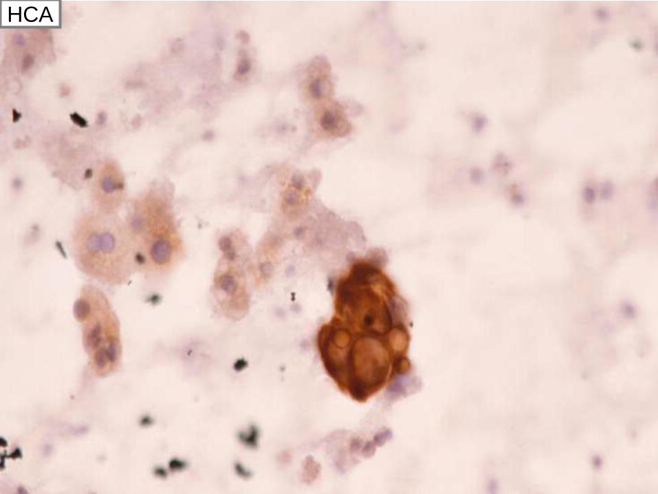
HCA
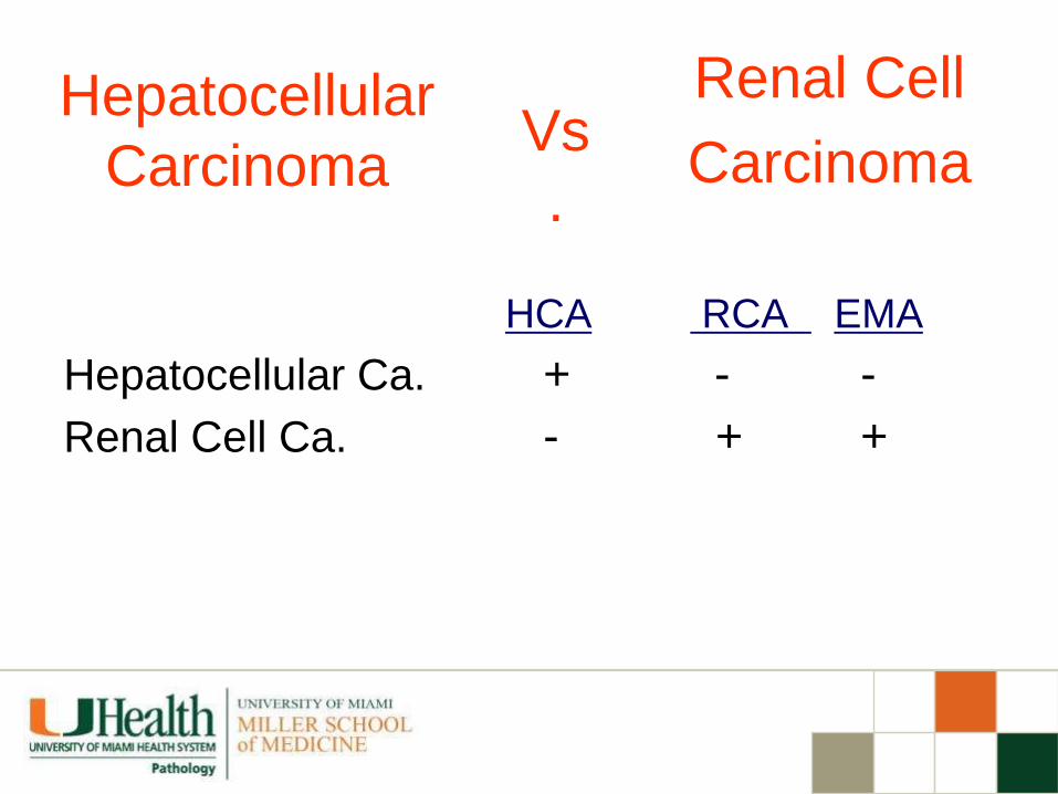
Renal Cell
Carcinoma Hepatocellular
Carcinoma Vs
.
HCA RCA EMA
Hepatocellular Ca. + - -
Renal Cell Ca. - + +


RCA
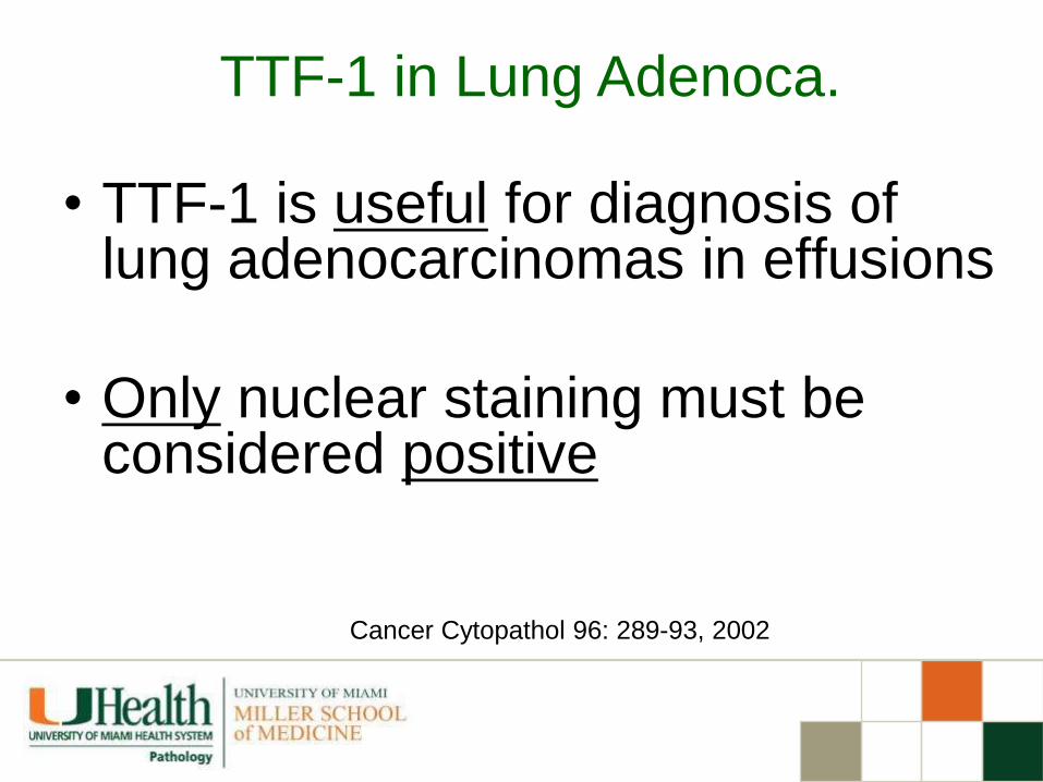
• TTF-1 is useful for diagnosis of lung adenocarcinomas in effusions
• Only nuclear staining must be considered positive
TTF-1 in Lung Adenoca.
Cancer Cytopathol 96: 289-93, 2002

TTF-1

TTF-1; Neg.

Is It Malignant ?
Yes
Adenocarcinoma
Site of Origin
Mesothelioma
Others
No ?
ICC
Small Cell Ca
Lymphoma
Squamous cell Ca
Others

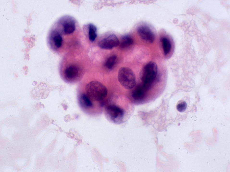
Small Cell Carcinoma
in Ascitic Fluid
Low Power
• Tight cell balls
• Indian file/chain
• Isolated cells may
be overlooked
High Power
• Nuclear molding
• Coarse chromatin
• Wrinkled nuclear
membrane
• Occasional cells
with nucleoli

Lung Carcinoma
Small Cell Non- Small vs
CK SYN CHR TTF-1
Non-Small + -/+ - +/-
Small Cell +(dot) + +/- +


CK
Differential Diagnosis
• Malignant lymphoma
• “Small blue cell tumors”
Small Cell Carcinoma
in Ascitic Fluid

Small Cell Ca - + +/- - -
LCA KER CHR DES NB
Lymphoma + - - - -
Rhabdomyosarcoma - - - + -
Neuroblastoma - - - - +
ICC in Differential Diagnosis of
Small Cell Malignancies

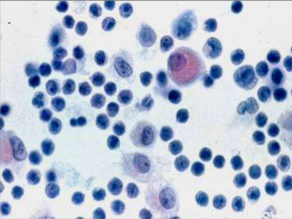
Malignant Lymphoma in Ascitic Fluid
Low Power
Isolated Cells


High Power • Nuclear variation in size and shape
• Nuclear indentation/convolution
• Vesicular nuclei with prominent nucleoli
• Individual cell necrosis (apoptosis)
• Scant, basophilic cytoplasm, rarely well
preserved
Malignant Lymphoma in Ascitic Fluid


CD20

CD 79

HHV8 associated lymphoma
Malignant Lymphoma in Ascitic Fluid

Lymphoma vs. Carcinoma vs.
Germinoma vs. Melanoma
Favor Lymphoma
• Only isolated cells
• Nuclear clefts
• Apoptotic cells
Immunocytochemistry
• LCA ( + )
• Keratin ( - )
• PLAP ( - )
• S100 ( - )

Small “Mature-Looking”
Lymphocytes in Effusions
Differential Diagnosis
• Chronic pleuritis (TB)
• Small cell lymphomas
• Chronic lymphocytic leukemia
• Waldenstrom’s macroglobulinemia
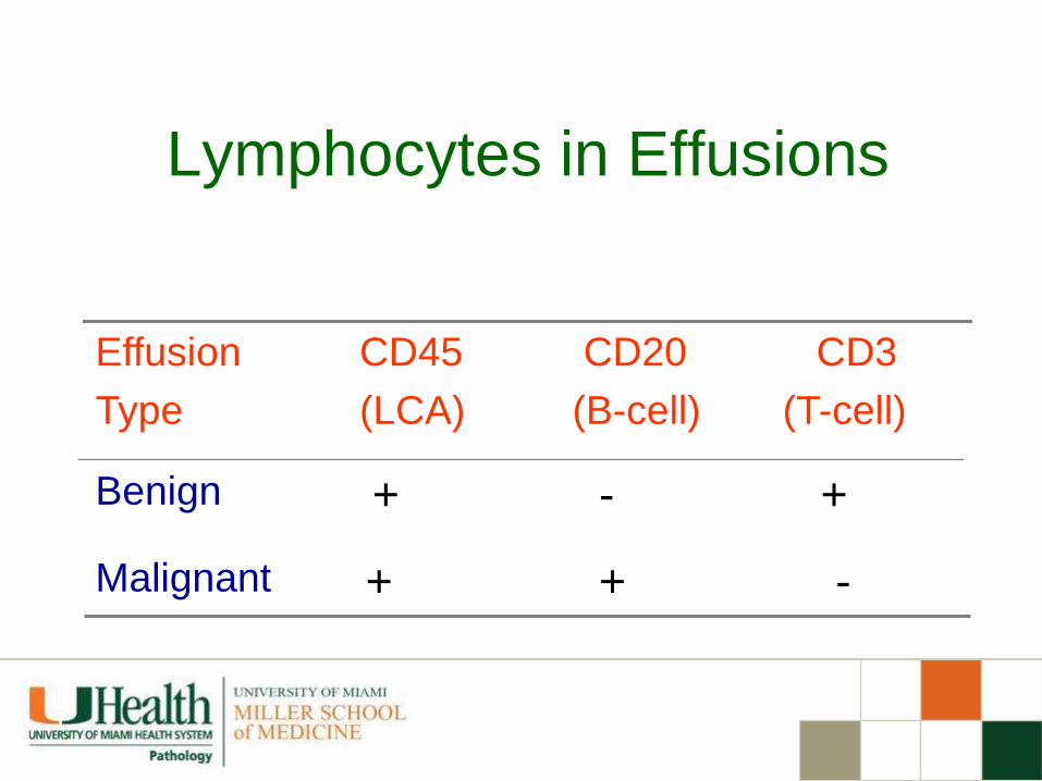
Lymphocytes in Effusions
- + + Malignant
+ - + Benign
CD3
(T-cell)
CD20
(B-cell)
CD45
(LCA)
Effusion
Type

CD3
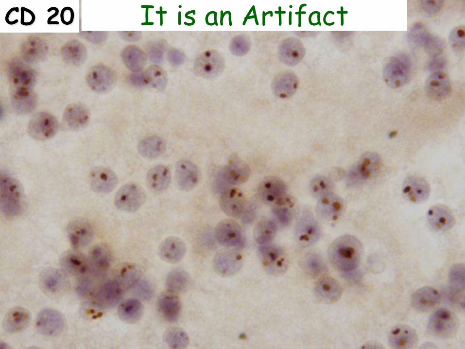
CD 20 It is an Artifact
Ancillary Techniques to Rule
Out Malignant Lymphoma
• Flow cytometry
• Gene rearrangement

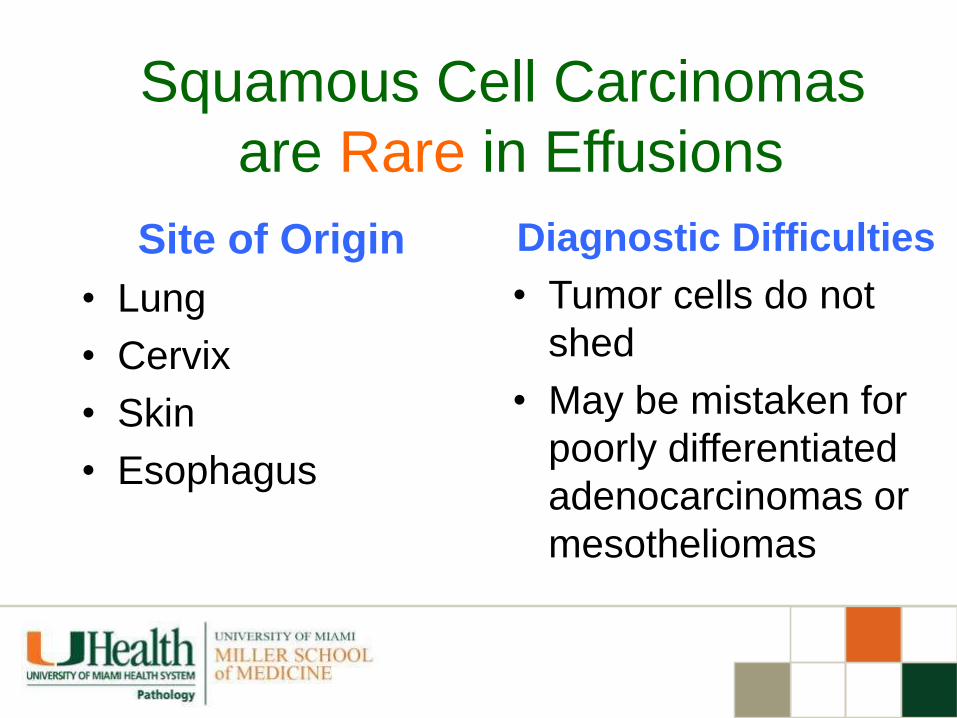
Squamous Cell Carcinomas
are Rare in Effusions
Site of Origin
• Lung
• Cervix
• Skin
• Esophagus
Diagnostic Difficulties
• Tumor cells do not
shed
• May be mistaken for
poorly differentiated
adenocarcinomas or
mesotheliomas

Squamous cell ca

Squamous cell ca
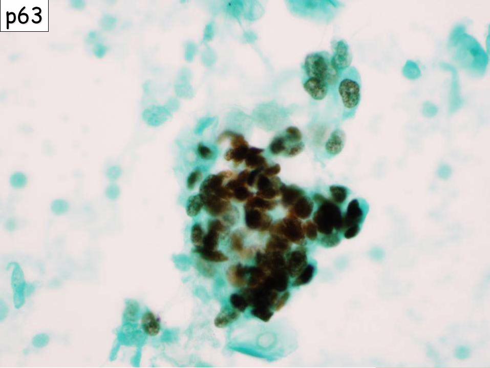
p63

Remember ! • Squamous carcinoma cells are usually
overlooked in body cavity fluid cytology - Only few cells shed
• They might be confused with necrotic /degenerative mesothelial cells
• p63 and p40 are very helpful to detect squamous cells
Cancer Cytopathol 2009; 117: 46-50
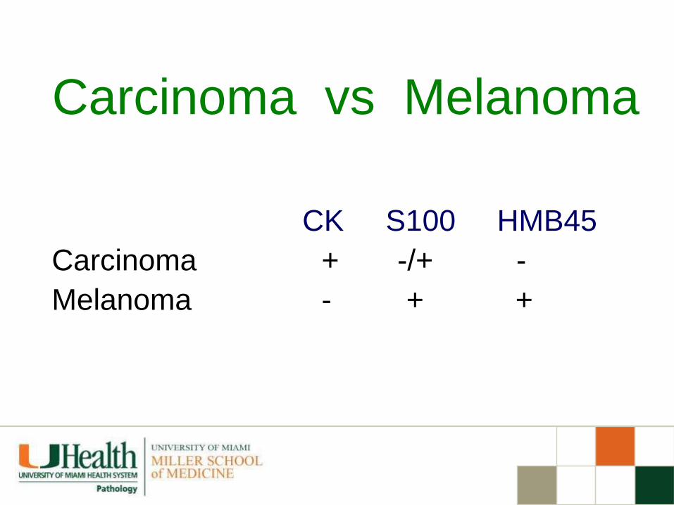
Carcinoma vs Melanoma
CK S100 HMB45
Carcinoma + -/+ -
Melanoma - + +


HMB 45

Melanoma Markers
• S100 Protein +++
• HMB-45 +++
• Melan-A ++
• Tyrosinase ++

ICC in Diagnostic Cytology
Applications
• Tumor Diagnosis/Classification
• Prognostic/Predictor Markers
• Target Therapy

ER-1D5

Ki-67

Detection of HER2 in cytology
ICC, FISH, CISH
Predictive Value
NOT standard of Care for
Breast CA
Diagn Cytopathol 1994; 11:262-265
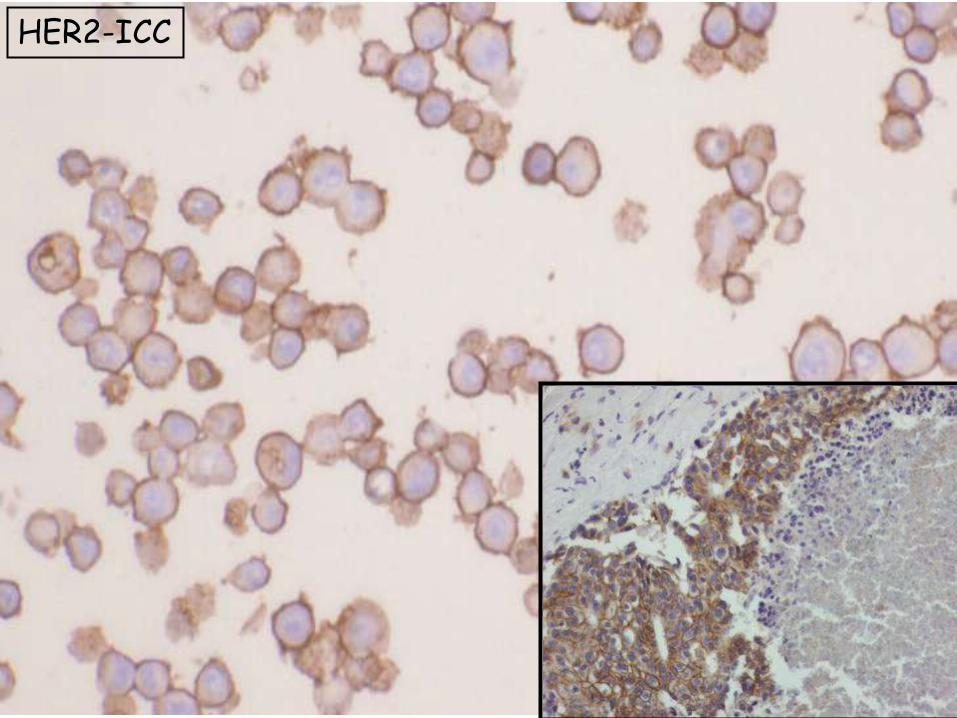
HER2-ICC
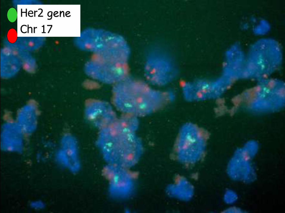
Her2 gene
Chr 17

HER2-CISH
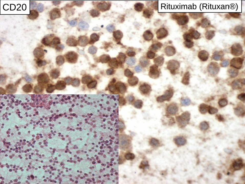
CD20 Rituximab (Rituxan®)
ICC in Diagnostic Cytology
Applications
• Tumor Diagnosis/Classification
• Prognostic/Predictor Markers
• Target Therapy

Arch Pathol Lab Med 2013, 137:668-684
– tyrosine kinase inhibitors (TKI) first-line
therapy in patients with advanced lung
adenocarcinoma with EGFR mutations
– adenocarcinomas with ALK
rearrangements are responsive to
crizotinib (AlK inhibitor).
– Patients with KRAS or BRAF mutation
do not respond to TKI, ALKI
NSCLC: Target Therapy
Arch Pathol Lab Med 2013, 137:668-684
– patients with adenocarcinoma or NSCLC,
not otherwise specified (NSCLC-
NOS),are more responsive to pemetrexed
than those squamous cell carcinoma
– squamous cell carcinoma is associated
with life-threatening hemorrhage in
patients treated with bevacizumab;
therefore, it is contraindicated in lung
cancer patients with this histology.
NSCLC: Target Therapy


TTF-1
Squamous Cell Carcinoma p63

ICC Limitations
• Large 3D cellular clusters in cytospin
samples
• Histiocytes, macrophages, cells in
mitosis , tumor giant cells
Look for single cells or smaller 2D groups
AM J Clin Pathol 1990; 94:470-475.

ICC Limitations
• Lack of internal control
• Negative results in ICC are
not as meaningful as positive
reactions
Diag Cytopathol 1986; 81-2, 1986

Final Words….
• Use our 3-step approach:
–Define a specific differential Dx
–Select a small panel of ICC markers
–Combine Cytomorphology and ICC
• ICC can be used on previously alcohol-fixed Pap-stained slides without de-staining
• The technique does not require any
modification of the routine ICC staining
protocol
Final Words….

Springer, 2007 Demos, 2011
•
ASCP Workshops
Diagnostic problems in body cavity fluid
cytology; a practical approach.
Immunocytochemistry in Diagnostic
Cytology:
Values and Limitations
Parvin Ganjei-Azar MD, FASCP
Mercè Jordà MD, PhD, FASCP

University of Miami Health System
Sylvester Comprehensive Cancer Center