ARTICULAR CARTILAGE REPAIR - The University of...
6
This presentation is the intellectual property of the author. Contact them for permission to reprint and/or distribute. ARTICULAR CARTILAGE REPAIR Dr. Matthew Murray Ortho San Antonio Orthopaedic Sports Medicine Arthroscopic Surgery Articular Cartilage Properties Injuries to Articular Cartilage Treatment of Articular Cartilage Lesions Traditional Techniques Marrow Stimulation Cell-Based Techniques Whole-Tissue Transplantation Emerging Technologies DeNovo Biocartilage CartiForm PRP and Stem Cells OUTLINE Hyaline Cartilage Caps the ends of bones that form synovial joints Contains predominantly Type II Collagen Thickness Varies in Different Joints Up to 2-4mm in the knee joint ARTICULAR CARTILAGE FUNCTIONS Both a cushion and slick surface for movement Allows bones to slide against each other in a joint Allows load bearing without permanent distortion Shock/Impact Absorbing Nourishment provided by synovial fluid, NOT underlying bone Withdrawal of synovial fluid can lead to rapid cartilage deterioration ARTICULAR CARTILAGE Limited intrinsic capacity for spontaneous healing Avascular Hypocellular Potential for considerable pain and disability if untreated Subsequent development of osteoarthritis ARTICULAR CARTILAGE LESIONS Must differentiate osteoarthritis versus focal articular cartilage lesion ARTICULAR CARTILAGE LESIONS
Transcript of ARTICULAR CARTILAGE REPAIR - The University of...

This presentation is the intellectual property of the author.Contact them for permission to reprint and/or distribute.
ARTICULAR CARTILAGE REPAIR
Dr. Matthew MurrayOrtho San AntonioOrthopaedic Sports MedicineArthroscopic Surgery
Articular Cartilage Properties
Injuries to Articular Cartilage
Treatment of Articular Cartilage Lesions Traditional Techniques Marrow Stimulation
Cell-Based Techniques
Whole-Tissue Transplantation
Emerging Technologies DeNovo
Biocartilage
CartiForm
PRP and Stem Cells
OUTLINE
Hyaline Cartilage Caps the ends of bones that form synovial joints
Contains predominantly Type II Collagen
Thickness Varies in Different Joints Up to 2-4mm in the knee joint
ARTICULAR CARTILAGE
FUNCTIONS Both a cushion and slick surface for movement
Allows bones to slide against each other in a joint
Allows load bearing without permanent distortion
Shock/Impact Absorbing
Nourishment provided by synovial fluid, NOT underlying bone Withdrawal of synovial fluid can lead to rapid cartilage deterioration
ARTICULAR CARTILAGE
Limited intrinsic capacity for spontaneous
healing Avascular
Hypocellular
Potential for considerable pain and
disability if untreated
Subsequent development
of osteoarthritis
ARTICULAR CARTILAGE LESIONS
Must dif ferentiate osteoarthritis versus focal articular carti lage lesion
ARTICULAR CARTILAGE LESIONS

This presentation is the intellectual property of the author.Contact them for permission to reprint and/or distribute.
300,000 knee cartilage procedures in the US annually
Insidious Onset Osteochondritis Dissecans (OCD) Dissection of the articular cartilage from underlying subchondral bone No clear cause Typically affects adolescents Worse prognosis in adults
Traumatic Onset (athletic activities) ACL injuries Patella dislocations
But, many are incidental and asymptomatic
ARTICULAR CARTILAGE LESIONS
Common association with structural abnormalities Limb Malalignment Excessive varus/valgus overloads compartment
Osteotomy
Ligamentous Instability ACL
Patellar Instability
Meniscal Deficiency Subtotal meniscectomy increases stresses 300%
Meniscal allograft transplantation
*neglecting these abnormalities wil l lead to
failure of any chondral repair procedure*
ARTICULAR CARTILAGE LESIONS
Physical Weight Loss
Activity Modification
Therapy
Bracing
Pharmacologic NSAIDS
Glucosamine/Chondroitin
Injections Steroids
Viscosupplementation
OCD – stable, juvenile lesions Protected weight-bearing, immobilization
NONOPERATIVE TREATMENT
Repair OCD Lesions bioabsorbable pins/screws
OPERATIVE TREATMENT
Marrow Stimulation Techniques (MSTs) Drilling
Abrasion Arthroplasty
MICROFRACTURE Relatively easy procedure
Single operation
Useful for smaller lesions (<2cm2)
Adjunct to ACL or MPFL surgery
70-80% satisfaction and RTP
BUT –
Type I Collagen (Fibrocartilage)
Results deteriorate over time
OPERATIVE TREATMENT
MICROFRACTURE

This presentation is the intellectual property of the author.Contact them for permission to reprint and/or distribute.
Cartilage Restoration (Cell-Based Technology) Autologous Chondrocyte Implantation (ACI) Articular cartilage harvest and re-implantation Type II collagen Useful for large lesions Good long-term clinical results BUT - 2 stage operation Very expensive $$$$$$$$$ Technically demanding, time consuming
MACI (Matrix-Assisted ACI) Cultured chondrocytes implanted onto a scaffold Still two-stage operation Not approved by FDA for use in United States
OPERATIVE TREATMENT
Cartilage Replacement (Whole-Tissue Transplantation)
Osteochondral Autograft Transplantation (OATS, Mosaicplasty) Tissue readily available
Inexpensive
BUT
Limited amount of available donor tissue
Donor site morbidity
Technically demanding procedure
OPERATIVE TREATMENT
Cartilage Replacement (Whole-Tissue Transplantation)
Osteochondral Allograft Transplantation No Donor site morbidity Useful for larger defects/even hemicondyle Ability to precisely match contour of condyle Eliminates “dead space” of mosaicplasty Useful when bone loss is present
BUT Expensive Limited graft availability Even more technically demanding Risk of disease transmission Risk of incomplete graft incorporation/rejection
OPERATIVE TREATMENT
OPERATIVE TREATMENT
Why do we need new techniques?? Microfracture Type I collagen
Not useful for larger lesions
Limited long-term results
OATS Donor site morbidity
Not useful for larger lesions
Technically demanding
Osteochondral Allograft Limited tissue available
Even more technically demanding
Potential for allogenic disease transmission/rejection
EMERGING TECHNOLOGIES
With all these emerging techniques Limited clinical data
Long-term effects unknown
EMERGING TECHNOLOGIES
This presentation is the intellectual property of the author.Contact them for permission to reprint and/or distribute.
DeNovo (Zimmer) Particulated Juvenile Cartilage
Higher density of chondrocytes
Type II Hyaline Cartilage
BUT- Expensive
Limited availability
Difficult to obtain insurance approval
Limited shelf-life (60 days)
EMERGING TECHNOLOGIES
DeNovo
Largest Study to date – 25 pts with 2 year f/u Farr et al AJSM 2014
Improved function
MRI shows normal cartilage by 2 years
BUT Mixture of Type II > Type I collagen
EMERGING TECHNOLOGIES
BioCartilage (Arthrex) Micronized articular cartilage allograft Mixed with PRP or Stem Cells
Cartilage extracellular matrix (type II collagen, proteoglycans, growth factors) No chondrocytes
Microfracture Augmenatation
EMERGING TECHNOLOGIES
Biocartilage Cheap
Readily available
Extended shelf life – 5 years
Can be performed arthroscopically
BUT
It’s Cheap and Easy May not always be the right treatment Bone loss
Uncontained lesion
Larger lesion
EMERGING TECHNOLOGIES
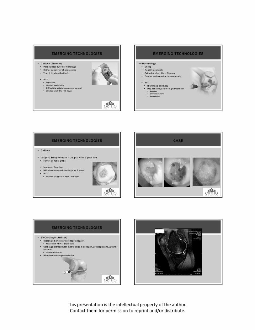
CASE
This presentation is the intellectual property of the author.Contact them for permission to reprint and/or distribute.
Cartiform (Arthrex) Porous osteochondral allograft AKA skin graft, MACI
Viable chondrocytes, growth factors
Preserves layers or normal articular cartilage
Easy to cut, contour
2 year shelf-life Can be frozen with 70% chondrocyte viability
BUT – Expensive
Limited clinical data
Open procedure
EMERGING TECHNOLOGIES
PRP – Platelet Rich Plasma Platelets – release growth factors Ability to attract MSCs, Macrophages, Fibroblasts Stimulate Cell Proliferation
Stem Cells Proliferative Potential Multipotentiality Ability to differentiate and mature into a different cell l ines
Osteocyte Chondrocyte Adipocyte
Peripheral Blood Stem Cells Mesenchymal Stem Cells Bone marrow Synovial tissue Periosteum Fat
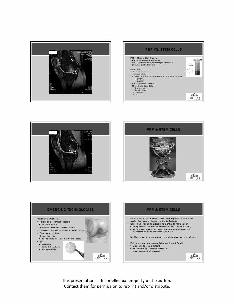
PRP VS. STEM CELLS
PRP & STEM CELLS
No evidence that PRP or Stem Cells injections alone are useful for focal articular cartilage lesions
Can be useful as an adjunct to cartilage restoration Study shows Stem cells as effective as ACI (Saw et al 2012) Study shows Stem Cells added to microfracture outperform
microfracture alone (Nejadnik et al 2010)
Neither proven to reverse or slow degenerative joint disease
Public perception versus Evidence-based Reality Expensive burden to patient Not covered by insurance companies Legal aspects/FDA approval
PRP & STEM CELLS

This presentation is the intellectual property of the author.Contact them for permission to reprint and/or distribute.
CONCLUSIONS
1. Articular cartilage lesions can lead to disability and osteoarthritis if untreated
2. Traditional repair/regenerative techniques are performed based on size of the lesion and activity level of the patient
3. When used appropriately, traditional techniques have yielded good results
4. Emerging Techniques for cartilage restoration have shown early encouraging results, but no long-term data
5. PRP and Stem Cells can be useful adjuncts to cartilage restoration, but are not indicated for isolated use
THANK YOU