
ARTICLE Platelet Biology & Its Disorders Whole exome ...orca.cf.ac.uk/101872/1/DII-168...
10
1170 haematologica | 2016; 101(10) Received: March 17, 2016. Accepted: June 10, 2016. Pre-published: June 16, 2016. ©2016 Ferrata Storti Foundation Check the online version for the most updated information on this article, online supplements, and information on authorship & disclosures: www.haematologica.org/content/101/10/1170 Material published in Haematologica is cov- ered by copyright. All rights reserved to the Ferrata Storti Foundation. Copies of articles are allowed for personal or internal use. Permission in writing from the publisher is required for any other use. Correspondence: [email protected] Ferrata Storti Foundation EUROPEAN HEMATOLOGY ASSOCIATION Haematologica 2016 Volume 101(10):1170-1179 ARTICLE Platelet Biology & Its Disorders doi:10.3324/haematol.2016.146316 I nherited thrombocytopenias are a heterogeneous group of disorders characterized by abnormally low platelet counts which can be asso- ciated with abnormal bleeding. Next-generation sequencing has pre- viously been employed in these disorders for the confirmation of sus- pected genetic abnormalities, and more recently in the discovery of novel disease-causing genes. However its full potential has not yet been exploited. Over the past 6 years we have sequenced the exomes from 55 patients, including 37 index cases and 18 additional family members, all of whom were recruited to the UK Genotyping and Phenotyping of Platelets study. All patients had inherited or sustained thrombocytope- nia of unknown etiology with platelet counts varying from 11x10 9 /L to 186x10 9 /L. Of the 51 patients phenotypically tested, 37 (73%), had an additional secondary qualitative platelet defect. Using whole exome sequencing analysis we have identified “pathogenic” or “likely patho- genic” variants in 46% (17/37) of our index patients with thrombocy- topenia. In addition, we report variants of uncertain significance in 12 index cases, including novel candidate genetic variants in previously unreported genes in four index cases. These results demonstrate that whole exome sequencing is an efficient method for elucidating potential pathogenic genetic variants in inherited thrombocytopenia. Whole exome sequencing also has the added benefit of discovering potentially pathogenic genetic variants for further study in novel genes not previ- ously implicated in inherited thrombocytopenia. Whole exome sequencing identifies genetic variants in inherited thrombocytopenia with secondary qualitative function defects Ben Johnson, 1 Gillian C. Lowe, 1 Jane Futterer, 1 Marie Lordkipanidzé, 1 David MacDonald, 1 Michael A. Simpson, 2 Isabel Sanchez-Guiú, 3 Sian Drake, 1 Danai Bem, 1 Vincenzo Leo, 4 Sarah J. Fletcher, 1 Ban Dawood, 1 José Rivera, 3 David Allsup, 5 Tina Biss, 6 Paula HB Bolton-Maggs, 7 Peter Collins, 8 Nicola Curry, 9 Charlotte Grimley, 10 Beki James, 11 Mike Makris, 4 Jayashree Motwani, 12 Sue Pavord, 13 Katherine Talks, 6 Jecko Thachil, 7 Jonathan Wilde, 14 Mike Williams, 12 Paul Harrison, 15 Paul Gissen, 16 Stuart Mundell, 17 Andrew Mumford, 18 Martina E. Daly, 4 Steve P. Watson, 1 and Neil V. Morgan 1 on behalf of the UK GAPP Study Group 1 Institute for Cardiovascular Sciences, College of Medical and Dental Sciences, University of Birmingham, UK; 2 Division of Genetics and Molecular Medicine, King's College, London, UK; 3 Centro Regional de Hemodonación, Universidad de Murcia, IMIB- Arrixaca, Murcia, Spain; 4 Department of Infection, Immunity and Cardiovascular Disease, University of Sheffield Medical School, University of Sheffield, UK; 5 Hull Haemophilia Treatment Centre, Hull and East Yorkshire Hospitals NHS Trust, Castle Hill Hospital, Hull, UK; 6 Department of Haematology, Royal Victoria Infirmary, Newcastle Upon Tyne, UK; 7 Department of Haematology, Manchester Royal Infirmary, Manchester, UK; 8 Arthur Bloom Haemophilia Centre, School of Medicine, Cardiff University, UK; 9 Oxford Haemophilia & Thrombosis Centre, Churchill Hospital, Oxford, UK; 10 Nottingham Haemophilia Centre, Nottingham University Hospital, UK; 11 Regional Centre for Paediatric Haematology, Leeds Children’s Hospital, UK; 12 Department of Haematology, Birmingham Children's Hospital, UK; 13 Department of Haematology, Oxford University Hospitals NHS Foundation Trust, UK; 14 Adult Haemophilia Centre, Queen Elizabeth Hospital, Birmingham, UK; 15 School of Immunity and Infection, College of Medical and Dental Sciences, University of Birmingham, UK; 16 Medical Research Council, Laboratory for Molecular Cell Biology, University College London, UK; 17 School of Physiology, Pharmacology and Neuroscience, University of Bristol, UK; and 18 School of Cellular and Molecular Medicine, University of Bristol, UK ABSTRACT
-
Upload
duonghuong -
Category
Documents
-
view
213 -
download
0
Transcript of ARTICLE Platelet Biology & Its Disorders Whole exome ...orca.cf.ac.uk/101872/1/DII-168...
1170 haematologica | 2016; 101(10)
Received: March 17, 2016.
Accepted: June 10, 2016.
Pre-published: June 16, 2016.
©2016 Ferrata Storti Foundation
Check the online version for the most updatedinformation on this article, online supplements,and information on authorship & disclosures:www.haematologica.org/content/101/10/1170
Material published in Haematologica is cov-ered by copyright. All rights reserved to theFerrata Storti Foundation. Copies of articlesare allowed for personal or internal use.Permission in writing from the publisher isrequired for any other use.
Correspondence: [email protected]
Ferrata StortiFoundation
EUROPEANHEMATOLOGYASSOCIATION
Haematologica 2016Volume 101(10):1170-1179
ARTICLE Platelet Biology & Its Disorders
doi:10.3324/haematol.2016.146316
Inherited thrombocytopenias are a heterogeneous group of disorderscharacterized by abnormally low platelet counts which can be asso-ciated with abnormal bleeding. Next-generation sequencing has pre-
viously been employed in these disorders for the confirmation of sus-pected genetic abnormalities, and more recently in the discovery ofnovel disease-causing genes. However its full potential has not yet beenexploited. Over the past 6 years we have sequenced the exomes from55 patients, including 37 index cases and 18 additional family members,all of whom were recruited to the UK Genotyping and Phenotyping ofPlatelets study. All patients had inherited or sustained thrombocytope-nia of unknown etiology with platelet counts varying from 11x109/L to186x109/L. Of the 51 patients phenotypically tested, 37 (73%), had anadditional secondary qualitative platelet defect. Using whole exomesequencing analysis we have identified “pathogenic” or “likely patho-genic” variants in 46% (17/37) of our index patients with thrombocy-topenia. In addition, we report variants of uncertain significance in 12index cases, including novel candidate genetic variants in previouslyunreported genes in four index cases. These results demonstrate thatwhole exome sequencing is an efficient method for elucidating potentialpathogenic genetic variants in inherited thrombocytopenia. Wholeexome sequencing also has the added benefit of discovering potentiallypathogenic genetic variants for further study in novel genes not previ-ously implicated in inherited thrombocytopenia.
Whole exome sequencing identifies geneticvariants in inherited thrombocytopenia withsecondary qualitative function defectsBen Johnson,1 Gillian C. Lowe,1 Jane Futterer,1 Marie Lordkipanidzé,1 DavidMacDonald,1 Michael A. Simpson,2 Isabel Sanchez-Guiú,3 Sian Drake,1 DanaiBem,1 Vincenzo Leo,4 Sarah J. Fletcher,1 Ban Dawood,1 José Rivera,3 DavidAllsup,5 Tina Biss,6 Paula HB Bolton-Maggs,7 Peter Collins,8 Nicola Curry,9
Charlotte Grimley,10 Beki James,11 Mike Makris,4 Jayashree Motwani,12 SuePavord,13 Katherine Talks,6 Jecko Thachil,7 Jonathan Wilde,14 Mike Williams,12
Paul Harrison,15 Paul Gissen,16 Stuart Mundell,17 Andrew Mumford,18 Martina E. Daly,4 Steve P. Watson,1 and Neil V. Morgan1 on behalf of the UK GAPP Study Group
1Institute for Cardiovascular Sciences, College of Medical and Dental Sciences,University of Birmingham, UK; 2Division of Genetics and Molecular Medicine, King'sCollege, London, UK; 3Centro Regional de Hemodonación, Universidad de Murcia, IMIB-Arrixaca, Murcia, Spain; 4Department of Infection, Immunity and CardiovascularDisease, University of Sheffield Medical School, University of Sheffield, UK; 5HullHaemophilia Treatment Centre, Hull and East Yorkshire Hospitals NHS Trust, Castle HillHospital, Hull, UK; 6Department of Haematology, Royal Victoria Infirmary, NewcastleUpon Tyne, UK; 7Department of Haematology, Manchester Royal Infirmary, Manchester,UK; 8Arthur Bloom Haemophilia Centre, School of Medicine, Cardiff University, UK;9Oxford Haemophilia & Thrombosis Centre, Churchill Hospital, Oxford, UK; 10NottinghamHaemophilia Centre, Nottingham University Hospital, UK; 11Regional Centre forPaediatric Haematology, Leeds Children’s Hospital, UK; 12Department of Haematology,Birmingham Children's Hospital, UK; 13Department of Haematology, Oxford UniversityHospitals NHS Foundation Trust, UK; 14Adult Haemophilia Centre, Queen ElizabethHospital, Birmingham, UK; 15School of Immunity and Infection, College of Medical andDental Sciences, University of Birmingham, UK; 16Medical Research Council, Laboratoryfor Molecular Cell Biology, University College London, UK; 17School of Physiology,Pharmacology and Neuroscience, University of Bristol, UK; and 18School of Cellular andMolecular Medicine, University of Bristol, UK
ABSTRACT
Introduction
Inherited thrombocytopenias (IT) are a heterogeneousgroup of disorders characterized by platelet counts ofless than 150x109/L in whole blood. Platelet counts areconsidered normal when maintained at levels between150x109/L and 450x109/L. This is achieved by homeosta-tic processes controlling platelet production (throm-bopoiesis), platelet senescence and platelet consump-tion/destruction. Pathogenic mutations can result in adisruption of these balanced processes causing IT.However, the clinical manifestations are often dependenton both a decreased platelet count and a qualitative oracquired platelet defect and can vary dramatically fromsevere and potentially life-threatening bleeding to nosymptoms. This variation is noted among individualsshown to have the same underlying genetic causes of dis-ease, suggesting that bleeding risk and phenotype arecomplex traits.1
The average incidence of IT is estimated to be approx-imately 270 cases per 1x10⁶ live births.2 To date there are27 individual IT disorders with known causative muta-tions registered within the Online Mendelian Inheritancein Man (OMIM) catalog, although 33 disease-causinggenes have been described.3
Genetic studies have played a major role in the diagno-sis and progressive understanding of IT. The genes impli-cated in the disease encode proteins that vary widely infunction and include transcription factors (ETV6, FLI1,GATA1, GFI1B and RUNX1) and proteins involved incytoskeleton rearrangement and organization (ACTN1,FLNA, GP1BA, GP1BB, GP9, TUBB1 and WAS).However, some protein functions currently remainunknown (SLFN14 and GNE).4-9. Although our knowl-edge of the causes of IT continues to grow, presently agenetic diagnosis is only reported in approximately 50%of individuals.10-12
So far, genetic investigation into IT has focused on can-didate gene sequencing and individual cases of wholeexome sequencing (WES) when a causative gene is notobvious.9 With 50% of patients currently undiagnosed, achange in the way we approach genetic diagnosis is nec-essary. Here we present the first, large-scale, WES-onlyapproach to patients with suspected IT. We demonstrateits application in determining possible genetic origins ofIT including identification of variants in novel candidatecausative genes. We combine this with an approachimplemented by the Genotyping and Phenotyping ofPlatelets (GAPP) study, which combines WES analysiswith extensive platelet phenotyping to create a completemethod of diagnosis and gene discovery in this subset ofpatients.
Methods
Study approvalThe UK-GAPP study was approved by the National Research
Ethics Service Committee of West Midlands–Edgbaston (RECreference: 06/MRE07/36) and participants gave written informedconsent in accordance with the Declaration of Helsinki. Thisstudy was registered at www.isrctn.org as #ISRCTN 77951167.The GAPP study is included in the National Institute of HealthResearch Non-Malignant Haematology study portfolio(ID9858).
Platelet counts, morphology and white blood cell counts
Results from patients’ samples were compared to ranges forhealthy volunteers for the specific method of morphology used.Platelet counts for light transmission aggregometry and flowcytometry analysis as well as mean platelet volume in platelet-richplasma were originally measured using the Beckman Coultercounter (n=44). Subsequently, platelet counts, morphology andwhite blood cell counts in whole blood were determined using theSysmex XN-1000 (n=11). The PLT-F channel was used to measureplatelet counts in whole blood and the immature platelet fraction.Mean platelet volume was determined from the impedance PLT-Ichannel. White blood cell counts were obtained using the SysmexXN-DIFF channel. All samples were tested against a normal rangewhich was established by measuring the counts for 40 healthyindividuals using the Sysmex XN-1000.
Platelet preparation and platelet function testingPlatelet function was assessed by light transmission aggregom-
etry, including lumiaggregometry, for samples with platelet countsin platelet-rich plasma of >1x10⁸/mL (n=13). An in-house flow-cytometry assay was developed to assess platelet function inpatients with platelet counts in platelet-rich plasma <1x10⁸/mL(n=22). Platelets from individuals with borderline platelet counts inplatelet-rich plasma, between 1.0 and 1.5x10⁸/mL, were assessedusing both assays (n=16).
Aggregometry was performed as previously described.13,14 Forflow cytometry, resting surface levels of CD42b, CD41 and GPVIwere assessed. The platelet-rich plasma was then stimulated withADP (3 and 30 µM), CRP (0.3 and 3 µg/mL) and PAR-1 peptide (10and 100 µM). Membrane expression of P-selectin (FITC-conjugat-ed mouse anti-human CD62P antibody, BD Pharmingen), a mark-er of platelet alpha granule release, as well as fluorescent fibrino-gen binding (a marker of integrin activation) was assessed by flowcytometry on an Accuri C6 flow cytometer. Incubation took placeat 37ºC for 2 min and was terminated by adding a 5-fold excess ofice-cold phosphate-buffered saline.
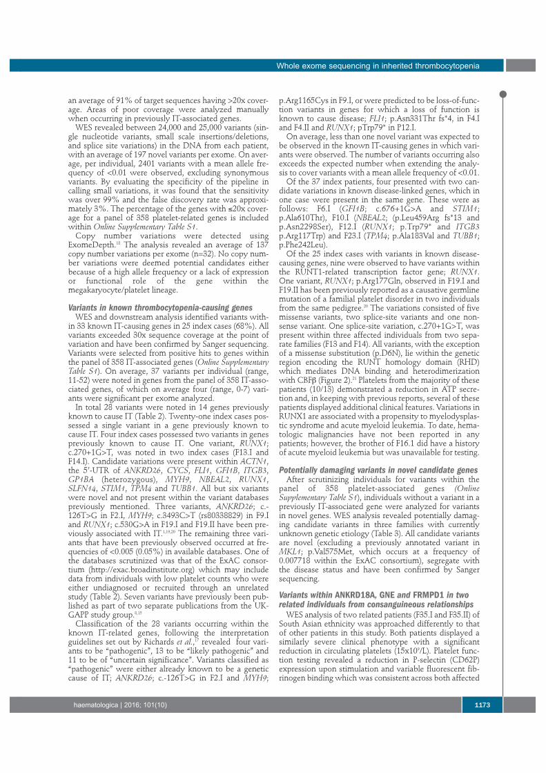
Whole exome sequencingWES and bioinformatics analysis were performed as described
previously8,15,16 (Figure 1). The pathogenicity of variants was determined and called using
the consensus guidelines as set out by the American College ofMedical Genetics and Genomics and the Association forMolecular Pathology (ACMG guidelines).17 Segregation was deter-mined by Sanger sequencing of candidate variants in both affectedand unaffected family members, when available, and the classifi-cation was adapted appropriately for the specific study and smallsample size.
Sanger sequencingTo verify candidate mutations and examine their segregation
among family members Sanger sequencing was performed usingstandard methods on an ABI 3730 automated sequencer, asdescribed previously.8
Results
Recruitment of patientsTo date, 55 patients with a suspected IT or sustained
reduced platelet counts have been enrolled from 25 UKHaemophilia Care Centres and investigated as part of theGAPP study. Before enrollment in the study, all patientsunderwent clinical and genetic work-up to exclude known
Whole exome sequencing in inherited thrombocytopenia
haematologica | 2016; 101(10) 1171
platelet disorders (including Bernard-Soulier syndromeand MYH9-related disorders, analyzed initially by bloodfilm), idiopathic thrombocytopenic purpura and othernon-platelet disorders including von Willebrand diseaseand inherited coagulation factor deficiencies. The patients’bleeding phenotypes are displayed in Table 1. WES wasperformed on genomic DNA from all patients, including37 index cases, all of whom met the study’s entry criteria.All patients, excluding F35.I and F35.II, were of whiteBritish or mixed British ethnicity. All results followingplatelet function testing and WES were reported back tothe referring hematology consultants to aid in geneticcounselling and disease management.
Platelet counts, morphology and function testingPatients were recruited with a platelet count in whole
blood, at the time of enrollment, of less than 150x109/L.Patients with platelet counts in the range of 150x109/L to200x109/L remained enrolled in the study if they showeda similar phenotype to related affected family membersand a platelet count below 150x109/L had been observedprior to enrollment (patients F4.II, F11.III, F13.I and F30.II).Platelet counts, mean platelet volume and immatureplatelet fraction are displayed in Table 1. Of the 55 recruit-ed patients, 12 were deemed to have a macrothrombocy-
topenia and three a microthrombocytopenia (Table 1).White cell counts were within the normal range(3.78x10⁹/L - 10.11x10⁹/L, n=40) in all patients analyzed(n=11).
Platelet function studies revealed the presence of a sec-ondary qualitative defect in addition to the low plateletcount in 37/51 (73%) of the 55 patients whose DNAunderwent WES and who were also available for plateletfunction testing (Table 1). Of the 37 patients with a sec-ondary qualitative defect, 89% (33/37) displayed defectsin both alpha and dense granule secretion. Five of thesepatients with an observed granule secretion defect werealso suspected to have an additional Gi defect because ofreduced responses to all concentrations of ADP. Theremaining four patients without an observable granulesecretion defect showed abnormalities in alternative path-ways (integrin activation, cyclooxygenase pathway andGPVI surface levels) in addition to low platelet counts(Table 1).
Whole exome sequencingWES was performed on genomic DNA from all 55
patients, including 37 index cases, following platelet func-tion testing. An average fold-coverage of 111 wasobserved across all DNA samples analyzed by WES with
B. Johnson et al.
1172 haematologica | 2016; 101(10)
Figure 1. Bioinformatics pipeline analysis of whole exome sequencing data.Initial WES analysis focused on comparison with a panel of 358 genes (OnlineSupplementary Table S1), after which screening of exome variants focused onnovel variants. Variants were classified using the ACMG consensus guidelines.
an average of 91% of target sequences having >20x cover-age. Areas of poor coverage were analyzed manuallywhen occurring in previously IT-associated genes.
WES revealed between 24,000 and 25,000 variants (sin-gle nucleotide variants, small scale insertions/deletions,and splice site variations) in the DNA from each patient,with an average of 197 novel variants per exome. On aver-age, per individual, 2401 variants with a mean allele fre-quency of <0.01 were observed, excluding synonymousvariants. By evaluating the specificity of the pipeline incalling small variations, it was found that the sensitivitywas over 99% and the false discovery rate was approxi-mately 3%. The percentage of the genes with ≤20x cover-age for a panel of 358 platelet-related genes is includedwithin Online Supplementary Table S1.
Copy number variations were detected usingExomeDepth.18 The analysis revealed an average of 137copy number variations per exome (n=32). No copy num-ber variations were deemed potential candidates eitherbecause of a high allele frequency or a lack of expressionor functional role of the gene within themegakaryocyte/platelet lineage.
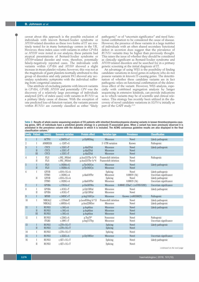
Variants in known thrombocytopenia-causing genesWES and downstream analysis identified variants with-
in 33 known IT-causing genes in 25 index cases (68%). Allvariants exceeded 30x sequence coverage at the point ofvariation and have been confirmed by Sanger sequencing.Variants were selected from positive hits to genes withinthe panel of 358 IT-associated genes (Online SupplementaryTable S1). On average, 37 variants per individual (range,11-52) were noted in genes from the panel of 358 IT-asso-ciated genes, of which on average four (range, 0-7) vari-ants were significant per exome analyzed.
In total 28 variants were noted in 14 genes previouslyknown to cause IT (Table 2). Twenty-one index cases pos-sessed a single variant in a gene previously known tocause IT. Four index cases possessed two variants in genespreviously known to cause IT. One variant, RUNX1;c.270+1G>T, was noted in two index cases (F13.I andF14.I). Candidate variations were present within ACTN1,the 5’-UTR of ANKRD26, CYCS, FLI1, GFI1B, ITGB3,GP1BA (heterozygous), MYH9, NBEAL2, RUNX1,SLFN14, STIM1, TPM4 and TUBB1. All but six variantswere novel and not present within the variant databasespreviously mentioned. Three variants, ANKRD26; c.-126T>G in F2.I, MYH9; c.3493C>T (rs80338829) in F9.Iand RUNX1; c.530G>A in F19.I and F19.II have been pre-viously associated with IT.1,19,20 The remaining three vari-ants that have been previously observed occurred at fre-quencies of <0.005 (0.05%) in available databases. One ofthe databases scrutinized was that of the ExAC consor-tium (http://exac.broadinstitute.org) which may includedata from individuals with low platelet counts who wereeither undiagnosed or recruited through an unrelatedstudy (Table 2). Seven variants have previously been pub-lished as part of two separate publications from the UK-GAPP study group.8,15
Classification of the 28 variants occurring within theknown IT-related genes, following the interpretationguidelines set out by Richards et al.,17 revealed four vari-ants to be “pathogenic”, 13 to be “likely pathogenic” and11 to be of “uncertain significance”. Variants classified as“pathogenic” were either already known to be a geneticcause of IT; ANKRD26; c.-126T>G in F2.I and MYH9;
p.Arg1165Cys in F9.I, or were predicted to be loss-of-func-tion variants in genes for which a loss of function isknown to cause disease; FLI1; p.Asn331Thr fs*4, in F4.Iand F4.II and RUNX1; pTrp79* in P12.I.
On average, less than one novel variant was expected tobe observed in the known IT-causing genes in which vari-ants were observed. The number of variants occurring alsoexceeds the expected number when extending the analy-sis to cover variants with a mean allele frequency of <0.01.
Of the 37 index patients, four presented with two can-didate variations in known disease-linked genes, which inone case were present in the same gene. These were asfollows: F6.I (GFI1B; c.676+1G>A and STIM1;p.Ala610Thr), F10.I (NBEAL2; (p.Leu459Arg fs*13 andp.Asn2298Ser), F12.I (RUNX1; p.Trp79* and ITGB3p.Arg117Trp) and F23.I (TPM4; p.Ala183Val and TUBB1;p.Phe242Leu).
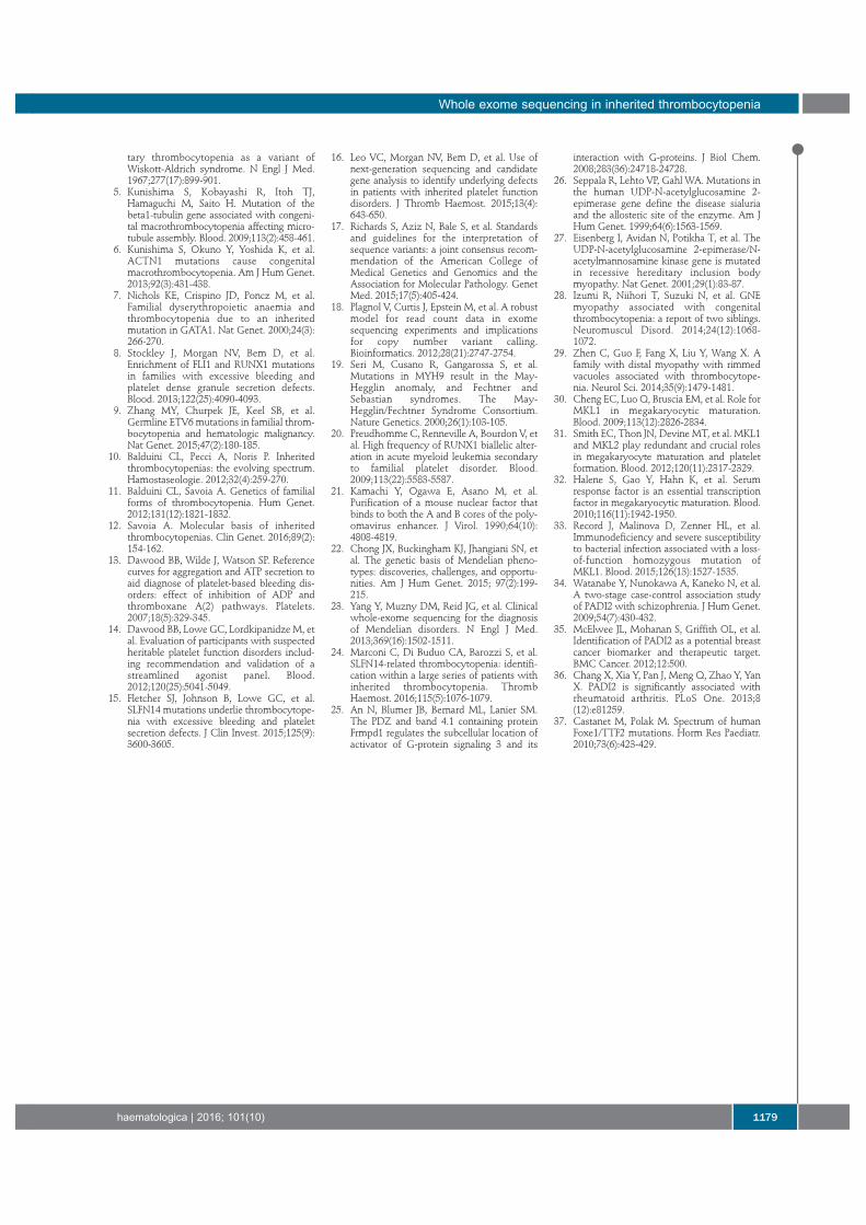
Of the 25 index cases with variants in known disease-causing genes, nine were observed to have variants withinthe RUNT1-related transcription factor gene; RUNX1.One variant, RUNX1; p.Arg177Gln, observed in F19.I andF19.II has been previously reported as a causative germlinemutation of a familial platelet disorder in two individualsfrom the same pedigree.20 The variations consisted of fivemissense variants, two splice-site variants and one non-sense variant. One splice-site variation, c.270+1G>T, waspresent within three affected individuals from two sepa-rate families (F13 and F14). All variants, with the exceptionof a missense substitution (p.D6N), lie within the geneticregion encoding the RUNT homology domain (RHD)which mediates DNA binding and heterodimerizationwith CBFβ (Figure 2).21 Platelets from the majority of thesepatients (10/13) demonstrated a reduction in ATP secre-tion and, in keeping with previous reports, several of thesepatients displayed additional clinical features. Variations inRUNX1 are associated with a propensity to myelodysplas-tic syndrome and acute myeloid leukemia. To date, hema-tologic malignancies have not been reported in anypatients; however, the brother of F16.1 did have a historyof acute myeloid leukemia but was unavailable for testing.
Potentially damaging variants in novel candidate genesAfter scrutinizing individuals for variants within the
panel of 358 platelet-associated genes (OnlineSupplementary Table S1), individuals without a variant in apreviously IT-associated gene were analyzed for variantsin novel genes. WES analysis revealed potentially damag-ing candidate variants in three families with currentlyunknown genetic etiology (Table 3). All candidate variantsare novel (excluding a previously annotated variant inMKL1; p.Val575Met, which occurs at a frequency of0.007718 within the ExAC consortium), segregate withthe disease status and have been confirmed by Sangersequencing.
Variants within ANKRD18A, GNE and FRMPD1 in tworelated individuals from consanguineous relationships
WES analysis of two related patients (F35.I and F35.II) ofSouth Asian ethnicity was approached differently to thatof other patients in this study. Both patients displayed asimilarly severe clinical phenotype with a significantreduction in circulating platelets (15x109/L). Platelet func-tion testing revealed a reduction in P-selectin (CD62P)expression upon stimulation and variable fluorescent fib-rinogen binding which was consistent across both affected
Whole exome sequencing in inherited thrombocytopenia
haematologica | 2016; 101(10) 1173
individuals. The patients were cousins born from consan-guineous relationships within a single consanguineouskindred so the analysis was focused on identification of ashared homozygous variant due to the recessive segrega-tion of disease. Three variants occurring withinANKRD18A; p.Glu799del, GNE; p.Gly447Arg andFRMPD1; p.Ala509Val were present in both affected indi-viduals and within a tightly linked region of homozygosi-ty on chromosome 9p. The variations within ANKRD18Aand GNE were novel according to the previously men-tioned databases whereas the variant in FRMPD1 has beenobserved at a frequency of 0.0003708 including 39 timeswithin the South Asian population (rs571037699). There isno ClinVar entry for this variant and all three variants areclassified as variants of “uncertain significance”.
One missense variant in the recently proposedinherited thrombocytopenia-linked gene, MKL1
One individual was shown to harbor a rare (frequency<0.01) missense variant within the MegakaryoblasticLeukaemia (translocation) 1 gene; MKL1. The variant wasthe only variant occurring within a gene of hemostatic rel-evance within 109 significant novel variants. The variant;
MKL1; c.1723G>A, p.Val575Met present in patient F37.Ihas been noted previously at a frequency of 0.0007718(allele count of 6/7774 in the ExAC consortium). Thepatient has a mild reduction in platelet count (130x109/L)and no secondary qualitative defects in platelet functionwere observed. The variant is classified as of “unknownsignificance”.
Novel missense candidate variants in PADI2 and TTF2Three affected individuals and four unaffected related
individuals of a large kindred were recruited to the study.Mild thrombocytopenia was observed within the familywith platelet counts ranging from 80x109/L to 186x109/L inthe three affected individuals. All three affected individu-als presented with a normal platelet size (7.9-8.6 fL) and amild reduction in secretion was observed in F30.I andF30.III but not in F30.II. All affected individuals shared asimilar bleeding phenotype, suffering from spontaneousepistaxis, excessive bruising and prolonged bleeding fromminor wounds. WES analysis revealed 14 novel or rare(frequency <0.01) variants shared between the threeaffected individuals. Sanger sequencing of all 14 variants infour unaffected related individuals narrowed down candi-
B. Johnson et al.
1174 haematologica | 2016; 101(10)
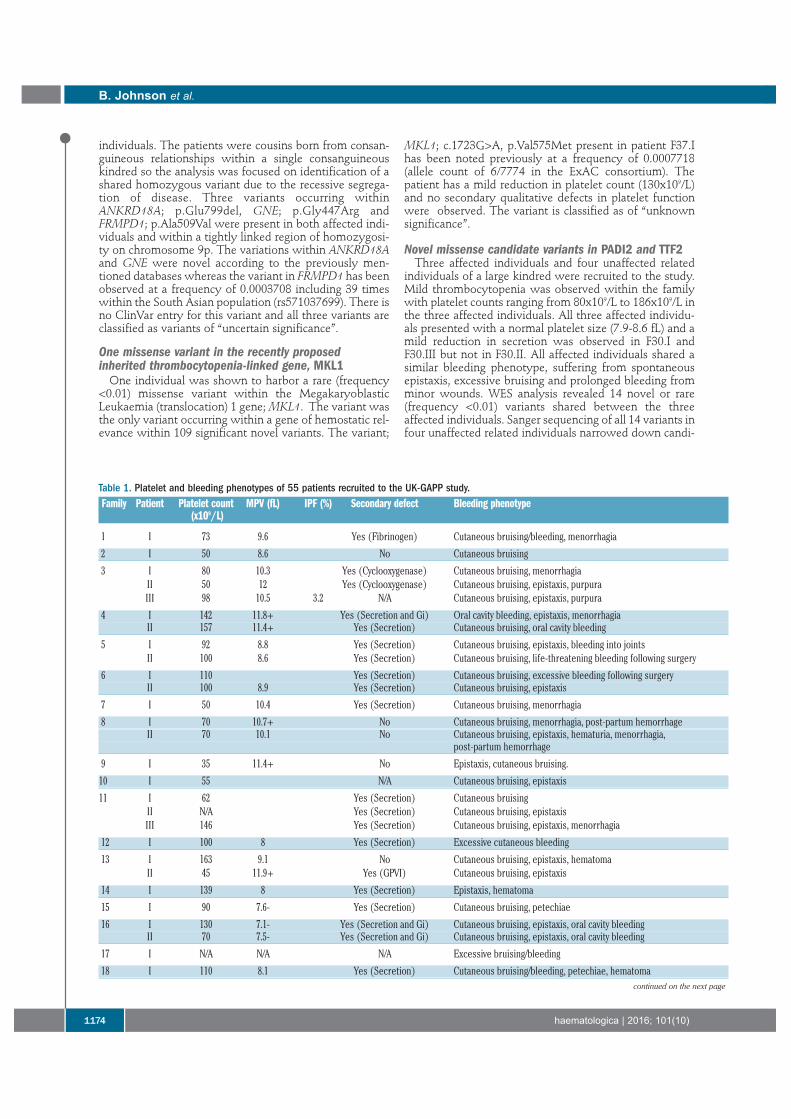
Table 1. Platelet and bleeding phenotypes of 55 patients recruited to the UK-GAPP study.Family Patient Platelet count MPV (fL) IPF (%) Secondary defect Bleeding phenotype
(x109/L)
1 I 73 9.6 Yes (Fibrinogen) Cutaneous bruising/bleeding, menorrhagia
2 I 50 8.6 No Cutaneous bruising
3 I 80 10.3 Yes (Cyclooxygenase) Cutaneous bruising, menorrhagiaII 50 12 Yes (Cyclooxygenase) Cutaneous bruising, epistaxis, purpuraIII 98 10.5 3.2 N/A Cutaneous bruising, epistaxis, purpura
4 I 142 11.8+ Yes (Secretion and Gi) Oral cavity bleeding, epistaxis, menorrhagiaII 157 11.4+ Yes (Secretion) Cutaneous bruising, oral cavity bleeding
5 I 92 8.8 Yes (Secretion) Cutaneous bruising, epistaxis, bleeding into jointsII 100 8.6 Yes (Secretion) Cutaneous bruising, life-threatening bleeding following surgery
6 I 110 Yes (Secretion) Cutaneous bruising, excessive bleeding following surgeryII 100 8.9 Yes (Secretion) Cutaneous bruising, epistaxis
7 I 50 10.4 Yes (Secretion) Cutaneous bruising, menorrhagia
8 I 70 10.7+ No Cutaneous bruising, menorrhagia, post-partum hemorrhageII 70 10.1 No Cutaneous bruising, epistaxis, hematuria, menorrhagia,
post-partum hemorrhage
9 I 35 11.4+ No Epistaxis, cutaneous bruising.
10 I 55 N/A Cutaneous bruising, epistaxis
11 I 62 Yes (Secretion) Cutaneous bruisingII N/A Yes (Secretion) Cutaneous bruising, epistaxisIII 146 Yes (Secretion) Cutaneous bruising, epistaxis, menorrhagia
12 I 100 8 Yes (Secretion) Excessive cutaneous bleeding
13 I 163 9.1 No Cutaneous bruising, epistaxis, hematomaII 45 11.9+ Yes (GPVI) Cutaneous bruising, epistaxis
14 I 139 8 Yes (Secretion) Epistaxis, hematoma
15 I 90 7.6- Yes (Secretion) Cutaneous bruising, petechiae
16 I 130 7.1- Yes (Secretion and Gi) Cutaneous bruising, epistaxis, oral cavity bleedingII 70 7.5- Yes (Secretion and Gi) Cutaneous bruising, epistaxis, oral cavity bleeding
17 I N/A N/A N/A Excessive bruising/bleeding
18 I 110 8.1 Yes (Secretion) Cutaneous bruising/bleeding, petechiae, hematomacontinued on the next page
dates to only two missense variants; PADI2 (p.Lys499Arg)and TTF2 (p.His1089Asp). Both variants segregate withdisease, not being present in the unaffected individuals.Both variants have been observed previously at a low fre-quency (<0.01) within the EXaC database (Table 3) and arecurrently classified as being of “uncertain significance”.
Discussion
Here we present the first, large-scale application of WESanalysis to patients with inherited bleeding diatheses pre-senting with thrombocytopenia of unknown etiology.
Platelet counts and phenotypic presentations varied con-siderably among the patients studied, which is consistentwith the variability observed in the spectrum of IT.However, the majority of patients (73%) were noted tohave a secondary qualitative defect in platelet functionwhich may explain the disproportionate bleeding whencompared to the patients’ platelet counts. A lack of consis-tency was noted in families 13 and 30, which apparentlyincluded affected individuals both with and withoutdefects in platelet function. Clinical complications areshared among the affected family members so this most
likely represents limitations in the sensitivity of plateletfunction testing or intra-familial variability.
Overall, when considering pathogenicity WES analysispositively predicted pathogenicity in 46% of index cases(17/37) (results classified as “pathogenic” or “likely patho-genic” in a gene consistent with the patients’ phenotypeand zygosity consistent with expected inheritance).Twenty-two percent of the index cases (8/37) had uncer-tain/possible pathogenicity (results classified as being of“uncertain significance” in known IT-causing genes). Theremaining 32% of index cases (12/37) had a negative pre-diction of pathogenicity (no convincing variants identifiedin known IT-causing genes). WES is not without its limita-tions and, as with any genetic analysis, all variants mustbe functionally confirmed as deleterious to the coded pro-tein. However, our positive variant discovery rate is com-parable to or exceeds the rates in previous large-scale WESclinical multicenter studies of Mendelian disorders.22,23
Focusing our genetic analysis on patients with unknownetiology of disease with minor prior genetic testing hasproduced a spectrum of variants different from that fromprevious, large-scale, targeted genetic studies of IT.Patients were recruited to the study with clinically diag-nosed bleeding disorders of unknown etiology. One
Whole exome sequencing in inherited thrombocytopenia
haematologica | 2016; 101(10) 1175
19 I 100 9 No Cutaneous bruising/bleedingII 100 9.2 No Cutaneous bruising/bleeding
20 I 89 13+ 17.5 Yes (Secretion and Gi) Cutaneous bruising
21 I 63 11.9 19.1 Yes (Secretion) Cutaneous bruising, epistaxis, hematomaII 83 11.9 24.3 Yes (Secretion) Cutaneous bruising, epistaxis, hematoma
22 I 74 11.2 Yes (Secretion and Gi) Cutaneous bruising/bleeding, hematuriaII 62 12.7+ 20.8 Yes (Secretion and Gi) Cutaneous bruising/bleeding, menorrhagia, post-partum hemorrhage,
hematomaIII 109 11 Yes (Secretion) Cutaneous bruising, hematoma, menorrhagia
23 I 119 11.1 Yes (Secretion) Cutaneous bruising/bleeding
24 I 104 9.6 No Menorrhagia, post-partum hemorrhageII 133 8.6 No Epistaxis
25 I 11 13.4+ Yes (Secretion and Gi) Cutaneous bruising
26 I 43 14+ No Cutaneous bruising, menorrhagia, oral cavity bleeding
27 I 100 10.3 Yes (Secretion) Cutaneous bruising/bleeding, epistaxis, oral cavity bleeding
28 I 25 8.5 Yes (Secretion) Cutaneous bruising
29 I 15 9.4 Yes (Secretion) Hematomas
30 I 137 7.9 Yes (Secretion) Cutaneous bruising, epistaxis, menorrhagiaII 186 8.6 No Cutaneous bruising, menorrhagia, hematuriaIII 80 8.2 Yes (Secretion) Cutaneous bruising
31 I 20 9.7 N/A Cutaneous bruising, epistaxis, oral cavity bleeding
32 I 15 9.5 20.2 No Cutaneous bruising
33 I 66 9.9 1.8 Yes (Secretion) Cutaneous bleeding
34 I 93 14.4+ 20.5 Yes (Secretion) Cutaneous bleeding, epistaxis
35 I 15 10.4 87 Yes (Secretion and other) Cutaneous bruising, epistaxis, hematomasII 14 15+ 83 Yes (Secretion and other) Cutaneous bleeding
36 I 104 13.3+ 17 No Menorrhagia
37 I 130 9.7 No Cutaneous bruising, epistaxis, menorrhagia
Average platelet count = 85x109/L (normal range 147-327x109/L, n=40). Average mean platelet volume (MPV) = 10 fL (normal range 7.8-12.69 fL, n=40). Immature platelet fraction (IPF)was available for 11 patients and varied between 1.8-87% (normal range 1.3-10.8%, n=40). Patients with an observed macro and micro thrombocytopenia are denoted by a + and -, respec-tively, following their most recent analyzed MPV. Secondary qualitative defects are abbreviated to the following; (Gi) - reduction in response upon ADP stimulation indicating a possible defectin the Gi pathway, (GPVI) – reduction in surface GPVI quantity. Each individual bleeding diathesis is summarized under bleeding phenotype.
continued from the previous page
caveat about this approach is the possible exclusion ofindividuals with known Bernard-Soulier syndrome orMYH9-related disorders as these two forms of IT are rou-tinely tested for in many hematology centers in the UK.However, three index cases with variants in either GP1BAor MYH9 were noted in our analysis; these patients hadatypical presentations of Bernard-Soulier syndrome orMYH9-related disorder and were, therefore, potentiallyfalsely-negatively reported cases. The individuals withvariants within GP1BA and MYH9 showed a slightincrease in mean platelet volume; however, this was not atthe magnitude of giant platelets normally attributed to thisgroup of disorders and only patient F9.I showed any sec-ondary syndromic symptoms with the individual suffer-ing from congenital cataracts.
One attribute of excluding patients with known variantsin GP1BA, GP1BB, MYH9 and potentially GP9 was thediscovery of a relatively large percentage of individualsanalyzed (24% of index cases) with variants in RUNX1 asa primary likely cause of disease. With the exception ofone predicted loss-of-function variant, the variants presentwithin RUNX1 are currently classified as either “likely
pathogenic” or of “uncertain significance” and need func-tional confirmation to be considered the cause of disease.However, the presence of these variants in a large numberof individuals with an often shared secondary functionaldefect in secretion does suggest that the prevalence ofRUNX1 variants may be higher than previously thought.This raises the issue of whether they should be consideredas clinically significant as Bernard-Soulier syndrome andMYH9-related disorders and be searched for in a primarygenetic screening at the initial diagnosis of IT.
An advantage of using WES is the possibility of findingcandidate variations in novel genes in subjects who do notpossess variants in known IT-causing genes. The determi-nation of whether these candidate variants are in factpathogenic relies on functional confirmation of the delete-rious effect of the variant. However, WES analysis, espe-cially with combined segregation analysis by Sangersequencing in extensive kindreds, can provide indicationsas to which variants may be of scientific and clinical rele-vance. This strategy has recently been utilized in the dis-covery of novel candidate variations in SLFN14 initially aspart of the GAPP study.15,24
B. Johnson et al.
1176 haematologica | 2016; 101(10)
Table 2. Results of whole exome sequencing analysis of 55 patients with inherited thrombocytopenia showing variants in known thrombocytopenia-caus-ing genes. 68% of individuals have a predicted genetic etiology in a previously IT-associated gene. When a variant has been previously observed it isannotated in the prevalence column with the database in which it is included. The ACMG consensus guideline results are also displayed in the finalclassification column.17
Family Patient Gene(s) Genomic variation Protein effect Variation type Prevalence Classification
1 I ACTN1 c.2647G>C p.Gly883Arg Missense Novel Likely pathogenic2 I ANKRD26 c.-126T>G 5'-UTR variation Known Pathogenic3 I CYCS c.155C>T p.Ala52Val Missense Novel Likely pathogenic
II CYCS c.155C>T p.Ala52Val Missense NovelIII CYCS c.155C>T p.Ala52Val Missense Novel
4 I FLI1 c.992_995del p.Asn331Thr fs*4 Frameshift deletion Novel PathogenicII FLI1 c.992_995del p.Asn331Thr fs*4 Frameshift deletion Novel
5 I FLI1 c.1028A>G p.Tyr343Cys Missense Novel Likely pathogenicII FLI1 c.1028A>G p.Tyr343Cys Missense Novel
6 I GFI1B c.814+1G>A Splicing Novel Likely pathogenicSTIM1 c.1828G>A p.Ala610Thr Missense 0.00019 (1k) Uncertain significance
II GFI1B c.814+1G>A Splicing Novel Likely pathogenicSTIM1 c.1828G>A p.Ala610Thr Missense 0.00019 (1k) Uncertain significance
7 I GP1BA c.1761A>C p.Gln587His Missense 0.00043 (EXaC) (rs570515282) Uncertain significance8 I GP1BA c.413G>T p.Gly138Val Missense Novel Likely pathogenic
II GP1BA c.413G>T p.Gly138Val Missense Novel9 I MYH9 c.3493C>T p.Arg1165Cys Missense Known (rs80338829) Pathogenic10 I NBEAL2 c.1376delT p.Leu459Arg fs*13 Frameshift deletion Novel Likely pathogenic
NBEAL2 c.6893A>G p.Asn2298Ser Missense Novel Likely pathogenic11 I RUNX1 c.16G>A p.Asp6Asn Missense Novel Likely pathogenic
II RUNX1 c.16G>A p.Asp6Asn Missense NovelIII RUNX1 c.16G>A p.Asp6Asn Missense Novel
12 I RUNX1 c.236G>A p.Trp79* Nonsense Novel PathogenicITGB3 c.349C>T p.Arg117Trp Missense Novel Uncertain significance
13 I RUNX1 c.270+1G>T Splicing Novel Likely pathogenicII RUNX1 c.270+1G>T Splicing Novel
14 I RUNX1 c.270+1G>T Splicing Novel15 I RUNX1 c.322G>A p.Gly108Ser Missense Novel Uncertain significance16 I RUNX1 c.427+1G>T Splicing Novel Likely pathogenic
II RUNX1 c.427+1G>T Splicing Novelcontinued on the next page
Family 35 is an interesting case of two affected relatedindividuals born from consanguineous relationships. Themolecular function of ANKRD18A is currently unknown,while FRMPD1 regulates the subcellular localization ofactivator of G-protein signaling 3 (AGS3).25 Both genes areexpressed weakly in hematopoietic cells. However, GNE,coding for an enzyme in the sialic acid biosynthetic path-way, is expressed in all cells of the hematopoietic lineage.There are currently 88 registered mutations in GNE in theHuman Genome Mutation Database(www.hgmd.cf.ac.uk). Mutations are known to be thegenetic cause of sialuria (OMIM269921) and hereditaryinclusion body myopathy (OMIM600737).26,27 Recently,two separate groups have reported patients with com-pound heterozygous variations in GNE, causing GNE-related myopathy with congenital thrombocytopenia.28,29
The platelet counts of the four reported affected individu-als were below 45x109/L; platelet volume measurementswere not recorded. None of the patients displayed signs ofmyopathy until mid-adolescence/early adulthood; F35.Iand F35.II are currently aged 10 and 6, respectively.Without functional characterization of the effects of eachvariation, we cannot definitively conclude the genetic eti-ology of these two individuals’ severe thrombocytopenia.However, WES analysis has allowed us to focus our effortson three potentially pathogenic variants in novel genes.
MKL1 was initially included in our panel of 358 genesfor post-WES analysis due to its role in megakaryocytematuration elucidated via its binding partner, serumresponse factor (SRF).30-32 Recently, the first case of ahomozygous mutation in MKL1 in a patient with severeimmunodeficiency and no hematologic malignancies wasreported.33 One interesting phenotypic presentation of theaffected individual was an intermittent mild thrombocy-topenia with low platelet counts in whole blood ofbetween 50x109/L and 150x109/L. Here we present a novelvariant within MKL1, at a highly conserved genetic site.The missense variant observed in F37.I represents the onlyvariant to occur in a gene with previous hematologicimplications. One further variant in MKL1 was observedin addition to a “likely pathogenic” frameshift causinginsertion within TUBB1 in patient F25.I. Due to the pre-dicted loss of function of the frameshift causing theTUBB1 variant it is unlikely that the variant with MKL1 isadditive to the phenotype of patient F25.I. However, thevariant of uncertain significance in patient F37.I is an inter-esting candidate to take forward for functional studies.
WES and segregation determination using Sangersequencing revealed candidate variants in PADI2 andTTF2 that segregate with disease in F30.I, F30.II andF30.III. The phenotypic presentations vary between thepatients but clinical presentations are consistent, which
Whole exome sequencing in inherited thrombocytopenia
haematologica | 2016; 101(10) 1177
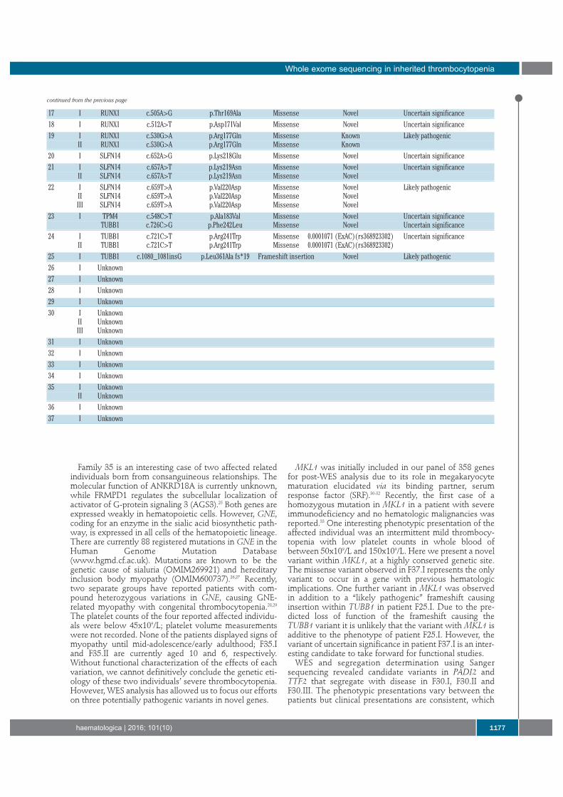
17 I RUNX1 c.505A>G p.Thr169Ala Missense Novel Uncertain significance18 I RUNX1 c.512A>T p.Asp171Val Missense Novel Uncertain significance19 I RUNX1 c.530G>A p.Arg177Gln Missense Known Likely pathogenic
II RUNX1 c.530G>A p.Arg177Gln Missense Known20 I SLFN14 c.652A>G p.Lys218Glu Missense Novel Uncertain significance21 I SLFN14 c.657A>T p.Lys219Asn Missense Novel Uncertain significance
II SLFN14 c.657A>T p.Lys219Asn Missense Novel22 I SLFN14 c.659T>A p.Val220Asp Missense Novel Likely pathogenic
II SLFN14 c.659T>A p.Val220Asp Missense NovelIII SLFN14 c.659T>A p.Val220Asp Missense Novel
23 I TPM4 c.548C>T p.Ala183Val Missense Novel Uncertain significanceTUBB1 c.726C>G p.Phe242Leu Missense Novel Uncertain significance
24 I TUBB1 c.721C>T p.Arg241Trp Missense 0.0001071 (ExAC)(rs368923302) Uncertain significanceII TUBB1 c.721C>T p.Arg241Trp Missense 0.0001071 (ExAC)(rs368923302)
25 I TUBB1 c.1080_1081insG p.Leu361Ala fs*19 Frameshift insertion Novel Likely pathogenic26 I Unknown27 I Unknown28 I Unknown29 I Unknown30 I Unknown
II UnknownIII Unknown
31 I Unknown32 I Unknown33 I Unknown34 I Unknown35 I Unknown
II Unknown36 I Unknown37 I Unknown
continued from the previous page
may reflect limitations in the sensitivity of platelet func-tion testing. Neither gene has previously been implicatedin hematologic abnormalities: mutations in PADI2 havebeen associated with schizophrenia, breast cancer andrheumatoid arthritis, while mutations in TTF2 have beenassociated with thyroid dysgenesis.34-37 WES analysis hastherefore provided us with the first steps for determiningthe impact of these two variants of uncertain significanceand whether they have the propensity to be disease caus-ing.
In summary, we show that WES can be applied to iden-tify the underlying genetic cause in known IT-causinggenes for patients with thrombocytopenia and unclear dis-ease etiology. We show similar positive detection rateswhen compared to prior targeted studies and, with theaddition of complementary functional studies, show animproved detection rate when compared to WES analysisof other developmental disorders. We also suggest theapplicability of WES in providing preliminary insight intonovel genes and their potential mechanism of actionthrough candidate variations of unknown significance.
This approach provides a foundation to enhance our cur-rent knowledge on megakaryopoiesis, platelet functionand platelet senescence/death through subsequent func-tional studies.
AcknowledgmentsWe thank the families for providing samples and our clinical
and laboratory colleagues for their help. This work was supportedby the British Heart Foundation (RG/PG/13/36/30275;RG/09/007), an MRC Doctoral Training Partnership grant (BJ),a Wellcome Trust Combined Training Programme Fellowship(093994) (GCL), the Healing Foundation (PH) and the PlateletCharity. We thank the NIHR Haematology Specialty Group fortheir help in recruiting to the study, and all our clinical investiga-tors and collaborators. The authors also acknowledge supportfrom the Department of Health via the National Institute forHealth Research (NIHR) Comprehensive Biomedical ResearchCentre Award to Guy's & St Thomas' NHS Foundation Trust inpartnership with King's College London and King's CollegeHospital NHS Foundation Trust. We thank the Queen ElizabethHospital Charity for funding the Sysmex XN-1000.
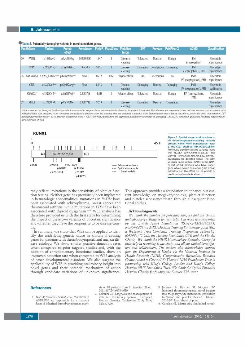
Table 3. Potentially damaging variants in novel candidate genes.FamilyGene Variant Protein Prevalence PhyloP PhastCons Mutation SIFT Provean PolyPhen-2 ACMG Classification
effect taster
30 PADI2 c.1496A>G p.Lys499Arg 0.000008681 1.647 1 Diseas-e Tolerated Neutral Benign PM Uncertaincausing (segregation) significance
TTF2 c.3265C>G p.His1089Asp 1.65E-05 5.131 1 Disease- Damaging Deleterious Damaging PM Uncertaincausing (segregation) , PP3 significance
35 ANKRD18A c.2395_2397delhom p.Glu799delhom Novel 0.772 0.965 Polymorphism NA Deleterious NA PM2, UncertainPP (segregation), PM6 significance
GNE c.1339G>Ahom p.Gly447Arghom Novel 5.343 1 Disease- Damaging Neutral Damaging PM2, Uncertaincausing PP (segregation), PM6 sgnificance
FRMPD1 c.1526C>Thom p.Ala509Valhom 0.0003708 -1.459 0 Polymorphism Tolerated Neutral Benign PP (segregation), UncertainPM6 significance
37 MKL1 c.1723G>A p.Val575Met 0.0007718 3.358 1 Disease- Damaging Neutral Damaging Uncertaincausing significance
When a variant has been previously observed it is annotated in the prevalence column with the database in which it is included. PhyloP scores vary between -14 and +6 and measure conservation at eachindividual base, sites predicted to be conserved are assigned a positive score, fast evolving sites are assigned a negative score. Mutationtaster uses a Bayes classifier to predict the effect of a mutation. SIFTdamaging prediction score= <0.05. Provean deleterious score = <-2.5. PolyPhen-2 predictions are appraised qualitatively as benign or damaging. The ACMG consensus guidelines, including supporting evi-dence, are also shown.
Figure 2. Spatial amino acid locations ofall thrombocytopenia-causing variantspresent within RUNT transcription factor1 (RUNX1) (RefSeq NP_001001890).Previously disease-causing variants foundthe HGMD (www.hgmd.cf.ac.uk) andClinVar (www.ncbi.nlm.nih.gov/clinvar/)databases are denoted above. The eightvariants found within RUNX1 in the GAPPcohort of 54 patients who have under-gone whole exome sequencing are denot-ed below and the effect on the protein orpredicted splice-site is shown.
References
1. Noris P, Perrotta S, Seri M, et al. Mutations inANKRD26 are responsible for a frequentform of inherited thrombocytopenia: analy-
sis of 78 patients from 21 families. Blood.2011;117(24):6673-6680.
2. Balduini CL. Diagnosis and management ofinherited thrombocytopenias. EuropeanHuman Genetics Conference 2014; 2014;Milan, Italy.
3. Johnson B, Fletcher SF, Morgan NV.Inherited thrombocytopenia: novel insightsinto megakaryocyte maturation, proplateletformation and platelet lifespan. Platelets.2016:1-7. Epub ahead of print.
4. Canales ML, Mauer AM. Sex-linked heredi-
B. Johnson et al.
1178 haematologica | 2016; 101(10)
tary thrombocytopenia as a variant ofWiskott-Aldrich syndrome. N Engl J Med.1967;277(17):899-901.
5. Kunishima S, Kobayashi R, Itoh TJ,Hamaguchi M, Saito H. Mutation of thebeta1-tubulin gene associated with congeni-tal macrothrombocytopenia affecting micro-tubule assembly. Blood. 2009;113(2):458-461.
6. Kunishima S, Okuno Y, Yoshida K, et al.ACTN1 mutations cause congenitalmacrothrombocytopenia. Am J Hum Genet.2013;92(3):431-438.
7. Nichols KE, Crispino JD, Poncz M, et al.Familial dyserythropoietic anaemia andthrombocytopenia due to an inheritedmutation in GATA1. Nat Genet. 2000;24(3):266-270.
8. Stockley J, Morgan NV, Bem D, et al.Enrichment of FLI1 and RUNX1 mutationsin families with excessive bleeding andplatelet dense granule secretion defects.Blood. 2013;122(25):4090-4093.
9. Zhang MY, Churpek JE, Keel SB, et al.Germline ETV6 mutations in familial throm-bocytopenia and hematologic malignancy.Nat Genet. 2015;47(2):180-185.
10. Balduini CL, Pecci A, Noris P. Inheritedthrombocytopenias: the evolving spectrum.Hamostaseologie. 2012;32(4):259-270.
11. Balduini CL, Savoia A. Genetics of familialforms of thrombocytopenia. Hum Genet.2012;131(12):1821-1832.
12. Savoia A. Molecular basis of inheritedthrombocytopenias. Clin Genet. 2016;89(2):154-162.
13. Dawood BB, Wilde J, Watson SP. Referencecurves for aggregation and ATP secretion toaid diagnose of platelet-based bleeding dis-orders: effect of inhibition of ADP andthromboxane A(2) pathways. Platelets.2007;18(5):329-345.
14. Dawood BB, Lowe GC, Lordkipanidze M, etal. Evaluation of participants with suspectedheritable platelet function disorders includ-ing recommendation and validation of astreamlined agonist panel. Blood.2012;120(25):5041-5049.
15. Fletcher SJ, Johnson B, Lowe GC, et al.SLFN14 mutations underlie thrombocytope-nia with excessive bleeding and plateletsecretion defects. J Clin Invest. 2015;125(9):3600-3605.
16. Leo VC, Morgan NV, Bem D, et al. Use ofnext-generation sequencing and candidategene analysis to identify underlying defectsin patients with inherited platelet functiondisorders. J Thromb Haemost. 2015;13(4):643-650.
17. Richards S, Aziz N, Bale S, et al. Standardsand guidelines for the interpretation ofsequence variants: a joint consensus recom-mendation of the American College ofMedical Genetics and Genomics and theAssociation for Molecular Pathology. GenetMed. 2015;17(5):405-424.
18. Plagnol V, Curtis J, Epstein M, et al. A robustmodel for read count data in exomesequencing experiments and implicationsfor copy number variant calling.Bioinformatics. 2012;28(21):2747-2754.
19. Seri M, Cusano R, Gangarossa S, et al.Mutations in MYH9 result in the May-Hegglin anomaly, and Fechtner andSebastian syndromes. The May-Hegglin/Fechtner Syndrome Consortium.Nature Genetics. 2000;26(1):103-105.
20. Preudhomme C, Renneville A, Bourdon V, etal. High frequency of RUNX1 biallelic alter-ation in acute myeloid leukemia secondaryto familial platelet disorder. Blood.2009;113(22):5583-5587.
21. Kamachi Y, Ogawa E, Asano M, et al.Purification of a mouse nuclear factor thatbinds to both the A and B cores of the poly-omavirus enhancer. J Virol. 1990;64(10):4808-4819.
22. Chong JX, Buckingham KJ, Jhangiani SN, etal. The genetic basis of Mendelian pheno-types: discoveries, challenges, and opportu-nities. Am J Hum Genet. 2015; 97(2):199-215.
23. Yang Y, Muzny DM, Reid JG, et al. Clinicalwhole-exome sequencing for the diagnosisof Mendelian disorders. N Engl J Med.2013;369(16):1502-1511.
24. Marconi C, Di Buduo CA, Barozzi S, et al.SLFN14-related thrombocytopenia: identifi-cation within a large series of patients withinherited thrombocytopenia. ThrombHaemost. 2016;115(5):1076-1079.
25. An N, Blumer JB, Bernard ML, Lanier SM.The PDZ and band 4.1 containing proteinFrmpd1 regulates the subcellular location ofactivator of G-protein signaling 3 and its
interaction with G-proteins. J Biol Chem.2008;283(36):24718-24728.
26. Seppala R, Lehto VP, Gahl WA. Mutations inthe human UDP-N-acetylglucosamine 2-epimerase gene define the disease sialuriaand the allosteric site of the enzyme. Am JHum Genet. 1999;64(6):1563-1569.
27. Eisenberg I, Avidan N, Potikha T, et al. TheUDP-N-acetylglucosamine 2-epimerase/N-acetylmannosamine kinase gene is mutatedin recessive hereditary inclusion bodymyopathy. Nat Genet. 2001;29(1):83-87.
28. Izumi R, Niihori T, Suzuki N, et al. GNEmyopathy associated with congenitalthrombocytopenia: a report of two siblings.Neuromuscul Disord. 2014;24(12):1068-1072.
29. Zhen C, Guo F, Fang X, Liu Y, Wang X. Afamily with distal myopathy with rimmedvacuoles associated with thrombocytope-nia. Neurol Sci. 2014;35(9):1479-1481.
30. Cheng EC, Luo Q, Bruscia EM, et al. Role forMKL1 in megakaryocytic maturation.Blood. 2009;113(12):2826-2834.
31. Smith EC, Thon JN, Devine MT, et al. MKL1and MKL2 play redundant and crucial rolesin megakaryocyte maturation and plateletformation. Blood. 2012;120(11):2317-2329.
32. Halene S, Gao Y, Hahn K, et al. Serumresponse factor is an essential transcriptionfactor in megakaryocytic maturation. Blood.2010;116(11):1942-1950.
33. Record J, Malinova D, Zenner HL, et al.Immunodeficiency and severe susceptibilityto bacterial infection associated with a loss-of-function homozygous mutation ofMKL1. Blood. 2015;126(13):1527-1535.
34. Watanabe Y, Nunokawa A, Kaneko N, et al.A two-stage case-control association studyof PADI2 with schizophrenia. J Hum Genet.2009;54(7):430-432.
35. McElwee JL, Mohanan S, Griffith OL, et al.Identification of PADI2 as a potential breastcancer biomarker and therapeutic target.BMC Cancer. 2012;12:500.
36. Chang X, Xia Y, Pan J, Meng Q, Zhao Y, YanX. PADI2 is significantly associated withrheumatoid arthritis. PLoS One. 2013;8(12):e81259.
37. Castanet M, Polak M. Spectrum of humanFoxe1/TTF2 mutations. Horm Res Paediatr.2010;73(6):423-429.
Whole exome sequencing in inherited thrombocytopenia
haematologica | 2016; 101(10) 1179


