Article a b a - SciELO
23
Article J. Braz. Chem. Soc., Vol. 21, No. 10, 1819-1824, 2010. Printed in Brazil - ©2010 Sociedade Brasileira de Química 0103 - 5053 $6.00+0.00 *e-mail: [email protected] 3’,8’’-Biisokaempferide, a Cytotoxic Biflavonoid and Other Chemical Constituents of Nanuza plicata (Velloziaceae) Meri Emili F. Pinto, a Marcelo Sobral da Silva,* ,a Elisabete Schindler, b José Maria Barbosa Filho, a Ramon dos Santos El-Bachá, b Marianna Vieira S. Castello-Branco, a Maria de Fatima Agra a and Josean Fechine Tavares a a Laboratório de Tecnologia Farmacêutica, Universidade Federal da Paraíba, CP 5009, 58051-970 João Pessoa-PB, Brazil b Laboratório de Neuroquímica e Biologia Celular (LabNq), Instituto de Ciências da Saúde (ICS), Universidade Federal da Bahia, 40110-902 Salvador-BA, Brazil Foi isolado das folhas de Nanuza plicata um novo biflavonóide, chamado 3’,8’’-biisocampferideo (1), juntamente com os conhecidos compostos amentoflavona (2), ácido patagônico (3), (4aR,5S,6R,8aR)-5-[2-(2,5-dihidro-5-metóxi-2-oxofuran-3-il)etil]-3,4,4a,5,6,7,8, 8a-octahidro-5,6,8a-trimetilnaftaleno-1-ácido carboxílico (4), cafeoilquinato de metila (5), ácido 3,5-di-cafeoilquínico (6) e luteolina (7). Os compostos 3, 4, 5 e 6 são relatados pela primeira vez em Velloziaceae. As estruturas dos compostos foram elucidadas com base em métodos espectroscópicos, especialmente RMN e EM. A citotoxicidade de 3’,8’’-biisocampferideo foi estudada em células de glioblastoma humano (GL-15). A concentração efetiva, que produziu morte em 50% das células após 72 h foi 36,5 μmol L -1 . Alterações na morfologia celular, incluindo retração e degradação de citoplasma, foram observadas quando as células foram tratadas com concentrações a partir de 20 μmol L -1 de 3’,8’’-biisocampferideo por 72 h. A novel biflavonoid, named 3’,8’’-biisokaempferide (1), along with the known compounds amentoflavone (2), patagonic acid (3), (4aR,5S,6R,8aR)-5-[2-(2,5-Dihydro-5-methoxy-2-oxofuran- 3-yl)ethyl]-3,4,4a,5,6,7,8,8a-octahydro-5,6,8a-trimethylnaphthalene-1-carboxylic acid (4), 5-caffeoylquinic acid methyl ester (5), 3,5-di-caffeoylquinic acid (6) and luteolin (7), were isolated of the leaves from Nanuza plicata. The compounds 3, 4, 5 and 6 are reported for the first time in Velloziaceae. The structures of the compounds were elucidated on the basis of spectroscopic methods, especially NMR and MS analyses. The cytotoxicity of 3’,8’’-biisokaempferide was studied in cultures of human glioblastoma GL-15 cells. The effective concentration, which killed 50 % of cells after 72 h was 36.5 μmol L -1 . Changes in cellular morphology, including retraction and degradation of cytoplasm, were observed when cells were treated with concentrations from 20 μmol L -1 of 3’,8’’-biisokaempferide for 72 h. Keywords: 3’,8’’-biisokaempferide, Nanuza plicata, cytotoxicity, Velloziaceae Introduction Velloziaceae is a family of monocotyledonous plants containing about 270 species of the tropical Brazilian flora occurring in rocky fields. 1 These plants live under conditions of high solar irradiation and low water availability. 2 Species of this family contain high amounts of diterpenes with a skeleton of the clerodane, 2 cleisthantane, isopimarane, 3 kaurane, 4 and labdane 5 types, besides flavonoids monoisoprenylated, C-alkylated and biflavonoids. 6-9 The genus Nanuza comprises three species, N. plicata, N. almeidae and N. luetzelburgii . Nanuza plicata (Mart.) L. B. Sm. & Ayensu (Synonymy: Vellozia plicata and Xerophyta plicata), is known commonly as “canela d’ema”. In earlier studies the compounds 3-geranyl-4-hydroxybenzoate 10-11 and the biflavonoid amentoflavone, 12 were isolated from species collected in the States of Rio de Janeiro and Minas Gerais, Brazil, respectively. In this work, we report the isolation and structure elucidation of the following compounds: two
Transcript of Article a b a - SciELO

Article J. Braz. Chem. Soc., Vol. 21, No. 10, 1819-1824, 2010.
Printed in Brazil - ©2010 Sociedade Brasileira de Química0103 - 5053 $6.00+0.00
*e-mail: [email protected]
3’,8’’-Biisokaempferide, a Cytotoxic Biflavonoid and Other Chemical Constituents of Nanuza plicata (Velloziaceae)
Meri Emili F. Pinto,a Marcelo Sobral da Silva,*,a Elisabete Schindler,b José Maria Barbosa Filho,a Ramon dos Santos El-Bachá,b Marianna Vieira S. Castello-Branco,a
Maria de Fatima Agraa and Josean Fechine Tavaresa
aLaboratório de Tecnologia Farmacêutica, Universidade Federal da Paraíba, CP 5009, 58051-970 João Pessoa-PB, Brazil
bLaboratório de Neuroquímica e Biologia Celular (LabNq), Instituto de Ciências da Saúde (ICS), Universidade Federal da Bahia, 40110-902 Salvador-BA, Brazil
Foi isolado das folhas de Nanuza plicata um novo biflavonóide, chamado 3’,8’’-biisocampferideo (1), juntamente com os conhecidos compostos amentoflavona (2), ácido patagônico (3), (4aR,5S,6R,8aR)-5-[2-(2,5-dihidro-5-metóxi-2-oxofuran-3-il)etil]-3,4,4a,5,6,7,8, 8a-octahidro-5,6,8a-trimetilnaftaleno-1-ácido carboxílico (4), cafeoilquinato de metila (5), ácido 3,5-di-cafeoilquínico (6) e luteolina (7). Os compostos 3, 4, 5 e 6 são relatados pela primeira vez em Velloziaceae. As estruturas dos compostos foram elucidadas com base em métodos espectroscópicos, especialmente RMN e EM. A citotoxicidade de 3’,8’’-biisocampferideo foi estudada em células de glioblastoma humano (GL-15). A concentração efetiva, que produziu morte em 50% das células após 72 h foi 36,5 μmol L-1. Alterações na morfologia celular, incluindo retração e degradação de citoplasma, foram observadas quando as células foram tratadas com concentrações a partir de 20 μmol L-1 de 3’,8’’-biisocampferideo por 72 h.
A novel biflavonoid, named 3’,8’’-biisokaempferide (1), along with the known compounds amentoflavone (2), patagonic acid (3), (4aR,5S,6R,8aR)-5-[2-(2,5-Dihydro-5-methoxy-2-oxofuran-3-yl)ethyl]-3,4,4a,5,6,7,8,8a-octahydro-5,6,8a-trimethylnaphthalene-1-carboxylic acid (4), 5-caffeoylquinic acid methyl ester (5), 3,5-di-caffeoylquinic acid (6) and luteolin (7), were isolated of the leaves from Nanuza plicata. The compounds 3, 4, 5 and 6 are reported for the first time in Velloziaceae. The structures of the compounds were elucidated on the basis of spectroscopic methods, especially NMR and MS analyses. The cytotoxicity of 3’,8’’-biisokaempferide was studied in cultures of human glioblastoma GL-15 cells. The effective concentration, which killed 50 % of cells after 72 h was 36.5 μmol L-1. Changes in cellular morphology, including retraction and degradation of cytoplasm, were observed when cells were treated with concentrations from 20 μmol L-1 of 3’,8’’-biisokaempferide for 72 h.
Keywords: 3’,8’’-biisokaempferide, Nanuza plicata, cytotoxicity, Velloziaceae
Introduction
Velloziaceae is a family of monocotyledonous plants containing about 270 species of the tropical Brazilian flora occurring in rocky fields.1 These plants live under conditions of high solar irradiation and low water availability.2 Species of this family contain high amounts of diterpenes with a skeleton of the clerodane,2 cleisthantane, isopimarane,3 kaurane,4 and labdane5 types,
besides flavonoids monoisoprenylated, C-alkylated and biflavonoids.6-9 The genus Nanuza comprises three species, N. plicata, N. almeidae and N. luetzelburgii. Nanuza plicata (Mart.) L. B. Sm. & Ayensu (Synonymy: Vellozia plicata and Xerophyta plicata), is known commonly as “canela d’ema”. In earlier studies the compounds 3-geranyl-4-hydroxybenzoate10-11 and the biflavonoid amentoflavone,12 were isolated from species collected in the States of Rio de Janeiro and Minas Gerais, Brazil, respectively. In this work, we report the isolation and structure elucidation of the following compounds: two
3’,8’’-Biisokaempferide, a Cytotoxic Biflavonoid and Other Chemical Constituents J. Braz. Chem. Soc.1820
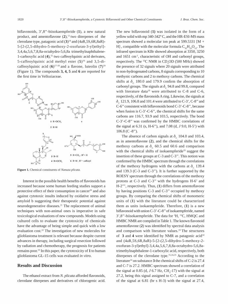
biflavonoids, 3’,8’’-biisokaempferide (1), a new natural product, and amentoflavone (2),13 two diterpenes of the clerodane type, patagonic acid (3)14 and (4aR,5S,6R,8aR)-5-[2-(2,5-dihydro-5-methoxy-2-oxofuran-3-yl)ethyl]-3,4,4a,5,6,7,8,8a-octahydro-5,6,8a trimethylnaphthalene-1-carboxylic acid (4),15 two caffeoylquinic acid derivates, 5-caffeoylquinic acid methyl ester (5)16 and 3,5-di-caffeoylquinic acid (6)17-18 and a flavone, luteolin (7)19 (Figure 1). The compounds 3, 4, 5 and 6 are reported for the first time in Velloziaceae.
Interest in the possible health benefits of flavonoids has increased because some human feeding studies support a protective effect of their consumption in cancer20 and also against cytotoxic insults induced by oxidative stress and amyloid b suggesting their therapeutic potential against neurodegenerative diseases.21 The replacement of animal techniques with non-animal ones is imperative in safe toxicological evaluations of new compounds. Models using cultured cells to evaluate the cytotoxicity of chemicals have the advantage of being simple and quick with a low evaluation cost.22 The investigation of new molecules for glioblastoma treatment is relevant because despite current advances in therapy, including surgical resection followed by radiation and chemotherapy, the prognosis for patients remains poor.23 In this paper, the cytotoxicity of 1 to human glioblastoma GL-15 cells was evaluated in vitro.
Results and Discussion
The ethanol extract from N. plicata afforded flavonoids, clerodane diterpenes and derivatives of chlorogenic acid.
The new biflavonoid (1) was isolated in the form of a yellow solid with mp 340-342º C, and the HR-ESI-MS mass spectrum showed a molecular ion peak at 599.5331 [M + H] , compatible with the molecular formula C
32H
23O
12. The
infrared spectrum in KBr showed absorption at 3350, 3250 and 1651 cm-1, characteristic of OH and carbonyl groups, respectively. The 13C NMR in CD
3OD (500 MHz) showed
the presence of 32 signals where 20 signals were attributed to non-hydrogenated carbons, 8 signals corresponding to 10 methynic carbons and 2 to methoxy carbons. The chemical shifts at d
C 180.0 and 179.9 confirm the absorption of
carbonyl groups. The signals at dC 94.8 and 99.8, compared
with literature data19 were attributed to C-8 and C-6, respectively, of the flavonoids A ring. Likewise, the signals at d
C 121.9, 106.8 and 101.4 were attributed to C-3’, C-8’’ and
C-6’’ consistent with biflavonoids bond C-3’-C-8’’, because when fusion is C-3’-C-6’’, the chemical shifts for the same carbons are 116.7, 93.9 and 103.5, respectively. The bond C-3’-C-8’’ was confirmed by the HMBC correlations of the signal at 6.31 (s, H-6’’), and 7.08 (d, J 9.0, H-5’) with 106.8 (C -8’’).
The absence of carbon signals at dC 104.0 and 103.4,
as in amentoflavone (2), and the chemical shifts for the methoxy carbons at d
C 60.5 and 60.6 and comparison
with the chemical shifts of isokaempferide19 suggest the insertion of these groups at C-3 and C-3’’. This notion was confirmed by the HMBC spectrum through the correlations of the methoxy hydrogens with the carbons at d
C 139.4
and 139.3 (C-3 and C-3’’). It is further supported by the ROESY spectrum through the correlations of the methoxy protons at C-3 and C-3’’ with the hydrogens H-6’ and H-2’’’, respectively. Thus, (1) differs from amentoflavone by having positions C-3 and C-3’’ occupied by methoxy groups. By comparing the chemical shifts of monomeric units of (1) with the literature could be characterized them as units isokaempferide. Therefore, (1) is a new biflavonoid with union C-3’-C-8’’ of isokaempferide, named 3’,8’’-biisokaempferide. The data for 1H, 13C, HMQC and HMBC NMR are compiled in Table 1. The known flavonoid amentoflavone (2) was identified by spectral data analysis and comparison with literature values.19 The structures of 3 and 4 were identified by NMR as patagonic acid14 and (4aR,5S,6R,8aR)-5-[2-(2,5-dihydro-5-methoxy-2-oxofuran-3-yl)ethyl]-3,4,4a,5,6,7,8,8a-octahydro-5,6,8a-trimethylnaphthalene-1-carboxylic acid, respectively, both diterpenes of the clerodane type.15,24,25 According to the literature14 on substance 3 the chemical shifts of C-2 is 27.4 and C-7 is 27.2. HMBC spectrum showed a correlation of the signal at 0.85 (d, J 6.7 Hz, CH
3-17) with the signal at
27.2, being this signal assigned to C-7, and a correlation of the signal at 6.81 (br s H-3) with the signal at 27.4,
Figure 1. Chemical constituents of Nanuza plicata.

Pinto et al. 1821Vol. 21, No. 10, 2010
which was assigned to C-2. Thus, 13C NMR data of 3 were unequivocally marked.
The spectral data analysis of 5 and 6 identified these compounds as 5-caffeoylquinic acid methyl ester26 and 3,5-di-caffeoylquinic acid,18 respectively, both reported for the first time in the family Velloziaceae. Luteolin (7) was identified by spectral data analysis and comparison with literature values.19
The effect of the concentration of compound 1 on the induction of cytotoxicity to human glioblastoma GL-15 cells was investigated (Figure 2). It was found that the minimal cytotoxic concentration was 20 μmol L-1 as compared to control group, killing 46.9% of cells after 72 h 3’,8’’-biisokaempferide -induced cytotoxicity was fitted to equation 1:
V = 4.987 + {115.113 / [1 + 10 (1.991 log [C] - 2.919)]} (1) (R2 = 0.9262)
in which V corresponds to cell viability normalized to data measured under control conditions, and [C] is the 3’,8’’-biisokaempferide concentration. The calculated effective concentration, which killed 50% (EC
50) of GL-15
cells after 72 h was 36.5 μmol L-1.
This is important to state that the same investigation carried out with amentoflavone showed that it does not possess any antitumoral activity against GL-15 cells until 600 μmol L-1 (data not shown). Since the difference between these compounds shown in formulas 1 and 2 (Figure 1) is the presence of two methoxy carbons, these groups enhance the induction of cytotoxicity to GL-15 cells.
The morphology of cells treated with 1.5 μmol L-1 3’,8’’-biisokaempferide was not modified after 72 h, when compared to the negative control group. Changes in cellular morphology were observed when cells were treated with 20 μmol L-1 3’,8’’-biisokaempferide for 72 h, some cells showed retraction and degradation of cytoplasm, others became round and big. Furthermore, some cells detached from plates. Cells treated with 200 μmol L-1 3’,8’’-biisokaempferide for 72 h presented an almost
Table 1. NMR data for 1a
Positions HMQC HMBC
1H 13C
2 - 158.1
3 - 139.4
4 - 180.0
5 - 162.9
6 6.14 (d, J 2.0) 99.8 C-5 , C-7, C-10
7 - 166.2
8 6.19 ( d, J 2.0) 94.8 C-7 , C-9, C-10
9 - 158.4
10 - 105.7
1’ - 122.7
2’ 8.16 (d, J 2.0) 134.6 C-2, C-4’, C-6’, C-8’’
3’ - 121.9
4’ - 161.1
5’ 7.08 (d, J 9.0) 118.4 C-1’ , C-3’ , C-4’
6’ 8.08 (dd, J 9.0; 2.0) 130.4 C-4’
2’’ - 157.2
3’’ - 139.3
4’’ - 179.9
5’’ - 162.1
6’’ 6.31 (s) 101.4 C-5’’, C-7’’, C-8’’
7’’ - 167.5
8’’ - 106.8
9’’ - 155.6
10’’ - 105.2
1’’’ - 122.6
2’’’ 7.78 (d, J 8.0) 131.3 C-2’’, C-4’’’
3’’’ 6.65 (d, J 8.0) 116.3 C-1’’’, C-4’’’
4’’’ - 161.4
5’’’ 6.65 (d, J 8.0) 116.3 C-4’’’
6’’’ 7.78 (d, J 8.0) 131.3 C-2’, C-4’’’
3-OCH3
3.73 (s) 60.5 C-3
3’’-OCH3
3.78 (s) 60.6 C-3’’
5-OH 12.74 (s)b -
5’’-OH 12.86 (s)b -
a Data obtained at 500 MHz, CD3OD; J in Hz and d in ppm; b Values
obtained in DMSO-d6.
Figure 2. 3’,8’’-biisokaempferide induced cytotoxicity to human glioblastoma GL-15 cells. GL-15 cells were exposed to 3’,8’’-biiso-kaempferide 1.5-200 μmol L-1 for 72 h, at 37 °C, and 5% CO
2, in 96-well
plates (n = 8 for each concentration). Cell viability was normalized to data measured under control conditions without 3’,8’’-biisokaempferide. * p < 0.05 versus control cells.

3’,8’’-Biisokaempferide, a Cytotoxic Biflavonoid and Other Chemical Constituents J. Braz. Chem. Soc.1822
complete degradation of cytoplasm, characterizing cellular death.
Temozolomide (TMZ) was the first chemotherapeutic agent approved for treatment of high-grade malignant gliomas in more than 20 years. In primary culture of human glioblastoma cells, differences in TMZ cytotoxicity was observed, the individualized EC
50 after 72 h showed
variability between patients, 450-900 mmol L-1.27 Although relatively uncommon, malignant gliomas are associated with disproportionately high morbidity and mortality, and despite optimal treatment, the median survival is only 12 to 15 months for patients with glioblastoma.28 Glioblastoma stem cells display strong capability of tumor’s resistance to TMZ.29 In this study, the 3’,8’’-biisokaempferide presented an EC
50 of 36.5 μmol L-1 in GL-15 cells, a cytotoxic
concentration beneath that found for TMZ.27
Experimental
General procedures
Melting points have not been corrected. IR spectra were recorded on a BOMEM-MB 100 spectrophotometer. 1H (500 MHz) and 13C (125 MHz) NMR spectra were recorded on a VARIAN-System spectrometer using DMSO-d
6,
CD3OD or CDCl
3 with TMS as internal standard. HRESI
mass spectra were obtained with a Brüker Daltonics UltrOTOF-Q, Billerica, MA, spectrometer using (H
2O,
Ar), 20 eV for MS and 45 eV for MS/MS in positive mode. Column chromatography with silica gel (Merck 0.063-0.20 mm) and Sephadex LH-20 (Sigma, USA); silica gel F254 G (Vetec) was used for preparative TLC, silica gel plates PF
254 7749 (Merck) was used for analytical TLC; with
visualization under UV (254 and 366 nm), or exposure to iodine vapor.
Plant material
The entire plant of Nanuza plicata (Mart.) L.B.Sm. & Ayensu (Velloziaceae) was collected in March 2007 in the semi-arid northeast region of Brazil, city of Serra Branca in Paraiba State. Voucher specimen (Agra et al.; 5730) is deposited at the Herbarium Prof. Lauro Pires Xavier, Universidade Federal da Paraíba, João Pessoa-PB, Brazil.
Extraction and isolation
The dry and pulverized plant material (2.0 kg) was extracted successively with 95% ethanol at room temperature. The solvent was removed under reduced pressure, obtaining the crude ethanol extract (60 g).
Eight grams of this extract were submitted to gel filtration through Sephadex LH 20, eluted with methanol followed by a methanol:chloroform (1:1), resulting in 76 fractions (A1-A76). The eluate from the column was monitored by analytical thin-layer chromatography and similar fractions were combined. The group of fractions A5-A7 (35 mg) was submitted to preparative TLC with chloroform:methanol (9:1), obtaining 1 (20 mg). The group of fractions A8-A9 (25 mg) was recrystallized from acetone obtaining 2 (15 mg). The fractions A10-A13 (100 mg) were chromatographed on a silica gel column utilizing chloroform:methanol as the mobile phase, with an increasing polarity gradient, obtaining 3 and 4 (10 mg), 5 (11 mg) and 6 (12 mg), respectively. The group of fractions A23-A28 was recrystallized from acetone, obtaining 7 (8 mg).
Glioblastoma cell cultures
Experiments were carried out using human glioblastoma GL-15 cell line of clonal origin, which has been previously established.30 GL-15 cell cultures were prepared as described previously.31 Cells were grown in a humidified 95% air and 5% CO
2 atmosphere at 37 ºC, and the culture
medium was replaced three times a week. At the time of the experiment, confluent cells were trypsinized and plated in a 96-well plate, at a final density of 3 × 10⁴ cells/cm2. Experiments were initiated 72 h after plating.
In vitro evaluation of 3’,8’’-biisokaempferide induced cytotoxicity
3’,8’’-biisokaempferide at concentrations between 1.5-200 mmol L-1, and amentoflavone at concentrations between 3-600 mmol L-1 were used to examine its cytotoxic effects. Eight replicates were used for each dose in a 96-well plate. Cells were exposed to 1 for 72 h. Cells viability was assessed using 3-(4,5-dimethylthiazol-2-yl)-2,5-diphenyltetrazolium bromide (MTT; Sigma, St. Louis, MO). MTT was dissolved at a concentration of 5 mg L-1 in sterile phosphate buffered saline (PBS) at room temperature, and the solution was further sterilized by passing through a 0.2 mm filter and stored at 4 °C in the dark. The final concentration of MTT added to each well was 1 mg L-1. After 2 h of incubation at 37 ºC, a same volume of lysis buffer was added. Lysis buffer was prepared as follows: 20% (m/v) sodium dodecyl sulphate (SDS) was dissolved at 37 ºC in a solution of 50% (v/v) dimethylformamide (DMF) and reagent grade water, pH 4.7. After an overnight incubation at room temperature, optical densities were measured at 580 nm. Cell viability was

Pinto et al. 1823Vol. 21, No. 10, 2010
normalized to data measured under control conditions, without 1.
Phase contrast microscopy
Cell morphology was evaluated by phase contrast microscopy using an inverted microscope Eclipse TS100 (Nikon, Tokyo, Japan). Photographs were taken by a Coolpix 4300 digital camera (Nikon) attached to the microscope. Nikon View version 6.1.0 was used to transfer images from the camera to a computer to be edited. The only process used to edit images was to transform color photographs into halftone pictures. A ruler with ticks every 10 mm (Olympus, Tokyo, Japan) was photographed under the same conditions. A new layer containing the ruler was added to pictures using Photo Impression 4.0 (ArcSoft, Fremont, USA).
Statistical analysis
The data for the concentration-response curve were analyzed using one-way ANOVA and groups were compared by Student-Newman-Keuls test. The data were fitted using nonlinear regression performed with GraphPad Prism software (San Diego, USA).
3’,8’’-biisokaempferide (1)Yellow amorphous powder; IR (KBr) ν
max/cm-1: 3350,
1651, 1500, 1357, 1980; HRMS-ESI m/z: 599.5331 (M + H) +, (calc. for C
32H
23O
12 599.5389), found 464.3662
(100%); 1H and 13C NMR spectral data: see Table 1.
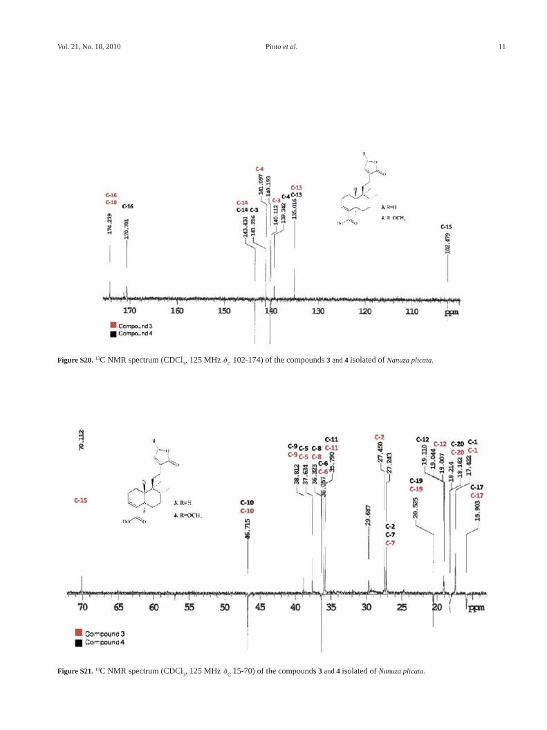
Patagonic acid (3)White amorphous powder; IR (KBr) ν
max/cm-1: 3433,
2958, 1751, 1678, 1458, 1261; 1H NMR (CDCl3; 500MHz):
d 1.70 (m, 2H-1), 1.45 (m, 2H-2), 6.81 (br s, H-3), 1.43 (m, H-6eq), 2.40 (m, H-6ax), 2.25 (m, 2H-7), 1.50 (m, H-8), 1.35 (m, H-10), 1.19 (m, H-11eq), 2.40 (m, H-11ax), 2,05 (m, H-12eq), 2.20 (m, H-12ax), 7.06 (br s, H-14), 4.79 (br s, 2H-15), 0.85 (d, 3H-17), 1.27 (s, 3H-19), 0.75 (s, 3H-20); NMR (CDCl
3; 500MHz): d 17.4 (CH
2-1), 27.4 (CH
2-
2), 140.1 (CH-3), 141.1 (C-4), 37.4 (C-5), 36.0 (CH2-6),
27.2 (CH2-7), 36.3 (CH-8), 38.7 (C-9), 46.7 (CH-10), 35.7
(CH2-11), 19.0 (CH
2-12), 135.0 (C-13), 143.4 (CH-14),
70.1 (CH2-15), 174.3 (C-16), 15.8 (CH
3-17), 174.2 (C-18),
20.5 (CH3-19), 18.1 (CH
3-20).
Supplementary Information
Supplementary data are available free of charge at http://jbcs.sbq.org.br, as PDF file.
Acknowledgments
The authors are grateful to CNPq, FAPESB and CAPES for scholarships and financial support, as well as to V. C. de O. Costa (UFPB) for the NMR spectra, and N. P. Lopes of Faculdade de Ciências Farmacêuticas-USP-Ribeirão Preto for the HR-ESIMS.
References
1. Stannard, B. L.; Flora of the Pico das Almas: Chapada da
Diamantina, Whitstable Litho Ltd: Great Britain, 1995.
2. Pinto, A. C.; Epifânio, R. A.; Zocher, D. H. T.; Biochem. Syst.
Ecol. 2004, 32, 597.
3. Pinto, A. C.; Patitucci, M. L.; Silva, R. S.; Queiroz, P. P. S.;
Kelecon, A.; Tetrahedron 1983, 39, 3351.
4. Pinto, A. C.; Pinchin, R.; Prado, S. K.; Phytochemistry 1983,
22, 2017.
5. Branco, A.; Pinto A. C.; Braz-Filho, R.; An. Acad. Bras. Cienc.
2004, 76, 505.
6. Branco, A.; Pereira, A. S.; Cardoso, J. N.; Aquino-Neto, F.
R.; Pinto, A. C.; Braz-Filho, R.; Phytochem. Anal. 2001, 12,
266.
7. Branco, A.; Pinto, A. C.; Ifa, D. R.; Braz-Filho, R.; J. Braz.
Chem. Soc. 2002, 13, 318.
8. Salatino, A.; Salatino, M. L. F.; Santos, D. Y. A. C.; Patrício,
M. C. B.; Genet. Mol. Biol. 2000, 23, 931.
9. Williams, C. A.; Harborne, J. B.; Menezes, N. L.; Biochem.
Syst. Ecol. 1991, 19, 483.
10. Riehl, C. A. S.; Pinto, A. C.; Figueroa-Villar, J. D; Nat. Prod.
Res. 2006, 20, 1225.
11. Riehl, C. A. S.; Pinto, A. C.; Kaiser, C. R.; Figueroa-Villar, J.
D.; Cruz, E. R.; Spectrosc. Lett. 2000, 33, 643.
12. Williams, C. A.; Harborne, J. B.; Tomas-Barberan, F. A.;
Phytochemistry 1987, 26, 2553.
13. Markham, K. R.; Sheppard, C.; Geiger, H.; Phytochemistry
1987, 26, 3335.
14. Rivera, A. P.; Faini, F.; Castillo, M.; J. Nat. Prod. 1988, 51,
155.
15. Krishna, V.; Singh, P.; Phytochemistry 1999, 52, 1341.
16. Barron, D.; Kaouadji, M.; Mariotte, A. M.; Z. Naturforsch.
1984, 39, 167.
17. Timmermann, B. N.; Hoffman, J. J.; Jolad, S. D.; Schram,
K. H.; Klenck, R. E.; Bates, R. B.; J. Nat. Prod. 1983, 46,
365.
18. Meira, M.; David, J. M.; David, J. P.; Araújo, S. V.;
Regis, T. L.; Giulietti, A. M.; Queiróz, L. P.; Quim. Nova
2008, 31, 751.
19. Agrawal, P. K.; Carbons-13 NMR of flavonoids, Elsevier: New
York, 1986.
20. Ross, J. A.; Kasum, C. M.; Annu. Rev. Nutr. 2002, 22, 19.

3’,8’’-Biisokaempferide, a Cytotoxic Biflavonoid and Other Chemical Constituents J. Braz. Chem. Soc.1824
21. Kang, S. S.; Lee, J. Y.; Choi, Y. K.; Song, S. S.; Kim, J. S.; Jeon,
S. J.; Han, Y. N.; Sonc, K. H.; Hana, B. H.; Bioorg. Med. Chem.
Lett. 2005, 15, 3588.
22. Matsuda, S.; Hisama, M.; Shibayama, H.; Itou, N.; Iwaki, M.;
Yakugaku Zasshi 2009, 129, 1113. (Coden: YKKZAJ ISSN:
0031-6903, AN 2009: 1314869 CaPlus).
23. Valensin, S.; Ghiron, C.; Lamanna, C.; Kremer, A.; Rossi, M.;
Ferruzzi, P.; Nievo, M.; Bakker, A.; BMC Cancer 2009, 9, 196.
24. Santos, A. G.; Perez, C. C.; Tininis, A. G.; Bolzani, V. S.;
Cavalheiro, A. J.; Quim. Nova 2007, 30, 1100.
25. Ahmad, V. U.; Khan, A.; Farooq, U.; Kousar, F.; Khan, S. S.;
Nawaz, S. A.; Abbasi, M. A.; Choudhary, M. I.; Chem. Pharm.
Bul. 2005, 53, 378.
26. Rumbero-Sanchez, A.; Vazquez, P.; Phytochemistry 1991, 30,
311.
27. Pédeboscq, S.; L’Azou, B.; Liguoro, D.; Pometan, J. P.; Cambar,
J.; Exp. Toxicol. Pathol. 2007, 58, 247.
28. Wen, P. Y.; Kesari, S.; N. Engl. J. Med. 2008, 359, 492.
29. Fu, Z.-G. J.; Liu, X.-M.; Liu, F.-R.; Chen, H.-L.; Shi, J. C.-S.;
Pang, H.-K.; Chen Ng, Z.-P.; Chin. Med. J. 2009, 122, 1255.
30. Bocchini, V.; Casalone, R.; Collini, P.; Rebel, G.; Lo Curto, F.;
Cell Tissue Res. 1991, 265, 73.
31. Planchenault, T.; Costa, S. L.; Fages, C.; Riche, D.; Charriére-
Bertrand, C.; Perzelova, A.; Barlovatz-Meimon, G.; Tardy, M.
Neurosci. Lett. 2001, 299, 140.
Submitted: December 18, 2009
Published online: June 11, 2010

Supplementary Inform
ationJ. Braz. Chem. Soc., Vol. 21, No. 10, S1-S17, 2010.
Printed in Brazil - ©2010 Sociedade Brasileira de Química0103 - 5053 $6.00+0.00
*e-mail: [email protected]
3’,8’’-Biisokaempferide, a Cytotoxic Biflavonoid, and Other Chemical Constituents of Nanuza plicata (Velloziaceae)
Meri Emili F. Pinto,a Marcelo Sobral da Silva,*,a Elisabete Schindler,b José Maria Barbosa Filho,a Ramon dos Santos El-Bachá,b Marianna Vieira S. Castello-Branco,a
Maria de Fatima Agraa and Josean Fechine Tavaresa
aLaboratório de Tecnologia Farmacêutica, Universidade Federal da Paraíba, CP 5009, 58051-970 João Pessoa-PB, Brazil
bLaboratório de Neuroquímica e Biologia Celular (LabNq), Instituto de Ciências da Saúde (ICS), Universidade Federal da Bahia, 40110-902 Salvador-BA, Brazil
Figure S1. IR spectrum (KBr) of the compound 1 isolated of Nanuza plicata.
3’,8’’-Biisokaempferide, a Cytotoxic Biflavonoid, and Other Chemical Constituents J. Braz. Chem. Soc.2
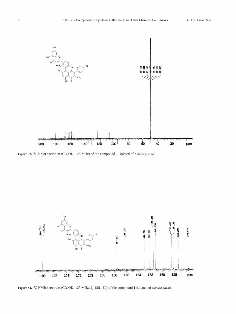
Figure S2. 13C NMR spectrum (CD3OD, 125 MHz) of the compound 1 isolated of Nanuza plicata.
Figure S3. 13C NMR spectrum (CD3OD, 125 MHz, d
C 156-180) of the compound 1 isolated of Nanuza plicata.

Pinto et al. 3Vol. 21, No. 10, 2010
Figure S4. 13C NMR spectrum (CD3OD, 125 MHz, d
C 94-139) of the compound 1 isolated of Nanuza plicata.
Figure S5. HR-ESI-MS spectrum of the compound 1 isolated of Nanuza plicata.

3’,8’’-Biisokaempferide, a Cytotoxic Biflavonoid, and Other Chemical Constituents J. Braz. Chem. Soc.4
Figure S6. 1H NMR spectrum (CD3OD, 500 MHz) of the compound 1 isolated of Nanuza plicata.
Figure S7. 1H NMR spectrum (CD3OD, 500 MHz, d
H 7.8-8.2) of the compound 1 isolated of Nanuza plicata.
Pinto et al. 5Vol. 21, No. 10, 2010
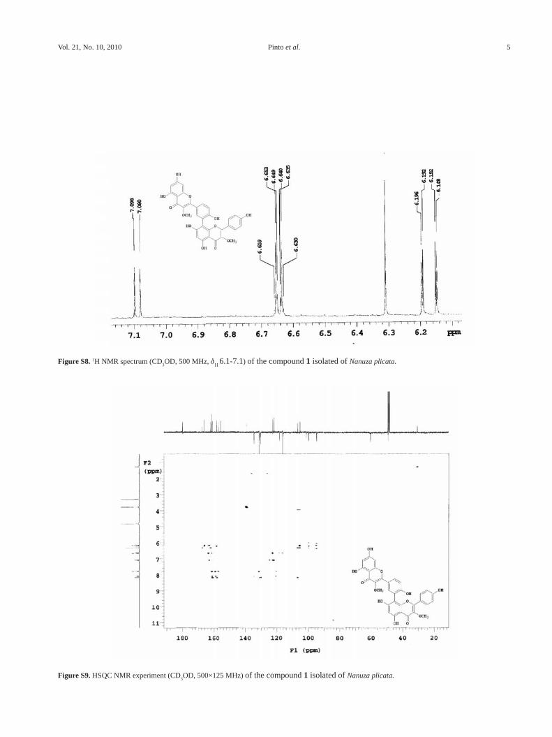
Figure S8. 1H NMR spectrum (CD3OD, 500 MHz, d
H 6.1-7.1) of the compound 1 isolated of Nanuza plicata.
Figure S9. HSQC NMR experiment (CD3OD, 500×125 MHz) of the compound 1 isolated of Nanuza plicata.
3’,8’’-Biisokaempferide, a Cytotoxic Biflavonoid, and Other Chemical Constituents J. Braz. Chem. Soc.6
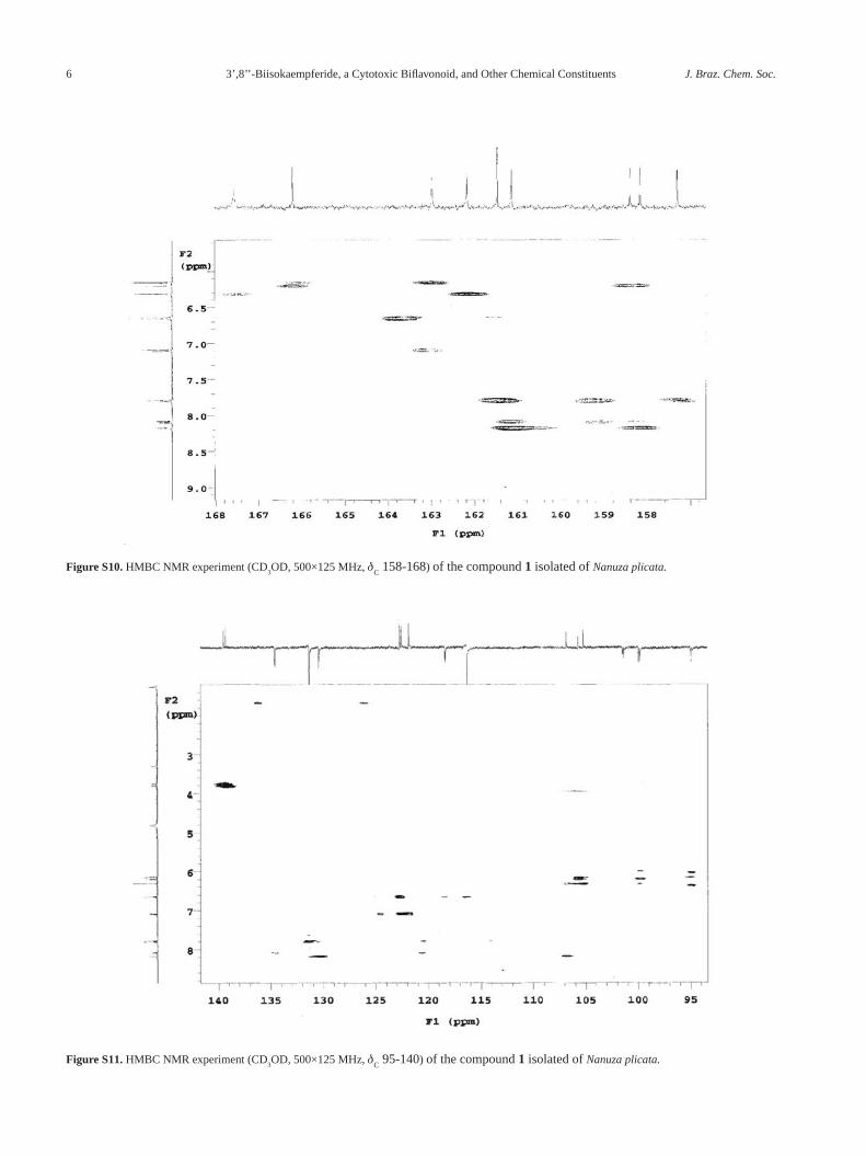
Figure S10. HMBC NMR experiment (CD3OD, 500×125 MHz, d
C 158-168) of the compound 1 isolated of Nanuza plicata.
Figure S11. HMBC NMR experiment (CD3OD, 500×125 MHz, d
C 95-140) of the compound 1 isolated of Nanuza plicata.
Pinto et al. 7Vol. 21, No. 10, 2010
Figure S12. HMBC NMR experiment (CD3OD, 500 x 125 MHz, d
C 138-140) of the compound 1 isolated of Nanuza plicata.
Figure S13. IR spectrum (KBr) of the compound 2 isolated of Nanuza plicata.

3’,8’’-Biisokaempferide, a Cytotoxic Biflavonoid, and Other Chemical Constituents J. Braz. Chem. Soc.8
Figure S14. 13C NMR spectrum (CD3OD, 125 MHz) of the compound 2 isolated of Nanuza plicata.
Figure S15. 13C NMR spectrum (CD3OD, 125 MHz) of the compound 2 isolated of Nanuza plicata.

Pinto et al. 9Vol. 21, No. 10, 2010
Figure S16. 13C NMR spectrum (CD3OD, 125 MHz, d
C 95-135) of the compound 2 isolated of Nanuza plicata.
Figure S17. 1H NMR spectrum (CD3OD, 500 MHz) of the compound 2 isolated of Nanuza plicata.

3’,8’’-Biisokaempferide, a Cytotoxic Biflavonoid, and Other Chemical Constituents J. Braz. Chem. Soc.10
Figure S18. 1H NMR spectrum (CD3OD, 500 MHz, d
H 6.1-7.9) of the compound 2 isolated of Nanuza plicata.
Figure S19. IR spectrum (KBr) of the compounds 3 and 4 isolated of Nanuza plicata
Pinto et al. 11Vol. 21, No. 10, 2010
Figure S20. 13C NMR spectrum (CDCl3, 125 MHz d
C 102-174) of the compounds 3 and 4 isolated of Nanuza plicata.
Figure S21. 13C NMR spectrum (CDCl3, 125 MHz d
C 15-70) of the compounds 3 and 4 isolated of Nanuza plicata.

3’,8’’-Biisokaempferide, a Cytotoxic Biflavonoid, and Other Chemical Constituents J. Braz. Chem. Soc.12
Figure S22. 1H NMR spectrum (CDCl3, 500 MHz) of the compounds 3 and 4 isolated of Nanuza plicata.
Figure S23. 1H NMR spectrum (CDCl3, 500 MHz, d
H 3.5-7.0) of the compounds 3 and 4 isolated of Nanuza plicata.
Pinto et al. 13Vol. 21, No. 10, 2010
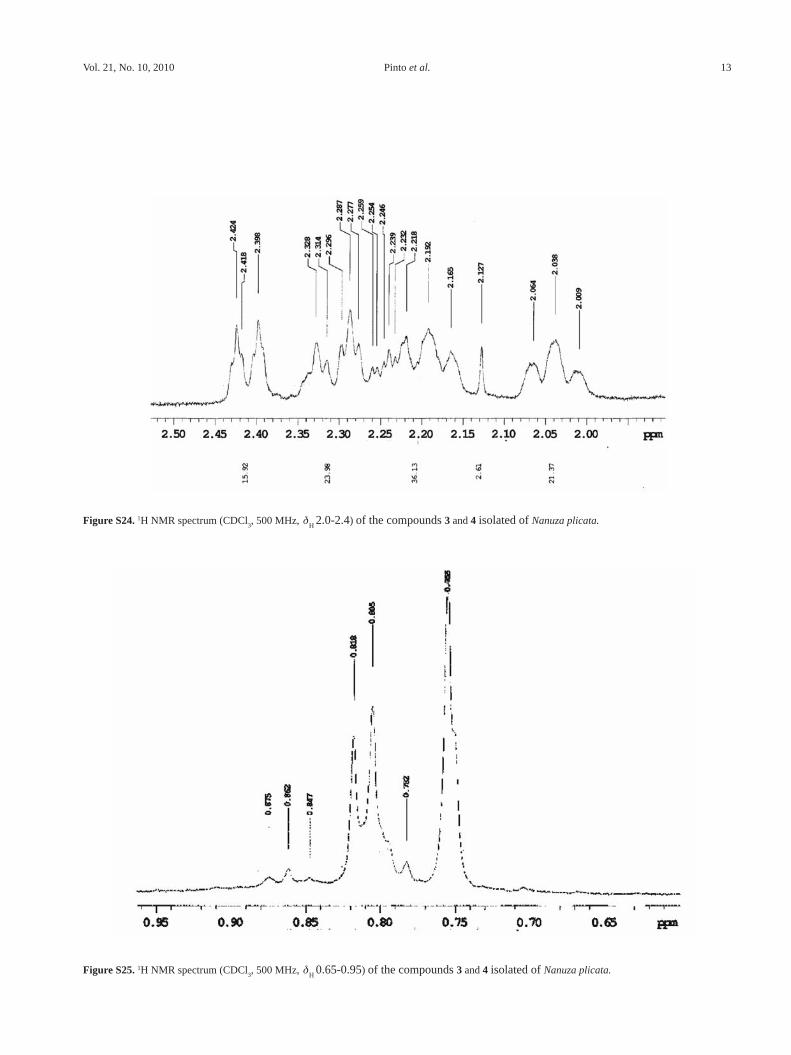
Figure S24. 1H NMR spectrum (CDCl3, 500 MHz, d
H 2.0-2.4) of the compounds 3 and 4 isolated of Nanuza plicata.
Figure S25. 1H NMR spectrum (CDCl3, 500 MHz, d
H 0.65-0.95) of the compounds 3 and 4 isolated of Nanuza plicata.

3’,8’’-Biisokaempferide, a Cytotoxic Biflavonoid, and Other Chemical Constituents J. Braz. Chem. Soc.14
Figure S26. HMQC NMR experiment (CDCl3, 500×125 MHz, d
C 140-144) of the compounds 3 and 4 isolated of Nanuza plicata.
Figure S27. HMQC NMR experiment (CDCl3, 500 x 125 MHz, d
C 15-45) of the compounds 3 and 4 isolated of Nanuza plicata.

Pinto et al. 15Vol. 21, No. 10, 2010
Figure S28. HMBC NMR experiment (CDCl3, 500 x 125 MHz, d
C 100-145) of the compounds 3 and 4 isolated of Nanuza plicata. * Correlation of
compounds 4.
Figure S29. HMBC NMR experiment (CDCl3, 500×125 MHz, d
C 15-50) of the compounds 3 and 4 isolated of Nanuza plicata.

3’,8’’-Biisokaempferide, a Cytotoxic Biflavonoid, and Other Chemical Constituents J. Braz. Chem. Soc.16
Figure S30. 13C NMR spectrum (CD3OD, 125 MHz) of the compound 7 isolated of Nanuza plicata.
Figure S31. 13C NMR spectrum (CD3OD, 125 MHz, d
C 95-184) of the compound 7 isolated of Nanuza plicata.

Pinto et al. 17Vol. 21, No. 10, 2010
Figure S32. 1H NMR spectrum (CD3OD, 500 MHz) of the compound 2 isolated of Nanuza plicata.
Figure S33. 1H NMR spectrum (CD3OD, 500 MHz, d
H 6.1-7.5) of the compound 7 isolated of Nanuza plicata.