Arterial complications
73
Vascular Ehlers Danlos Syndrom When Do Vascular MD need to think about ? Emmanuel Messas M.D.,Ph.D.,F.E.S.C The reference center of rare vascular diseases Paris, 19 Mars 2014 College Français de Pathologie Vasculaire Paris,France
Transcript of Arterial complications

Vascular Ehlers Danlos Syndrom
When Do Vascular MD need to think about ?
Emmanuel Messas M.D.,Ph.D.,F.E.S.C
The reference center of rare vascular diseases
Paris, 19 Mars 2014
College Français de Pathologie Vasculaire
Paris,France

Mme N HN, 33 ANS
Pr E. Messas
Centre de Référence des Maladies Vasculaires Rares
Médecine Vasculaire/HTA
HEGP, APHP, Paris
www.maladiesvasculairesrares.com

Mme N HN, 33 ANS
• Douleur aiguë de l’HCD avec malaise lipothymique chez une patiente sans antécédent personnel notable
• Transfert aux urgences du CHU d’Avicennes où survient rapidement un choc hémorragique avec hémopéritoine et HRP massifs. Bilan CT : rupture artère splénique. Ligature chirurgicale de l’artère splénique et résection partielle du pancréas.

Mme N HN, 33 ANS
• Bilan lésionnel
dissection artère rénale droite, hématome disséquant artère rénale
gauche avec infarctus rénaux, dissection tronc cœliaque et artère
hépatique, dissection carotides internes post-bulbaires et aspect
très irrégulier séquellaire
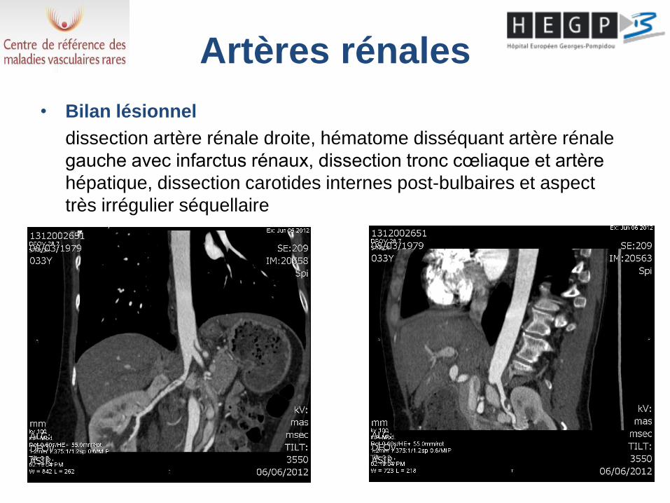
Artères rénales
• Bilan lésionnel
dissection artère rénale droite, hématome disséquant artère rénale
gauche avec infarctus rénaux, dissection tronc cœliaque et artère
hépatique, dissection carotides internes post-bulbaires et aspect
très irrégulier séquellaire
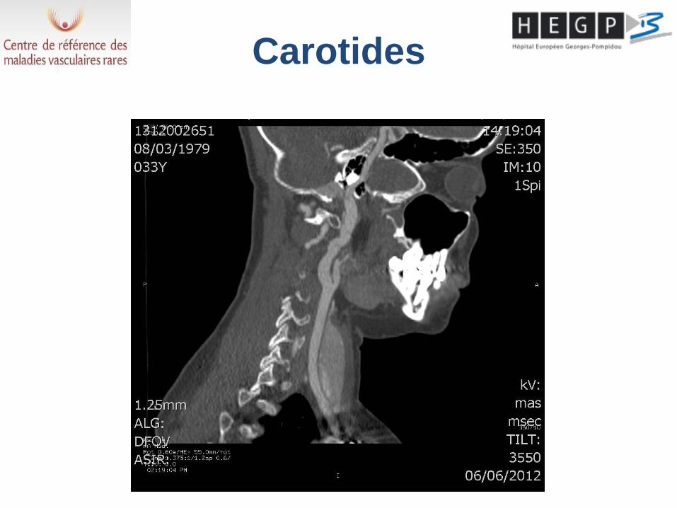
Carotides
Hypothèse diagnostique
• Dysplasie fibromusculaire
Le caractère irrégulier des artères carotidiennes et
l’association d’une atteinte des TSA et des artères
rénales/digestives font suspecter une DFM et la patiente
est adressée au centre de référence pour la prise en
charge
Mais
La rupture artérielle spontanée et les lésions multiples
des artères de moyen calibre chez un adulte jeune
permet d’emblée de poser le diagnostic de SED
vasculaire

Anamnèse
Antécédents:
• Aucun sauf tendance alopécique ancienne étiquetée « androgénique »
• G0P0
• Fratrie de 5, sans histoire familiale suspecte identifiable
Examen clinique
Peau fine ++ tendance ecchymotique depuis l’enfance, lacis vasculaires décolleté et membres +++, instabilité ATM sans fragilité gingivale, cicatrisation chéloïde plutôt que papyracée, phénotype du visage évocateur, acrogérie mineure
Soit 4 critères diagnostics majeurs en faveur d’un SED vasculaire
Test génétique
Confirmation de la maladie: mutation d’épissage dans le gène COL3A1 (c.2446-2A>G)
c.2446-2A>G c.2446-2A>G

Conclusion
Le Syndrome d’Ehlers-Danlos vasculaire est une maladie héréditaire artérielle révélée typiquement chez l’adulte jeune par un accident
artériel aigu.
La survenue d’une rupture artérielle spontanée, et à fortiori en présence de lésions artérielles satellites doit dans ce contexte faire
d’emblée suspecter le diagnostic de SED vasculaire, même en l’absence d’histoire familiale évocatrice

Ehlers Danlos syndrom
• Heterogeneous group of multiple genetic disease of conjunctive tissues which have in common :
- Skin Hyperextensibility
- Joint hypermobility
- Easy Bruising and tissular fragility
• The clinical diagnosis of Ehlers–Danlos syndrome type IV, the vascular type, is made on the basis of four clinical criteria:
- easy bruising,
- thin skin with visible veins,
- characteristic facial features,
and rupture of arteries, uterus, or intestines

Ehlers Danlos Syndromes
Villefranche Classification
Beighton P et al. Am J Med Genet 1998

vEDS diagnosis
Beighton et al Am J Med Genet. 1998;77:31-7.
« The presence of any two or more of the major criteria is
highly indicative of the diagnosis, and laboratory testing
is strongly recommended. »
Genetic feature and Epidemiology
• vEDS is uncommon (the precise incidence and prevalence are not known about about 1/150,000), and in part because of its rarity, the diagnosis is often made only after a catastrophic complication or at postmortem examination.
• Autosomal dominant disorder with high level of neomutation (50%).
• The diagnosis is confirmed by the demonstration that
cultured fibroblasts synthesize abnormal type III
procollagen molecules or by the identification of a
mutation in the gene for type III pro-collagen (COL3A1 )
an essential constituent of arterial wall, skin,articular
capsuls, uterin and gastrointestin wall specially sigmoid
colon
• The high arterial and tissular fragility are responsible of the
extrem gravity of the disease

Type 3 Procollagene
•Fibrillar Collagene formed by homotrimer of proa1(III) chains with
triple triple-helical domain and C and N terminal globular
• Triple helical stability linked to the conservation of its primary repetative
structure: Amino acid triplet starting with Glycine
•In each instance, the effect of an abnormal allele product is amplified, since
type III procollagen is a homotrimer.
•As a consequence, an abnormal product from one allele is expected to
result in seven of eight molecules being abnormal.
44 Kb Chr 2q31
COL3A1

Gene basis
• The causative gene, COL3A1 (accession numbers 193023938–
193062278 in the Human Genome Project Working Draft at UCSC),
located at 2q31-q32, contains 52 exons (of which exons 4 and 5
are fused to create a single exon) distributed over 44 kb (Chu and
Prockop 1993).
• New mutations are common, and approximately one-half of
identified individuals with the condition have no apparent family
history of the disorder.
• Approximately two-thirds of the mutations are single-nucleotide
substitutions that result in substitutions for glycine residues in
the triple-helical domain of the proa1(III) chain. Most of the rest are
splice- site mutations that result in exon skipping, and a small
number are larger genomic deletions (Kuivaniemi et al. 1997).
• No correlations have been discerned between the nature or
location of the mutation and the type or frequency of major
complications (Pepin et al. 2000).
• SEDv autosomal dominant
- with dominant negative effect
- with haplo - insufficiency
- Private mutation (and more than 50%
of neomutation )
• Genetic analysis was at the beginning
informative in 60% of the case , now it is
more closed to 90%.
• Ongoing trial on genotype/phenotype
analysis
Genetic Analyis
Result

Hôpital Européen Georges Pompidou,Paris,France
NATIONAL CENTER FOR RARE VASCULAR DISEASE
Cohort of 160 index cases vEDS
followed since 2006
The reference center of rare vascular diseases
www.maladiesvasculairesrares.com
Pr X Jeunemaitre
Dr M Frank Pr E Messas
Dr T Mirault

Patients characteristics

Onset of clinical events in respect of mutation type

Diagnosis
Clinical consensus not validated • Only the detection of known pathogenic mutation of Col3A1
gene provide diagnosis evidence.
• In case of negative genetic test : Collegial study of the clinical
course and radiology exam can performed the diagnosis
(specially in presence of two majors clinical factors)
• Interest of echotracking to identify patient with IMT less than 400
µm
• In case of clinical suspicion (two major criteria or one major
criteria associated with family history ) genetic test is proposed
and result provided during a multidiscplinary consultation
(clinician, geneticist and psychiatry)
• Explication of the different method of diagnosis, of the heritability
and transmission of the disease with the need of genealogical tree
and prophylactic measure

Clinical Presentation of vEDS
– Two type are typically described:
- Ecchymotic type
- Acrogeric type with facial morphotype
characteristics (face of « Madone »
associated with an early aging of the
extremity)
– Skin hyperextensibility is mild and joint
hypermobility is limited to the small distal
articulation

Facial Morphotype
« Face of madone »
- Thin noise, horizontal lip mildly ourled
- Hollow Cheek, tight firm lobeless ears
-Prominent staring eyes because of a paucity
Adipose tissue


Morphotype facial

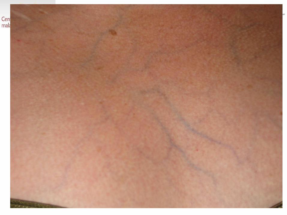
Dermal Sign
Translucent skin
- Pale skin sometimes velvety to the touch
- Subcutaneous veinous pattern particularly apparent
on the anterior part of the chest

Cutaneous signs
Skin fragility
- Spontaneous bruising ,
- Sometimes delayed healing with scare having a thin
atrophic papyraceous appearanceon friction points.
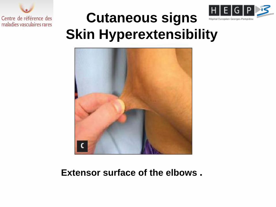
Cutaneous signs
Skin Hyperextensibility
Extensor surface of the elbows .

Signs skin
Acrogeria
Skin thin and retracted ends of the tendons
giving hands and feet look emaciated,
prematurely aged.


Articular signs
• Large joints hypermobile rare in SEDv
• Moderate joint laxity affecting the small joints of the
fingers and toes.
• The notion of congenital dislocation of the hip, a
talipes equinovarus or simply in a cavus
have the value of minor sign
• Repeated sprains or dislocations of the shoulders,
joints and ankles are sometimes in the foreground.
• Early osteoarthritis linked to hypermobility is a source of
pain and joint stiffness.
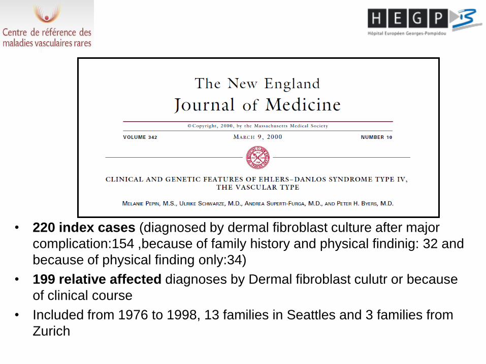
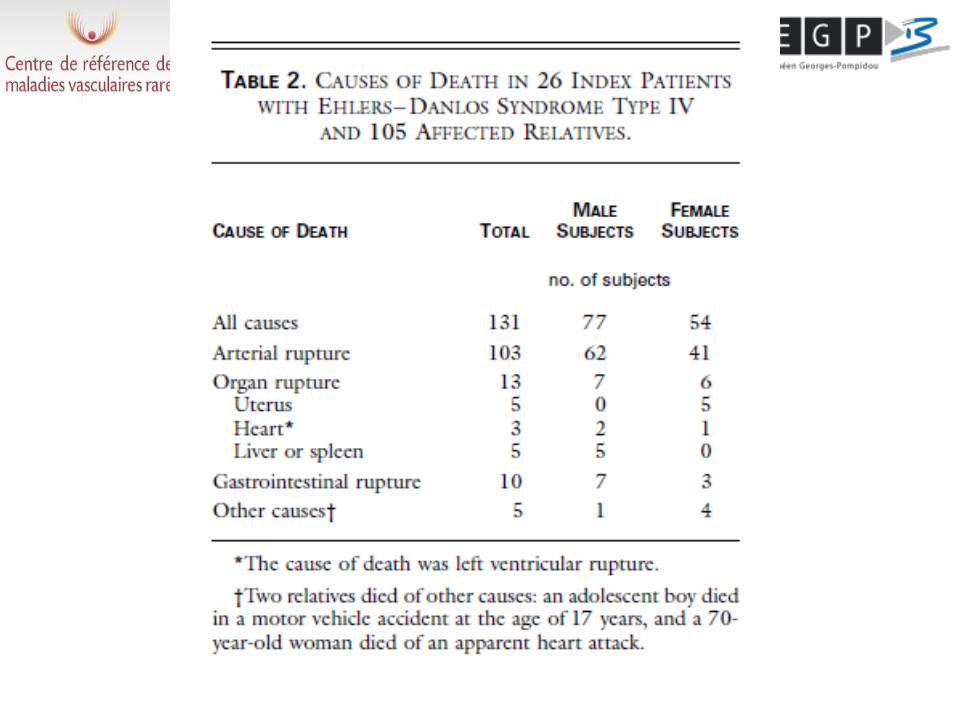
• 220 index cases (diagnosed by dermal fibroblast culture after major
complication:154 ,because of family history and physical findinig: 32 and
because of physical finding only:34)
• 199 relative affected diagnoses by Dermal fibroblast culutr or because
of clinical course
• Included from 1976 to 1998, 13 families in Seattles and 3 families from
Zurich

Kaplan–Meier Estimates of Overall Survival among 374 Subjects
with vascular Ehlers–Danlos Syndrome.
From Pepin M et al. N Engl J Med 2000;342;673-80

Age at the Time of a First Complication among 207 Index Patients.
From Pepin M et al. N Engl J Med 2000;342;673-80
- 25% of patients before the age of 20 - 80% before 40
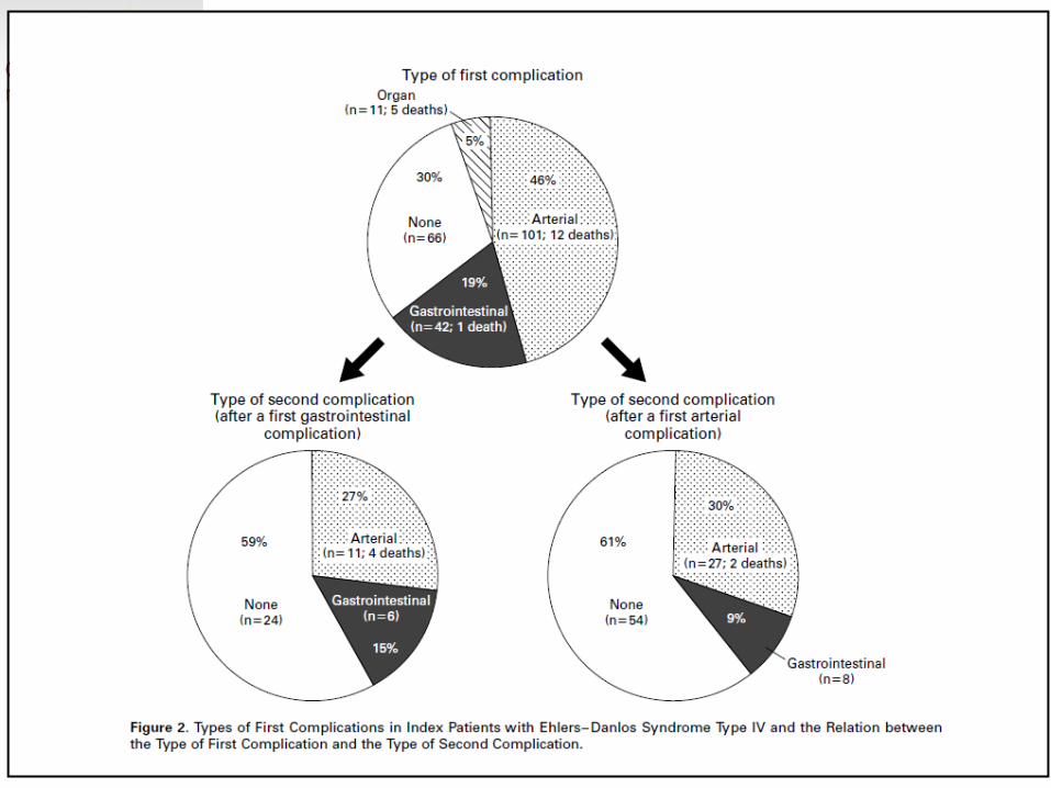
Visceral complications
• Arterial complications
• Gastrointestinal complications
• Obstetric complications
• Respiratory complications
• Venous complications

Arterial complications
• Dissecting aneurysms and dissections
• Arterial rupture
• Arteriovenous fistulas (carotid-cavernous
sinus)
• Occur in arteries of normal caliber
• Medium caliber arteries preferentially with

Copyright © 2007 by the American Roentgen Ray Society
Zilocchi, M. et al. Am. J. Roentgenol. 2007;189:712-719
Spectrum and distribution of vascular findings in Ehlers-Danlos syndrome: total of 83 abnormal vascular findings including ectasia, aneurysm, dissection, and occlusion

Angiographic aspects
of Spontaneous Complications during vSED

Aspect of dissecting
aneurysm by Echo Doppler

Endovascular treatment.
An arteriovenous fistula
leak stent
ARTERIAL PHENOTYPE
IN vEDS

Boutouyrie et al ,Circulation 2004

Boutouyrie et al ,Circulation 2004

Boutouyrie et al ,Circulation 2004

BIOMECANICAL OF ARTERY WALL PROPERTY
• Marfan Syndrom
- Mostly Aorta
- Decreased distensibility
- Increased stiffness index
- Increased pulse wave velocity
• Vascular EDS
- Mostly peripheral artery
- No change in Distensibility and stiffness
- Decrease of WALL Thickness
- Increase of wall stress
Boutouyrie et al ,Circulation 2004
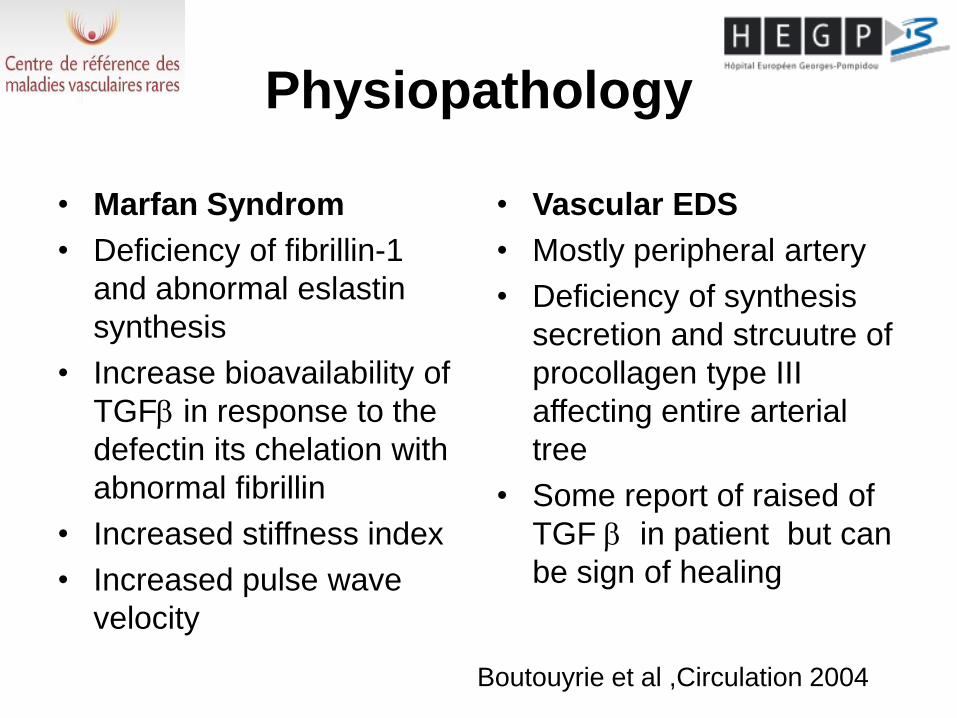
Physiopathology
• Marfan Syndrom
• Deficiency of fibrillin-1
and abnormal eslastin
synthesis
• Increase bioavailability of
TGF in response to the
defectin its chelation with
abnormal fibrillin
• Increased stiffness index
• Increased pulse wave
velocity
• Vascular EDS
• Mostly peripheral artery
• Deficiency of synthesis
secretion and strcuutre of
procollagen type III
affecting entire arterial
tree
• Some report of raised of
TGF in patient but can
be sign of healing
Boutouyrie et al ,Circulation 2004

« We use in our center the threshold
of IMT less than 400 µm as minor
parameter to motivate gene testing »

Back To History ….
Institute
Curie
Pierre Curie
Theory of
Piezo-electricity
Paul Langevin - 1914
First Sonar Experiment
Precursor of Modern
Medical Echography
Langevin Radiation Force
E.S.P.C.I.
Visit www.loa.espci.fr/epom/epom_main.html
Mathias
Fink
Director of LOA
(PR. ESPCI)

ELASTOGRAPHY ULTRASOUND
with conventional vascular probe 1. Ultra fast imaging: 5-10000 frames per sec
Conventional US time
0 s 1 s
A 30 ms Experiment !! Ultrafast US
2. Virtual palpation without external vibrator
- Sending a wave train ("push") focused from the probe
for vibration - Visualization of the shear wave through
ultrafast mode -Determining the Young's modulus from the speed of
the shear wave

vEDS patients Controls p
Right PWV
Increase (%) 7.13 [-11.78; 22.04] 28.53 [6.08; 34.48] 0.0201
PWV Slope
(m/s/mmHg) 1.08 [-1.31; 3.09] 3.16 [1.30; 5.85] 0.0198
Left PWV
Increase (%) 9.90 [3.07; 21.04] 16.27 [12.23; 28.45] 0.0475
PWV Slope
(m/s/mmHg) 1.44 [0.37; 2.80] 3.17 [1.70; 5.66] 0.0050
PWS late - PWS early
-----------------------------
SBP - DBP
PWV slope

1 normal
2 elastin deficiency
3 collagen deficiency
Sheridan WS,.Mechanical characterization of a customized
decellularized scaffold for vascular tissue engineering.
J Mech Behav Biomed Mater. 2012 Apr;8:58-70.
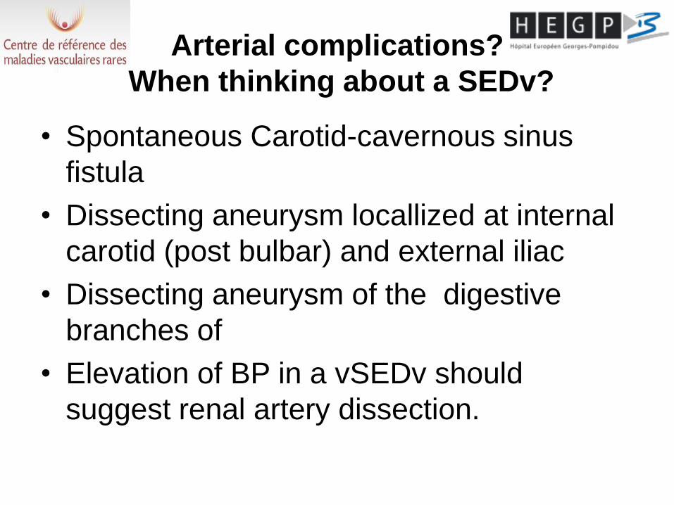
Arterial complications?
When thinking about a SEDv?
• Spontaneous Carotid-cavernous sinus
fistula
• Dissecting aneurysm locallized at internal
carotid (post bulbar) and external iliac
• Dissecting aneurysm of the digestive
branches of
• Elevation of BP in a vSEDv should
suggest renal artery dissection.

Arterial complications
Conservative Treatment • Conservative treatment with no treatment most often
Bed rest and analgesic, local compression, transfusion
Avoid puncture
Indications of endovascular treatments are:
- Selective embolization of an artery for hemostasis goal
- Occlusion carotid-cavernous fistula
Indication for surgery as a last resort:
- Bypass for limb salvage.
- Difficulty for clamping, hemostasis and suturing of vessels
- venous bypass proscribed
- Anastomoses performed without tension point reinforced by separate horizontal
"pledgets".
-skin suture is made of separate points nonabsorbable suture left in place for an
extended period.

Digestive complications
• Spontaneous rupture of the gastrointestinal tract (80% of the sigmoid)
• The episode is fatal in 2% of cases
• High risk of recurrence: 50% of colonic perforations
• Inguinal hernias, umbilical, hiatal, diaphragmatic and white line hernias are common.
• Spontaneous rupture of the spleen and liver (rare)
• Acute abdomen: abdominal CT need urgently.
• Pneumothorax • Hémoptysia • Pulmonary blebs
Pulmonary Complications

Obstetric complications
• Maternal mortality to 11.5% in relation to uterine rupture
during labor or vascular ruprture in post partum.
• Little specific data on the subject
• Current study VEDOC (Frank et al) • Discussion on:
- Routine Caesarean after 32 weeks
- If vaginal delivery: forceps and strengthening perineal - Beta blocker prescription during labor
-Type of anesthesia: epidural avoid
• Contraception: IUDs prevent
Venous complications
• Early onset varicose veins
• Stripping of the great saphenous vein to
avoisd.
• Deep thrombophlebitis secondary to
prolonged immobility

Evolution and Prognosis
• Median survival is 48 years
• Causes :
- arterial rupture + + + (unpredictable) : 78,5%
- gastrointestinal perforation : 7,5%
-uterine rupture, cardiac or hepatosplenic : 10%
• Heterogeneity between and within family
• The age of death in the series of Pepin ranged from
6-73 years.
• In this study, the patient's prognosis is better than
that of the related suffering.
• Maternal mortality: 11.5%
Initial assessment of the lesions • Echo Doppler of aorta and lower limb and of
carotid artery by experienced operator for
this type of pathology
• Angio CT scanner
• Study of the mitral valve apparatus by
echocardiography.
• Annual Hospitalisation and semi-annual
consultation.

Prophylactic measures
• Document "passport AFSED" or care card issued by
the CRMVR
• Safe puncture
• Intramuscular and KT subclavian central set against
• Anti hypertension (preferred beta blocker Celiprolol)
• Sports with flexion contraindicated (cycling, skiing)
• Transit with regular laxative.
• Rectal temperature and colonoscopy to avoid.
• IUD not recommended
Genetic Counseling
• Autosomal dominant
• Two systematic reviews of genetic testing
• Problem of detection of minor
• Related issue of testing (COL3A1 not detected in the
index case)
• Problem of prenatal screening (trophoblast puncture
between 8 and 12 weeks) or preimplantation genetic
diagnosis required prior identification of the causal
mutation in one of the two parents.

Medication
• Since Bbest study all SEDv under Celectol
200mg x 2 / d
• Interest AT2 Inhibitor in case of need to
control blood pressure
• No etiological treatment
• Experimental studies on the benefit of
doxycycline or cell therapy.
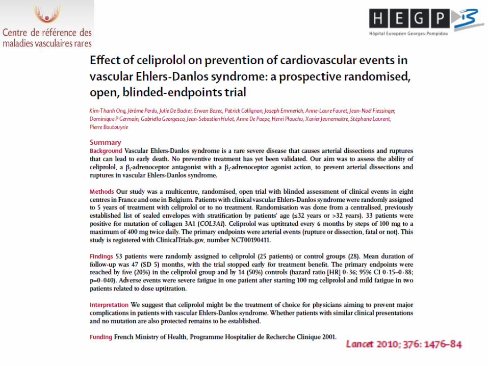
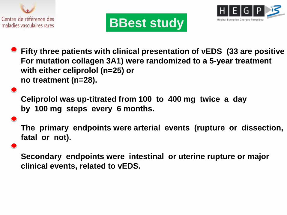
BBest study
Fifty three patients with clinical presentation of vEDS (33 are positive
For mutation collagen 3A1) were randomized to a 5-year treatment
with either celiprolol (n=25) or
no treatment (n=28).
Celiprolol was up-titrated from 100 to 400 mg twice a day
by 100 mg steps every 6 months.
The primary endpoints were arterial events (rupture or dissection,
fatal or not).
Secondary endpoints were intestinal or uterine rupture or major
clinical events, related to vEDS.

Mean duration of follow-up was 47 (± 15) months.
The primary endpoint was reached by 5 patients (20%) in the
celiprolol group and by 14 patients (50%) in the control
group (hazard ratio, 0.36; 95% CI, 0.15 to 0.88; P=0.04).
Primary plus secondary endpoints occurred in 6 patients (24%)
in the celiprolol group and in 17 patient (61%) in the control
group (hazard ratio, 0.31; 95% CI, 0.14 to 0.71; P=0.0097).
Twenty-nine patients carrying mutation for COL3A1 gene were
also protected since 1/10 presented a primary event under
celiprolol versus 11/19 in the control group (p=0.025).
BBest study

Kaplan-Meier curves of event-free survival of all patients
in PROBE design

The reference center of rare vascular diseases
Vascular Medicine – HTA
Pr Emmanuel Messas
Dr Tristan Mirault
Pr Jean-Noël Fiessinger
Pr Joseph Emmerich
Pr Pierre-François Plouin
Dr Nicolas Denarié
Assistants of department
Genetic
Pr Xavier Jeunemaitre
Dr Michaël Frank
Dr Anne-Laure Fauret
Diane Molière Psychiatry
Dr Khadija Lahlou-Laforêt
Lymphology Department
Dr Stéphane Vignes
Secretariat : Marie-Christine Nain

11 CENTRES OF EXPERTISE
Ghent

www.maladiesvasculairesrares.com
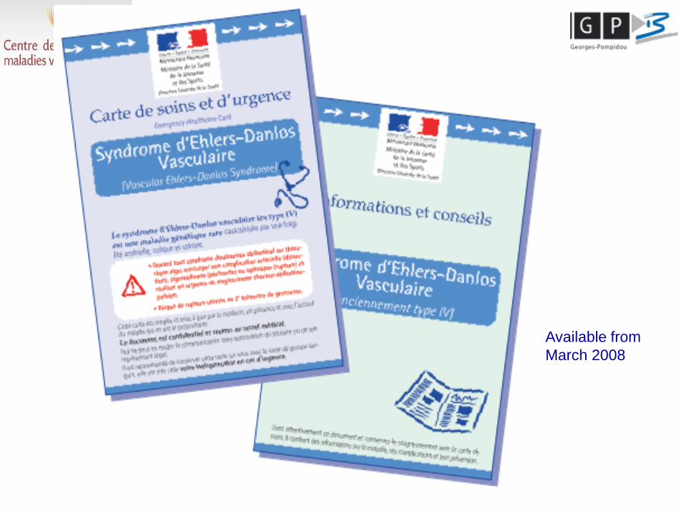
Available from
March 2008

Conclusions
• Ehlers Danlos syndrome consist of a hetrerogeneous
group of inherited connective tissue disorders.
• The vascular type is most severe becasue of its potential
vascular, and hollow organe dissection or rupture.
• Cardiologist should think about this rare vascualr
disease in case of early severe vascular or, cardiac
event in young patient witrhout cardiovascualr risk
• Atypical arterial site of the dissection its associatio to
digestive and or uterine complication shouldf also make
think of this disease
• Interest of Genetic testing and familial tree
• The rule is less invasive treatment
• Interest of Celiprolol for all suspected vEDS patient