are secondary growth - University of …Topic 16. Secondary Growth Introduction: Secondary growth...
12
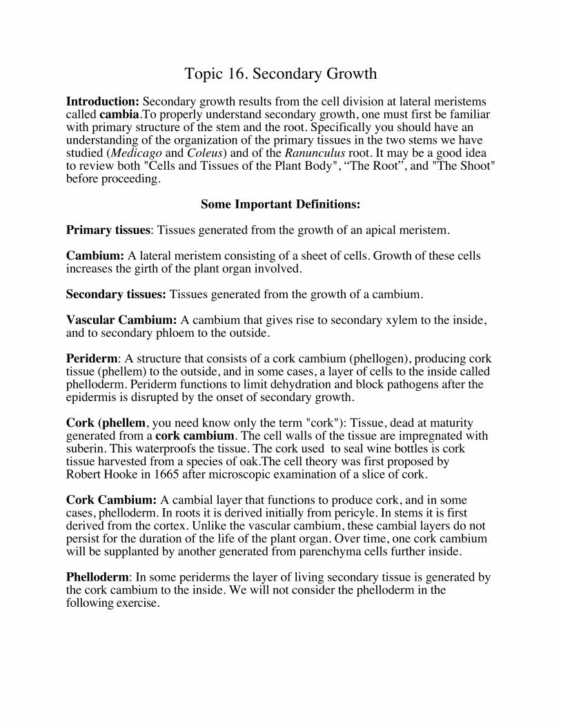
Topic 16. Secondary Growth Introduction: Secondary growth results from the cell division at lateral meristems called cambia.To properly understand secondary growth, one must first be familiar with primary structure of the stem and the root. Specifically you should have an understanding of the organization of the primary tissues in the two stems we have studied (Medicago and Coleus) and of the Ranunculus root. It may be a good idea to review both "Cells and Tissues of the Plant Body", “The Root”, and "The Shoot" before proceeding. Some Important Definitions: Primary tissues: Tissues generated from the growth of an apical meristem. Cambium: A lateral meristem consisting of a sheet of cells. Growth of these cells increases the girth of the plant organ involved. Secondary tissues: Tissues generated from the growth of a cambium. Vascular Cambium: A cambium that gives rise to secondary xylem to the inside, and to secondary phloem to the outside. Periderm: A structure that consists of a cork cambium (phellogen), producing cork tissue (phellem) to the outside, and in some cases, a layer of cells to the inside called phelloderm. Periderm functions to limit dehydration and block pathogens after the epidermis is disrupted by the onset of secondary growth. Cork (phellem, you need know only the term "cork"): Tissue, dead at maturity generated from a cork cambium. The cell walls of the tissue are impregnated with suberin. This waterproofs the tissue. The cork used to seal wine bottles is cork tissue harvested from a species of oak.The cell theory was first proposed by Robert Hooke in 1665 after microscopic examination of a slice of cork. Cork Cambium: A cambial layer that functions to produce cork, and in some cases, phelloderm. In roots it is derived initially from pericyle. In stems it is first derived from the cortex. Unlike the vascular cambium, these cambial layers do not persist for the duration of the life of the plant organ. Over time, one cork cambium will be supplanted by another generated from parenchyma cells further inside. Phelloderm: In some periderms the layer of living secondary tissue is generated by the cork cambium to the inside. We will not consider the phelloderm in the following exercise.
Transcript of are secondary growth - University of …Topic 16. Secondary Growth Introduction: Secondary growth...
Topic 16. Secondary Growth
Introduction: Secondary growth results from the cell division at lateral meristemscalled cambia.To properly understand secondary growth, one must first be familiarwith primary structure of the stem and the root. Specifically you should have anunderstanding of the organization of the primary tissues in the two stems we havestudied (Medicago and Coleus) and of the Ranunculus root. It may be a good ideato review both "Cells and Tissues of the Plant Body", “The Root”, and "The Shoot"before proceeding.
Some Important Definitions:
Primary tissues: Tissues generated from the growth of an apical meristem.
Cambium: A lateral meristem consisting of a sheet of cells. Growth of these cellsincreases the girth of the plant organ involved.
Secondary tissues: Tissues generated from the growth of a cambium.
Vascular Cambium: A cambium that gives rise to secondary xylem to the inside,and to secondary phloem to the outside.
Periderm: A structure that consists of a cork cambium (phellogen), producing corktissue (phellem) to the outside, and in some cases, a layer of cells to the inside calledphelloderm. Periderm functions to limit dehydration and block pathogens after theepidermis is disrupted by the onset of secondary growth.
Cork (phellem, you need know only the term "cork"): Tissue, dead at maturitygenerated from a cork cambium. The cell walls of the tissue are impregnated withsuberin. This waterproofs the tissue. The cork used to seal wine bottles is corktissue harvested from a species of oak.The cell theory was first proposed byRobert Hooke in 1665 after microscopic examination of a slice of cork.
Cork Cambium: A cambial layer that functions to produce cork, and in somecases, phelloderm. In roots it is derived initially from pericyle. In stems it is firstderived from the cortex. Unlike the vascular cambium, these cambial layers do notpersist for the duration of the life of the plant organ. Over time, one cork cambiumwill be supplanted by another generated from parenchyma cells further inside.
Phelloderm: In some periderms the layer of living secondary tissue is generated bythe cork cambium to the inside. We will not consider the phelloderm in thefollowing exercise.
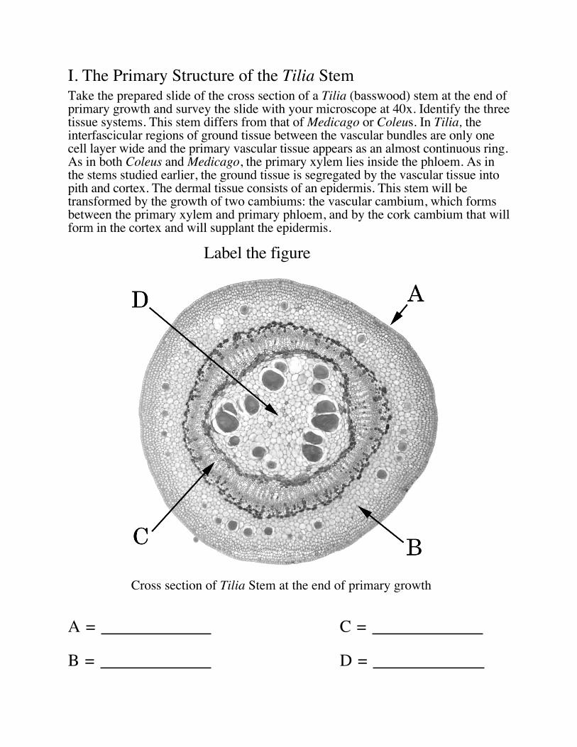
I. The Primary Structure of the Tilia StemTake the prepared slide of the cross section of a Tilia (basswood) stem at the end ofprimary growth and survey the slide with your microscope at 40x. Identify the threetissue systems. This stem differs from that of Medicago or Coleus. In Tilia, theinterfascicular regions of ground tissue between the vascular bundles are only onecell layer wide and the primary vascular tissue appears as an almost continuous ring.As in both Coleus and Medicago, the primary xylem lies inside the phloem. As inthe stems studied earlier, the ground tissue is segregated by the vascular tissue intopith and cortex. The dermal tissue consists of an epidermis. This stem will betransformed by the growth of two cambiums: the vascular cambium, which formsbetween the primary xylem and primary phloem, and by the cork cambium that willform in the cortex and will supplant the epidermis.
Label the figure
Cross section of Tilia Stem at the end of primary growth
A = C =
B = D =
Draw the intact epidermis.
Draw a primary vessel element.
II. Secondary Tissues of an older Tilia Stem
Take the prepared slide of the cross sections of Tilia (basswood) stems: 1, 2 and3-year sections on the same slide. Survey the section of the 1-year old stem withyour microscope at 40x. Starting in the middle of the stem, identify the following: pith; xylem tissue, phloem tissue, cortex, periderm, and epidermis.
Now switch to 400x. Carefully study the boundary of the xylem with the pith. Theinnermost xylem is primary. The outermost is secondary. The boundary betweenthe two is subtle: the secondary xylem starts where the xylem rays begin. Xylemrays are areas of parenchyma of the secondary xylem running radially through thestem. Starting at the outside of the cylinder of xylem, locate a ray and follow ittowards the pith. Where it ends marks the location of the primary xylem.Study the boundary between the xylem and the phloem. This marks the location ofthe vascular cambium. Note that on either side of the vascular cambium are rtheyoungest secondary tissues. The farther the tissues lie from the vascular cambium,the older the tissue. This means that as we move outward through the xylem wemove progressively from older to younger xylem. However, as we moveoutward through the phloem we move progressively from younger to olderphloem.
The outermost phloem is the protophloem and is marked by a continuous ring offibers. These fibers differentiated from cells in the primary phloem that maturedafter the onset of secondary growth.Outside of the phloem is the cortex. In the cortex a periderm has formed to theoutside. The periderm consists of a cork cambium together with the cork tissuederived from that cambium. Note the epidermis being sloughed off the stem.

Label the figure
Cross Section of a One-year Old Tilia Stem
A = E =
B = F =
C = G =
D = H =
Section After Three Years Growth.
Switch back to 40x and survey the 3-year old Tilia section. The obvious changesvisible here are the growth rings present in the secondary xylem, and the growth ofcertain rays in the phloem forming wedge-shaped regions of parenchyma. Thesequence of tissues outlined before are the same from the center outward: pith,primary xylem, secondary xylem, vascular cambium, secondary phloem, primaryphloem, cortex, and periderm.
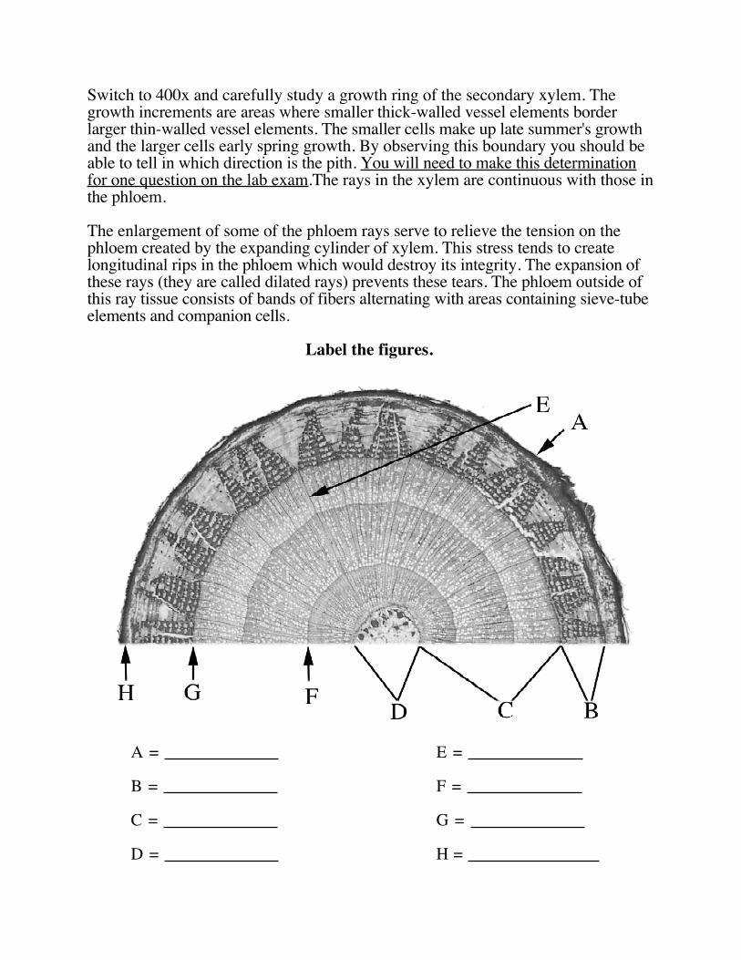
Switch to 400x and carefully study a growth ring of the secondary xylem. Thegrowth increments are areas where smaller thick-walled vessel elements borderlarger thin-walled vessel elements. The smaller cells make up late summer's growthand the larger cells early spring growth. By observing this boundary you should beable to tell in which direction is the pith. You will need to make this determinationfor one question on the lab exam.The rays in the xylem are continuous with those inthe phloem.
The enlargement of some of the phloem rays serve to relieve the tension on thephloem created by the expanding cylinder of xylem. This stress tends to createlongitudinal rips in the phloem which would destroy its integrity. The expansion ofthese rays (they are called dilated rays) prevents these tears. The phloem outside ofthis ray tissue consists of bands of fibers alternating with areas containing sieve-tubeelements and companion cells.
Label the figures.
A = E =
B = F =
C = G =
D = H = ____________

A = F =
B = G =
C = H =
D = I =
E =
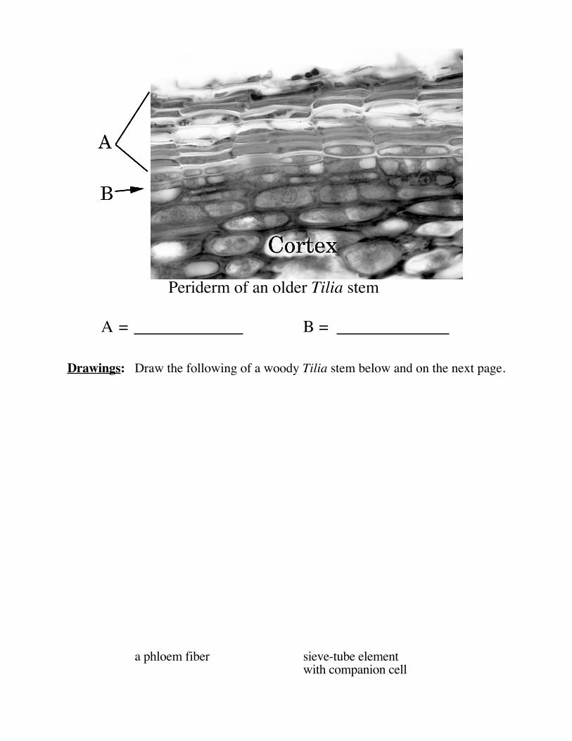
Periderm of an older Tilia stem
A = B =
Drawings: Draw the following of a woody Tilia stem below and on the next page.
a phloem fiber sieve-tube elementwith companion cell

cork cells adjacent vessel elements acrosswith cork cambium a growth ring
Parenchyma in Parenchyma in aa xylem ray non dilated phloem ray
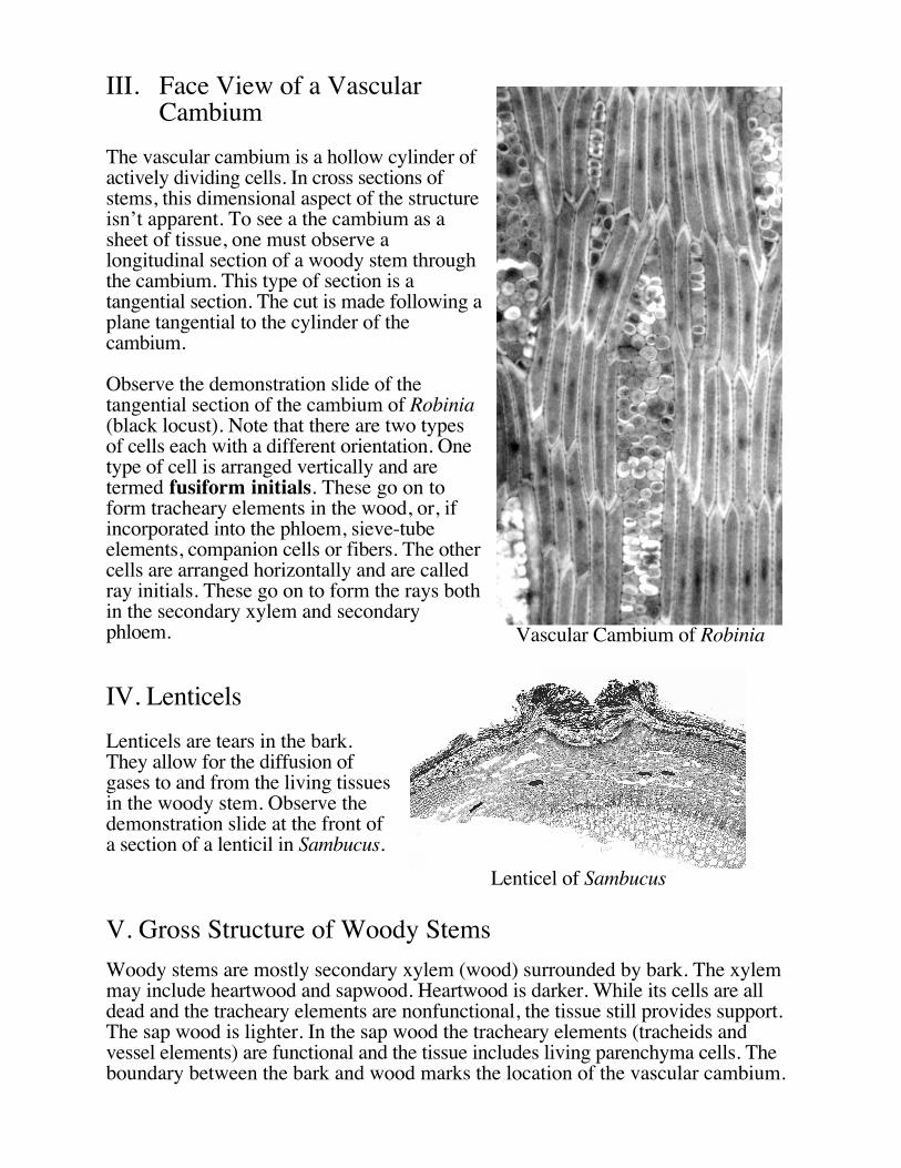
III. Face View of a VascularCambium
The vascular cambium is a hollow cylinder ofactively dividing cells. In cross sections ofstems, this dimensional aspect of the structureisn’t apparent. To see a the cambium as asheet of tissue, one must observe alongitudinal section of a woody stem throughthe cambium. This type of section is atangential section. The cut is made following aplane tangential to the cylinder of t
Vascular Cambium of Robinia
hecambium.
Observe the demonstration slide of thetangential section of the cambium of Robinia(black locust). Note that there are two typesof cells each with a different orientation. Onetype of cell is arranged vertically and aretermed fusiform initials. These go on toform tracheary elements in the wood, or, ifincorporated into the phloem, sieve-tubeelements, companion cells or fibers. The othercells are arranged horizontally and are calledray initials. These go on to form the rays bothin the secondary xylem and secondaryphloem.
IV. Lenticels
Lenticels are tears in the bark.They allow for the diffusion ofgases to and from the living tissuesin the woody stem. Observe thedemonstration slide at the front ofa section of a lenticil in Sambucus.
Lenticel of Sambucus
V. Gross Structure of Woody Stems
Woody stems are mostly secondary xylem (wood) surrounded by bark. The xylemmay include heartwood and sapwood. Heartwood is darker. While its cells are alldead and the tracheary elements are nonfunctional, the tissue still provides support.The sap wood is lighter. In the sap wood the tracheary elements (tracheids andvessel elements) are functional and the tissue includes living parenchyma cells. Theboundary between the bark and wood marks the location of the vascular cambium.

The bark itself is divided into two regions by the cork cambium: the living areainside the cork cambium is the inner bark, and the dead tissue outside is the outerbark. Evidence of earlier cork cambia can be easily discerned in some woody stems.Take a section of oak wood from the front bench and identify the following: pith,heart wood, sap wood, vascular cambium, xylem rays, inner bark, and theouter bark. If you can’t locate these all of the above, ask your TA for help.
Label the Figures.
Cross Section of Bur Oak Stem
A = E =
B = F =
C = G =
D = H =
I =

Detail of the Bark of Bur Oak
A = D =
B = E =
C = F =
VI. Secondary Growth in Roots
The onset of secondary growth in roots is somewhat different than that in stems.This is well illustrated in your text on page 601 in figure 25-16. The pericycle playsan important role in secondary growth. It both forms the periderm and also splicestogether the pieces of vascular cambium at the protoxylem poles where it isdiscontinuous. Because the first periderm is formed by the pericycle, all the tissuesoutside and including the endodermis are sloughed off immediately with the onset ofsecondary growth.
VIa. Secondary Growth in Tilia: Locate the prepared slide of Tilia root andobserve with your microscope. Identify the vascular cambium, secondaryxylem, and secondary phloem. Can you identify the star-shaped mass ofprimary xylem inside this root? Identify the periderm, vascular cambium,primary xylem, secondary xylem, and secondary phloem.
From what primary tissue is the secondary tissue surrounding the phloemderived?
_________________________________

VIb. The carrot: Carrots are roots with lots of secondary growth. At their core, theyare composed of xylem tissue surrounded by phloem tissue. Make a crosssection of a carrot and observe it with a dissection microscope. Note the rayswhich are indicative of secondary tissues. Identify the vascular cambium,primary xylem, secondary xylem, and secondary phloem.
From what primary tissue is the secondary tissue surrounding the phloemderived?
________________________________
Draw a vessel element in your carrot section.
Oak wood and the inner core of carrots are both examples of xylem tissue. Whyis one hard and tough while the other is crisp and delicious? Can you relate thisto the different functions of roots vs. stems?
Cross Section of a Ranunculus Root at the End of Primary Growth (Left).
At the onset of secondary growth in the root, only the vascular cylinder with thepericycle is retained (right).