Are Probiotics Effective in Targeting Alcoholic Liver ...
13
Are Probiotics Effective in Targeting Alcoholic Liver Diseases? Meegun Hong 1 & Dae Hee Han 1 & Jitaek Hong 1 & Dong Joon Kim 1 & Ki Tae Suk 1 # Springer Science+Business Media, LLC, part of Springer Nature 2018 Abstract Alcoholic liver disease (ALD) encompasses a broad spectrum of disorders including steatosis, steatohepatitis, fibrosis, and cirrhosis. Despite intensive research in the last two decades, there is currently no Food and Drug Administration-approved therapy for treating ALD. Several studies have demonstrated the importance of the gut-liver axis and gut microbiome on the pathogenesis of ALD. Alcohol may induce intestinal dysbiosis and increased intestinal permeability, which in turn result in increased levels of pathogen-associated molecular patterns such as lipopolysaccharide (LPS) and translocation of microbial products from the gut to the liver (bacterial translocation). LPS is an inflammatory signal that activates toll-like receptor 4 on Kupffer cells, contributing to the inflammation observed in ALD. Recently, probiotics have been shown to be effective in reducing or preventing the progression of ALD. A potential mechanism is that the probiotics transforms the composition of intestinal microbiota, which leads to reductions in alcohol-induced dysbiosis, intestinal permeability, bacterial transloca- tion, endotoxemia, and consequently, the development of ALD. While transformation of intestinal microbiota by probiotics appears to be a promising therapeutic strategy for the treatment of intestinal barrier dysfunction, there is a scarcity of research that studies probiotics in the context of ALD. In this review, we discuss the potential therapeutic applications of probiotics in the treatment of ALD. Keywords Alcoholic liver disease . Microbiota . Probiotics . Dysbiosis . Gut Introduction Worldwide, alcohol consumption ranks third among the vari- ous risk factors for disease and disability. It is responsible for 2.5 million deaths annually, constituting 4% of all deaths worldwide [1]. The large absolute increase in alcohol con- sumption has led to a rapid increase in alcohol-related diseases and accidents. Alcoholic liver disease (ALD) is responsible for approximately 25% of deaths resulting from alcohol con- sumption [2, 3]. Chronic alcohol ingestion is known to cause steatohepatitis, fibrosis, cirrhosis, and cancer [ 4 – 7 ]. Regardless of the spectrum of ALD, abstaining from alcohol prevents progression of the disease, improves the survival rate, and decreases the need for liver transplantation [8]. The pathophysiology of ALD varies according to the stage of the disease and the presence of genetic and nongenetic factors that affect its onset and clinical progression [9]. However, pathophysiological importance between ALD and gut microbiota is not yet fully understood. Most reports came from animal studies, and little clinical data are available in ALD. Further clinical studies are needed in the future. Activation of Kupffer cells has been identified as an essen- tial element in the pathogenesis of ALD [10, 11]. Alcohol induces bacterial overgrowth (especially that of gram- negative bacteria) and the translocation of the endotoxin lipo- polysaccharide (LPS) from the gut to the liver [12, 13]. Alcohol has been known to disrupt the gut barrier function, which consequently promotes the translocation of microbial LPS from the lumen of the intestines to the portal vein, where it travels to the liver. Kupffer cells and macrophages recruited to the liver can be activated by bacterial endotoxin such as LPS through toll-like receptor (TLR) 4. The levels of LPS in the portal vein and in the systemic circulation are increased with excessive alcohol intake [14, 15]. These observations suggest that gut-derived LPS is the central mediator of inflam- mation in alcoholic steatohepatitis [16]. Moderate alcohol consumption has also been identified as a strong risk factor Meegun Hong, Dae Hee Han, and Jitaek Hong share co-first author. * Ki Tae Suk [email protected] 1 Department of Internal Medicine, Hallym University Chuncheon Sacred Heart Hospital, Hallym University College of Medicine, Gyo-dong, Chuncheon 24253, South Korea Probiotics and Antimicrobial Proteins https://doi.org/10.1007/s12602-018-9419-6
Transcript of Are Probiotics Effective in Targeting Alcoholic Liver ...

Are Probiotics Effective in Targeting Alcoholic Liver Diseases?
Meegun Hong1& Dae Hee Han1
& Jitaek Hong1& Dong Joon Kim1
& Ki Tae Suk1
# Springer Science+Business Media, LLC, part of Springer Nature 2018
AbstractAlcoholic liver disease (ALD) encompasses a broad spectrum of disorders including steatosis, steatohepatitis, fibrosis, andcirrhosis. Despite intensive research in the last two decades, there is currently no Food and Drug Administration-approvedtherapy for treating ALD. Several studies have demonstrated the importance of the gut-liver axis and gut microbiome on thepathogenesis of ALD. Alcohol may induce intestinal dysbiosis and increased intestinal permeability, which in turn result inincreased levels of pathogen-associated molecular patterns such as lipopolysaccharide (LPS) and translocation of microbialproducts from the gut to the liver (bacterial translocation). LPS is an inflammatory signal that activates toll-like receptor 4 onKupffer cells, contributing to the inflammation observed in ALD. Recently, probiotics have been shown to be effective inreducing or preventing the progression of ALD. A potential mechanism is that the probiotics transforms the composition ofintestinal microbiota, which leads to reductions in alcohol-induced dysbiosis, intestinal permeability, bacterial transloca-tion, endotoxemia, and consequently, the development of ALD. While transformation of intestinal microbiota byprobiotics appears to be a promising therapeutic strategy for the treatment of intestinal barrier dysfunction, there is ascarcity of research that studies probiotics in the context of ALD. In this review, we discuss the potential therapeuticapplications of probiotics in the treatment of ALD.
Keywords Alcoholic liver disease . Microbiota . Probiotics . Dysbiosis . Gut
Introduction
Worldwide, alcohol consumption ranks third among the vari-ous risk factors for disease and disability. It is responsible for2.5 million deaths annually, constituting 4% of all deathsworldwide [1]. The large absolute increase in alcohol con-sumption has led to a rapid increase in alcohol-related diseasesand accidents. Alcoholic liver disease (ALD) is responsiblefor approximately 25% of deaths resulting from alcohol con-sumption [2, 3]. Chronic alcohol ingestion is known to causesteatohepatitis, fibrosis, cirrhosis, and cancer [4–7].Regardless of the spectrum of ALD, abstaining from alcoholprevents progression of the disease, improves the survivalrate, and decreases the need for liver transplantation [8].
The pathophysiology of ALD varies according to the stageof the disease and the presence of genetic and nongeneticfactors that affect its onset and clinical progression [9].However, pathophysiological importance between ALD andgut microbiota is not yet fully understood. Most reports camefrom animal studies, and little clinical data are available inALD. Further clinical studies are needed in the future.
Activation of Kupffer cells has been identified as an essen-tial element in the pathogenesis of ALD [10, 11]. Alcoholinduces bacterial overgrowth (especially that of gram-negative bacteria) and the translocation of the endotoxin lipo-polysaccharide (LPS) from the gut to the liver [12, 13].Alcohol has been known to disrupt the gut barrier function,which consequently promotes the translocation of microbialLPS from the lumen of the intestines to the portal vein, whereit travels to the liver. Kupffer cells and macrophages recruitedto the liver can be activated by bacterial endotoxin such asLPS through toll-like receptor (TLR) 4. The levels of LPS inthe portal vein and in the systemic circulation are increasedwith excessive alcohol intake [14, 15]. These observationssuggest that gut-derived LPS is the central mediator of inflam-mation in alcoholic steatohepatitis [16]. Moderate alcoholconsumption has also been identified as a strong risk factor
Meegun Hong, Dae Hee Han, and Jitaek Hong share co-first author.
* Ki Tae [email protected]
1 Department of Internal Medicine, Hallym University ChuncheonSacred Heart Hospital, Hallym University College of Medicine,Gyo-dong, Chuncheon 24253, South Korea
Probiotics and Antimicrobial Proteinshttps://doi.org/10.1007/s12602-018-9419-6

for small intestinal bacterial overgrowth [17]. Figure 1 ex-plains this process of alcohol consumption bringing aboutchanges in the intestinal milieu and inducing consequentdownstream immune responses in the liver.
Metabolites produced by bacteria, such as short chain fattyacids, volatile organic compounds, and bile acids, are in-volved in ALD pathology [18, 19]. Alcohol fed mice exhibit-ed decreased expression of bacterial genes involved in thebiosynthesis of saturated fatty acids and decreased levels ofsaturated long-chain fatty acids [18]. In the comparison of thefecal metabolites, 13 biomarkers are related to ALD and themost discriminating molecules were bile acid derivatives andfatty acids [19].
A previous study demonstrated the elevated alcohol con-centration in the breathing air of ob/ob mice and demonstratedthat this alcohol concentration can be reduced by gut micro-bial changes with neomycin [20]. The increased alcohol-producingmicrobiota and serum-ethanol concentrations in pa-tients with nonalcoholic steatohepatitis and the well-knownrole of ethanol metabolism in oxidative stress and liver inflam-mation strongly suggest that alcohol-producing microbiotaplay an important role in the pathophysiology of nonalcoholicsteatohepatitis [21]. Results from several research groups sup-port that some microbiota in nonalcoholic fatty liver diseasepatients produce alcohol. In other words, alcohol metabolismmay represent an important triggering factor in nonalcoholicfatty liver disease pathogenesis [22, 23].
Probiotics has been associated with beneficial effects onthe gut; probiotics can transiently colonize the gastrointestinaltract, correct dysbiosis, inhibit the growth and virulence of
enteric pathogens, and decrease the expression of TLR 4 onKupffer cells [24, 25]. Some reports have demonstrated thetherapeutic potential of probiotics in clinical studies and ani-mal models of ALD.
Alcohol-Associated Changes of the GutMicrobiota: Animal and Clinical Studies
As the gut is directly linked to the liver through the portal tract,the dysbiosis in gut microbiotas can result liver disease includ-ing ALD. Theoretically, modulation of gut microbiota(dysbiosis) to healthy state by the administration of probioticscan be effective in the therapy of ALD and recent evidenceshave been released.
Animal Studies
The interaction between the microbiota and the host liver isassociated with the pathogenesis of ALD. It has been demon-strated that alcohol can alter the compositions of themicrobiome such as small intestinal bacterial overgrowthand impair intestinal integrity and barrier function by directtoxic effect [26, 27]. Many studies have demonstrated therelationship between the gut and ALD. One study, inducedALD in C57BL/6 J mice using the Tsukamoto-French model,which involves continuous intragastric feeding of an isocalo-ric diet or alcohol for 3 weeks, and demonstrated that alcoholcauses intestinal dysbiosis, reducing the capacity of themicrobiome to synthesize saturated long-chain fatty acid and
Fig. 1 Probiotics and alcoholicliver disease. Alcoholconsumption leads to prominentchanges in the composition ofmicrobiomes which induce smallintestinal bacterial overgrowthand dysbiosis. And, probioticshas the ability to transform theintestinal microbiota communitycompositions, which leads to thereduction of alcohol-induceddysbiosis, intestinal permeability,bacterial translocation,endotoxemia, and thedevelopment of ALD [72]
Probiotics & Antimicro. Prot.

the proportion of Lactobacillus species [18]. In another studyutilizing the Tsukamoto-French model, ethanol feeding hadshown to reduce the numbers of Lactobacillus [28].Intragastric feeding of ethanol with continuous infusion hasbeen associated with the reduction of operational taxonic unitof several Firmicutes (namely Lactococcus, Pediococcus,Lactobacillus, and Leuconostoc) and an increase inVerrucomicrobia and Bacteroides such as Bacteroidales andPorphyromonadaceae [29]. Another study using a liquid eth-anol diet in a mouse model utilizing Lieber-DeCarli demon-strated alcohol-induced phyla shifts involving a significantreduction in Firmicutes but remarkable expansion ofProteobacteria and Actinobacteria [30]. In this study, theyobserved an increase of Lactobacillus conversely to someothers groups and a decrease in Bacteroides andParabacteroides. A study utilizing an in vivo rat modelshowed that a 12-week liquid ethanol diet results in signifi-cantly higher numbers of Escherichia coli but significantlylower numbers of Lactobacillius and Bifidobacterium [31](Table 1). However, analysis methods for the microbiota com-position in these different studies are different (pyrosequenc-ing on the 454 FLX Titanium platform, real-time PCR, orIllumina HiSeq 2000 platform), so results can be changedand standard methodology with upgraded technic is need forthe metagenomics.
Clinical Studies
In human studies, quantitative and qualitative changes in theintestinal microbiota can occur in subjects with moderate al-cohol consumption and patients with alcoholic cirrhosis[32–34]. Clinical researches have demonstrated an increasein the number of E. coli but a decrease in the numbers ofLactobacillus, Lactococcus, Periococcus, Leuconostoc [35],Clostridium leptum [36], and Bacteroidaceae [37]. Typically,the mean abundance of Bacteroidaceae from Bacteroideteswas decreased in the alcoholic groups compared with thehealthy control and the groups were statistically significantlydifferent. A previous study on patients with alcoholic hepatitisdemonstrated that alcohol consumption increases the numbersof Bifidobacterium, Streptococci, and Enterobacteria but de-creases the number of Atopobium [38]. Alcohol-dependentpatients with high-intestinal permeability were observed withdrastic decreases in the numbers of Ruminococcus,Faecalibacterium, Subdoligranulum, Oscillibacter, andAnaerofilum [34]. Other studies have shown that cirrhosisis associated with decreased abundance of Bacteroidaceaeand Veillonellaceae particularly in patients with alcohol-induced liver cirrhosis [39]. In conclusion, chronic alcoholconsumption leads to prominent changes in microbiomewhich induces intestinal bacterial overgrowth anddysbiosis (Table 2).
Probiotics in Alcoholic Liver Disease
Probiotics Products
Most probiotics contain Lactobacillus and Bifidobacterium,both of which are saccharolytic bacteria that can ferment car-bohydrates to lactic acid; lactic acid is known to be effective ininhibiting the growth of pathogenic bacteria. In addition, py-ruvate produced from fermentation can be utilized by certaincolonic anaerobes in the production of beneficial short-chainfatty acids [25]. Lactobacillus strains, LAP5 and LF33, havebeen found to inhibit the growth of E. coli and Salmonellatyphimurium in vitro [40], while another strain, NP51, wasshown to reduce the number of E. coli O157:H7 in fecal sam-ples of beef cattle [41, 42]. B. animalis MB5 andL. rhamnosus Gorbach-Goldin (GG) were demonstrated toprotect intestinal cells from inflammation caused by E. coli[43]. Lactobacillius strains commonly found in yogurt andprobiotic supplements include the following: L. acidophilus,L. bulgaricus, L. rhamnosus GG, L. plantarum, L. reuteri,L. salivarius, L. casei, and L. johnsonii. The following strainsof Bifidobacterium are commonly utilized as probiotics:B. bifidum, B. lactis, B. longum, and B. breve (Table 3).
Animal Studies
Many studies utilizing alcohol-treated experimentalmodels have evaluated probiotics as a therapeutic approachthat can potentially reduce bacterial translocation, improveintestinal microbiota, and fortify the gut barrier (Table 4).For instance, oral administration of heat-killed L. brevisSBC8803 increased expression of cytoprotective Hsp25mRNA in the small intestine and prevented ethanol-induced overexpression of TNF (tumor necrosis factor)-αand sterol regulatory element-binding proteins at themRNA level in the liver [44].
In a rat gavage model of ALD, treatment withL. rhamnosusGG has been associated with a robust reductionin-alcohol induced gut hyperpermeability, necroinflammationsystemic oxidative stress, and severity of alcoholicsteatohepatits [45]. Another study utilizing oral gavage onALD-induced rats revealed that administration ofL. plantarum ameliorates ALD by suppression of molecularinflammatory markers, particularly the IL (interleukin)-12/p40 subunit [46]. Further, L. rhamnosus GG treatment hasbeen found to have protective effects on Lieber-DeCarli alco-hol diet induced hepatic inflammation; these observed effectsare closely associated with the attenuation of TNF-α produc-tion via inhibition of TLR4- and TLR5-mediated endotoxinactivation [47], regulation of hypoxia inducible factor (HIF)-targeted epithelial barrier-protective factor [48], increase offatty acid β-oxidation [49], and the reduction of de novo lipo-genesis and hepatic apoptosis [49]. Remodeling of the
Probiotics & Antimicro. Prot.

Table1
Changes
inintestinalmicrobiotain
anim
als
Animal
Conditio
nCom
parison
Method
Phylum
Class
Order
Fam
ilyGenus/species
C57BL/6
J(age
8weeks)
theTsukam
oto-
French
model
Contin
uous
intragastric
feedingof
isocaloric
dietor
alcoholfor
3weeks
16SrRNAgene
pyrosequencing,
andquantitative
real-tim
ePCR,
cecum
samples
Firmicutes
↓Bacteroidetes
↑Bacilli
Lactobacillales
Lactobacillaceae
Lactobacillus
↓L.rham
nosus↓
[28]
C57BL/6
JIntragastric
Tsukam
oto-
French
model
Isocalorisdietand
24.8g/kg/day
alcohol1
weeks
DNApergram
offeces
Firmicutes
Verrucom
icrobia
Bacilli
Verrucom
icrobiae
Lactobacillales
Verrucom
icrobiales
Lactobacillaceae
Verrucom
icrobiaceae
Lactobacillus
↓Akkermansia
muciniphila
↓
[29]
C57/B6
(age
8weeks)
Intragastricfeeding
ofalcohol
Isocalorisdietand
30.9g/kg/day
alcohol3
weeks
16SrRNAgene
pyrosequencing,
andquantitative
real-tim
ePCR,
cecum
samples
Bacteroidetes
↑Firmicutes
↓Verrucom
icrobia↑
Bacteroidetes
Bacilli
Bacteroidales
↑La
ctobacillales
Bacteroidaceae
Porphyrom
onadaceae↑
Lactobacillaceae
Leuconostocaceae
Bacteroides
↑La
ctococcus↓
Pediococcus
↓La
ctobacillus
↓Leuconostoc↓
[30]
C57BL/6
N(age
8–10
weeks)
Lieber-DeC
arli
liquiddiet5%
ethanold
iet
8weeks
IsocaloricLieber-
DeC
arliliq
uiddiet
with
maltose
dextrin
16SrRNAgene
sequencing,
stoolsam
ples
Actinobacteria↑
Proteobacteria↑
Firmicutes
↑Bacteriodetes
↓
Actinobacteria
Betaproteobacteria
Clostridia
Actinom
ycetales
Burkholderiales
Clostridiales
Corynebacteriaceae
Alcaligenaceae
Rum
inococcaceae
↓La
chnospiraceae↓
Corynebacterium
↑Alcaligenes
↑La
ctobacillus
↑
[31]
Wistarrats
(age
8weeks)
theliq
uid
ethanold
iet
Liquidethanold
iet
contained35%
energy
asethanol,
isocaloricnorm
alliq
uiddietfor
12weeks.
Qutitativ
ecultu
ring,
fecalsam
ples
Firmicutes
Actinobacteria
Probacteria
Bacilli
Actinobacteria
Gam
maproteobacteria
Lactobacillales
Bifidobacteriales
Enterobacteriales
Lactobacillaceae
Bifidobacteriaceae
Enterobacteriaceae
Lactobacillus
↓Bifidobacterium↓
Escherichia
coli↑
[32]
↑,increase
incondition
alcoholic
diseaserelativ
eto
condition
control;↓,
decrease
incondition
alcoholic
diseaserelativ
eto
condition
control;rRNAribosomeribonucleicacid;PCRpolymerasechain
reactio
n
Probiotics & Antimicro. Prot.
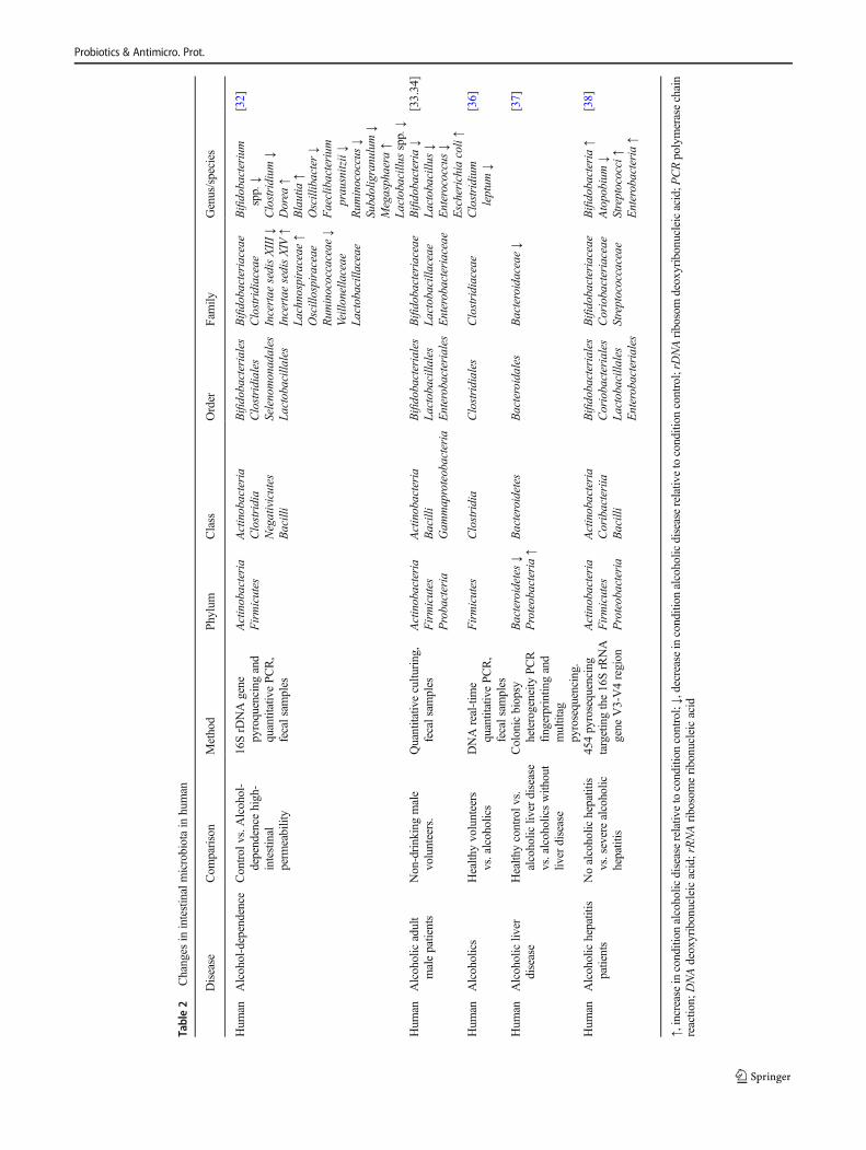
Table2
Changes
inintestinalmicrobiotain
human
Disease
Com
parison
Method
Phylum
Class
Order
Fam
ilyGenus/species
Hum
anAlcohol-dependence
Control
vs.A
lcohol-
dependence
high-
intestinal
perm
eability
16SrD
NAgene
pyroquencing
and
quantitativePCR,
fecalsam
ples
Actinobacteria
Firmicutes
Actinobacteria
Clostridia
Negativicutes
Bacilli
Bifidobacteriales
Clostridiales
Selenomonadales
Lactobacillales
Bifidobacteriaceae
Clostridiaceae
Incertae
sedisXIII↓
Incertae
sedisXIV
↑La
chnospiraceae↑
Oscillospiraceae
Rum
inococcaceae
↓Veillonellaceae
Lactobacillaceae
Bifidobacterium
spp.↓
Clostridium
↓Dorea
↑Blautia
↑Oscillibacter↓
Faeclibacterium
prausnitzii↓
Rum
inococcus↓
Subdoligranulum↓
Megasphaera
↑La
ctobacillus
spp.↓
[32]
Hum
anAlcoholicadult
malepatients
Non-drinkingmale
volunteers.
Quantitativ
ecultu
ring,
fecalsam
ples
Actinobacteria
Firmicutes
Probacteria
Actinobacteria
Bacilli
Gam
maproteobacteria
Bifidobacteriales
Lactobacillales
Enterobacteriales
Bifidobacteriaceae
Lactobacillaceae
Enterobacteriaceae
Bifidobacteria
↓La
ctobacillus
↓Enterococcus↓
Escherichia
coli↑
[33.34]
Hum
anAlcoholics
Health
yvolunteers
vs.alcoholics
DNAreal-tim
equantitativePCR,
fecalsam
ples
Firmicutes
Clostridia
Clostridiales
Clostridiaceae
Clostridium
leptum
↓[36]
Hum
anAlcoholicliv
erdisease
Health
ycontrolv
s.alcoholic
liver
disease
vs.alcoholicswith
out
liver
disease
Colonicbiopsy
heterogeneity
PCR
fingerprintin
gand
multitag
pyrosequencing.
Bacteroidetes
↓Proteobacteria↑
Bacteroidetes
Bacteroidales
Bacteroidaceae↓
[37]
Hum
anAlcoholichepatitis
patients
Noalcoholic
hepatitis
vs.severealcoholic
hepatitis
454pyrosequencing
targetingthe16SrRNA
gene
V3-V4region
Actinobacteria
Firmicutes
Proteobacteria
Actinobacteria
Coribacteriia
Bacilli
Bifidobacteriales
Coriobacteriales
Lactobacillales
Enterobacteriales
Bifidobacteriaceae
Coriobacteriaceae
Streptococcaceae
Bifidobacteria
↑Atopobium
↓Streptococci↑
Enterobacteria↑
[38]
↑,increase
incondition
alcoholic
diseaserelativ
etocondition
control;↓,decrease
incondition
alcoholic
diseaserelativ
etocondition
control;rD
NAribosomdeoxyribonucleicacid;P
CRpolymerasechain
reactio
n;DNAdeoxyribonucleicacid;rRNAribosomeribonucleicacid
Probiotics & Antimicro. Prot.

structure of the microbial community in response toL. rhamnosus GG probiotic treatment may be a contributingfactor in the reduction of intestinal permeability, which con-sequently leads to the attenuation of bacterial translocationand endotoxemia [30]. Wang et al. [50] have demonstratedthat L. rhamnosus GG pretreatment has a protective roleagainst the deleterious effects of binge alcohol exposure,which involves HIF adaptation signaling and mucus protec-tive gene regulation (associated with the expression of tightjunction proteins and adaptors).
VSL#3 Bifidobacteriaceae (B. longum, B. infantis, andB. breve), Lactobacillaceae (L. acidophilus, L. paracasei,L. bulgaricus, and L. plantarum), and Streptococcusthermophilus (3 × 10¹¹/g of viable lyophilized bacteria) treat-ment has been shown to prevent endotoxin and other bacterialproducts in the gut lumen from passing into the portal circu-lation. In addition, the treatment also decreased the productionof TNF-α and increased the expression of tight junction pro-teins in a rat model [51]. Another study utilizing a rat modelhas shown that supplementation with L. acidophilus,
L. bulgaricus, B. bifidum, B. longum, and Streptococcusthermophilus during ethanol exposure can inhibit the eleva-tion of plasma endotoxin levels by normalizing intestinal per-meability and fecal microbial compositions [31].
In two separate studies utilizing mouse models, the ef-ficacy of probiotics has been implicated directly in thecontext of ALD. When a probiotics diet consisting ofL. rhamnosus and L. acidophilus (Lacidofil®) was admin-istered to a mouse model with ALD for 4 weeks, TLR4levels were found to be significantly lower in the groupstreated with probiotics compared with the control group[52, 53]. The TLR4 pathway has been previously describedas the central component through which ALD-inducedchanges in microbiota can cause eventual inflammatoryresponses and damage the liver. Administration ofprobiotics can also decrease the levels of deleterious cyto-kines such as IL-1β and TNF-α [53]. These results alignwith the finding that probiotics have an inhibitory effect onthe TLR4 pathway which is associated with the release ofpro-inflammatory cytokines.
Table 3 Probiotics productsProduct Company Stain
Duolac Gold probiotics Cell Biotech Co., Ltd. Bifidobacterium bifidum (KCTC 12199BP),
Bitidobacterium lactis (KTCT 11904BP),
Bifidobacterium longum (KCTC 12200BP),
Lactobacillus acidophilus (KCTC 11906BP),
Lactobacillus rhamnosus (KCTC 12202BP),
Streptococcus thermophilus (KCTC 11870BP)
5 × 10⁹ viable cells
Yakult® Yakult Honsha Co., Ltd. Lactobacillus casei Shirota at a concentrationof 108/mL
Lacidofil® Pharmbio Korea Lactobacillus rhamnosus R0011,
Lactobacillus acidophilus R0052
2 × 10⁹ CFU
Lactowel Chong Kun Dang
PharmaceuticalCorporation
Lactobacillus subtilis, Streptococcus faecium
5.0 × 10⁸ CFU
VSL#3 VSL Pharmaceuticals, Ft. Bifidobacterium longum, Bifidobacterium infantis,Bifidobacterium breve, Lactobacillus acidophilus,Lactobacillus paracasei, Lactobacillusbulgaricus,Lactobacillus plantarum,
Streptococcus thermophilus
3 × 10¹¹/g of viable lyophilized bacteria
LP299V Adock Ingram Lactobacillus plantarum 299v, 109 CFU
BioGaia BioGaia® Lactobacillus reuteri DSM 17938, 1 × 10⁸ CFU
Normia® JGL Lactobacillus rhamnosus GG (LGG®),
Bifidobacterium (BB-12®) in the concentrationof 108 to 1010
BAlgibif^ and BAlgilac^ Microgen/Imbio Bifidobacterium bifidum 0.9 × 10⁸ CFU,
Lactobacillus plantarum 8PA3 0.9 × 10⁹ CFU
CFU colony-forming unit
Probiotics & Antimicro. Prot.

Table4
Probioticsin
alcoholic
liver
diseaseanim
al
Animal
Conditio
nCom
parison
Serum
Liver
Intestine
Microbiota
C57BL/6
N(age
7weeks)
Lieber-DeC
arlidietalcoholand
water
4or
5weeks,ethanol-containing
diet-fed
Lactobacillus
brevis
Heat-killedL.
brevisSB
C8803
100
or500mg/kg/day,4
weeks
with
alcohol
Totalcholesterol
↓Triacylglycerol
↓SREBP1↓
SREBP2↓
Hsp25
↑[44]
Sprague-Daw
leyrats
Gavaged
with
alcoholtwicedaily
(8g/kg)for10
weeks
Oncedaily
gavage
of2.5×10
7liv
eL.
rham
nosusGorbach-G
oldin,
10weeks
with
alcohol
Necrosis↓
inflam
mation↓
MPO↓
Carbonyl↓
Nitrotyrosine↓
Fat↓
Carbonyl↓
Nitrotyrosine↓
[45]
Wistarrats
(200–250
g)14
g/kg
35%
alcohol1
0weeks
through
oralgavage
L.plantarum,109
CFU/m
L,
8weeks
with
alcohol
Endotoxin
↑NF-κB↑
IL-12/p40↓
[46]
C57BL/6
NLieber-DeC
arlidiet,5%
alcohol
for8weeks
L.rham
nosusGG
109CFU
/day,last2
weeks
oftheexperiment.
MPOactiv
ity↓
TNF-α↓
Cyp2E
1↓
TLR5↓
[47]
C57BL/6
N(age
8weeks)
Lieber-DeC
arlidietcontaining
5%alcoholfor
8weeks
L.rham
nosusGG
109CFU
/day,2
weeks
ALT
↓LPS↓
TG↓
VEGF↑
ITF↑
HIF-2α↑
ZO-1
↑Claudin-1
↑Occludin↑
[48]
C57BJ/6N(m
ale)
Lieber-DeC
arlidietcontaning5%
alcoholfor
8weeks
L.rham
nosusGG,
109CFU
/day,last2
weeks
oftheexperiment.
AST↓
ALT
↓FFA
↓
TG↓
FFA
↓SREBP-1c↑
SCD-1
↑PPA
R-α
↑PG
C-1α↑
CPT-1↑
p-AMPKα↑
p-ACC↑
[49]
C57BL/6
N(age
8–10
weeks,m
ale)
Lieber-DeC
arliliq
uiddiet5%
ethanol
diet8weeks,isocaloricLieber-DeC
arli
liquiddietwith
maltose
dextrin
L.rham
nosusGG
109CFU
/mLlast2weeks
oftheexperiment.
LPS↓
ALT
↓ZO-1
↑claudin-1↑
Symplekin
↑p130
↑
Actinobacteria↓
Rum
inococcaceae
↑Corynebacterium
↓Alcaligenes
↓La
ctobacillus
↑
[30]
C57BL/6
N(age
9weeks,m
ale)
Alcohol
at6g/kg
was
administered
viagavage
L.rham
nosusGG
109CFU
,5days
during
theexperiment
TG↓
ROS↓
TNF-α↓
MPO↓
ROS↓
Ileum
perm
eability↓
Fordrin
↑Sy
mplekin
↑HIF-2α↑
HIF-1α↑
ZO-1
↑Claudin-1
↑Occludin↑
[50]
Probiotics & Antimicro. Prot.

Clinical Studies
A clinical study with alcoholic cirrhosis patients has suggestedthat administration of L. casei Shirota (6.5 × 10⁹ CFU, threetimes daily for 4 weeks, with abstinence state) can restoreneutrophil phagocytic capacity in cirrhosis, possibly by alter-ing IL-10 secretion and TLR4 expression [54]. In a cohortstudy on alcoholic liver cirrhosis patients, Yakult 400 contain-ing L. casei (with abstinence state) was shown to significantlyincrease serum levels of liver-specific rapid-turnover proteintransthyretin and decrease the level of h-c-reactive protein(CRP). In addition, alcohol-induced deterioration of gut florawas improved with the increase of protein production inducedby probiotic treatment [55].
In a study involving mild (not severe) alcoholic hepatitispatients, intake of L. subtilis and S. faecium (Lactowel, 5.0 ×10⁸ CFU, without drinking alcohol during study period) re-duced gut-derived microbial LPS and E. coli [53]. In thisstudy, patients who were > 20 years old had liver function testwith an aspartate aminotransferase (AST)/alanine aminotrans-ferase (ALT) > 1 and elevated AST (ALT) level and an alcoholconsumption history of more than 40 g/day for women and60 g/day for men during the 7 days before screening weredefined as a mild alcoholic hepatitis. Administration ofprobiotics (Duolac Gold probiotics containing B. bifidum,B. lactis, B. longum, L. acidophilus, L. rhamnosus, andS. thermophilus) for 4 weeks in chronic liver disease patients(24 ALD patients) resulted in the alleviation of small intestinalbacterial overgrowth and digestive symptoms. In addition, thelevels of fecalB. lactis, L. rhamnosus, and L. acidophiluswereincreased [56].
In ALD patients, probiotics (BAlgibif^ and BAlgilac^ con-taining B. bifidum and L. plantarum) therapy led to an increasein fecal levels of Bifidobacteria and Lactobacilli and decreasein serumASTwith standard therapy [35]. They concluded thatshort-term oral supplementation with B. bifidum andL. plantarum 8PA3 was associated with restoration of thebowel flora and greater improvement in alcohol-induced liverinjury than standard therapy alone taken together; probioticshave the ability to transform composition of the intestinalmicrobiota community, which leads to the reduction ofalcohol-induced dysbiosis, intestinal permeability, bacterialtranslocation, endotoxemia, and the development of ALD.Transformation of intestinal microbiota should be consideredas a therapeutic strategy against intestinal barrier dysfunctionand the development of ALD. (Table 5).
Future Research Directions in Alcoholic LiverDisease
As the direct connection between the intestines and the liver,the gut-liver axis, gut microbiota, and associated dysbiosisT
able4
(contin
ued)
Animal
Conditio
nCom
parison
Serum
Liver
Intestine
Microbiota
Wistarrats
(age
6–8weeks)
5g/kg
40%
alcoholthrough
stom
ach
feedingevery12
h,threetim
esVSL
#30.6g/kg,30min
priorto
administrationof
alcohol
TNF-α
↓Endotoxin
↓Occludin↑
ZO-1
↑[51]
Wistarrats
(age
8weeks)
Liquidethanold
ietcontained
35%
energy
asethanol,isocaloricnorm
alliq
uiddietfor12
weeks.
Synbiotic
powderwith
L.acidophilus,
L.bulgaricus,B
.bifidum,B
.longum,
andS.thermophiles,12
weeks
Endotoxin
↓TG↓
TNF-α↓
IL-10↑
Relativeintesity
oflactulose↓
Latobacillu
s↑
Bifidobacterium↑
[31]
C57BL/6
(age
6weeks)
5g/kg
/day
ethanolo
ralg
avage,plus
LPSinjection11
weeks
Lacidofil®
(L.rhamnosusR0011
and
acidophilusR0052,last2
weeks
oftheexperiment.
TLR4↓
TNF-α↓
Restoratio
nof
microvilli
ofintestine
[53]
↑,increase
incondition
alcoholic
diseaserelativ
eto
treatm
entprobiotics;↓,
decrease
incondition
alcoholic
diseaserelativ
eto
treatm
entprobiotics;CFUcolony-formingunit;
SREBP,
sterol
regulatory
elem
ent-bindingprotein;Hsp25,heatshock
protein25;M
PO,m
yeloperoxidase;N
F-κB,nuclearfactorkappa-lig
ht-chain-enhancerofactivated
Bcells;IL,interleukin;TN
F,tumornecrosisfactor;C
yP2E
1,cytochromeP4
50family
2subfam
ilyEmem
ber1;TL
R,toll-lik
ereceptor;A
LT,alanine
transaminase;LP
S,lip
opolysaccharide;TG
,triglyceride;VEGF,
vascularendothelialgrowthfactor;ITF
,intestin
altrefoilfactor;HIF,hypoxia-induciblefactor;ZO
,zonula
occludens;AST,aspartateam
inotransferase;FFT,
free
fatty
acid;SC
D-1,stearoyl-coenzym
eA
desaturase
1;PPA
R,peroxisomeproliferator-
activ
ated
receptor;PRC,PPA
Rgammacoactiv
ator;CPT,
carnitine
palm
itoyltransferase;
AMPK,5′
adenosinemonophosphate-activated
proteinkinase;ACC,adenoidcystic
carcinom
a;ROS,
reactiv
eoxygen
species
Probiotics & Antimicro. Prot.

have been known as another regulators in the pathophysi-ology of ALD. As a result, new therapeutic approaches formodulation of gut microbiota have been proposed and theeffectiveness of new therapies including probiotics, prebi-otics, synbiotics, fecal microbiota transplantation (FMT),bile acid regulation, and absorbent has been demonstratedin recent several studies. Recent report demonstrated thatethanol exposure diminishes intestinal Akkermansiamuciniphila abundance in both mice and humans and canbe recovered in experimental ALD by oral supplementa-tion of A. muciniphila.
FMT has been of interest in its therapeutic potential forseveral diseases, such as irritable bowel syndrome, metabolicsyndrome, Clostridium difficile infection (CDI), Crohn’s dis-ease, ALD, non-alcoholic fatty liver disease, non-alcoholicsteatohepatitis, neuro-developmental disorders, autoimmunediseases, and allergic diseases [57–59]. The use of FMT inCDI patients was associated with a significant decrease inthe serum level of pro-inflammatory cytokines (TNF-α, IL-1β, IL-6, IL-8, and IL-12) and CRP and normalized fecalcalprotectin [60]. A fructose-rich diet-induced metabolic syn-drome in a rat model of obesity, treated with antibiotics(Ampicilin and neomycin) and FMT (stools from control an-imals), showed reduction in plasma non-esterified fatty acids,LPS, and TNF-α in both groups of fructose-fed rats [61]. Astudy of FMT- and prebiotic (pectin)-treatedmice receiving analcohol diet showed that levels of alanine aminotransferase,triglyceride, and pro-inflammatory cytokines such as TNF-α,IL-1β, CCL2, and TGF-β level in the liver were similar to thatof mice fed the control diet [62]. A severe alcoholic hepatitispilot study found that 1 week of FMT improved liver functionand survival at 1 year [63]. Ren et al. [64] demonstrated thatFMT-induced hepatitis B virus e-antigen (HBeAg) clearance
significantly increased in patients who had persistently posi-tive HBeAg even after long-term antiviral treatment.
Prebiotics are defined as Ba non-digestible food ingredientthat beneficially affects the host by selectively stimulating thegrowth and/or activity of one or a limited number of bacteriain the colon, and thus improves host health.^ [65]. Prebiotics(lactulose) treatment improves psychometric functions in he-patic cirrhosis patients with subclinical hepatic encephalopa-thy [66]. Prebiotics (oats) may prevent alcohol-induced oxi-dative tissue damage, disruption of intestinal barrier integrity,up-regulation of iNOS, and nitric oxide overproduction in thecolonic mucosa [67]. Fructo-oligosaccharides and galacto-oligosaccharide prevented the deleterious effects on behavior,cytokine (such as TNF-α and IL-6) release, and microbiotainduced by chronic psychosocial stress. Previous report dem-onstrated that prebiotics (fructooligosaccharides) improve al-coholic steatohepatitis by inducing Reg3g expression and re-ducing intestinal bacterial overgrowth [29]. Moreover chang-es in microbial community, coupled with increased cecalweight and total bacterial numbers, lead to higher levels ofshort-chain fatty acids in the cecum [68]. Specifically, thepotential synergy between probiotics and prebiotics improvesthe intestinal microbial environment, activates host immunefunction, and prevents or treats diarrhea, inflammatory boweldisease, and hepatic steatosis [69, 70].
Antibiotics are able to affect gut microbiota compositionsignificantly, which consequently leads to clinical manifesta-tions, with either a Beubiotic^ effect or a Bdysbiotic^ effect.Antimicrobials may reduce the population of deleterious bac-teria, decreasing the amount of LPS released and diminishingthe associated inflammatory response [71, 72]. A clinicalstudy on obesity demonstrated that short-term antibiotics(vancomycin) treatment markedly affected microbial diversity
Table 5 Probiotics in alcoholic liver disease-clinical trials
Disease Comparison Serum Microbiota
Human Alcoholic cirrhosis Lactobacillus caseiShirota 6.5 × 10⁹ CFU,3 times daily for 4 weeks
Neutrophil TLR4 ↓TNFR1 ↓TNFR2 ↓IL-10 ↓
[54]
Human Alcoholic liver cirrhosis Yakult 400, 4 weeks h-CRP ↓ Clostridium coccoides ↑Bacteroides fragilis ↑Enterobacteriaceae ↓
[55]
Human Alcoholic liver disease(AST/ALT > 1)
Lactowel, 7 days LPS ↓TNF-α ↓
E.coli ↓ [56]
Human Alcoholic liver disease Duolac gold probiotics B. lactis ↑L. rhamnosus ↑L. acidophilus ↑
[57]
Human Alcoholic liver disease BAlgibif^ and BAlgilac^ AST ↓ Bifidobacteria ↑Lactobacilli ↑Enterococci ↑
[35]
↑, increase in condition alcoholic disease relative to treatment probiotics; ↓, decrease in condition alcoholic disease relative to treatment probiotics;CFU,colony-forming unit; TLR, toll-like receptor; TNFR, tumor necrosis factor receptor; IL, interleukin; h-CRP, hypersensitive C-reactive protein; AST,aspartate aminotransferase; ALT, alanine transaminase; LPS, lipopolysaccharide; TNF, tumor necrosis factor
Probiotics & Antimicro. Prot.

and composition, which was accompanied by a reduced con-version of primary to secondary bile acids and a lower pro-duction of short-chain fatty acids in the gut [73]. I. Bergheimet al. [74] demonstrated that chronic fructose intake associatedwith NAFLD and antibiotics (polymyxin and neomycin) treat-ment markedly reduced hepatic lipid accumulation and hepat-ic triglyceride levels. Treatment with rifaximin improved theclinical status of patients with NASH through the inhibition ofLPS production, which was associated with reduced serumaspartate aminotransferase, alanine aminotransferase, gammaglutamyl transpeptidase, ferritin, and low-density lipoproteinlevels [75]. Treatment of alcohol-related decompensated cir-rhosis with rifaximin significantly improved the hepatic ve-nous pressure gradient values, significantly decreased plasmaendotoxin levels in patients, improved survival, and decreasedhepatic encephalopathy [76, 77]. Two months’ treatment withadjunctive antibiotics (norfloxacin) acted synergistically withpropranolol to reduce TNF-α in peripheral and in hepaticvenous blood and portal pressure in patients with cirrhosis[78]. Over a 6-month period, rifaximin treatment in hepaticencephalopathy significantly reduced the risk of hospitaliza-tion and breakthrough episodes [79]. Long-term treatmentwith antibiotics (norfloxacin and neomycin) significantly im-proved fasting cyclic activity, reduced the duration of orocecaltransit time, and small-intestinal bacterial overgrowth [80].
With the above, we suggest that FMT, prebiotics, and an-tibiotics may be potential treatment options for ALD and theymay help promote the reestablishment of gut homeostasis.Future therapeutic advances may employ engineered microbi-ota and pharmabiotics that can produce anti-inflammatorypeptides such as IL-10 or beneficial antioxidants, finally ef-fective in the treatment of ALD. It is hypothesized that thera-pies change the gut microbiota by employing antibiotics,probiotics, or fecal transplants will be advanced by treatmentsinvolving more precise microbial communications,engineered individual microbiotas, or drugs that inhibit orpromote secretion of specific metabolites.
Currently, we still do not have a strong proof of a cause-effect relationship about what comes first; increased perme-ability and then dysbiosis or dysbiosis inducing increased per-meability. We do also not exactly know how direct alcoholtoxicity contributes these changes. In addition, ALD can bedeveloped without gut microbiota change and all ALD pa-tients do not have same microbiota composition. Therefore,therapeutic mechanism of new therapeutic options might bedemonstrated by further clinical trials [81].
Conclusion
Further studies are needed to better understand the involve-ment of gut microbiota in ALD and the relationship betweenalcohol administration and changes in gut microbiota.
Treatments that can alter the gut microbiota needed to be de-veloped to prevent alcohol-induced gut leakiness and the de-velopment of ALD, the latter of which involve mechanismssuch as alcohol-induced intestinal and systemic oxidativestress. Although an ever-increasing number of probioticstrains and related products are being identified as being po-tential therapeutic against ALD, the precise mechanisms un-derlying the role of probiotics in regulating gut microbiota,intestinal barrier function, gut-brain axis, and the pathogenesisof ALD warrant further investigation.
Author’s Contribution Meegun Hong: analysis and interpretation of thedata, collection and assembly of data, drafting of the article. Ki Tae Suk:conception and design, critical revision of the article for important intel-lectual content, final approval of the article. Dae Hee Han and JitaekHong: critical revision of the article for important intellectual content.Dong Joon Kim: provision of study materials.
Funding information This research was supported by Hallym UniversityResearch Fund, Korea National Research Foundation (NRF-2015R1C1A1A01053232 and NRF-2018M3A9F3020956), and HallymUniversity Research Fund 2016 (HURF-2016-60).
Compliance with Ethical Standards
Conflict of Interest The authors declare that they have no conflict ofinterest.
Abbreviations ALD, alcoholic liver disease; LPS, lipopolysaccharide;TLR, Toll-like receptor; TNF, tumor necrosis factor; IL, interleukin;FMT, fecal microbiota transplantation; GG, Gorbach-Goldin; CDI,Clostridium difficile infection; CRP, C-reactive protein; AST, aspartateaminotransferase; ALT, alanine aminotransferase
References
1. Room R, Babor T, Rehm J (2005) Alcohol and public health.Lancet 365(9458):519–530. https://doi.org/10.1016/S0140-6736(05)17870-2
2. Sanyal AJ (2011) NASH: a global health problem. Hepatol Res41(7):670–674. https://doi.org/10.1111/j.1872-034X.2011.00824.x
3. Moreau R (2016) Acute-on-chronic liver failure: a new syndromein cirrhosis. Clin Mol Hepatol 22(1):1–6. https://doi.org/10.3350/cmh.2016.22.1.1
4. Pares A, Caballeria J, Bruguera M et al (1986) Histological courseof alcoholic hepatitis. Influence of abstinence, sex and extent ofhepatic damage. J Hepatol 2(1):33–42
5. Stewart S, Jones D, Day CP (2001) Alcoholic liver disease: newinsights into mechanisms and preventative strategies. Trends MolMed 7(9):408–413
6. ThurmanRG (1998) II. Alcoholic liver injury involves activation ofKupffer cells by endotoxin. Am J Phys 275(4 Pt 1):G605–G611
7. Ji SB, Lee SS, Jung HC et al (2016) AKorean patient with Guillain-Barre syndrome following acute hepatitis E whose cholestasis re-solved with steroid therapy. Clin Mol Hepatol 22(3):396–399.https://doi.org/10.3350/cmh.2015.0039
8. Veldt BJ, Laine F, Guillygomarc'h A et al (2002) Indication of livertransplantation in severe alcoholic liver cirrhosis: quantitative eval-uation and optimal timing. J Hepatol 36(1):93–98
Probiotics & Antimicro. Prot.

9. European Association for the Study of The Liver (2012) EASLclinical practical guidelines: management of alcoholic liver disease.J Hepatol 57(2):399–420. https://doi.org/10.1016/j.jhep.2012.04.004
10. Wheeler MD, Kono H, Yin M, Nakagami M, Uesugi T, Arteel GE,Gäbele E, Rusyn I, Yamashina S, Froh M, Adachi Y, Iimuro Y,Bradford BU, Smutney OM, Connor HD, Mason RP, Goyert SM,Peters JM, Gonzalez FJ, Samulski RJ, Thurman RG (2001) Therole of Kupffer cell oxidant production in early ethanol-inducedliver disease. Free Radic Biol Med 31(12):1544–1549
11. Enomoto N, Ikejima K, Bradford BU et al (2000) Role of Kupffercells and gut-derived endotoxins in alcoholic liver injury. JGastroenterol Hepatol 15 Suppl(D20–25)
12. Petrasek J, Csak T, Szabo G (2013) Toll-like receptors in liverdisease. Adv Clin Chem 59:155–201
13. Shah VH (2015) Managing alcoholic liver disease. Clin MolHepatol 21(3):212–219. https://doi.org/10.3350/cmh.2015.21.3.212
14. Uesugi T, Froh M, Arteel GE, Bradford BU, Thurman RG (2001)Toll-like receptor 4 is involved in the mechanism of early alcohol-induced liver injury in mice. Hepatology 34(1):101–108. https://doi.org/10.1053/jhep.2001.25350
15. Petrasek J, Mandrekar P, Szabo G (2010) Toll-like receptors in thepathogenesis of alcoholic liver disease. Gastroenterol Res Pract2010:1–12. https://doi.org/10.1155/2010/710381
16. Szabo G (2015) Gut-liver axis in alcoholic liver disease.Gastroenterology 148(1):30–36. https://doi.org/10.1053/j.gastro.2014.10.042
17. Gabbard SL, Lacy BE, Levine GM, Crowell MD (2013) The im-pact of alcohol consumption and cholecystectomy on small intesti-nal bacterial overgrowth. Dig Dis Sci 59:638–644. https://doi.org/10.1007/s10620-013-2960-y
18. Chen P, Torralba M, Tan J, Embree M, Zengler K, Stärkel P, vanPijkeren JP, DePew J, Loomba R, Ho SB, Bajaj JS, Mutlu EA,Keshavarzian A, Tsukamoto H, Nelson KE, Fouts DE, Schnabl B(2015) Supplementation of saturated long-chain fatty acids main-tains intestinal eubiosis and reduces ethanol-induced liver injury inmice. Gastroenterology 148(1):203–214 e216. https://doi.org/10.1053/j.gastro.2014.09.014
19. Kirpich IA, Petrosino J, Ajami N, FengW,Wang Y, Liu Y, Beier JI,Barve SS, Yin X, Wei X, Zhang X, McClain CJ (2016) Saturatedand unsaturated dietary fats differentially modulate ethanol-inducedchanges in gut microbiome and metabolome in a mouse model ofalcoholic liver disease. Am J Pathol 186(4):765–776. https://doi.org/10.1016/j.ajpath.2015.11.017
20. Cope K, Risby T, Diehl AM (2000) Increased gastrointestinal eth-anol production in obese mice: implications for fatty liver diseasepathogenesis. Gastroenterology 119(5):1340–1347
21. Zhu L, Baker SS, Gill C, Liu W, Alkhouri R, Baker RD, Gill SR(2013) Characterization of gut microbiomes in nonalcoholicsteatohepatitis (NASH) patients: a connection between endogenousalcohol and NASH. Hepatology 57(2):601–609. https://doi.org/10.1002/hep.26093
22. Zhu L, Baker RD, Zhu R, Baker SS (2016) Gut microbiota producealcohol and contribute to NAFLD. Gut 65(7):1232. https://doi.org/10.1136/gutjnl-2016-311571
23. Sookoian S, Pirola CJ (2017) Genetic predisposition in nonalcohol-ic fatty liver disease. Clin Mol Hepatol 23(1):1–12. https://doi.org/10.3350/cmh.2016.0109
24. Sherman PM, Ossa JC, Johnson-Henry K (2009) Unraveling mech-anisms of action of probiotics. Nutr Clin Pract 24(1):10–14. https://doi.org/10.1177/0884533608329231
25. Mizock BA (2015) Probiotics. Dis Mon 61(7):259–290. https://doi.org/10.1016/j.disamonth.2015.03.011
26. Schnabl B, Brenner DA (2014) Interactions between the intestinalmicrobiome and liver diseases. Gastroenterology 146(6):1513–1524. https://doi.org/10.1053/j.gastro.2014.01.020
27. Szabo G, Bala S (2010) Alcoholic liver disease and the gut-liveraxis. World J Gastroenterol 16(11):1321–1329
28. Hartmann P, Chen P, Wang HJ, Wang L, McCole DF, Brandl K,Stärkel P, Belzer C, Hellerbrand C, Tsukamoto H, Ho SB, SchnablB (2013) Deficiency of intestinal mucin-2 ameliorates experimentalalcoholic liver disease in mice. Hepatology 58(1):108–119. https://doi.org/10.1002/hep.26321
29. Yan AW, Fouts DE, Brandl J et al (2011) Enteric dysbiosis associ-ated with a mouse model of alcoholic liver disease. Hepatology53(1):96–105. https://doi.org/10.1002/hep.24018
30. Bull-Otterson L, Feng W, Kirpich I, Wang Y, Qin X, Liu Y,Gobejishvili L, Joshi-Barve S, Ayvaz T, Petrosino J, Kong M,Barker D, McClain C, Barve S (2013) Metagenomic analyses ofalcohol induced pathogenic alterations in the intestinal microbiomeand the effect of lactobacillus rhamnosus GG treatment. PLoS One8(1):e53028. https://doi.org/10.1371/journal.pone.0053028
31. Chiu WC, Huang YL, Chen YL, Peng HC, Liao WH, Chuang HL,Chen JR, Yang SC (2015) Synbiotics reduce ethanol-induced he-patic steatosis and inflammation by improving intestinal permeabil-ity and microbiota in rats. Food Funct 6(5):1692–1700. https://doi.org/10.1039/c5fo00104h
32. Bajaj JS, Heuman DM, Hylemon PB, Sanyal AJ, White MB,Monteith P, Noble NA, Unser AB, Daita K, Fisher AR, SikaroodiM, Gillevet PM (2014) Altered profile of human gut microbiome isassociated with cirrhosis and its complications. J Hepatol 60(5):940–947. https://doi.org/10.1016/j.jhep.2013.12.019
33. Gabbard SL, Lacy BE, Levine GM, Crowell MD (2014) The im-pact of alcohol consumption and cholecystectomy on small intesti-nal bacterial overgrowth. Dig Dis Sci 59(3):638–644. https://doi.org/10.1007/s10620-013-2960-y
34. Leclercq S, Matamoros S, Cani PD, Neyrinck AM, Jamar F, StärkelP, Windey K, Tremaroli V, Bäckhed F, Verbeke K, de Timary P,Delzenne NM (2014) Intestinal permeability, gut-bacterialdysbiosis, and behavioral markers of alcohol-dependence severity.Proc Natl Acad Sci U S A 111(42):E4485–E4493. https://doi.org/10.1073/pnas.1415174111
35. Kirpich IA, Solovieva NV, Leikhter SN, Shidakova NA, LebedevaOV, Sidorov PI, Bazhukova TA, Soloviev AG, Barve SS, McClainCJ, CaveM (2008) Probiotics restore bowel flora and improve liverenzymes in human alcohol-induced liver injury: a pilot study.Alcohol 42(8):675–682. https://doi.org/10.1016/j.alcohol.2008.08.006
36. Tuomisto S, Pessi T, Collin P, Vuento R, Aittoniemi J, Karhunen PJ(2014) Changes in gut bacterial populations and their translocationinto liver and ascites in alcoholic liver cirrhotics. BMCGastroenterol 14:40. https://doi.org/10.1186/1471-230X-14-40
37. Mutlu EA, Gillevet PM, Rangwala H, Sikaroodi M, Naqvi A,Engen PA, Kwasny M, Lau CK, Keshavarzian A (2012) Colonicmicrobiome is altered in alcoholism. Am J Physiol GastrointestLiver Physiol 302(9):G966–G978. https://doi.org/10.1152/ajpgi.00380.2011
38. Llopis M, Cassard AM,Wrzosek L, Boschat L, Bruneau A, FerrereG, Puchois V, Martin JC, Lepage P, le Roy T, Lefèvre L, LangelierB, Cailleux F, González-Castro AM, Rabot S, Gaudin F, AgostiniH, Prévot S, Berrebi D, Ciocan D, Jousse C, Naveau S, Gérard P,Perlemuter G (2016) Intestinal microbiota contributes to individualsusceptibility to alcoholic liver disease. Gut 65(5):830–839. https://doi.org/10.1136/gutjnl-2015-310585
39. Kakiyama G, Hylemon PB, Zhou H, Pandak WM, Heuman DM,Kang DJ, Takei H, Nittono H, Ridlon JM, Fuchs M, Gurley EC,Wang Y, Liu R, Sanyal AJ, Gillevet PM, Bajaj JS (2014) Colonicinflammation and secondary bile acids in alcoholic cirrhosis. Am J
Probiotics & Antimicro. Prot.

Physiol Gastrointest Liver Physiol 306(11):G929–G937. https://doi.org/10.1152/ajpgi.00315.2013
40. Tsai CC, Hsih HY, Chiu HH, Lai YY, Liu JH, Yu B, Tsen HY(2005) Antagonistic activity against Salmonella infection in vitroand in vivo for two lactobacillus strains from swine and poultry. IntJ Food Microbiol 102(2):185–194. https://doi.org/10.1016/j.ijfoodmicro.2004.12.014
41. Younts-Dahl SM, Osborn GD, Galyean ML et al (2005) Reductionof Escherichia coli O157 in finishing beef cattle by various doses oflactobacillus acidophilus in direct-fed microbials. J Food Prot68(1):6–10
42. Peterson RE, Klopfenstein TJ, Moxley RA et al (2007) Efficacy ofdose regimen and observation of herd immunity from a vaccineagainst Escherichia coli O157:H7 for feedlot cattle. J Food Prot70(11):2561–2567
43. Roselli M, Finamore A, Britti MS, Mengheri E (2006) Probioticbacteria Bifidobacterium animalis MB5 and lactobacillusrhamnosus GG protect intestinal Caco-2 cells from theinflammation-associated response induced by enterotoxigenicEscherichia coli K88. Br J Nutr 95(6):1177–1184
44. Segawa S, Wakita Y, Hirata H, Watari J (2008) Oral administrationof heat-killed lactobacillus brevis SBC8803 ameliorates alcoholicliver disease in ethanol-containing diet-fed C57BL/6N mice. Int JFood Microbiol 128(2):371–377. https://doi.org/10.1016/j.ijfoodmicro.2008.09.023
45. Forsyth CB, Farhadi A, Jakate SM, Tang Y, Shaikh M,Keshavarzian A (2009) Lactobacillus GG treatment amelioratesalcohol-induced intestinal oxidative stress, gut leakiness, and liverinjury in a rat model of alcoholic steatohepatitis. Alcohol 43(2):163–172. https://doi.org/10.1016/j.alcohol.2008.12.009
46. Arora S, Kaur IP, Chopra K et al (2014) Efficiency of double lay-ered microencapsulated probiotic to modulate proinflammatorymolecular markers for the management of alcoholic liver disease.Mediators Inflamm 2014:715130. https://doi.org/10.1155/2014/715130
47. Wang Y, Liu Y, Kirpich I, Ma Z, Wang C, Zhang M, Suttles J,McClain C, Feng W (2013) Lactobacillus rhamnosus GG reduceshepatic TNFalpha production and inflammation in chronic alcohol-induced liver injury. J Nutr Biochem 24(9):1609–1615. https://doi.org/10.1016/j.jnutbio.2013.02.001
48. Wang Y, Kirpich I, Liu Y, Ma Z, Barve S, McClain CJ, Feng W(2011) Lactobacillus rhamnosus GG treatment potentiates intestinalhypoxia-inducible factor, promotes intestinal integrity and amelio-rates alcohol-induced liver injury. Am J Pathol 179(6):2866–2875.https://doi.org/10.1016/j.ajpath.2011.08.039
49. Zhang M, Wang C, Wang C, Zhao H, Zhao C, Chen Y, Wang Y,McClain C, Feng W (2015) Enhanced AMPK phosphorylationcontributes to the beneficial effects of lactobacillus rhamnosusGG supernatant on chronic-alcohol-induced fatty liver disease. JNutr Biochem 26(4):337–344. https://doi.org/10.1016/j.jnutbio.2014.10.016
50. Wang Y, Liu Y, Sidhu A, Ma Z, McClain C, Feng W (2012)Lactobacillus rhamnosus GG culture supernatant ameliorates acutealcohol-induced intestinal permeability and liver injury. Am JPhysiol Gastrointest Liver Physiol 303(1):G32–G41. https://doi.org/10.1152/ajpgi.00024.2012
51. Chang B, Sang L, Wang Y et al (2013) The protective effect ofVSL#3 on intestinal permeability in a rat model of alcoholic intes-tinal injury. BMC Gastroenterol 13:151. https://doi.org/10.1186/1471-230X-13-151
52. Bang CS, Hong SH, Suk KT, Kim JB, Han SH, Sung H, Kim EJ,Kim MJ, Kim MY, Baik SK, Kim DJ (2014) Effects of Korean redginseng (Panax ginseng), urushiol (Rhus vernicifera Stokes), andprobiotics (lactobacillus rhamnosus R0011 and lactobacillus aci-dophilus R0052) on the gut-liver axis of alcoholic liver disease. J
Ginseng Res 38(3):167–172. https://doi.org/10.1016/j.jgr.2014.04.002
53. Hong M, Kim SW, Han SH, Kim DJ, Suk KT, Kim YS, Kim MJ,Kim MY, Baik SK, Ham YL (2015) Probiotics (lactobacillusrhamnosus R0011 and acidophilus R0052) reduce the expressionof toll-like receptor 4 in mice with alcoholic liver disease. PLoSOne 10(2):e0117451. https://doi.org/10.1371/journal.pone.0117451
54. Stadlbauer V, Mookerjee RP, Hodges S, Wright GAK, Davies NA,Jalan R (2008) Effect of probiotic treatment on deranged neutrophilfunction and cytokine responses in patients with compensated alco-holic cirrhosis. J Hepatol 48(6):945–951. https://doi.org/10.1016/j.jhep.2008.02.015
55. Koga H, Tamiya Y, Mitsuyama K, Ishibashi M, Matsumoto S,Imaoka A, Hara T, Nakano M, Ooeda K, Umezaki Y, Sata M(2013) Probiotics promote rapid-turnover protein production byrestoring gut flora in patients with alcoholic liver cirrhosis.Hepatol Int 7(2):767–774. https://doi.org/10.1007/s12072-012-9408-x
56. Kwak DS, Jun DW, Seo JG, ChungWS, Park SE, Lee KN, Khalid-Saeed W, Lee HL, Lee OY, Yoon BC, Choi HS (2014) Short-termprobiotic therapy alleviates small intestinal bacterial overgrowth,but does not improve intestinal permeability in chronic liver dis-ease. Eur J Gastroenterol Hepatol 26(12):1353–1359. https://doi.org/10.1097/MEG.0000000000000214
57. Borody TJ, Campbell J (2012) Fecal microbiota transplantation:techniques, applications, and issues. Gastroenterol Clin North Am41(4):781–803. https://doi.org/10.1016/j.gtc.2012.08.008
58. Sidhu SS, Goyal O, Kishore H, Sidhu S (2017) New paradigms inmanagement of alcoholic hepatitis: a review. Hepatol Int 11:255–267. https://doi.org/10.1007/s12072-017-9790-5
59. Smits LP, Bouter KE, deVosWMet al (2013) Therapeutic potentialof fecal microbiota transplantation. Gastroenterology 145(5):946–953. https://doi.org/10.1053/j.gastro.2013.08.058
60. Konturek PC, Koziel J, DieterichW, Haziri D,Wirtz S, Glowczyk I,Konturek K, Neurath MF, Zopf Y (2016) Successful therapy ofClostridium difficile infection with fecal microbiota transplantation.J Physiol Pharmacol 67(6):859–866
61. Di Luccia B, Crescenzo R, Mazzoli A et al (2015) Rescue ofFructose-Induced Metabolic Syndrome by antibiotics or Faecaltransplantation in a rat model of obesity. PLoS One 10(8):e0134893. https://doi.org/10.1371/journal.pone.0134893
62. Ferrere G, Wrzosek L, Cailleux F, Turpin W, Puchois V, Spatz M,Ciocan D, Rainteau D, Humbert L, Hugot C, Gaudin F, NoordineML, Robert V, Berrebi D, Thomas M, Naveau S, Perlemuter G,Cassard AM (2017) Fecal microbiota manipulation preventsdysbiosis and alcohol-induced liver injury in mice. J Hepatol66(4):806–815. https://doi.org/10.1016/j.jhep.2016.11.008
63. Philips CA, Pande A, Shasthry SM, Jamwal KD, Khillan V,Chandel SS, Kumar G, Sharma MK, Maiwall R, Jindal A,Choudhary A, Hussain MS, Sharma S, Sarin SK (2017) Healthydonor fecal microbiota transplantation in steroid-ineligible severealcoholic hepatitis: a pilot study. Clin Gastroenterol Hepatol 15(4):600–602. https://doi.org/10.1016/j.cgh.2016.10.029
64. Ren YD, Ye ZS, Yang LZ et al (2016) Fecal microbiota transplan-tation induces hepatitis B virus e-antigen (HBeAg) clearance inpatients with positive HBeAg after long-term antiviral therapy.Hepatology. https://doi.org/10.1002/hep.29008
65. Gibson GR, Roberfroid MB (1995) Dietary modulation of the hu-man colonic microbiota: introducing the concept of prebiotics. JNutr 125(6):1401–1412
66. Watanabe A, Sakai T, Sato S, Imai F, Ohto M, Arakawa Y, Toda G,Kobayashi K, Muto Y, Tsujii T, Kawasaki H, Okita K, Tanikawa K,Fujiyama S, Shimada S (1997) Clinical efficacy of lactulose incirrhotic patients with and without subclinical hepatic
Probiotics & Antimicro. Prot.

encephalopathy. Hepatology 26(6):1410–1414. https://doi.org/10.1053/jhep.1997.v26.pm0009397979
67. Tang Y, Forsyth CB, Banan A, Fields JZ, Keshavarzian A (2009)Oats supplementation prevents alcohol-induced gut leakiness in ratsby preventing alcohol-induced oxidative tissue damage. JPharmacol Exp Ther 329(3):952–958. https://doi.org/10.1124/jpet.108.148643
68. Burokas A, Arboleya S, Moloney RD et al (2017) Targeting themicrobiota-gut-brain axis: prebiotics have anxiolytic and antide-pressant-like effects and reverse the impact of chronic stress inmice. Biol Psychiatry 82(7):472–487. https://doi.org/10.1016/j.biopsych.2016.12.031
69. Ohshima T, Kojima Y, Seneviratne CJ, Maeda N (2016)Therapeutic application of Synbiotics, a fusion of probiotics andprebiotics, and Biogenics as a new concept for oral Candida infec-tions: a mini review. Front Microbiol 7:10. https://doi.org/10.3389/fmicb.2016.00010
70. Olveira G, Gonzalez-Molero I (2016) An update on probiotics,prebiotics and symbiotics in clinical nutrition. Endocrinol Nutr63(9):482–494. https://doi.org/10.1016/j.endonu.2016.07.006
71. Ianiro G, Tilg H, Gasbarrini A (2016) Antibiotics as deep modula-tors of gut microbiota: between good and evil. Gut 65(11):1906–1915. https://doi.org/10.1136/gutjnl-2016-312297
72. Sung H, Kim SW, HongM, Suk KT (2016) Microbiota-based treat-ments in alcoholic liver disease. World J Gastroenterol 22(29):6673–6682. https://doi.org/10.3748/wjg.v22.i29.6673
73. Reijnders D, Goossens GH, Hermes GD et al (2016) Effects of gutmicrobiota manipulation by antibiotics on host metabolism in obesehumans: a randomized double-blind placebo-controlled trial. CellMetab 24(1):63–74. https://doi.org/10.1016/j.cmet.2016.06.016
74. Bergheim I, Weber S, Vos M, Krämer S, Volynets V, Kaserouni S,McClain CJ, Bischoff SC (2008) Antibiotics protect againstfructose-induced hepatic lipid accumulation in mice: role of endo-toxin. J Hepatol 48(6):983–992. https://doi.org/10.1016/j.jhep.2008.01.035
75. Gangarapu V, Ince AT, Baysal B, Kayar Y, Klç U, Gök Ö, Uysal Ö,Şenturk H (2015) Efficacy of rifaximin on circulating endotoxinsand cytokines in patients with nonalcoholic fatty liver disease. Eur JGastroenterol Hepatol 27(7):840–845. https://doi.org/10.1097/MEG.0000000000000348
76. Vlachogiannakos J, Saveriadis AS, Viazis N et al (2009) Intestinaldecontamination improves liver haemodynamics in patients withalcohol-related decompensated cirrhosis. Aliment Pharmacol Ther29(9):992–999. https://doi.org/10.1111/j.1365-2036.2009.03958.x
77. Vlachogiannakos J, Viazis N, Vasianopoulou P, Vafiadis I,Karamanolis DG, Ladas SD (2013) Long-term administration ofrifaximin improves the prognosis of patients with decompensatedalcoholic cirrhosis. J Gastroenterol Hepatol 28(3):450–455. https://doi.org/10.1111/jgh.12070
78. Gupta N, Kumar A, Sharma P, Garg V, Sharma BC, Sarin SK(2013) Effects of the adjunctive probiotic VSL#3 on portalhaemodynamics in patients with cirrhosis and large varices: a ran-domized trial. Liver Int 33(8):1148–1157. https://doi.org/10.1111/liv.12172
79. Bass NM, Mullen KD, Sanyal A, Poordad F, Neff G, Leevy CB,Sigal S, Sheikh MY, Beavers K, Frederick T, Teperman L,Hillebrand D, Huang S, Merchant K, Shaw A, Bortey E, ForbesWP (2010) Rifaximin treatment in hepatic encephalopathy. N EnglJ Med 362(12) :1071–1081. ht tps : / /doi .org /10.1056/NEJMoa0907893
80. Madrid AM,HurtadoC,VenegasM, Cumsille F, Defilippi C (2001)Long-term treatment with cisapride and antibiotics in liver cirrhosis:effect on small intestinal motility, bacterial overgrowth, and liverfunction. Am J Gastroenterol 96(4):1251–1255. https://doi.org/10.1111/j.1572-0241.2001.03636.x
81. Chen H, Shen F, Sherban A, Nocon A, Li Y, Wang H, Xu MJ, RuiX, Han J, Jiang B, Lee D, Li N, Keyhani-Nejad F, Fan JG, Liu F,Kamat A, Musi N, Guarente L, Pacher P, Gao B, Zang M (2018)DEPTOR suppresses lipogenesis and ameliorates hepatic steatosisand acute-on-chronic liver injury in alcoholic liver disease.Hepatology. https://doi.org/10.1002/hep.29849
Probiotics & Antimicro. Prot.