Architecture and conservation of the bacterial DNA replication machinery, an underexploited drug
23
University of Wollongong Research Online Faculty of Science - Papers (Archive) Faculty of Science, Medicine and Health 2012 Architecture and conservation of the bacterial DNA replication machinery, an underexploited drug target Andrew Robinson University of Wollongong, [email protected] Rebecca J. Causer University of Wollongong, [email protected] Nicholas E. Dixon University of Wollongong, [email protected] Research Online is the open access institutional repository for the University of Wollongong. For further information contact the UOW Library: [email protected] Publication Details Robinson, A., Causer, R. J. & Dixon, N. E. (2012). Architecture and conservation of the bacterial DNA replication machinery, an underexploited drug target. Current Drug Targets, 13 (3), 352-372.
Transcript of Architecture and conservation of the bacterial DNA replication machinery, an underexploited drug

University of WollongongResearch Online
Faculty of Science - Papers (Archive) Faculty of Science, Medicine and Health
2012
Architecture and conservation of the bacterialDNA replication machinery, an underexploiteddrug targetAndrew RobinsonUniversity of Wollongong, [email protected]
Rebecca J. CauserUniversity of Wollongong, [email protected]
Nicholas E. DixonUniversity of Wollongong, [email protected]
Research Online is the open access institutional repository for the University of Wollongong. For further information contact the UOW Library:[email protected]
Publication DetailsRobinson, A., Causer, R. J. & Dixon, N. E. (2012). Architecture and conservation of the bacterial DNA replication machinery, anunderexploited drug target. Current Drug Targets, 13 (3), 352-372.

Architecture and conservation of the bacterial DNA replication machinery,an underexploited drug target
Abstract"New antibiotics with novel modes of action are required to combat the growing threat posed by multi-drugresistant bacteria. Over the last decade, genome sequencing and other high-throughput techniques haveprovided tremendous insight into the molecular processes underlying cellular functions in a wide range ofbacterial species. We can now use these data to assess the degree of conservation of certain aspects of bacterialphysiology, to help choose the best cellular targets for development of new broad-spectrum antibacterials.DNA replication is a conserved and essential process, and the large number of proteins that interact toreplicate DNA in bacteria are distinct from those in eukaryotes and archaea; yet none of the antibiotics incurrent clinical use acts directly on the replication machinery. Bacterial DNA synthesis thus appears to be anunderexploited drug target. However, before this system can be targeted for drug design, it is important tounderstand which parts are conserved and which are not, as this will have implications for the spectrum ofactivity of any new inhibitors against bacterial species, as well as the potential for development of drugresistance. In this review we assess similarities and differences in replication components and mechanismsacross the bacteria, highlight current progress towards the discovery of novel replication inhibitors, andsuggest those aspects of the replication machinery that have the greatest potential as drug targets."
Keywordsarchitecture, drug, underexploited, machinery, replication, dna, bacterial, conservation, target, CMMB
DisciplinesLife Sciences | Physical Sciences and Mathematics | Social and Behavioral Sciences
Publication DetailsRobinson, A., Causer, R. J. & Dixon, N. E. (2012). Architecture and conservation of the bacterial DNAreplication machinery, an underexploited drug target. Current Drug Targets, 13 (3), 352-372.
This journal article is available at Research Online: http://ro.uow.edu.au/scipapers/2996

352 Current Drug Targets, 2012, 13, 352-372
1389-4501/12 $58.00+.00 © 2012 Bentham Science Publishers
Architecture and Conservation of the Bacterial DNA Replication Machinery, an Underexploited Drug Target
Andrew Robinson, Rebecca J. Causer and Nicholas E. Dixon*
School of Chemistry, University of Wollongong, Australia
Abstract: New antibiotics with novel modes of action are required to combat the growing threat posed by multi-drug resistant bacteria. Over the last decade, genome sequencing and other high-throughput techniques have provided tremendous insight into the molecular processes underlying cellular functions in a wide range of bacterial species. We can now use these data to assess the degree of conservation of certain aspects of bacterial physiology, to help choose the best cellular targets for development of new broad-spectrum antibacterials.
DNA replication is a conserved and essential process, and the large number of proteins that interact to replicate DNA in bacteria are distinct from those in eukaryotes and archaea; yet none of the antibiotics in current clinical use acts directly on the replication machinery. Bacterial DNA synthesis thus appears to be an underexploited drug target. However, before this system can be targeted for drug design, it is important to understand which parts are conserved and which are not, as this will have implications for the spectrum of activity of any new inhibitors against bacterial species, as well as the potential for development of drug resistance. In this review we assess similarities and differences in replication components and mechanisms across the bacteria, highlight current progress towards the discovery of novel replication inhibitors, and suggest those aspects of the replication machinery that have the greatest potential as drug targets.
Keywords: DnaB, DnaC, DnaE, DNA polymerase IIIC, DnaG primase, helicase.
INTRODUCTION
The overuse of antibiotics during the past 60 years has exerted strong selective pressure on pathogenic bacteria, dri-ving many to develop effective mechanisms of drug resist-ance [1]. Among the most notorious examples are methici-llin-resistant strains of Staphylococcus aureus (MRSA) and vancomycin-resistant Enterococcus spp. (VRE), both Gram-positives. An equal or perhaps greater threat, however, comes from Gram-negative bacteria like Acinetobacter and Pseudomonas spp., some strains of which are multi- or even pan-drug resistant [2-4]. Resistant bacteria have developed diverse strategies to evade antibiotic therapy and most worryingly, appear to be developing resistance against an ever-widening spectrum of antibiotic compounds [5]. There is thus an urgent need for the development of new antibiotics with entirely new modes of action to treat infections caused by these highly resistant bacteria [1, 2, 6-8]. Unfortunately, the development of novel antimicrobial compounds has all but ceased in recent years, in part because existing antibio-tics were so effective prior to the widespread dissemination of drug resistant strains [2, 9]. Efforts to develop entirely novel antibiotics have been hampered by the inherent diffi-culty of discovering appropriate cellular targets and func-tional lead compounds. Most antibiotics developed in recent years have been simple modifications of older compounds, aimed primarily at circumventing problems with resistance [10].
The past decade has seen an explosion of data that greatly enhance our knowledge of bacterial physiology [11-
*Address correspondence to this author at the School of Chemistry, University of Wollongong, NSW 2522, Australia; Tel: (61)-2-42214346; Fax: (61)-2-42214287; E-mail: [email protected]
17]. High-throughput genome sequencing initiatives have generated more than 1000 complete bacterial genomes [18]. Many hundreds more are near completion. High-throughput gene knockout studies have been used to determine the essentiality of each individual gene in 14 different bacterial species [14]. For well-studied model organisms, such as Escherichia coli, large-scale attempts are being made to map the entire cellular protein-protein interaction network [19]. Structural genomics initiatives have now determined three-dimensional structures for many hundreds of bacterial proteins [15]. In addition to these high-throughput studies, researchers using more traditional approaches have made many exciting discoveries in recent years. A highlight is the use of fluorescence microscopy to study the actions of individual proteins inside living bacterial cells, which has added clarity to support decades of in vitro studies [20]. Crucially, the data derived from genome sequencing and other high-throughput studies now allow us to extrapolate much of the information derived from traditional work with model organisms to other bacteria, including species that act as human pathogens [21].
Are there new opportunities for the discovery of novel antibiotic compounds buried within all these new data? Now is an ideal time to collate this information and use it to assess which among cellular processes might serve as useful targets for drug discovery studies. In general, the biological targets of antibiotics are: (i) essential for growth and propagation of bacterial cells, (ii) conserved across a wide range of human pathogens, and (iii) not present, or distinct from correspond-ing processes, in humans. Promisingly, there remain some cellular systems in bacteria that satisfy these criteria, yet are not the targets of any current antibiotics. These systems might therefore include new targets for the rational design or discovery of novel antibiotic compounds.

Bacterial DNA Replication as a Drug Target Current Drug Targets, 2012, Vol. 13, No. 3 353
The replication of chromosomal DNA is one such pro-cess. It is one of the most fundamental processes carried out by bacteria, yet currently only one functional class of anti-biotics (the DNA gyrase inhibitors) targets DNA replication, and even then the mode of action is indirect [22]. The mechanisms underlying bacterial DNA replication are now well understood, particularly in E. coli [23-25]. DNA repli-cation is carried out by a highly dynamic complex called the replisome, comprised of at least 13 different proteins (Table 1). Complete replisome complexes from E. coli and Bacillus subtilis have been reconstituted from individually purified components and are fully functional in vitro [26, 27]. Minimal replicases have been assembled for other bacteria, namely the Gram-positive pathogens S. aureus [28] and Streptococcus pyogenes [29], the Gram-negative pathogen Pseudomonas aeruginosa [30] and the hyperthermophile Aquifex aeolicus [31]. Three-dimensional structures are now available for nearly all of the individual protein modules and even for some of the replisomal sub-complexes. The majo-rity of protein-protein interactions have been mapped and are being studied in increasingly finer detail [32]. With an abundance of genome sequence data available, we can now extrapolate our understanding of E. coli DNA replication to other organisms [21].
Could new antibiotics be designed that target conserved aspects of the DNA replication machinery? If so, will it be possible to avoid the development of resistance encountered so often in the past? In this review we summarize current understanding of bacterial DNA replication, use genome sequence data to map the conservation of replication com-ponents across the bacteria, summarize recent efforts to develop DNA replication inhibitors and identify unexploited components that are most likely to be useful as targets for drug discovery and rational drug design.
BACTERIAL DNA REPLICATION
Although (often) functionally equivalent, the proteins that replicate chromosomal DNA in bacteria (Table 1) are distinct in sequence and structure from those in eukaryotes and archaea. Despite their enormous genetic diversity, all bacteria appear to share essentially the same mechanisms of chromosomal replication and most of the replication proteins are sufficiently conserved to be readily identified in trans-lated genome sequences. Most bacteria contain a single cir-cular chromosome, within which replication is initiated at a single site, the origin of replication, oriC [33]. The two strands of the template DNA are separated at the origin, yielding two fork structures. Replicative DNA polymerases (replicases) and accessory proteins are assembled onto each of these forks, and synthesize new DNA bidirectionally around the circular chromosome (Fig. 1A) until the two replication forks meet in the terminus region (Ter), located approximately opposite the origin. This eventually yields two copies of the bacterial chromosome, each containing one strand from the parental chromosome and one nascent strand.
The best-studied bacterial replication system is that of E. coli whose mechanism, for the most part, serves as a model for all bacteria. In E. coli, oriC is recognized first by the replication initiator protein DnaA, which exists in forms that
contain tightly bound ATP or ADP. The origin contains a series of five 9-bp sequence repeats known as DnaA (or R) boxes, to which DnaA-ATP and DnaA-ADP bind, as well as three additional sites (I boxes) that are specific for the ATP-bound form [34, 35]. DnaA appears to remain associated with boxes R1, R2 and R4 for most of the cell cycle. At the onset of a round of DNA replication, binding of ATP-bound DnaA molecules to the remaining sites (R3, I1, I2 and I3) leads to separation of the two template DNA strands at a nearby AT-rich region. Four separate systems regulate this process, ensuring that replication is initiated only once dur-ing each cell cycle [34]. Following strand separation, one ring-shaped hexamer of the replicative helicase DnaB (DnaB6) is loaded onto each of the DNA strands in the same orientation and each proceeds to unwind the parental DNA duplex, creating replication forks that move away from the origin in opposite directions. The replicase, DNA poly-merase III holoenzyme (Pol III HE), associates with the forks and synthesizes both new DNA strands, leaving two completed duplex structures in its wake. Like most DNA polymerases, Pol III cannot begin DNA synthesis on a single-stranded DNA template; it can only extend pre-existing DNA or RNA primers. It is the DnaG primase that first associates with DnaB at the replication fork and constructs short RNA primers, which are then extended by Pol III to build each new DNA strand.
Following decades of contention, the stoichiometry of individual components within active replisomes has recently been measured in living E. coli cells [36]. While DnaG primase was not quantified in this study, existing structural and biochemical evidence indicate that three molecules are likely to be present at each fork [37-39]. Each replisome is comprised of two main sub-complexes: the primosome ([DnaB6][DnaG]3), and Pol III HE ([���]3[�3��’(��)][2]3) (Fig 1B). The Pol III HE in turn contains three different subcomplexes: a single clamp loader complex (�3��’��) tethers three core polymerases (���), each of which when actively synthesizing DNA is associated with a dimeric -sliding clamp. Three 2 dimers are present at each replication fork [36]; the third could be associated with either the clamp loader or with the third ��� core. Each Pol III HE thus contains three polymerase cores that are tethered together through the clamp loader complex, and to their DNA subs-trates by -sliding clamps. Each polymerase � subunit is accompanied by a proofreading � subunit, an exonuclease that removes errantly incorporated nucleotides at the 3’ end of the newly synthesized strand. DNA synthesis by Pol III core is carried out only in the 5’�3’ direction; thus synthesis of one nascent strand (the leading strand) is continuous while synthesis of the other strand (the lagging strand) is discon-tinuous. Synthesis on the lagging strand occurs in four stages. Firstly DnaG primase associates with the DnaB helicase (which translocates on the lagging strand), recog-nizes a trinucleotide recognition sequence and produces an RNA primer (of up to 14 nt). This happens about once every 1000 nt during lagging strand synthesis. A -sliding clamp is loaded at the newly primed site by the clamp loader complex, onto which a Pol III core then associates. The Pol III core synthesizes new DNA until it reaches the preceding primer, producing an ~1 kb Okazaki fragment. The Pol III core is then transferred onto a subsequent (upstream) -

354 Current Drug Targets, 2012, Vol. 13, No. 3 Robinson et al.
Table 1. Bacterial DNA Replication Proteins
Protein Function Phylogenetic
distribution Essentiality*
Enzymatic
activity†
Interaction partners Structures‡ (organism)*
References
Initiation complex
DnaA initiator all bacteria Ab, Bs, Ec, Fn, Hi, Mg, Mp, Mt, Pa,
Sa ATPase DnaB, Hda, DiaA, Dps,
HU, DNA 2HCB (Aa), 1L8Q (Aa), 2E0G
(Ec), 2Z4R (Tm) [161, 170-176]
Hda initiation suppressor
�/ /-proteobacteria
Ec, Pa ATPase , DnaA 3BOS (Sh) [162, 177]
YabA initiation suppressor
firmicutes D � , DnaA – [178]
DnaB helicase co-loader
firmicutes, tenericutes
Bs, Sa � DnaC (helicase), DnaD, DnaI
– [55, 179]
DnaC helicase loader
enterobacteria, Aquificae
Ec, St ATPase DnaB (helicase), DNA 3ECC (Aa) [49, 53, 54]
DnaD DNA remodelling
firmicutes Bs, Sa, Sn � DnaB, DnaI 2V79 (Bs) [55, 63, 180]
DnaI helicase loader
firmicutes Bs, Sa, Sn ATPase DnaC (helicase), DnaB, DnaD, DNA
2K7R (Bs), 2W58 (Gk) [58, 181]
Primosome
DnaB/DnaC DNA helicase all bacteria Ab, Bs, Ec, Fn, Hi, Mg, Mp, Mt, Pa,
Sa, Sn ATPase DnaA, DnaC/DnaI,
DnaG, �, Rep, DNA
2Q6T (Ta), 2VYE (Gk), 2R6A (Gs), 1B79 (Ec), 1JWE (Ec),
2R5U (Mt)
[37, 53, 54, 63, 132, 139, 182-
186]
DnaG DNA primase all bacteria Ab, Bs, Ec, Fn, Hi, Mg, Mp, Mt, Pa,
Sa, Sn RNA primase DnaB, SSB, DNA
1D0Q (Gs), 1DDE (Ec), 1EQN (Ec), 3B39 (Ec), 2R6A (Gs),
1Z8S (Gs), 2HAJ (Ec), 1T3W (Ec), 2AU3 (Aa)
[37, 39, 50, 54, 78, 137, 139, 140,
187-189]
DNA Pol III core
DnaE � subunit,
polymerase activity
all bacteria Ab, Bs, Ec, Fn, Hi, Mg, Mp, Mt, Pa,
Sa, Sn DNA polymerase �, , � , DNA 2HNH (Ec), 2HPI (Ta), 3E0D
(Ta) [26, 164, 190-
194]
DnaQ � subunit,
proofreading activity
�//-proteobacteria
Ab, Ec, Fn, Hi, Pa exonuclease �, �, DNA 1J53 (Ec), 2IDO (Ec) [192, 195-197]
HolE � subunit enterobacteria D � � 2AXD (Ec), 2AE9 (Ec) [198-200]
PolC polymerase activity
firmicutes, tenericutes
Bs, Mg, Mp, Sa DNA
polymerase/ exonuclease
, �, DNA 3F2B (Gk), 2P1J (Tm) [27, 28, 65, 75, 163, 190]
DNA Pol III clamp loader complex
DnaX � and subunits
all bacteria Ab, Bs, Ec, Fn, Hi, Mg, Mp, Mt, Pa,
Sa, Sn ATPase �, �, �’, , �, DnaB 2AYA (Ec), 3GLI (Ec), 1NJ5
(Ec), 1XXH (Ec) [76, 201-204]
HolA � subunit all bacteria Ab, Bs, Ec, Fn, Hi, Mp, Pa
� �, �’, , 3GLI (Ec), 1XXH (Ec), 1JQL (Ec)
[76, 203, 205]
HolB �’ subunit all bacteria Ab, Bs, Ec, Fn, Hi, Mg, Mp, Pa, Sa, Sn
ATPase �, �, 3GLI (Ec), 1XXH (Ec) [76, 203]
HolC � subunit �//-proteobacteria
Hi � �, SSB 1EM8 (Ec) [78, 83, 206]
HolD � subunit -proteobacteria D � �, �/ 1EM8 (Ec) [76, 84, 206]
Other replication proteins
DnaN sliding
clamp all bacteria
Ab, Bs, Ec, Fn, Hi, Mg, Pa, Sa
�
�, �, Hda, UmuC, UmuD, DinB1, MutS,
MutL, DNA ligase, PolA, PolB, DNA
2POL (Ec), 1JQL (Ec), 2AVT (Sp)
[26, 45, 172, 191, 192, 205, 207-
212]

Bacterial DNA Replication as a Drug Target Current Drug Targets, 2012, Vol. 13, No. 3 355
(Table 1) Contd…..
Protein Function Phylogenetic
distribution Essentiality*
Enzymatic
activity†
Interaction partners Structures‡ (organism)*
References
SSB ssDNA binding
all bacteria Ab, Bs, Ec, Fn, Hi, Mg, Mp, Pa, Sa, Sn
�
DnaG, �, RecQ, TopB, UmuC, RecJ, PriA,
RecO, exonuclease I, GroEL, DNA
1EQQ (Ec), 1Z9F (Tm), 2VW9 (Hp), 1SE8 (Dr), 1UE1 (Mt),
2FXQ (Ta)
[46, 78, 79, 81-83, 167, 213-225]
PolA DNA polymerase I
all bacteria Ab, Bs, Ec, Fn, Hi, Mg, Mp, Mt, Sa
DNA polymerase/ exonuclease
, DNA 1DPI (Ec), 1KLN (Ec), 1KFS (Ec), 3BDP (Gs), 3KTQ (Ta)
[190, 210, 226-230]
LigA DNA ligase all bacteria Ab, Bs, Ec, Fn, Hi, Mg, Mp, Mt, Pa,
Sa, Sn DNA ligase , DNA
2OWO (Ec), 1DGS (Tf), 1B04 (Gs), 1TA8 (Ef), 3JSL (Sa),
1ZAU (Mt) [210, 231-235]
DNA gyrase
DNA supercoiling
all bacteria Ab, Bs, Ec, Fn, Hi, Mg, Mp, Mt, Pa,
Sa, Sn
topoisomerase ATPase
GyrI, CcdB, DNA 2WL2 (Ec), 2XCQ (Sa), 2XCS (Sa), 2XCT (Sa)
[103, 106]
Tus terminator enterobacteria D � DNA 2EWJ (Ec) [43]
RTP terminator some bacillales D � DNA 2EFW (Bs), 1BM9 (Bs), 1F4K (Bs)
[44, 236, 237]
* Organism designations: Aa, Aquifex aeolicus; Ab, Acinetobacter baylyi; Bs, Bacillus subtilis; Dr, Deinococcus radiodurans; Ec, Escherichia coli; Ef, Enterococcus faecalis; Fn, Francisella novicida; Gk, Geobacillus kaustophilus; Gs, Geobacillus stearothermophilus; Hi, Haemophilus influenzae; Hp, Helicobacter pylori; Mt, Mycobacterium tuberculosis; Mg, Mycoplasma genitalium; Mp, Mycoplasma pulmonis; Pa, Pseudomonas aeruginosa; Sa, Staphylococcus aureus; Sm, Shewanella amazonensis; Sn, Streptococcus pneumoniae; Sp, Streptococcus pyogenes; St, Salmonella typhimurium; Ta, Thermus aquaticus; Tf, Thermus filiformis; Tm, Thermatoga maritima; Vc, Vibrio cholerae; D, dispensable; gene not found to be essential in any organism. † �, no activity or no structure available ‡ Codes shown are Protein Data Bank accession number
loaded primer to begin synthesizing a new fragment. The RNA primers between fragments are removed by ribo-nuclease HI and/or DNA polymerase I and replaced with DNA by the latter enzyme. Finally the DNA fragments are joined by DNA ligase to create a continuous lagging DNA strand. Throughout DNA synthesis, available template strands are coated by tetramers of the single-stranded DNA-binding protein, SSB, which maintains contact with the replisome through interactions with primase and the � subunit of the clamp loader. Despite the complexity of this process, DNA synthesis occurs with remarkable fidelity and at an astonishing rate, close to 1000 nt per second [40]. The bidirectionality of chromosome synthesis means that replication of the 4.6 Mb E. coli chromosome is completed in about 40 min. Coordinated leading and lagging strand synthesis requires that the replisome be highly dynamic, with components frequently being switched from one part of the template to another. This is made possible through a complex series of protein-protein interactions, some of which are only transient, that coordinate the various enzymatic modules within the replisome assembly.
Three-dimensional structures have now been determined for practically all the functional modules within the bacterial replisome, using proteins from E. coli and/or other orga-nisms (Table 1). The network of protein-protein interactions has been largely determined. Inclusive of initiation com-plexes, replisome components, SSB, DNA polymerase I and DNA ligase, replication of the bacterial chromosome at the very least requires nine distinct enzymatic activities and ten different protein-protein contacts to be made [32]. These numbers are even greater when essential, but non-universal, components (e.g. helicase loaders) are included. Each of these activities/interactions represents a potential target site
for interference by antibiotic compounds. Despite this, very few inhibitors specific for bacterial DNA replication com-ponents are available and the replication machinery remains an underexploited target for antibacterial chemotherapy. It is important, however, to consider our knowledge of DNA replication in the context of all bacteria, not just model organisms, if we are to choose DNA replication proteins as targets for drug discovery or rational drug design.
CONSERVATION OF DNA REPLICATION COMPONENTS IN BACTERIA
With the abundance of bacterial genome sequence data now available, the sequence conservation and phylogenetic distribution of DNA replication proteins is becoming clear (Table 1). There is a basic replication module that is present in all bacteria and probably arose early in evolutionary history: the DnaA replication initiator, DnaB helicase (called DnaC in firmicutes like Bacillus spp.), DnaG primase, Pol III � (plus an additional, related PolC in some organisms), 2, � (part of PolC when PolC is present), �3, � and �’, SSB, DNA polymerase I and DNA ligase. This set of proteins consti-tutes all of the components necessary to carry out and coordinate leading and lagging strand synthesis on a double-stranded DNA template [26, 27]. Other modules are res-tricted to particular phylogenetic groups and are likely therefore to have evolved more recently, presumably helping to regulate replication events. Some attributes, such as the fusion of ribonuclease HI to the � subunit of Pol III core [21, 41], have a very restricted distribution (in this case to the family Moraxellacae). Certain other properties, such as the presence of replication termination systems in the Enterobacteriacae (Tus-Ter [42, 43]) and firmicutes (RTP-

356 Current Drug Targets, 2012, Vol. 13, No. 3 Robinson et al.
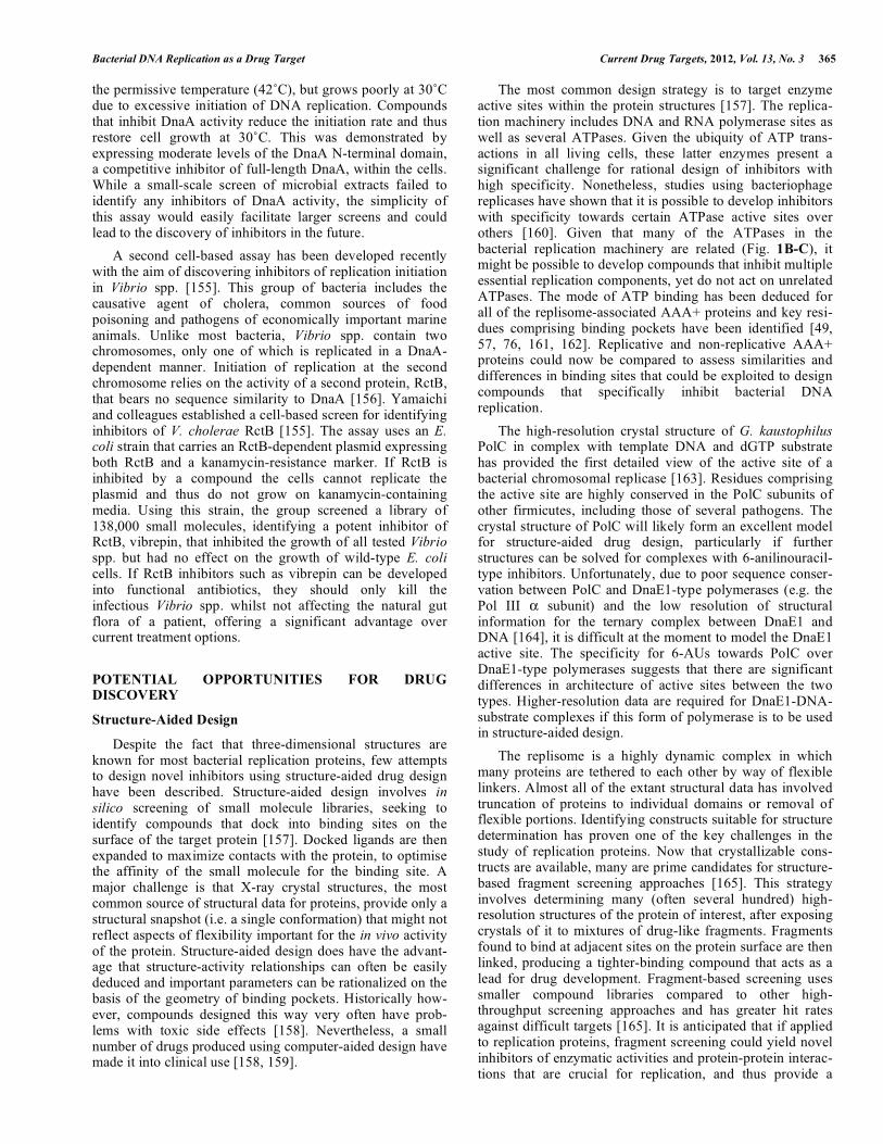
Fig. (1). Architecture and conservation of bacterial replisomes. (A) Bidirectional replication of a circular bacterial chromosome initiates at oriC and terminates opposite. Green circles denote replisomes at replication forks. (B) Model for leading and lagging strand synthesis at a replication fork in E. coli. (C) Overlaid ribbon diagrams of the AAA+ domains of DnaA (PDB: 2HCB, blue), DnaC (PDB: 3ECC, green), DnaI (PDB: 2W58, pink) and Hda (PDB: 3BOS, cyan). The position of the ATP analog AMP-PCP (colored by atom type: C, yellow; N, blue; O, red; P, orange) and a Mg2+ ion (gray sphere) within the DnaA structure is shown. (D) Overlaid ribbon diagrams of the AAA+-like domains of the clamp loader subunits � (blue), � (magenta), and �’ (green). Coordinates were derived from PDB: 3GLI. The positions of ADP (colored by atom type, as above for ATP), the phosphate transition state analog BeF3 (Be, magenta; F, cyan) and a Mg2+ ion (gray sphere) within the � subunit are shown. (E) Phylogenetic tree based on the sequences of DnaC/DnaI helicase loader proteins. The tree was constructed using the neighborhood-joining tree method in Geneious (Biomatters, Auckland, New Zealand), using the Jukes-Cantor genetic distance model and employing the bootstrap method with 100,000 replicates. The sequence of E. coli DnaA was included as an outgroup. Colored boxes indicate helicase loader families (enterobacteria DnaC-type, cyan; Aquificae DnaC-type, pink; firmicute DnaI-type, green). (F) Ribbon diagrams showing filaments of Aquifex aeolicus DnaC (PDB: 3ECC) formed by P61 crystal packing [49]. (G) Model for polymerase handover during lagging strand synthesis in Bacillus subtilis [27]. Panels (C), (D) and (F) were created using PyMOL [169].
Escherichia coliKlebsiella pneumoniae
DnaA
Salmonella entericaPersephonella marinaAquifex aeolicusHydrogenobacter thermophilus
Clostridium botulinumStreptococcus pyogenes
Bacillus subtilisStaphylococcus aureus
0.2
DnaC
DnaC
DnaI
E
i. primer extension
helicase
primase
τ δδ'
SSB
Okazaki
fragment
leading strand
lagging strand
5’
3’
5’
3’PolC
PolC
DnaE
3
ii. elongation
5’
3’
5’
3’helicase
primase
τ δδ'
SSB
Okazaki
fragment
leading strand
lagging strandPolC
PolC
DnaE
3
C D
F
Geading strandle
DnaB helicase
DnaG primase
Pol III
α
α
α β
β β
ττ
τ δδ'
SSB
Okazaki fragment
Pol I
DNA ligase
leading strand
lagging strand
fork progression
fork progressionA B
5’
3’
5’
3’RNA
primer
oriC
Ter
ε
ε
ε

Bacterial DNA Replication as a Drug Target Current Drug Targets, 2012, Vol. 13, No. 3 357
Ter [44]), are not essential for cell growth and are therefore of limited interest with respect to drug discovery. Only those components that are essential in those organisms that have them are discussed further.
In general the sequences of DNA replication proteins are moderately conserved among all bacteria. The genetically distant E. coli and B. subtilis, for example, share 11–49% sequence identity with respect to the proteins comprising the basic replication module. Overall the sequences of these proteins are less conserved than those of proteins involved in other informational processes such as RNA transcription and protein translation, but more conserved than proteins that function in metabolic pathways and other non-informational processes such as cell division [21]. Within the replication proteins, sequence motifs that encompass enzymatic sites are far more conserved than regions that mediate protein-protein interactions. Similarly, whole proteins that contain enzy-matic active sites tend to be more conserved than those that only mediate protein-protein interactions. Notable exceptions to this are the highly conserved protein-protein interaction systems present on the -sliding clamp and SSB [21, 45, 46].
Several DNA replication proteins are paralogs of each other or share structurally related domains. Many belong to the AAA+ family of ATPases, which harness the energy liberated by ATP hydrolysis to perform mechanical func-tions [47]. Within particular replication sub-complexes the AAA+ proteins are clearly related to each other. The initiator protein DnaA, for example, is closely related to the Hda initiation regulator and (when they are present) the helicase loaders DnaC and DnaI (Fig. 1C) [48, 49]. Similarly, the �, � and �’ subunits of the clamp loader complex [47], while not all enzymatically active as ATPases, share significant sequence homology and have similar overall structures (Fig. 1D). These similarities suggest that the members of each sub-complex often share a common ancestor; they presume-ably arose through ancient gene duplication events, even-tually diverging to take on distinctive roles in the replication process.
Structural homology is also seen among individual domains of some replication proteins. The N-terminal domain of the replicative helicase and the C-terminal domain of DNA primase, which interact to facilitate RNA primer synthesis, share a common fold despite negligible sequence similarity [39, 50, 51]. To date this fold has only been observed in these two proteins. Interestingly, weak sequence homology has recently been revealed for domains of two other replication proteins; the DnaB and DnaD replication initiator accessory proteins found in firmicutes (Table 1). Using Hidden Markov Model-based homology searches, Marston and colleagues were able to recognise two distinct homologous domains, DDBH1 and DDBH2, within these proteins [52]. DnaD has a DDBH1-DDBH2 domain structure while DnaB includes two DDBH2 domains and is arranged DDBH1-(DDBH2)2. Despite their extreme sequence diver-gence, these domains share common attributes within the two proteins: the DDBH1 domains facilitate subunit tetra-merization while the DDBH2 domains bind to DNA (double-stranded DNA for DnaD and the C-terminal domain of DnaB; single-stranded DNA for the first DDBH2 domain of DnaB) and consequently facilitate protein oligomerization.
Loading of the Replicative Helicase at oriC
The process of loading the replicative helicase at the origin has primarily been studied in two model organisms, the Gram-negative enterobacterium E. coli and the Gram-positive firmicute B. subtilis. In both, the helicase associates with DnaA during loading. In E. coli, DnaA-dependent recruitment of DnaB at oriC requires the helicase to be in a complex ([DnaB6][DnaC]6) with its loader protein DnaC [53]. DnaC suppresses the ATPase activity of DnaB and remains bound throughout the loading process. Once loaded, primase associates with the helicase, triggering the release of DnaC to enable strand separation by the helicase to commence [54]. Loading of the replicative helicase (called DnaC) in B. subtilis follows a similar mechanism except that the helicase loader is called DnaI and the two additional proteins described above, DnaB and DnaD, are also required [55]. Smits and colleagues have recently delineated the order of protein associations at oriC that precede helicase loading in this organism [55]. DnaA binds first to oriC. The DNA remodeling protein DnaD is then recruited, either through interaction with DnaA or through direct interaction with non-B-form DNA within the DnaA-coated oriC. Membrane-associated DnaB molecules are then recruited, probably through direct interaction with DnaD. Finally, two DnaC helicase-DnaI helicase loader complexes associate with the origin, at which point the two helicase hexamers are loaded onto the DNA. The E. coli DnaC and B. subtilis DnaI loader proteins share significant sequence homology, suggesting that this final step might be somewhat conserved between the two organisms. Interestingly however, relatives of these loader proteins can only be found in a few select groups of organisms outside of the enterobacteria and firmicutes.
Members of the Aquificae, a small group of hyper-thermophilic Gram-negative rods that includes the model organism Aquifex aeolicus [56], maintain a helicase loader known as DnaC that is a homolog of E. coli DnaC and B. subtilis DnaI [49]. As with E. coli, no relatives of B. subtilis DnaB or DnaD are present in these organisms. For this reason, the Aquificae DnaC proteins have been considered functionally equivalent to E. coli DnaC. Phylogenetic sequence analysis demonstrates, however, that Aquificae DnaC proteins are only distantly related to the other DnaC/I helicase loaders (Fig. 1E). This suggests that Aquificae DnaC proteins represent their own distinct family of helicase loaders and that they might have unique characteristics relative to members of the other two families.
Collectively, the DnaC/I-type helicase loaders are two-domain proteins, with helicase binding being mediated through the N-terminal domain while ATPase activity and DNA binding occurs within the C-terminal AAA+ domain. Crystal structures of the C-terminal domains of A. aeolicus DnaC [49] and Geobacillus kaustophilus DnaI [57] reveal very similar overall structures (Fig. 1C). Sequence homo-logy within this region suggests that the C-terminal domain of E. coli DnaC is probably also similar to the equivalent A. aeolicus and G. kaustophilus domains. The A. aeolicus domain was seen to form helical filaments in the crystalline state (Fig. 1F), reminiscent of filaments previously observed for ATP-bound DnaA from E. coli [49]. This helical arrangement is thought to represent a structure important for

358 Current Drug Targets, 2012, Vol. 13, No. 3 Robinson et al.
helicase loading in vivo [49]. The fact that the C-terminal domains of helicase loaders are well conserved across the three families suggests that E. coli DnaC and B. subtilis DnaI probably also adopt helical arrangements during helicase loading.
The solution structure of the N-terminal domain of B. subtilis DnaI determined by NMR, which revealed a novel zinc-binding fold, provides the only structural definition of a helicase loader N-terminal domain [58]. The sequences of the N-terminal domains are conserved within each of the three helicase loader families; however these domains in E. coli and A. aeolicus DnaCs are clearly unrelated to that of B. subtilis DnaI. For both E. coli DnaC and B. subtilis DnaI, it is this N-terminal domain that facilitates binding to the helicase [53, 59]. Further work is required to determine if the structural basis of the helicase-helicase loader interactions is in any way conserved among the three helicase loader families.
The fact that DnaC/I-type helicase loaders are not conserved among all bacteria implies that organisms outside of the enterobacteria, firmicutes and Aquificae must either not require specialized helicase loader proteins or utilise proteins that are unrelated to DnaC/I for this function [21]. In support of the former scenario, Soni and colleagues have shown that the helicase from Helicobacter pylori, an organism that lacks an identifiable helicase loader, can be used to complement a temperature-sensitive dnaC mutant of E. coli [60]. This implies that the H. pylori helicase can be loaded onto the E. coli oriC using the E. coli machinery in the absence of a functional DnaC. One possibility is that DnaA could itself be sufficient for loading of the replicative helicase at oriC in organisms that lack helicase loaders. Indeed the helicases from two such organisms, Pseudomonas putida and P. aeruginosa can be loaded onto oriV of the RK2 plasmid in vitro using only DnaA [61]. In the same system, the E. coli helicase required DnaC to be present for loading onto oriV [61]. Another possibility is that organisms without DnaC/I might utilise replication restart complexes analogous to the PriA/PriB/DnaT system found in E. coli [62] to load the helicase at both origin and non-origin sequences. In fact in B. subtilis, the DnaB and DnaD proteins are required for loading the helicase both during initiation of DNA replication and for replication restart [63]. Interest-ingly, homologs of B. subtilis DnaB are maintained within members of the tenericutes, which lack homologs of DnaD and DnaI (Table 1). In this case it is tempting to speculate that DnaB might assist other, as yet unidentified, helicase loaders in loading the helicase at oriC.
While helicase loaders are not found in all bacteria, they form an essential part of the DNA replication machinery in those organisms that have them (Table 1). Importantly, this includes several important human pathogens, such as E. coli, Salmonella typhimurium, Bacillus anthracis, Listeria monocytogenes, Clostridium botulinum, Staphylococcus aureus and Streptococcus pyogenes. For these organisms, helicase loaders might represent useful antibacterial drug targets. One could imagine, however, that relatively simple modifications to other helicase loading pathways (such as those involved in replication restart) might allow bacteria to survive should their DnaC/DnaI pathways be inhibited.
Ultimately this may limit the usefulness of helicase loader inhibitors as antibiotics.
DnaE1 versus PolC + DnaE3 Mechanisms
The majority of bacterial genomes sequenced to date contain a single type of replicative polymerase, the Pol III � subunit, known under the polymerase classification scheme developed by Zhao and colleagues as DnaE1 [64]. It has long been known, however, that the Gram-positive B. subtilis uses two related, but distinct, replicase types known as PolC and DnaE3 [27, 64]. DnaE3 is homologous to DnaE1 and has a similar domain organization, while PolC is more distantly related to DnaE1, contains an additional �-related proofreading domain, and has a dissimilar arrange-ment of functional domains [64]. Both PolC and DnaE3 are required for DNA replication in B. subtilis [65]. Based on the observation that DnaE3 deprivation immediately halts lag-ging strand synthesis, while allowing leading strand synthesis to continue, it was originally hypothesized that PolC and DnaE3, respectively, constitute separate leading and lagging strand replicases [65], akin to the eukaryotic polymerases Pol � and Pol � [66]. Recently however, reconstitution of a functional B. subtilis replisome in vitro has shown that this is not the case [27].
After identifying 13 proteins likely to comprise the replisome of B. subtilis (polymerases PolC and DnaE3, SSB, PriA, DnaG primase, DnaC helicase, DnaB, DnaD, and DnaI helicase loaders, the clamp, and the clamp loader subunits �, �, and �’), Sanders and colleagues were able to purify each of these components and use them to carry out coordinated leading and lagging strand synthesis on a synthetic minicircle DNA template [27]. All 13 proteins were found to be required for lagging strand synthesis while DnaE3 and primase could be omitted for leading strand synthesis. It was found that both PolC and DnaE3 could extend DNA primers, but only DnaE3 could extend RNA primers like those laid down by primase. Inclusion of both PolC and DnaE3 had a strong synergistic effect on the rate of lagging strand synthesis. These observations are consistent with a model in which DnaE3 does not act as a dedicated lagging-strand polymerase, but rather acts to extend the RNA primers laid down by primase with a short stretch of DNA so that PolC can continue DNA synthesis (Fig 1G). The role of DnaE3 is thus analogous to that of the eukaryotic Pol �, which extends RNA primers with DNA before handing them over to the lagging strand Pol � [27]. As PolC/DnaE3 are present in all firmicutes and tenericutes, this model probably applies to all organisms within these groups.
Clamp Loader Subunits of DNA Polymerase III
At the heart of the Pol III holoenzyme (minimally [��]3[�3��’][2]2) is the heteropentameric clamp loader complex (�3��’). As its name suggests, this complex func-tions to load -sliding clamps onto DNA primer-templates, bestowing leading- and lagging-strand processivity on the replicase [67]. Each of the three � subunits of the clamp loader also binds to the � subunit of a polymerase core, thus tethering three polymerases together to coordinate synthesis of the two strands [36]. In E. coli the dnaX gene encoding the � subunit includes sequence elements that induce a

Bacterial DNA Replication as a Drug Target Current Drug Targets, 2012, Vol. 13, No. 3 359
ribosomal frameshift with approximately 50% efficiency [68-72]. This frameshift gives rise to a truncated form of �, known as , comprising the N-terminal three domains out of five in the full-length � protein; can replace � to form complexes that maintain clamp loading activity, but since it is the C-terminus of � that contacts �, cannot multimerize � subunits to form a functional replicase. It has long been thought that the clamp loader complex in E. coli contains two � subunits and one , thus yielding a Pol III HE with two polymerase cores ([���]2[�2��’][2]2). Recently however, the Leake and Sherratt groups have quantified each of the Pol III HE subunits at replication forks in living E. coli cells [36], presenting strong evidence that in vivo, replicases contain three � subunits and three ��� cores. The authors instead propose that clamp loader complexes containing subunits could function in other processes that require loading of -sliding clamps, such as in DNA repair [36]. The programmed ribosomal frameshift encoded within the E. coli dnaX gene is also conserved in the related organism Salmonella typhimurium [73]; in fact similar frameshift sequences are found within dnaX genes among all the enterobacteria. The dnaX gene of the hyperthermophilic Gram-negative organism Thermus thermophilus also produces both � and products [74]. In this case is produced by transcriptional slippage rather than a ribosomal frameshift. Most other bacteria, however, do not maintain frameshift signatures within their dnaX genes and may not, therefore, produce subunits. Indeed the dnaX genes from B. subtilis, S. aureus, S. pyogenes, P. aeruginosa and A. aeolicus produce only full-length � products when expressed in recombinant E. coli systems [27-31, 75]. It appears likely that all bacteria maintain three � subunits within clamp loader complexes at replication forks and thus utilise trimeric polymerases to replicate their chromosomes.
In addition to �, � and �’ subunits, the clamp loader complexes of -proteobacteria include a heterodimeric sub-complex made up of the � and � subunits (Table 1). This is tethered to the more universal clamp loader assembly (�3��’) through an interaction between the N-terminus of � and domain III from all three of the � subunits [76, 77]. The � subunit interacts with the C-terminus of SSB, providing a mechanism for the handover of primed template DNA from primase to Pol III HE during lagging strand synthesis [78-82]. Recently, the link between SSB and Pol III afforded by the �� heterodimer has been shown to be crucial for strand displacement activity of Pol III HE [83]. It has also been demonstrated that the interaction of � with � significantly stimulates clamp-loading activity in E. coli and P. aeruginosa [30, 76, 77, 84]. Interestingly, while only one �� heterodimer can associate with each clamp loader complex, there are on average four �� heterodimers at each replication fork in live E. coli cells, compared with one �3��’ complex [36]. This suggests that additional �� heterodimers associate with SSB at replication forks, awaiting handover to Pol III HE.
The gene encoding �, holC, is dispensable in most organisms (Table 1) [14]) and can be disrupted in E. coli, although such disruptions show some growth defects [85]. The holD gene, which encodes � is dispensable in E. coli [85] but essential in both Acinetobacter baylyi [86] and P. aeruginosa [87]. The � proteins in these latter organisms are much longer than their E. coli counterpart and might
therefore carry additional functions that are essential for survival [21, 84]. That the genes encoding � and/or � can be disrupted in E. coli and other bacteria suggests that their role in DNA replication is probably regulatory rather than being part of some fundamental mechanism. Interestingly, genes encoding � appear to be distributed across more of the bacteria (�, and -proteobacteria) than genes encoding � (-proteobacteria only; see Table 1). The � proteins have highly divergent sequences [21]; thus it is possible that the � homologs in and -proteobacteria have simply diverged beyond the detection limit of sequence similarity searches, including sensitive Hidden Markov Model-based methods [88, 89]. If there is a genuine discrepancy in the phylogenetic distribution of � and � subunits, however, this suggests that � may have evolved to interact directly with � and/or have extra functions outside of clamp loader complexes.
Gene Duplications
Genome sequencing has revealed that many bacteria contain genes coding for multiple versions of certain replica-tion proteins. Many of these proteins are potential drug targets and are discussed in detail in the next section. The � subunit of Pol III, which provides the major DNA poly-merase activity of the replisome, exists in at least four variants termed DnaE1, DnaE2, DnaE3 and PolC [64]. The first three variants have a similar sequential arrangement of PHP, polymerase and OB-fold domains within their primary sequences, yet can be classified into three different groups using sequence analysis. PolC proteins have regions homo-logous to the domains found in DnaE-type polymerases, as well as an additional proofreading exonuclease domain. The arrangement of domains is different however, appearing in the primary sequence in the order OB-fold, PHP domain, exonuclease and finally the polymerase domain. As dis-cussed above, all bacteria outside of the firmicutes and tenericutes have DnaE1, while organisms within these groups have both PolC and DnaE3. In contrast, DnaE2 does not conform to phylogenetic boundaries and can co-exist with DnaE1 or PolC and DnaE3. The error-prone DnaE2 variant forms part of a LexA-regulated adaptive mutagenesis cassette, which appears to have originated in the actinobac-teria but has since disseminated throughout other bacterial groups through lateral gene transfer [90]. DnaE2 variants are found in the important human pathogens Mycobacterium tuberculosis and P. aeruginosa and are likely to play a role in the development of antibiotic resistance in these orga-nisms [91, 92].
Other duplications of DNA replication proteins have more mysterious functions. The genomes of many Bacillus spp. contain a second copy of the dnaN gene, dnaN-2, cod-ing for a -sliding clamp (DnaN-2) that shares approxi-mately 40% sequence homology with its canonical-type -sliding clamp (DnaN-1) [93]. In E. coli the -clamp is known to bind to the � subunit of Pol III as well as a host of DNA repair enzymes, thus playing a role in both replication and repair [45]. In B. anthracis, deletion of dnaN-2 produces a phenotype indistinguishable from wild-type, whereas deletion of dnaN-1 results in a mutator phenotype [93]. This, together with the fact that a dnaN-1/dnaN-2 double mutant could not be produced, suggests that DnaN-1 functions in both DNA replication and repair, while DnaN-2 can

360 Current Drug Targets, 2012, Vol. 13, No. 3 Robinson et al.
participate in replication but is deficient in one or more DNA repair functions.
Duplications of genes encoding the � subunit of Pol III (dnaQ), DNA primase (dnaG) and SSB (ssb) can also be found within the genomes of a variety of diverse bacteria, not conforming to any obvious phylogenetic boundaries. While some attempts have been made to characterize the products of these duplicated genes, their biological roles remain unclear [94, 95]. Nevertheless, it is important to consider the existence of these gene duplications if the canonical versions of the proteins they encode are to be considered as drug targets, in particular whether any func-tional redundancy may provide a selective advantage to their hosts when challenged by antibiotics. On the other hand, if two essential proteins with different functions are suffi-ciently similar to be targeted by the same drug, then this would provide a substantial barrier to selection of resistant mutants in either.
EXISTING INHIBITORS AND CURRENT SCREEN-ING STRATEGIES
Of all the antibiotics in current clinical use, only two classes inhibit the process of DNA replication. The quino-lone and aminocoumarin drugs inhibit the action of DNA gyrase, a type II toposiomerase that introduces negative supercoils in DNA ahead of replication forks to enable continued strand separation by the helicase during DNA replication [96]. Given that the replication machinery includes so many other proteins with functions essential for bacterial viability, it is likely that at least some are useful drug targets. The replisome is very much an under-explored target for drug development, however, and few attempts to discover specific inhibitors have been described. One of the major challenges in discovering DNA replication inhibitors has been the need for biochemical assays that can be used for high-throughput screening. Important advances have been made in this area recently and are likely to yield new inhibi-tors in the near future. This section describes inhibitors that have been identified so far and the current strategies used for discovering new antagonists of replication activity.
DNA Gyrase Inhibitors
Gyrase (a type IIA DNA topoisomerase) plays an essential role in DNA replication, actively underwinding the double-stranded template DNA ahead of the replication fork to obviate effects of positive supercoiling induced by the progressing replisome. It acts by cutting both DNA strands, passing double-stranded DNA through the gap, then religat-ing the original ends [97]. It is a heterotetrameric enzyme comprised of two different subunits: GyrA, which binds DNA and carries out strand cleavage/ligation, and GyrB, which hydrolyses ATP to drive the supercoiling reaction. Gyrase is the cellular target of several different classes of bacteriocidal agents, including the highly-successful fluoro-quinolone antibiotics, coumarins and cyclothialidines that are currently undergoing drug development, as well as a range of naturally-occurring bacterial toxins [22, 97]. Much of the current research on gyrase inhibitors focuses on modifying existing scaffolds to address issues with drug resistance, toxic side effects and poor cellular penetration, as
well as to broaden the range of bacterial species that com-pounds are active against [22, 97, 98]. Structure-aided design and virtual screening techniques have proven somewhat successful in delivering new classes of gyrase inhibitors, some of which show broad-spectrum antibacterial activity and reduced rates of resistance relative to exisiting drugs [99, 100]. Most recently, there has been exciting progress with two novel classes of gyrase inhibitors that show great promise for development into new antibiotics in the near future.
The novel antibiotic simocyclinone D8 was originally identified in extracts from the antibiotic-producing bacterium Streptomyces antibioticus Tü 6040 [101]. This compound has a chlorinated aminocoumarin group linked to an angu-cyclic polyketide moiety through a tetraene linker and a D-olivose sugar (Fig. 2A). The presence of the aminocoumarin group suggested DNA gyrase to be the cellular target and simocyclinone D8 was shown to potently inhibit the E. coli enzyme in vitro [102]. Unlike the aminocoumarin antibiotics however, simocyclinone D8 was found not to inhibit the ATPase activity of the GyrB subunit, nor did it stimulate the formation of unproductive cleavage complexes within the GyrA subunit as seen with quinolone compounds, suggesting an entirely novel mode of inhibition [102]. Edwards and colleagues have recently determined the crystal structure of simocyclinone D8 in complex with the N-terminal domain of the GyrA subunit, revealing binding pockets for the amino-coumarin and polyketide moieties close to, but distinct from a previously identified quinolone-binding site [103]. This represents a significant step in the development of simocyc-linones into clinically useful antibiotics, as structure-based activity relationships can now be explored. In addition to this binding site within the GyrA subunit, biochemical studies have suggested the existence of a second low-affinity bind-ing site within GyrB [104]. Further work is now required to explore the possibility of cooperative binding at the GyrA and GyrB sites. Interestingly, while simocyclinone D8 shows poor activity against Gram-negative laboratory bacteria and is thus being viewed as a potential treatment only for infec-tions caused by Gram-positives, it has recently been found to have potent activity against a number of Gram-negatives isolated from clinical samples [105]. This observation is likely to assist in identifying parameters that can be opti-mized to broaden the spectrum of activity of simocyclinones, and adds to the promise of these compounds becoming clinically useful antibiotics.
A very promising new antibiotic that acts on gyrase has been recently described by GlaxoSmithKline researchers [106]. GSK299423 (Fig. 2B) is a member of the novel bac-terial topoisomerase inhibitors (NBTI) family of compounds, which has also been explored by Novoxel [107] and Johnson & Johnson [108, 109]. GSK299423 strongly inhibits the activity of S. aureus and E. coli gyrase in vitro (IC50 = 14–100 nM), making it more than 2000 times more potent than the widely used drug ciprofloxacin (a fluoroquinolone) [106]. GSK299423 shows potent antibacterial activity against a range of Gram-positive and Gram-negative pathogens (MIC = 0.016–8 μg/mL), including strains that are resistant to fluoroquinolones. The crystal structure of the gyrase-DNA-GSK299423 complex has been determined using the S. aureus enzyme, revealing that the compound binds between the active sites of the two GyrA subunits,

Bacterial DNA Replication as a Drug Target Current Drug Targets, 2012, Vol. 13, No. 3 361
away from known binding sites for quinolone-type inhibitors [106]. GSK299423 appears to inhibit the catalytic cycle of gyrase by stabilizing a pre-cleavage enzyme-DNA complex, an entirely novel mode of gyrase inhibition. Taken together, the potency of GSK299423, its broad-spectrum activity and its lack of cross-resistance with existing fluoroquinolones suggest the NBTIs represent exciting candidates for development into a much-needed new class of antibiotics with a novel mode of action.
Inhibitors of DNA Polymerase Activity
DNA polymerases are the targets of several important anti-viral and anti-cancer drugs, yet few inhibitors exist for the equivalent bacterial enzymes [110]. To date, there are no known inhibitors that are specific for DnaE1-type poly-merases (e.g. the � subunit of E. coli Pol III). Two classes of compounds are known to effectively inhibit the PolC-type polymerases found in the firmicutes and tenericutes: 6-anilinouracils (6-AUs) and quinazolin-2-ylamino-quinazolin-4-ols (BisQuinols). Members of a third class of compounds, the dichlorobenzylguanines, have been shown to inhibit the activities of DnaE1-, DnaE3- and PolC-type polymerases, although no analysis of their antimicrobial properties has been published since their syntheses were described six years ago [111].
The 6-anilinouracils are the oldest and most developed class of DNA polymerase inhibitors [112-114]. These compounds are competitive inhibitors of dGTP binding to PolC [115], forming a three-hydrogen-bond base pair with cytosine residues in the template DNA (Fig. 3A). Many variations have been made on the original 6-AU scaffold, producing several compounds that potently inhibit PolC activity and bacterial growth in vitro [115-119]. Historically however, 6-AUs have proven modest inhibitors of bacterial growth in animal infection models due to poor solubility and bioavailability [120]. Most compounds that have demons-trated activity in vivo have been tested using intraperitoneal dosing, which largely circumvents the problems associated with poor sample solubility. Recently, Svenstrup and col-leagues addressed this issue by varying 3-substituents on the 6-AU scaffold with a view to improving aqueous solubility,
and in doing so have produced two compounds (Fig. 3B) that show strong activity against S. aureus and Enterococcus faecalis using intravenous dosing in a mouse infection model [118]. It is hoped that this advance in understanding of the structure-activity relationships of 6-AUs will facilitate the development of clinically useful compounds.
Members of the second class of PolC inhibitors, the BisQuinols, were discovered recently using high-throughput fluorescence-based inhibition assays [121]. The methodo-logy underlying these assays is described later in this section. It was predicted that BisQuinols might base pair with cyto-sine residues in DNA templates and thus be competitive with dGTP substrates, as observed for the 6-AUs. Biochemical analysis revealed, however, that BisQuinols were uncom-petitive with respect to nucleotide substrates, instead show-ing competitive behaviour against template DNA. The mechanism of PolC inhibition by BisQuinols must therefore be different to that of 6-AU compounds and appears to be more analogous to that of non-nucleoside reverse transcript-tase inhibitors used to treat HIV infections [121, 122]. Importantly, the initial series of BisQuinol compounds also showed significant inhibition of the eukaryotic Pol � and thus appears to show poor selectivity for bacterial DNA polymerases [121]. Nevertheless, one representative com-pound (Fig. 3C) was found to be somewhat effective in rescuing mice from a lethal intraperitoneal S. aureus infection, thus demonstrating a degree of in vivo efficacy. A better understanding of the structure-activity relationships for this novel class of PolC inhibitors is now required to assess their potential for development into functional antibiotics.
A promising new strategy for the development of PolC inhibitors is to covalently link 6-anilinouracil derivatives to fluoroquinolones to create hybrid PolC-DNA gyrase inhibi-tors. These hybrid compounds maintain dual anti-PolC and anti-DNA gyrase activity in vivo and have greater or equal potency against whole bacterial cells than their isolated 6-AU or fluoroquinolone parent compounds [116, 123, 124]. In fact when testing one representative hybrid (Fig. 3D), Butler and colleagues observed that the fusion to fluro-quinolone actually increased the anti-PolC activity of the 6-
OHO
Cl
OHHN
O
O
OOOH3C
O
OH OH OH
OH
CH3
HOOO
OH3C
N
O
NHN
N
S
O
A
B
Fig. (2). Chemical structures of new inhibitors of DNA gyrase. (A) Simocyclinone D8 [101-105]. (B) GSK299423 [106].

362 Current Drug Targets, 2012, Vol. 13, No. 3 Robinson et al.
AU moiety [116]. Even more promisingly, in cases where each of the parent compounds was only weakly active in isolation, i.e. for drug-resistant organisms, the hybrid com-pound was found to offer greater potency than an equimolar mixture of its parents. The hybrid compound was thus effective against a broader range of bacterial strains than either parent, an attribute also observed for oxazolidinone-quinolone hybrid protein synthesis/DNA gyrase inhibitors [125, 126]. The use of hybrid inhibitors intuitively should
provide greater protection against the development of drug resistance by target mutagenesis than traditional single-target inhibitors – this would require co-mutation of two loci in the bacterial population. This indeed appears to be the case for Butler’s hybrid compound, for which resistance in S. aureus was seen to develop far more slowly than resistance to either parent compound in a simple pure culture system [116].
Several bacterial replisomes have now been reconstituted and are functional in vitro [26-28, 30, 127]. For E. coli and
N
NH
R
O
O
NH
N
N OHN
H
N
NH
O
O
NH
N
O
N
O
N
NH
O
O
NH
SS
N N
H3C
AB
N
N
NH
N
NMeO
OHC
N
NH
O
O
NH
N
N N
F
F
O
COOHD
N
HNN
E
N
HO
N
N S
O O
O
NH
N
O
O S
Br
O2N
O
NH
NH
O
O S
N
N S
OO
O OH
N O
NN
NN
SH
Fig. (3). DNA polymerase inhibitors. (A) Three-hydrogen-bond interaction between 6-anilinouracil compounds and a cytosine residue within DNA [115]. Chemical structures of (B) new 6-anilinouracil derivatives with improved aqueous solubility [118], (C) a BisQuinol inhibitor of PolC [121], (D) a hybrid PolC/DNA gyrase inhibitor [116], and (E) novel replisome inhibitors identified by high-throughput screening [127].

Bacterial DNA Replication as a Drug Target Current Drug Targets, 2012, Vol. 13, No. 3 363
B. subtilis, this includes all the machinery required for both leading and lagging strand synthesis [26, 27]. For other species, more minimal replisomes capable only of leading strand synthesis have been assembled [28, 30]. Recently, Dallmann and colleagues described a high-throughput assay to screen for inhibitors of reconstituted replicase activity, making use of an intercalating fluorescent dye to monitor conversion of single-stranded DNA substrates into a double-stranded product [127]. This approach has a significant advantage over assays that focus on isolated replication components, in that inhibitors of any part of the replication machinery can be identified through a common endpoint. These workers also included specificity assays to test if compounds bind to DNA, or inhibit viral and eukaryotic DNA polymerases, RNA polymerase, an unrelated eukaryo-tic ATPase or an unrelated control enzyme, -galactosidase. Using these assays, they screened a small library of 2000 compounds and identified seven (Fig. 3E) that act as specific inhibitors of bacterial replisomes and kill bacterial cells by inhibiting DNA replication. This high-throughput assay approach thus provides an efficient means to generate lead compounds for further drug development, and could be applied equally well for screening natural product and commercially available libraries of drug-like compounds.
Inhibitors of Primosome Functions
Outside of DNA gyrase and the replicative polymerases, another replication subcomplex to be targeted for drug dis-covery has been the primosome, comprised of the DnaB/C helicase and DnaG primase (see Table 1). These proteins play critical roles in lagging strand synthesis and are essen-tial for bacterial growth [40, 85, 128]. These proteins are targets for some naturally occurring DNA replication inhibitors. The dietary flavonoid myricetin (Fig. 4A), for instance, has antimicrobial activity and has been shown to potently inhibit the replicative helicase, but not primase [129]. The bacterial alarmone compound (p)ppGpp on the other hand, is now known to inhibit the activity of primase, thereby halting DNA replication as part of the stringent response [130, 131]. The existence of these natural, low molecular weight inhibitors lends hope that the primosome subcomplex might be a viable target for drug discovery.
The replicative helicase has two functional domains. The N-terminal domain recruits primase to the replication fork, while helicase activity stems from the C-terminal domain. The helicase domain belongs to the RecA-type family of ATPases [132], which includes the bacterial replication/ recombination/repair proteins RepA [133] and RecA [134], the transcription termination factor Rho [135] as well as the eukaryotic DNA repair proteins Rad51 and DMC1 [136]. On the other hand, primase has a central RNA polymerase domain that is not related to other RNA polymerases and has a uniquely-shaped active site [137]. This domain is flanked by an N-terminal zinc-binding domain, which binds to specific trinucleotide initiation sequences on the template DNA, and a C-terminal domain that binds to the helicase [138]. Primase interacts with the helicase to enable primer synthesis on the lagging strand in vivo [32]. This interaction stimulates the activity of both proteins in vitro [139] and varies in strength, ranging from a weak and transient complex as observed for the E. coli proteins [140] to a highly
stable complex as for the G. stearothermophilus proteins [37].
In recent years high-throughput biochemical assays have been developed to measure the activities of helicase and primase, as well as the mutually stimulatory effects of their interaction [141-143]. Application of these assays in drug discovery has been limited, however, and has produced only a few inhibitors [144-146]. The Förster resonance energy transfer (FRET) assay for DnaB helicase activity developed by Zhang and colleagues [141], for instance, has been used by researchers at Targanta Therapeutics to screen a library of 230,000 commercially available compounds for inhibitors of P. aeruginosa DnaB [147]. The Pseudomonas DnaB helicase was used as it can be efficiently loaded onto substrate DNA molecules in the absence of a helicase loader protein, in contrast to the equivalent helicases from E. coli and B. subtilis [61, 148]. Their screen revealed a triaminotriazine compound that inhibited the helicase with IC50 = 5 μM (Fig. 4B). Despite showing potent inhibition of the P. aeruginosa DnaB in vitro, neither the newly identified compound nor analogs prepared to explore effects on cytotoxicity were found to inhibit the growth of P. aeruginosa cells, despite the fact that a permeability barrier-deficient strain was used [147]. Some compounds were, however, moderately active in inhibiting the growth of S. aureus, S. pneumoniae and a hyper-susceptible strain of E. coli. Unfortunately, this series of compounds exhibited a range of undesirable properties, including cytotoxicity towards HeLa cells and loss of antimicrobial activity in the presence of serum, that ultimately deem them unsuitable as lead compounds for antibiotic development. To date this represents the only reported systematic attempt to identify specific inhibitors of replicative helicase function.
DNA primase has attracted slightly more attention as a drug target, although success has again been limited. To date all searches for inhibitors have utilized the scintillation proximity assay for primase activity developed by Zhang and colleagues [141]. The assay is based on the ability of primase to incorporate radioactive 3H-labeled NTP substrates into an RNA product, which is then captured using a biotin tag on the template DNA strand to which the RNA product remains hybridized. Researchers at Schering Plough used this assay to screen extracts of various plants for inhibitors of E. coli primase [144], unearthing two novel phenolic saccharides from Polygonum cusoidatum that produced IC50 values < 5 μM (Fig. 4C). Unfortunately, these compounds appear to act by binding to the template DNA rather than to primase itself and are therefore not appropriate lead compounds [129, 144]. In another similar study conducted by Schering Plough researchers, a novel bicyclic macrolide compound, Sch 642305 (Fig. 4D), was isolated from the fungus Penicillium verrucosum and found to inhibit E. coli primase with an IC50 of 70 μM [146]. Promisingly, Sch 642305 also inhibited the growth of the E. coli strain HS294 (defective lipopolysaccharide layer and disrupted acrAB efflux pump) with an MIC of 40 μg/mL. As yet no attempts have been reported to establish the mode of Sch 642305 inhibition. Presumably this is due to difficulty in obtaining sufficient material for biochemical analysis, although several synthetic approaches for production of Sch 642305 have now been described [149-151]. Before Sch 642305 can be considered as a lead compound, further work is required to

364 Current Drug Targets, 2012, Vol. 13, No. 3 Robinson et al.
determine its mode of action, as well as its efficacy against other bacterial strains in both in vitro cell growth and in vivo animal infection models.
Making use of the high-resolution crystal structure of the RNA polymerase domain of E. coli primase [137], Agarwal and colleagues at Achillion Pharmaceuticals used virtual screening to identify three potentially ‘druggable’ sites within primase [145]. They selected 79 compounds predicted to bind at these sites, and using the scintillation proximity assay [152] identified four inhibitors with IC50 values of 7–50 μM [145]. None of these compounds was active in inhibiting the growth of E. coli cells, however. The initial four compounds were used to deduce a three-dimensional pharmacophore, which was then used to search a database of commercially available compounds for other potential inhibitors. From this search, a further 34 chemically diverse compounds were selected for enzyme inhibition assays, revealing eight additional compounds that inhibited primase with IC50 values < 100 μM. Three of these also inhibited the growth of E. coli, with MIC values of 4–64 μg/mL (Fig. 4E). Analogs of these compounds were then studied with these assays, allowing structure-activity relationships to be
deduced for each chemical series [145]. Further work is now required to assess the efficacy of these compounds against other bacterial strains for antibiotic development.
Inhibitors of the Initiation of DNA Replication
A relatively new approach towards the discovery of DNA replication inhibitors is to target the initiation stage. In (almost) all bacteria, the first step in replicating the chromosome is the binding of multiple DnaA molecules at the origin, which ultimately leads to separation of the two template strands [153]. Since this step signifies commitment to a complete round of replication, the amount and activity of DnaA is tightly regulated within the cell. Insufficient DnaA leads to under-initiation, while excess leads to over-initiation. Both disrupt cell growth. Currently, no antibacterial agents target the initiation process.
Fossum and colleagues have recently developed a robust cell-based assay to screen for inhibitors of DnaA activity [154]. The assay utilises an E. coli strain containing a novel dnaA allele that produces an over-active DnaA variant and confers a cold-sensitive phenotype. This strain grows well at
N
N
N
NH
NH
NH
N
O
O2N
Cl
A
HO
HO
HOO
O
O
OH
OHOHO
OH
OH
HO O
OH
OH
O
OH
OH
HO
HO OH
O
O
B
C
O
O
HHO
O
H
O
N
N
O
ND
N
S
N
N
S
HN
N
N
N
N
N
O
OOH
HO
OH
OH
OH
E
Fig. (4). Chemical structures of inhibitors of primosome functions. (A) The DnaB helicase inhibitor, myricetin [129]. (B) A triaminotriazine inhibitor of DnaB helicase [147]. (C) Phenolic saccharide inhibitors of DnaG primase [144]. (D) Sch 642305, an inhibitor of DnaG primase [146]. (E) Inhibitors of DnaG primase produced using structure-aided design [145].
Bacterial DNA Replication as a Drug Target Current Drug Targets, 2012, Vol. 13, No. 3 365
the permissive temperature (42˚C), but grows poorly at 30˚C due to excessive initiation of DNA replication. Compounds that inhibit DnaA activity reduce the initiation rate and thus restore cell growth at 30˚C. This was demonstrated by expressing moderate levels of the DnaA N-terminal domain, a competitive inhibitor of full-length DnaA, within the cells. While a small-scale screen of microbial extracts failed to identify any inhibitors of DnaA activity, the simplicity of this assay would easily facilitate larger screens and could lead to the discovery of inhibitors in the future.
A second cell-based assay has been developed recently with the aim of discovering inhibitors of replication initiation in Vibrio spp. [155]. This group of bacteria includes the causative agent of cholera, common sources of food poisoning and pathogens of economically important marine animals. Unlike most bacteria, Vibrio spp. contain two chromosomes, only one of which is replicated in a DnaA-dependent manner. Initiation of replication at the second chromosome relies on the activity of a second protein, RctB, that bears no sequence similarity to DnaA [156]. Yamaichi and colleagues established a cell-based screen for identifying inhibitors of V. cholerae RctB [155]. The assay uses an E. coli strain that carries an RctB-dependent plasmid expressing both RctB and a kanamycin-resistance marker. If RctB is inhibited by a compound the cells cannot replicate the plasmid and thus do not grow on kanamycin-containing media. Using this strain, the group screened a library of 138,000 small molecules, identifying a potent inhibitor of RctB, vibrepin, that inhibited the growth of all tested Vibrio spp. but had no effect on the growth of wild-type E. coli cells. If RctB inhibitors such as vibrepin can be developed into functional antibiotics, they should only kill the infectious Vibrio spp. whilst not affecting the natural gut flora of a patient, offering a significant advantage over current treatment options.
POTENTIAL OPPORTUNITIES FOR DRUG DISCOVERY
Structure-Aided Design
Despite the fact that three-dimensional structures are known for most bacterial replication proteins, few attempts to design novel inhibitors using structure-aided drug design have been described. Structure-aided design involves in silico screening of small molecule libraries, seeking to identify compounds that dock into binding sites on the surface of the target protein [157]. Docked ligands are then expanded to maximize contacts with the protein, to optimise the affinity of the small molecule for the binding site. A major challenge is that X-ray crystal structures, the most common source of structural data for proteins, provide only a structural snapshot (i.e. a single conformation) that might not reflect aspects of flexibility important for the in vivo activity of the protein. Structure-aided design does have the advant-age that structure-activity relationships can often be easily deduced and important parameters can be rationalized on the basis of the geometry of binding pockets. Historically how-ever, compounds designed this way very often have prob-lems with toxic side effects [158]. Nevertheless, a small number of drugs produced using computer-aided design have made it into clinical use [158, 159].
The most common design strategy is to target enzyme active sites within the protein structures [157]. The replica-tion machinery includes DNA and RNA polymerase sites as well as several ATPases. Given the ubiquity of ATP trans-actions in all living cells, these latter enzymes present a significant challenge for rational design of inhibitors with high specificity. Nonetheless, studies using bacteriophage replicases have shown that it is possible to develop inhibitors with specificity towards certain ATPase active sites over others [160]. Given that many of the ATPases in the bacterial replication machinery are related (Fig. 1B-C), it might be possible to develop compounds that inhibit multiple essential replication components, yet do not act on unrelated ATPases. The mode of ATP binding has been deduced for all of the replisome-associated AAA+ proteins and key resi-dues comprising binding pockets have been identified [49, 57, 76, 161, 162]. Replicative and non-replicative AAA+ proteins could now be compared to assess similarities and differences in binding sites that could be exploited to design compounds that specifically inhibit bacterial DNA replication.
The high-resolution crystal structure of G. kaustophilus PolC in complex with template DNA and dGTP substrate has provided the first detailed view of the active site of a bacterial chromosomal replicase [163]. Residues comprising the active site are highly conserved in the PolC subunits of other firmicutes, including those of several pathogens. The crystal structure of PolC will likely form an excellent model for structure-aided drug design, particularly if further structures can be solved for complexes with 6-anilinouracil-type inhibitors. Unfortunately, due to poor sequence conser-vation between PolC and DnaE1-type polymerases (e.g. the Pol III � subunit) and the low resolution of structural information for the ternary complex between DnaE1 and DNA [164], it is difficult at the moment to model the DnaE1 active site. The specificity for 6-AUs towards PolC over DnaE1-type polymerases suggests that there are significant differences in architecture of active sites between the two types. Higher-resolution data are required for DnaE1-DNA-substrate complexes if this form of polymerase is to be used in structure-aided design.
The replisome is a highly dynamic complex in which many proteins are tethered to each other by way of flexible linkers. Almost all of the extant structural data has involved truncation of proteins to individual domains or removal of flexible portions. Identifying constructs suitable for structure determination has proven one of the key challenges in the study of replication proteins. Now that crystallizable cons-tructs are available, many are prime candidates for structure-based fragment screening approaches [165]. This strategy involves determining many (often several hundred) high-resolution structures of the protein of interest, after exposing crystals of it to mixtures of drug-like fragments. Fragments found to bind at adjacent sites on the protein surface are then linked, producing a tighter-binding compound that acts as a lead for drug development. Fragment-based screening uses smaller compound libraries compared to other high-throughput screening approaches and has greater hit rates against difficult targets [165]. It is anticipated that if applied to replication proteins, fragment screening could yield novel inhibitors of enzymatic activities and protein-protein interac-tions that are crucial for replication, and thus provide a

366 Current Drug Targets, 2012, Vol. 13, No. 3 Robinson et al.
starting point for the development of novel antibiotics that target the DNA replication machinery.
Protein-Protein Interaction Hubs
The portions of replication proteins that form protein-protein contacts are generally poorly conserved among bacterial genera. There are, however, two protein-protein interaction systems in bacterial replication that are extremely well conserved – those based around the -sliding clamp and SSB [21]. The �-sliding clamp is known to bind (at least) to DNA polymerases I, II, III, IV and V, DNA ligase, Hda, MutS and MutL in E. coli, acting to recruit these proteins to their sites of action on double-stranded DNA during replication and repair processes [21, 45]. As far as is known, each of these proteins binds to the same hydrophobic groove on the �-sliding clamp by way of a pentameric or hexameric peptide motif, often located at or close to the N- or C-terminus [45]. The hydrophobic groove on the �-sliding clamp and its binding motifs in other proteins are strongly conserved across bacterial species [21]. An analogous mechanism is also shared by the PCNA sliding clamp of archaea and eukaryotes, although the consensus sequence recognised by PCNA is different from that of the bacterial �-binding motifs. The strong conservation of this protein binding mechanism across bacteria, together with the essentiality of many of the interactions formed at this single site, makes the protein-binding groove of the �-sliding clamp an attractive target for rational antibiotic design.
The O’Donnell group has recently developed a high-throughput fluorescence polarization anisotropy assay for identifying inhibitors of �-clamp-DNA polymerase interac-tions [166]. The assay is based on the displacement of a fluorescent TAMN-labeled peptide, derived from the E. coli Pol III � subunit, from its binding site on the �-clamp. From a library of >30,000 small molecules, 91 inhibitors were identified. These were then screened in a DNA replication assay, revealing 19 compounds that specifically inhibited the -dependent activity of Pol III HE. A subset of these com-pounds was also active in displacing a PolC-derived peptide from S. pyogenes , consistent with the high level of con-servation of protein-binding-site residues in subunits from diverse bacteria [21]. One compound, RU7 (Fig. 5A), was further capable of differential inhibition of binding of various polymerases to , inhibiting the -dependent acti-vities of Pol III and Pol II, but not Pol IV [166]. Importantly, RU7 was found not to inhibit PCNA-dependent DNA syn-thesis by a model eukaryotic polymerase, yeast Pol �. The O’Donnell compounds seem to be useful leads that could eventually be developed into a new class of bacterial DNA replication inhibitors.
SSB also interacts with many DNA replication and repair proteins by way of a conserved peptide motif, recruiting them to their sites of action on single-stranded DNA [21, 46]. These interactions are mediated by the final 6–9 residues at the C-terminus of SSB [79], which in E. coli has the sequence -MDFDDDIPF. These residues are highly conserved across a wide range of bacterial species, indicating that this system is probably used universally by bacteria. A crystal structure of the terminal SSB peptide in complex with exonuclease I shows the final two residues of SSB bind within a hydrophobic pocket on the exonuclease I surface
[167]. It is anticipated that its binding to other proteins occurs in a similar fashion. By employing a fluorescence polarization anisotropy assay similar to that developed for monitoring -clamp interactions [166], Keck and colleagues screened a library of >50,000 small molecules, identifying four (Fig. 5B) that disrupt the interaction between the SSB C-terminus and exonuclease I [168]. Of these, two contained groups analogous to the Phe residue at the C-terminus of SSB. Interestingly, these same two compounds inhibited SSB interactions with RecQ and PriA, each structurally unrelated to exonuclease I, demonstrating at least some similarity in the arrangement of SSB binding sites in these proteins. Given that the SSB protein interaction system is conserved in bacteria, but not present in eukaryotes, it may be possible to develop these compounds into selective inhibitors of bacterial DNA replication.
N
S
OO
OO
OH
Br Br
A
O
O O
NH
Cl
FF
F
N
N
O
N
S Cl
Br
S
N
O
S
O
O
SN
O
S O
O
B
Fig. (5). Chemical structures of protein-protein interaction
inhibitors. (A) RU7, an inhibitor of -sliding clamp interactions [166]. (B) Inhibitors of interactions mediated by the conserved C-terminal peptide of SSB [168].

Bacterial DNA Replication as a Drug Target Current Drug Targets, 2012, Vol. 13, No. 3 367
The �-clamp and SSB interaction hubs are promising novel drug targets. If inhibitors of these interactions can be designed, they should be capable of simultaneously dis-rupting several functions critical to the survival of a bacterial cell. Such inhibitors should also have broad-spectrum activity as these modules are highly conserved. Finally, and perhaps most crucially, the fact that numerous proteins interact at a single site on each of these hubs should help to preclude development of resistance to such inhibitors – development of resistance by target modification would require simultaneous mutation of many different essential sites. The accumulation of point mutations that decrease the affinity of inhibitory compounds for their target is a common route to antibiotic resistance. In the case of protein-protein interaction sites, any mutation occurring on one interacting partner needs compensatory mutation(s) on another to maintain the integrity of the (essential) interaction site. For this to occur in the �-clamp and SSB protein-binding sites, it would necessitate the unlikely acquisition of simultaneous compensatory mutations in the binding sites of many or all of the numerous proteins that bind at these sites.
PERSPECTIVES
The lack of antibiotics that target the bacterial DNA replication machinery is perhaps surprising given that it is one of the best characterized and understood of complex biological processes. Unlike many other essential processes, few useful inhibitors of replication proteins have been discovered within natural product extracts. One explanation might be that inhibiting bacterial replisomes is inherently difficult and that even Nature has struggled to develop small molecule inhibitors against them. Until recently, however, suitable assays to test for specific inhibition of the replica-tion machinery have not been widely available, so useful replication inhibitors might simply have gone undiscovered. A series of assays has now been developed that allow for high-throughput screening for inhibitors. Together with an abundance of protein structural data and a solid understand-ing of the protein-protein interactions present within active replisomes, it is hoped that selective inhibitors can be deve-loped in the near future. We must however, learn from lessons of the past and choose targets that minimize oppor-tunities for the development of drug resistance. One way that this can be achieved is by incorporating genome sequence data into the target selection process, to ensure that only highly conserved components of the replication machinery are marked for drug development. With this in mind, the enzymatic sites found in the replicative DNA polymerases, primase, helicase and various AAA+ proteins, as well as the highly conserved protein interaction hubs formed around the -sliding clamp and SSB are likely to have the most potential as targets for drug discovery and rational design.
ACKNOWLEDGEMENTS
This work was supported by a project grant No. 455597 from the National Health and Medical Research Council (Australia). N.E.D. holds an Australian Research Council Australian Professorial Fellowship. We are grateful for com-ments of one of the referees, which significantly improved the manuscript.
REFERENCES
[1] Nikaido H. Multidrug resistance in bacteria. Annu Rev Biochem 2009; 78: 119-46.
[2] Talbot GH. What is in the pipeline for Gram-negative pathogens? Expert Rev Anti-Infect Ther 2008; 6: 39-49.
[3] Peleg AY, Seifert H, Paterson DL. Acinetobacter baumannii: emergence of a successful pathogen. Clin Microbiol Rev 2008; 21: 538-82.
[4] Calhoun JH, Murray CK, Manring MM. Multidrug-resistant organisms in military wounds from Iraq and Afghanistan. Clin Orthop Relat Res 2008; 466: 1356-62.
[5] Gootz TD. The global problem of antibiotic resistance. Crit Rev Immunol 2010; 30: 79-93.
[6] Ceri H, Olson ME, Turner RJ. Needed, new paradigms in antibiotic development. Expert Opin Pharmacother 2010; 11: 1233-7.
[7] Jagusztyn-Krynicka EK, Wyszynska A. The decline of antibiotic era – new approaches for antibacterial drug discovery. Pol J Microbiol 2008; 57: 91-8.
[8] Bax R, Mullan N, Verhoef J. The millennium bugs – the need for and development of new antibacterials. Int J Antimicrob Agents 2000; 16: 51-9.
[9] Norrby SR, Nord CE, Finch R. Lack of development of new antimicrobial drugs: a potential serious threat to public health. Lancet Infect Dis 2005; 5: 115-9.
[10] Devasahayam G, Scheld WM, Hoffman PS. Newer antibacterial drugs for a new century. Expert Opin Investig Drugs 2010; 19: 215-34.
[11] Rossolini GM, Thaller MC. Coping with antibiotic resistance: contributions from genomics. Genome Med 2010; 2: 15.
[12] Suzuki N, Inui M, Yukawa H. Random genome deletion methods applicable to prokaryotes. Appl Microbiol Biotechnol 2008; 79: 519-26.
[13] Reznikoff WS, Winterberg KM. Transposon-based strategies for the identification of essential bacterial genes. Methods Mol Biol 2008; 416: 13-26.
[14] Zhang R, Lin Y. DEG 5.0, a database of essential genes in both prokaryotes and eukaryotes. Nucleic Acids Res 2009; 37: D455-8.
[15] Weigelt J. Structural genomics – impact on biomedicine and drug discovery. Exp Cell Res 2010; 316: 1332-8.
[16] Chautard E, Thierry-Mieg N, Ricard-Blum S. Interaction networks: from protein functions to drug discovery. A review. Pathol Biol (Paris) 2009; 57: 324-33.
[17] Vidal M. A unifying view of 21st century systems biology. FEBS Lett 2009; 583: 3891-4.
[18] Kanehisa M, Araki M, Goto S, et al. KEGG for linking genomes to life and the environment. Nucleic Acids Res 2008; 36: D480-4.
[19] Su C, Peregrin-Alvarez JM, Butland G, et al. Bacteriome.org – an integrated protein interaction database for E. coli. Nucleic Acids Res 2008; 36: D632-6.
[20] Gitai Z. New fluorescence microscopy methods for microbiology: sharper, faster, and quantitative. Curr Opin Microbiol 2009; 12: 341-6.
[21] Robinson A, Brzoska AJ, Turner KM, et al. Essential biological processes of an emerging pathogen: DNA replication, transcription, and cell division in Acinetobacter spp. Microbiol Mol Biol Rev 2010; 74: 273-97.
[22] Bradbury BJ, Pucci MJ. Recent advances in bacterial topoisomerase inhibitors. Curr Opin Pharmacol 2008; 8: 574-81.
[23] Langston LD, Indiani C, O'Donnell M. Whither the replisome: emerging perspectives on the dynamic nature of the DNA replication machinery. Cell Cycle 2009; 8: 2686-91.
[24] Yao NY, O'Donnell M. SnapShot: The replisome. Cell 2010; 141: 1088-1088 e1.
[25] Pomerantz RT, O'Donnell M. Replisome mechanics: insights into a twin DNA polymerase machine. Trends Microbiol 2007; 15: 156-64.
[26] O'Donnell M, Studwell PS. Total reconstitution of DNA polymerase III holoenzyme reveals dual accessory protein clamps. J Biol Chem 1990; 265: 1179-87.
[27] Sanders GM, Dallmann HG, McHenry CS. Reconstitution of the B.
subtilis replisome with 13 proteins including two distinct replicases. Mol Cell 2010; 37: 273-81.
[28] Bruck I, Georgescu RE, O'Donnell M. Conserved interactions in the Staphylococcus aureus DNA PolC chromosome replication machine. J Biol Chem 2005; 280: 18152-62.

368 Current Drug Targets, 2012, Vol. 13, No. 3 Robinson et al.
[29] Yang F, Dicker IB, Kurilla MG, Pompliano DL. PolC-type polymerase III of Streptococcus pyogenes and its use in screening for chemical inhibitors. Anal Biochem 2002; 304: 110-6.
[30] Jarvis TC, Beaudry AA, Bullard JM, Janjic N, McHenry CS. Reconstitution of a minimal DNA replicase from Pseudomonas
aeruginosa and stimulation by non-cognate auxiliary factors. J Biol Chem 2005; 280: 7890-900.
[31] Bruck I, Yuzhakov A, Yurieva O, et al. Analysis of a multicomponent thermostable DNA polymerase III replicase from an extreme thermophile. J Biol Chem 2002; 277: 17334-48.
[32] Schaeffer PM, Headlam MJ, Dixon NE. Protein–protein interactions in the eubacterial replisome. IUBMB Life 2005; 57: 5-12.
[33] Egan ES, Fogel MA, Waldor MK. Divided genomes: negotiating the cell cycle in prokaryotes with multiple chromosomes. Mol Microbiol 2005; 56: 1129-38.
[34] Mott ML, Berger JM. DNA replication initiation: mechanisms and regulation in bacteria. Nat Rev Microbiol 2007; 5: 343-54.
[35] Leonard AC, Grimwade JE. Building a bacterial orisome: emergence of new regulatory features for replication origin unwinding. Mol Microbiol 2005; 55: 978-85.
[36] Reyes-Lamothe R, Sherratt DJ, Leake MC. Stoichiometry and architecture of active DNA replication machinery in Escherichia
coli. Science 2010; 328: 498-501. [37] Bailey S, Eliason WK, Steitz TA. Structure of hexameric DnaB
helicase and its complex with a domain of DnaG primase. Science 2007; 318: 459-63.
[38] Mitkova AV, Khopde SM, Biswas SB. Mechanism and stoichiometry of interaction of DnaG primase with DnaB helicase of Escherichia coli in RNA primer synthesis. J Biol Chem 2003; 278: 52254-61.
[39] Oakley AJ, Loscha KV, Schaeffer PM, et al. Crystal and solution structures of the helicase-binding domain of Escherichia coli primase. J Biol Chem 2005; 280: 11495-504.
[40] Tanner NA, Hamdan SM, Jergic S, et al. Single-molecule studies of fork dynamics in Escherichia coli DNA replication. Nat Struct Mol Biol 2008; 15: 170-6.
[41] Barbe V, Vallenet D, Fonknechten N, et al. Unique features revealed by the genome sequence of Acinetobacter sp. ADP1, a versatile and naturally transformation competent bacterium. Nucleic Acids Res 2004; 32: 5766-79.
[42] Neylon C, Kralicek AV, Hill TM, Dixon NE. Replication termination in Escherichia coli: Structure and anti-helicase activity of the Tus-Ter complex. Microbiol Mol Biol Rev 2005; 69: 501-26.
[43] Mulcair MD, Schaeffer PM, Oakley AJ, et al. A molecular mousetrap determines polarity of termination of DNA replication in E. coli. Cell 2006; 125: 1309-19.
[44] Vivian JP, Porter CJ, Wilce JA, Wilce MCJ. An asymmetric structure of the Bacillus subtilis replication terminator protein in complex with DNA. J Mol Biol 2007; 370: 481-91.
[45] Wijffels G, Dalrymple B, Kongsuwan K, Dixon NE. Conservation of eubacterial replicases. IUBMB Life 2005; 57: 413-9.
[46] Shereda RD, Kozlov AG, Lohman TM, Cox MM, Keck JL. SSB as an organizer/mobilizer of genome maintenance complexes. Crit Rev Biochem Mol Biol 2008; 43: 289-318.
[47] Davey MJ, Jeruzalmi D, Kuriyan J, O'Donnell M. Motors and switches: AAA+ machines within the replisome. Nat Rev Mol Cell Biol 2002; 3: 826-35.
[48] Davey MJ, O'Donnell M. Replicative helicase loaders: ring breakers and ring makers. Curr Biol 2003; 13: R594-6.
[49] Mott ML, Erzberger JP, Coons MM, Berger JM. Structural synergy and molecular crosstalk between bacterial helicase loaders and replication initiators. Cell 2008; 135: 623-34.
[50] Syson K, Thirlway J, Hounslow AM, Soultanas P, Waltho JP. Solution structure of the helicase-interaction domain of the primase DnaG: a model for helicase activation. Structure 2005; 13: 609-16.
[51] Soultanas P. The bacterial helicase-primase interaction: a common structural/functional module. Structure 2005; 13: 839-44.
[52] Marston FY, Grainger WH, Smits WK, et al. When simple sequence comparison fails: the cryptic case of the shared domains of the bacterial replication initiation proteins DnaB and DnaD. Nucleic Acids Res 2010; 38: 6930-42.
[53] Galletto R, Jezewska MJ, Bujalowski W. Interactions of the Escherichia coli DnaB helicase hexamer with the replication factor the DnaC protein. Effect of nucleotide cofactors and the ssDNA on
protein-protein interactions and the topology of the complex. J Mol Biol 2003; 329: 441-65.
[54] Makowska-Grzyska M, Kaguni JM. Primase directs the release of DnaC from DnaB. Mol Cell 2010; 37: 90-101.
[55] Smits WK, Goranov AI, Grossman AD. Ordered association of helicase loader proteins with the Bacillus subtilis origin of replication in vivo. Mol Microbiol 2010; 75: 452-61.
[56] Deckert G, Warren PV, Gaasterland T, et al. The complete genome of the hyperthermophilic bacterium Aquifex aeolicus. Nature 1998; 392: 353-8.
[57] Tsai K-L, Lo Y-H, Sun Y-J, Hsiao C-D. Molecular interplay between the replicative helicase DnaC and its loader protein DnaI from Geobacillus kaustophilus. J Mol Biol 2009; 393: 1056-69.
[58] Loscha KV, Jaudzems K, Ioannou C, et al. A novel zinc-binding fold in the helicase interaction domain of the Bacillus subtilis DnaI helicase loader. Nucleic Acids Res 2009; 37: 2395-404.
[59] Ioannou C, Schaeffer PM, Dixon NE, Soultanas P. Helicase binding to DnaI exposes a cryptic DNA-binding site during helicase loading in Bacillus subtilis. Nucleic Acids Res 2006; 34: 5247-58.
[60] Soni RK, Mehra P, Mukhopadhyay G, Dhar SK. Helicobacter pylori DnaB helicase can bypass Escherichia coli DnaC function in vivo. Biochem J 2005; 389: 541-8.
[61] Caspi R, Pacek M, Consiglieri G, Helinski DR, Toukdarian A, Konieczny I. A broad host range replicon with different requirements for replication initiation in three bacterial species. EMBO J 2001; 20: 3262-71.
[62] Lopper M, Boonsombat R, Sandler SJ, Keck JL. A hand-off mechanism for primosome assembly in replication restart. Mol Cell 2007; 26: 781-93.
[63] Bruand C, Farache M, McGovern S, Ehrlich SD, Polard P. DnaB, DnaD and DnaI proteins are components of the Bacillus subtilis replication restart primosome. Mol Microbiol 2001; 42: 245-55.
[64] Zhao X-Q, Hu J-F, Yu J. Comparative analysis of eubacterial DNA polymerase III alpha subunits. Genomics Proteomics Bioinformatics 2006; 4: 203-11.
[65] Dervyn E, Suski C, Daniel R, et al. Two essential DNA polymerases at the bacterial replication fork. Science 2001; 294: 1716-9.
[66] Kunkel TA, Burgers PM. Dividing the workload at a eukaryotic replication fork. Trends Cell Biol 2008; 18: 521-7.
[67] Jeruzalmi D, O'Donnell M, Kuriyan J. Crystal structure of the processivity clamp loader gamma () complex of E. coli DNA polymerase III. Cell 2001; 106: 429-41.
[68] Blinkowa AL, Walker JR. Programmed ribosomal frameshifting generates the Escherichia coli DNA polymerase III subunit from within the � subunit reading frame. Nucleic Acids Res 1990; 18: 1725-9.
[69] Flower AM, McHenry CS. The subunit of DNA polymerase III holoenzyme of Escherichia coli is produced by ribosomal frameshifting. Proc Natl Acad Sci USA 1990; 87: 3713-7.
[70] Tsuchihashi Z. Translational frameshifting in the Escherichia coli
dnaX gene in vitro. Nucleic Acids Res 1991; 19: 2457-62. [71] Tsuchihashi Z, Kornberg A. Translational frameshifting generates
the subunit of DNA polymerase III holoenzyme. Proc Natl Acad Sci USA 1990; 87: 2516-20.
[72] Larsen B, Gesteland RF, Atkins JF. Structural probing and mutagenic analysis of the stem-loop required for Escherichia coli
dnaX ribosomal frameshifting: programmed efficiency of 50%. J Mol Biol 1997; 271: 47-60.
[73] Blinkova A, Burkart MF, Owens TD, Walker JR. Conservation of the Escherichia coli dnaX programmed ribosomal frameshift signal in Salmonella typhimurium. J Bacteriol 1997; 179: 4438-42.
[74] Yurieva O, Skangalis M, Kuriyan J, O'Donnell M. Thermus
thermophilis dnaX homolog encoding - and �-like proteins of the chromosomal replicase. J Biol Chem 1997; 272: 27131-9.
[75] Bruck I, O'Donnell M. The DNA replication machine of a Gram-positive organism. J Biol Chem 2000; 275: 28971-83.
[76] Simonetta KR, Kazmirski SL, Goedken ER, et al. The mechanism of ATP-dependent primer-template recognition by a clamp loader complex. Cell 2009; 137: 659-71.
[77] Anderson SG, Williams CR, O'Donnell M, Bloom LB. A function for the � subunit in loading the Escherichia coli DNA polymerase sliding clamp. J Biol Chem 2007; 282: 7035-45.
[78] Yuzhakov A, Kelman Z, O'Donnell M. Trading places on DNA – a three-point switch underlies primer handoff from primase to the replicative DNA polymerase. Cell 1999; 96: 153-63.

Bacterial DNA Replication as a Drug Target Current Drug Targets, 2012, Vol. 13, No. 3 369
[79] Kozlov AG, Jezewska MJ, Bujalowski W, Lohman TM. Binding specificity of Escherichia coli single-stranded DNA binding protein for the � subunit of DNA pol III holoenzyme and PriA helicase. Biochemistry 2010; 49: 3555-66.
[80] Downey CD, McHenry CS. Chaperoning of a replicative polymerase onto a newly assembled DNA-bound sliding clamp by the clamp loader. Mol Cell 2010; 37: 481-91.
[81] Witte G, Urbanke C, Curth U. DNA polymerase III � subunit ties single-stranded DNA binding protein to the bacterial replication machinery. Nucleic Acids Res 2003; 31: 4434-40.
[82] Glover BP, McHenry CS. The �� subunits of DNA polymerase III holoenzyme bind to single-stranded DNA-binding protein (SSB) and facilitate replication of an SSB-coated template. J Biol Chem 1998; 273: 23476-84.
[83] Yuan Q, McHenry CS. Strand displacement by DNA polymerase III occurs through a �-�-� link to single-stranded DNA-binding protein coating the lagging strand template. J Biol Chem 2009; 284: 31672-9.
[84] Jarvis TC, Beaudry AA, Bullard JM, Ochsner U, Dallmann HG, McHenry CS. Discovery and characterization of the cryptic � subunit of the pseudomonad DNA replicase. J Biol Chem 2005; 280: 40465-73.
[85] Baba T, Ara T, Hasegawa M, et al. Construction of Escherichia coli
K-12 in-frame, single-gene knockout mutants: the Keio collection. Mol Syst Biol 2006; 2: 2006 0008. doi:10.1038/ msb4100050.
[86] de Berardinis V, Vallenet D, Castelli V, et al. A complete collection of single-gene deletion mutants of Acinetobacter baylyi ADP1. Mol Syst Biol 2008; 4: 174.
[87] Jacobs MA, Alwood A, Thaipisuttikul I, et al. Comprehensive transposon mutant library of Pseudomonas aeruginosa. Proc Natl Acad Sci USA 2003; 100: 14339-44.
[88] Söding J, Biegert A, Lupas AN. The HHpred interactive server for protein homology detection and structure prediction. Nucleic Acids Res 2005; 33: W244-8.
[89] Söding J. Protein homology detection by HMM-HMM comparison. Bioinformatics 2005; 21: 951-60.
[90] Erill I, Campoy S, Mazon G, Barbé J. Dispersal and regulation of an adaptive mutagenesis cassette in the bacteria domain. Nucleic Acids Res 2006; 34: 66-77.
[91] Abella M, Campoy S, Erill I, Rojo F, Barbé J. Cohabitation of two different lexA regulons in Pseudomonas putida. J Bacteriol 2007; 189: 8855-62.
[92] Boshoff HI, Reed MB, Barry CE 3rd, Mizrahi V. DnaE2 polymerase contributes to in vivo survival and the emergence of drug resistance in Mycobacterium tuberculosis. Cell 2003; 113: 183-193.
[93] Yang H, Miller JH. Deletion of dnaN1 generates a mutator phenotype in Bacillus anthracis. DNA Repair (Amst) 2008; 7: 507-14.
[94] Zhang S, Yang H, Li L, Tian Y, Tan H. Novel ssDNA-binding properties of SSB2 and SSB3 from Thermoanaerobacter
tengcongensis. Wei Sheng Wu Xue Bao 2009; 49: 453-9. [95] Li J, Liu J, Zhou L, Pei H, Zhou J, Xiang H. Two distantly
homologous DnaG primases from Thermoanaerobacter tengcongensis exhibit distinct initiation specificities and priming activities. J Bacteriol 2010; 192: 2670-81.
[96] Black MT, Coleman K. New inhibitors of bacterial topoisomerase GyrA/ParC subunits. Curr Opin Investig Drugs 2009; 10: 804-10.
[97] Sissi C, Palumbo M. In front of and behind the replication fork: bacterial type IIA topoisomerases. Cell Mol Life Sci 2010; 67:2001-4.
[98] Miller JR, Herberg JT, Tomilo M, McCroskey MC, Feilmeier BJ. Streptococcus pneumononiae gyrase ATPase: development and validation of an assay for inhibitor discovery and characterization. Anal Biochem 2007; 365: 132-43.
[99] Ostrov DA, Hernández Prada JA, Corsino PE, Finton KA, Le N, Rowe TC. Discovery of novel DNA gyrase inhibitors by high-throughput virtual screening. Antimicrob Agents Chemother 2007; 51: 3688-98.
[100] Oblak M, Kotnik M, Solmajer T. Discovery and development of ATPase inhibitors of DNA gyrase as antibacterial agents. Curr Med Chem 2007; 14: 2033-47.
[101] Schimana J, Fiedler HP, Groth I, et al. Simocyclinones, novel cytostatic angucyclinone antibiotics produced by Streptomyces antibioticus Tü 6040. I. Taxonomy, fermentation, isolation and biological activities. J Antibiot (Tokyo) 2000; 53: 779-87.
[102] Flatman RH, Howells AJ, Heide L, Fiedler HP, Maxwell A. Simocyclinone D8, an inhibitor of DNA gyrase with a novel mode of action. Antimicrob Agents Chemother 2005; 49: 1093-100.
[103] Edwards MJ, Flatman RH, Mitchenall LA, et al. A crystal structure of the bifunctional antibiotic simocyclinone D8, bound to DNA gyrase. Science 2009; 326:1415-8.
[104] Sissi C, Vazquez E, Chemello A, Mitchenall LA, Maxwell A, Palumbo M. Mapping simocyclinone D8 interaction with DNA gyrase: evidence for a new binding site on GyrB. Antimicrob Agents Chemother 2010; 54: 213-20.
[105] Richter SN, Frasson I, Palumbo M, Sissi C, Palù G. Simocyclinone D8 turns on against Gram-negative bacteria in a clinical setting. Bioorg Med Chem Lett 2010; 20: 1202-4.
[106] Bax BD, Chan PF, Eggleston DS, et al. Type IIA topoisomerase inhibition by a new class of antibacterial agents. Nature 2010; 466: 935-40.
[107] Black MT, Stachyra T, Patel D, et al. Mechanism of action of the antibiotic NXL101, a novel nonfluoroquinolone inhibitor of bacterial type II topoisomerases. Antimicrob Agents Chemother 2008; 52: 3339-49.
[108] Wiener JJM, Gomez L, Venkatesan H, et al. Tetrahydroindazole inhibitors of bacterial type II topoisomerases. Part 2: SAR development and potency against multidrug-resistant strains. Bioorg Med Chem Lett 2007; 17: 2718-22.
[109] Gomez L, Hack MD, Wu J, et al. Novel pyrazole derivatives as potent inhibitors of type II topoisomerases. Part 1: synthesis and preliminary SAR analysis. Bioorg Med Chem Lett 2007; 17: 2723-7.
[110] Berdis AJ. DNA polymerases as therapeutic targets. Biochemistry 2008; 47: 8253-60.
[111] Wright GE, Brown NC, Xu W-C, et al. Active site directed inhibitors of replication-specific bacterial DNA polymerases. Bioorg Med Chem Lett 2005; 15: 729-32.
[112] Brown NC. 6-(p-hydroxyphenylazo)-uracil: a selective inhibitor of host DNA replication in phage-infected Bacillus subtilis. Proc Natl Acad Sci USA 1970; 67: 1454-61.
[113] Wright GE, Brown NC. Deoxyribonucleotide analogs as inhibitors and substrates of DNA polymerases. Pharmacol Ther 1990; 47: 447-97.
[114] Tarantino PM Jr., Zhi C, Gambino JJ, Wright GE, Brown NC. 6-Anilinouracil-based inhibitors of Bacillus subtilis DNA polymerase III: antipolymerase and antimicrobial structure-activity relationships based on substitution at uracil N3. J Med Chem 1999; 42: 2035-40.
[115] Wright GE, Brown NC. Inhibitors of Bacillus subtilis DNA polymerase III. 6-Anilinouracils and 6-(alkylamino)uracils. J Med Chem 1980; 23: 34-8.
[116] Butler MM, Lamarr WA, Foster KA, et al. Antibacterial activity and mechanism of action of a novel anilinouracil-fluoroquinolone hybrid compound. Antimicrob Agents Chemother 2007; 51: 119-27.
[117] Kuhl A, Svenstrup N, Ladel C, et al. Biological characterization of novel inhibitors of the Gram-positive DNA polymerase IIIC enzyme. Antimicrob Agents Chemother 2005; 49: 987-95.
[118] Svenstrup N, Ehlert K, Ladel C, Kuhl A, Häbich D. New DNA polymerase IIIC inhibitors: 3-subtituted anilinouracils with potent antibacterial activity in vitro and in vivo. ChemMedChem 2008; 3: 1604-15.
[119] Zhi C, Long Z-Y, Gambino J, et al. Synthesis of substituted 6-anilinouracils and their inhibition of DNA polymerase IIIC and Gram-positive bacterial growth. J Med Chem 2003; 46: 2731-9.
[120] Rose Y, Ciblat S, Reddy R, et al. Novel non-nucleobase inhibitors of Staphylococcus aureus DNA polymerase IIIC. Bioorg Med Chem Lett 2006; 16: 891-6.
[121] Guiles J, Sun X, Critchley IA, et al. Quinazolin-2-ylamino-quinazolin-4-ols as novel non-nucleoside inhibitors of bacterial DNA polymerase III. Bioorg Med Chem Lett 2009; 19: 800-2.
[122] Balzarini J. Current status of the non-nucleoside reverse transcriptase inhibitors of human immunodeficiency virus type 1. Curr Top Med Chem 2004; 4: 921-44.
[123] Zhi C, Long Z-Y, Manikowski A, et al. Synthesis and antibacterial activity of 3-substituted-6-(3-ethyl-4-methylanilino)uracils. J Med Chem 2005; 48: 7063-74.
[124] Zhi C, Long Z-Y, Manikowski A, et al. Hybrid antibacterials. DNA polymerase-topoisomerase inhibitors. J Med Chem 2006; 49: 1455-65.

370 Current Drug Targets, 2012, Vol. 13, No. 3 Robinson et al.
[125] Hubschwerlen C, Specklin J-L, Baeschlin DK, et al. Structure-activity relationship in the oxazolidinone-quinolone hybrid series: influence of the central spacer on the antibacterial activity and the mode of action. Bioorg Med Chem Lett 2003; 13: 4229-33.
[126] Gordeev MF, Hackbarth C, Barbachyn MR, et al. Novel oxazolidinone-quinolone hybrid antimicrobials. Bioorg Med Chem Lett 2003; 13: 4213-6.
[127] Dallmann HG, Fackelmayer OJ, Tomer G, et al. Parallel multiplicative target screening against divergent bacterial replicases: Identification of specific inhibitors with broad spectrum potential. Biochemistry 2010; 49: 2551-62.
[128] Tanner NA, Loparo JJ, Hamdan SM, Jergic S, Dixon NE, van Oijen AM. Real-time single-molecule observation of rolling-circle DNA replication. Nucleic Acids Res 2009; 37: e27.
[129] Griep MA, Blood S, Larson MA, Koepsell SA, Hinrichs SH. Myricetin inhibits Escherichia coli DnaB helicase but not primase. Bioorg Med Chem 2007; 15: 7203-8.
[130] Maci�g M, Kochanowska M, �y�e� R, W�grzyn G, Szalewska-Paasz A. ppGpp inhibits the activity of Escherichia coli DnaG primase. Plasmid 2010; 63: 61-7.
[131] Wang JD, Sanders GM, Grossman AD. Nutritional control of elongation of DNA replication by (p)ppGpp. Cell 2007; 128: 865-75.
[132] Bailey S, Eliason WK, Steitz TA. The crystal structure of the Thermus aquaticus DnaB helicase monomer. Nucleic Acids Res 2007; 35: 4728-36.
[133] Niedenzu T, Roleke D, Bains G, Scherzinger E, Saenger W. Crystal structure of the hexameric replicative helicase RepA of plasmid RSF1010. J Mol Biol 2001; 306: 479-87.
[134] Story RM, Weber IT, Steitz TA. The structure of the E. coli recA protein monomer and polymer. Nature 1992; 355: 318-25.
[135] Thomsen ND, Berger JM. Running in reverse: the structural basis for translocation polarity in hexameric helicases. Cell 2009; 139: 523-34.
[136] Leipe DD, Aravind L, Grishin NV, Koonin EV. The bacterial replicative helicase DnaB evolved from a RecA duplication. Genome Res 2000; 10: 5-16.
[137] Keck JL, Roche DD, Lynch AS, Berger JM. Structure of the RNA polymerase domain of E. coli primase. Science 2000; 287: 2482-6.
[138] Larson MA, Griep MA, Bressani R, Chintakayala K, Soultanas P, Hinrichs SH. Class-specific restrictions define primase interactions with DNA template and replicative helicase. Nucleic Acids Res 2010; 38: 7167-78.
[139] Chintakayala K, Larson MA, Griep MA, Hinrichs SH, Soultanas P. Conserved residues of the C-terminal p16 domain of primase are involved in modulating the activity of the bacterial primosome. Mol Microbiol 2008; 68: 360-71.
[140] Su X-C, Schaeffer PM, Loscha KV, Gan PHP, Dixon NE, Otting G. Monomeric solution structure of the helicase-binding domain of Escherichia coli DnaG primase. FEBS J 2006; 273: 4997-5009.
[141] Zhang Y, Yang F, Kao Y-C, Kurilla MG, Pompliano DL, Dicker IB. Homogenous assays for Escherichia coli DnaB-stimulated DnaG primase and DnaB helicase and their use in screening for chemical inhibitors. Anal Biochem 2002; 304: 174-9.
[142] Earnshaw DL, Moore KJ, Greenwood CJ, et al. Time-resolved fluorescence energy transfer DNA helicase assays for high throughput screening. J Biomol Screen 1999; 4: 239-48.
[143] Koepsell SA, Hanson S, Hinrichs SH, Griep MA. Fluorometric assay for bacterial primases. Anal Biochem 2005; 339: 353-5.
[144] Hegde VR, Pu H, Patel M, et al. Two new bacterial DNA primase inhibitors from the plant Polygonum cuspidatum. Bioorg Med Chem Lett 2004; 14: 2275-7.
[145] Agarwal A, Louise-May S, Thanassi JA, et al. Small molecule inhibitors of E. coli primase, a novel bacterial target. Bioorg Med Chem Lett 2007; 17: 2807-10.
[146] Chu M, Mierzwa R, Xu L, et al. Isolation and structure elucidation of Sch 642305, a novel bacterial DNA primase inhibitor produced by Penicillium verrucosum. J Nat Prod 2003; 66: 1527-30.
[147] McKay GA, Reddy R, Arhin F, et al. Triaminotriazine DNA helicase inhibitors with antibacterial activity. Bioorg Med Chem Lett 2006; 16: 1286-90.
[148] Zhong Z, Helinski D, Toukdarian A. A specific region in the N terminus of a replication initiation protein of plasmid RK2 is required for recruitment of Pseudomonas aeruginosa DnaB helicase to the plasmid origin. J Biol Chem 2003; 278: 45305-10.
[149] Ishigami K. Synthetic studies of natural 10-membered lactones, mueggelone, microcarpalide, and Sch 642305, which have interesting bioactivities. Biosci Biotechnol Biochem 2009; 73: 971-9.
[150] Mehta G, Shinde HM. Enantioselective total synthesis of bioactive natural product (+)-Sch 642305: a structurally novel inhibitor of bacterial DNA primase and HIV-1 Tat transactivation. Chem Commun (Camb) 2005; 3703-5.
[151] Wilson EM, Trauner D. Concise synthesis of the bacterial DNA primase inhibitor (+)-Sch 642305. Org Lett 2007; 9: 1327-9.
[152] Earnshaw DL, Pope AJ. FlashPlate scintillation proximity assays for characterization and screening of DNA polymerase, primase, and helicase activities. J Biomol Screen 2001; 6: 39-46.
[153] Kaguni JM. DnaA: controlling the initiation of bacterial DNA replication and more. Annu Rev Microbiol 2006; 60: 351-75.
[154] Fossum S, De Pascale G, Weigel C, Messer W, Donadio S, Skarstad K. A robust screen for novel antibiotics: specific knockout of the initiator of bacterial DNA replication. FEMS Microbiol Lett 2008; 281: 210-4.
[155] Yamaichi Y, Duigou S, Shakhnovich EA, Waldor MK. Targeting the replication initiator of the second Vibrio chromosome: towards generation of Vibrionaceae-specific antimicrobial agents. PLoS Pathog 2009; 5: e1000663.
[156] Duigou S, Knudsen KG, Skovgaard O, Egan ES, Løbner-Olesen A, Waldor MK. Independent control of replication initiation of the two Vibrio cholerae chromosomes by DnaA and RctB. J Bacteriol 2006; 188: 6419-24.
[157] Song CM, Lim SJ, Tong JC. Recent advances in computer-aided drug design. Brief Bioinform 2009; 10: 579-91.
[158] Mandal S, Moudgil M, Mandal SK. Rational drug design. Eur J Pharmacol 2009; 625: 90-100.
[159] Mitrasinovic PM. Advances in the structure-based design of the influenza A neuraminidase inhibitors. Curr Drug Targets 2010; 11: 315-26.
[160] Eng K, Scouten-Ponticelli SK, Sutton M, Berdis A. Selective inhibition of DNA replicase assembly by a non-natural nucleotide: exploiting the structural diversity of ATP-binding sites. ACS Chem Biol 2010; 5: 183-94.
[161] Erzberger JP, Mott ML, Berger JM. Structural basis for ATP-dependent DnaA assembly and replication-origin remodeling. Nat Struct Mol Biol 2006; 13: 676-83.
[162] Xu Q, McMullan D, Abdubek P, et al. A structural basis for the regulatory inactivation of DnaA. J Mol Biol 2009; 385: 368-80.
[163] Evans RJ, Davies DR, Bullard JM, et al. Structure of PolC reveals unique DNA binding and fidelity determinants. Proc Natl Acad Sci USA 2008; 105: 20695-700.
[164] Wing RA, Bailey S, Steitz TA. Insights into the replisome from the structure of a ternary complex of the DNA polymerase III �-subunit. J Mol Biol 2008; 382: 859-69.
[165] Jhoti H, Cleasby A, Verdonk M, Williams G. Fragment-based screening using X-ray crystallography and NMR spectroscopy. Curr Opin Chem Biol 2007; 11: 485-93.
[166] Georgescu RE, Yurieva O, Kim S-S, Kuriyan J, Kong X-P, O'Donnell M. Structure of a small-molecule inhibitor of a DNA polymerase sliding clamp. Proc Natl Acad Sci USA 2008; 105: 11116-21.
[167] Lu D, Keck JL. Structural basis of Escherichia coli single-stranded DNA-binding protein stimulation of exonuclease I. Proc Natl Acad Sci USA 2008; 105: 9169-74.
[168] Lu D, Bernstein DA, Satyshur KA, Keck JL. Small-molecule tools for dissecting the roles of SSB/protein interactions in genome maintenance. Proc Natl Acad Sci USA 2010; 107: 633-8.
[169] DeLano WL. The PyMOL molecular graphics system. 2002, DeLano Scientific, San Carlos, CA.
[170] Chodavarapu S, Felczak MM, Yaniv JR, Kaguni JM. Escherichia
coli DnaA interacts with HU in initiation at the E. coli replication origin. Mol Microbiol 2008; 67: 781-92.
[171] Chodavarapu S, Gomez R, Vicente M, Kaguni JM. Escherichia coli Dps interacts with DnaA protein to impede initiation: a model of adaptive mutation. Mol Microbiol 2008; 67: 1331-46.
[172] Kato J, Katayama T. Hda, a novel DnaA-related protein, regulates the replication cycle in Escherichia coli. EMBO J 2001; 20: 4253-62.
[173] Keyamura K, Fujikawa N, Ishida T, et al. The interaction of DiaA and DnaA regulates the replication cycle in E. coli by directly

Bacterial DNA Replication as a Drug Target Current Drug Targets, 2012, Vol. 13, No. 3 371
promoting ATP–DnaA-specific initiation complexes. Genes Dev 2007; 21: 2083-99.
[174] Erzberger JP, Pirruccello MM, Berger JM. The structure of bacterial DnaA: implications for general mechanisms underlying DNA replication initiation. EMBO J 2002; 21: 4763-73.
[175] Abe Y, Jo T, Matsuda Y, Matsunaga C, Katayama T, Ueda T. Structure and function of DnaA N-terminal domains: specific sites and mechanisms in inter-DnaA interaction and in DnaB helicase loading on oriC. J Biol Chem 2007; 282: 17816-27.
[176] Ozaki S, Kawakami H, Nakamura K, et al. A common mechanism for the ATP-DnaA-dependent formation of open complexes at the replication origin. J Biol Chem 2008; 283: 8351-62.
[177] Kawakami H, Su'etsugu M, Katayama T. An isolated Hda-clamp complex is functional in the regulatory inactivation of DnaA and DNA replication. J Struct Biol 2006; 156: 220-9.
[178] Cho E, Ogasawara N, Ishikawa S. The functional analysis of YabA, which interacts with DnaA and regulates initiation of chromosome replication in Bacillus subtilis. Genes Genet Syst 2008; 83: 111-25.
[179] Grainger WH, Machón C, Scott DJ, Soultanas P. DnaB proteolysis in vivo regulates oligomerization and its localization at oriC in Bacillus subtilis. Nucleic Acids Res 2010; 38: 2851-64.
[180] Schneider S, Carneiro MJVM, Ioannou C, Soultanas P, Paoli M. Crystallization and X-ray diffraction analysis of the DNA-remodel-ling protein DnaD from Bacillus subtilis. Acta Crystallogr 2007; F63: 110-3.
[181] Tsai K-L, Lo Y-H, Sun Y-J, Hsiao C-D. Molecular interplay between the replicative helicase DnaC and its loader protein DnaI from Geobacillus kaustophilus. J Mol Biol 2009; 393: 1056-69.
[182] Lo Y-H, Tsai K-L, Sun Y-J, Chen W-T, Huang C-Y, Hsiao C-D. The crystal structure of a replicative hexameric helicase DnaC and its complex with single-stranded DNA. Nucleic Acids Res 2009; 37: 804-14.
[183] Gao D, McHenry CS. � binds and organizes Escherichia coli replication proteins through distinct domains. Domain IV, located within the unique C terminus of �, binds the replication fork, helicase, DnaB. J Biol Chem 2001; 276: 4441-6.
[184] Weigelt J, Brown SE, Miles CS, Dixon NE, Otting G. NMR structure of the N-terminal domain of E. coli DnaB helicase: implications for structure rearrangements in the helicase hexamer. Structure 1999; 7: 681-90.
[185] Fass D, Bogden CE, Berger JM. Crystal structure of the N-terminal domain of the DnaB hexameric helicase. Structure 1999; 7: 691-8.
[186] Biswas T, Tsodikov OV. Hexameric ring structure of the N-terminal domain of Mycobacterium tuberculosis DnaB helicase. FEBS J 2008; 275: 3064-71.
[187] Podobnik M, McInerney P, O'Donnell M, Kuriyan J. A TOPRIM domain in the crystal structure of the catalytic core of Escherichia
coli primase confirms a structural link to DNA topoisomerases. J Mol Biol 2000; 300: 353-62.
[188] Corn JE, Pelton JG, Berger JM. Identification of a DNA primase template tracking site redefines the geometry of primer synthesis. Nat Struct Mol Biol 2008; 15: 163-9.
[189] Corn JE, Pease PJ, Hura GL, Berger JM. Crosstalk between primase subunits can act to regulate primer synthesis in trans. Mol Cell 2005; 20: 391-401.
[190] Dalrymple BP, Kongsuwan K, Wijffels G, Dixon NE, Jennings PA. A universal protein-protein interaction motif in the eubacterial DNA replication and repair systems. Proc Natl Acad Sci USA 2001; 98: 11627-32.
[191] Reems JA, Wood S, McHenry CS. Escherichia coli DNA polymerase III holoenzyme subunits �, , and directly contact the primer-template. J Biol Chem 1995; 270: 5606-13.
[192] McHenry CS. DNA polymerase III holoenzyme of Escherichia
coli: components and function of a true replicative complex. Mol Cell Biochem 1985; 66: 71-85.
[193] Lamers MH, Georgescu RE, Lee S-G, O'Donnell M, Kuriyan J. Crystal structure of the catalytic � subunit of E. coli replicative DNA polymerase III. Cell 2006; 126: 881-92.
[194] Bailey S, Wing RA, Steitz TA. The structure of T. aquaticus DNA polymerase III is distinct from eukaryotic replicative DNA polymerases. Cell 2006; 126: 893-904.
[195] Perrino FW, Harvey S, McNeill SM. Two functional domains of the � subunit of DNA polymerase III. Biochemistry 1999; 38: 16001-9.
[196] Hamdan S, Carr PD, Brown SE, Ollis DL, Dixon NE. Structural basis for proofreading during replication of the Escherichia coli chromosome. Structure 2002; 10: 535-46.
[197] Kirby TW, Harvey S, DeRose EF, et al. Structure of the Escherichia coli DNA polymerase III �-HOT proofreading complex. J Biol Chem 2006; 281: 38466-71.
[198] Keniry MA, Park AY, Owen EA, et al. Structure of the � subunit of Escherichia coli DNA polymerase III in complex with the � subunit. J Bacteriol 2006; 188: 4464-73.
[199] Pintacuda G, Park AY, Keniry MA, Dixon NE, Otting G (2006) Lanthanide labeling offers fast NMR approach to 3D structure determinations of protein–protein complexes. J Am Chem Soc 2006; 128: 3696-702.
[200] Mueller GA, Kirby TW, DeRose EF, Li D, Schaaper RM, London RE. Nuclear magnetic resonance solution structure of the Escherichia coli DNA polymerase III � subunit. J Bacteriol 2005; 187: 7081-9.
[201] Jergic S, Ozawa K, Su X-C, et al. The unstructured C-terminus of the � subunit of Escherichia coli DNA polymerase III holoenzyme is the site of interaction with the � subunit. Nucleic Acids Res 2007; 35: 2813-2824.
[202] Su X-C, Jergic S, Keniry MA, Dixon NE, Otting G. Solution structure of Domains IVa and V of the � subunit of Escherichia coli DNA polymerase III and interaction with the � subunit. Nucleic Acids Res 2007; 35: 2825-32.
[203] Podobnik M, Weitze TF, O'Donnell M, Kuriyan J. Nucleotide-induced conformational changes in an isolated Escherichia coli DNA polymerase III clamp loader subunit. Structure 2003; 11: 253-63.
[204] Kazmirski SL, Podobnik M, Weitze TF, O'Donnell M, Kuriyan J. Structural analysis of the inactive state of the Escherichia coli DNA polymerase clamp-loader complex. Proc Natl Acad Sci USA 2004; 101: 16750-5.
[205] Jeruzalmi D, Yurieva O, Zhao Y, et al. Mechanism of processivity clamp opening by the delta subunit wrench of the clamp loader complex of E. coli DNA polymerase III. Cell 2001; 106: 417-28.
[206] Gulbis JM, Kazmirski SL, Finkelstein J, Kelman Z, O'Donnell M, Kuriyan J. Crystal structure of the chi:psi sub-assembly of the Escherichia coli DNA polymerase clamp-loader complex. Eur J Biochem 2004; 271: 439-49.
[207] López de Saro FJ, Marinus MG, Modrich P, O'Donnell M. The sliding clamp binds to multiple sites within MutL and MutS. J Biol Chem 2006; 281: 14340-9.
[208] Duzen JM, Walker GC, Sutton MD. Identification of specific amino acid residues in the E. coli processivity clamp involved in interactions with DNA polymerase III, UmuD and UmuD'. DNA Repair (Amst) 2004; 3: 301-12.
[209] López de Saro FJ, Georgescu RE, O'Donnell M. A peptide switch regulates DNA polymerase processivity. Proc Natl Acad Sci USA 2003; 100: 14689-94.
[210] López de Saro FJ, O'Donnell M. Interaction of the sliding clamp with MutS, ligase, and DNA polymerase I. Proc Natl Acad Sci USA 2001; 98: 8376-80.
[211] Kong X-P, Onrust R, O'Donnell M, Kuriyan J. Three-dimensional structure of the subunit of E. coli DNA polymerase III holoenzyme: a sliding DNA clamp. Cell 1992; 69: 425-37.
[212] Argiriadi MA, Goedken ER, Bruck I, O'Donnell M, Kuriyan J. Crystal structure of a DNA polymerase sliding clamp from a Gram-positive bacterium. BMC Struct Biol 2006; 6: 2.
[213] Suski C, Marians KJ. Resolution of converging replication forks by RecQ and topoisomerase III. Mol Cell 2008; 30: 779-89.
[214] Arad G, Hendel A, Urbanke C, Curth U, Livneh Z. Single-stranded DNA-binding protein recruits DNA polymerase V to primer termini on RecA-coated DNA. J Biol Chem 2008; 283: 8274-82.
[215] Han ES, Cooper DL, Persky NS, et al. RecJ exonuclease: substrates, products and interaction with SSB. Nucleic Acids Res 2006; 34: 1084-91.
[216] Cadman CJ, McGlynn P. PriA helicase and SSB interact physically and functionally. Nucleic Acids Res 2004; 32: 6378-87.
[217] Kantake N, Madiraju MV, Sugiyama T, Kowalczykowski SC. Escherichia coli RecO protein anneals ssDNA complexed with its cognate ssDNA-binding protein: A common step in genetic recombination. Proc Natl Acad Sci USA 2002; 99: 15327-32.
[218] Genschel J, Curth U, Urbanke C. Interaction of E. coli single-stranded DNA binding protein (SSB) with exonuclease I. The carboxy-terminus of SSB is the recognition site for the nuclease. Biol Chem 2000; 381: 183-92.
[219] Laine PS, Meyer RR. Interaction of the heat shock protein GroEL of Escherichia coli with single-stranded DNA-binding protein:

372 Current Drug Targets, 2012, Vol. 13, No. 3 Robinson et al.
suppression of ssb-113 by groEL46. J Bacteriol 1992; 174: 3204-11.
[220] Matsumoto T, Morimoto Y, Shibata N, et al. Roles of functional loops and the C-terminal segment of a single-stranded DNA binding protein elucidated by X-ray structure analysis. J Biochem 2000; 127: 329-35.
[221] DiDonato M, Krishna SS, Schwarzenbacher R, et al. Crystal structure of a single-stranded DNA-binding protein (TM0604) from Thermotoga maritima at 2.60 Å resolution. Proteins 2006; 63: 256-60.
[222] Chan K-W, Lee Y-J, Wang C-H, Huang H, Sun Y-J. Single-stranded DNA-binding protein complex from Helicobacter pylori suggests an ssDNA-binding surface. J Mol Biol 2009; 388: 508-19.
[223] Bernstein DA, Eggington JM, Killoran MP, Misic AM, Cox MM, Keck JL. Crystal structure of the Deinococcus radiodurans single-stranded DNA-binding protein suggests a mechanism for coping with DNA damage. Proc Natl Acad Sci USA 2004; 101: 8575-80.
[224] Saikrishnan K, Jeyakanthan J, Venkatesh J, et al. Structure of Mycobacterium tuberculosis single-stranded DNA-binding protein. Variability in quaternary structure and its implications. J Mol Biol 2003; 331: 385-93.
[225] Jedrzejczak R, Dauter M, Dauter Z, Olszewski M, Dlugolecka A, Kur J. Structure of the single-stranded DNA-binding protein SSB from Thermus aquaticus. Acta Crystallogr 2006; D62: 1407-12.
[226] Ollis DL, Brick P, Hamlin R, Xuong NG, Steitz TA. Structure of large fragment of Escherichia coli DNA polymerase I complexed with dTMP. Nature 1985; 313: 762-6.
[227] Beese LS, Derbyshire V, Steitz TA. Structure of DNA polymerase I Klenow fragment bound to duplex DNA. Science 1993; 260: 352-5.
[228] Beese LS, Steitz TA. Structural basis for the 3'–5' exonuclease activity of Escherichia coli DNA polymerase I: a two metal ion mechanism. EMBO J 1991; 10: 25-33.
[229] Kiefer JR, Mao C, Braman JC, Beese LS. Visualizing DNA replication in a catalytically active Bacillus DNA polymerase crystal. Nature 1998; 391: 304-7.
[230] Li Y, Korolev S, Waksman G. Crystal structures of open and closed forms of binary and ternary complexes of the large fragment of Thermus aquaticus DNA polymerase I: structural basis for nucleotide incorporation. EMBO J 1998; 17: 7514-25.
[231] Nandakumar J, Nair PA, Shuman S. Last stop on the road to repair: structure of E. coli DNA ligase bound to nicked DNA-adenylate. Mol Cell 2007; 26: 257-71.
[232] Lee JY, Chang C, Song HK, et al. Crystal structure of NAD+-dependent DNA ligase: modular architecture and functional implications. EMBO J 2000; 19: 1119-29.
[233] Singleton MR, Hakansson K, Timson DJ, Wigley DB. Structure of the adenylation domain of an NAD+-dependent DNA ligase. Structure 1999; 7: 35-42.
[234] Han S, Chang JS, Griffor M. Structure of the adenylation domain of NAD+-dependent DNA ligase from Staphylococcus aureus. Acta Crystallogr 2009; F65: 1078-82.
[235] Srivastava SK, Tripathi RP, Ramachandran R. NAD+-dependent DNA ligase (Rv3014c) from Mycobacterium tuberculosis. Crystal structure of the adenylation domain and identification of novel inhibitors. J Biol Chem 2005; 280: 30273-81.
[236] Bussiere DE, Bastia D, White SW. Crystal structure of the replication terminator protein from B. subtilis at 2.6 Å. Cell 1995; 80: 651-60.
[237] Wilce JA, Vivian JP, Hastings AF, et al. Structure of the RTP-DNA complex and the mechanism of polar replication fork arrest. Nat Struct Biol 2001; 8: 206-10.
Received: January 10, 2011 Revised: November 03, 2011 Accepted: November 05, 2011
PMID: 22206257