Arabidopsis VIRE2 INTERACTING PROTEIN2 Is … · Arabidopsis VIRE2 INTERACTING PROTEIN2 Is Required...
15
Arabidopsis VIRE2 INTERACTING PROTEIN2 Is Required for Agrobacterium T-DNA Integration in Plants W Ajith Anand, a Alexander Krichevsky, b Sebastian Schornack, c Thomas Lahaye, c Tzvi Tzfira, d Yuhong Tang, a Vitaly Citovsky, b and Kirankumar S. Mysore a,1 a Plant Biology Division, Samuel Roberts Noble Foundation, Ardmore, Oklahoma 73401 b Department of Biochemistry and Cell Biology, State University of New York, Stony Brook, New York 11794 c Institute of Genetics, Martin Luther University, D-06099 Halle (Saale), Germany d Department of Molecular, Cellular, and Developmental Biology, University of Michigan, Ann Arbor, Michigan 48109 Agrobacterium tumefaciens–mediated genetic transformation is an efficient tool for genetic engineering of plants. VirE2 is a single-stranded DNA binding Agrobacterium protein that is transported into the plant cell and presumably protects the T-DNA from degradation. Using a yeast two-hybrid system, we identified Arabidopsis thaliana VIRE2-INTERACTING PROTEIN2 (VIP2) with a NOT domain that is conserved in both plants and animals. Furthermore, we provide evidence supporting VIP2 interaction with VIP1, a basic domain/leucine zipper motif–containing protein required for nuclear import and integration of T-DNA. Virus-induced gene silencing of VIP2 in Nicotiana benthamiana and characterization of the Arabidopsis vip2 mutant (At vip2) demonstrate that VIP2 is required for Agrobacterium-mediated stable transformation but not for transient trans- formation. Assays based upon a promoter-trap vector and quantification of T-DNA integration further confirmed VIP2 in- volvement in T-DNA integration. Interestingly, VIP2 transcripts were induced to a greater extent over prolonged periods after infection with a T-DNA transfer-competent Agrobacterium strain compared with the transfer-deficient Agrobacterium strain. Transcriptome analyses of At vip2 suggest that VIP2 is likely a transcriptional regulator, and the recalcitrancy to transformation in At vip2 is probably due to the combination of muted gene expression response upon Agrobacterium infection and repression of histone genes resulting in decreased T-DNA integration events. INTRODUCTION Agrobacterium tumefaciens is a soil-borne phytopathogen that causes crown gall disease in plants. This disease is the mani- festation of transfer, integration, and expression of oncogenes on a specific region of the T-DNA in susceptible hosts (reviewed in Gelvin, 2003; Anand and Mysore, 2006; Tzfira and Citovsky, 2006). Apart from T-DNA, several Agrobacterium encoded pro- teins, such as VirD2, VirE2, VirE3, and VirF, are also translocated into plants (Vergunst et al., 2003; Cascales and Christie, 2004; Christie, 2004). The current consensus is that Agrobacterium separately translocates the VirD2–T-strand and VirE2 and that the VirD2–T-strand–VirE2 complex (T-complex) assembles in the plant cell (Vergunst et al., 2000; Cascales and Christie, 2004). VirD2 remains tightly attached to the 59 end of the nicked T-DNA region, while the remaining single-stranded DNA is covered stoichiometrically with VirE2, protecting the T-strand from exo- nucleolytic degradation in planta. The T-complex is subsequently imported into the nucleus most likely through interactions with other host proteins, such as VIP1 (Tzfira et al., 2001) and importin a (Ballas and Citovsky, 1997). Once inside the plant nucleus, the T-complex is stripped of its proteins probably through targeted proteolysis involving the SCF virF ubiquitin complex (Tzfira et al., 2004). The T-DNA most likely relies on host DNA repair machin- ery for its conversion into double-stranded T-DNA intermediates and their recognition by proteins such as histone H2A (Mysore et al., 2000; Li et al., 2005a), histone H3 (Anand et al., 2007), and KU80 (Li et al., 2005b) for integration into the host chromosome. To better characterize the functions of VirE2 in T-DNA transfer and integration, plant proteins that specifically associate with VirE2 were identified by screening the Arabidopsis thaliana cDNA library against VirE2 in the yeast two-hybrid system (Tzfira et al., 2001). Two VirE2-interacting proteins (VIPs) were identified and designated as VIP1 and VIP2 (Tzfira et al., 2000). Functional characterization of VIP1 through antisense and overexpression approaches implicated its requirement for T-DNA and VirE2 nu- clear import via the importin a-dependent pathway (Tzfira et al., 2001; Tzfira and Citovsky, 2002). Here, we report the involvement of VIP2 in T-DNA integration. VIP2 encodes a NOT (for negative on TATA-less) domain–containing protein that interacts with VirE2 and is required for Agrobacterium-mediated plant transforma- tion. VIP2 silenced and knockout plants are defective in stable T-DNA transformation but not in transient transformation. The amount of integrated T-DNAs in VIP2-silenced Nicotiana ben- thamiana plants was significantly less than in nonsilenced plants. On the basis of the above observations, we conclude that VIP2 plays an important role in Agrobacterium-mediated plant trans- formation by facilitating T-DNA integration into plant chromosomes. 1 To whom correspondence should be addressed. E-mail ksmysore@ noble.org; fax 580-224-6692. The author responsible for distribution of materials integral to the findings presented in this article in accordance with the policy described in the Instructions for Authors (www.plantcell.org) is: Kirankumar S. Mysore ([email protected]). W Online version contains Web-only data. www.plantcell.org/cgi/doi/10.1105/tpc.106.042903 This article is published in The Plant Cell Online, The Plant Cell Preview Section, which publishes manuscripts accepted for publication after they have been edited and the authors have corrected proofs, but before the final, complete issue is published online. Early posting of articles reduces normal time to publication by several weeks. The Plant Cell Preview, www.aspb.org ª 2007 American Society of Plant Biologists 1 of 14
Transcript of Arabidopsis VIRE2 INTERACTING PROTEIN2 Is … · Arabidopsis VIRE2 INTERACTING PROTEIN2 Is Required...
Arabidopsis VIRE2 INTERACTING PROTEIN2 Is Required forAgrobacterium T-DNA Integration in Plants W
Ajith Anand,a Alexander Krichevsky,b Sebastian Schornack,c Thomas Lahaye,c Tzvi Tzfira,d Yuhong Tang,a
Vitaly Citovsky,b and Kirankumar S. Mysorea,1
a Plant Biology Division, Samuel Roberts Noble Foundation, Ardmore, Oklahoma 73401b Department of Biochemistry and Cell Biology, State University of New York, Stony Brook, New York 11794c Institute of Genetics, Martin Luther University, D-06099 Halle (Saale), Germanyd Department of Molecular, Cellular, and Developmental Biology, University of Michigan, Ann Arbor, Michigan 48109
Agrobacterium tumefaciens–mediated genetic transformation is an efficient tool for genetic engineering of plants. VirE2 is a
single-stranded DNA binding Agrobacterium protein that is transported into the plant cell and presumably protects the T-DNA
from degradation. Using a yeast two-hybrid system, we identified Arabidopsis thaliana VIRE2-INTERACTING PROTEIN2
(VIP2) with a NOT domain that is conserved in both plants and animals. Furthermore, we provide evidence supporting VIP2
interaction with VIP1, a basic domain/leucine zipper motif–containing protein required for nuclear import and integration of
T-DNA. Virus-induced gene silencing of VIP2 in Nicotiana benthamiana and characterization of the Arabidopsis vip2 mutant
(At vip2) demonstrate that VIP2 is required for Agrobacterium-mediated stable transformation but not for transient trans-
formation. Assays based upon a promoter-trap vector and quantification of T-DNA integration further confirmed VIP2 in-
volvement in T-DNA integration. Interestingly, VIP2 transcripts were induced to a greater extent over prolonged periods
after infection with a T-DNA transfer-competent Agrobacterium strain compared with the transfer-deficient Agrobacterium
strain. Transcriptome analyses of At vip2 suggest that VIP2 is likely a transcriptional regulator, and the recalcitrancy to
transformation in At vip2 is probably due to the combination of muted gene expression response upon Agrobacterium
infection and repression of histone genes resulting in decreased T-DNA integration events.
INTRODUCTION
Agrobacterium tumefaciens is a soil-borne phytopathogen that
causes crown gall disease in plants. This disease is the mani-
festation of transfer, integration, and expression of oncogenes
on a specific region of the T-DNA in susceptible hosts (reviewed
in Gelvin, 2003; Anand and Mysore, 2006; Tzfira and Citovsky,
2006). Apart from T-DNA, several Agrobacterium encoded pro-
teins, such as VirD2, VirE2, VirE3, and VirF, are also translocated
into plants (Vergunst et al., 2003; Cascales and Christie, 2004;
Christie, 2004). The current consensus is that Agrobacterium
separately translocates the VirD2–T-strand and VirE2 and that
the VirD2–T-strand–VirE2 complex (T-complex) assembles in the
plant cell (Vergunst et al., 2000; Cascales and Christie, 2004).
VirD2 remains tightly attached to the 59 end of the nicked T-DNA
region, while the remaining single-stranded DNA is covered
stoichiometrically with VirE2, protecting the T-strand from exo-
nucleolytic degradation in planta. The T-complex is subsequently
imported into the nucleus most likely through interactions with
other host proteins, such as VIP1 (Tzfira et al., 2001) and importin a
(Ballas and Citovsky, 1997). Once inside the plant nucleus, the
T-complex is stripped of its proteins probably through targeted
proteolysis involving the SCFvirF ubiquitin complex (Tzfira et al.,
2004). The T-DNA most likely relies on host DNA repair machin-
ery for its conversion into double-stranded T-DNA intermediates
and their recognition by proteins such as histone H2A (Mysore
et al., 2000; Li et al., 2005a), histone H3 (Anand et al., 2007), and
KU80 (Li et al., 2005b) for integration into the host chromosome.
To better characterize the functions of VirE2 in T-DNA transfer
and integration, plant proteins that specifically associate with
VirE2 were identified by screening the Arabidopsis thaliana cDNA
library against VirE2 in the yeast two-hybrid system (Tzfira et al.,
2001). Two VirE2-interacting proteins (VIPs) were identified and
designated as VIP1 and VIP2 (Tzfira et al., 2000). Functional
characterization of VIP1 through antisense and overexpression
approaches implicated its requirement for T-DNA and VirE2 nu-
clear import via the importin a-dependent pathway (Tzfira et al.,
2001; Tzfira and Citovsky, 2002). Here, we report the involvement
of VIP2 in T-DNA integration. VIP2 encodes a NOT (for negative
on TATA-less) domain–containing protein that interacts with VirE2
and is required for Agrobacterium-mediated plant transforma-
tion. VIP2 silenced and knockout plants are defective in stable
T-DNA transformation but not in transient transformation. The
amount of integrated T-DNAs in VIP2-silenced Nicotiana ben-
thamiana plants was significantly less than in nonsilenced plants.
On the basis of the above observations, we conclude that VIP2
plays an important role in Agrobacterium-mediated plant trans-
formation by facilitating T-DNA integration into plant chromosomes.
1 To whom correspondence should be addressed. E-mail [email protected]; fax 580-224-6692.The author responsible for distribution of materials integral to thefindings presented in this article in accordance with the policy describedin the Instructions for Authors (www.plantcell.org) is: Kirankumar S.Mysore ([email protected]).W Online version contains Web-only data.www.plantcell.org/cgi/doi/10.1105/tpc.106.042903
This article is published in The Plant Cell Online, The Plant Cell Preview Section, which publishes manuscripts accepted for publication after they
have been edited and the authors have corrected proofs, but before the final, complete issue is published online. Early posting of articles reduces
normal time to publication by several weeks.
The Plant Cell Preview, www.aspb.org ª 2007 American Society of Plant Biologists 1 of 14

Furthermore, transcriptome analyses showed that many genes
were constitutively differentially expressed in the At vip2 knock-
out, and gene expression response to Agrobacterium infection
was muted in At vip2 compared with wild-type Arabidopsis
plants. These data provided insights into the possible role of VIP2
as a transcription regulator.
RESULTS
Identification of At VIP2
VIPs were identified using the yeast two-hybrid screen with an
Arabidopsis cDNA library as prey and the Agrobacterium VirE2
protein as bait as described (Tzfira et al., 2001). Three VirE2-
interacting clones belonged to the same cDNA, which was des-
ignated At VIP2. Coexpression of the largest clone of At VIP2 and
VirE2 (Figure 1A), but not of lamin C (Figure 1C) or topoisomerase
I (Figure 1D), indicated that only At VIP2 and VirE2 coexpression
activated the HIS3 and b-galactosidase (Figure 1E) reporter
genes. The interaction of At VIP2 with VirE2 was specific because
it did not occur with lamin C and DNA topoisomerase I, known as
nonspecific activators in the two-hybrid system best suited to
eliminate false positive interactions (Bartel et al., 1993; Park and
Sternglanz, 1998). At VIP2 did not interact with VirD2 (data not
shown) that is thought to function differently from VirE2 during
the T-DNA nuclear import (Guralnick et al., 1996), which further
reinforces its specific interaction. Interestingly, At VIP2 also in-
teracted with At VIP1, a previously identified VirE2-interacting
protein (Tzfira et al., 2001), in the yeast two-hybrid system (Fig-
ures 1B and 1E).
Sequence analysis of the At VIP2 cDNA predicted a single
open reading frame (ORF) encoding a protein of 556 amino acids.
The deduced amino acid sequence of At VIP2 contains a con-
served C-terminal domain for NOT genes (NOT2/NOT3/NOT5;
Collart and Struhl, 1994; Oberholzer and Collart, 1998) (Figure
1F). The VIP2 gene (At5g59710; genomic sequence of At VIP2
carries 11 exons and 10 introns) in the Arabidopsis database is
represented by two splice variant cDNAs (GenBank accession
numbers AK117230 and AF295433; Wang and Brendel, 2006). At
VIP2 is also annotated as a transcription regulator NOT2/NOT3/
NOT5 protein (GenBank accession number NM_125363). There
are at least two other proteins with a NOT domain in Arabidopsis
Figure 1. At VIP2–VirE2 and At VIP2–At VIP1 interactions in the Two-Hybrid System and Amino Acid Sequences of At VIP2 and Nb VIP2.
(A) At VIP2 þ VirE2.
(B) At VIP2 þ At VIP1.
(C) At VIP2 þ human lamin C.
(D) At VIP2 þ topoisomerase I.
(E) b-Galactosidase assay. From left to right: At VIP2 þ VirE2, At VIP2 þ At VIP1, At VIP2 þ human lamin C, and At VIP2 þ topoisomerase I.
Cells shown in (A) to (D) were grown in the absence of His, Trp, and Leu, and cells shown in (E) were grown in the absence of Trp and Leu.
(F) Multiple sequence alignment by ClustalW (1.81) of amino acid sequences of full-length proteins for At VIP2 and Nb VIP2. The identical amino acids
are shown in red, conserved amino acids in blue, semiconserved amino acids in green, and the divergent amino acids in black. The shaded area
represents the C-terminal NOT domain between the two proteins.
2 of 14 The Plant Cell
(GenBank accession numbers NM_100644 and NM_121828)
that have 15 and 61% similarity to At VIP2, respectively (see Sup-
plemental Figure 1 online). The NOT domain of VIP2 is conserved
among plants and animals (see Supplemental Figure 1 online).
At VIP2 Is Imported into the Plant Cell Nucleus
We examined the subcellular localization of GFP-tagged At VIP2
in epidermal cells of tobacco and onion along with another fluo-
rescent reporter, DsRed2 (known to partition between the cell
cytoplasm and the nucleus; Dietrich and Maiss, 2002; Goodin
et al., 2002; Schultheiss et al., 2003). GFP-At VIP2 was imported
into the nucleus of onion (Allium cepa) and tobacco (Nicotiana
tabacum) cells displaying a predominantly intranuclear accu-
mulation as determined by confocal microscopy with optical
sections through the cell nucleus (see Supplemental Figures 2A
and 2D online). Combined image of GFP-At VIP2 and DsRed2
fluorescence showed overlapping signal (yellow color) within the
cell nucleus, confirming GFP-At VIP2 localization within the nu-
cleus (see Supplemental Figures 2C and 2F online). These results
were consistent with the previous report that showed that in
transgenic Arabidopsis plants, the yellow fluorescent protein
(YFP)–tagged At VIP2, expressed under its native promoter and
terminator sequences, accumulated within the cell nucleus (Tian
et al., 2004).
Silencing of Nb VIP2 by Virus-Induced Gene Silencing in
N. benthamiana Results in Smaller Crown Galls
Due to the unavailability of an Arabidopsis vip2 mutant at the ini-
tial stages of this study, we used a virus-induced gene silencing
(VIGS)–based reverse genetics approach (Burch-Smith et al.,
2004; Anand et al., 2007) to investigate whether VIP2 is required
for Agrobacterium-mediated plant transformation. A fragment
representing part of the Nb VIP2 gene (414 bp in length) was
amplified by PCR from N. benthamiana cDNA, using primers
specific to tomato (Solanum lycopersicum) VIP2 (Sl VIP2; Gen-
Bank accession number BG130671), and cloned into tobacco
rattle virus (TRV)–based VIGS vectors (Liu et al., 2002a, 2002b).
The reduction of Nb VIP2 transcripts was quantified by semi-
quantitative RT-PCR (see Supplemental Figure 3A online) and by
real-time quantitative RT-PCR (qRT-PCR) analyses. Only 23% 6
4% mRNA of Nb VIP2 was detected in gene-silenced plants
compared with TRV:00 (virus without the insert) inoculated plants.
To test whether VIP2 is required for Agrobacterium infectivity,
the stems of Nb VIP2–silenced, TRV:00-inoculated, and wild-type
(no virus inoculation) plants were infected with oncogenic strain
A. tumefaciens A348 as described (Anand et al., 2007). We ob-
served relatively smaller tumors incited on the shoots of Nb
VIP2–silenced plants compared with the tumors on the TRV:00-
inoculated plants or wild-type plants (see Supplemental Figure
3B online).
Nb VIP2 Is Required for Stable Transformation
The ability of Nb VIP2–silenced plants to develop tumors on leaf
disks was tested following inoculation with strain A. tumefaciens
A348. Tumors were quantified by counting the number of tumors/
leaf disk and by measuring the weight of leaf disks with tumors
(Figures 2A and 2B; see Supplemental Figure 4 online). The
tumor-inducing capability was severely attenuated in Nb VIP2–
silenced plants compared with TRV:00 and wild-type plants.
To rule out the possibility that the reduction in number of
tumors produced in Nb VIP2–silenced plants could have resulted
from the downregulation of genes involved in phytohormone re-
sponses, we inoculated leaf disks from the Nb VIP2–silenced and
TRV:00 plants with a disarmed strain A. tumefaciens GV2260
containing the binary vector pCAS1 (Nam et al., 1999) that con-
tains a nos-bar gene as a selectable marker. Approximately 33%
of the leaf disks derived from the Nb VIP2–silenced plants sur-
vived the glufosinate ammonium (GF) selection and produced
small transgenic GF-resistant calli on callus-inducing medium
(CIM). In the case of TRV:00 and wild-type control plants, 100%
of the leaf disks survived GF selection and produced predom-
inantly large GF-resistant calli (Figure 2C).
Uninfected leaf disks of Nb VIP2–silenced plants were able to
form calli, at an equal efficiency as that of TRV:00 plants, on non-
selective CIM (Figure 2D). Thus, silencing of the VIP2 gene ap-
parently does not interfere with essential plant cellular functions
pertaining to cell division. These data clearly indicate that silenc-
ing of VIP2 in N. benthamiana attenuates Agrobacterium-mediated
stable transformation.
We investigated whether the Nb VIP2–silenced plants can be
efficiently transformed by Agrobacterium-independent transfor-
mation techniques. Leaf disks from the TRV:00 and Nb VIP2–
silenced plants were biolistically transformed with 35S:gus
(uidA-intron) or Ubi:bar constructs for transient and stable trans-
formation, respectively. No differences were detected for the
transient expression of b-glucuronidase (GUS) in the Nb VIP2–
silenced plants and TRV:00-infected plants (see Supplemental
Figure 5A online). No significant differences were also seen in the
number of leaf disks producing GF-resistant calli on TRV:00-
inoculated (76% 6 9%) and Nb VIP2–silenced plants (67% 6
7%). The presence of the bar gene in the GF-resistant calli was
confirmed by PCR (data not shown). These results suggest that
VIP2 gene silencing in N. benthamiana did not affect both tran-
sient and stable transformation by particle bombardment.
Nb VIP2–Silenced Plants Are Partially Blocked at the T-DNA
Integration Step
To identify the step at which VIP2 is involved in Agrobacterium-
mediated transformation, we inoculated leaf disks derived from
the Nb VIP2–silenced N. benthamiana and TRV:00 plants with a
disarmed strain A. tumefaciens GV2260 containing the binary
vector pBISN1 (carries on its T-DNA a uidA-intron gene encoding
GUS; Nam et al., 1999). The 5-bromo-4-chloro-3-indolyl b-D-
glucuronide (X-Gluc) staining and GUS activity on the leaf disks
of Nb VIP2–silenced plants were not significantly different than
TRV:00 plants at 2 and 3 d after inoculation (DAI; Figures 3A and
3B), suggesting that there was no deficiency in transient trans-
formation in the silenced plants. Also, no qualitative differences
in the transient GUS expression were detected, when the uidA-
intron gene was delivered by agroinfiltration, in the Nb VIP2–
silenced and TRV:00 plants (see Supplemental Figure 5B online).
Leaf disks from Nb VIP2–silenced plants showed less X-Gluc
VIP2 Is Required for T-DNA Integration 3 of 14

staining and only 61 to 65% GUS activity compared with leaf
disks derived from the TRV:00 plants at 5 to 10 DAI (Figures 3A
and 3B). This represents a combination of both transient and
stable GUS expression. Thus, we concluded that Nb VIP2 gene
silencing partially blocked the later stages (T-DNA integration) of
Agrobacterium-mediated transformation.
To provide additional evidence that the T-DNA integration was
blocked in Nb VIP2–silenced plants, we inoculated the leaf disks
derived from Nb VIP2–silenced and TRV:00 plants with a dis-
armed strain A. tumefaciens GV2260 containing the binary vector
pKM1 (Mysore et al., 1998) carrying a promoterless uidA-intron
gene and a 35S:luciferase (luc)-intron gene within the T-DNA.
Here, the expression of the uidA gene in plants is dependent
upon T-DNA integration downstream of a plant promoter, while
the luc gene can express transiently irrespective of T-DNA inte-
gration. Significantly less GUS activity was detected on the leaf
disks of Nb VIP2–silenced plants at 9 to 15 DAI compared with
the TRV:00 plants (Figure 3C; see Supplemental Figure 6A on-
line). As a positive control for Agrobacterium infectivity, we de-
tected the expression of the luc gene in the representative leaf
disks derived from the same experiment for Nb VIP2–silenced
and TRV:00 plants by semiquantitative RT-PCR (see Supple-
mental Figure 6B online).
To provide direct evidence for deficiency in T-DNA integration
in Nb VIP2–silenced plants, DNA gel blot analyses (Mysore et al.,
2000) were performed on high molecular weight DNA extracted
from cell cultures of Nb VIP2–silenced and TRV:00 plants in-
fected with a disarmed strain A. tumefaciens GV2260 containing
the binary vector pBISN1 (Nam et al., 1999). The differences in
the amount of T-DNA, containing uidA-intron gene, integrated
into the genomes of Nb VIP2–silenced and TRV:00 plants was
determined by hybridizing the above-mentioned DNA blot with
radiolabeled uidA gene. DNA from Nb VIP2–silenced plants
showed weaker signals compared with DNA from TRV:00 plants
(Figure 3D). Nb H3–silenced plants recently have been shown to
be deficient in T-DNA integration (Anand et al., 2007). DNA from
Nb H3–silenced plants infected with A. tumefaciens GV2260
containing pBISN1 was used as control. We confirmed that the
plant DNA samples were free of contaminating Agrobacterium
DNA by performing quantitative DNA PCR (qPCR) using the bac-
terial chromosomal gene Atu0972 as previously described (Anand
et al., 2007). The same DNA gel blot was stripped and rehybri-
dized with the radiolabeled Nb RAR1 gene to demonstrate that
similar amounts of DNA were loaded in lanes with DNA from Nb
VIP2 and Nb H3 cultures with respect to DNA from TRV:00 cul-
tures (Figure 3D). Furthermore, we support the above results
by quantifying the relative amount of T-DNA integrated into the
genome by real-time qPCR as described (Li et al., 2005b; Anand
et al., 2007) on genomic DNA extracted from the calli generated
on leaf disks infected with the disarmed strain of Agrobacterium
containing the binary vector pBISN1. The amount of PCR pro-
ducts specific to the uidA gene, determined by qPCR, was ;63%
less in Nb VIP2–silenced plants compared with TRV:00 plants
(Figure 3E). Semiquantitative PCR amplifications were also per-
formed using primers specific to uidA exons bordering an intron
and primers specific to a bacterial chromosome to show the
Figure 2. Agrobacterium Transformation Assays in Nb VIP2–Silenced Plants.
(A) Leaf disk tumorigenesis assay. Leaf disks of the Nb VIP2–silenced plants and TRV:00 (control) plants were inoculated with tumorigenic strain
A. tumefaciens A348 and incubated on hormone-free Murashige and Skoog (MS) medium.
(B) Quantification of tumors. The number of tumors produced per leaf disk was counted 3 weeks after inoculation. Data represent the mean of two
experiments with a minimum of 150 leaf disks each per treatment with their SE values shown as error bars. Asterisk denotes significant difference
compared with controls using Fisher’s least significant difference test at P ¼ 0.05.
(C) Stable transformation assay. Leaf disks from the silenced and TRV:00 plants were infected with a nontumorigenic strain A. tumefaciens GV2260
harboring the binary vector pCAS1 and incubated on CIM with GF.
(D) Effect of VIP2 gene silencing on cell division. The effect of gene silencing on cell division was evaluated by placing uninoculated leaf disks from the
silenced and TRV:00 plants on a nonselective CIM. All the experiments were done with at least five biological replicates and repeated two times, and the
results were consistent among the replicates. Photographs shown in (A), (C), and (D) were taken 4 weeks after Agrobacterium inoculation.
4 of 14 The Plant Cell
specific amplification of the integrated T-DNA molecule (see
Supplemental Figure 6C online). Based on these results, we sug-
gest that VIP2 plays a crucial role in T-DNA integration.
Nb VIP2 Is Induced by Agrobacterium Infection
The Nb VIP2 gene was induced up to twofold 12 h after infection
(HAI) with both an avirulent strain A. tumefaciens A136 (lacks
Ti plasmid) and a T-DNA transfer competent strain A. tumefaciens
GV2260, carrying pBISN1 when compared with the mock-
inoculated N. benthamiana (Figure 4). Nb VIP2 transcripts remained
elevated up to 36 HAI in leaves inoculated with A136 but de-
creased to basal levels at 48 HAI. In the leaves infected with
GV2260, elevated transcript levels of Nb VIP2 were maintained
up to 48 HAI and were twofold to threefold more than those de-
tected in A136-infected leaves (Figure 4). These results suggest
that the transfer-competent strain of Agrobacterium induces
VIP2 gene expression to a greater extent than the avirulent strain.
Nb VIP2 Interacts with VirE2 Both in Vitro and in Planta
The N. benthamiana gene corresponding to Nb VIP2 was cloned
by rapid amplification of cDNA ends (see Supplemental Methods
online). The ORF of Nb VIP2 is 1812 bp in length, encoding a pro-
tein of 603 amino acid residues (GenBank accession number
DQ000202). Sequence alignment of the Nb VIP2 and At VIP2
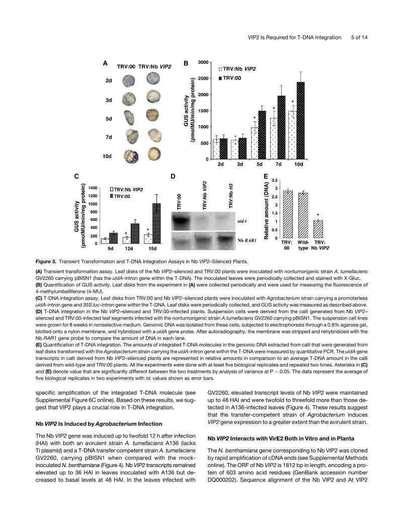
Figure 3. Transient Transformation and T-DNA Integration Assays in Nb VIP2–Silenced Plants.
(A) Transient transformation assay. Leaf disks of the Nb VIP2–silenced and TRV:00 plants were inoculated with nontumorigenic strain A. tumefaciens
GV2260 carrying pBISN1 (has the uidA-intron gene within the T-DNA). The inoculated leaves were periodically collected and stained with X-Gluc.
(B) Quantification of GUS activity. Leaf disks from the experiment in (A) were collected periodically and were used for measuring the fluorescence of
4-methylumbelliferone (4-MU).
(C) T-DNA integration assay. Leaf disks from TRV:00 and Nb VIP2–silenced plants were inoculated with Agrobacterium strain carrying a promoterless
uidA-intron gene and 35S:luc-intron gene within the T-DNA. Leaf disks were periodically collected, and GUS activity was measured as described above.
(D) T-DNA integration in the Nb VIP2–silenced and TRV:00-infected plants. Suspension cells were derived from the calli generated from Nb VIP2–
silenced and TRV:00-infected leaf segments infected with the nontumorigenic strain A.tumefaciens GV2260 carrying pBISN1. The suspension cell lines
were grown for 8 weeks in nonselective medium. Genomic DNA was isolated from these cells, subjected to electrophoresis through a 0.8% agarose gel,
blotted onto a nylon membrane, and hybridized with a uidA gene probe. After autoradiography, the membrane was stripped and rehybridized with the
Nb RAR1 gene probe to compare the amount of DNA in each lane.
(E) Quantification of T-DNA integration. The amounts of integrated T-DNA molecules in the genomic DNA extracted from calli that were generated from
leaf disks transformed with the Agrobacterium strain carrying the uidA-intron gene within the T-DNA were measured by quantitative PCR. The uidA gene
transcripts in calli derived from Nb VIP2–silenced plants are represented in relative amounts in comparison to an average T-DNA amount in the calli
derived from wild-type and TRV:00 plants. All the experiments were done with at least five biological replicates and repeated two times. Asterisks in (C)
and (E) denote value that are significantly different between the two treatments by analysis of variance at P ¼ 0.05. The data represent the average of
five biological replicates in two experiments with SE values shown as error bars.
VIP2 Is Required for T-DNA Integration 5 of 14

protein sequences showed 69% sequence identity with a con-
served C-terminal NOT domain (Figure 1F). Nb VIP2 carries two
in-frame insertions of five and 32 amino acids that are lacking in
At VIP2 (Figure 1F).
Nb VIP2 also interacted with VirE2 in a yeast two-hybrid
system, and this interaction was specific since the Nb VIP2 in-
teraction did not occur with the nonspecific interactors such as
DNA topoisomerase I and lamin C (see Supplemental Figure 7
online). We further demonstrated that Nb VIP2 can interact with
VirE2 in planta using biomolecular fluorescence complementa-
tion (BiFC; Walter et al., 2004). BiFC vectors were modified to
make it GATEWAY ready (see Methods). The interaction between
N-tagged Nb VIP2 (pSPYNE:Nb VIP2) and C-tagged VirE2
(pSPYCE:VirE2) was observed as yellow fluorescence from the
reconstitution of YFP (Figure 5). Two different controls were used
for BiFC: first, we made a translational fusion of full-length VirE2
including the stop codon (designated as VirE2*) with cYFP in
pSPYCE; second, we cloned the full-length transcriptional fac-
tor TGA2 into pSPYCE. In the first control, no VirE2-cYFP fusion
protein would be synthesized, resulting in the failure of the re-
constitution of YFP fluorescence when the two interactors are
brought together. The second control facilitates identification of
nonspecific interaction of VIP2 with transcription factors. YFP
fluorescence was not detected in leaves coinfiltrated with
pSPYNE:Nb VIP2 and pSPYCE:VirE2* or pSPYNE:Nb VIP2 and
pSPYCE:TGA2 (Figure 5).
The Arabidopsis vip2 Mutant Is Defective in T-DNA
Integration but Not in Transient T-DNA Expression
Recently, we were able to identify an Arabidopsis T-DNA mutant
line (At vip2; GABI_676A06; T-DNA insertion in the second exon)
(Rosso et al., 2003) (Figure 6A) that does not produce At VIP2
transcripts (Figure 6B). To further confirm the results obtained
from Nb VIP2–silenced plants, we performed root transformation
assays on At vip2 plants (Nam et al., 1999; Mysore et al., 2000).
Upon infection with an oncogenic strain A. tumefaciens A208, At
vip2 produced fewer tumors (38% 6 3% of the infected roots
formed tumors) compared with the wild-type plants (87% 6 5%
of the infected roots formed tumors) (Figure 6C; see Supple-
mental Table 1 online). However, no significant differences were
observed between the wild type and At vip2 for transient GUS
expression at 2 DAI (Figure 6D). Stable GUS expression in At vip2
was only 25% 6 6% of the GUS expression observed in the
wild-type plants (Figure 6D; see Supplemental Table 1 online).
We also performed stable transformation assay with the strain
A. tumefaciens GV3101 containing pCAS1. Significantly reduced
numbers of GF-resistant calli were observed in the At vip2 mutant
(33% 6 1% of infected roots formed GF-resistant calli) relative
to the wild-type plants (83% 6 3% of infected roots formed
Figure 4. Differential Gene Expression of Nb VIP2 upon Infection with
Agrobacterium.
Individual leaves of two separate N. benthamiana plants were syringe
(needleless) infiltrated with either an avirulent strain Agrobacterium A136
(lacks Ti plasmid; cannot transfer T-DNA) or a T-DNA transfer-competent
strain A. tumefaciens GV2260 carrying pBISN1. Leaf samples from the
infiltrated area were collected at different times after inoculation, and
total RNA was isolated for real-time quantitative PCR. RNA from the
buffer-infiltrated N. benthamiana leaves collected at 12 HAI was used as
a calibrator to determine the relative amount of Nb VIP2 transcripts.
Samples were pooled together from two independent experiments, and
the average of two technical replicates is shown.
Figure 5. In Planta Interaction of Nb VIP2 with VirE2.
The top panels depict the YFP fluorescence, and the bottom panels
represent the epifluorescence images of epidermal leaf cells from the
same leaf infiltrated with Agrobacterium suspension cultures harboring
the indicated proteins. Individual leaves of N. benthamiana plants were
syringe (needleless) infiltrated with Agrobacterium suspension cultures
singly or in the following combinations: pCAMBIA1390-35S:YFP, pSPY-
NE:VIP2, pSPYCE:VirE2, pSPYNE:VIP2/pSPYCE:VirE2, pSPYNE:VIP2/
pSPYCE:TGA2, and pSPYNE:VIP2/pSPYCE:VirE2*. Wild-type 35S:YFP
and fusion protein pSPYNE:VIP2/pSPYCE:VirE2 are both localized to the
nucleus of plant cells, while pSPYNE:VIP2/pSPYCE:VirE2* carrying the
VirE2 stop codon and pSPYNE:VIP2/pSPYCE:TGA2 did not produce any
fluorescence. All the images are from a single confocal section. Bars ¼10 mm.
6 of 14 The Plant Cell

GF-resistant calli). Root segments derived from both the wild-
type and At vip2 mutant plants were able to form calli at similar
frequencies on nonselective CIM (data not shown). These results
further support the role of VIP2 in T-DNA integration in another
plant species.
Transcriptome Analyses Suggest That VIP2 Plays a Role
in Transcription Regulation
To gain insight on the biological role of VIP2 in plants, a com-
prehensive survey of global gene expression was done using the
Arabidopsis whole-genome Affymetrix gene chip (ATH1) to quan-
tify the spatio-temporal variations in transcript abundance be-
tween wild-type Columbia-0 (Col-0) and At vip2. Comparative
analyses between Col-0 and At vip2 showed 4241 genes to be
constitutively differentially expressed with a false discovery rate
(FDR) <10%. Out of the 4241 differentially expressed genes,
2157 genes had more transcript abundance in At vip2 compared
with Col-0, whereas 2084 genes had more transcript abundance
in Col-0 compared with At vip2 (see Supplemental Table 2 on-
line). Functional classification of the 4241 differentially expressed
genes indicated genes involved in a variety of functions, and the
majority (28.7%) of them encode proteins of unknown function
(see Supplemental Figure 8 online). These data support our hy-
pothesis that VIP2 plays a direct or indirect role in transcription
regulation of many genes. Interestingly, upon careful examina-
tion of the transcriptome data, we found a majority of the 52 genes
encoding histones or histone-associated proteins to be consti-
tutively repressed in the At vip2 compared with Col-0 plants
(Figure 7; see Supplemental Table 3 online). Although the tran-
script differences of some of the histone genes were less than
twofold, their expression profiles were obviously different in At
vip2 and Col-0 plants (Figure 7). The exact expression values of
these genes are shown in Supplemental Table 3 online. Histones
Figure 6. Identification of At vip2 and Transformation Assays in the Mutant.
(A) The full-length genomic sequence of the At VIP2 gene (exons are shaded) showing the T-DNA insertion in the second exon of the Arabidopsis T-DNA
mutant line (At vip2; GABI_676A06).
(B) RNA gel blot analysis confirms the absence of At VIP2 transcripts in At vip2. Five micrograms of RNA extracted from leaves was fractionated on a
formaldehyde-agarose gel, blotted onto a nylon membrane, and probed with 32P-labeled At VIP2 gene (top panel). Ethidium bromide–stained gel
showing rRNA suggests equal amounts of total RNA were loaded in each lane (bottom panel).
(C) Roots of wild-type and vip2 mutant plants were infected with a tumorigenic strain A. tumefaciens A208 (nopaline strain), and tumors incited on the
roots were visualized and scored 4 weeks after infection.
(D) Transient and stable GUS expression. Roots of the wild-type and At vip2 plants were inoculated with a strain A. tumefaciens GV3101 carrying the
uidA-intron gene within the T-DNA. The inoculated roots were periodically collected and stained with X-Gluc. All the experiments were repeated two
times.
VIP2 Is Required for T-DNA Integration 7 of 14

have already been implicated in Agrobacterium-mediated plant
transformation (Mysore et al., 2000; Yi et al., 2002, 2006; Li et al.,
2005a; Anand et al., 2007).
We monitored the differential expression of genes in the At vip2
mutant and Col-0 in response to Agrobacterium infection by in-
filtrating the leaves with a disarmed strain A. tumefaciens GV3101
harboring the uidA-intron gene as described (Wroblewski et al.,
2005). Strikingly, under the same selection condition, fewer genes
were differentially expressed in At vip2, at 48 and 72 HAI, com-
pared with the number of genes that were differentially expressed
in the Col-0 plant at the same time points (see Supplemental
Table 4 online). The fact that we were not able to achieve 100%
Figure 7. Expression Profile for the 52 Differentially Expressed Histone or Histone-Associated Genes Represented in the ATH1 Gene Chips in Col-0
and At vip2.
Color code represents expression values of ratio between At vip2 and Col-0, wherein red and green indicate up- and downregulation of genes,
respectively. Each horizontal line displays the expression data for one gene. Data were clustered with correlation using the TIGR Multiple Experiment
Viewer.
8 of 14 The Plant Cell
transformation in the infiltrated plants, based on the GUS histo-
chemical staining (see Supplemental Figure 9 online), could have
diluted the effect on differential gene expression upon Agro-
bacterium infection. Nevertheless, our data suggest that At vip2
is significantly muted in its response, based on differential gene
expression, to Agrobacterium infection. These results further
validate the role of VIP2 in transcriptional regulation and Agro-
bacterium-mediated plant transformation.
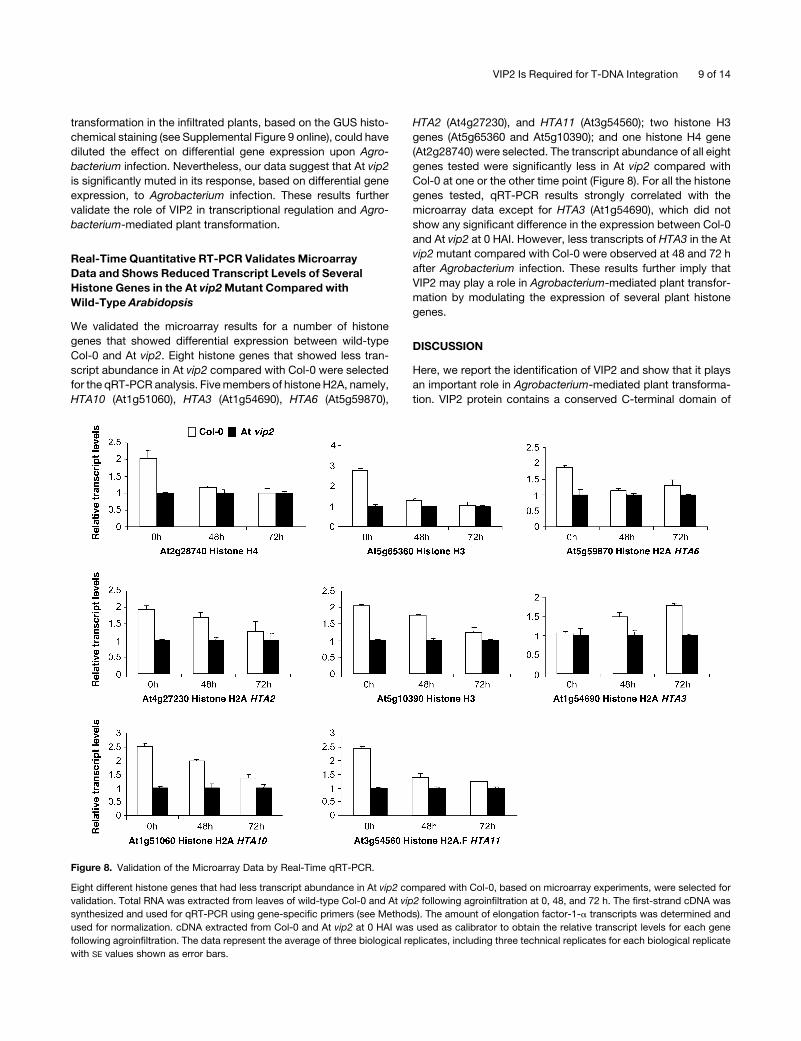
Real-Time Quantitative RT-PCR Validates Microarray
Data and Shows Reduced Transcript Levels of Several
Histone Genes in the At vip2 Mutant Compared with
Wild-Type Arabidopsis
We validated the microarray results for a number of histone
genes that showed differential expression between wild-type
Col-0 and At vip2. Eight histone genes that showed less tran-
script abundance in At vip2 compared with Col-0 were selected
for the qRT-PCR analysis. Five members of histone H2A, namely,
HTA10 (At1g51060), HTA3 (At1g54690), HTA6 (At5g59870),
HTA2 (At4g27230), and HTA11 (At3g54560); two histone H3
genes (At5g65360 and At5g10390); and one histone H4 gene
(At2g28740) were selected. The transcript abundance of all eight
genes tested were significantly less in At vip2 compared with
Col-0 at one or the other time point (Figure 8). For all the histone
genes tested, qRT-PCR results strongly correlated with the
microarray data except for HTA3 (At1g54690), which did not
show any significant difference in the expression between Col-0
and At vip2 at 0 HAI. However, less transcripts of HTA3 in the At
vip2 mutant compared with Col-0 were observed at 48 and 72 h
after Agrobacterium infection. These results further imply that
VIP2 may play a role in Agrobacterium-mediated plant transfor-
mation by modulating the expression of several plant histone
genes.
DISCUSSION
Here, we report the identification of VIP2 and show that it plays
an important role in Agrobacterium-mediated plant transforma-
tion. VIP2 protein contains a conserved C-terminal domain of
Figure 8. Validation of the Microarray Data by Real-Time qRT-PCR.
Eight different histone genes that had less transcript abundance in At vip2 compared with Col-0, based on microarray experiments, were selected for
validation. Total RNA was extracted from leaves of wild-type Col-0 and At vip2 following agroinfiltration at 0, 48, and 72 h. The first-strand cDNA was
synthesized and used for qRT-PCR using gene-specific primers (see Methods). The amount of elongation factor-1-a transcripts was determined and
used for normalization. cDNA extracted from Col-0 and At vip2 at 0 HAI was used as calibrator to obtain the relative transcript levels for each gene
following agroinfiltration. The data represent the average of three biological replicates, including three technical replicates for each biological replicate
with SE values shown as error bars.
VIP2 Is Required for T-DNA Integration 9 of 14

NOT2/NOT3/NOT5 proteins (Figure 1F). NOT2/NOT3/NOT5
domain–containing genes were identified from Saccharomyces
cerevisiae via genetic screens for increased transcription from
TATA-less promoters (Oberholzer and Collart, 1998; Collart, 2003).
The NOT proteins are an integral component of the CCR4 (for
carbon catabolite repression) transcriptional complex sharing
overlapping functions (Liu et al., 1998) and are believed to be
involved in both positive and negative regulation of gene ex-
pression in yeast (Collart, 2003; Collart and Timmers, 2004). The
yeast Not2p and Drosophila Rga proteins that contain NOT
domains are well studied and are thought to mediate intranuclear
interactions between chromatin components and the transcrip-
tional complex (Collart and Struhl, 1993; Frolov et al., 1998;
Collart, 2003). The function of NOT domain–containing proteins
in plants is not known. On the basis of its similarity to yeast and
animal NOT proteins, we speculate VIP2 to be a transcriptional
regulator.
The involvement of VIP2 in Agrobacterium-mediated plant
transformation was shown using Nb VIP2–silenced N. benthami-
ana and an Arabidopsis T-DNA knockout, At vip2. Our observa-
tions that Nb VIP2–silenced and At vip2 knockout plants were
recalcitrant to Agrobacterium-mediated stable transformation
but not to biolistic transformation further suggest that VIP2 is
specifically required for Agrobacterium-mediated stable plant
transformation. Even though the amount of stably integrated
T-DNA in the Nb VIP2–silenced plants was significantly lower
compared with the wild-type plants, the Nb VIP2–silenced plants
did not show any deficiency for transient transformation. These
results implicated a role of VIP2 in T-DNA integration. The pro-
longed induction of the VIP2 transcripts by the T-DNA transfer
competent strain of Agrobacterium compared with the induction
by the avirulent strain is consistent with the hypothesis that
Agrobacterium modulates host gene expression to facilitate the
genetic transformation event (Ditt et al., 2001; Veena et al., 2003;
Hwang and Gelvin, 2004). The magnitude of Nb VIP2 induction
reported is likely an underestimation due to the fact that not all
cells in the infiltrated area were transformed. The specific inter-
action of VirE2 and Nb VIP2 was confirmed in planta by BiFC.
Even though we predominantly detected YFP fluorescence in the
nucleus, we seldom observed YFP fluorescence in the cyto-
plasm due to VirE2–Nb VIP2 interaction. It is likely that the VirE2–
Nb VIP2 interaction happens in the cytoplasm and then the
protein complex along with other interactors gets localized to the
nucleus. Nevertheless, our data suggest that VirE2 interacts with
Nb VIP2 in planta, and further studies are needed to precisely
identify the cellular compartment where the initial interaction
takes place.
The transcriptome analyses of wild-type Arabidopsis and At
vip2 identified 4241 differentially expressed genes with an FDR of
0.1, spanned across different functional groups. This was not a
big surprise to us, and the data fit well with our speculation that
VIP2 is likely a transcriptional regulator. This conclusion is based
on circumstantial evidence, and we have not provided direct
evidences to suggest its role as a transcriptional regulator. Nev-
ertheless, At vip2 was muted in its response (based on differ-
ential gene expression) to Agrobacterium infection compared
with wild-type plants. This muted response of At vip2 to Agro-
bacterium infection probably contributes to its recalcitrance to
Agrobacterium-mediated stable plant transformation. It is impor-
tant to note that At vip2 did not have any deficiency in transient
transformation (2 to 3 DAI); therefore, the differential gene ex-
pression that we observed at 2 and 3 d after Agrobacterium
infection was not due to differences in transformation efficiency
between At vip2 and Col-0 plants. Strikingly, we found that tran-
scripts of many histone genes were less abundant in At vip2
compared with wild-type plants (Figures 7 and 8), which could be
another factor contributing to the transformation recalcitrance
phenotype of At vip2. Our data support earlier speculation that
histone proteins are involved in Agrobacterium-mediated trans-
formation, especially in the step of T-DNA integration (Mysore
et al., 2000; Yi et al., 2002, 2006; Li et al., 2005a; Anand et al.,
2007).
Based on the pfam domain prediction, VIP2 from Arabidopsis
and N. benthamiana have a conserved NOT2/NOT3/NOT5
C-terminal domain, which we speculate forms complexes similar
to Ccr4-Not in plants, and participates in diverse cellular functions
in plant development, including chromatin remodeling, that will
be addressed elsewhere. The core histones (H2A, H2B, H3, and
H4) form the building blocks of the nucleosome, which are the
fundamental repeating units of chromatin. Earlier observations
have suggested that once inside the nucleus, the Agrobacterium
T-DNA–associated proteins likely interact with the nucleosome
assembly. VIP1, a bZIP transcription factor, was shown to in-
teract with Arabidopsis histone H2A in planta (Li et al., 2005a).
Furthermore, it has been shown that chromatin-associated pro-
teins, such as histone H2A (Mysore et al., 2000), and chromatin
assembly factor 1 (Endo et al., 2006; Kirik et al., 2006) play a role
in T-DNA integration in plants. Core histone proteins are evolu-
tionarily conserved and undergo posttranslational modification,
implicating them in regulating gene expression (for review, see
Fischle et al., 2003). The fact that NOT2/NOT3/NOT5 proteins
form a nuclear complex that mediates intranuclear interactions
between chromatin components and the transcriptional complex
in many eukaryotes (Collart and Struhl, 1993; Frolov et al., 1998;
Collart, 2003) and our finding that the histone gene transcripts
are less abundant in At vip2 further provides evidence for the
biological role of VIP2 in Agrobacterium-mediated plant trans-
formation. We hypothesize that Agrobacterium takes advantage
of VIP2 to integrate its T-DNA into the plant chromosome. It is
tempting to speculate that VIP1, VIP2, and VirE2 function as a
multiprotein complex that plays a crucial role in T-DNA nuclear
import, intranuclear transport of the T-complex, and integration
of T-DNA into host genome. The above findings open up a new
area of research that could be directed toward the functional
characterization of VIP2 in plants and its biological relevance to
plant development and transformation.
METHODS
Yeast Two-Hybrid Assay
The yeast two-hybrid assays were performed as previously described
(Tzfira et al., 2001) and are detailed in the Supplemental Methods online.
The Nb VIP2 gene was cloned into pGAD424 as a PstI fragment, and its
product was shown to interact with pBTM116-VirE2 in a yeast two-hybrid
system.
10 of 14 The Plant Cell
VIGS and in Planta Tumor Assay
Plant material, bacterial culture conditions, cloning of the Nb VIP2 gene
into the TRV-VIGS vector, and sequence conformation and protocols for
VIGS were performed as described (Ryu et al., 2004; Anand et al., 2007)
with minor modifications (see Supplemental Methods online). Shoots
of the gene-silenced plants and empty vector control plants (TRV:00),
3 weeks after TRV infection, were inoculated by puncturing the stem using
a needle with a suspension culture of a tumorigenic strain Agrobacterium
tumefaciens (A348) containing the octopine type Ti plasmid (pTiA6). Tu-
mors on shoots were observed 4 weeks after Agrobacterium infection.
Leaf Disk Transformation Assays
Leaf disk transformation assays were performed as described (Anand
et al., 2007). Briefly, axenic leaf disks (15 to 20 for each plant) were
incubated with different strains of Agrobacterium (see Results) for 15 min,
blotted on sterile filter paper, cocultivated with the bacteria at 258C for 2 d
in the dark, and transferred onto either MS medium (Gibco-BRL) for tu-
morigenesis assay or to CIM (4.32 g/L MS minimal salts, 1 mL/L vitamin
stock, 100 mg/L myo-inositol, 20 g/L glucose, 0.5 mg/L 2,4-D, 0.3 mg/mL
kinetin, 5 mg/L indole-3-acetic acid, and 0.8% phytagar with antibiotics)
containing cefotaxamine (200 mg/mL) and tricarcillin (100 mg/mL) for
stable and transient transformation assays. GF (5 mg/mL) was included in
CIM for stable transformation assay. The cultures were incubated at
25.0 6 2.08C with a 16-h photoperiod at 70% humidity at 150 mE s�1 m�2
light intensity. Transient transformation assays (histochemical GUS stain-
ing and quantification of GUS activity) were performed as described
(Jefferson et al., 1987; Anand et al., 2007).
Agrobacterium-Independent Transformation Assays
The efficacy of Agrobacterium independent transformation methods in
the Nb VIP2–silenced leaves was tested by particle bombardment. DNA
(1 mg) was adsorbed onto 10 mg of 1-mm gold particles (Bio-Rad) and
bombarded at 150 p.s.i. into the leaf epidermis of greenhouse-grown
Nicotiana benthamiana plants, followed by incubation for 24 to 48 h at
258C in dark. For the transient transformation assay, leaf disks from the
TRV:00-inoculated and Nb vip2–silenced plants were biolistically trans-
formed with a 35S:uidA (pAHC20) construct. Bombarded leaves were
stained with X-Gluc staining solution (50 mM NaH2PO4, 10 mM Na2�EDTA,
300 mM mannitol, and 2 mM X-Gluc, pH 7.0) 48 to 72 h after bombardment
and viewed under a Bio-Rad confocal microscope for GUS-expressing
spots. For the stable transformation assay, the leaf disks from the TRV:00
and Nb VIP2–silenced plants were biolistically transformed with Ubi:bar
(pAHC20 cassette). The transformed leaf disks were selected on media
supplemented with GF for 8 weeks, and the presence of the bar gene was
detected by PCR in a few representative GF-resistant calli as described
earlier (Anand et al., 2003b)
RNA Extraction, PCR, T-DNA Integration Assay, and Differential
Gene Expression
RNA extraction, first-strand cDNA synthesis, semiquantitative RT-PCR,
and qRT-PCR were performed using standard protocols as described
(Ryu et al., 2004; see Supplemental Methods online). The RNA gel blot
analyses were performed using standard protocols (Anand et al., 2003a)
on total RNA extracted from leaves of Col-0 and the At vip2 mutant. For the
T-DNA integration assay, we performed DNA gel blot analysis (Mysore
et al., 2000) on the genomic DNA extracted from suspension cell lines
generated from the calli produced on nonselective medium by leaf disks
of Nb VIP2–silenced plants and TRV:00 plants infected with disarmed
strain A. tumefaciens GV2260 carrying pBISN1 (see Results). Suspension
cells were cultured for 8 weeks, in the presence of timentin, by periodic
transferring into fresh media to remove bacterial contamination. Repre-
sentative suspension cells were collected and stained with X-Gluc solu-
tion to check for the presence and expression of the uidA-gene in the
transgenic cells. The DNA gel blots were hybridized with radiolabeled
probes of the uidA and Nb RAR1 genes for detecting the integrated T-DNA
and as control for DNA loading, respectively. For real-time quantification
of integrated T-DNA, genomic DNA was extracted from calli (collected
from a pool of two independent experiments, with five biological replicates
each) produced on leaf disks of Nb VIP2–silenced and TRV:00 plants that
were transformed with strain A. tumefaciens GV2260 carrying pBISN1.
Calli were washed with dimethyl sulfoxide (15% [w/v]) several times by
vortexing to remove any attached bacteria, and DNA was extracted using
DNAzol (Invitrogen) according to the manufacturer’s instructions. Quan-
titative DNA PCRs were performed on duplicate DNA samples using the
GUS gene and Agrobacterium chromosomal gene (Atu0792) specific
primers (for primer details, see Anand et al., 2007) to check for the
presence of the uidA gene and bacterial DNA contamination. Duplicate
samples were analyzed by qPCR with the primers GUS-FP (59-AGG-
TGCACGGGAATATTTCG-39) and GUS-RP (59-ACGCGTCGGGTCGA-
GTT-39) to determine the abundance of integrated T-DNA. As a loading
control for silenced and nonsilenced plants, parallel qPCR reactions using
Nb Ef1a primers Nb Ef1F (59-TGAGGCTCTTGACCAGATTAATGA-39) and
Nb Ef1R (59-GTAAACATCCTGAAGTGGAAGACGTA-39) were performed.
For differential gene expression analyses of Nb VIP2, individual leaves
of two separate N. benthamiana plants were infiltrated (using a needleless
syringe) with an avirulent strain Agrobacterium A136 or a T-DNA transfer
competent strain A. tumefaciens GV2260 carrying pBISN1 or the infiltra-
tion buffer. Samples were collected at different time points after inoculation
and were subjected to qRT-PCR using primers Nb VIP2F (59-AAGGTGG-
GAATGCTGATTATGC-39) and Nb VIP2R (59-TCTTCCCATTGAGAAG-
TGTTGCT-39), in parallel with Nb Ef1a primers as loading control. The
experiments were repeated twice.
Characterization of At vip2
Seeds of Arabidopsis thaliana wild-type Col-0 and T-DNA insertion mutant
GABI_676A06 (At vip2) were germinated, and the roots were subjected to
transient and stable Agrobacterium-mediated transformation assays as
described (Nam et al., 1999; Zhu et al., 2003). RNA gel blot analyses were
performed on RNA extracted from homozygous At vip2 plants and wild-
type Col-0 plants using the radiolabeled probe of the At VIP2 gene. RT-
PCR reactions were performed on cDNAs prepared from the mutant using
primer combinations (At VIP2F, 59-TGGTTCGGGCAGATCGTTTAC-
TGC-39; At VIP2R, 59-GCAAGCTTGGTCTCTTTTCC-39) to determine the
presence of At VIP2 transcript. In vitro tumorigenesis assays were per-
formed on the axenic root segments by infecting with oncogenic strain
A.tumefaciensA208containinganopaline-typeTiplasmid (pTiT37), cocul-
tivated for 48 h in dark at room temperature, transferred to a hormone-free
MS media supplemented with cefotaxamine and tricarcillin, and the tu-
mor numbers and phenotypes were recorded 4 to 5 weeks after infection.
Transient and stable GUS expression assays and the GF-resistant calli
assay were performed as detailed earlier (Li et al., 2005b) using the dis-
armed strain A. tumefaciens GV3101 containing either pBISN1 or pCAS1.
Expression Profiling in the vip2 Mutant in Response to
Agrobacterium Infection
The Affymetrix microarrays (Arabidopsis ATH1 genome array) were used
in the expression profiling study involving At vip2 and Col-0 plants. The
wild-type Col-0 and At vip2 mutant plants were syringe (needleless)
infiltrated with the disarmed strain A. tumefaciens GV3101 (OD600 ;0.2).
Samples were collected at 0, 48, and 72 HAI based on the previous report
(Ditt et al., 2006) showing strong differential gene expression at 48 HAI in
Arabidopsis suspension cell cultures following Agrobacterium infection.
VIP2 Is Required for T-DNA Integration 11 of 14
Preliminary experiments on agroinfiltration in Arabidopsis indicated that
the earliest GUS expression was observed at 48 HAI, with the GUS
expression even stronger at 72 HAI, which is indicative of higher trans-
formation frequency. Leaf samples were individually pooled from the
infected plants (10 plants for each time point) for RNA extraction, and few
representative leaves were stained with X-Gluc to confirm GUS expres-
sion. Total RNA was extracted from two independent biological replicates
as described earlier (Ryu et al., 2004). RNA was further cleaned with the
RNeasy mini kit (Qiagen) following the manufacturer’s instructions, and
the quality check was performed using the Bioanalyzer 2100 (Agilent
Technologies). Affymetrix chip labeling, hybridization, and scanning pro-
cedures followed the instructions provided in the Affymetrix manual (www.
affymetrix.com/support/technical/manual/expression_manual.affx).
Data Analysis, Gene Clustering, and Validation of the
Microarray Data
Leaf disk transformation data were subjected to analysis of variance us-
ing JMP software version 4.0.4 (SAS Institute) or by analysis of variance.
When a significant result using F-test was obtained at P ¼ 0.05, sepa-
ration of treatment means was determined by Fisher’s protected least
significant difference.
To obtain genes differentially expressed between the wild type and
mutant, pairwise comparisons were performed for microarray data ob-
tained from Col-0 and At vip2 at the same treatment conditions. Genes
responsive to Agrobacterium treatment in both Col-0 and At vip2 were
identified by comparing the treated samples with their 0 h control ac-
cordingly. For each comparison, the normalized data were imported into
an Excel sheet, and differential genes were selected using the Associative
Analysis algorithm developed by Dozmorov and Centola (2003). In this
analysis, the Bonferroni adjusted P value threshold for Student’s t test
was set at 0.05/N, n ¼ 22,000, the number of probe sets in the reference
group (Dozmorov and Centola, 2003). The corrected P value ensures that
the overall false positive among the multiple comparisons are controlled
under 0.05. To account for multiple hypotheses testing, Q values, an es-
timation of FDR, were also calculated for each probe set using EDGE soft-
ware (http://www.biostat.washington.edu/software/jstorey/edge/; Storey
and Tibshirani, 2003). The Q value for a particular probe set reflects the
proportion of false positives incurred among all probe sets as or more
significant than the one being measured (Storey and Tibshirani, 2003). For
comparative gene expression analyses of histone and histone-related
genes between Col-0 and the At vip2 mutant following Agrobacterium
treatment, all 52 histones and histone-related genes known to be expressed
(having the presence of calli in both replicates) were clustered and visual-
ized using TIGR Multiple Experiment Viewer (http://www.tm4.org/mev.html).
A few differentially regulated histone genes identified from the tran-
scriptome analysis were selected for validation of the results by qRT-PCR
(see Supplemental Table 5 online for primer details). The data from two
of the biological replicates used for microarray analysis and an indepen-
dent third biological replicate each with three technical replicates were
analyzed to quantify the relative transcript levels in At vip2 and Col-0
plants.
BiFC Assay
Agrobacterium binary BiFC vectors (Walter et al., 2004), pSPYNE-35S
and pSPYCE-35S with YFP dissected into two parts (the N-terminal
[nYFP] and the C-terminal [cYFP]) were used to generate GATEWAY-
compatible derivatives. A blunt-end GATEWAY cassette, reading frame
B (Invitrogen), was inserted into the EcoRV site of pBluescript SKIIþ(Stratagene) to obtain pBGB-EH. Subsequently, an XbaI-XhoI fragment of
pBGB-EH was transferred into the XbaI-XhoI sites of pSPYNE-35S and
pSPYCE-35S to obtain pSPYNE-35S_GW and pSPYCE-35S_GW, re-
spectively. For fusion protein analyses, the full-length Nb VIP2 ORF was
cloned into the nYFP construct (pSPYNE:VIP2) and the translational
fusion of VirE2 into the cYFP construct with (pSPYCE:VirE2*) and without
(pSPYCE:VirE2) the stop codon. We also cloned the transcriptional factor
TGA2 gene into the cYFP construct (pSPYCE:TGA2). These constructs
were transformed into strain A. tumefaciens GV2260 by electroporation.
We used pCAMBIA1390 harboring the 35S:YFP in GV2260 as a positive
control. All the strains of Agrobacterium were grown in Luria-Bertani
media under appropriate antibiotics overnight, induced with acetosyr-
ingone (100 mg/mL) for 4 h at room temperature. Agrobacterium strains
containing individual constructs were mixed at a 1:1 ratio and infiltrated
(;1.0 OD) into the leaves of 3- to 4-week-old N. benthamiana plants. The
infiltrated plants were placed in the dark for 72 h, and leaf sections were
examined by a Leica TCS SP2 AOBS confocal laser scanning microscope
with the samples excited at 514 nm at 72 HAI. These experiments were
repeated twice.
Accession Numbers
Sequence data from this article can be found in the GenBank/EMBL data
libraries under accession numbers AF295433 (At VIP2) and DQ000202
(Nb VIP2).
Supplemental Data
The following materials are available in the online version of this article.
Supplemental Figure 1. Alignment of Proteins with the NOT Domain.
Supplemental Figure 2. Nuclear Localization of At VIP2 in Plant
Cells.
Supplemental Figure 3. Semiquantitative RT-PCR Analyses and in
Planta Tumor Assays.
Supplemental Figure 4. Quantification of Tumors in Nb VIP2–
Silenced Plants.
Supplemental Figure 5. Nb VIP2–Silenced Plants Transformed by
Alternate Methods.
Supplemental Figure 6. T-DNA Integration Assay.
Supplementaly Figure 7. Nb Vip2–VirE2 Interaction.
Supplemental Figure 8. Classification of the 4241 Differentially Ex-
pressed Genes.
Supplemental Figure 9. Transient Expression of the uidA-Intron
Gene in Leaves of Arabidopsis Plants.
Supplemental Table 1. Quantitative Analyses of Transient and Stable
Transformations in Arabidopsis At vip2 Mutant and Wild-Type Plants.
Supplemental Table 2. Differentially Expressed Genes between Col-0
and At vip2 at the Same Treatment Conditions and between 0 h Con-
trol and Treatments in Either Col-0 or At vip2.
Supplemental Table 3. Differentially Expressed Histone and Histone-
Associated Genes in At vip2 and Col-0 in Response to Agrobacterium
Infection.
Supplemental Table 4. Differential Gene Expression in the Wild-Type
Col-0 and the At vip2 Mutant in Response to Agrobacterium Infection.
Supplemental Table 5. Primer Sequences of Genes Used for Re-
validation of the Microarray Data by qRT-PCR.
Supplemental Methods.
ACKNOWLEDGMENTS
We thank S. Gelvin, S. Uppalapati, and M. Udvardi for critical reading of
the manuscript. We also thank S.P. Dinesh-Kumar for providing GATEWAY
12 of 14 The Plant Cell

ready TRV-VIGS vectors, Stan Gelvin for many strains of Agrobacterium,
Miki Hartwell and Candice Jones for their assistance with in vitro assays,
and Stacy Allen for his assistance with the microarray experiments. This
work was supported by Noble Foundation and by National Science
Foundation Award 0445799 (K.S.M.). The work in the V.C. laboratory is
supported by grants from the National Institutes of Health, the National
Science Foundation, the USDA, the U.S.–Israel Binational Agricultural
Research and Development Fund, and the U.S.–Israel Binational Science
Foundation. The confocal system used at the Noble Foundation was from
an equipment grant from the National Science Foundation (DBI 0400580).
Received March 30, 2006; revised February 4, 2007; accepted April 27,
2007; published May 11, 2007.
REFERENCES
Anand, A., and Mysore, K.S. (2006). Agrobacterium-biology and crown
gall disease. In Plant-Associated Bacteria, S.S. Gnanamanickam, ed
(Dordrecht, The Netherlands: Springer Science), pp. 359–384.
Anand, A., Trick, H.N., Gill, B.S., and Muthukrishnan, S. (2003b).
Stable transgene expression and random gene silencing in wheat.
Plant Biotechnol. J. 1: 241–252.
Anand, A., Zarir, V., Ryu, C.M., Kang, L., del-Pozo, O., Martin, G.B.,
and Mysore, K.S. (2007). Identification of plant genes involved in
Agrobacterium-mediated transformation by using virus-induced gene
silencing as a functional genomics tool. Mol. Plant Microbe Interact.
20: 41–52.
Anand, A., Zhou, T., Trick, H.N., Gill, B.S., Bockus, W.W., and
Muthukrishnan, S. (2003a). Greenhouse and field testing of trans-
genic wheat plants stably expressing genes for thaumatin-like protein,
chitinase and glucanase against Fusarium graminearum. J. Exp. Bot.
54: 1101–1111.
Ballas, N., and Citovsky, V. (1997). Nuclear localization signal binding
protein from Arabidopsis mediates nuclear import of Agrobacterium
VirD2 protein. Proc. Natl. Acad. Sci. USA 94: 10723–10728.
Bartel, P., Chien, C., Sternglanz, R., and Fields, S. (1993). Elimination
of false positives that arise in using the two-hybrid system. Biotech-
niques 14: 920–924.
Burch-Smith, T.M., Anderson, J.C., Martin, G.B., and Dinesh-Kumar,
S.P. (2004). Applications and advantages of virus-induced gene
silencing for gene function studies in plants. Plant J. 39: 734–746.
Cascales, E., and Christie, P.J. (2004). Definition of a bacterial type IV
secretion pathway for a DNA substrate. Science 304: 1170–1173.
Christie, P.J. (2004). Type IV secretion: The Agrobacterium VirB/D4 and
related conjugation systems. Biochim. Biophys. Acta 1694: 219–234.
Collart, M.A. (2003). Global control of gene expression in yeast by the
Ccr4-Not complex. Gene 313: 1–16.
Collart, M.A., and Struhl, K. (1993). CDC39 an essential nuclear protein
that negatively regulates transcription and differentially affects the
constitutive and inducible his3 promoters. EMBO J. 12: 177–186.
Collart, M.A., and Struhl, K. (1994). NOT1 (CDC39), NOT2 (CDC35),
NOT3 and NOT4 encode a global-negative regulator of transcription
that differentially affects TATA-element utilization. Genes Dev. 8:
525–537.
Collart, M.A., and Timmers, H.T. (2004). The eukaryotic Ccr4-Not
complex: A regulatory platform integrating mRNA metabolism with
cellular signaling pathways? Prog. Nucleic Acid Res. Mol. Biol. 77:
289–322.
Dietrich, C., and Maiss, E. (2002). Red fluorescent protein DsRed from
Discosoma sp. as a reporter protein in higher plants. Biotechniques
32: 286–293.
Ditt, R.F., Kerr, F.K., de Figueiredo, P., Delrow, J., Comai, L., and
Nester, E.W. (2006). The Arabidopsis thaliana transcriptome in re-
sponse to Agrobacterium tumefaciens. Mol. Plant Microbe Interact.
19: 665–681.
Ditt, R.F., Nester, E.W., and Comai, L. (2001). Plant gene expression
response to Agrobacterium tumefaciens. Proc. Natl. Acad. Sci. USA
98: 10954–10959.
Dozmorov, I., and Centola, M. (2003). An associative analysis of gene
expression array data. Bioinformatics 22: 204–211.
Endo, M., Ishikawa, Y., Osakabe, K., Nakayama, S., Kaya, H., Araki,
T., Shibahara, K., Abe, K., Ichikawa, H., Valentine, L., Hohn, B.,
and Toki, S. (2006). Increased frequency of homologous recombina-
tion and T-DNA integration in Arabidopsis CAF-1 mutants. EMBO J.
25: 5579–5590.
Fischle, W., Wang, Y., and Allis, C.D. (2003). Histone and chromatin
cross-talk. Curr. Opin. Cell Biol. 15: 172–183.
Frolov, M.V., Benevolenskaya, E.V., and Birchler, J.A. (1998).
Regena (Rga), a Drosophila homolog of the global negative tran-
scriptional regulator CDC36 (NOT2) from yeast, modifies gene ex-
pression and suppresses position effect variegation. Genetics 148:
317–330.
Gelvin, S.B. (2003). Agrobacterium-mediated plant transformation: The
biology behind the ‘‘gene-jockeying’’ tool. Microbiol. Mol. Biol. Rev.
67: 16–37.
Goodin, M.M., Dietzgen, R.G., Schichnes, D., Ruzin, S., and Jackson,
A.O. (2002). pGD vectors: Versatile tools for the expression of green
and red fluorescent protein fusions in agroinfiltrated plant leaves. Plant
J. 31: 375–383.
Guralnick, B., Thomsen, G., and Citovsky, V. (1996). Transport of DNA
into the nuclei of Xenopus oocytes by a modified VirE2 protein of
Agrobacterium. Plant Cell 8: 363–373.
Hwang, H.-H., and Gelvin, S.B. (2004). Plant proteins that interact with
VirB2, the Agrobacterium tumefaciens pilin protein, mediate plant
transformation. Plant Cell 16: 3148–3167.
Jefferson, R.A., Kavanagh, T.A., and Bevan, M.W. (1987). GUS
fusions: b-Glucuronidase as a sensitive and versatile gene fusion
marker in higher plants. EMBO J. 6: 901–907.
Kirik, A., Pecinka, A., Wendeler, E., and Reiss, B. (2006). The
chromatin assembly factor subunit FASCIATA1 is involved in homol-
ogous recombination in plants. Plant Cell 18: 2431–2442.
Li, J., Krichevsky, A., Vaidya, M., Tzfira, T., and Citovsky, V. (2005a).
Uncoupling of the functions of the Arabidopsis VIP1 protein in tran-
sient and stable plant genetic transformation by Agrobacterium. Proc.
Natl. Acad. Sci. USA 102: 5733–5738.
Li, J., Vaidya, M., White, C., Vainstein, A., Citovsky, V., and Tzfira, T.
(2005b). Involvement of KU80 in T-DNA integration in plant cells. Proc.
Natl. Acad. Sci. USA 102: 19231–19236.
Liu, H.-Y., Badarinarayana, V., Audino, D.C., Rappsilber, J., Mann,
M., and Denis, C.L. (1998). The NOT proteins are part of the CCR4
transcriptional complex and affect gene expression both positively
and negatively. EMBO J. 17: 1096–1106.
Liu, Y., Schiff, M., and Dinesh-Kumar, P. (2002a). Virus-induced gene
silencing in tomato. Plant J. 31: 777–786.
Liu, Y., Schiff, M., Marathe, R., and Dinesh-Kumar, P. (2002b).
Tobacco Rar1, EDS1 and NPR1/NIMI1 like genes are required for
N-mediated resistance to tobacco mosaic virus. Plant J. 30: 415–429.
Mysore, K.S., Bassuner, B., Deng, X.B., Darbinian, N.S., Motchoulski,
A., Ream, W., and Gelvin, S.B. (1998). Role of the Agrobacterium
tumefaciens VirD2 protein in T-DNA transfer and integration. Mol.
Plant Microbe Interact. 11: 668–683.
Mysore, K.S., Nam, J., and Gelvin, S.B. (2000). An Arabidopsis histone
H2A mutant is deficient in Agrobacterium T-DNA integration. Proc.
Natl. Acad. Sci. USA 97: 948–953.
VIP2 Is Required for T-DNA Integration 13 of 14

Nam, J., Mysore, K.S., Zheng, C., Knue, M.K., Matthysse, A.G., and
Gelvin, G.B. (1999). Identification of T-DNA tagged Arabidopsis
mutants that are resistant to transformation by Agrobacterium. Mol.
Gen. Genet. 261: 429–438.
Oberholzer, U., and Collart, M.A. (1998). Characterization of NOT5 that
encodes a new component of the Not protein complex. Gene 207:
61–69.
Park, H., and Sternglanz, R. (1998). Two separate conserved domains
of eukaryotic DNA topoisomerase I bind to each other and reconsti-
tute enzymatic activity. Chromosoma 107: 211–215.
Rosso, M.G., Li, Y., Strizhov, N., Reiss, B., Dekker, K., and Weisshaar,
B. (2003). An Arabidopsis thaliana T-DNA mutagenized population
(GABI-Kat) for flanking sequence tag-based reverse genetics. Plant
Mol. Biol. 53: 247–259.
Ryu, C.-M., Anand, A., Kang, L., and Mysore, K.S. (2004). Agrodrench:
A novel and effective agroinoculation method for virus-induced gene
silencing in roots and diverse Solanaceous species. Plant J. 40:
322–331.
Schultheiss, H., Dechert, C., Kogel, K.H., and Huckelhoven, R.
(2003). Functional analysis of barely RAC/ROP G-protein family
members in susceptibility to the powdery mildew fungus. Plant J.
36: 589–601.
Storey, J.D., and Tibshirani, R. (2003). Statistical significance for
genomewide studies. Proc. Natl. Acad. Sci. USA 100: 9440–9445.
Tian, G.-W., et al. (2004). High-throughput fluorescent tagging of full-
length Arabidopsis gene products in planta. Plant Physiol. 135: 25–38.
Tzfira, T., and Citovsky, V. (2002). Partners-in-infection: Host proteins
involved in the transformation of plant cells by Agrobacterium. Trends
Cell Biol. 12: 121–129.
Tzfira, T., and Citovsky, V. (2006). Agrobacterium-mediated genetic
transformation of plants: Biology and biotechnology. Curr. Opin.
Biotechnol. 17: 147–154.
Tzfira, T., Li, J., Lacroix, B., and Citovsky, V. (2004). Agrobacterium
T-DNA integration: Molecules and models. Trends Genet. 20: 375–383.
Tzfira, T., Rhee, Y., Chen, M.-H., Kunik, T., and Citovsky, V. (2000).
Nucleic acid transport in plant-microbe interactions: The molecules
that walk through the Walls. Annu. Rev. Microbiol. 54: 187–219.
Tzfira, T., Vaidya, M., and Citovsky, V. (2001). VIP1, an Arabidopsis
protein that interacts with Agrobacterium VirE2, is involved in VirE2
nuclear import and Agrobacterium infectivity. EMBO J. 20: 3596–
3607.
Veena, Jiang, H., Doerge, R.W., and Gelvin, S.B. (2003). Transfer of
T-DNA and Vir proteins to plant cells by Agrobacterium tumefaciens
induces expression of host genes involved in mediating transforma-
tion and suppresses host defense gene expression. Plant J. 35:
219–226.
Vergunst, A.C., Schrammeijer, B., den Dulk-Ras, A., de Vlaam,
C.M.T., Regensburg-Tuink, T.J., and Hooykaas, P.J. (2000). VirB/
D4-dependent protein translocation from Agrobacterium into plant
cells. Science 290: 979–982.
Vergunst, A.C., van Lier, M.C.M., den Dulk-Ras, A., and Hooykaas,
P.J. (2003). Recognition of the Agrobacterium VirE2 translocation
signal by the VirB/D4 transport system does not require VirE1. Plant
Physiol. 133: 978–988.
Walter, M., Chaban, C., Schutze, K., Batistic, O., Weckermann, K.,
Nake, C., Blazevic, D., Grefen, C., Schumacher, K., Oecking, C.,
Harter, K., and Kudla, J. (2004). Visualization of protein interactions
in living plant cells using bimolecular fluorescence complementation.
Plant J. 40: 428–438.
Wang, B.-B., and Brendel, V. (2006). Genomewide comparative anal-
ysis of alternative splicing in plants. Proc. Natl. Acad. Sci. USA 103:
7175–7180.
Wroblewski, T., Tomczak, A., and Michelmore, R. (2005). Optimiza-
tion of Agrobacterium-mediated transient assays of gene expression
in lettuce, tomato and Arabidopsis. Plant Biotechnol. J. 3: 259–273.
Yi, H., Sardesai, N., Fujinuma, T., Chan, C.W., Veena, and Gelvin,
S.B. (2006). Constitutive expression exposes functional redundancy
between the Arabidopsis histone H2A gene HTA1 and other H2A gene
family members. Plant Cell 18: 1575–1589.
Yi, H.C., Mysore, K.S., and Gelvin, S.B. (2002). Expression of the
Arabidopsis histone H2A-1 gene correlates with susceptibility to
Agrobacterium transformation. Plant J. 32: 285–298.
Zhu, Y., et al. (2003). Identification of Arabidopsis rat mutants. Plant
Physiol. 132: 494–505.
14 of 14 The Plant Cell

DOI 10.1105/tpc.106.042903; originally published online May 11, 2007;Plant Cell
Vitaly Citovsky and Kirankumar S. MysoreAjith Anand, Alexander Krichevsky, Sebastian Schornack, Thomas Lahaye, Tzvi Tzfira, Yuhong Tang,
Integration in Plants T-DNAAgrobacterium VIRE2 INTERACTING PROTEIN2 Is Required for Arabidopsis
This information is current as of July 2, 2018
Supplemental Data /content/suppl/2007/05/10/tpc.106.042903.DC1.html
Permissions https://www.copyright.com/ccc/openurl.do?sid=pd_hw1532298X&issn=1532298X&WT.mc_id=pd_hw1532298X
eTOCs http://www.plantcell.org/cgi/alerts/ctmain
Sign up for eTOCs at:
CiteTrack Alerts http://www.plantcell.org/cgi/alerts/ctmain
Sign up for CiteTrack Alerts at:
Subscription Information http://www.aspb.org/publications/subscriptions.cfm
is available at:Plant Physiology and The Plant CellSubscription Information for
ADVANCING THE SCIENCE OF PLANT BIOLOGY © American Society of Plant Biologists