Arabidopsis MAP kinase 4 regulates salicylic acid- and jasmonic
15
Arabidopsis MAP kinase 4 regulates salicylic acid- and jasmonic acid/ethylene-dependent responses via EDS1 and PAD4 Peter Brodersen 1 , Morten Petersen 1 , Henrik Bjørn Nielsen 2 , Shijiang Zhu 1 , Mari-Anne Newman 3 , Kevan M. Shokat 4 , Steffen Rietz 5 , Jane Parker 5 and John Mundy 1,* 1 Institute of Molecular Biology, Copenhagen University, Øster Farimagsgade 2A, DK-1353 Copenhagen K, Denmark, 2 Center for Biological Sequence Analysis, BioCentrum-DTU, Building 208, Technical University of Denmark, DK-2800 Lyngby, Denmark, 3 Institute of Plant Biology, Royal Veterinary and Agricultural University, Thorvaldsenvej 40, DK-1871 Frederiksberg, Denmark, 4 Department of Cellular and Molecular Pharmacology, University of California San Francisco, CA 94143–0450, USA, and 5 Department of Plant–Microbe Interactions, Max-Planck Institute for Plant Breeding Research, Carl-von-Linne ´ Weg 10, 50829 Cologne, Germany Received 9 December 2005; revised 6 April 2006; accepted 11 April 2006. * For correspondence (fax þ45 35322128; e-mail [email protected]). Summary Arabidopsis MPK4 has been implicated in plant defense regulation because mpk4 knockout plants exhibit constitutive activation of salicylic acid (SA)-dependent defenses, but fail to induce jasmonic acid (JA) defense marker genes in response to JA. We show here that mpk4 mutants are also defective in defense gene induction in response to ethylene (ET), and that they are more susceptible than wild-type (WT) to Alternaria brassicicola that induces the ET/JA defense pathway(s). Both SA-repressing and ET/JA-(co)activating functions depend on MPK4 kinase activity and involve the defense regulators EDS1 and PAD4, as mutations in these genes suppress de-repression of the SA pathway and suppress the block of the ET/JA pathway in mpk4. EDS1/PAD4 thus affect SA–ET/JA signal antagonism as activators of SA but as repressors of ET/JA defenses, and MPK4 negatively regulates both of these functions. We also show that the MPK4–EDS1/PAD4 branch of ET defense signaling is independent of the ERF1 transcription factor, and use comparative microarray analysis of ctr1, ctr1/ mpk4, mpk4 and WT to show that MPK4 is required for induction of a small subset of ET-regulated genes. The regulation of some, but not all, of these genes involves EDS1 and PAD4. Keywords: hormone interactions, MAP kinase, pathogen responses. Introduction Plants are able to activate immune responses upon recog- nition of invading pathogens. Recognition may occur via gene-for-gene interactions in which a plant resistance (R) gene product interacts with or detects the action of a cognate pathogen avirulence (Avr) factor (Nimchuk et al., 2003). R– Avr interactions induce rapid resistance responses at the infection site that are often mediated by salicylic acid (SA). Many virulent pathogens also induce basal defense re- sponses that involve SA, and loss of basal defense causes hyper-susceptibility to virulent pathogens. In addition to initiation of local defenses, R protein activation can lead to an immune state in systemic tissues termed systemic acquired resistance (SAR). SAR develop- ment in Arabidopsis correlates with expression of the pathogenesis-related (PR) genes PR1, PR2 and PR5, and involves micro-oxidative bursts and SA accumulation in systemic tissues (Alvarez et al., 1998; Malamy et al., 1990; Uknes et al., 1992). The role of SA in plant immunity is supported by the fact that exogenous SA, or high-level endogenous SA accumulation by expression of bacterial SA synthases, induce SAR-like resistance and PR gene expres- sion (Verberne et al., 2000). Conversely, SAR is impaired in the SA-deficient mutants eds5 and sid2 (Nawrath and Me ´ traux, 1999). SA depletion by transgenic expression of 532 ª 2006 The Authors Journal compilation ª 2006 Blackwell Publishing Ltd The Plant Journal (2006) 47, 532–546 doi: 10.1111/j.1365-313X.2006.02806.x
Transcript of Arabidopsis MAP kinase 4 regulates salicylic acid- and jasmonic

Arabidopsis MAP kinase 4 regulates salicylic acid- andjasmonic acid/ethylene-dependent responses via EDS1 andPAD4
Peter Brodersen1, Morten Petersen1, Henrik Bjørn Nielsen2, Shijiang Zhu1, Mari-Anne Newman3, Kevan M. Shokat4, Steffen
Rietz5, Jane Parker5 and John Mundy1,*
1Institute of Molecular Biology, Copenhagen University, Øster Farimagsgade 2A, DK-1353 Copenhagen K, Denmark,2Center for Biological Sequence Analysis, BioCentrum-DTU, Building 208, Technical University of Denmark, DK-2800 Lyngby,
Denmark,3Institute of Plant Biology, Royal Veterinary and Agricultural University, Thorvaldsenvej 40, DK-1871 Frederiksberg, Denmark,4Department of Cellular and Molecular Pharmacology, University of California San Francisco, CA 94143–0450, USA, and5Department of Plant–Microbe Interactions, Max-Planck Institute for Plant Breeding Research, Carl-von-Linne Weg 10, 50829
Cologne, Germany
Received 9 December 2005; revised 6 April 2006; accepted 11 April 2006.*For correspondence (fax þ45 35322128; e-mail [email protected]).
Summary
Arabidopsis MPK4 has been implicated in plant defense regulation because mpk4 knockout plants exhibit
constitutive activation of salicylic acid (SA)-dependent defenses, but fail to induce jasmonic acid (JA) defense
marker genes in response to JA.We show here thatmpk4mutants are also defective in defense gene induction
in response to ethylene (ET), and that they are more susceptible than wild-type (WT) to Alternaria brassicicola
that induces the ET/JA defense pathway(s). Both SA-repressing and ET/JA-(co)activating functions depend on
MPK4 kinase activity and involve the defense regulators EDS1 and PAD4, as mutations in these genes suppress
de-repression of the SA pathway and suppress the block of the ET/JA pathway in mpk4. EDS1/PAD4 thus
affect SA–ET/JA signal antagonism as activators of SA but as repressors of ET/JA defenses, and MPK4
negatively regulates both of these functions. We also show that the MPK4–EDS1/PAD4 branch of ET defense
signaling is independent of the ERF1 transcription factor, and use comparativemicroarray analysis of ctr1, ctr1/
mpk4, mpk4 and WT to show that MPK4 is required for induction of a small subset of ET-regulated genes. The
regulation of some, but not all, of these genes involves EDS1 and PAD4.
Keywords: hormone interactions, MAP kinase, pathogen responses.
Introduction
Plants are able to activate immune responses upon recog-
nition of invading pathogens. Recognition may occur via
gene-for-gene interactions in which a plant resistance (R)
gene product interacts with or detects the action of a cognate
pathogen avirulence (Avr) factor (Nimchuk et al., 2003). R–
Avr interactions induce rapid resistance responses at the
infection site that are often mediated by salicylic acid (SA).
Many virulent pathogens also induce basal defense re-
sponses that involve SA, and loss of basal defense causes
hyper-susceptibility to virulent pathogens.
In addition to initiation of local defenses, R protein
activation can lead to an immune state in systemic tissues
termed systemic acquired resistance (SAR). SAR develop-
ment in Arabidopsis correlates with expression of the
pathogenesis-related (PR) genes PR1, PR2 and PR5, and
involves micro-oxidative bursts and SA accumulation in
systemic tissues (Alvarez et al., 1998; Malamy et al., 1990;
Uknes et al., 1992). The role of SA in plant immunity is
supported by the fact that exogenous SA, or high-level
endogenous SA accumulation by expression of bacterial SA
synthases, induce SAR-like resistance and PR gene expres-
sion (Verberne et al., 2000). Conversely, SAR is impaired in
the SA-deficient mutants eds5 and sid2 (Nawrath and
Metraux, 1999). SA depletion by transgenic expression of
532 ª 2006 The AuthorsJournal compilation ª 2006 Blackwell Publishing Ltd
The Plant Journal (2006) 47, 532–546 doi: 10.1111/j.1365-313X.2006.02806.x

bacterial nahG salicylate hydroxylase also impairs SAR
induction, although nahG expression has pleiotropic effects
beyond SA catabolism (Heck et al., 2003; van Wees and
Glazebrook, 2003). Other defense-related hormones such as
ethylene (ET) and jasmonic acid (JA) appear to be dispen-
sable for SAR activation (Lawton et al., 1995; Pieterse et al.,
1998).
Some signal transducers and transcriptional activators of
SA-mediated responses have been identified. Many of these
proteins are involved in local R-controlled responses, SAR,
and maintenance of basal defenses, whereas others only
have demonstrated roles in certain SA-mediated defense
responses. Long-distance SAR signaling involves the activ-
ities of at least two apoplastic proteins. The non-specific
lipid transfer-like protein DIR1 is required for an as yet
undefined branch of SAR that is independent of systemic SA
accumulation (Maldonado et al., 2002), while the CDR1
protease is involved in triggering SA accumulation (Xia
et al., 2004). SA accumulation is negatively regulated by the
MAP kinase MPK4 (Petersen et al., 2000), and in many cases
requires the aminotransferase ALD1 and the action of the
interacting EDS1, PAD4 and SAG101 proteins that are
essential components of basal resistance (Falk et al., 1999;
Feys et al., 2001, 2005; Jirage et al., 1999; Song et al., 2004).
EDS1 and PAD4 participate in a defense amplification loop
that responds to SA and reactive oxygen intermediate-
derived signals (Rusterucci et al., 2001). Mechanisms of SA
perception remain unclear, although a catalase, carbonic
anhydrase and methylsalicylate esterase have been purified
as SA-binding proteins (Forouhar et al., 2005; Slaymaker
et al., 2002). The BTB/ankyrin repeat protein NPR1 is central
to SA signal transduction, as npr1 mutants are non-respon-
sive to exogenous SA (Cao et al., 1997). NPR1 translocates to
the nucleus in the presence of SA and its actions include
stimulation of the DNA-binding activity of the TGA family of
leucine zipper transcription factors that bind to the PR1
promoter to activate transcription (Fan and Dong, 2002;
Johnson et al., 2003). SA-dependent, NPR1- independent
defense responses also exist, and may involve the tran-
scription factor Why1 whose DNA-binding activity is in-
duced by SA independently of NPR1 (Desveaux et al., 2004).
SA-mediated defense responses provide protection from
biotrophic fungi, oomycetes and bacteria such as Erysiphe
orontii, Peronospora parasitica and Pseudomonas syringae.
In contrast, defense against many necrotrophic fungi does
not involve SA, but relies on ET and JA accumulation and
signaling. Although it is unclear how necrotrophic fungi are
recognized by plants, infection by these pathogens initiates
a systemic defense system mediated by ET and JA, and
associated with expression of the defensin PDF1.2 (Penni-
nckx et al., 1996). ET signaling involves a family of mem-
brane-anchored receptors (ETR1, ETR2, EIN4, ERS1, and
ERS2), the ETR1-associated protein kinase CTR1 that negat-
ively regulates ET signaling, the family of labile EIN3-like
transcription factors whose turnover is controlled by
SCFEBP1/EBP2 ubiquitin ligases, and other factors whose
biochemical functions are unclear (Guo and Ecker, 2004).
JA signaling is less well understood, but involves the
ubiquitin ligase SCFCOI1 and the JA-conjugating enzyme
JAR1 (Devoto and Turner, 2003). ET and JA defense sign-
aling converge on induction of the histone deacetylase
HDA19 and the transcription factor ERF1. HDA19 is required
for Alternaria brassicicola resistance, and its over-expres-
sion causes ERF1 induction (Zhou et al., 2005). ERF1 over-
expression in wild-type (WT), ET- and JA-insensitive genetic
backgrounds is sufficient to induce PDF1.2 expression and
resistance to several necrotrophic fungi (Berrocal-Lobo
et al., 2002; Lorenzo et al., 2003; Solano et al., 1998). The
secreted lipase GLIP1 with anti-fungal activity is a physio-
logically relevant target of the ET/JA defense pathway, as
GLIP1 is induced by both hormones, and glip1 mutants
exhibit enhanced susceptibility to A. brassicicola infection
(Oh et al., 2005).
PDF1.2 serves as a useful marker for ET/JA pathway
activation, but defense responses mediated by ET and JA
also involve aspects distinct from PDF1.2 induction. For
example, the R2R3 Myb transcription factor BOS1 is
induced in a JA-dependent manner by Botrytis cinerea
infection, and is required for resistance to at least two
necrotrophic fungi. Nonetheless, PDF1.2 induction occurs
normally in bos1 mutants upon B. cinerea infection
(Mengiste et al., 2003).
While distinct, the SA-, ET- and JA-mediated defense
systems interact in complex ways. Overlap in gene induction
between SA, JA and ET treatments is significant (Schenk
et al., 2000), and the induction of some genes exhibits SA–
JA and/or SA–ET synergism (Lawton et al., 1994; Xu et al.,
1994), while some wound-related, JA-induced genes exhibit
ET–JA antagonism (Norman-Setterblad et al., 2000). A third
systemic defense system, induced systemic resistance (ISR),
is an example of the compatibility and independence of SA
and ET/JA signaling, as ISR requires JA and ET signaling as
well as NPR1, and can be induced with SAR to produce
additive resistance effects (Pieterse et al., 1998; van Wees
et al., 2000). Nonetheless, antagonistic interactions between
signaling via SA and ET/JA are well documented. For
example, the necrotroph-induced genes ERF1, PDF1.2,
b-CHI and PR4 are synergistically induced by ET and JA,
but JA induction of PDF1.2 can be inhibited by SA (Lorenzo
et al., 2003; Norman-Setterblad et al., 2000). Mutual antag-
onism between SA and ET/JA was also evident from a
microarray study of defense-related mutants infected with P.
syringae pv. maculicola (Glazebrook et al., 2003). This
showed that expression of a cluster of SA-related genes,
including PR1, was increased in ET- and JA-insensitive
mutants, while ET/JA-related genes showed increased
expression in SA pathway mutants. Inhibition of SA signa-
ling by JA also occurs, as activation of JA signaling in
MPK4 regulation of defenses via EDS1 and PAD4 533
ª 2006 The AuthorsJournal compilation ª 2006 Blackwell Publishing Ltd, The Plant Journal, (2006), 47, 532–546

tomato enhances susceptibility to virulent P. syringae pv.
tomato DC3000 (Pst DC3000; Zhao et al., 2003), while JA-
insensitive mutants exhibit increased pathogen-induced SA
levels and resistance in both Arabidopsis and tomato (Kloek
et al., 2001; Zhao et al., 2003). Pst DC3000 uses the JA
agonist coronatine as a virulence factor, and may thereby
hijack antagonistic functions in the host to suppress the SA
defense mechanism that combats its infection.
Despite evidence for SA–ET/JA antagonism, the under-
lying molecular mechanisms remain ill-defined. In Ara-
bidopsis, genetic evidence suggests involvement of NPR1,
the transcription factors ERF1 and WRKY70, and the MAP
kinase MPK4 in the control of antagonism (Berrocal-Lobo
et al., 2002; Li et al., 2004; Petersen et al., 2000; Spoel
et al., 2003). Unsaturated fatty acid-derived signals may
also play a role, as ssi2 mutants, defective in a plastidic
fatty acid desaturase, exhibit partially SA-dependent PR1
expression and Pst DC3000 resistance, and strongly
reduced, but oleic acid-rescuable, PDF1.2 expression in
response to JA (Kachroo et al., 2001; Shah et al., 2001).
Formal genetic interpretations place NPR1 and WRKY70
as positive regulators of SA signaling, and as negative
regulators of ET/JA signaling, while the opposite is true
for ERF1 and MPK4. However, these observations do not
clarify how antagonism is controlled, and, apart from a
genetic interaction between WRKY70 and NPR1 in the
suppression of PDF1.2, it is unclear how the actions of
these factors are connected.
We showed previously that mpk4 mutants constitutively
express SA-mediated resistance responses but are blocked
in defensin expression by JA (Petersen et al., 2000). MAP
kinases (MAPKs) are conserved in eukaryotic signal trans-
duction where they orchestrate responses to extracellular
stresses and developmental cues via phosphorylation of
substrate proteins including transcription factors. In most
cases, MAPK activity is controlled by sequential activation of
three protein kinases, by which an MAPK kinase kinase
(MAPKKK) activates an MAPK kinase (MAPKK) that in turn
activates an MAPK by phosphorylation of conserved Thr and
Tyr residues in the so-called MAPK T-loop (Madhani et al.,
1997). We have recently described the MPK4 substrate
MKS1, a nuclear protein that interacts with two WRKY
transcription factors (Andreasson et al., 2005). The molecu-
lar phenotypes of plants over- or under-expressing MKS1
indicate that it mediates some effects of MPK4 on SA-
mediated resistance responses but has little if any effect on
responses mediated by JA.
Here we dissect the function of MPK4 in the SA–ET/JA
defense network in further detail. We show that MPK4 kinase
activity is central to both SAR repression and ET/JA defense
induction, and that both processes involve EDS1 and PAD4
downstream of MPK4. Our data therefore place EDS1 and
PAD4 as regulators of the antagonism between the SA- and
ET/JA-mediated defense systems.
Results
MPK4 is required for defensin expression and resistance to
Alternaria
The inducibility of PDF1.2 mRNA accumulation by both ET
and JA prompted us to test whether MPK4 is also required for
ET-mediated PDF1.2 expression.mpk4/nahGwas included in
this analysis to remove potential interference with the ET/JA
pathway by high SA levels in mpk4. Both mpk4 and mpk4/
nahG exhibited strongly reduced PDF1.2 accumulation in
response to ET compared with WT backgrounds (Figure 1a).
The ET/JA-regulated defense pathway is required for
PDF1.2 expression and resistance following infection by
necrotrophic fungi including Alternaria brassicicola (Penni-
nckx et al., 1996; Thomma et al., 1998). To test whether the
block of PDF1.2 expression in mpk4 reflected a broader
defect in ET/JA defense induction, the resistance of mpk4
and mpk4/nahG to A. brassicicola was assessed. In contrast
to Ler and nahG, mpk4 and mpk4/nahG developed clear
disease symptoms and supported growth of fungal hyphae
(Figure 1b). Increased susceptibility was also observed
when plants had been pre-treated with methyl jasmonate
(MeJA) to induce the ET/JA defense pathway (data not
shown). Increased susceptibility was accompanied by
reduced PDF1.2 expression in non-infected leaves (Fig-
ure 1c). Thus, MPK4 is required for local resistance to
A. brassicicola infection and systemic PDF1.2 induction
mediated by the ET/JA defense pathway. We note that the
mpk4/nahG lines used are a mixed background between Ler
and Col-0, raising the possibility that the enhanced suscep-
tibility of mpk4/nahG compared with nahG strains in Col-0
and Ler may be due to genetic variation other than the mpk4
mutation. This possibility is unlikely as MPK4/nahG lines
from the same cross did not exhibit the hyper-susceptibility
observed in mpk4/nahG.
To examine whether MPK4 is required for ET and JA
signaling in a broader developmental context, we tested
induction of two growth responses to these hormones in
mpk4 seedlings. mpk4 exhibited both a seedling triple
response to application of 50 lM of the ET precursor
1-aminocyclopropane-1-carboxylic acid (ACC), as well as
inhibition of root growth by 1–100 lM MeJA (data not
shown). This indicates that MPK4 is not required for all ET
and JA responses.
MPK4 kinase activity is required for both SA and ET/JA
pathway regulation
MAP kinases may regulate their targets by both kinase
activity-dependent and -independent mechanisms (Bardwell
et al., 1998; Madhani et al., 1997). It is therefore possible that
the control of SA- and ET/JA-dependent defenses by MPK4
have different requirements for MPK4 kinase activity. To
534 Peter Brodersen et al.
ª 2006 The AuthorsJournal compilation ª 2006 Blackwell Publishing Ltd, The Plant Journal, (2006), 47, 532–546
examine this possibility, we expressed two inactive, HA-
epitope-tagged MPK4 mutants, mpk4AEF and mpk4K72R, in
the mpk4 null background. mpk4AEFcannot be activated by
T-loop phosphorylation, while mpk4K72R is catalytically
inactive but can be phosphorylated in the T-loop. In some
MAP kinases, T-loop phosphorylation is important for both
kinase activation and kinase-independent modulation of
interactions with regulatory targets (Bardwell et al., 1998).
Western blotting and immunoprecipitation kinase assays
confirmed that both mutant forms were expressed to the
same levels as WT HA-epitope-tagged MPK4, and that they
had no detectable kinase activity (Figure 2a).
We then examined the SA- and ET/JA-related phenotypes
of these lines expressing mutant kinase forms. We previ-
ously showed that mpk4AEF is unable to complement the
dwarf and constitutive PR1 expression phenotypes of the
mpk4 knockout mutant, suggesting that MPK4 kinase activ-
ity is required for repression of SA-dependent defenses
(Petersen et al., 2000). This was confirmed by analysis of
mpk4K72R, which also exhibited dwarfism, high-level accu-
mulation of total SA (the sum of free and glucose-conju-
gated), and strong expression of PR1 (Figure 2b,c). PDF1.2
expression in mpk4K72R and mpk4AEF in response to ET and
JA was then used to examine involvement of MPK4 kinase
activity in the ET/JA pathway. In addition, PDF1.2 induction
in response to ET and JA was as severely blocked in
mpk4K72R and mpk4AEFas in the mpk4 null mutant (Fig-
ure 2d,e), and both mutants showed hypersusceptibility to
A. brassicicola similar to the mpk4 null mutants (Figure S1).
This indicates that MPK4 kinase activity affects both the SA
and ET/JA defense pathways.
To assess the impact of MPK4 kinase activity on the SA
and JA/ET defense pathways more directly, we used a
conditional loss-of-function MPK4 allele constructed accord-
ing to a chemical–genetic system for protein kinases (Bishop
et al., 2000). In this system, a specific point mutation that
enlarges the ATP-binding pocket is introduced into the
kinase. This mutation sensitizes the kinase to inhibition by
bulky C3–1’-naphtyl (NaPP1) or C3–1’-naphtylmethyl
(NMPP1) derivatives of the Src tyrosine kinase family
inhibitor PP1. NaPP1 and NMPP1 are not efficient inhibitors
of WT protein kinases. The corresponding binding pocket
residue in MPK4 is Y124. Therefore, HA-epitope-tagged
MPK4Y124Gand MPK4Y124A mutants were constructed and
Ler mpk40 48 120 0 48 120
PDF1.2
nahG mpk4nahG0 48 120 0 48 120
PDF1.2
mpk4Ler0 24 48 0 24 48
PDF1.2
PDF1.2
mpk4nahGnahG0 24 48 0 24 48
(a)
(b)
(c)
nahG
mpk4
mpk4nahG
Ler
Figure 1. MPK4 is required for activation of the ethylene (ET)/jasmonic acid
(JA) defense pathway.
(a) Induction of PDF1.2 mRNA in response to ET. RNA from 3-week-old plants
treated with 50 p.p.m. ET for 24 and 48 h was blotted and hybridized to 32P-
labeled labeled probes synthesized using a PDF1.2 (At5g44420) cDNA
fragment as template.
(b) Growth of Alternaria brassicicola on wild-type (WT) and mpk4 leaves.
Three droplets (15 ll) containing 2.5 · 105 spores per ml were placed on three
leaves of 3-week-old plants. Leaves were examined 1 week after inoculation.
The left panel shows symptoms of A. brassicicola leaf infections, the right
panel shows staining of dead plant cells and fungal structures by trypan blue.
The experiment was repeated twice with similar results.
(c) Induction of PDF1.2 mRNA in non-infected, systemic leaves upon local
infection with A. brassicicola. Non-infected leaf tissue was harvested at the
times indicated. Spore inoculation was performed as in (b) and RNA analysis
as in (a).
MPK4 regulation of defenses via EDS1 and PAD4 535
ª 2006 The AuthorsJournal compilation ª 2006 Blackwell Publishing Ltd, The Plant Journal, (2006), 47, 532–546

expressed in the mpk4 background. Both mutants fully
complemented the morphological mpk4 phenotypes, and
had WT kinase activity levels when immunoprecipitated
from naıve plants (data not shown). In addition, MPK4Y124G
had SA levels as low as mpk4 mutants expressing transgenic
WT MPK4 (Figure 2b). Both MPK4Y124Gand MPK4Y124A, but
not WT MPK4, were inhibited by NaPP1 (Figure 3a) and less
potently by NMPP1 (not shown) in in vitro kinase assays with
MPK4 versions immunopurified from total protein extracts.
MPK4Y124Gshowed stronger NaPP1 inhibition than
MPK4Y124A and was chosen for in vivo experiments.
The involvement of MPK4 kinase activity in SA-dependent
defenses was investigated by spraying plants with NaPP1
and measuring PR1 expression 20 h later. Compared with
the in vitro assay described above, 100-fold higher NaPP1
concentrations were used for these in vivo experiments, as
previously described in yeast (Bishop et al., 2000). PR1
mRNA accumulated specifically in MPK4Y124G plants in an
NaPP1-dose-dependent manner after 20 h (Figure 3b). To
evaluate the role of MPK4 kinase activity in the ET/JA
pathway, ET treatments for 16 h were performed in the
presence or absence of NaPP1. In WT backgrounds, PDF1.2
was induced regardless of the presence of NaPP1, while
PDF1.2 induction was strongly reduced by NaPP1 in
MPK4Y124G (Figure 3c). These results indicate that condi-
tional loss of MPK4 kinase activity affects both SA and ET/JA
responses over the relatively short time frames of 16–20 h.
Y124A WT0 10.1 0 10.1 0 10.1 µM
NaPP1
MBP
Y124G
0 10100 010 100
Y124GWT
PR1
µM
NaPP1
(a)
(b)
(c)
rRNA
+ +– + +– + +–+– – +– – +– –
Ler WT Y124G
PDF1.2
NaPP1
rRNA
ET
Figure 3. Analyses of inhibitor-sensitive MPK4 alleles.
(a) NaPP1 inhibition of kinase activities immunoprecipitated from total protein
extracts. HA-tagged MPK4 versions were immunoprecipitated from 200 lg of
total protein extract. Immunoprecipitates were incubated with a mock
solution (0.01% DMSO), 100 nM NaPP1 or 1 lM NaPP1 for 10 min prior to
in-solution phosphorylation reactions with MBP as substrate.
(b) Accumulation of PR1 mRNA in response to NaPP1 application. Solutions
containing either 1% DMSO, 10 lM or 100 lM NaPP1 in 1% DMSO were
sprayed onto leaves of 3-week-old plants, and RNA was extracted 20 h
later.
(c) Induction of PDF1.2 mRNA in response to ET in the presence or
absence of NaPP1. Three-week old plants were sprayed with mock or
100 lM NaPP1 solutions, and a 16 h treatment with 50 p.p.m. ET was
started 1 h later.
Ler MPK4-HA
mpk4 mpk4-K72R
mpk4-AEF
PR1
MPK4-HA
mpk4-AEF
mpk4-K72R
Ler
Activity
Protein
MPK4-HA
mpk4-AEF
mpk4-K72R
mpk4 MPK4-HA
mpk4-AEF
mpk4-K72R
mpk40 48 0 48 0 48 0 48MeJA 0 16 0 16 0 16 0 16ET
PDF1.2 PDF1.2
0
10 000
20 000
30 000
40 000
50 000
60 000
70 000
Ler MPK4-HA
mpk4 mpk4-K72R
mpk4-AEF
MPK4-Y124G
To
tal S
A/(
ng
/g F
W)
(a)
(c)
(d) (e)
(b)
Figure 2. Analyses of kinase-dead MPK4 versions expressed in the mpk4 background.
(a) Activity and expression level of MPK4 versions. HA-tagged MPK4 versions were immunoprecipitated from 200 lg of total protein extract, and the
immunoprecipitates were divided for activity assay using 32P-labeled ATP and myelin basic protein (MBP) as substrate, or Western analysis using anti-HA
antibodies.
(b) Accumulation of total SA. Three-week-old leaves were subjected to metabolite extraction and glucosidase treatment, and the total SA (sum of free and glucose-
conjugated) was quantified by comparison of UV-VIS absorption spectra with SA-spiked sid2 controls following high performance liquid chromatography
fractionation.
(c) Accumulation of PR1 mRNA. RNA was extracted from 3-week-old leaves, and blots hybridized to 32P-labeled, PR1-specific probes (At2g14610).
(d) Induction of PDF1.2 mRNA in response to methyl jasmonate (MeJA). Three-week old plants were treated with 50 lM MeJA for 48 h.
(e) Induction of PDF1.2 mRNA in response to ET. Three-week old plants were treated with 50 p.p.m. ET for 16 h.
536 Peter Brodersen et al.
ª 2006 The AuthorsJournal compilation ª 2006 Blackwell Publishing Ltd, The Plant Journal, (2006), 47, 532–546

EDS1 and PAD4 function downstream of MPK4 in SA-
dependent defense regulation
Epistatic relationships between mpk4 and other defense-
related mutants were examined to assess the relative posi-
tion(s) of MPK4 in the SA and ET/JA signaling networks. For
analysis of the SA pathway, eds1–2 and pad4–2 (both in Ler)
were used because they exhibit attenuated SA accumulation
and enhanced susceptibility to virulent pathogens including
Pst DC3000 (Feys et al., 2001). Both mpk4/eds1–2 and mpk4/
pad4–2 partially suppressed dwarfism, and this suppression
was more pronounced than that in mpk4/nahG (Figure 4a).
In addition, mpk4/eds1–2 and mpk4/pad4–2 exhibited strong
suppression of SA accumulation, PR1 expression and
resistance to Pst DC3000 (Figure 4b–d). Notably, mpk4/eds1–
2 showed nearly complete suppression of these phenotypes,
while suppression in mpk4/pad4 was less complete. The
residual dwarfism, Pst DC3000 resistance and PR1 expres-
sion were apparently not due to redundancy between EDS1
and PAD4 because mpk4/pad4–2/eds1–2 triple mutants
exhibited stronger morphological defects than either double
mutant, and had resistance and PR1 expression phenotypes
similar to mpk4/eds1–2 (data not shown). These data indi-
cate that EDS1 and PAD4 act positively downstream of MPK4
in the control of SA levels and related defenses. Importantly,
mpk4/eds1 and mpk4/pad4 exhibited stronger suppression
of morphological defects, but weaker suppression of SA
accumulation, than mpk4/nahG. This indicates that EDS1
and PAD4 can affect the morphological phenotype of mpk4
via SA-independent mechanisms.
MPK4 acts downstream or independently of ERF1 in ET/JA
defense regulation
To further analyse the relationship between MPK4 and the
ET/JA signaling network, we examined its relationship to
CTR1 and ERF1. This revealed that the high level of PDF1.2
accumulation in the ctr1–2 mutant was completely sup-
pressed in the ctr1–2/mpk4 double mutants, while ctr1–2 and
ctr1–2/mpk4 both accumulated similar levels of ERF1 mRNA
(Figure 5a). These results indicate that MPK4 functions
downstream or independently of both CTR1 and ERF1.
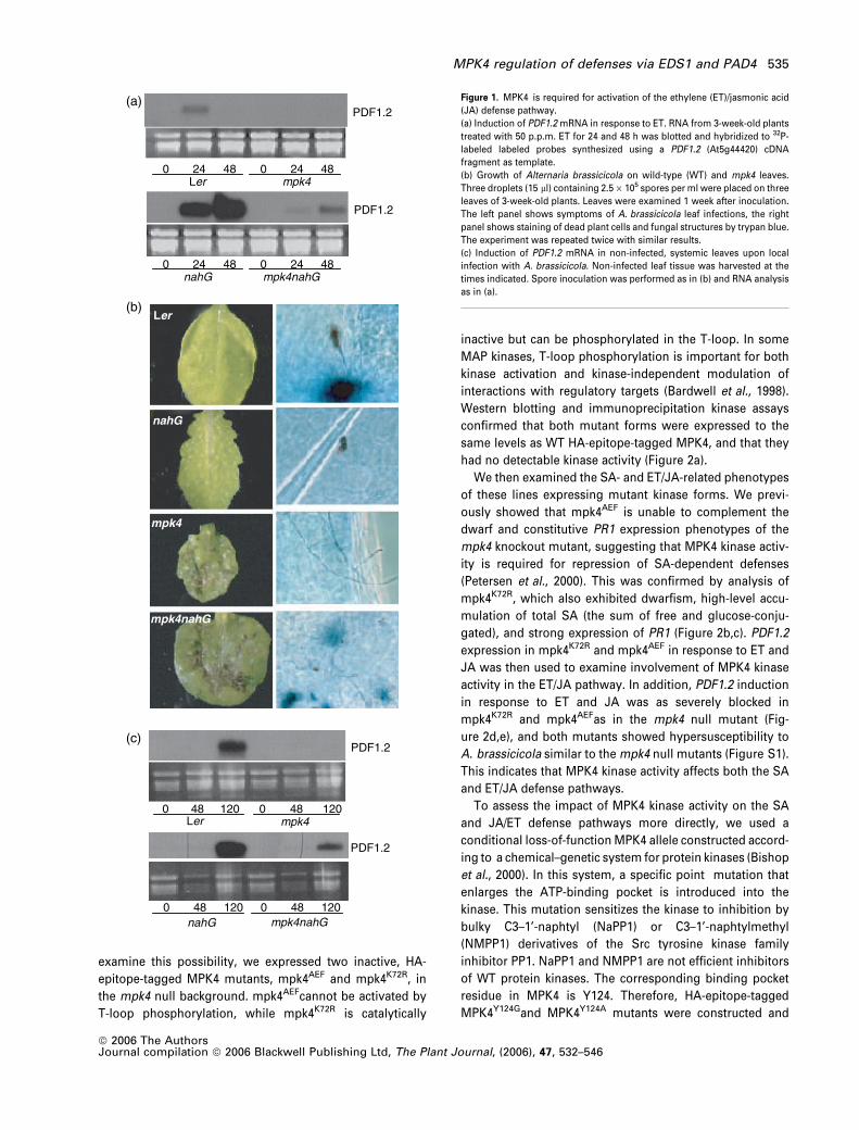
Two approaches were used to confirm the disproportion-
ate ERF1 and PDF1.2 expression in mpk4 backgrounds. First,
ERF1 and PDF1.2 mRNA accumulation following ET induc-
tion was examined in mpk4 and mpk4/nahG. This showed
that while PDF1.2 expression was blocked in mpk4 and
mpk4/nahG, ERF1 mRNA levels were either constitutively
elevated (mpk4) or normally induced by ET (mpk4/nahG)
(Figure 5b). Second, we transformed a 35S:ERF1 construct
into mpk4 heterozygotes and isolated several lines with
constitutive ERF1 expression. The levels of ERF1 and PDF1.2
mRNA were then examined in WT and mpk4 dwarf plants
segregating from these 35S:ERF1 transgenic lines. This
Ler mpk4 eds1 mpk4eds1
mpk4pad4
pad4
(a)
100
101
102
103
104
105
106
107
108
109
0 1 2 3 4 5
eds1
pad4
mpk4
mpk4eds1
mpk4pad4
Ler
(d)
PR1
rRNA
0
5000
10 000
15 000
20 000
Ler mpk4 eds1 mpk4eds1
pad4 mpk4pad4
nahG mpk4nahG
To
tal S
A /
(ng
/gF
W)(b)
(c)
mpk4mpk4 nahG
mpk4 pad4mpk4 eds1
Ler
Figure 4. Mutations in EDS1 and PAD4 suppress SA-dependent defense
activation in mpk4.
(a) Representative pictures of phenotypes of 3-week-old WT, mpk4, mpk4/
nahG, mpk4/pad4 and mpk4/eds1 versus Ler WT. Scale bar is 1 cm.
(b) Accumulation of total SA. Extraction and quantification were performed as
in Figure 2(b).
(c) Accumulation of PR1 mRNA in leaves of 18-day-old plants. Phosphori-
mager quantification of hybridization signals showed that the PR1 signal
intensities in mpk4/eds1 and mpk4/pad4 were approximately 5% and
approximately 10%, respectively, of that in mpk4.
(d) Growth of Pst DC3000 (CFU cm)2 leaf area). Pst DC3000 was vacuum-
infiltrated at 105 CFU ml)1 into leaves and bacterial growth quantified by
counting colony-forming units at the indicated time points. Each point is the
average of triplicate samples and the entire experiment was repeated three
times with similar results.
MPK4 regulation of defenses via EDS1 and PAD4 537
ª 2006 The AuthorsJournal compilation ª 2006 Blackwell Publishing Ltd, The Plant Journal, (2006), 47, 532–546
showed that accumulation of PDF1.2 mRNA was signifi-
cantly reduced (4–7-fold when quantified by PhosphorI-
mager analysis) in mpk4 compared with WT siblings
(Figure 5c). This result further suggests that MPK4 influen-
ces ET, and most likely JA, signaling downstream or
independently of ERF1. Given the proximity of ERF1 to ET/
JA transcriptional responses, ERF1 could be a target of
MPK4. However, MPK4 and ERF1 did not interact in a yeast
two-hybrid assay, and were not co-immunoprecipitated
from total protein extracts (data not shown).
MPK4 effects on PDF1.2 expression and Alternaria resistance
are mediated by EDS1 and PAD4
We next analysed whether MPK4 affects PDF1.2 expression
via a pathway contributing to the ET/JA defense network
downstream of ERF1. If so, EDS1 and PAD4 could act as
repressors in such a pathway as expression of PDF1.2 in
cpr6–1/eds1 and cpr6–1/pad4 was strongly enhanced com-
pared with cpr6–1, while neither pad4 nor eds1 single mu-
tants accumulated high levels of PDF1.2 mRNA (Clarke et al.,
2001; Jirage et al., 2001). We therefore tested whether
induction of PDF1.2 was restored in mpk4/eds1–2 and mpk4/
pad4–2 double mutants. Significant PDF1.2 mRNA accumu-
lation was detected in mpk4/eds1–2 in response to MeJA at
24 h after hormone application, whereas little PDF1.2 mRNA
was detected in mpk4/pad4 and in SA-depleted mpk4/nahG
(Figure 6a). Interestingly, PDF1.2 mRNA accumulation was
partially restored in mpk4/pad4 at 7 h after MeJA applica-
tion, indicating that the effects of pad4 are, at least in part,
epistatic to mpk4 (Figure S2). This double mutant analysis
indicates that EDS1 and PAD4 act as repressors of PDF1.2
induction by MeJA downstream of MPK4, and suggests that
EDS1 plays a more important role than PAD4 in such PDF1.2
repression.
We also tested the involvement of EDS1 and PAD4 in ET
signaling by monitoring PDF1.2 mRNA accumulation in ctr1–
2 and ctr1–2/mpk4 mutants into which the eds1–2, pad4–2
and eds1–2/pad4–2 alleles had been introduced. While the
results of this analysis were more complex, they were
consistent with a model in which EDS1 and PAD4 repress
PDF1.2 expression downstream of MPK4 (Figure 6b). First,
PDF1.2 accumulation in the ctr1–2 background was signifi-
cantly increased in the absence of PAD4 or of both PAD4 and
EDS1. Second, mutation of EDS1 bypassed the requirement
of MPK4 for PDF1.2 induction, while full PDF1.2 induction
was dependent on MPK4 in the pad4 single mutant back-
ground. In contrast, the low PDF1.2 level in ctr1/eds1
suggests that EDS1 has an activating as well as repressive
role in ET-related induction of PDF1.2. Despite this excep-
tion, the results indicate that PAD4 and EDS1 act to repress
PDF1.2 downstream of MPK4 in ET/JA signaling.
To analyse the physiological relevance of the above
differences in gene expression, we tested the resistance of
mpk4/pad4 and mpk4/eds1 to A. brassicicola infection. Both
double mutants were markedly less susceptible than mpk4
single mutants and the SA-depleted mpk4/nahG line,
although more hyphal growth and sporulation was ob-
served on mpk4/pad4 than on pad4 and WT Ler (Figure 6c).
Collectively, these results are consistent with a model in
which PDF1.2 expression and A. brassicicola resistance are
regulated by a pathway requiring MPK4 activity. This
pathway is mediated by the repressive effects of EDS1 and
PAD4, and functions in addition to, or downstream of, the
activating pathway mediated by ERF1 (Berrocal-Lobo et al.,
2002; Lorenzo et al., 2004).
Enhanced EDS1 protein accumulation in mpk4 mutants
We examined whether MPK4 affects EDS1 protein accumu-
lation by immunodetection in extracts of mpk4 single and
double mutants. All mpk4 backgrounds tested, including
SA-deficient mpk4/nahG, accumulated high levels of EDS1
(Figure 6d), although EDS1 levels were considerably higher
in mpk4 than in mpk4/nahG or mpk4/pad4. Thus, although
SA may contribute to EDS1 accumulation in mpk4 via in-
creased EDS1 mRNA accumulation, increased EDS1 protein
levels in mpk4 are not due solely to high SA levels (Falk
et al., 1999; Feys et al., 2001). EDS1 may therefore be more
directly regulated by MPK4. However, recombinant EDS1
was not an in vitro substrate of MPK4 immuno-purified from
plant extracts (data not shown). Nonetheless, the correlation
between high EDS1 levels and reduced PDF1.2 induction
(mpk4 and mpk4/nahG), and the reversion of PDF1.2 induc-
tion by the eds1 mutation, suggest that EDS1 abundance or
activity may be regulated by JA and/or ET via MPK4. We note
ctr1 ctr1mpk4
Ler
(a)
(b)
PDF1.2
0 0 0 024 24 24 24Ler mpk4 nahG mpk4nahG
ERF1
EF-1α
EF-1α
ERF1
PDF1.2
WT
35S:ERF1Line 33
35S:ERF1Line 41
Ler Ler mpk4 Ler mpk4mpk4
(c)
PDF1.2ERF1
rRNA
Figure 5. MPK4 regulates ET induction of PDF1.2 downstream or indepen-
dently of CTR1 and ERF1.
(a) Accumulation of PDF1.2 and ERF1 mRNA in ctr1, ctr1/mpk4 and Ler; 2 lg of
polyA RNA was loaded per lane.
(b) Accumulation of PDF1.2 and ERF1 mRNA in response to 50 p.p.m. ET;
20 lg of total RNA was loaded per lane.
(c) Accumulation of PDF1.2 and ERF1 mRNA in mpk4 and WT backgrounds
over-expressing ERF1; 20 lg of total RNA was loaded per lane.
538 Peter Brodersen et al.
ª 2006 The AuthorsJournal compilation ª 2006 Blackwell Publishing Ltd, The Plant Journal, (2006), 47, 532–546

that EDS1 or PAD4 protein levels were not affected by ET or
MeJA, although EDS1 levels were significantly lower in ctr1–
2 than in WT Col-0 (not shown). In conclusion, high EDS1
protein levels may explain many of the SA, ET and JA def-
ense defects observed in mpk4 mutants, but mechanistic
links between MPK4 activity and EDS1 accumulation remain
unknown.
Global analysis of MPK4-dependent, ET-related genes
The action of MPK4, and possibly EDS1 and PAD4, in ET
signaling was further characterized by comparing the tran-
scriptomes of ctr1–2, ctr1–2/mpk4, mpk4 and WT (Ler and
Col-0 samples). We used a two-factor ANOVA design with
three replicates of each category yielding P values for dif-
ferentially expressed genes in ctr1 and mpk4 and for inter-
action effects between ctr1 and mpk4. P-value cut-offs of
0.005 for the two main effects, and 0.01 for interaction ef-
fects, resulted in only one predicted false positive (see
Experimental procedures).
We focused on two classes of genes with significantly
different expression levels among the four genotypes. Class-
I represented the MPK4-dependent set of ET-related genes
whose mRNAs over-accumulate in ctr1–2 compared with
WT, but where this difference is suppressed by mpk4. Of the
22 810 genes represented on the array, only 48 Class-I genes
were identified (Table 1). Many (35) of these genes exhibited
a pattern in which mpk4 mutation alone led to significant
under-expression relative to WT, such that expression in
ctr1/mpk4 became correspondingly lower than in ctr1
(Figure 7a). Most of the Class-I genes have no known
function. Apart from PDF1.2, only the bHLH transcription
factor BEE1 has been associated with ET responses as it is
induced by ACC (Friedrichsen et al., 2002). We did not
identify genes whose repression in ctr1–2 versus WT
required MPK4, indicating that MPK4 acts as an activator
rather than a repressor of the induction of ET effectors.
The accumulation of mRNAs encoded by 78 Class-II genes
was different from WT in both ctr1–2 and ctr1–2/mpk4, but
similar to WT in mpk4 (Table 2). The accumulation of
mRNAs of two of these genes (EBP, b-CHI) in the ctr1
mutant backgrounds was shown to be independent of MPK4
by Northern blotting (Figure S3). This analysis also revealed
that the mRNAs of these genes did not significantly
(a)
(b)
(c)
(d)
Ler mpk4
00 24 24
nahG mpk4 nahG
00 24 24
eds1 mpk4 eds1
00 24 24
pad4 mpk4 pad400 24 24
PDF1.2
PDF1.2
MeJA
mpk4Ler
mpk4nahGnahG
mpk4pad4pad4
mpk4eds1eds1
MeJA
EF-1α
ctr1pad4eds1
Col Ler mpk4 ctr1 ctr1mpk4
ctr1pad4
ctr1mpk4pad4
ctr1eds1
ctr1mpk4eds1
ctr1mpk4pad4eds1
PDF1.2
EF-1α
Ler mpk4 nahG mpk4nahG
pad4 mpk4pad4
RbcL
α−EDS1
Figure 6. EDS1 and PAD4 function downstream of MPK4 in regulating
PDF1.2 induction and Alternaria brassicicola resistance.
(a) Accumulation of PDF1.2 mRNA in response to MeJA in mpk4, mpk4/nahG,
mpk4/eds1 and mpk4/pad4. Three-week-old plants were treated with 50 lM
MeJA for 24 h.
(b) Accumulation of PDF1.2 mRNA in ctr1 backgrounds defective in EDS1,
PAD4 and/or MPK4; 20 lg of total RNA was loaded per lane. To avoid
saturation of the ctr1 pad4 signal, the exposure was much shorter than the
autoradiogram of Figure 5(a). This explains the apparent low intensity of the
PDF1.2 signal in ctr1.
(c) Growth of A. brassicicola on mpk4, mpk4/nahG, mpk4/eds1 and mpk4/
pad4. Leaves were infected with three 15 ll droplets of A. brassicicola spores
at 2.5 · 105 spores per ml. Hyphal growth was revealed by trypan blue
staining 7 days after inoculation. Six to eight plants of each genotype were
infected, and there was little individual variation in hyphal growth among
plants of a genotype.
(d) Accumulation of EDS1 protein in mpk4, mpk4/nahG and mpk4/pad4. At
longer exposures, EDS1 protein was also detected in Ler. The lower panel
shows the RUBISCO large subunit (RbcL) from an identically loaded
Coomassie-stained gel run in parallel with the Western blot.
MPK4 regulation of defenses via EDS1 and PAD4 539
ª 2006 The AuthorsJournal compilation ª 2006 Blackwell Publishing Ltd, The Plant Journal, (2006), 47, 532–546

over-accumulate in ctr1/pad4 or ctr1/eds1. A relatively large
group of MPK4-independent genes was expected because
developmental defects typical of ctr1–2 plants were retained
in ctr1–2/mpk4 double mutants. However, MPK4-independ-
ent genes also included known or putative defense-related
genes such as b-CHI and several putative R genes
(At5g17880, At5g17890, At5g36930, and At1g59124), indica-
ting that MPK4 influences the expression of only a subset of
ET-dependent defense genes.
PAD4 and EDS1 are involved in regulating some, but not all,
MPK4-dependent genes
To determine whether the regulation of MPK4-dependent,
ET-related genes generally involves PAD4 and EDS1, we used
real-time RT-PCR to test the expression of some of the Class-I
genes in the series of ctr1mutants into whichmpk4,pad4 and
eds1 alleles had been introduced. This analysis identified one
Table 1 Class-I genes with MPK4-dependent over-expression inctr1
Gene Description
At5g44420 Defensin PDF1.2aAt2g26020 Defensin PDF1.2bAt5g61160 Anthocyanin 5-aromatic acyltransferase-likeAt1g73330 Protease inhibitor DR4At1g18400 bHLH transcription factor BEE1At3g14210 Putative myrosinase-associated proteinAt5g65390 Arabinogalactan-protein AGP7At1g78970 Lupeol synthase LUP1At1g20190 Expansin EXP11At3g60290 OxidoreductaseAt2g40670 Response regulator ARR16At5g22460 Esterase/lipase/thioesterase familyAt2g06850 Xyloglucan endotransglycosylase EXGT-A1At2g36870 Putative xyloglucan endotransglycosylaseAt1g44830 AP2 transcription factorAt4g02290 Endo-1,4-beta glucanase-likeAt4g21410 Ser/Thr kinase-likeAt4g37800 Putative xyloglucan endotransglycosylaseAt1g65290 Acyl carrier protein familyAt1g27460 Calmodulin-binding protein-likeAt2g47880 GlutaredoxinAt5g10430 Arabinogalactan-protein AGP4At5g60920 Phytochelatin synthetase-like COBAt3g16370 GDSL-motif lipase/hydrolase proteinAt5g48900 Pectate lyaseAt4g25260 Pectin esterase-likeAt2g38180 GDSL-motif lipase/hydrolase protein
Functionally annotated genes upregulated in ctr1 and suppressed bympk4 according to P-value criteria (see Experimental procedures). Afull list of Class-I genes is given in Table S1.
(a)
1.0 0.1 1.8 4.70.6
70
4.30.1 1.6
8.40.4
0.0
10.0
20.0
30.0
40.0
50.0
60.0
70.0
80.0A
t5g
6116
0
1.01.2
0.1
2.0
1.3
2.3
0.7
1.8
0.8
2.7
0.2
0.0
0.5
1.0
1.5
2.0
2.5
3.0
3.5
BE
E1
1.01.4
0.1
3.1
0.5
4.0
0.41.0
0.7
1.4
0.2
0.0
1.0
2.0
3.0
4.0
5.0
6.0
At5
g57
760
1.0
0.40.2
2.0
0.6
1.5
0.8
0.30.5
1.0
0.5
0.0
0.5
1.0
1.5
2.0
2.5
Col-0
Ler
mpk
4ctr
1c1
m4
c1p4
c1m
4p4
c1e1
c1m
4e1
c1p4
e1
c1m
4p4e
1
At5
g24
570
(b)
Figure 7. mRNA accumulation patterns in WT and mutants determined by
transcriptomics and real-time quantitative PCR.
(a) Boxplots illustrating gene expression profiles of class I (left) and class-II
(right) genes in WT (Col-0 and Ler), ctr1, mpk4 and ctr1/mpk4 plants.
Horizontal lines in boxes indicate median gene expression intensity in a
given genotype, horizontal box edges indicate quartiles, and upper and lower
bars indicate two standard deviations from median. The y-axes are scaled
gene expression values and the unit is standard deviations from mean
expression (z-score).
(b) Real-time PCR quantification of relative expression levels of MPK4-
dependent genes whose regulation involves PAD4 and EDS1 (At5g61160) or
proceeds independently of PAD4 and EDS1 (BEE1, At5g57760, and
At5g24570). c1, ctr1; m4, mpk4; p4, pad4; e1, eds1. Error bars indicate
standard deviations of triplicate, linearly transformed CT data.
540 Peter Brodersen et al.
ª 2006 The AuthorsJournal compilation ª 2006 Blackwell Publishing Ltd, The Plant Journal, (2006), 47, 532–546

additional mRNA with significant hyper-accumulation in ctr1/
pad4 (At5g61160, encoding an anthocyanin-5-aromatic acyl
transferase-like protein, AACT). Similar to PDF1.2, AACT
mRNA also accumulated to low levels in ctr1/eds1, but
differed in that it exhibited a requirement for MPK4 in the
pad4/eds1 double mutant background (Figure 7b). In addi-
tion, the expression of three genes (bHLH transcription factor
BEE1, and At5g57760 and At5g24570 encoding unknown
proteins) was not stimulated by pad4 and/or eds1 mutation,
although their full induction in ctr1 was confirmed to depend
on MPK4 (Figure 7b). This indicates that the set of MPK4-
dependent genes does not constitute a regulon, but consists
of differently regulated subgroups of genes. This is consis-
tent with our inability to identify conserved promoter ele-
ments among all Class-I genes.
Discussion
Negative regulatory role of MPK4 activity in the SA defense
pathway
We previously proposed that MPK4 negatively regulates SA-
dependent defense responses via its basal kinase activity
due to the activation of SA-dependent defenses in the mpk4
knockout and in a kinase-inactive mpk4 mutant (Petersen
et al., 2000). This model is consistent with protein kinase
inhibitor studies in tobacco showing that Ser/Thr kinase
inhibition led to accumulation of SA and to nahG-sup-
pressible PR1 expression (Conrath et al., 1997). However,
the dwarf stature of mpk4 plants left open the possibility that
deregulation of defenses was an indirect consequence of the
loss of MPK4 kinase activity even though such phenotypes
are common among mutants with constitutive expression of
SA defenses, and their penetrance correlates with defense
expression in mutants such as cpr1 and bon1 (Clarke et al.,
2001; Jirage et al., 2001; Yang and Hua, 2004).
Here we examine the relationship between MPK4 activity
and defense regulation in more detail. We can exclude the
possibility that PR1 expression arises solely as a conse-
quence of developmental defects in mpk4 mutants, because
PR1 was induced upon specific inhibition of MPK4 activity in
plants with WT morphology (Figure 3). Although this indi-
cates that MPK4 inactivation is sufficient to activate the SA
defense pathway in a WT plant, it is still unclear whether
such inactivation is required for activation of the pathway.
Likewise, these results on conditional MPK4 inactivation do
not exclude other indirect effects of MPK4 inhibition leading
to activation of the SA pathway. A gain-of-function analysis
of the requirement for MPK4 inactivation in SA-dependent
defense activation would help address both questions, but
our attempts to obtain constitutively active MPK4 variants
have so far failed.
EDS1 and PAD4 in the SA-ET-JA defense network
In addition to its role as a negative regulator of the SA
pathway, MPK4 is involved in regulating ET/JA-dependent
defenses. We show that our initial report of reduced PDF1.2
Table 2 Class-II genes with MPK4-independent over- or under-expression in ctr1
Gene Description
At2g38530 Non-specific lipid transfer protein 2 (LTP 2)At3g16770 AP2 transcription factor EBPAt4g30290 Putative xyloglucan endotransglycosylaseAt1g62380 ACC oxidase ACO2At5g17880 TIR-NBS-LRR class R-protein-likeAt5g36930 TIR-NBS-LRR class R-protein-likeAt1g63880 TIR-NBS-LRR class R-protein-likeAt5g57240 Oxysterol-binding protein-likeAt1g33790 Jacalin lectin familyAt3g12500 Basic endochitinase PR3At3g09260 Beta-glucosidase-likeAt2g16060 Non-symbiotic hemoglobin AHB1At3g22840 Early light-induced protein ELIP2At5g17890 TIR-NBS-LRR class R-protein-likeAt4g16260 Glucan endo-1,3-beta-glucosidase-likeAt5g36220 Cytochrome P450 CYP91A1At2g02850 Plastocyanin-likeAt3g44970 Cytochrome P450At5g02760 Protein phosphatase 2CAt4g14690 Early light-induced protein ELIP1At5g05750 DnaJ protein familyAt1g62770 Pectin esterase-likeAt3g16430 Myrosinase binding protein-likeAt5g64570 Glycosyl hydrolaseAt1g73830 bHLH transcription factor BEE3At5g67060 bHLH transcription factor bHLH088At3g23150 Ethylene receptor-related ETR2At2g30520 ROOT PHOTOTROPISM 2 RPT2At1g79000 Acetyltransferase-related protein 2 PCAT2At1g10480 C2H2-type zinc finger protein ZFP5At5g50570 Squamosa promoter binding protein-likeAt5g22510 Alkaline/neutral invertaseAt3g14230 AP2 transcription factorAt5g25350 F-box LRR proteinAt1g59700 Glutathione-S-transferase-likeAt1g31580 CXC750At3g18280 Lipid transfer protein/protease inhibitor familyAt1g59124 CC-NBS-LRR class R-protein-likeAt3g43600 Aldehyde oxidase 2 AAO2At1g14210 Ribonuclease-likeAt1g55920 Serine acetyltransferase SAT1At1g69310 WRKY family transcription factor WRKY57At3g17510 CBL-interacting protein kinase 1 CIPK1At3g57410 VILLIN 3 VLN3At4g34250 Fatty acid elongase 1 FAE1At1g31600 OxidoreductaseAt2g27050 EIN3-like transcription factor EIL1At5g25890 IAA28At4g27300 S-locus protein kinase-likeAt3g58550 Lipid transfer protein/protease inhibitor family
Functionally annotated genes differentially expressed in ctr1 but notaffected by MPK4. A full list of Class-II genes is given in Table S2.
MPK4 regulation of defenses via EDS1 and PAD4 541
ª 2006 The AuthorsJournal compilation ª 2006 Blackwell Publishing Ltd, The Plant Journal, (2006), 47, 532–546

mRNA induction in response to JA extends to the ET re-
sponse, and that MPK4 kinase activity is required for PDF1.2
induction by both JA and ET. A block of PDF1.2 expression in
mpk4 is also seen in response to A. brassicicola infection.
This reflects a physiologically important defect in induction
of ET/JA-dependent defenses, because resistance to
A. brassicicola is lost in mpk4, mpk4/nahG and, to some
degree, in mpk4/pad4 mutants.
The analysis of genetic interactions between MPK4, EDS1
and PAD4 supports a model of how MPK4 activity is required
for both repression of SA and activation of ET/JA defenses.
In this model, EDS1, and PAD4 to a lesser extent, are central
to the antagonism between the SA and ET/JA defense
pathways, acting as positive regulators of SA accumulation
and negative regulators of ET/JA defense signaling. Both of
these functions are negatively influenced, perhaps indi-
rectly, by MPK4 activity. In the absence of MPK4 activity,
EDS1 and PAD4 are effective as SAR activators mainly
through SA amplification, and as ET/JA defense repressors
via a function that does not rely on SA accumulation
(Figure 8). Such an SA-independent function of PAD4 was
previously suggested based on expression profiling experi-
ments following bacterial infection of pad4 mutants and
mutants impaired in SA biosynthesis (Glazebrook et al.,
2003). This model is also consistent with the fact that eds1,
pad4 and nahG all suppress SA accumulation and Pst
DC3000 resistance in mpk4, and that eds1, but not nahG,
restores A. brassicicola resistance and PDF1.2 inducibility by
MeJA in mpk4. We note, however, that while our genetic
data are consistent with this model, they do not exclude
alternative scenarios for the actions of MPK4, EDS1 and
PAD4 relative to each other in defense signaling.
The strong reduction of PDF1.2 expression in mpk4
mutants that over-express transgenic ERF1 suggests that
the repressive effects of MPK4–EDS1/PAD4 are mediated
either downstream or independently of ERF1. As dis-
cussed below, given the poor overlap between ERF1-
induced genes and MPK4-dependent, ET-related genes,
this repression probably occurs independently of ERF1.
Inhibition of the repressive effects on PDF1.2 expression
of EDS1/PAD4 clearly requires MPK4 activity, although it
apparently does not involve induction of MPK4 activity
above basal levels, because we have not detected
enhanced MPK4 activity is response to MeJA, ET or in
ctr1 mutant backgrounds (P. Brodersen, unpublished
results). Rather, it is likely to involve the action of other
factors, as hyper-accumulation of PDF1.2 mRNA is seen in
ctr1–2/pad4–2 and ctr1–2/pad4–2/eds1–2 compared with
ctr1–2 that all have active MPK4.
A model in which EDS1 and PAD4 act as direct repressors
of ET/JA signaling, in addition to more indirect effects via
elevated SA levels, is consistent with the analysis of PDF1.2
expression in cpr6 mutants. cpr6–1/eds1–2 exhibits strongly
enhanced PDF1.2 expression compared with cpr6–1 even
upon application of exogenous SA (Clarke et al., 2001). In
addition, as NPR1 is required for positive feedback induction
of EDS1 and PAD4 by SA (Falk et al., 1999; Jirage et al.,
1999), the failure of SA to repress JA induction of PDF1.2 in
npr1–1 (Spoel et al., 2003), as well as the hyper-induction of
PDF1.2 in cpr6/npr1 (Clarke et al., 2000), may be in part due
to impaired EDS1 and PAD4 induction. Similarly, it is
possible that NPR1-dependent repression of PDF1.2 by
WRKY70 (Li et al., 2004) involves enhanced expression of
EDS1 and PAD4.
The EDS1 and PAD4 proteins both consist of multiple
domains of unknown function. Taken together with their
functions in both SA and ET/JA defense regulation, this
raises the same question of genetic separability that we have
attempted to address for MPK4. Answering this question
and, if possible, assigning SA- or ET/JA-related functions to
specific domains in EDS1 and PAD4, are goals for future
research that could make use of the mpk4/eds1 and mpk4/
pad4 mutants described here.
MPK4-dependent, ET-related genes
The MPK4-dependent set of ET response genes is narrow,
and does not comprise all defense-related ET response
genes. For example, the induction of b-CHI and several
putative R genes was independent of MPK4. Some genes in
the MPK4-dependent set, including PDF1.2 and a few cell
wall proteins or modifying enzymes, have known or possible
defense-related functions, but their relationships to ET
responses are unknown.
Figure 8. A model for MPK4 action in plant defense pathways.
Central points include:
EDS1 and PAD4 act as activators of the SA pathway and repressors of the ET/
JA pathway
MPK4 negatively regulates both activities of EDS1 and PAD4
The repressive pathway acting in the ET/JA defense system is independent of
known activating pathways converging on ERF1
The repressive pathway influences only a subset of known genes associated
with the ET/JA defense pathway (such as PDF1.2 and anthocyanin-5-aromatic
acyl transferase, AACT).
542 Peter Brodersen et al.
ª 2006 The AuthorsJournal compilation ª 2006 Blackwell Publishing Ltd, The Plant Journal, (2006), 47, 532–546

ERF1 induction is an important event in the activation of
the ET/JA defense system that depends on activators such
as EIN2, EIN3 and COI1 (Lorenzo et al., 2003), and can be
triggered by ctr1 mutation (Solano et al., 1998). Our micro-
array data suggest that, rather than acting downstream of
ERF1 in the activating pathway, MPK4 acts in a repressive
pathway independent of this activating pathway. First, the
overlap of ET-related genes induced by ERF1 over-expres-
sion (Lorenzo et al., 2003) with MPK4-dependent, ET-related
genes is very limited. Second, the inducing effect of the ctr1
mutation, and the repressive effect of the mpk4 mutation,
are largely additive for many of the MPK4-dependent, ET-
related genes revealed by the microarray analysis.
For some of the MPK4-dependent genes, repression
appears to be mediated at least in part by PAD4 and EDS1.
This is the case for PDF1.2 and AACT. Such genes may be
involved in the defense response to necrotrophic fungi,
because resistance to A.brassicicola in mpk4 is largely
restored by pad4 or eds1 mutation. PDF1.2 is clearly associ-
ated with defense responses, and the same may be true for
AACT, as it is one of a small set of genes that are hyper-
induced by JA in the jin1 mutant that exhibits enhanced
resistance to necrotrophic fungi (Lorenzo et al., 2004; R.
Solano, CNB-CSIC,Madrid, Spain,personalcommunication).
The regulation of several other MPK4-dependent genes,
including the BEE1 transcription factor, does not involve
EDS1 and PAD4. It is currently unclear whether these genes
are defense-associated, or involved in other MPK4-depend-
ent, ET-regulated processes. Nonetheless, the fact that BEE1
is included in this set of genes suggests that their induction
involves enhanced BEE1 expression. Significantly, the BEE
family of transcription factors comprising the three closely
related BEE1, BEE2 and BEE3 genes (Friedrichsen et al.,
2002) is required for ET induction of at least one gene in this
set (P. Brodersen, J. Mundy, J. Nemhauser and J. Chory,
Salk Institute, La Jolla, USA, unpublished data). The possible
involvement of this gene set in the ET/JA defense pathway is
currently under investigation.
Experimental procedures
DNA constructs
Triple C-terminally HA-tagged MPK4 versions were constructed asdescribed previously (Petersen et al., 2000). The Quick-Change kit(Stratagene, La Jolla, CA, USA) was used for site-directed muta-genesis. A 35S–ERF1–nos construct in pROK2 obtained from JosephEcker (Solano et al., 1998) was used as template in a PCR reactionwith 5’-phosphorylated 35S and nos primers, and the product wascloned into the SmaI site of pCAMBIA3300.
Plant constructions
mpk4/eds1–2 and mpk4/pad4–2. mpk4 heterozygotes werecrossed to eds1–2 and pad4–2. F1 and F2 plants were allowed to self,
and families heterozygous for mpk4 and homozygous for eds1–2 orpad4–2 were selected on kanamycin and by PCR with primersdetecting the eds1–2 deletion (Falk et al., 1999) or the pad4–2frameshift after DNA sequencing (Jirage et al., 1999). Double mu-tants segregating from these families were identified by phenotype,confirmed by PCR, amplified and used for subsequent analyses.
ctr1–2/mpk4/pad4–2 and ctr1–2/mpk4/eds1–2. ctr1–2 plantsheterozygous for mpk4 were crossed to eds1–2 and pad4–2. F1
plants were kanamycin-selected and selfed. In F2, kanamycin-resistant ctr1–2 homozygotes were identified by phenotype and al-lowed to self. F3 families homozygous for eds1–2 or pad4–2 werethen selected by PCR, and triple mutants maintained as mpk4heterozygotes.
ctr1–2/mpk4/pad4–2/eds1–2 andmpk4/pad4–2/eds1–2. A ctr1–2/mpk4/pad4–2 triple heterozygous plant (above) was crossed to eds1–2, and a quadruple heterozygote, identified in F1 by kanamycinselection and PCR detecting the ctr1–2 deletion (Kieber et al., 1993)and the eds1–2 and pad4–2 alleles as described above, was allowedto self. Among 140 kanamycin-resistant F2 progeny, a single pad4/eds1 recombinant heterozygous for ctr1–2 and pad4–2, but homozy-gous for eds1–2, was identified. Kanamycin-resistant F3 progenyhomozygous for ctr1–2, or lacking the ctr1–2 allele, were identified byphenotype and PCR, and pad4–2 homozygotes were selected by PCR,giving rise to ctr1–2/mpk4/pad4–2/eds1–2 and mpk4/pad4–2/eds1–2families.
Plant treatments
Plants were grown in growth chambers under long days (16 h light/8 h darkness) for all treatments other than P. syringae infections forwhich short-day regimes were used (8 h light/16 h darkness). Dayand night temperatures were 21 and 16�C, respectively.
For ET inductions, plants were kept in 11 l polycarbonate jars(Nalgene, Rochester, NY, USA) sealed with silicon grease, and0.54 ll ET was injected with a 27G syringe through a rubbermembrane. MeJA inductions were performed as previously des-cribed (Petersen et al., 2000).
NaPP1 was synthesized as described previously (Bishop et al.,2000) and dissolved in DMSO at 10 mM. For plant treatments, thisstock was diluted to 100 lM in water containing 0.01% Silwet andsprayed onto leaves of 2–3-week-old plants. For mock treatments,1% DMSO in water with 0.01% Silwet was used.
Pst DC3000 growth curves were determined as described byPetersen et al. (2000).
Alternaria brassicicola strain MUCL 20297 was grown on 0.5%potato dextrose for 14–20 days until sporulation was dense. Sporeswere suspended in water, filtered through Miracloth and their titredetermined by Fuchs–Rosenthal cytometer counting. Spores wereapplied to leaves of 3-week-old plants in three 15 ll droplets per leafat 2.5 · 105 spores per ml, and symptoms were evaluated 7 dayslater.
RNA analysis
Total RNA was extracted by Trizol Reagent (Invitrogen, Carlsbad,CA, USA). Northern blotting and synthesis of radiolabeled probeswas performed according to standard protocols. cDNA templatesfor PR1 and PDF1.2 were amplified by PCR as described previously(Petersen et al., 2000). A cDNA fragment specific for ERF1 wasamplified from 35S–ERF1 in pROK2 (Solano et al., 1998), cloned in
MPK4 regulation of defenses via EDS1 and PAD4 543
ª 2006 The AuthorsJournal compilation ª 2006 Blackwell Publishing Ltd, The Plant Journal, (2006), 47, 532–546

antisense orientation in front of the T7 promoter (Promega, Madi-son, WI, USA) in pGEM-T-easy and used as a template for in vitrotranscription incorporating radiolabeled 32P-UTP.
For reverse transcription (RT) and quantitative PCR analysis, RNAsamples were first treated with RQ1 DNase (Promega, Madison, WI,USA). RT reactions were done with 1 lg of RNA and 0.5 lg of (dT)21
primer at 42�C with 0.1 unit of reverse transcriptase (Promega) and 2units of RNasin (Promega) for 1 h in 20 ll reactions. QuantitativePCR was performed using the SYBR Green protocol (AppliedBiosystems, Foster City, CA, USA) with 10 pmol of each primerand a 0.5 ll aliquot of RT reaction product in a 25 ll reaction.Quantitative PCR reactions were performed in triplicate and aver-aged for each line individually. Quantification of the threshold cycle(CT) values obtained by quantitative PCR analysis was achieved bythe 2�DDC
T method (Livak and Schmittgen, 2001) after verifying thatthe value CT(ubiquitin)-CT(target) remained constant for each of thetarget genes tested over a 100-fold cDNA dilution series.
SA measurements
Total SA was extracted and quantified as described by Newmanet al. (2001).
Kinase assays
MPK4 versions were immunoprecipitated with 12CA5 anti-HA anti-body as described previously (Petersen et al., 2000). After threewashes in immunoprecipitation buffer and one wash in kinase as-say buffer, immunoprecipitates were incubated in 30 ll kinasebuffer (20 mM Tris, pH 7.5, 2 mM EGTA, 30 mM MgCl2, 1 mM
Na3VO4, 50 lM ATP) with 5 lg myelin basic protein and 3 lCi of 32P-ATP (3000 Ci mmol)1) at 30�C for 30 min. Reactions were stoppedby addition of SDS sample buffer and products resolved by SDS–PAGE. For inhibition assays with NaPP1 and NMPP1, immunopre-cipitates were incubated with or without inhibitor in kinase assaybuffer for 10 min on ice before addition of substrates. MPK4-HA andEDS1 Western blots were performed as described previously (Feyset al., 2001; Petersen et al., 2000).
Microarray hybridization and analysis
Total RNA was isolated from three independent replicates of ctr1–2,mpk4, ctr1–2/mpk4 and WT (one Col-0 sample, two Ler samples).The RNA was amplified and hybridized to 12 Affymetrix microarraysaccording to Affymetrix protocols (Affymetrix UK Ltd., HighWycombe, UK). Raw intensity data was normalized using R imple-mentation of qspline (Gautier et al., 2004; Workman et al., 2002). Animplementation of the logit-t method in the statistical language R(Lemon et al., 2003), applying two-way ANOVA instead of a t-test,was used to calculate statistical significances of differential geneexpression. False-positive rates were estimated by recalculating P-values with permuted sample categories. This procedure wasrepeated four times, generating four sets of 22 810 permuted P-values. The P-value cut-off was chosen so that only one permuted P-value was lower than the cut-off. The resulting P-value cut-offs were0.005 for the main two effects and 0.01 for the interaction effect.Gene expression index values were calculated using perfect match-only implementation (Gautier et al., 2004) of the method introducedby Li and Wong (2001). Gene expression profiles from significantlydifferentially expressed genes were clustered by partitioningaround medoids (PAM) clustering (k ¼ 12). Classes I and II corres-pond to PAM clusters 10 and 7, respectively. The data (raw and gene
P-values) are publicly accessible from ArrayExpress under acces-sion number E-MEXP-174 at http://www.ebi.ac.uk/arrayexpress/query/entry.
Acknowledgements
We thank J. Ecker for providing the 35S–ERF1 plasmid, and W.Brokaert for A. brassicicola. L. Navarro and Z. Nimchuk are thankedfor critical reading of the manuscript. This work was supported bygrants to P.B. from the Faculty of Science, University of Copenha-gen, and to J.M. from the Danish Research Councils (23–01–0145)and the European Union (hprnct200000093).
Supplementary Material
The following supplementary material is available for this articleonline:Figure S1. Mutants expressing inactive MPK4 proteins are hyper-susceptible to Alternaria brassicicola.Figure S2. Mutations in PAD4 or EDS1 suppress the block of PDF1.2induction in mpk4.Figure S3. MPK4-independent induction of two class-II genes.Table S1 Full list of Class-I genesTable S2 Full list of Class-II genesThis material is available as part of the online article from http://www.blackwell-synergy.com
References
Alvarez, M.E., Pennell, R.I., Meijer, P.J., Ishikawa, A., Dixon, R.A.
and Lamb, C. (1998) Reactive oxygen intermediates mediate asystemic signal network in establishment of plant immunity. Cell,92, 773–784.
Andreasson, E., Jenkins, T., Brodersen, P. et al. (2005) The MAPkinase substrate MKS1 is a regulator of plant defense responses.EMBO J. 24, 2579–2589.
Bardwell, L., Cook, J.G., Voora, D., Baggott, D.M., Martinez, A.R.
and Thorner, J. (1998) Repression of yeast Ste12 transcriptionfactor by direct binding of unphosphorylated Kss1 MAPK and itsregulation by the Ste7 MEK. Genes Dev. 12, 2887–2898.
Berrocal-Lobo, M., Molina, A. and Solano, R. (2002) Constitutiveexpression of ETHYLENE-RESPONSE-FACTOR1 confers resist-ance to several necrotrophic fungi. Plant J. 29, 23–32.
Bishop, A.C., Ubersax, J.A., Petsch, D.T. et al. (2000) A chemicalswitch for inhibitor-sensitive alleles of any protein kinase. Nature,407, 395–401.
Cao, H., Glazebrook, J., Clarke, J.D., Volko, S. and Dong, X. (1997)The Arabidopsis NPR1 gene that controls systemic acquiredresistance encodes a novel protein containing ankyrin repeats.Cell, 88, 57–63.
Clarke, J.D., Volko, S.M., Ledford, H., Ausubel, F.M. and Dong, X.
(2000) Roles of salicylic acid, jasmonic acid, and ethylene in cpr-induced resistance in Arabidopsis. Plant Cell, 12, 2175–2190.
Clarke, J.D., Aarts, N., Feys, B.J., Dong, X. and Parker, J.E. (2001)Constitutive disease resistance requires EDS1 in the Arabidopsismutants cpr1 and cpr6 and is partially EDS1-dependent in cpr5.Plant J. 26, 409–420.
Conrath, U., Silva, H. and Klessig, D.F. (1997) Protein dephospho-rylation mediates salicylic acid expression of PR-1 genes in to-bacco. Plant J. 11, 747–757.
Desveaux, D., Subramaniam, R., Despres, C., Mess, J.-N., Levesque,
C., Fobert, P.R., Dangl, J.L. and Brisson, N. (2004) A ‘Whirly’
544 Peter Brodersen et al.
ª 2006 The AuthorsJournal compilation ª 2006 Blackwell Publishing Ltd, The Plant Journal, (2006), 47, 532–546

transcription factor is required for salicylic acid-dependent dis-ease resistance in Arabidopsis. Dev. Cell, 6, 229–240.
Devoto, A. and Turner, J.G. (2003) Regulation of jasmonate-medi-ated plant responses in Arabidopsis.Ann.Bot. (Lond),92, 329–337.
Falk, A., Feys, B.F., Forst, L.N., Jones, J.D.G., Daniels, M.J. and
Parker, J.E. (1999) EDS1, an essential component of R-gene-mediated disease resistance in Arabidopsis has homology toeukaryotic lipases. Proc. Natl Acad Sci. USA, 96, 3292–3297.
Fan, W. and Dong, X. (2002) In vivo interaction between NPR1 andtranscription factor TGA2 leads to salicylic acid-mediated geneactivation in Arabidopsis. Plant Cell, 14, 1377–1389.
Feys, B.J., Moisan, L.J., Newman, M.-A. and Parker, J.E. (2001) Di-rect interaction between the Arabidopsis disease resistancesignaling proteins, EDS1 and PAD4. EMBO J. 20, 5400–5411.
Feys, B.J., Wiermer, M., Bhat, R.A., Moisan, L.J., Medina-Escobar,
N., Neu, C., Cabral, A. and Parker, J.E. (2005) Arabidopsis SEN-ESCENCE-ASSOCIATED GENE101 stabilizes and signals withinan ENHANCED DISEASE SUSCEPTIBILITY1 complex in plant in-nate immunity. Plant Cell, 17, 2601–2613.
Forouhar, F., Yang, Y., Kumar, D. et al. (2005) Structural and bio-chemical studies identify tobacco SABP2 as a methyl salicylateesterase and implicate it in plant innate immunity. Proc. NatlAcad. Sci. USA, 102, 1773–1778.
Friedrichsen, D.M., Nemhauser, J., Muramitsu, T., Maloof, J.N.,
Alonso, J., Ecker, J.R., Furuya, M. and Chory, J. (2002) Threeredundant brassinosteroid early response genes encode putativebHLH transcription factors required for normal growth. Genetics,162, 1445–1456.
Gautier, L., Cope, L., Bolstad, B.M. and Irizarry, R.A. (2004) Affy –analysis of Affymetrix GeneChip data at the probe level. Bioin-formatics, 20, 307–315.
Glazebrook, J., Chen, W., Estes, B., Chang, H.-S., Nawrath, C.,
Metraux, J.-P., Zhu, T. and Katagiri, F. (2003) Topology of thenetwork integrating salicylate and jasmonate signal transduc-tion derived from global expression phenotyping. Plant J. 34,217–228.
Guo, H. and Ecker, J.R. (2004) The ethylene signaling pathway: newinsights. Curr. Opin. Plant Biol. 7, 40–49.
Heck, S., Grau, T., Buchala, A., Metraux, J.-P. and Nawrath, C. (2003)Genetic evidence that expression of NahG modifies defencepathways independent of salicylic acid biosynthesis in the Ara-bidopsis–Pseudomonas syringae pv. tomato interaction. Plant J.36, 342–352.
Jirage, D., Tootle, T.L., Reuber, T.L., Frost, L.N., Feys, B.F., Parker,
J.E., Ausubel, F.M. andGlazebrook, J. (1999) Arabidopsis thalianaPAD4 encodes a lipase-like gene that is important for salicylic acidsignaling. Proc. Natl Acad. Sci. USA, 96, 13583–13588.
Jirage, D., Zhou, N., Cooper, B., Clarke, J.D., Dong, X. and Glaze-
brook, J. (2001) Constitutive salicylic acid-dependent signaling incpr1 and cpr6 mutants requires PAD4. Plant J. 26, 395–407.
Johnson, C., Boden, E. and Arias, J. (2003) Salicylic acid and NPR1induce the recruitment of trans-activating TGA factors to a def-ense gene promoter in Arabidopsis. Plant Cell, 15, 1846–1858.
Kachroo, P., Shanklin, J., Shah, J., Whittle, E.J. and Klessig, D.F.
(2001) A fatty acid desaturase modulates the activation of defensesignaling pathways in plants. Proc. Natl Acad. Sci. USA, 98, 9448–9453.
Kieber, J.J., Rothenberg, M., Roman, G., Feldmann, K.A. and Ecker,
J.R. (1993) CTR1, a negative regulator of the ethylene responsepathway in Arabidopsis, encodes a member of the raf family ofprotein kinases. Cell, 72, 427–441.
Kloek, A.P., Verbsky, M.L., Sharma, S.B., Schoelz, J.E., Vogel, J.,
Klessig, D.F. and Kunkel, B.N. (2001) Resistance to Pseudomonassyringae conferred by an Arabidopsis thaliana coronatine-insen-
sitive (coi1) mutation occurs through two distinct mechanisms.Plant J. 26, 509–522.
Lawton, K., Potter, S.L., Uknes, S. and Ryals, J. (1994) Acquiredresistance signal transduction in Arabidopsis is ethylene inde-pendent. Plant Cell, 6, 581–588.
Lawton, K.A., Weymann, K., Friedrich, L., Vernooij, B., Uknes, S. and
Ryals, J. (1995) Systemic acquired resistance in Arabidopsisrequires salicylic acid but not ethylene. Mol. Plant–MicrobeInteract. 8, 863–870.
Lemon, W.J., Liyanarachchi, S. and You, M. (2003) A high per-formance test of differential gene expression for oligonucleotidearrays. Genome Biol. 4, R67. http://genomebiology.com/2003/4/10/R67.
Li, C. and Wong, W.H. (2001) Model-based analysis of oligonucle-otide arrays: expression index computation and outlier detection.Proc. Natl Acad. Sci. USA, 98, 31–36.
Li, J., Brader, G. and Palva, E.T. (2004) The WRKY70 transcriptionfactor: a node of convergence for jasmonate-mediated andsalicylate-mediated signals in plant defense. Plant Cell, 16,319–331.
Livak, K.J. and Schmittgen, T.D. (2001) Analysis of relative geneexpression data using real-time quantitative PCR and the2()DD C(T)) method. Methods, 25, 402–408.
Lorenzo, O., Piqueras, R., Sanchez-Serrano, J.J. and Solano, R.
(2003) ETHYLENE RESPONSE FACTOR1 integrates signals fromethylene and jasmonate pathways in plant defense. Plant Cell, 15,165–178.
Lorenzo, O., Chico, J.M., Lopez-Fernandez, L.A., Sanchez-Serrano,
J.J. and Solano, R. (2004) JASMONATE-INSENSITIVE1 encodes aMyc transcription factor essential to discriminate between dif-ferent jasmonate-regulated defence responses in Arabodipsis.Plant Cell, 16, 1938–1950.
Madhani, H.D., Styles, C.A. and Fink, G.R. (1997) MAP kinases withdistinct inhibitory functions impart signaling specificity duringyeast differentiation. Cell, 91, 673–684.
Malamy, J., Carr, J.P., Klessig, D.F. and Raskin, I. (1990) Salicylicacid: a likely endogenous signal in the resistance response oftobacco to viral infection. Science, 250, 1002–1004.
Maldonado, A.M., Doerner, P., Dixon, R.A., Lamb, C.J. and Cameron,
R.K. (2002) A putative lipid transfer protein involved in systemicresistance signaling in Arabidopsis. Nature, 419, 399–403.
Mengiste, T., Chen, X., Salmeron, J. and Dietrich, R. (2003) TheBOTRYTIS-SUSCEPTIBLE1 gene encodes an R2R3MYB tran-scription factor protein that is required for biotic and abiotic stressresponses in Arabidopsis. Plant Cell, 15, 2551–2565.
Nawrath, C. and Metraux, J.-P. (1999) Salicylic acid induction-defi-cient mutants of Arabidopsis express PR-2 and PR-5 and accu-mulate high levels of camalexin after pathogen inoculation. PlantCell, 11, 1393–1404.
Newman, M.-A., von Roepenack-Lahaye, E., Parr, A., Daniels,
M.J. and Dow, J.M. (2001) Induction of hydroxycinnamoyl–tyramine conjugates in pepper by Xanthomonas campestris: aplant defense response activated by hrp gene-dependent and -independent mechanisms. Mol. Plant–Microbe Interact. 14,785–792.
Nimchuk, Z., Eulgem, T., Holt, B.F. and Dangl, J.L. (2003) Recogni-tion and response in the plant immune system. Annu. Rev. Genet.37, 579–609.
Norman-Setterblad, C., Vidal, S. and Palva, E.T. (2000) Interactingsignal pathways control defense gene expression in Arabidopsisin response to cell wall-degrading enzymes from Erwiniacarotovora. Mol. Plant–Microbe Interact. 13, 430–438.
Oh, I.S., Park, A.R., Bae, M.S., Kwon, S.J., Kim, Y.S., Lee, J.E., Kang,
N.Y., Lee, S., Cheong, H. and Park, O.K. (2005) Secretome analysis
MPK4 regulation of defenses via EDS1 and PAD4 545
ª 2006 The AuthorsJournal compilation ª 2006 Blackwell Publishing Ltd, The Plant Journal, (2006), 47, 532–546

reveals an Arabidopsis lipase involved in defense against Alter-naria brassicicola. Plant Cell, 17, 2832–2847.
Penninckx, I.A., Eggermont, K., Terras, F.R.G., Thomma, B.P., De
Samblanx, G.W., Buchala, A., Metraux, J.-P., Manners, J.M. and
Broekaert, W.F. (1996) Pathogen-induced systemic activation of aplant defensin gene in Arabidopsis follows a salicylic acid-inde-pendent pathway. Plant Cell, 8, 2309–2323.
Petersen, M., Brodersen, P., Naested, H. et al. (2000) ArabidopsisMAP kinase 4 negatively regulates systemic acquired resistance.Cell, 103, 1111–1120.
Pieterse, C.M., vanWees, S.C., van Pelt, J.A., Knoester, M., Laan, R.,
Gerrits, H., Weisbeek, P.J. and van Loon, L.C. (1998) A novelsignaling pathway controlling induced systemic resistance inArabidopsis. Plant Cell, 10, 1571–1580.
Rusterucci, C., Aviv, D.H., Holt, B.F., Dangl, J.L. and Parker, J.E.
(2001) The disease resistance signaling components EDS1 andPAD4 are essential regulators of the cell death pathway controlledby LSD1 in Arabidopsis. Plant Cell, 13, 2211–2224.
Schenk, P.M., Kazan, K., Wilson, I., Anderson, J.P., Richmond, T.,
Somerville, S.C. and Manners, J.M. (2000) Coordinated plantdefense responses in Arabidopsis revealed by microarray analy-sis. Proc. Natl Acad. Sci. USA, 97, 11655–11660.
Shah, J., Kachroo, P., Nandi, A. and Klessig, D.F. (2001) A recessivemutation in the Arabidopsis SSI2 gene confers SA- and NPR1-independent expression of PR genes and resistance againstbacterial and oomycete pathogens. Plant J. 25, 563–574.
Slaymaker, D.H., Navarre, D.A., Clark, D., del Pozo, O., Martin, G.B.
and Klessig, D.F. (2002) The tobacco salicylic acid-binding pro-tein-3 (SABP3) is the chloroplast carbonic anhydrase, whichexhibits antioxidant activity and plays a role in the hypersensitivedefense response. Proc. Natl Acad. Sci. USA, 99, 11640–11645.
Solano, R., Stepanova, A., Chao, Q. and Ecker, J.R. (1998) Nuclearevents in ethylene signaling: a transcriptional cascade mediatedby ETHYLENE-INSENSITIVE3 and ETHYLENE-RESPONSE-FAC-TOR1. Genes Dev. 12, 3703–3714.
Song, J.T., Lu, H., McDowell, J.M. and Greenberg, J.T. (2004) A keyrole for ALD1 in activation of local and systemic defenses inArabidopsis. Plant J. 40, 200–212.
Spoel, S.H., Koornneef, A., Claessens, S.M. et al. (2003) NPR1modulates cross-talk between salicylate- and jasmonate-dependent defense pathways through a novel function in thecytosol. Plant Cell, 15, 760–770.
Thomma, B.P., Eggermont, K., Penninckx, I.A., Mauch-Mani, B.,
Vogelsang, R., Cammue, B.P. and Broekaert, W.F. (1998) Separate
jasmonate-dependent and salicylate-dependent defense-re-sponse pathways in Arabidopsis are essential for resistance todistinct microbial pathogens. Proc. Natl Acad. Sci. USA, 95,15107–15111.
Uknes, S., Mauch-Mani, B., Moyer, M., Potter, S., Williams, S.,
Dincher, S., Chandler, D., Slusarenko, A., Ward, E. and Ryals, J.
(1992) Acquired resistance in Arabidopsis. Plant Cell, 4, 645–656.
Verberne, M.C., Verpoorte, R., Bol, J.F., Mercado-Blanco, J. and
Linthorst, H.J. (2000) Overproduction of salicylic acid in plants bybacterial transgenes enhances pathogen resistance. Nature Bio-technol. 18, 779–783.
van Wees, S.C. and Glazebrook, J. (2003) Loss of non-host resist-ance of Arabidopsis NahG to Pseudomonas syringae pv.phaseolica is due to degradation products of salicylic acid. PlantJ. 33, 733–742.
van Wees, S.C., de Swart, E.A., van Pelt, J.A., van Loon, L.C. and
Pieterse, C.M. (2000) Enhancement of induced resistance bysimultaneous activation of salicylate-and jasmonate-dependentdefense pathways in Arabidopsis thaliana. Proc. Natl Acad. Sci.USA, 97, 8711–8716.
Workman, C., Jensen, L.J., Jarmer, H., Berka, R., Gautier, L., Niel-
sen, H.B., Saxild, H.H., Nielsen, C., Brunak, S. and Knudsen, S.
(2002) A new non-linear normalization method for reducingvariability in DNA microarray experiments. Genome Biol. 3, R48.http://genomebiology.com/2002/3/9/research/0048
Xia, Y., Suzuki, H., Borevitz, J., Blount, J., Guo, Z., Patel, K., Dixon,
R.A. and Lamb, C. (2004) An extracellular aspartic proteasefunctions in Arabidopsis disease resistance signaling. EMBO J.23, 980–988.
Xu, Y., Chang, P.-F., Liu, D., Narasimhan, M.L., Raghothama, K.G.,
Hasegawa, P.M. and Bressan, R.A. (1994) Plant defense genes aresynergistically induced by ethylene and methyl jasmonate. PlantCell, 6, 1077–1085.
Yang, S. and Hua, J. (2004) A haplotype-specific resistance generegulated by BON1. Plant Cell, 16, 1060–1071.
Zhao, Y., Thilmony, R., Bender, C.L., Schaller, A., He, S.Y. and Howe,
G.A. (2003) Virulence systems of Pseudomonas syringae pv. to-mato promote bacterial speck disease in tomato by targeting thejasmonate signaling pathway. Plant J. 36, 485–499.
Zhou, C., Zhang, L., Duan, J., Miki, B. and Wu, K. (2005) HISTONEDEACETYLASE19 is involved in jasmonic acid and ethylenesignaling of pathogen response in Arabidopsis. Plant Cell, 17,1196–1204.
546 Peter Brodersen et al.
ª 2006 The AuthorsJournal compilation ª 2006 Blackwell Publishing Ltd, The Plant Journal, (2006), 47, 532–546