Aptamers for pharmaceuticals and their application in environmental ...
30
Aptamers for pharmaceuticals and their application in environmental analytics Beate Strehlitz & Christine Reinemann & Soeren Linkorn & Regina Stoltenburg Received: 27 July 2011 /Accepted: 10 November 2011 /Published online: 17 December 2011 # The Author(s) 2011. This article is published with open access at Springerlink.com Abstract Aptamers are single-stranded DNA or RNA oligonucleotides, which are able to bind with high affinity and specificity to their target. This property is used for a multitude of applications, for instance as molecular recognition elements in biosensors and other assays. Biosensor application of aptamers offers the possibility for fast and easy detection of environmental relevant substances. Pharmaceutical residues, deriving from human or animal medical treatment, are found in surface, ground, and drinking water. At least the whole range of frequently administered drugs can be detected in noticeable concentrations. Biosensors and assays based on aptamers as specific recognition elements are very convenient for this application because aptamer development is possible for toxic targets. Commonly used biological receptors for biosensors like enzymes or antibodies are mostly unavailable for the detection of pharmaceuticals. This review describes the research activities of aptamer and sensor developments for pharmaceutical detection, with focus on environmental applications. Keywords Aptamer . Small organic molecule . Pharmaceutical . Biosensor . Environmental analysis Introduction Aptamers Even though, until now, aptamers have been developed mainly for medical applications or clinical diagnostics, they are well suited as novel biological recognition elements for the detection of pharmaceutical residues in the environment because of their specific properties. Aptamers are short single-stranded oligomers (ssDNA or RNA), which are able to bind their target molecules with high specificity and selectivity. Binding occurs because of their specific and complex three-dimensional shape characterized by stems, loops, bulges, hairpins, pseudoknots, triplexes, or quadruplexes. The aptamer-target binding results from structure compatibility, stacking of aromatic rings, elec- trostatic and van der Waals interactions, and hydrogen bondings, or from a combination of these effects [1, 2]. Initially, RNA aptamer development was described for bacteriophage T4 DNA polymerase [3] and organic dyes Cibacron blue and Reactive blue 4 [4]. These publications described for the first time the evolutionary process to select aptamers starting with of a big variety of oligonu- cleotides in a so-called library (ca. 10 15 different structures) by repeated rounds consisting of the steps (a) binding between target molecule and library, (b) elution of the bound oligonucleotides, and (c) their amplification. The resulting pool of pre-selected oligonucleotides forms the starting pool of the following round. This process is called systematic evolution of ligands by exponential enrichment (SELEX) and mimicks the Darwinian principle. The first DNA aptamers were described only 2 years later for Cibacron blue, Reactive blue 4, and Reactive green 19 [5]. B. Strehlitz (*) : C. Reinemann : S. Linkorn : R. Stoltenburg UFZ-Helmholtz Centre for Environmental Research, Permoserstr. 15, 04318 Leipzig, Germany e-mail: [email protected] Bioanal Rev (2012) 4:1–30 DOI 10.1007/s12566-011-0026-1
Transcript of Aptamers for pharmaceuticals and their application in environmental ...

Aptamers for pharmaceuticals and their applicationin environmental analytics
Beate Strehlitz & Christine Reinemann &
Soeren Linkorn & Regina Stoltenburg
Received: 27 July 2011 /Accepted: 10 November 2011 /Published online: 17 December 2011# The Author(s) 2011. This article is published with open access at Springerlink.com
Abstract Aptamers are single-stranded DNA or RNAoligonucleotides, which are able to bind with highaffinity and specificity to their target. This property isused for a multitude of applications, for instance asmolecular recognition elements in biosensors and otherassays. Biosensor application of aptamers offers thepossibility for fast and easy detection of environmentalrelevant substances. Pharmaceutical residues, derivingfrom human or animal medical treatment, are found insurface, ground, and drinking water. At least the wholerange of frequently administered drugs can be detectedin noticeable concentrations. Biosensors and assaysbased on aptamers as specific recognition elements arevery convenient for this application because aptamerdevelopment is possible for toxic targets. Commonlyused biological receptors for biosensors like enzymes orantibodies are mostly unavailable for the detection ofpharmaceuticals. This review describes the researchactivities of aptamer and sensor developments forpharmaceutical detection, with focus on environmentalapplications.
Keywords Aptamer . Small organic molecule .
Pharmaceutical . Biosensor . Environmental analysis
Introduction
Aptamers
Even though, until now, aptamers have been developedmainly for medical applications or clinical diagnostics, theyare well suited as novel biological recognition elements forthe detection of pharmaceutical residues in the environmentbecause of their specific properties. Aptamers are shortsingle-stranded oligomers (ssDNA or RNA), which areable to bind their target molecules with high specificityand selectivity. Binding occurs because of their specificand complex three-dimensional shape characterized bystems, loops, bulges, hairpins, pseudoknots, triplexes, orquadruplexes. The aptamer-target binding results fromstructure compatibility, stacking of aromatic rings, elec-trostatic and van der Waals interactions, and hydrogenbondings, or from a combination of these effects [1, 2].
Initially, RNA aptamer development was described forbacteriophage T4 DNA polymerase [3] and organic dyesCibacron blue and Reactive blue 4 [4]. These publicationsdescribed for the first time the evolutionary process toselect aptamers starting with of a big variety of oligonu-cleotides in a so-called library (ca. 1015 different structures)by repeated rounds consisting of the steps (a) bindingbetween target molecule and library, (b) elution of thebound oligonucleotides, and (c) their amplification. Theresulting pool of pre-selected oligonucleotides forms thestarting pool of the following round. This process is calledsystematic evolution of ligands by exponential enrichment(SELEX) and mimicks the Darwinian principle. The firstDNA aptamers were described only 2 years later forCibacron blue, Reactive blue 4, and Reactive green 19 [5].
B. Strehlitz (*) :C. Reinemann : S. Linkorn :R. StoltenburgUFZ-Helmholtz Centre for Environmental Research,Permoserstr. 15,04318 Leipzig, Germanye-mail: [email protected]
Bioanal Rev (2012) 4:1–30DOI 10.1007/s12566-011-0026-1
Since then, aptamers for very diverse targets of differentmolecule classes and sizes were developed. Proteins are thepredominant aptamer selection targets, but aptamers are alsodescribed for larger targets like whole cells, viruses, andtissues, or smaller targets like small organic molecules [6].The SELEX principle was modified with a lot of variations,and most of them have their own names. The process ofaptamer selection with its variants is not in the focus of thisreview. To get an overview, the reader is referred to ourformer review article [2] and similar articles [7–10].
One of the biggest advantages of aptamers in comparisonto other biological recognition elements is the possibility todevelop them for toxic substances as the frequently usedbiological recognition elements enzymes and antibodiescannot be developed for toxic targets. Pharmaceuticals areshown to have poisonous effects at least when used in highdoses. Therefore, the development of antibodies for pharma-ceuticals is a difficult thing to deal with. Aptamers aredescribed for a great variety of pharmaceuticals with medicalapplication. Some of them are used in detection systems butare mostly utilized for the measurement in blood or other bodyfluids. A new application field for aptamers is the detection ofpharmaceutical residues in the environment, which have to bedetermined in a fast and simple way. On the other hand,aptamers can be pharmaceuticals by themselves. Theseaptamer therapeutics are used because of the high affinitiesto their target and specificities comparable to those ofmonoclonal antibodies for therapeutical treatments [11]. Themost successful therapeutic application of an aptamer hasbeen the adaptation of an antivascular endothelial growthfactor aptamer [12]. The PEGylated form of this aptamer(called pegaptanib) is used as the medicinal active compo-nent in a drug for treatment of age-related wet maculardegeneration. The pharmaceutical product Macugen®(pegaptanib sodium injection) from Pfizer Inc./OSI Pharma-ceuticals was approved in December 2004 (USA) andJanuary 2006 (Europe) [11, 13–15]. Aptamer therapeuticswill not be reviewed in this paper. The focus of ourexamination lies on aptamers able to bind to pharmaceut-icals, which are used for human and animal treatment andcan be found in surface and ground waters as well as indrinking water. Pesticides are another group of waterpollutants, identified as an environmental problem muchearlier than pharmaceuticals. They are not considered in thisreview.
Pharmaceuticals in the environment
Pharmaceuticals belong to the trace contaminants in waterwhich are large in number, low in quantity, huge ininterference, and high in toxicity and presenting highchallenges for detection on site and in real time [16]. Theenormous amount of about 95% of the pharmaceuticals
administered to humans is excreted unchanged or asdecomposition or conversion product in urine or stool.Additionally, the disposal of leftover pharmaceuticals viatoilet or sink is still going strong. By this way, humanpharmaceuticals reach the wastewater treatment plants overthe path of the wastewater. Common wastewater filtertechnologies do not remove all of the pharmaceuticalresidues, which finally arrive at the surface water bodies.A total of 95% of the pharmaceuticals found in theenvironment derive from the treatment of humans and 5%of animals [17]. The pharmaceuticals for animal treatmentoften go directly to the soil with the urine of grazinganimals or by fertilization using stall manure and liquidmanure and drain away into the ground water or by surfacerunoff into conterminous water bodies.
Although the pharmaceutical residues in the watercycle are mostly in the range of nanograms or micro-grams per liter, the implications of this presence aremainly unknown. Anyhow, aquatic plants and animalsare exposed to the pharmaceutical residues during theirwhole life time. Endocrine-disrupting substances inlakes and rivers, for instance, lead to feminization ofmale fish [18].
Drinking water is often made from ground and surfacewater (in Germany 76.2% ground water, 13.3% surfacewater, 10.5% others like bank filtrate) [19]. In this way,pharmaceutical residues are directly imbibed by humans butalso enter the food and beverage industry. The entry ofantibiotics into the environment can lead to antibioticresistances. Infections of humans and animals with resistantbacteria are life-threatening because they can no longer betreated with antibiotics. Although the concentration of mostpharmaceuticals in drinking water is below therapeuticdoses and below the threshold values, the consequences tohuman health of a permanent uptake of pharmaceuticals insmallest amounts are still not investigated [20].
Aptamer-based systems for measurement, enrichment,and elimination of pharmaceuticals
Aptamers can act as specific affinity binders to targetmolecules. Their application is comparable to antibodies.Aptamers developed for pharmaceuticals can be used asbiological receptors in assays, test systems, or biosensorsfor the measurement of these pharmaceuticals in solutions[21]. Moreover, they can be used for the enrichment of thetarget substances, especially before measurements in solutionswith very low concentrations. Filtration and elimination oftarget substances out of a water matrix is possible by theapplication of aptamers [22].
The fast and easy detection of pharmaceutical residuesmay help to differentiate between unpolluted and pollutedwater regarding surface, ground, and drinking water.
2 B. Strehlitz, et al.

Appropriate aptamer-based measuring systems may be usedfor:
– Automatic monitoring of the functionality of waste-water treatment plants regarding pharmaceuticaldegradation
– Analysis of immission pathways of pharmaceuticalsinto the environment
– Construction of components of wastewater treatmentplants, like filters, to test the filtration effect usingmarkers
– Inspection of incoming components, like the rawmaterial “water” (production water as well as drinkingwater) in the food production
Very important for all these aptamer applications are theirspecial properties such as reversible binding and reversiblefolding which enables regeneration of the aptamer-targetbinding.
Until now, standard methods for the determination ofpharmaceuticals are based mainly on methods like high-performance liquid chromatography, gas chromatography(GC), GC coupled with mass spectroscopy, or liquidchromatography coupled with mass spectroscopy. All thesemethods are laborious, time-consuming, and need highlyskilled personnel. Aptamer-based systems for pharmaceut-icals detection would offer an alternative for fast and easydetection of the substances. Moreover, when the aptamersare coupled with very easy detection systems (for instancecolorimetric assays), an in-field screening of the relevantsubstance in environmental samples would be possible.Below, recent developments of RNA aptamers and theirproperties are presented.
RNA aptamers for pharmaceuticals
Most of the investigations on RNA–drug interactions arededicated to antibiotics. Emerging bacterial resistances forexisting antibiotics pose a constant threat to human health.In order to develop new drugs by a rational approach, it isnecessary to gain detailed knowledge of the mode of actionof existing antibiotics. Numerous antibiotics are known tointeract with different functional RNA species like rRNA[23, 24], self-splicing introns [25], and hammerheadribozymes [26]. These interactions obstruct the normalfunction of the respective RNA and associated molecules.However, a detailed understanding of the underlyingprinciples that govern the molecular recognition betweenantibiotics and RNA is lacking. Characterization of the fulllength of naturally occurring RNA is rather tedious or evenunfeasible. So small binding RNA aptamers that are moreamenable to structural characterization are used as anelegant alternative. These studies contribute plenty of
information on how different RNAs are able to bind tosmall molecules. In contrast to antibiotics, the interactionsbetween RNA and other classes of molecules such aspsychotropics are only infrequently examined. Althoughmodifications that render RNA aptamers more stable toenvironmental conditions can be easily introduced, theapplication as molecular recognition elements is more anexception than the rule. This field of application isrelinquished to the more suited DNA aptamers.
RNA aptamers for aminoglycoside antibiotics
Tobramycin
In order to investigate small RNA structures that are able torecognize aminoglycosides, RNA capable of binding totobramycin was identified using an in vitro SELEXapproach [27]. After six cycles of selection, 18 clones weresequenced. All of the clones were unique showing noobvious consensus sequence. Clone W13 bound to a singletobramycin molecule with a dissociation constant of3.0 μM. The pool from the sixth round was subjected toadditional seven selection cycles. From the new pool, twoclasses of consensus sequences with substantial sequencehomology were obtained. From each class, one representa-tive (X1 and J6) was chosen for further study. Bothsequences displayed an additional high affinity binding siteresulting in a 2:1 stoichiometry between tobramycin and theaptamers. The KD values for the binding to the high affinitysites were 2 nM for J6 and 3 nM for X1, respectively,whereas the corresponding KD values for the low affinitysites were 6 μM for J6 and 16 μM for X1. The consensussequences were predicted to form stem-loop structureswhich in turn were expected to constitute the bindingregions. Minimal structures truncated to the stem-loopmotifs (X1sl and J6sl) confirmed a 2:1 binding stoichiometry.The high affinity binding sites were shown to have slightlyreduced affinities for tobramycin compared to the originallyselected aptamers.
In a subsequent study X1, J6 and an additional X3sequence were characterized in more detail [28]. Thepredicted stem-loop structures were confirmed to be thetobramycin binding regions. Competition experiments withneomycin, gentamicin, and erythromycin revealed affinitiesthat were three to six orders of magnitude lower for thesemolecules than for tobramycin. NMR solution structures ofrelated versions of the selected aptamers confirmed thestem-loop structures as the tobramycin binding sites andprovided a detailed picture of the specifics of the binding[29, 30].
A simplified 39-mer version of J6 termed J6e with adissociation constant of 58 nM discriminated betweensimilar aminoglycosides as well [31]. A trinucleotide bulge
Aptamers for pharmaceuticals 3

in the stem of the structure turned out to be essential forbinding. Substitutions that resulted in a shortening of theloop were not tolerated.
Investigation of yet another J6-related sequence (J6f1)showed that tobramycin is recognized by the bases of theaptamer and does not interact with the phosphate backbone[32]. The trinucleotide bulge is not directly involved in therecognition of tobramycin but is necessary to pry open thedouble helical region in order to allow tobramycin to accessthe bases of the aptamer.
Aptamers for tobramycin that could be converted intobeacon aptamers were selected using a selection processdesigned to obtain sequences that undergo a conformationalchange upon the binding of tobramycin [33]. Two sequencefamilies were obtained after 14 selection cycles. Thesesequences were unrelated to the previously selectedaptamers [27]. One clone from each family was convertedinto a beacon aptamer (BA14-1 and BA14-2). Increase influorescence intensity upon tobramycin binding was foundto be modest but reproducible. KD values were approxi-mately 16 μM for BA14-2 and 500 μM for BA14-1.Paromomycin was shown to exhibit no response.
A new selection cycle using a partially randomizedversion of the previously selected BA14-2 aptamer [33] as astarting point was carried out by Vandenengel and Morse[34]. A mutational variant of BA14-2 termed A415256showed a five times lower minimal detection limit (2 μM)for tobramycin compared to the original BA14-2 (10 μM).The detection limit for kanamycin B was determined to be30 μM but with a significantly decreased efficiency.Kanamycin A was barely detectable at 30 μM whereasamikacin and paromomycin showed no response.
Neomycin B
Neomycin B binds to different RNA targets including 16SrRNA, the group I intron, and the hammerhead ribozyme.One possible explanation for this behavior is that theseRNA structures share a common structural motif.
In order to identify structural requirements for neomycinB binding, small RNA sequences suited for neomycin Bbinding were isolated by in vitro selection [35]. Eightrounds of selection resulted in 21 aptamer clones whichshared a 13-nucleotide consensus sequence in approximate-ly half of the cases. The conserved region folded into astem-loop structure. The loop consisted of a five baseconsensus sequence whereas the stems varied in sequenceand in length. However, sequences that did not share theconsensus sequence folded into stem-loop structures aswell. The overall stem-loop structure could be confirmed toprovide the neomycin B binding site. The KD value for the
clone neo5 was determined to be 115 nM. The relatedparomomycin bound with a 100 times lower affinity.Binding of the aptamer to neomycin B was found to beMg2+ dependent. Based on the facts that neomycin B wasrecognized by different selected RNA molecules withvariations in the consensus sequence and that no consensussequence was found compared to natural occurring RNA,the authors concluded that the molecular three-dimensionalshape of the RNA molecules is the crucial factor forneomycin B binding rather than a specific RNA sequence.
Details of the molecular recognition between theaptamers and neomycin B were investigated by otherresearch groups. The NMR structure of a complex betweena 23-mer version of the selected aptamers and neomycin Brevealed that the rings I and II of the neomycin B moleculeare anchored within a RNA binding pocket [36]. Binding ofneomycin to this pocket is governed by hydrogen bonding[37]. The directional characteristics of hydrogen bondsprovide an explanation of the observed variations inaffinities of aptamers for aminoglycosides that differslightly in stereochemistry and in the position of theirfunctional groups. Two additional low affinity binding sitescould be discovered using spectroscopic methods [38].These sites are nonspecific and result from electrostaticinteractions between the RNA backbone and the neomycinmolecule.
An additional selection using a different selectionscheme was successfully carried out providing furtherinsights into sequence requirements for neomycin Bbinding [39]. Five rounds of selection using a poolincluding a 47-nucleotide sequence from the 16S rRNAwith 30% degeneracy per position were successful inyielding binding sequences for neomycin B. The selectedsequences were no longer able to fold into the wild-typesecondary structure (motif A). Sequences corresponding tothe consensus sequence folded into the same hairpin motif(motif B) as the previously selected aptamers that wereobtained using a completely randomized library [35]. Thedissociation constant for a motif B aptamer (clone22) wasdetermined to be 0.5 μM. Motif A sequences were shownto have lower affinities. The selection showed thatoptimized sequences for neomycin B that bind with higheraffinities compared to natural occurring RNA can be readilyobtained by in vitro selection.
Kanamycin A
An evolutionary relationship between naturally occurringfunctional RNA molecules would provide an explanationfor the observed interactions of aminoglycoside antibioticswith diverse functional RNA. In order to estimate the
4 B. Strehlitz, et al.

diversity of RNA sequences that are able to bind to theaminoglycoside antibiotic kanamycin A (and lividomycin,see below), an in vitro selection was performed by Lato etal. [40]. Four selection cycles resulted in an RNA pool thatwas estimated to contain approximately 106 differentsequences for kanamycin A binding. Because of this greatnumber, only a spot check of individual sequences waspossible. No duplications and obvious sequence motifscould be recognized. Secondary structure predictionsrevealed a multitude of single and multiple stem-loops,internal loops, multiarm junctions, and stems with orwithout bulges. A predominant motif was not observed.Affinity elution was used to determine binding constantswhich were estimated to be no more than 220 nM.Specificity tests showed that members of the kanamycinfamily (dibekacin and amikacin) bind tighter to the selectedRNA than the less similar ribostamycin or the unrelatedstreptomycin. Although kanamycin A and kanamycin Bdiffer only by one amino group, some of the selectedsequences were able to distinguish between these mole-cules. Comparison to aminoglycoside binding sites onnaturally occurring RNA species exhibited no structuralsimilarities despite of the functional similarities. Theauthors concluded from the fact that there is a multitudeof structures for kanamycin A binding that differentunrelated RNA species could have evolved to bind toaminoglycosides, and therefore, a single RNA ancestor fortoday’s functional RNA molecules is rather unlikely. Thisconclusion seems to be open for discussion. Four rounds ofselection may not be sufficient to effectively narrow downthe pool to the best binding sequences. A small subset ofhigh affinity binders may be hidden in the highly divergentpool and is most likely to be missing in the characterizationprocedures. Therefore, potential similarities to naturaloccurring RNA structures are very likely to escape thenotice of the authors.
Kanamycin B
To gain insight into the general rules that govern therecognition between RNA and different aminoglycosideantibiotics, kanamycin B was chosen as a target for in vitroselection [41]. Kanamycin B was considered to be apromising target because it differs only by an additionalhydroxyl group from tobramycin for which tightly bindingaptamers were already selected [27]. Twelve clones fromthe ninth selection cycle were investigated. Three sequen-ces were found to be identical, but no further sequencehomology with the remaining sequences was discovered.The triplicated RNA (K8) was the best binding sequencewith a KD of 180 nM. K8 was able to discriminate between
kanamycin B and paromomycin by a factor of approxi-mately 10. The dissociation constant for kanamycin A was4.4 μM suggesting an important role for the 2-amino groupin the A-ring of kanamycin B for the target recognition.Tobramycin was found to bind even tighter to K8 (KD=11.6 nM) compared to the actual selection target kanamycinB. Secondary structure prediction of K8 revealed amolecule composed of stem-loop structures containingseveral bulges. A 22-nucleotide minimal version waspredicted to form a short stem with a bulge and a loop atthe 5′-end of the aptamer. A non-canonical base pair wassuggested to lead to a widening of the groove that hosts theaminoglycoside. The identities of the bases in the loop werefound to be essential for aminoglycoside binding.
Lividomycin
In the same study that was performed to select aptamers forkanamycin A (see above), lividomycin was an alternativetarget to explore if different functional RNA species aredescendants of a common ancestor [40]. Four selectioncycles revealed a large variety of binding sequenceswithout any recognizable conserved sequences or second-ary structure motifs. Six binding sequences were chosen forfurther investigation. Specificity was evaluated usingrelated and unrelated aminoglycoside antibiotics. Membersfrom the same family like neomycin and paromomycinwere found to bind more tightly to the selected sequencesthan less similar molecules such as ribostamycin andkanamycin. KD values of individual aptamers were foundto be 300 nM on average or less. The authors’ conclusionthat the obtained results suggest a multitude of differentbinding motifs for aminoglycoside antibiotics suffers fromthe same drawback mentioned in the kanamycin Aparagraph. Four selection rounds are most likely inadequateto identify the best binding motifs so the conclusions drawnfrom the highly divergent fourth round pool may beinaccurate.
Another attempt including additional rounds of selectionwas performed to discover potential binding sites innaturally occurring RNA [42]. The selection was conductedin analogy to the original one [40], but six additionalrounds with increased stringency were performed. Incontrast to the previous selection, 12 of the 13 clonesinvestigated were found to be identical. A stem-loopstructure was predicted for the dominant species. Elementsof the constant 3′-region are involved in forming a longstem structure. Regrettably no truncation experiments werecarried out, so it remains unclear if the invariable primerregion is necessary for target binding. Comparison of theobtained sequence with genomic data revealed that the
Aptamers for pharmaceuticals 5

selected aptamer is similar to a part of the stomatitis virus.As apparent from this selection, the strategy to search fornew potential targets for existing drugs rather than search-ing for new drugs seems to be a promising approach.
Streptomycin
Streptomycin interacts with ribosomal RNA and therebyinterferes with translation. Inhibition of the self-splicing ofgroup I intron RNA is also known. RNA sequences capableof binding streptomycin were selected to address the basicquestion how RNA molecules are able to bind to theirrespective ligand [43]. Four rounds of selection wereperformed before splitting the obtained pool. One part ofthe pool was subjected to three additional cycles. The otherpart was subjected to a counterselection step againstbluensomycin followed by three cycles without counter-selection. From the counterselection procedure, a 22-nucleotide sequence (motif 1) without sequence variationswas obtained. The procedure without any counterselectionsteps resulted in a 26-nucleotide sequence (motif 2) withsequence variations in only two bases. Three sequences thatdiffered from motif 1 and motif 2 bound to streptomycinand suggested the formation of alternative binding motifsfor the antibiotic. Minimal binding sequences for motif 1and motif 2 consisted of 46 and 41 bases, respectively. Themotif 1 minimer showed a strong discrimination betweenstreptomycin and bluensomycin. This indicates that thesingle counterselection step was effective. The motif 2minimer showed a lower affinity for streptomycin com-pared to the motif 1 minimer and bound to both antibioticsbut had a preference for streptomycin. Mg2+ was shown tobe an essential cofactor because no binding with eithermotif was detected in the absence of the ion. Conforma-tional changes upon ligand binding in the presence of Mg2+
were observed for motif 1 but not for motif 2. Secondarystructure prediction revealed two asymmetric internal loopsseparated by a stem. The structure is capped by a hairpinloop. No similarities to natural binding sites were found.
The X-ray structure of the binding complex showed thatstreptomycin is inserted in a pocket that includes elementsfrom both asymmetric loops [44]. A total of three cationbinding sites were suggested to stabilize an unusualconformation of the 5′-site.
RNA aptamers for tetracycline antibiotics
Doxycycline
Aptamers for the antibiotic doxycycline were selectedexploiting the allosteric inhibition of a hammerheadribozyme fused to a randomized RNA library [45]. Clonesfrom cycles 10, 13, and 16 were sequenced. Eight different
sequence classes were identified. After the cycles 10 and13, an error-prone PCR step was introduced in order toincrease the complexity of the enriched pool. The inhibitionvalues Ki for the four clones that showed the best inhibitionis in the range of 20 to 70 nM. Specificity of the selectedsequences was tested using tetracycline which differs onlyby one hydroxyl group from the original target doxycy-cline. Two clones showed an at least 10,000-fold discrim-ination between the two related substances. The tworemaining clones exhibited a slightly enhanced discrimina-tion of four to five times. The authors suggested theformation of a different binding pocket for these lowdiscrimination sequences. The clone with the highestaffinity (16–05) but with only modest discriminationabilities was subjected to further investigation in order toidentify the minimal binding motif. A truncated sequenceconsisting exclusively of the randomized region was notsufficient for the binding to doxycycline. A helix domain ofthe fused hammerhead substructure was found to be anecessary component for the correct recognition of thetarget.
Tetracycline
Tetracycline interferes with the binding of the aminoacetyl-tRNA to the ribosomal A-site at the 30S ribosomal subunitand therefore inhibits the bacterial protein synthesis.Because the details of this interaction were poorly under-stood, a selection was performed with the aim to obtainaptamers against tetracycline with an affinity comparable tothat of the small ribosomal subunit [46]. Binding sequencesfrom round 13 and 14 were sequenced. The sequence cb28was chosen for further analysis because its KD value of1 μM was in the desired affinity range. The lead cleavagepattern suggested that the aptamer changes conformationupon ligand binding. In the absence of Mg2+, no bindingwas observed. Secondary structure prediction revealed anarrangement of several stems and loops. Some of themcould be deleted without a loss of function. A minimalversion of cb28 consisting of 60 nucleotides was predictedto form a stem-loop structure. Different tetracyclines wereused to test the specificity of the aptamer. Class II tetracyclineswhich do not efficiently inhibit bacterial translation boundonly poorly to cb28. Class I tetracyclines which inhibitprokaryotic translation bound with good affinities with theexception of doxycycline and minocycline. These findingssuggested that the hydroxyl group at position 6 in thetetracycline molecule is a prerequisite for binding. A differentbinding mechanism for the aptamer and the ribosome wasconcluded. The authors suggested tetracycline binding tointerhelical regions and accommodation in a three-wayjunction instead of the simple stem-loop motif usuallyobserved for aminoglycoside antibiotic binding sites. The
6 B. Strehlitz, et al.

same motif could be identified in the selected ribozymefor the tetracycline antibiotic doxycycline [45].
The X-ray cocrystal structure of the cb28 minimerrevealed the formation of a non-canonical pseudoknot[47]. The structure was stabilized by tetracycline andseveral tightly bound divalent cations. The antibiotic itselfbound to the RNA as a magnesium chelate. This form ofbinding is known from tetracycline binding to the 30Ssubunit of the bacterial ribosome as well. In comparison toother small molecule aptamers, the proposed three-helixjunction is unique and similar to naturally occurringriboswitches.
RNA aptamers for further antibiotics
Viomycin
Viomycin is a small cyclic peptide antibiotic that interfereswith prokaryotic protein synthesis as well as group I intronself-splicing. A selection of RNA sequences that bind toviomycin was conducted in order to investigate themolecular basis of the recognition of viomycin by RNA[48]. Seven rounds of selection resulted in 23 sequences.Only one of these sequences was selected multiple times. Astretch of 14 nucleotides showed a close relationship fordifferent clones at the primary sequence level. Bases at bothends of this region were able to form base pairs.Accordingly, a stem-loop structure was proposed. Dissoci-ation constants for sequences containing this consensussequence were in the range of 11 to 21 μM. A truncatedversion consisting only of the conserved loop and a six-base stem was not sufficient for viomycin binding.Additional investigations revealed that a shortage from the3′-end of the aptamer was not tolerated. Bases located inthis region have the potential to base pair with theconserved loop structure. This pairing results in theformation of a pseudoknot structure. Natural target sites ofviomycin are proposed to fold into pseudoknot structures aswell [49]. The authors concluded that viomycin hasspecificity for pseudoknot structures and is able torecognize particular pseudoknots.
Linezolid
Linezolid is an oxazolidinone antibiotic that interferes withbacterial protein synthesis by inhibition of the ribosomalfunction. A linezolid–neomycin conjugate was successfullyused to select linezolid binding sequences from a genomiclibrary [50]. Eight rounds of selection were performed, andthree clones that were selected multiple times were chosenfor further investigation. Structure prediction revealed atypical stem-loop motif. Dissociation constants of truncatedversions of the selected aptamers binding to the hetero-
conjugate were in the range of 66 to 260 nM. Neomycinwhich is part of the heteroconjugate bound with an affinity ofabout 300 nM. Due to the weak affinity of linezolid to theselected sequences, a direct binding could not be observed.Neomycin bound exclusively to the stem region. The bindingof the heteroconjugate was extended to the terminal loop, andaccordingly, this site was assigned to be the binding site forlinezolid. Surface plasmon resonance (SPR) measurementsusing the strongest binding sequence (L2-12) resulted in anaffinity of 5.4 mM for linezolid. The affinity in the presence ofneomycin was 0.45 mM which supported the assumption ofdifferent binding sites for both molecules.
Chloramphenicol
Chloramphenicol inhibits the peptidyl transferase activity inprokaryotes by association with the 23S rRNA which isinvolved in the peptide bond formation. Recognition ofchloramphenicol by RNA was studied investigating RNAmolecules that are able to bind to the antibiotic [51]. Twolibraries differing in the length of the random region weresubjected to the selection procedure. After 12 rounds ofselection, 96 aptamer clones were investigated of which 74were shown to be unique. KD values ranged from 200 μM toless than 2 μM. The best binding sequences were the bestrepresented sequences in the enriched pool. Nine sequenceswere predicted to fold into helices containing two asymmetricbulges formed by four to six adenosines located across asingle adenosine. A minimal 50-nucleotide version containingboth asymmetric bulges (Cam1) and a 33-nucleotide versioncontaining only one asymmetric bulge (Cam2) were prepared.Cam1 had an affinity comparable to the full-length RNAwhereas Cam2 showed no activity. Five additional rounds ofselection using Cam1 with a 15% mutagenesis rate perposition were performed. Domains that were supposed toform the binding sites such as the successive adenosines inthe bulges were found to be invariant or nearly invariant.Reselection of a mutated version of Cam2 showed activityafter six rounds. No resemblance to the original Cam2molecules was observed. The sequences seemed to havediverged to other binding motifs that were not furtherevaluated. NMR studies revealed a 1:1 stoichiometry betweenCam1 and chloramphenicol. This result is rather surprisingbecause chloramphenicol itself is not a symmetrical molecule.One explanation is that both binding sites might not beaccessible at the same time. But the lack of activity of Cam2contradicts this assumption. An alternative explanation is theformation of tertiary interactions between both of theadenosine elements that provide a single binding site. Theauthors proposed this as a mechanism for the chloramphen-icol binding in the 23S rRNA where the formation of asimilar structure would bring into contact three regions thatare thought to be involved in chloramphenicol binding.
Aptamers for pharmaceuticals 7

Moenomycin A
Moenomycins inhibit the bacterial cell wall synthesis byinteracting with the transglycosylase resulting in theprevention of chain elongation of the formed polysaccha-ride strand. One strategy to discover new drugs is to searchfor biosynthetic precursors of already existing drugs. In thenext step, these precursors may be used to construct semi-synthetic libraries which in turn are screened for activecompounds [52]. Because there are no classical antibodiesfor derivatives of the antibiotic moenomycin A, a screeningfor RNA sequences that are able to bind to moenomycin Awas performed [52]. A total of 12 selection cycles usingthree slightly different approaches resulted in approximately100 clones to be investigated. The selected RNA moleculeswere dominated by two motifs. The invariable primer bindingsites were shown to be not necessary for binding. Dissociationconstants were determined to be in the range of 300 to 400 nM.Unfortunately, secondary structures were not examined socomparison to other small molecule binding RNAmotifs is notpossible. Competition assays demonstrated that the selectedaptamers recognize the disaccharide portion of moenomycinA. Fluorescence correlation spectroscopy using a tetramethyl-rhodamine derivative of moenomycin was applied to study theformation of the RNA–moenomycin complex [53]. Onerepresentative of the previously selected aptamers [52] (A6)was shown to have a KD value of 437 nM. This value is ingood agreement with the 320 nM for the same aptamerobtained using affinity chromatography.
RNA aptamers for pharmaceuticals with psychotropiceffects
Codeine
Codeine is a constituent of opium andwidely used as a narcoticdrug but is often abused for its euphoric and depressant effects[54]. Possible applications of codeine binding aptamers insensor systems or as tools for biotechnological purposesprompted Win et al. to select aptamers against codeine [55].Fifteen selection cycles including three error-prone PCR stepsin rounds 11 to 13 and a counterselection step againstmorphine yielded about 60 clones. Five identical sequencesand two sets with one-nucleotide difference were identified.The remaining sequences were shown to be unique. Aconsensus sequence or structural similarity between thedifferent sequences could not be observed, but short stretchesof identical bases were found in many clones. Sequences withthe highest affinities (FC5 and FC45) were chosen for furtheranalysis. KD values for binding in solution were 4.5 μM forFC5 and 47 μM for FC45. A higher specificity for codeinecompared to morphine was reflected by lower affinities formorphine (25 μM for FC5 and 212 μM for FC45). The
successful molecular discrimination is based on a singlemethyl group by which codeine differs from morphine. FC5showed a higher affinity for thebaine compared to codeinewhereas FC45 had a higher affinity for codeine. The authorssuggested that this behavior is a consequence of theimmobilization procedure which allowed no presentation ofdifferential functional groups. This in turn implies that theselected aptamers are able to recognize conformationaldifferences between thebaine and codeine. A truncated 41-nucleotide version of FC5 (FC5L) including the randomregion and the 5′ constant region exhibited an affinity forcodeine comparable to the 59-nucleotide parent sequence. Theanalog 44 minimer of FC45 (FC45L) bound to codeine withan almost identical affinity as the original aptamer. Thepredicted structures of the minimal aptamers revealed stem-loop structures which differed from other known stem-loopstructures in the length of the stem region which is composedof only two to three base pairs. Modifications that extendedthe stems were tolerated as demonstrated by comparableaffinities for codeine.
Theophylline
Theophylline is a naturally occurring alkaloid used in thetreatment of asthma and bronchitis [56]. A theophyllineaptamer was obtained by Jenison et al. after eight selectioncycles [57]. A simultaneous selection introduced a counter-selection step against the related caffeine in round five. Aconsensus secondary structural motif revealed a stem-loopstructure which encompasses the binding region. An RNAsequence from the pool that was subjected to the counter-selection against caffeine (TCT8-4) was chosen to determinethe binding characteristics. The affinity to theophylline wasdetermined to be 0.6 μM. No binding was detected in theabsence of magnesium ions. A truncated version containing38 nucleotides (mTCT8-4) was used to determine the minimalbinding requirements. This minimal version showed anenhanced affinity of 0.1 μM. Caffeine and nine other xanthinederivatives were chosen for competition experiments. Theaffinities ranged from three times lower affinity for CP-theophylline to a 10,000 times lower affinity for caffeine.From these assays, the authors concluded that the N-7hydrogen is essential for theophylline binding. NMR meas-urements showed a conformational change upon ligandbinding and revealed a 1:1 stoichiometry between the selectedaptamer and theophylline.
Dopamine
Dopamine is a catecholamine neurotransmitter which issupposed to play an essential role in degenerative disordersof the central nervous system culminating in diseases likeschizophrenia [58], Huntington’s disease [59], and Parkinson’s
8 B. Strehlitz, et al.

disease [60]. RNA aptamers for dopamine were selected byMannironi et al. [61]. After nine rounds of selection, 44 cloneswere sequenced and 20 of them were found to be identical.No sequence homology between the different clones could beobserved so that various sequences of RNA that are able tobind to dopamine seem to exist. The most abundant clonedopa2 with an affinity of 2.8 μM was chosen for furtheranalysis. Secondary structure predictions showed an arrange-ment of four stem-loop motifs. Truncation at the 3′-endeliminating one stem-loop and parts of another stem weretolerated without a loss of function. The 5′-end including theinvariable primer region was necessary for target binding.This truncated version termed dopa2/c.1 showed an increasedaffinity (1.6 μM) compared to the parent aptamer. Bindingexperiments with various dopamine analogs indicated that thealiphatic chain and the hydroxyl group at position 3 areessential for the ligand-target recognition. Six additionalrounds using a pool of partially randomized versions ofdopa2 were performed. Sequence comparison between theoriginal and the reselected sequences hinted at tertiaryinteractions between two of the loop structures. The bindingpocket was proposed to be formed by canonical or wobblebase pairing between the two loop structures. For the correctfolding of the aptamer, magnesium ions were found to beessential. The minimal binding sequence had a length of 57nucleotides which is relatively large compared to aptamersselected for other small molecules. RNA sequences that areable to bind to this neurotransmitter were successfully selectedalthough there are no known interactions between nucleicacids and dopamine in vivo.
The described RNA aptamers are almost exclusivelyused as tools for the unraveling of molecular recognitionprocesses occurring between functional RNA moleculesand antibiotics. The required structural information can bemore easily assessed for small RNA aptamers compared tothe large natural RNAs. Stem-loop structures seem espe-cially suited for the accommodation of aminoglycosideantibiotics. RNA aptamers for antibiotics that do not belongto the aminoglycoside class exhibit alternative structuralmotifs like pseudoknot structures (viomycin), asymmetricbulges connected by a stem (chloramphenicol), or a three-helix junction (doxycycline).
Furthermore, RNA aptamers can be successfully con-verted into beacon aptamers which respond with enhancedfluorescence intensity to the presence of the respectivetarget. Alternatively, RNA aptamers are used to determineconsensus sequences for the binding of the target inquestion. Alignments with genetic data reveal potentialnew target structures for already existing drugs. Utilizationas antibodies in order to search for precursors of existingdrugs which in turn can be screened for potential activity isanother promising field of application.
DNA aptamers for pharmaceuticals
The following chapter reviews articles on DNA aptamersdeveloped to detect pharmaceuticals. Since DNA aptamersare appropriate for various purposes in different researchareas, examples described in this section include thoseaimed at medical, biotechnological, or environmentalapplications.
DNA aptamers for antibiotics
Anthracyclines
Anthracyclines are a class of antibiotics derived fromStreptomyces bacteria and frequently used as cytostatics inchemotherapy to treat various kinds of cancers [62]. A highpercentage (∼37%) of these administrated anthracyclines isexcreted unmetabolized in urine [63]. If not degraded oreliminated, they may accumulate in the environment andpossibly reach the drinking water or contaminate the soil.
Wochner et al. [64] selected and characterized DNAaptamers with high affinity and specificity for the mostcommon anthracyclines daunomycin and its hydroxylderivate doxorubicin. The aptamer development was carriedout by a semiautomatic in vitro selection [65] by usingdaunomycin as target, immobilized on magnetic beads.After ten rounds of selection, a group of daunomycin-binding aptamers with high-sequence similarity and a highG-content was identified and characterized. Competition ofbinding studies with fluorescent dye-linked aptamer assay[66], enzyme-linked aptamer assay (ELAA), and surfaceplasmon resonance assay (Biacore X) determined the bestbinder and its specificity for daunomycin and doxorubicin.KD values for daunomycin were 20 nM for the best binder(clone 10.10) and 272 nM for the 41-mer truncatedsequence 10.10v. The truncation experiments exposed thebinding region as a G-rich domain with contiguous,stabilizing sequence parts.
Tetracyclines
Tetracyclines are a class of broad spectrum antimicrobialagents, which comprise various tetracycline nucleus derivatesdiffering only in the presence or absence of –H and –OHgroups around the tetracycline backbone. Tetracyclines areapplied in human therapy, animal husbandry, aquafarming,and fruit crop production. They are extensively used inveterinary medicine and as growth promoters for animals.Only a small percentage of the administered antibiotics ismetabolized in the animals. Accordingly, a large fraction iseither accumulated in tissues or excreted. Hence, tetracyclinesare found in food products like meat, milk, and eggs [67] or inthe environment [68].
Aptamers for pharmaceuticals 9

Niazi et al. [69, 70] selected ssDNA aptamers fortetracyclines by a combination of the Toggle-SELEX [71]and FluMag-SELEX [72] methods. Four rounds of FluMag-SELEX with oxytetracycline immobilized on magnetic beadswere accomplished before carrying out a counter SELEXwith tetracycline. Each of the resulting fractions wassubjected to a separate SELEX procedure. In one of theseSELEX procedures [70], oxytetracycline beads were used,and after eight rounds of selection and a subsequent counterSELEX to doxycycline, five different types of sequenceswere found. Four of these bound specifically to oxytetracy-cline in a low nanomolar range (KD=9.61±0.3 nM foraptamer no. 4, 12.08±2.25 nM for aptamer no. 5, 56.84±3.62 nM for aptamer no. 20, and 121.1±5.3 nM for aptamerno. 2; performed by equilibrium dialysis). Aptamer no.20 was highly specific for oxytetracycline with no orinsignificant binding to tetracycline and doxycycline,followed by no. 5 and 4. Aptamers no. 2 and 4 showed topossess a binding capacity of about 3–5% with tetracyclineand doxycycline, but they also showed a considerably higherbinding preference to oxytetracycline. The best binders werepredicted to form G-quartet structures.
The other SELEX procedure [69] was carried out in twophases: In the first part, four SELEX rounds withoxytetracycline-modified beads were performed, with a tetra-cycline counterselection. In the following part of selection,tetracycline modified beads were used and a counterselectionwith doxycycline was done. After 12 SELEX rounds (all inall), 20 tetracycline group-specific aptamers were obtainedwhich bound more than one analog of tetracycline antibiotics.Seven of these aptamers featured high affinities for the basictetracycline backbone. Affinity constants of the aptamers weredetermined in the range of 63 to 483 nM (values see Table 1).The palindromic sequence motif GGTGTGG or one of itstruncated forms seems to be essential for binding to thetetracycline backbone, as one of them was present in all of theaptamers, including those binding to oxytetracycline.
Kanamycins
Kanamycins are a group of structural closely related amino-glycosidic antibiotics used to treat a broad variety of bacterialinfections, especially in veterinary medicine. Despite theirimpressive clinical effectiveness, kanamycins are potentiallyototoxic and nephrotoxic in humans and animals. Thus,monitoring the level of kanamycin residues in food ordrinking water is essential for the maintenance of publichealth [73]. Commercially available kanamycin is a mixtureof kanamycins A, B, and C but contains mainly kanamycinA (∼75%). Another frequently used antibiotic of the kanamy-cin group is tobramycin.
Recently, DNA aptamers for kanamycin were selectedby Song et al. [74]. The selection procedure was carried out
by affinity chromatography, in which commercially availablekanamycin was immobilized on cyanogen bromide-activatedsepharose beads. After nine rounds of in vitro selection andamplification, six groups of aptamers with specificity forkanamycin and its derivatives were enriched. The bindingaptamers had a high G-content, and the authors found aconsensus region composed of a stem-loop with a double G-residue in the loop that was shown to be necessary for binding.Binding assays with fluorescence-labeled oligonucleotidesrevealed affinity constants of 85.6 nM for aptamer Kana2 asthe aptamer with the strongest binding affinity to kanamycinout of the six selected aptamer groups. A minimized 21-merfrom the Kana2 aptamer (called Ky2) containing theconserved secondary structure showed an affinity constant of78.8 nM for kanamycin. Ky2 also bound to both kanamycin B(KD=84.5 nM) and tobramycin (KD=103 nM). No bindingcould be detected to streptomycin, sulfadimethoxine, andampicillin. The Ky2 aptamer has been applied in a goldnanoparticle-based colorimetric, simple, and easy-to-handlekanamycin assay, allowing detection of kanamycin at a levelas low as 25 nM.
DNA aptamers for pharmaceuticals with psychotropiceffects
Cocaine
Cocaine (benzoylmethylecgonine) is a crystalline tropanealkaloid and a derivative of ecgonine. Cocaine acts as avery strong stimulant of the central nervous system and asan appetite suppressant. It is a popular illegal drug withhigh addictive potential. Current studies pointed out theproblematical contamination of cocaine in surface andwaste waters in various countries [75–78].
Stojanovic et al. selected a cocaine-specific aptamer andwere the first to describe its application [79–81]. The aptamerundergoes a formidable structure change while binding tococaine. Because of this feature, a multitude of researchersapplied this aptamer in various analytical systems [82–85].However, there is no publication describing the actualaptamer selection. The cocaine aptamer called MNS-4.1seems to provide the initial aptamer structure with theproposed cocaine binding pocket located in the center of athree-way junction structure [80]. The dissociation constantof this aptamer is given with ∼0.4 to 10 μM [80]. Theaptamer MNS-7.9 was constructed by shortening one of thestems. It retained significant, albeit reduced affinity forcocaine with KD∼20 μM [80]. Moreover, the aptamer MNS-4.1 was separated at a predicted loop into two subunits C2-Dand F-C1 with KD∼200 μM [79]. Most of the multitudeaptamer-based cocaine biosensors and assays are based onthese aptamer structures. They are described in more detail inthe “Aptamer-based detection systems” section.
10 B. Strehlitz, et al.
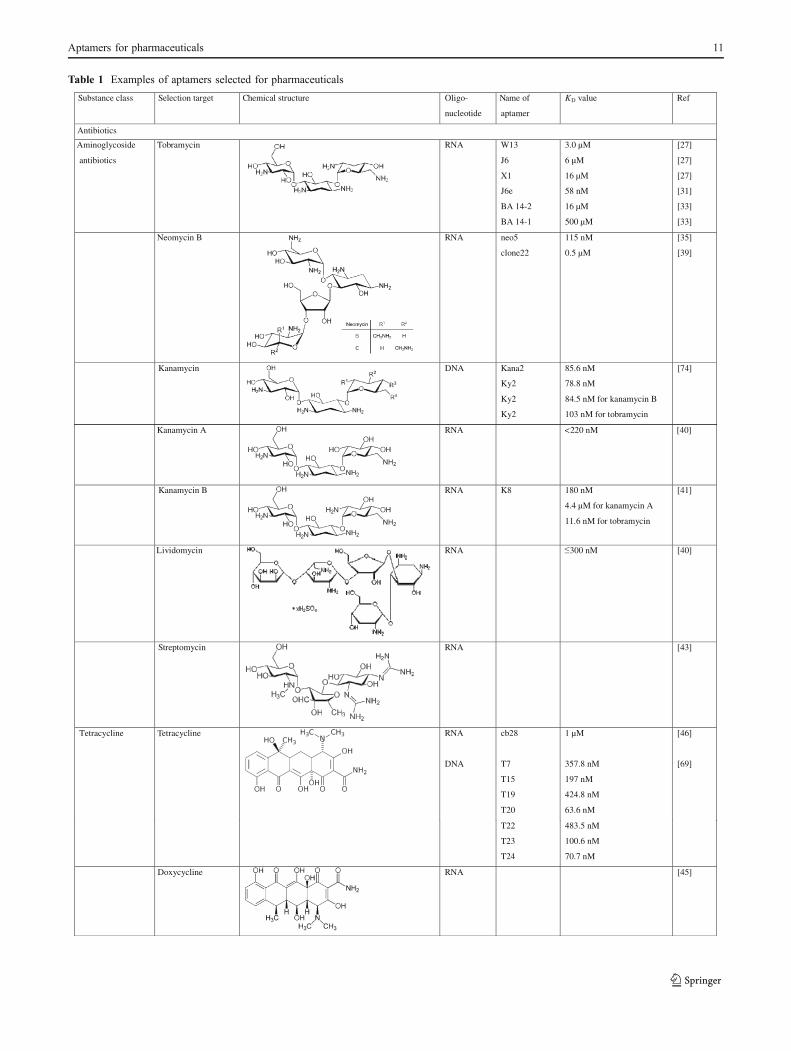
Table 1 Examples of aptamers selected for pharmaceuticals
Substance class Selection target Chemical structure -ogilO
nucleotide
Name of
aptamer
KD value Ref
Antibiotics
Aminoglycoside
antibiotics
Tobramycin RNA W13
J6
X1
J6e
BA 14-2
BA 14-1
3.0 µM
6 µM
16 µM
58 nM
16 µM
500 µM
[27]
[27]
[27]
[31]
[33]
[33]
Neomycin B RNA neo5
clone22
115 nM
0.5 µM
[35]
[39]
Kanamycin DNA Kana2
Ky2
Ky2
Ky2
85.6 nM
78.8 nM
84.5 nM for kanamycin B
103 nM for tobramycin
[74]
Kanamycin A RNA <220 nM [40]
Kanamycin B RNA K8 180 nM
4.4 µM for kanamycin A
11.6 nM for tobramycin
[41]
Lividomycin RNA 300 nM [40]
Streptomycin ]34[ANR
Tetracycline Tetracycline RNA
DNA
cb28
T7
T15
T19
T20
1 µM
357.8 nM
197 nM
424.8 nM
63.6 nM
[46]
[69]
T22
T23
T24
483.5 nM
100.6 nM
70.7 nM
Doxycycline ]54[ANR
Aptamers for pharmaceuticals 11

Oxytetracycline DNA No. 4
No. 5
No. 20
No. 2
9.61± 0 3 nM
12.08 ± 2.25 nM
56.84 ±3.62 nM
121.1±5.3 nM
[70]
Anthracycline Daunomycin
(Daunorubicin)
DNA 10.10
10.10v
20 nM
272 nM
[64]
Further antibiotics Viomycin RNA 11–21µM [48]
Substance class Selection target Chemical structure -ogilO
nucleotide
Name of
aptamer
KD value Ref
Linezolid RNA
L2-12
66 – 260 nM for linezolid
neomycin heteroconjugate
300 nM for neomycin
5.4 mM
[50]
Chloramphenicol RNA 2 – 200 µM [51]
Moenomycin A RNA
(A6)
(A6)
300 – 400 nM
437 nM by FCS
320 nM by affinity
chromatography
[52]
[53]
Pharmaceuticals
with psychotropic
effects
Codeine RNA FC5
FC45
FC5
FC45
4.5 µM
47 µM
25 µM for morphine
212 µM
[55]
Theophylline RNA TCT8-4
mTCT8-4
0.6 µM
0.1 µM
[57]
Cocaine DNA
DNA
DNA
DNA
MNS-4.1
MNS-7.9
F7.9
~0.4 - 10µM
~20 µM
~100 µM
100 ± 9 µM
4.6 ± 0.3 nM
134.4 ± 7.2 µM
[80]
[80]
[80]
[85]
[147]
[152]
Table 1 (continued)
12 B. Strehlitz, et al.

Thalidomide DNA T5-B
T5-1-B
T5-1a
113 µM
133 µM
1.05 ± 0.59 µM
[89]
Dopamine RNA dopa2
dopa2/c.1
2.8 µM
1.6 µM
[61]
Analgetics Diclofenac DNA D10/DA24
D22
D16
D3
100.64 ± 40.5 nM
166.34 ± 57.9 nM
148.73 ±15.5 nM
42.7 ± 15.9 nM
[94]
Substance class Selection target Chemical structure -ogilO
nucleotide
Name of
aptamer
KD value Ref
Ibuprofen (racematic
form)
DNA IBA2
IBA8
IBA12
IBA4
IBA17
IBA4
IBA17
3.0 µM (for racematic form)
5.2 µM (for racematic form)
3.2 µM (for racematic form)
1.5 µM (for (S)-isomer)
3.8 µM (for (S)-isomer)
2.4 µM (for racematic form)
6.8 µM (for racematic form)
[97]
Hormones Estradiol DNA 0.1 – 3 µM
0.13 µM
[102]
Somatropin DNA 218 nM [105]
Insulin ]801[AND
Vasopressin DNA D-Aptamer
L-Aptamer
0.9 µM for D-vasopressin
1.2 µM for L-vasopressin
[116]
Table 1 (continued)
Aptamers for pharmaceuticals 13

Thalidomide
Thalidomide (also known under the brand names Contergan®in Germany and Kevadon® in Canada and the USA)was introduced as a sedative drug in the late 1950s, butwas withdrawn from the market in 1960s due to itsstrong teratogenic and neuropathogenic effects. Thalid-omide exists in two enantiomeric forms, the (R)- and(S)-isomers which cause the differences in its biologicalactivity. While the enantiomers can interconvert (race-mize) in vivo [86], their pathogenic mechanisms are stillnot understood. Recently, thalidomide has been consideredas a potential drug for various diseases such as autoimmunediseases, AIDS, Hansen’s disease, and some cancers [87, 88].
Shoji et al. [89] generated a modified DNA aptamerthat binds the (R)-isomer of a thalidomide derivate withhigh enantioselectivity. The aptamer selection wasdesigned using the racematic thalidomide derivate, whichwas conjugated with biotin and immobilized on astreptavidin gel. A modified DNA library was appliedwhich contained cationic ammonium groups attached by ahydrophobic hexamethylene linker at the thymidineresidues (THM). Fifteen rounds of selection were carriedout, and the five aptamers with the highest bindingaffinities to thalidomide showed a high guanine content.Tests with the best binding aptamer exhibited that themodified group THM was indispensable for the binding tothalidomide. An affinity constant of 113 μM wasdetermined for the selected aptamer T5-B (biotinylated)by SPR. The SPR measurements were carried out with aderivate of thalidomide which was conjugated with a PEGderivative as a weight tag. Truncated versions of the bestbinding aptamer T5 (T5-1, T5-2, and T5-3) were used todetermine the thalidomide-binding site which was expected toform a stem-loop structure. The fragment T5-1 was suggested
as the binding site of the aptamer T5. The dissociationconstant for the aptamer fragment T5-1-B determined by SPRmeasurements was 133 μM which is almost the same as thatof the parent aptamer, T5-B. Studies with fluorescencetitration revealed a high enantioselective binding behavior ofthe truncated aptamer T5-1a which recognized the (R)-isomerof thalidomide. The dissociation constant of T5-1a wasestimated by fluorescence titration and was found to be1.05±0.59 μM.
DNA aptamers for analgetics
Diclofenac
Diclofenac (DCF) is a nonsteroidal anti-inflammatory drugwith analgetic, antiphlogistic, antipyretic, and antirheumaticproperties. It is widely used to treat pain and inflammatorydisorders including musculoskeletal complaints (arthritis,rheumatoid arthritis, polymyositis, dental pain, etc.). Diclofe-nac belongs to the most frequently detected pharmaceuticallyactive compounds in the water cycle [90] causing critical sideeffects for humans and also harmful environmental impacts.Published are, e.g., noxious effects on vultures [91, 92] andon freshwater fish species [93].
Joeng et al. [94] produced DCF-binding aptamers by usingthe FluMag-SELEX process [72]. After nine selectionrounds including counterselection steps with the structureanalog 2-anilinophenylacetic acid (2APA) and 4-amino-3,5-dichlorbenzoic acid, an enrichment of three major groupsbased on sequence similarity could be shown. Secondarystructure analysis revealed typical stem-loop structures. Thebest binders D10/DA24, D16, D22, and D3 exhibitedspecificity to the selection target DCF. Aptamers D22 andD16 possessed higher specificities to 2APA than for DCF.2APA is a structural analog of DCF lacking two chlorine
Gonadoliberin RNA
RNA
Spiegelmer
DNA
DNA
Spiegelmer
A10
A10 truncated
A10 truncated
Spiegelmer
S42
S42 truncated
S42 truncated
Spiegelmer
92 ± 12 nM for D-GnRH
263 nM for D-GnRH
190 nM for L-GnRH
55 ± 7 nM for D-GnRH
45 nM for D-GnRH
45 nM for L-GnRH
[122]
Substance class Selection target Chemical structure -ogilO
nucleotide
Name of
aptamer
KD value Ref
Table 1 (continued)
14 B. Strehlitz, et al.

atoms. For one of the aptamers (D3), specificity to DCF andno significant binding to 2APA could be shown. For all ofthese aptamers, no binding activity was detectable to 4-amino-3,5-dichlorobenzoic acid (chemically not related) andto naked beads. The determined KD values for immobilizeddiclofenac averaged between 42.7 and 166.34 nM and aregiven in detail in Table 1.
Ibuprofen
Ibuprofen is another representative nonsteroidal anti-inflammatory drug which offers analgetic, antiphlogistic,antipyretic, and antirheumatic properties. It is prevalentlyused for arthritis, primary dysmenorrhea, and fever. Due tothe chirality of ibuprofen, there are two enantiomers withdifferent physiological effects. The (S)-(+)-ibuprofen wasfound to be the active form both in vitro and in vivo [95],while (R)-(−)-ibuprofen seems to be teratogenic [96].
To generate an enantioselective DNA aptamer for ibupro-fen, Kim et al. [97] carried out a FluMag-SELEX [72] processwith racematic ibuprofen. Ten rounds of in vitro selectionincluding counterselection steps with other nonsteroidal anti-inflammatory drugs (fenoprofen, flubiprofen, naproxen) wereperformed. After this procedure, five different sequences(mostly G-rich) could be found. They could be divided intotwo sequence groups depending on the found consensusregions. The aptamers of the first group (IBA2, IBA8,IBA12) showed binding affinity to the racemic mixture ofibuprofen but were not able to bind pure (S)-ibuprofen. Onthis account, the authors concluded that they are specific forthe (R)-isomer. The affinity constants for this sequence groupwere determined by an affinity elution assay to be 3.0 μM(IBA2), 5.2 μM (IBA8), and 3.2 μM (IBA12). The aptamersof the second group (IBA4, IBA17) possessed bindingaffinities to the (S)-isomer (KD=1.5 μM (IBA4) and 3.8 μM(IBA17)) as well as to the racematic form (KD=2.4 μM(IBA4) and 6.8 μM (IBA17)) of ibuprofen. Neither the firstsequence group nor the second showed any binding affinity tothe other tested nonsteroidal anti-inflammatory drugs feno-profen, flubiprofen, and naproxen or to oxytetracycline.
DNA aptamers for hormones
Estradiol
Estradiol (also 17β-estradiol or E2) is one of the predominantsex hormones belonging to the class of steroid hormones.Derivatives of estradiol are the most common estrogeningredients in combined oral contraceptive pills. Estradiolitself has been widely applied in animal fattening for itsanabolic effects [98]. It is well-known that endocrine-disrupting chemicals, including estradiol, have harmfuleffects on aquatic organisms (e.g. [99, 100]). Also humans
are affected by chronic exposure since these chemicals reachthe natural aquatic systems and the drinking water [101].
Kim et al. [102] obtained 17β-estradiol-specific ssDNAaptamers after seven cycles of selection and enzymaticenrichment in a SELEX process. The aptamers wereanalyzed concerning their affinity to estradiol. Based on theequilibrium filtration method, affinity constants for the tenobtained aptamers were determined to be in a range of 0.1 to3 μM, with a KD=0.13 μM for the best binding oligonucle-otide. Secondary structure analysis indicated a stem-loopstructure for this estradiol aptamer. The aptamer was used inan electrochemical detection method by immobilizing theaptamer on a gold electrode chip. In that way, the authorscould show that the aptamer had no cross-reactivity to smallorganic chemicals with structural similarities to estradiol(methoxynaphthalene, aminoanthraquinone).
Huy et al. [103] used this aptamer for a novel separationand enrichment method of 17β-estradiol from aquaticsamples by application of aptamer-anchored microbeads.To achieve this, the estradiol-specific aptamers were amino-tagged and covalently attached to isothiocyanate modifiedglass beads. The experiments revealed a specific binding andenrichment of 17β-estradiol from spiked water samples. Nobinding to the antibiotics chloramphenicol and 3-[[(4-carboxyphenyl) methylene] amino]-2-oxazolidinone and aweak cross-reactivity to the estrogen diethylstilbestrol wasdetected. The results of this study demonstrate the ability ofaptamer-based affinity methods for the separation andenrichment of chemicals from environmental water samples.
Somatropin
Somatropin is the recombinant produced human growthhormone (rhGH). In contrast, the natural human growthhormone (hGH) is called somatotropin. Both are anionic, non-glycosylated four helix-bundle proteins. Somatropin has astrong anabolic effect. Therefore, it is often misused byathletes to enhance their performance. But it is also applied asmedication to treat hypopituitary dwarfism, injuries, bonefractures, bleeding ulcers, and burns [104].
Calik et al. [105] selected aptamers for somatropin usingthe so-called single-step ligand evolution by temperaturegradient method. The characteristic step of this method isan elution of the target-bound oligonucleotides by atemperature gradient. In this way, the authors assumed thataptamers with higher specificity to the target are separatedfrom those aptamers with lower specificity. At first, a negativeselection step with extracellular proteins of Bacillus subtiliswas performed because the aim of the aptamer developmentwas to separate and purify rhGH (somatropin) during theproduction process from B. subtilis fermentation broth. Afterthis pre-selective step, the obtained oligonucleotide pool wasincubated with somatropin microparticles. The somatropin-
Aptamers for pharmaceuticals 15

binding oligonucleotides were eluted stepwise by atemperature gradient within a range of T=55 °C to 95 °C.At a temperature of 85 °C, eight aptamer sequences withhigh affinity to somatropin were eluted. The KD value of thebest binder was determined to be 218 nM by equilibriumbinding analysis. By utilization of this aptamer immobilizedon microparticles, the authors were able to separate andpurify rhGH (somatropin) from the B. subtilis fermentationbroth with a purity of 99.8%.
Bruno et al. [106] developed aptamers, which are able todiscriminate between somatropin (rhGH) and somatotropin(hGH) and bind these hormones in different levels. Theaptamers were obtained after five rounds of a microbead-based SELEX procedure with rhGH and hGH as targets.Eight of the found aptamer sequences indicated the abilityto discriminate recombinant produced somatropin (rhGH)from natural somatotropin (hGH). Sequence analysesdiscovered GGGTG as the most common sequence seg-ment. Specificity tests by enzyme-linked aptamer assaywere carried out to determine cross-reactivity to proteins orpeptides of human body fluids and others. Regrettably, thebest rhGH discriminatory aptamers exhibited a cross-reactivity to human myoglobin and to BSA (but not to bonecollagen peptides and to an unrelated viral envelope peptide),which makes them inappropriate for measurements of rhGH/hGH in body fluids.
Insulin
The well-known peptide hormone insulin has centralfunctions in carbohydrate and fat metabolism in vertebrates.Dysfunctions in insulin balance lead to Diabetes mellitusand related metabolic disorders. Worldwide, millions ofpatients with diabetes depend on external insulin for theirsurvival because the hormone is no longer producedinternally. For this widespread clinical use, biosynthetic“human” insulin is manufactured recombinantly [107].
To select insulin-binding aptamers, Yoshida et al. [108]applied an ssDNA library which was expected to formvarious kinds of G-quartet structures. This approach waschosen because an insulin-linked polymorphic region (ILPR)in the human insulin gene promoter was found that can forman intramolecular G-quartet structure [109, 110]. The in vitroselection itself was carried out by aptamer blotting. In orderto do this, human insulin as the target was immobilized on amodified polyethersulfone affinity membrane. By using thismethod, the binding of the oligonucleotides to the targetprotein could be visualized. After six rounds of selection,three aptamer sequences were obtained. Investigations of thebinding ability to insulin in solution by fluorescencepolarization measurement exhibited that two of the aptamershad a higher affinity to insulin than ILPR (which can becalled “natural insulin-binding aptamer”). Circular dichroism
spectrum measurements revealed the aptamer folding into aG-quartet structure while binding to insulin. Complementary,the authors developed an aptameric enzyme subunit (AES)by connecting the selected insulin-binding aptamer with athrombin-inhibiting aptamer [111] for insulin detection.Using this AES, it was possible to detect insulin by measuringenzymatic activity of thrombin.
Vasopressin
Vasopressin (also arginine vasopressin or antidiuretic hormone)is a potent endogenous peptide hormone that controls there-absorption of molecules in the tubules of the kidneys byaffecting the tissue’s permeability. It also increases peripheralvascular resistance, which in turn increases arterial bloodpressure. It plays a key role in homeostasis and the regulationof water, glucose, and salts in the blood. It acts as aneurotransmitter in the brain to control the circadian rhythm,thermoregulation, and adrenocorticotropic hormone release[112–114]. The therapeutic use of vasopressin has becomeincreasingly important in intensive care, in the managementof cranial diabetes insipidus, bleeding abnormalities, esoph-ageal variceal hemorrhage, asystolic cardiac arrest, and septicshock [115].
Williams et al. [116] generated a mirror-image ssDNAaptamer (L-DNA aptamer, similar to Spiegelmers [117]) toachieve a nuclease-insensitive ligand. The aptamer selectionwas carried out using the “selection–reflection” strategy.First step of this procedure is the production of an enantiomer(D-isomer) of the cyclic L-peptide arginine vasopressin. This
D-isomer of vasopressin was used as target for the selectionof natural D-ssDNA aptamers. The SELEX process wasrealized by affinity chromatography [118]. The implementedoligonucleotide library was produced with a raised G-contentbecause the authors expected G-quartet structures forbinding. Consequently, it was not surprising that the receivedvasopressin aptamers exhibited a high G-content. Thebinding region could be defined as a stem with an internalloop of 20 nt that contains guanine nucleotides at conservedpositions. The truncated version of the D-ssDNA aptamer,containing only the binding region, was mirror-imaged into its
L-form and tested for its ability to bind natural L-vasopressin.This L-aptamer exhibited a more than 100-fold preference forvasopressin compared to oxytocin which is the closestknown human analog (differing only in two peptide residues).Dissociation constants were ascertained by equilibriumdialysis experiments and were determined to be 0.9 μMfor D-aptamer/D-vasopressin and 1.2 μM for L-aptamer/L-vasopressin. Stability and nuclease insensitivity of the D-/L-aptamer was proofed with the following results: The L-aptamer stayed unaffected within 10 days, and the D-aptamerwas degraded after 10 s by purified nucleases. Furthermore,the L-aptamer was not degraded in human serum and only to
16 B. Strehlitz, et al.

a small extent in calf serum. Additionally, the bioactivity ofthe L-aptamer as a vasopressin antagonist was confirmed incell culture.
Gonadoliberin
Gonadoliberin is a peptide hormone (also gonadotropin-releasing hormone I (GnRH) or gonadorelin) responsible forthe release of follicle-stimulating hormone and luteinizinghormone [119]. Therapeutically, it is often used in cases offertility dysfunctions [120]. Gonadoliberin analogs areapplied to treat breast or prostate carcinoma, endometriosis,and precocious puberty [121].
Leva et al. [122] assembled GnRH-specific RNA and DNASpiegelmers. Initially, a SELEX process to get natural D-RNAand D-ssDNA ligands for mirror-imaged gonadoliberin (D-GnRH) was implemented. In both cases, the selection forRNA and DNA aptamers was carried out by affinity chroma-tography using D-GnRH immobilized on thiol-modifiedsepharose. After six rounds of the RNA-SELEX, only onemajor binding sequence (named A10) was found which bound
D-GnRH with a KD value of 92±12 nM (determined byequilibrium dialysis). Eight rounds of the DNA-SELEX led tonine different sequences which were able to bind GnRH. A KD
value of 55±7 nM could be determined for the DNA aptamerwith the highest affinity (named S42). The selected RNA andDNA variants showed no similarity to each other, neitherconcerning their primary sequence nor their proposed secondarystructure. Truncations to ascertain the minimal binding domainindicated that 48 nt of the RNA aptamer form a three-way helixjunction with four unpaired nucleotides at the branching point,while the DNA aptamer (68 nt) forms a G-quadruplex structureflanked by two stems. Further substitutions and deletions led to a60-nt DNA aptamer which bound to D-GnRH with KD=45 nM.The affinity constant of the Spiegelmer to L-GnRH was thesame. Both constants were determined by equilibrium dialysis.The affinity constants for the truncated RNA aptamer and itsSpiegelmer were determined by isothermal calorimetry. The50-nt truncation of the RNA aptamer A10 showed KD=263 nM to D-GnRH, whereas the Spiegelmer L-RNA bound to
L-GnRH with KD=190 nM. Truncation of the RNA aptamer toa 50-mer decreased the binding affinity to half, whereastruncation of the best binding DNA sequence to a 60-mer didnot alter the binding affinity [122].
Specificity tests were carried out by SPR real-time kineticmeasurements. Both Spiegelmers exposed high specificity forgonadoliberin since the exchange of a single amino acidresulted in a dramatic loss of binding affinity (shown forchicken LHRH). The RNA Spiegelmer also recognized theGnRH analog buserelin, albeit with reduced affinity, whilethe DNA Spiegelmer showed nearly no binding to buserelin.No binding activity could be detected to the completelyunrelated peptides vasopressin and oxytocin. Cell experi-
ments clearly demonstrated that both Spiegelmers were ableto inhibit the binding of GnRH to its cell surface receptor.
In contrast to the development of RNA aptamers, DNAaptamers for pharmaceuticals are often selected for diagnosticand analytical applications. DNA aptamers are thereforewidely used to analyze body fluids. However, they are equallyvaluable for the detection of pharmaceutical residues in foodor in the environment. Moreover, some research groupsdevelop protocols not to implement aptamers in measurementand detection systems, but for application as affinity compo-nents in enrichment and purification methods. In contrast toDNA aptamers, the development of Spiegelmers is generallyfocused on a therapeutic application.
Independent of both the designated application and theselection method, it is remarkable that most DNA aptamersdeveloped for pharmaceuticals exhibit a high guaninecontent. In several cases, G-quadruplex structures wereidentified as the binding regions, and it is noteworthy thatalso the “natural” aptamer for insulin (the insulin-linkedpolymorphic region in the human insulin gene promoter)forms an intramolecular G-quartet structure [109, 110].Several studies indicate these structures to form highlycompetent binding configurations, especially for smallbioactive molecules [123–125]. Considering the innumera-ble possibilities for the potential formation of G-quadruplexstructures on the genomic level, these structures may occurmore often in living cells than currently anticipated. Besidesthat, G-quadruplexes seem to be very convenient ligands forsmall molecules with pharmaceutical properties.
Aptamer-based detection systems
In comparison to other biological recognition elements, suchas antibodies, aptamers are more stable to biodegradation andless vulnerable to denaturation. Their target binding isregenerative. Therefore, they are prime candidates forbiosensors and assays in a number of applications, such asenvironmental monitoring and medical diagnostics [126]. Thekey challenge to their successful application is the transfor-mation of the aptamer-binding events into physicallydetectable signals. To meet the challenge, a number ofmethods have been developed. Some aptamers bind theirtarget by a contemporaneous structure switch. Such biomo-lecular switches enable transduction of the binding event intoa specific output signal in a single step and without theaddition of exogenous reagents and without the use of labels[127]. They can be used also in connection with markers(labels) fixed to the structure. These markers can be forinstance redox active or optically active. The bindingbetween the structure switching aptamer and its targetinduces a change of steric configuration of the aptamer
Aptamers for pharmaceuticals 17

causing a change in the measuring signal. Aptamer modifi-cation with electrochemically or optically active labels isvery easy because the chemical synthesis procedures usedfor aptamer production provide a lot of standard methods forchemical binding of reporter molecules or reactive groups.
Most of the aptamer-based detection systems are describedwith one of the well-known aptamers for thrombin or cocaineserving as model aptamers. Cocaine belongs to the psycho-tropic pharmaceuticals, and therefore, these aptamer-basedsensors are included in this review.
Biosensors couple the biological recognition of theanalyte by a biological receptor (which may be anaptamer—“aptasensor” [128]) with a chemical or physicaltransducer. The characteristic feature of the biosensor is,according to the International Union of Pure and AppliedChemistry definition, the direct contact of the recognition
element and the transducer [129]. The aptamer-basedsystems for the detection of pharmaceuticals reviewed inthis article include not only biosensors which fulfill thisdefinition but also optical assays with separated aptamer-based detection and signal recognition, for instance incolorimetric assays or test strips. The sections below givedetailed descriptions about the aptamer-based sensors andassays, summarized at a glance in Table 2.
Electrochemical aptasensors
Electrochemical transducers offer some advantages in com-parison to optical and other transducers such as ultrahighsensitivity and simplicity [130]. Electrochemical transducersare the most frequently used in commercialized biosensors,for instance for blood glucose testing. The small size of
Table 2 Examples of aptamer-based biosensors and assays for pharmaceutical detection
Target/measurement of
DNA/RNAaptamer
Ref of aptamerdescription
Transducer/assay Limit of detection Linear range (μM) Ref ofsensor/assay
Oxytetracycline DNA [70] ec, chip system, labeled 0.005–0.1 [137]
Tetracycline DNA [69] ec, screen-printed electrode,CV, SWV, label-free
10 nM 0.01–10 [139]
Neomycin RNA [35] ec, competitive impedimetric,label-free
0.75–500 [140]
Tobramycin RNA [27] ec, competitive impedimetric,label-free
ATA 0.7 μM ATA 1.4–70 [141]FATA 0.4 μM FATA 0.8–34.9
Aminoglycoside RNA [27] ec, labeled 2–6 [142]
Cocaine DNA [80] Optical, colorimetric, labeled 50–500 [126]
Cocaine DNA [80] Optical, FRET, labeled 10–4,000 in bloodserum
[80]
Cocaine DNA [80] Optical, colorimetric dipstick 10 μM [149]
Cocaine DNA [80] ec, microfluidic, labeled 10–100 in bloodserum
[85]
Cocaine DNA [80] Optical, FRET, labeled 10 μM 10–1,000 This publication
Cocaine DNA [79] Optical, FRET, labeled 10–1,250 [79]
Cocaine DNA [79] Optical, FRET, labeled 1 μM 1–1,000 [143]Optical, excimer fluorescence,labeled
100 μM 100–1,000
ec, LSV, labeled 10 μM 10–1,000
Cocaine DNA [80] ec, CV, labeled <10 μM [82]
Cocaine DNA [79] ec, pulse voltammetry, label-free 0.1 μM 0.1–38.3 [145]
Cocaine DNA [79] ec, chronocoulometric, label-free 70 nM 0.1–26.3 [130]
Cocaine DNA [80] Optical, colorimetric, labeled 50 nM 0.1–20 [153]
Cocaine DNA [80] Electrogenerated chemiluminescence,labeled
1 nM 0.005–0.3 [146]
Cocaine DNA [80] Optical, fluorescence, rollingcircle amplification, labeled
0.48 nM 0.001–0.05 [154]
Cocaine DNA [82] Electrogenerated chemiluminescence,labeled
10 pM [147]
Vasopressin L-DNA [116] Optical, SERS, label-free 5.2 μU/ml [155]
Dopamine RNA [61] Optical, colorimetric, ELAA,labeled
1 pM 0.00005–0.1 [156]
17β-Estradiol DNA [102] ec, CV, SWV, label-free 0.00001–0.001 [102]
CV cyclic voltammetry, ec electrochemical, ELAA enzyme-linked aptamer-based assay, FRET fluorescence resonance energy transfer, LSV linearsweep voltammetry, SERS surface-enhanced Raman spectroscopy, SWV square wave voltammetry
18 B. Strehlitz, et al.

electrodes and the possibility to miniaturize the detectionunit as sensor or chip system and to integrate them with theelectronics of the device permit the construction of hand-helddevices or in-field/at-side devices. Aptamer-based biosensorswith electrochemical transduction [131–133] and also withvarious amplification methods of the aptamer-based signal-tracing are described [130].
Sassolas et al. have reviewed articles about electrochemicalaptasensor development written in the years between 2004and 2009 [134]. The article presents clearly drawn schemesof the different configurations of labeled and non-labeleddetection and between signal-on and signal-off electrochem-ical detection principles and describes advantages anddisadvantages of them.
Sadik et al. describe in their review dealing withbiomolecular recognition by use of electrochemical techni-ques various electrochemical aptasensors that were developedrecently [135]. Electrochemical detection of the aptamerreaction with the target is based on labels such as enzymes,nanoparticles, quantum dots, or redox active molecules, oron a binding-induced label-free detection. A reagentless,electrochemical aptamer-based sensor (E-AB sensor) wasdeveloped [82, 133, 136], which is a platform for aptamer-based detection. One of the ends of the aptamer is immobi-lized at the electrode surface. At the other end, a redox activelabel is attached to the aptamer, which is able to undergo aredox reaction with the electrode. In the case of binding of thetarget molecule to the aptamer, its conformation is changedand the distance between the redox active label and theelectrode surface is decreased, which in turn causes a changein the electrochemical signal. Even though—regardingpharmaceuticals—aptamers were developed for many sub-stances and substance classes, only a few of them are in factused in electrochemical biosensors. In the sections below,electrochemical aptasensors for antibiotics, psychotropics,and hormones are described as they are found in the literature.
Antibiotics
An electrochemical aptamer-based chip system, consisting ofa gold working electrode chip with eight electrodes in paralleland using the K3Fe(CN)6 redox mediator (comparable to thesystem described in [102]) was used for oxytetracyclinedetection. Kim et al. immobilized the thiol-modified oxytet-racycline specific DNA aptamer no. 14 described in [70] ona gold interdigitated array [137]. The reference and counterelectrode used are not specified in their work. The redoxreaction between ferrocyanide and ferricyanide is measuredby cyclic voltammetry (CV) and square wave voltammetry(SWV) before and after binding of oxytetracycline to theimmobilized aptamers. The dynamic range of the sensor liesbetween 5 and 100 nM oxytetracycline. Structurally relatedtetracycline derivatives such as doxycycline and tetracycline
were distinguished from oxytetracycline [137]. Sensorregeneration was achieved by use of 2 M NaCl [137, 138].
Kim et al. [139] used a screen-printed gold electrodewith silver reference electrode and gold counter electrodefor the immobilization of the tetracycline binding DNAaptamer T20, whose selection was described in [69]. Theaptamer was extended by a poly-T tail for flexibility andspacing at the 5′-end and labeled with biotin to immobilizeit at the streptavidin-modified electrode. The same detectionprinciple as in the sensor example above was used: Theredox reaction of the redox mediator K3Fe(CN)6 wasmeasured by CV and SWV at the aptamer-modifiedelectrode before and after binding of the target. Thedecrease of the current quantifies the target bound to theaptamer. This label-free sensor showed a dynamic range fortetracycline detection of 10 nM to 10 μM and a limit ofdetection of 10 nM tetracycline. The aptamer used in thisaptasensor can weakly recognize other tetracyclines likeoxytetracycline and doxycycline [69], so the sensor haslower specificity and can detect tetracycline and itsderivatives [139].
Faradaic impedance spectroscopy was used for neomycindetection with a competitive RNA aptamer sensor [140]. Theneomycin B-specific RNA aptamer [35, 37] was stabilizedby 2′-O-methylation. The aptamer was bound to neomycin Bimmobilized on a gold electrode. The assay procedureconsists of 5-min incubation of the electrode in the affinitysolution with varying concentrations of neomycin B andsubsequent transformation to the measuring solution, whichcontains the redox probe [Fe(CN)6]
3−/4−. The impedancespectrum at a bias potential of +0.225 V (Ag/AgCl/KClsat)using a frequency range from 10 kHz to 100 mHz and analternating current amplitude of 5 mV is measured. Theincrease of neomycin B in the affinity solution causes anincreasing aptamer displacement from the electrode which ismeasured as an impedance change [140]. The linear range ofthe label-free sensor is from 0.75 to 500 μM neomycin B.The measurement is fast because the aptamer displacementby neomycin B requires only 5 min. The electrode surfacecan be regenerated. In concentrated neomycin solution, allremaining aptamers are released. Further rebinding of theaptamer results in a fresh aptamer-bound surface [140]. Thesensor was stored in neomycin solution. One week storageand 20 measurement cycles are possible without significantloss in sensitivity. The practical test was done in milk spikedwith neomycin. The sample pretreatment consisted in 1:5dilution of the sample in affinity buffer and ultracentrifugation.The linear range for the measurement in milk is between25 μM and 2.5 mM neomycin B [140].
A comparable RNA aptamer-based displacement impedi-metric sensor for tobramycin detection was described byGonzalez-Fernandez et al. [141]. The RNA aptamer developedby Wang and Rando [27] was stabilized by 2′-O-methylation
Aptamers for pharmaceuticals 19

in the same way as the neomycin RNA aptamer mentionedbefore and described in [140]. Two types of aptamermethylation were tested, the fully modified aptamer (FATA)is methylated at each base, in contrast to the partially modifiedaptamer called ATA. The aim of this study was to detecttobramycin in human serum. Both aptamers were tested in thedisplacement assay by use of the impedimetric sensor whichworked as described before for neomycin detection [140]. Thechanges in the electron transfer resistance that resulted frombiosensing events between the redox probe ([Fe(CN)6]
3−/4−)and the modified electrode were measured as the impedancespectra (at a bias potential of +0.225 V (Ag/AgCl/KClsat)using a frequency range from 10 kHz to 100 mHz and analternating current amplitude of 5 mV). In this way, theanalyte tobramycin could be detected in solution by thedisplacement of aptamers bound to the tobramycin-modifiedsensor surface. The ATA aptamer showed a limit of detectionof 0.7 μM and a linear range of 1.4 to 70 μM for tobramycin.Regeneration of the sensor surface was done by incubation inconcentrated tobramycin solution for 1 h, removing all of theaptamer molecules from the electrode surface. Subsequently,the tobramycin-modified surface can be re-saturated by theaptamer and re-used for measurement. The sensor could beused for 1 week when stored between measurements in theregeneration solution (conc. tobramycin solution) at 4 °C. Theselectivity was tested with respect to the aminoglycosideskanamycin, neomycin B, paromomycin, and streptomycin.The tobramycin–ATA aptamer sensor was able to discriminatebetween tobramycin and kanamycin, paromomycin, andstreptomycin. However, neomycin B could be detected bythe sensor producing signals nearly the same as tobramycin.By use of the fully methylated aptamer (FATA), a limit ofdetection of 0.4 μM and a linear range of 0.8 to 34.9 μMwere found, which were in good agreement with the results ofATA aptamer, suggesting similar affinities for both aptamersfor its target tobramycin. The measurement of tobramycin inhuman serum was possible in the therapeutic concentrationrange [141].
An RNA aptamer-based E-AB sensor for aminoglycosideantibiotics detection is able to detect tobramycin, kanamycin,and gentamicin. This aptamer does not discriminate betweenthese three aminoglycosides binding all three with similaraffinities. This sensor was developed for point-of-carediagnostics. It allows measurement of aminoglycoside con-centrations over the therapeutically relevant low micromolarrange (2 to 6 μM). Methylene blue (MB) is used as the redoxtag at the 3′-end of the RNA aptamer. The binding of theaminoglycosides to the electrode-bound aptamer causes adecrease in Faradaic current from the attached redox tag.Nucleases in serum samples are removed by ultrafiltration,which is possible for the detection of the small aminoglycosidemolecules. The measurement process with sample pretreat-ment is complete in less than 30 min [142].
Pharmaceuticals with psychotropic effects
A rapid electrochemical aptamer biosensor for cocainedetection is based on the target-induced conformationalchange of the aptamer [82]. Baker et al. used the cocaineDNA aptamer F7.9D described by Stojanovic et al. [80]and coupled a MB redox tag to it. These modified aptamerswere immobilized on a gold electrode by self-assembly viaan alkanethiol group. The methylene blue reduction peak ofthe AC voltammogram increases with increasing cocaineconcentration. The authors assume that in the absence ofthe target cocaine the aptamer remains partially unfolded,but with addition of cocaine the aptamer folds into a typicalthree-way junction structure causing an alteration of theMB reduction peak. The sensor is “signal on,” in contrast tothe decreasing signal for the thrombin sensor described oneyear before [131]. The thrombin sensor construction wasthe same, whereas the thrombin aptamer shows a differentfolding reaction on target binding. The thrombin DNAaptamer folds into a G-quadruplex structure in the presenceof the target [131] in contrast to the cocaine aptamer whichfolds into a three-way junction structure [82]. In the firstcase the signal changes are caused by the MB tag, whichveers away from the sensor surface, and in the second casethe MB tag approaches the electrode surface. The detectionlimit of the cocaine biosensor is given to be below 10 μM.The sensor enables an easy detection of 500 μM cocaine infetal calf serum or human saliva [82].
A microfluidic electrochemical aptamer-based sensor(MECAS) was developed [85] based on the same cocaineaptamer F7.9D [80] which was labeled with MB forelectrochemical sensing of the target binding as describeddirectly before [82]. The MECAS chip consists of three goldelectrodes at which the thiolated aptamer is immobilized, aplatinum counter electrode and a platinum reference electrodewithin a 750-nl detection chamber. By use of this micro-fabricated electrochemical aptamer biosensor, the continuous,real-time detection of cocaine in undiluted, unmodified bloodserum in the physiological range was shown [85]; 10 to100 μM cocaine in blood serum was measured, and thedissociation constant for the aptamer binding was determinedto be 100±9 μM, which is identical to that for the aptamer insolution [80, 85].
Another MECAS is reported by Du et al. [130] forsimultaneous detection of ATP and cocaine as model targets.Here we will only review the cocaine detection by thismethod, because ATP is not a relevant pharmaceuticalresidue which has to be detected by environmental monitor-ing. Two cocaine aptamer fragments derived from [79] wereused. This MECAS chip consists of four groups of three-electrode systems. Each group of three-electrode systemsconsisted of two gold working electrodes, which shared onegold counter electrode and one Ag reference electrode.
20 B. Strehlitz, et al.

Polydimethylsiloxane-based microfluidic channel framescover the glass chip. One subunit of the cocaine aptamer isimmobilized on the gold surface of a working electrodedesired for cocaine detection. The second subunit of theaptamer is immobilized on gold nanoparticles (Au-NP).These marked Au-NP are pumped together with the cocainesolution into the chip system for detection. In case of cocainebinding, an agglomerate is formed at the electrode surfaceconsisting of the cocaine molecule bound between the twosubunits of the cocaine aptamer together with the Au-NPload bearing some more aptamer subunits. Subsequently, theelectrochemical probe [Ru(NH3)6]
3+ (RuHex) binds to thesurface-confined DNA via electrostatic interaction and ismeasured by chronocoulometry. The Au-NP are used toproduce an amplified electrochemical signal and improve thesensitivity of the sensor. The detection limit for cocaine of thissensor is 70 nM and the linear range is 0.1 to 26.3 μM [130].
Freeman et al. [143] showed three different possibilitiesto use the self-assembling cocaine aptamer subunits [79].The electrochemical cocaine detection by use of thesesubunits and a methylene blue label is described here; theoptical methods based on FRET and excimer fluorescenceare described in the following chapter “Optical test systems:biosensors and assays.” The electrochemical cocaineaptamer sensor consists of the thiol-modified aptamersubunit C2-D from [79] immobilized on a gold electrodeand the methylene blue (MB+)—modified subunit F-C1[79]. Linear sweep voltammograms were measured by useof a graphite counter electrode and a saturated calomelreference electrode (SCE). In the absence of cocaine, a lowintensity voltammetric response is observed, which mightbe attributed to a minute nonspecific binding of the MB+ tothe electrode surface [143]. In the presence of cocaine, thesynergistically stabilized supramolecular complex, consistingof cocaine and both aptamer subunits, is formed on theelectrode surface. The confinement of the redox label(methylene blue) at the electrode surface enables an electro-chemical readout of the formation of the cocaine aptamercomplex correlating to the cocaine concentration [143]. Thedetection limit of this sensor equates to 10 μM, the linearmeasuring range is 10 μM to 1 mM. Other studies havereported the electrochemical detection of analytes by a redox-labeled aptamer, which is immobilized on the electrode. Afteraddition of the analyte (target), the aptamer-target bindingcauses structural alignment of the redox label in respect to theelectrode [82, 143, 144]. In the absence of the analyte thiselectrode arrangement of the redox label causes a largeelectrical background response whereas the structural rear-rangement caused by the presence of analyte only leads tosmall voltammetric changes [143].
A label-free electrochemical aptasensor was constructed byuse of a layer-by-layer self-assembled multilayer withferrocene-appended poly(ethyleneimine) (Fc-PEI) on an
indium tin oxide (ITO) array electrode [145]. Ferrocene isthe redox mediator which is incorporated in the electrodesurface. Fc-PEI and gold nanoparticles (Au-NP) are assem-bled layer by layer on the electrode surface. The cocaineaptamer fragments from [79] are used. One cocaine aptamerfragment, which is labeled with a SH-group, is coupled tothe outermost Au-NP layer. The other cocaine aptamerfragment is able to hybridize partly with the first aptamerfragment and binds to the target cocaine. This binding causesa decreasing in the differential pulse voltammetry signal ofFc-PEI. The big advantage of this sensor construction is theincorporation of the electrode mediator in the electrodesurface. The sensor has a linear range of 0.1 μM to 38.3 μMand a detection limit of 0.1 μM. It was applied in spiked realsamples of human plasma, human serum, human saliva, andhuman urine (25% biological fluids). The recovery of3.8 μM cocaine in the 25% biological fluids was between101.4 and 105.8% and 96.1 and 102.7%, respectively, whichmeans that the measurement of cocaine in biological fluids ispossible by use of this aptasensor.
Electrogenerated chemiluminescence (ECL) techniquewas used with the cocaine aptamer F7.9D from [80] labeledat its 3′ terminus with the ruthenium complex Ru(bpy)2(dcbpy)NHS [146]. This aptamer is immobilized onto a gold electrodesurface via thiol-Au interactions. As in some other applica-tions, the cocaine aptamer [80] remains partially unfolded inthe absence of target, resulting in a small ECL signal due tothe distance between electrode surface and tag. In thepresence of target, the aptamer folds in the three-wayjunction structure and thus a strong ECL signal is measureddue to the tag moving closer to the electrode surface. TheECL signal is generated by polarization of the electrode to+0.8 V in 0.1 M phosphate-buffered saline (PBS) containing0.1 M tripropylamine (TPA). Sensor characteristics are thedetection limit of 1 nM cocaine, a linear measuring range of5 nM to 0.3 μM cocaine, and no cross-reactivity to heroinand caffeine. The sensor is regenerable in 2 ml of 0.10 MPBS (pH 7.40) for four minutes with two times and byextensively washing with water according to [82]. Long-termstorage stability was proved by storage at 4 °C over 10 days,with no significant change of the measuring signal for 50 nMcocaine [146].
In a subsequent work an electrogenerated chemilumi-nescence (ECL) sensor for cocaine detection uses thecocaine aptamer labeled with a ruthenium derivative(cocaine aptamer-Ru1) [147]. The 30-mer cocaine aptamerfrom Baker et al. [82], which is in fact based on the cocaineaptamer described by Stojanovic et al. [80], was modifiedwith spacers and the Ru1 complex (which is not specifiedin the article). The cocaine aptamer-Ru1 is covalentlycoupled to the surface of a paraffin-impregnated graphiteelectrode that had been covalently modified with amonolayer of 4-aminobenzene sulfonic acid using electro-
Aptamers for pharmaceuticals 21

chemical oxidation. ECL-based detection of cocaine wasperformed with a potential-step excitation from an initialpotential of 0 V to a constant potential +1.35 V vs. Ag/AgCl ina 0.1 M TPA—0.1 M PBS (pH 7.4) solution. As describedabove, the cocaine aptamer folds in the well-known three-way junction structure with cocaine binding, which bringsthe Ru 1 modification nearer to the electrode. Quantificationof cocaine is based on the ECL peak intensity change. TheECL emissions were detected with a photomultiplier tube.With this cocaine aptamer-Ru1-based sensor, the bindingconstant between cocaine and its aptamer was estimated tobe 4.6±0.3 nM [147]. The selectivity was tested withcaffeine and heroin, which did not interfere with cocaine.The sensor was reusable (RSD=2.8%, n=7) and had a goodlong-term storage stability at 4 °C (recovery of 96.8% and92.3% after 21 and 30 days). This cocaine sensor has adetection limit of 10 pM for cocaine, which is 2 orders ofmagnitude lower than that of the above mentioned ECLcocaine aptamer sensor with self-assembled aptamer-Ru(bpy)3
2+ [146].
Hormones
An electrochemical aptasensor for 17β-estradiol consists ofthe DNA aptamer-modified gold working electrode chip(eight electrodes in parallel) using the biotin-streptavidincoupling system, a platinum counter electrode, and an Ag/AgCl reference electrode [102]. K3Fe(CN)6 is used insolution as the electron mediator. CV and SWV are used forthe measurement of the redox reaction between ferrocya-nide and ferricyanide. Binding of 17β-estradiol moleculesto the aptamers causes measurable changes in electricalcurrent. The linear range of the biosensor was 0.01 to 1 nM17β-estradiol [102].
Optical test systems: biosensors and assays
Optical signals such as fluorescence and color are preferredfor affinity-based recognition in biosensors because theyare easily detectable with high sensitivity and minimalinstrumentation. Fluorescence-based detection found grow-ing interest for real-time in-field measurement because ofrecent advances in miniaturization of the fluorescenceexcitation sources and emission detectors [16].
The signals of colorimetric sensors and assays can bedetected by the naked eye under some circumstances. Bythis way, the need of instruments is eliminated which makesthe application easier for unskilled personnel and householdapplication [16].
Au-NP possess a number of useful properties, such asvery high extinction coefficients and distance dependentcolor display (aggregated particles are blue or purple,dispersed particles are red). Highly sensible and simple
tests like colorimetric assays or lateral flow-based test stripscan be constructed [16]. For competitive aptamer-based Au-NP colorimetric sensors, the affinity ratio of the aptamers toboth unmodified Au-NP and target (KD(AuNP)/KD(Target))was detected as a tool to design more sensitive smallmolecule detection systems [148]. The lateral flow tech-nology is used in some common applied antibody tests, asthe well-known commercially available pregnancy test kit.The user friendliness of this format allows its application byusers without laboratory background. No precise transfor-mation of solutions is necessary. The result is shown moreor less as a “yes/no” answer, but with clear discriminationbecause of the negative controls incorporated. Such a lateralflow system is described for aptamer nanoparticle-baseddisaggregation reaction for cocaine detection [149].
Pharmaceuticals with psychotropic effects
The frequently used cocaine aptamer was introduced byStojanovic and coworkers in the years 2000 to 2002 withthree assays. At first, an aptamer self-assembly withfluorescence quenching was described [79]. In this case,the aptamer sequence was divided in two subunits: One(named C2-D) is labeled with a 3′-dabcyl quencher and theother (named F-C1) with 5′-6-carboxy-fluorescein fluoro-phore [79]. The two subunits behaved as a self-assemblingfluorescent cocaine assay, which was able to report cocaineconcentrations in the range of 10 to 1250 μM in therespective selection buffer solution [79]. The fluoresceinemission at 518 nm (λex=472 nm) was quenched to 65% ofthe initial value with 1 mM cocaine. The assay showedselectivity for cocaine over its metabolites benzoyl-ecgoninand ecgonine methyl ester [79].
The second assay is based on the cocaine aptamer MNS-4.1 [80]. MNS-4.1 consists of three stems S1, S2, S3 withthe assumed cocaine binding pocket located in thelipophilic cavity in the center of the three-way junctionstructure. They engineered an instability in one of the stemsof this aptamer which results in a structure that is dependenton the stabilization provided by ligand binding to form thethree-way junction. Double end labeling of the opened stemwith 5′-fluorescein fluorophore and 3′-dabcyl as quencherresulted in the F7.9D cocaine aptamer. This assay operatesfor cocaine detection in serum in the micromolar range(10 μM to 4 mM) and is characterized by a dissociationconstant of ∼100 μM [80]. In their third article, Stojanovicand Landry [81] introduced a colorimetric dye displacementassay. The cocaine aptamer MNS-4.1 [80] is incubated withthe cyanine dye diethylthiatricarbocyanine iodide, whichcomplexed to the aptamer in the sequence region of thecocaine binding pocket. In the presence of cocaine, the dyeis replaced and cocaine binds to the aptamer. A change inthe absorption spectra of the dye can be detected depending
22 B. Strehlitz, et al.

on cocaine concentration. The authors stated that thesensitivity and selectivity of this colorimetric assay wasslightly better than that of the corresponding fluorescentassay for cocaine [80, 81].
An assay based on the same aptamer subunits from [79]was described which makes use of the quantum dots (QDs)stimulated fluorescence resonance energy transfer reactionin connection with aptamer-target binding and formation ofthe stable cocaine–aptamer supramolecular structure [143].CdSe/ZnS quantum dots were bound to the C2-D subunit ofthe cocaine aptamer [79], and the Atto 590 dye was boundto the F-C1 subunit from [79]. In the presence of cocaine,the supramolecular cocaine–aptamer complex is synergisti-cally stabilized by the base pairing of the tethered chains,giving rise to the stable cocaine–aptamer supramolecularstructure. This results in the FRET process between theQDs and the dye acceptor [143]. Luminescence spectrawere measured and the assay was characterized by adetection limit of 1 μM and a linear measuring range upto 1 mM cocaine [143]. In a further variation, the authorsuse the formation of the pyrene excimer structure and itscharacteristic fluorescence. Pyrene molecules are bound toboth aptamer subunits. In the absence of cocaine, thefluorescence spectra are characterized by pyrene monomeremission peaks. Upon the addition of cocaine, the fluores-cence of pyrene monomers decreases, and the characteristicfluorescence of the pyrene excimer at λ=480 nm isobserved. This assay showed a detection limit of 100 μMand a linear measuring range up to 1 mM cocaine [143].
For fast colorimetric sensing, a general sensor designbased on aptamers and gold nanoparticles was described[126]. The big advantage of simple colorimetric assays isthat they render analytical instruments unnecessary whatmakes the test easier and cheaper in most cases. The opticalproperties of metallic nanoparticles are characterized bytheir distance dependence and high extinction coefficients.Thus, they were used for DNA-related colorimetric assaysbased on Watson–Crick base pairing [150]. Aptamers maybe also used in combination with Au nanoparticles [151]. Amore general method for sensor design was described forcocaine detection based on the reported cocaine aptamer[80], with slight modifications [126]. The sensing principle
is based on the disassembly of Au nanoparticle aggregatesby binding of cocaine to the aptamer. The nanoparticleaggregates containing three components [126]: a linkerDNA, representing the aptamer with a sequence extensionat the 5′-end and two thiol-modified oligonucleotides,which are coupled to separate Au nanoparticles and areboth complementary to the 5′-region of the linker DNA. Sothe linker connects the two thiol-modified sequences andaggregates the two types of Au nanoparticles. In solution,these aggregates show a faint purple color. When cocaine isadded, the aptamer changes its configuration with bindingto cocaine. The linking between the two types of nano-particles opens up and the nanoparticles dissociate awayfrom each other, which causes a color change from purpleto red. This color change is concentration dependent.Cocaine concentrations in the range between 50 and500 μM were detected by use of this assay [126]. Thedetection of cocaine in the environment (e.g., in river water)by aptamer biosensors was not described until now.
An aptamer-based cocaine assay was developed in ourgroup with the aim for application in environmentalanalytics. In this case, the DNA cocaine aptamer [79–81]was modified with markers for FRET-based detection of thebinding reaction by intermolecular aptamer beacon forma-tion. The modifications of the aptamer sequence were donein the style of Liu et al. [126] whereas an additionalfluorescence marker was added at the 5′-end. The aptamerworks in this assay in connection with a second comple-mentary oligonucleotide which bears a DABCYL quencherat the 3′-end (see Table 3). Figure 1 shows the function ofthis aptamer-based cocaine assay. The measuring solutioncontains two DNA molecules—the aptamer and thequencher oligomer. In the absence of cocaine, the aptamerhas a partial unfolded structure, and the quencher oligomeris able to hybridize to the aptamer. Fluorophore andquencher come into close proximity. Energy transfer fromthe excited fluorophore to the quencher molecule takesplace and causes fluorescence deletion. The fluorescence inthe measuring solution reaches a minimum. In the presenceof cocaine, the aptamer folds in the typical three-wayjunction structure and a stable aptamer-binding complex isformed. The quencher oligomer is displaced from the aptamer
Table 3 Sequences of the cocaine aptamer (original [79–81] and modified [152]) and quencher oligonucleotide [152] used for a cocaine assaywith FRET-based detection
Anti-cocaine aptamer (original [79–81]) 5′-GGGAGACAAGGATAAATCCTTCAATGAAGTGGGTCGACA-3′ 39 nt
Modified anti-cocaine aptamer 5′-Fl–ATCTCGGGAGACAAGGATAAATCCTTCAATGAAGTGGGTCTCCC-3′ 44 nt
+
Complementary quencher oligo 5′-GTCTCCCGAGAT-D-3′ 12 nt
The functionality of the assay is depicted in Fig. 1. Letters in bold are added nucleotides, letters in italic are replaced nucleotides, and underlinedare complementary sequence parts of the aptamer
Fl Fluorescein marker, D Dabcyl marker
Aptamers for pharmaceuticals 23

and released into the surrounding solution. Fluorophoreand quencher are now far away from each other. Thefluorescence intensity increases in dependence of the cocaineconcentration in the measuring solution. The functionality ofthis assay was shown in binding experiments with cocaineconcentrations in the range of 0 to 5000 μM cocaine (Fig. 2).The detection limit for cocaine was 10 μM, and the linearrange was 10 μM to 1 mM cocaine. Non-linear regressionanalysis of the binding results was used for calculation of theKD value which was determined to be 134.4±7.2 μM(Fig. 2) for the aptamer in this assay [152].
Another colorimetric assay for cocaine is also readable bythe naked eye [153]. Two fragments of the cocaine aptamer[80] are used. One is thiolated and covalently bound toamine-functionalized magnetic nanoparticles (MNP). Theother fragment is labeled with a G-quadruplex DNAzymewhich can bind hemin and possesses peroxidase-like activity.This DNAzyme can catalyze the H2O2-mediated oxidation of3,3,5,5-tetramethylbenzidine sulfate (TMB). The same reac-tion catalyzed by horseradish peroxidase (HRP) is often usedin colorimetric assays, for instance ELISA tests, because of
Fig. 1 Functionality of aFRET-based cocaine assay byuse of the modified cocaineaptamer and a quencheroligonucleotide [152]. In theabsence of cocaine, the quencheroligomer is able to hybridize tothe partially unfolded aptamer.Fluorophore and quencher comeinto close proximity and fluores-cence is deleted. The fluorescencein the measuring solution reachesa minimum. In the presence ofcocaine, the aptamer folds in thetypical three-way junctionforming a stable aptamer-bindingcomplex. The quencher oligomeris displaced from the aptamerand released into the surroundingsolution. Fluorophore andquencher are now far away fromeach other. The fluorescenceintensity increases in dependenceof the cocaine concentrationin the measuring solution
Fig. 2 Measuring curve for cocaine detection by measurement offluorescence change in the aptamer-based cocaine assay (Fig. 1), c[cocaine]=0–5 mM [152]. The saturation curve was used for thedetermination of KD=134.4±7.2 μM by non-linear regression analysis
24 B. Strehlitz, et al.

its sensitive color change. When all three, the magneticbeads bearing one aptamer fragment, the other aptamerfragment conjugated with the DNAzyme, and cocaine comeinto contact in the measuring solution, cocaine binds to theaptamer strands which fold into the three-way junctionstructure on the surface of MNPs. The MNPs could beseparated from the excess of aptamer fragment-DNAzymecomplexes in solution by use of an external magnet.Afterwards, hemin is added to the MNPs solution whichcauses formation of the DNAzyme, which effectivelycatalyzes the H2O2–mediated oxidation of TMB, giving riseto a change in solution color. In the absence of cocaine, theDNAzyme-conjugated aptamer fragment does not bind to theMNP; thus, no background signal caused by this effect canbe found [153]. The color change of TMB as the measuringsignal was detected by the naked eye or by measuring theabsorption after terminating the TMB-H2O2 reaction byaddition of 1 M H2SO4. The assay has a linear range from100 nM to 20 μM cocaine and a detection limit of 50 nM.Selectivity was checked with methadone, ecgonine, andpethidine which did not show an observable color changecompared to the background [153]. The assay was tested forapplication in human plasma, serum, and urine. It provides asimple experimental process, low background signal, andgood selectivity. Its detection limit (50 nM) is lower than thatof most colorimetric cocaine aptamer sensors or tests.
A “dipstick” test based on the same Au-NP—cocaineaptamer [79–81]—linker—based assembly [126] wasdescribed in [149]. Additionally, some nanoparticles weremodified with biotin for capturing using streptavidin. Thedipstick consisted of four pads: the wicking pad which isdipped in the solution to be tested. The following conjuga-tion pad is loaded with the Au nanoparticle–cocaine aptameraggregates which are of purple color. Test solution withoutcocaine causes no disaggregation, and the aggregates do notchange their color and are not able to leave the conjugationpad. Cocaine in the test solution causes disaggregation, andthe smaller scattered nanoparticles with aptamer-boundcocaine are able to diffuse into the membrane pad. Thismembrane is loaded with a stripe of streptavidin. The biotin-marked nanoparticles are captured within this stripe giving ared color because of their disaggregated state. The followingabsorption pad collects all the remaining nanoparticles,which are not captured by streptavidin, resulting in a slightlyred color. The detection limit for cocaine in buffer solutionwas determined to be 10 μM. This lateral flow assay was alsoused for determination of cocaine in untreated human bloodserum. In this case, 10 μl of the serum sample was addeddirectly to the conjugation pad to rehydrate and react with thenanoparticle aggregates. After 20 s, the wicking pad wasdipped into running buffer solution. The sensitivity in serumwas 20 times lower compared to that in buffer solution [149].This lateral flow method should be a general approach
adaptable to other aptamers, as far as the target binding causesa structure switch of the aptamer so that the nanoparticledisaggregation can take place as described in [126].
A sensitive fluorescence-based method for quantitativecocaine detection is based on an aptamer and rolling circleamplification [154]. A cocaine binding aptamer which is inits core region very similar to that described in [80] wasused. The aptamer was immobilized at Au-NP which werebound to magnetic beads. A short DNA strand withstructure compatibility to a part of the aptamer sequencewas hybridized to the aptamer. In the presence of cocaine,the short DNA strand was displaced from aptamer owing tothe cocaine aptamer binding. Next, the short DNA strandwas separated by magnetic beads and used to originaterolling circle amplification as primer. The end products ofrolling circle amplification were detected by fluorescencesignal generation upon molecular beacons hybridizing withthe end products of rolling circle amplification. Cocaine wasdetected in the range of 0.5 nM to 1.0 μM by fluorescencemeasurement, with a detection limit of 0.48 nM cocaine and alinear range of the assay from 1 to 50 nM [154]. Compared toother aptamer-based assays and sensors for cocaine detec-tion, this method possesses a very good sensitivity. However,its practical use is limited because its mode of operation ismore complicated.
Hormones
An optofluidic surface-enhanced Raman spectroscopy(SERS) device for on chip detection of vasopressin based onan L-DNA aptamer developed by Williams et al. [116] wasdescribed [155]. Vasopressin was fluorescein isothiocyanate(FITC)-labeled, and the aptamer was incorporated in ametallic multilayer nanotube SERS active substrate. SERSsignals from the FITC-labeled vasopressin which was boundto the incorporated aptamers were measured. The detectionlimit was 5.2 μU/ml vasopressin in PBS buffer solution.
A competitive ELAA for dopamine detection in serumwas developed by use of the RNA aptamer for dopamine[61, 156]. The aptamer was 3′-biotin-labeled. The ELAAconsists of the immobilization of the biotin-labeled aptamerin the wells of a neutravidin-coated microtiter plate.Dopamine is added to the wells, and the plate is incubatedat 4 °C for 1 h while shaking. Afterward, dopamine–HRPconjugate in assay buffer is added and incubated at 4 °C for15 min at room temperature. Unbound dopamine–HRPconjugate is removed by washing. TMB in substrate bufferis added to each well and incubated for 10 min at roomtemperature. The color development is stopped by 2 MH2SO4, and absorbance is measured at 450 nm [156]. Theassay conditions for the competitive ELAA method wereoptimized for dopamine detection in serum. The test ischaracterized by a limit of detection of 1 pM and a dynamic
Aptamers for pharmaceuticals 25

range of 50 pM to 0.1 μM dopamine. Tests showed somecross-reactivity to norepinephrine (3.68%), whereas 3-methoxytyramine, epinephrine, homovanillic acid, and 3,4-dihydroxyphenylacetic acid showed very little cross-reactivity(<1%) [156]. In comparison to an ELISA method that usesantibodies, the ELAA is more selective and sensitive andshows a better linear range. It should be mentioned thatWalsh and DeRosa demonstrated that the DNA homolog ofthe RNA aptamer which was used in this ELAA assay isable to bind dopamine with improved affinity and similarspecificity [157], but was not tested in the ELAA assayuntil now.
Conclusions
The astonishing broad range of pharmaceuticals for whichaptamers were developed includes mainly antibiotics (RNAand DNA aptamers), psychotropics (RNA and DNAaptamers), analgetics (DNA aptamers), and hormones(DNA aptamers). The aim of RNA aptamer developmentfor pharmaceuticals was often to elucidate the mechanism ofaction of a specific drug, whereas DNA aptamer developmentwas dedicated to detection of pharmaceuticals in clinicaldiagnostics.
Biosensors and assays based on aptamers were developedmostly using optical and electrochemical detection of thebinding signal between aptamer and target. For the detectionof antibiotics, mainly electrochemical detection was de-scribed, whereas the cocaine aptamer is used in a big varietyof electrochemical and optical sensors and assays. Thestructure of the cocaine aptamer with its unique sequenceforming a three-way junction and the corresponding structureswitch on target binding make it particularly suitable as amodel aptamer for the development of sensors and assays.Particularly noticeable for the cocaine aptamer-based systemsis the generally higher sensitivity of electrogenerated chemi-luminescence over electrochemical sensors and over otheroptical assays.
Aptamer-based sensors and assays for the detection ofpharmaceuticals were, until now, developed generally eitheras model systems or aiming at the application in clinicaldiagnostics for the detection of drugs in human blood, serum,or urine. The use of aptamers for environmental application,especially for the detection of pharmaceutical residues insurface and ground waters, is not yet in the focus of thedevelopment of biosensors and assays. Only in a few articlesthe application of pharmaceutical-specific aptamers as affinitytags for enrichment and purification is mentioned.
However, the health risk of pharmaceuticals in theenvironment, which are found mostly in very small concen-trations but with permanent exposure to humans and animals,is not yet adequately explored. Consolidated knowledge about
emission, distribution, degradation, formation of metabolites,and adverse environmental effects is needed. The develop-ment of alternative measuring systems for online and in-fieldpharmaceuticals detection is necessary. Aptamer-based sen-sors and assays could be an alternative to the conventionalmethods of chemical analysis.
Acknowledgments Funding by Arbeitsgemeinschaft industriellerForschungsvereinigungen e.V. (AiF) Pro INNO II (KF 0011004DA6)and Federal Ministry of Education and Research (01RB0805B and03X0094B) is kindly acknowledged. We thank Nadia Nikolaus forhelpful discussion and critical reading of the manuscript.
Open Access This article is distributed under the terms of theCreative Commons Attribution Noncommercial License which per-mits any noncommercial use, distribution, and reproduction in anymedium, provided the original author(s) and source are credited.
References
1. Hermann T, Patel DJ (2000) Adaptive recognition by nucleicacid aptamers. Science 287(5454):820–825
2. Stoltenburg R, Reinemann C, Strehlitz B (2007) SELEX-A (r)evolutionary method to generate high-affinity nucleic acidligands. Biomol Eng 24(4):381–403
3. Tuerk C, Gold L (1990) Systematic evolution of ligands byexponential enrichment: RNA ligands to bacteriophage T4 DNApolymerase. Science 249(4968):505–510
4. Ellington AD, Szostak JW (1990) In vitro selection of RNAmolecules that bind specific ligands. Nature 346(6287):818–822
5. Ellington AD, Szostak JW (1992) Selection in vitro of single-stranded DNA molecules that fold into specific ligand-bindingstructures. Nature 355(6363):850–852
6. Sun S (2000) Technology evaluation: SELEX, Gilead SciencesInc. Curr Opin Mol Ther 2(1):100–105
7. Djordjevic M (2007) SELEX experiments: new prospects,applications and data analysis in inferring regulatory pathways.Biomol Eng 24(2):179–189
8. Gopinath SCB (2007) Methods developed for SELEX. AnalBioanal Chem 387(1):171–182
9. Klug SJ, Famulok M (1994) All you wanted to know aboutSELEX. Mol Biol Rep 20(2):97–107
10. Mairal T, Ozalp VC, Lozano Sanchez P, Mir M, Katakis I,O’Sullivan CK (2008) Aptamers: molecular tools for analyticalapplications. Anal Bioanal Chem 390(4):989–1007
11. Lee JF, Stovall GM, Ellington AD (2006) Aptamer therapeuticsadvance. Curr Opin Chem Biol 10(3):282–289
12. Tucker CE, Chen LS, Judkins MB, Farmer JA, Gill SC, DroletDW (1999) Detection and plasma pharmacokinetics of an anti-vascular endothelial growth factor oligonucleotide-aptamer(NX1838) in rhesus monkeys. J Chromatogr B 732(1):203–212
13. Chapman JA, Beckey C (2006) Pegaptanib: a novel approach toocular neovascularization. Ann Pharmacother 40(7–8):1322–1326
14. Maberley D (2005) Pegaptanib for neovascular age-relatedmacular degeneration. Issues Emerg Health Technol (76):1–4
15. Siddiqui MAA, Keating GM (2005) Pegaptanib—in exudativeage-related macular degeneration. Drugs 65(11):1571–1577
16. Wernette DP, Liu JW, Bohn PW, Lu Y (2008) Functional-DNA-based nanoscale materials and devices for sensing tracecontaminants in water. MRS Bull 33(1):34–41
26 B. Strehlitz, et al.
17. Kern K (2010) Rechtliche Regulierung der Umweltrisiken vonHuman- und Tierarzneimitteln (Vol. 8, Berliner StoffrechtlicheSchriften). Lexxion Verlagsgesellschaft, Berlin
18. Hansen PD, Dizer H, Hock B, Marx A, Sherry J, McMaster M etal (1998) Vitellogenin—a biomarker for endocrine disruptors.Trac-Trends in Analytical Chemistry 17(7):448–451
19. Bundesministeriums für Gesundheit und Umweltbundesamt(2005) Bericht des Bundesministeriums für Gesundheit und desUmweltbundesamtes an die Verbraucherinnen und Verbraucherüber die Qualität von Wasser für den menschlichen Gebrauch(Trinkwasser) in Deutschland. Bundesministeriums für Gesundheitund Umweltbundesamt, Bonn
20. Ternes TA, Siegrist H, Berthold G (2005) Arzneimittelrückstände:ein Problem für die nächste Generation? Eine zusammenfassendeBetrachtung. In: Umweltbundesamt (ed) Arzneimittel in derUmwelt—Zu Risiken und Nebenwirkungen fragen Sie dasUmweltbundesamt (29/05). UBA-Texte, Dessau, pp 101–113
21. Tombelli S, Mascini M (2010) Aptamers biosensors forpharmaceutical compounds. Comb Chem High ThroughputScreen 13(7):641–649
22. Kim M, Um HJ, Bang S, Lee SH, Oh SJ, Han JH et al (2009)Arsenic removal from Vietnamese groundwater using the arsenic-binding DNA aptamer. Environ Sci Technol 43(24):9335–9340
23. Noller HF (1991) Ribosomal RNA and translation. Annu RevBiochem 60:191–227
24. Kohanski MA, Dwyer DJ, Collins JJ (2010) How antibiotics killbacteria: from targets to networks. Nat RevMicrobiol 8(6):423–435
25. Schroeder R, Waldsich C, Wank H (2000) Modulation of RNAfunction by aminoglycoside antibiotics. EMBO J 19(1):1–9
26. Tor Y, Hermann T, Westhof E (1998) Deciphering RNArecognition: aminoglycoside binding to the hammerhead ribozyme.Chem Biol 5(11):R277–R283
27. Wang Y, Rando RR (1995) Specific binding of aminoglycosideantibiotics to RNA. Chem Biol 2(5):281–290
28. Wang Y, Killian J, Hamasaki K, Rando RR (1996) RNAmolecules that specifically and stoichiometrically bind amino-glycoside antibiotics with high affinities. Biochemistry 35(38):12338–12346
29. Jiang L, Suri AK, Fiala R, Patel DJ (1997) Saccharide–RNArecognition in an aminoglycoside antibiotic–RNA aptamercomplex. Chem Biol 4(1):35–50
30. Jiang L, Patel DJ (1998) Solution structure of the tobramycin–RNA aptamer complex. Nat Struct Biol 5(9):769–774
31. Hamasaki K, Killian J, Cho J, Rando RR (1998) Minimal RNAconstructs that specifically bind aminoglycoside antibiotics withhigh affinities. Biochemistry 37(2):656–663
32. Cho JY, Hamasaki K, Rando RR (1998) The binding site of aspecific aminoglycoside binding RNA molecule. Biochemistry37(14):4985–4992
33. Morse DP (2007) Direct selection of RNA beacon aptamers.Biochem Biophys Res Commun 359(1):94–101
34. Vandenengel JE, Morse DP (2009) Mutational analysis of asignaling aptamer suggests a mechanism for ligand-triggeredstructure-switching. Biochem Biophys Res Commun 378(1):51–56
35. Wallis MG, von Ahsen U, Schroeder R, FamulokM (1995) A novelRNA motif for neomycin recognition. Chem Biol 2(8):543–552
36. Jiang LC, Majumdar A, Hu WD, Jaishree TJ, Xu WK, Patel DJ(1999) Saccharide–RNA recognition in a complex formed betweenneomycin B and an RNA aptamer. Structure 7(7):817–827
37. Cowan JA, Ohyama T, Wang DQ, Natarajan K (2000)Recognition of a cognate RNA aptamer by neomycin B:quantitative evaluation of hydrogen bonding and electrostaticinteractions. Nucleic Acids Res 28(15):2935–2942
38. Stampfl S, Lempradl A, Koehler G, Schroeder R (2007)Monovalent ion dependence of neomycin B binding to an
RNA aptamer characterized by spectroscopic methods. Chem-BioChem 8(10):1137–1145
39. Famulok M, Hüttenhofer A (1996) In vitro selection analysis ofneomycin binding RNAs with a mutagenized pool of variants ofthe 16S rRNA decoding region. Biochemistry 35(14):4265–4270
40. Lato SM, Boles AR, Ellington AD (1995) In-vitro selection ofRNA lectins—using combinatorial chemistry to interpret ribozymeevolution. Chem Biol 2(5):291–303
41. Kwon M, Chun SM, Jeong S, Yu J (2001) In vitro selection ofRNA against kanamycin B. Molecular Cell 11(3):303–311
42. Lato SM, Ellington AD (1996) Screening chemical libraries fornucleic-acid-binding drugs by in vitro selection: a test case withlividomycin. Mol Divers 2(1–2):103–110
43. Wallace ST, Schroeder R (1998) In vitro selection andcharacterization of streptomycin-binding RNAs: recognitiondiscrimination between antibiotics. RNA 4(1):112–123
44. Tereshko V, Skripkin E, Patel DJ (2003) Encapsulating streptomy-cin within a small 40-mer RNA. Chem Biol 10(2):175–187
45. Piganeau N, Jenne A, Thuillier V, Famulok M (2000) Anallosteric ribozyme regulated by doxycycline. Angew Chem IntEd 39(23):4369–4373
46. Berens C, Thain A, Schroeder R (2001) A tetracycline-bindingRNA aptamer. Bioorg Med Chem 9(10):2549–2556
47. Xiao H, Edwards TE, Ferré-D’Amaré AR (2008) Structural basisfor specific, high-affinity tetracycline binding by an in vitro evolvedaptamer and artificial riboswitch. Chem Biol 15(10):1125–1137
48. Wallis MG, Streicher B, Wank H, von Ahsen U, Clodi E,Wallace ST et al (1997) In vitro selection of a viomycin-bindingRNA pseudoknot. Chem Biol 4(5):357–366
49. Rogers J, Chang AH, von Ahsen U, Schroeder R, Davies J(1996) Inhibition of the self-cleavage reaction of the humanhepatitis delta virus ribozyme by antibiotics. J Mol Biol 259(5):916–925
50. Kim HJ, Kwon M, Yu J (2007) Elucidation of the RNA target oflinezolid by using a linezolid–neomycin B heteroconjugate andgenomic SELEX. Bioorg Med Chem 15(24):7688–7695
51. Burke DH, Hoffman DC, Brown A, Hansen M, Pardi A, Gold L(1997) RNA aptamers to the peptidyl transferase inhibitorchloramphenicol. Chem Biol 4(11):833–843
52. Schürer H, Stembera K, Knoll D, Mayer G, Blind M, Förster HHet al (2001) Aptamers that bind to the antibiotic moenomycin A.Bioorg Med Chem 9(10):2557–2563
53. Schürer H, Buchynskyy A, Korn K, Famulok M, Welzei P, HahnU (2001) Fluorescence correlation spectroscopy as a new methodfor the investigation of aptamer/target interactions. Biol Chem382(3):479–481
54. Jensen S, Hansen AC (1993) Abuse of codeine separated fromover-the-counter drugs containing acetylsalicylic acid and codeine.Int J Legal Med 105(5):279–281
55. Win MN, Klein JS, Smolke CD (2006) Codeine-binding RNAaptamers and rapid determination of their binding constantsusing a direct coupling surface plasmon resonance assay. NucleicAcids Res 34(19):5670–5682
56. Barnes PJ (2003) Theophylline: new perspectives for an olddrug. Am J Respir Crit Care Med 167(6):813–818
57. Jenison RD, Gill SC, Pardi A, Polisky B (1994) High-resolutionmolecular discrimination by RNA. Science 263(5152):1425–1429
58. Condray R, Yao JK (2011) Cognition, dopamine and bioactivelipids in schizophrenia. Front Biosci (Schol Ed) 3:298–330
59. Roos RAC (2010) Huntington’s disease: a clinical review.Orphanet J Rare Dis 5(1):40
60. Savica R, Rocca WA, Ahlskog JE (2010) When does Parkinsondisease start? Arch Neurol 67(7):798–801
61. Mannironi C, Di Nardo A, Fruscoloni P, Tocchini-Valentini GP(1997) In vitro selection of dopamine RNA ligands. Biochemistry36(32):9726–9734
Aptamers for pharmaceuticals 27

62. Zagotto G, Gatto B, Moro S, Sissi C, Palumbo M (2001)Anthracyclines: recent developments in their separation andquantitation. J Chromatogr B: Biomed Sci Appl 764(1–2):161–171
63. Alberts DS, Bachur NR, Holtzman JL (1971) The pharmacokineticsof daunomycin in man. Clin Pharmacol Ther 12(1):96–104
64. Wochner A, Menger M, Orgel D, Cech B, Rimmele M, ErdmannVA et al (2008) A DNA aptamer with high affinity andspecificity for therapeutic anthracyclines. Anal Biochem 373(1):34–42
65. Wochner A, Cech B, Menger M, Erdmann VA, Glökler J (2007)Semi-automated selection of DNA aptamers using magneticparticle handling. Biotechniques 43(3):344–353
66. Wochner A, Glokler J (2007) Nonradioactive fluorescencemicrotiter plate assay monitoring aptamer selections. Biotechniques42(5):578–582
67. Pastor-Navarro N, Maquieira A, Puchades R (2009) Review onimmunoanalytical determination of tetracycline and sulfonamideresidues in edible products. Anal Bioanal Chem 395(4):907–920
68. Jorgensen SE, Halling-Sorensen B (2000) Drugs in the environ-ment. Chemosphere 40(7):691–699
69. Niazi JH, Lee SJ, Gu MB (2008) Single-stranded DNA aptamersspecific for antibiotics tetracyclines. Bioorg Med Chem 16(15):7245–7253
70. Niazi JH, Lee SJ, Kim YS, Gu MB (2008) ssDNA aptamers thatselectively bind oxytetracycline. Bioorg Med Chem 16(3):1254–1261
71. White R, Rusconi C, Scardino E, Wolberg A, Lawson J,Hoffman M et al (2001) Generation of species cross-reactiveaptamers using “toggle” SELEX. Mol Ther 4(6):567–574
72. Stoltenburg R, Reinemann C, Strehlitz B (2005) FluMag-SELEXas an advantageous method for DNA aptamer selection. AnalBioanal Chem 383(1):83–91
73. Jin Y, Jang JW, Han CH, Lee MH (2006) Development ofimmunoassays for the detection of kanamycin in veterinaryfields. J Vet Sci 7(2):111–117
74. Song KM, Cho M, Jo H, Min K, Jeon SH, Kim T et al (2011)Gold nanoparticle-based colorimetric detection of kanamycinusing a DNA aptamer. Anal Biochem 415(2):175–781
75. Huerta-Fontela M, Galceran MT, Ventura F (2008) Stimulatorydrugs of abuse in surface waters and their removal in aconventional drinking water treatment plant. Environ SciTechnol 42(18):6809–6816
76. Kasprzyk-Hordern B, Dinsdale RM, Guwy AJ (2008) Theoccurrence of pharmaceuticals, personal care products, endocrinedisruptors and illicit drugs in surface water in South Wales, UK.Water Res 42(13):3498–3518
77. van Nuijs AL, Pecceu B, Theunis L, Dubois N, Charlier C,Jorens PG et al (2009) Cocaine and metabolites in waste andsurface water across Belgium. Environ Pollut 157(1):123–129
78. Zuccato E, Chiabrando C, Castiglioni S, Calamari D, Bagnati R,Schiarea S et al (2005) Cocaine in surface waters: a newevidence-based tool to monitor community drug abuse. EnvironHealth 4:14
79. Stojanovic MN, de Prada P, Landry DW (2000) Fluorescentsensors based on aptamer self-assembly. J Am Chem Soc 122(46):11547–11548
80. Stojanovic MN, de Prada P, Landry DW (2001) Aptamer-basedfolding fluorescent sensor for cocaine. J Am Chem Soc 123(21):4928–4931
81. Stojanovic MN, Landry DW (2002) Aptamer-based colorimetricprobe for cocaine. J Am Chem Soc 124(33):9678–9679
82. Baker BR, Lai RY, Wood MS, Doctor EH, Heeger AJ, PlaxcoKW (2006) An electronic, aptamer-based small-molecule sensorfor the rapid, label-free detection of cocaine in adulterated samplesand biological fluids. J Am Chem Soc 128(10):3138–3139
83. Cekan P, Jonsson EO, Sigurdsson ST (2009) Folding of thecocaine aptamer studied by EPR and fluorescence spectroscopiesusing the bifunctional spectroscopic probe C. Nucleic Acids Res37(12):3990–3995
84. Chen J, Jiang J, Gao X, Liu G, Shen G, Yu R (2008) A newaptameric biosensor for cocaine based on surface-enhancedRaman scattering spectroscopy. Chemistry - A European Journal14(27):8374–8382
85. Swensen JS, Xiao Y, Ferguson BS, Lubin AA, Lai RY, HeegerAJ et al (2009) Continuous, real-time monitoring of cocaine inundiluted blood serum via a microfluidic, electrochemicalaptamer-based sensor. J Am Chem Soc 131(12):4262–4266
86. Teo SK, Denny KH, Stirling DI, Thomas SD, Morseth S,Hoberman AM (2004) Effects of thalidomide on developmental,peri- and postnatal function in female New Zealand white rabbitsand offspring. Toxicol Sci 81(2):379–389
87. Hashimoto Y (1998) Novel biological response modifiersderived from thalidomide. Curr Med Chem 5(3):163–178
88. Little RF, Wyvill KM, Pluda JM, Welles L, Marshall V, Figg WDet al (2000) Activity of thalidomide in AIDS-related Kaposi’ssarcoma. J Clin Oncol 18(13):2593–2602
89. Shoji A, Kuwahara M, Ozaki H, Sawai H (2007) Modified DNAaptamer that binds the (R)-isomer of a thalidomide derivativewith high enantioselectivity. J Am Chem Soc 129(5):1456–1464
90. Cleuvers M (2004) Mixture toxicity of the anti-inflammatorydrugs diclofenac, ibuprofen, naproxen, and acetylsalicylic acid.Ecotoxicol Environ Saf 59(3):309–315
91. Oaks JL, Gilbert M, Virani MZ, Watson RT, Meteyer CU,Rideout BA et al (2004) Diclofenac residues as the cause ofvulture population decline in Pakistan. Nature 427(6975):630–633
92. Shultz S, Baral HS, Charman S, Cunningham AA, Das D,Ghalsasi GR et al (2004) Diclofenac poisoning is widespread indeclining vulture populations across the Indian subcontinent.Proc Biol Sci 271(Suppl 6):S458–S460
93. Schwaiger J, Ferling H, Mallow U, Wintermayr H, Negele RD(2004) Toxic effects of the non-steroidal anti-inflammatory drugdiclofenac. Part I: Histopathological alterations and bioaccumu-lation in rainbow trout. Aquat Toxicol 68(2):141–150
94. Joeng CB, Niazi JH, Lee SJ, Gu MB (2009) ssDNA aptamersthat recognize diclofenac and 2-anilinophenylacetic acid. BioorgMed Chem 17(15):5380–5387
95. Evans AM (1992) Enantioselective pharmacodynamics andpharmacokinetics of chiral non-steroidal anti-inflammatorydrugs. Eur J Clin Pharmacol 42(3):237–256
96. Nielsen GL, Sorensen HT, Larsen H, Pedersen L (2001) Risk ofadverse birth outcome and miscarriage in pregnant users of non-steroidal anti-inflammatory drugs: population based observation-al study and case–control study. BMJ 322(7281):266–270
97. Kim YS, Hyun CJ, Kim IA, Gu MB (2010) Isolation andcharacterization of enantioselective DNA aptamers for ibuprofen.Bioorg Med Chem 18(10):3467–3473
98. Noppe H, Le Bizec B, Verheyden K, De Brabander HF (2008)Novel analytical methods for the determination of steroidhormones in edible matrices. Anal Chim Acta 611(1):1–16
99. Cargouet M, Perdiz D, Mouatassim-Souali A, Tamisier-KarolakS, Levi Y (2004) Assessment of river contamination byestrogenic compounds in Paris area (France). Sci Total Environ324(1–3):55–66
100. Servos MR, Bennie DT, Burnison BK, Jurkovic A, McInnis R,Neheli T et al (2005) Distribution of estrogens, 17beta-estradioland estrone, in Canadian municipal wastewater treatment plants.Sci Total Environ 336(1–3):155–170
101. Wagner M, Oehlmann J (2009) Endocrine disruptors in bottledmineral water: total estrogenic burden and migration from plasticbottles. Environ Sci Pollut Res Int 16(3):278–286
28 B. Strehlitz, et al.

102. Kim YS, Jung HS, Matsuura T, Lee HY, Kawai T, Gu MB(2007) Electrochemical detection of 17[beta]-estradiol usingDNA aptamer immobilized gold electrode chip. Biosens Bioelectron22(11):2525–2531
103. Huy GD, Jin N, Yin BC, Ye BC (2011) A novel separation andenrichment method of 17beta-estradiol using aptamer-anchoredmicrobeads. Bioprocess Biosyst Eng 34(2):189–195
104. Tritos NA, Mantzoros CS (1998) Recombinant human growthhormone: old and novel uses. Am J Med 105(1):44–57
105. Calik P, Balci O, Ozdamar TH (2009) Human growth hormone-specific aptamer identification using improved oligonucleotideligand evolution method. Protein Expr Purif 69(1):21–28
106. Bruno JG, Carrillo MP, Phillips T, Edge A (2011) Discriminationof recombinant from natural human growth hormone using DNAaptamers. J Biomol Tech 22(1):27–36
107. Ladisch MR, Kohlmann KL (1992) Recombinant human insulin.Biotechnol Prog 8(6):469–478
108. Yoshida W, Mochizuki E, Takase M, Hasegawa H, Morita Y,Yamazaki H et al (2009) Selection of DNA aptamers againstinsulin and construction of an aptameric enzyme subunit forinsulin sensing. Biosens Bioelectron 24(5):1116–1120
109. Connor AC, Frederick KA, Morgan EJ, McGown LB (2006)Insulin capture by an insulin-linked polymorphic region G-quadruplex DNA oligonucleotide. J Am Chem Soc 128(15):4986–4991
110. Hammond-Kosack MC, Docherty K (1992) A consensus repeatsequence from the human insulin gene linked polymorphicregion adopts multiple quadriplex DNA structures in vitro. FEBSLett 301(1):79–82
111. Yoshida W, Sode K, Ikebukuro K (2006) Aptameric enzymesubunit for biosensing based on enzymatic activity measurement.Anal Chem 78(10):3296–3303
112. Doris PA (1984) Central cardiovascular regulation and the roleof vasopressin: a review. Clin Exp Hypertens A 6(12):2197–2217
113. Martin GJ, Ehrenreich J (1964) Oxytocin and vasopressin,posterior pituitary hormones. A review. J Germantown Hosp12:20–31
114. Schapiro H, Britt LG (1972) The action of vasopressin on thegastrointestinal tract. A review of the literature. Am J Dig Dis 17(7):649–667
115. Chen P (2002) Vasopressin: new uses in critical care. Am J MedSci 324(3):146–154
116. Williams KP, Liu XH, Schumacher TN, Lin HY, Ausiello DA,Kim PS et al (1997) Bioactive and nuclease-resistant L-DNAligand of vasopressin. Proceedings of the National Academy ofSciences of the United States of America-Physical Sciences 94(21):11285–11290
117. Klussmann S, Nolte A, Bald R, Erdmann VA, Furste JP (1996)Mirror-image RNA that binds D-adenosine. Nat Biotechnol 14(9):1112–1115
118. Gold L, Polisky B, Uhlenbeck O, Yarus M (1995) Diversity ofoligonucleotide functions. Annu Rev Biochem 64:763–797
119. Kiesel L (1993) Molecular mechanisms of gonadotrophinreleasing hormone-stimulated gonadotrophin secretion. HumReprod 8(Suppl 2):23–28
120. Jennings JC, Moreland K, Peterson CM (1996) In vitrofertilisation. A review of drug therapy and clinical management.Drugs 52(3):313–343
121. Leone M, Valenti G (1987) GnRH analogs: experimental andclinical review. Minerva Endocrinologica 12(4):305–316
122. Leva S, Lichte A, Burmeister J, Muhn P, Jahnke B, Fesser D et al(2002) GnRH binding RNA and DNA Spiegelmers: a novelapproach toward GnRH antagonism. Chem Biol 9(3):351–359
123. Arola A, Vilar R (2008) Stabilisation of G-quadruplex DNA bysmall molecules. Curr Top Med Chem 8(15):1405–1415
124. Han H, Hurley LH (2000) G-quadruplex DNA: a potential targetfor anti-cancer drug design. Trends Pharmacol Sci 21(4):136–142
125. Simonsson T (2001) G-quadruplex DNA structures—variationson a theme. Biol Chem 382(4):621–628
126. Liu JW, Lu Y (2006) Fast colorimetric sensing of adenosine andcocaine based on a general sensor design involving aptamers andnanoparticles. Angew Chem Int Ed 45(1):90–94
127. Vallee-Belisle A, Plaxco KW (2010) Structure-switching biosen-sors: inspired by Nature. Curr Opin Struct Biol 20(4):518–526
128. O’Sullivan CK (2002) Aptasensors—the future of biosensing.Anal Bioanal Chem 372(1):44–48
129. Scheller FW (2007) From biosensor to biochip. FEBS J 274(21):5451
130. Du Y, Chen C, Zhou M, Dong S, Wang E (2011) Microfluidicelectrochemical aptameric assay integrated on-chip: a potentiallyconvenient sensing platform for the amplified and multiplexanalysis of small molecules. Anal Chem 83(5):1523–1529
131. Xiao Y, Lubin AA, Heeger AJ, Plaxco KW (2005) Label-freeelectronic detection of thrombin in blood serum by using anaptamer-based sensor. Angew Chem Int Ed 44(34):5456–5459
132. Willner I, Zayats M (2007) Electronic aptamer-based sensors.Angew Chem Int Ed Engl 46(34):6408–6418
133. Zuo XL, Song SP, Zhang J, Pan D, Wang LH, Fan CH (2007) Atarget-responsive electrochemical aptamer switch (TREAS) forreagentless detection of nanomolar ATP. J Am Chem Soc 129(5):1042–1043
134. Sassolas A, Blum LJ, Leca-Bouvier BD (2009) Electrochemicalaptasensors. Electroanalysis 21(11):1237–1250
135. Sadik OA, Aluoch AO, Zhou A (2009) Status of biomolecularrecognition using electrochemical techniques. Biosens Bioelectron24(9):2749–2765
136. Cash KJ, Ricci F, Plaxco KW (2009) An electrochemical sensorfor the detection of protein-small molecule interactions directlyin serum and other complex matrices. J Am Chem Soc 131(20):6955–6957
137. Kim YS, Niazi JH, Gu MB (2009) Specific detection ofoxytetracycline using DNA aptamer-immobilized interdigitatedarray electrode chip. Anal Chim Acta 634(2):250–254
138. Radi AE, Sanchez JLA, Baldrich E, O’Sullivan CK (2006)Reagentless, reusable, ultrasensitive electrochemical molecularbeacon aptasensor. J Am Chem Soc 128(1):117–124
139. Kim YJ, Kim YS, Niazi JH, Gu MB (2010) Electrochemicalaptasensor for tetracycline detection. Bioprocess Biosyst Eng 33(1):31–37
140. de-los-Santos-Alvarez N, Lobo-Castanon MJ, Miranda-OrdieresAJ, Tunon-Blanco P (2007) Modified-RNA aptamer-basedsensor for competitive impedimetric assay of neomycin B. JAm Chem Soc 129(13):3808–3809
141. Gonzalez-Fernandez E, de-los-Santos-Alvarez N, Lobo-CastanonMJ, Miranda-Ordieres AJ, Tunon-Blanco P (2011) Impedimetricaptasensor for tobramycin detection in human serum. BiosensBioelectron 26(5):2354–2360
142. Rowe AA, Miller EA, Plaxco KW (2010) Reagentless measurementof aminoglycoside antibiotics in blood serum via an electrochemical,ribonucleic acid aptamer-based biosensor. Anal Chem 82(17):7090–7095
143. Freeman R, Li Y, Tel-Vered R, Sharon E, Elbaz J, Willner I(2009) Self-assembly of supramolecular aptamer structures foroptical or electrochemical sensing. Analyst 134(4):653–656
144. Xiao Y, Piorek BD, Plaxco KW, Heeger AJ (2005) A reagentlesssignal-on architecture for electronic, aptamer-based sensors viatarget-induced strand displacement. J Am Chem Soc 127(51):17990–17991
145. Du Y, Chen C, Yin J, Li B, Zhou M, Dong S et al (2010) Solid-state probe based electrochemical aptasensor for cocaine: a
Aptamers for pharmaceuticals 29
potentially convenient, sensitive, repeatable, and integratedsensing platform for drugs. Anal Chem 82(4):1556–1563
146. Li Y, Qi H, Peng Y, Yang J, Zhang C (2007) Electrogeneratedchemiluminescence aptamer-based biosensor for the deter-mination of cocaine. Electrochem Commun 9(10):2571–2575
147. Sun B, Qi H, Ma F, Gao Q, Zhang C, Miao W (2010) Doublecovalent coupling method for the fabrication of highly sensitiveand reusable electrogenerated chemiluminescence sensors. AnalChem 82(12):5046–5052
148. Kim YS, Kim JH, Kim IA, Lee SJ, Gu MB (2011) Theaffinity ratio—its pivotal role in gold nanoparticle-basedcompetitive colorimetric aptasensor. Biosens Bioelectron 26(10):4058–4063
149. Liu JW, Mazumdar D, Lu Y (2006) A simple and sensitive“dipstick” test in serum based on lateral flow separation ofaptamer-linked nanostructures. Angew Chem Int Ed 45(47):7955–7959
150. Niemeyer C (2001) Nanoparticles, proteins, and nucleic acids:biotechnology meets materials science. Angew Chem Int Ed40:4128–4158
151. Katz E, Willner I (2004) Integrated nanoparticle–biomoleculehybrid systems: synthesis, properties, and applications. AngewChem Int Ed Engl 43(45):6042–6108
152. Stoltenburg R, Reinemann C, Strehlitz B (2010) Erkennung vonUmweltschadstoffen durch Aptamere. Laborwelt 2:6–9
153. Du Y, Li B, Guo S, Zhou Z, Zhou M, Wang E et al (2011) G-Quadruplex-based DNAzyme for colorimetric detection ofcocaine: using magnetic nanoparticles as the separation andamplification element. Analyst 136(3):493–497
154. Ma C, Wang W, Yang Q, Shi C, Cao L (2011) Cocaine detectionvia rolling circle amplification of short DNA strand separated bymagnetic beads. Biosens Bioelectron 26(7):3309–3312
155. Huh YS, Erickson D (2010) Aptamer based surface enhancedRaman scattering detection of vasopressin using multilayernanotube arrays. Biosens Bioelectron 25(5):1240–1243
156. Park H, Paeng IR (2011) Development of direct competitiveenzyme-linked aptamer assay for determination of dopamine inserum. Anal Chim Acta 685(1):65–73
157. Walsh R, DeRosa MC (2009) Retention of function in the DNAhomolog of the RNA dopamine aptamer. Biochem Biophys ResCommun 388(4):732–735
30 B. Strehlitz, et al.