Approach to a case of anemia in childrens
59
APPROACH TO A CASE OF ANEMIA IN CHILDREN BY DR. VIJAYAVANI DR.KRISHNA
-
Upload
krishna-yadarala -
Category
Education
-
view
334 -
download
1
Transcript of Approach to a case of anemia in childrens

APPROACH TO A CASE OF ANEMIA IN CHILDREN
BY
DR. VIJAYAVANI
DR.KRISHNA

• Anemia is generally defined as a reduction in the O2 carrying capacity
leading to tissue hypoxia.
• Ideally it is the ‘functional anemia’ at the tissue level which is more
important, as is very well demonstrated in case of congenital cyanotic
heart disease.
• However, there is no good marker of ‘functional anemia’ and hence
one has to rely on values of red cell mass or haemoglobin levels to
define anemia.

• DEFINITION
Anemia is defined as reduction in the blood hemoglobin concentration
2 standard deviations below the mean for the normal population with
respect to age, gender is known as anemia.


• APPROACH TO A CHILD WITH ANEMIA
A. Is the patient anemic?
B. How severely is he/she affected?
C. What is the cause and type of anemia?
D. What treatment should he/she be offered?

• Causes of Pallor in Children, Based on Etiologic Mechanism
I. Anemia
II. Decreased Tendency of the Skin to Pigment
A. Physiologic (fair-skinned individuals)
B. Limited sun exposure
III. Alteration of the Consistency of the SubcutaneousTissue
A. Edematous states
Increased intravascular hydrostatic pressure(e.g., congestive heart failure)
Decreased intravascular oncotic pressure(hypoproteinemia)
Increased vascular permeability (e.g., vasculitis)
B. Hypothyroidism

• IV. Decreased Perfusion of the Cutaneous/Mucosal Vasculature
• A. Hypotension
Cardiogenic shock (pump failure or rhythm disturbance)
Hypovolemia (blood loss, dehydration)
Anaphylaxis
Sepsis
Acute adrenal insufficiency
Vasovagal syncope
B. Vasoconstriction
• Increased sympathetic activity (hypoglycemia, pheochromocytoma)
• Neurologic complications (head trauma, seizures,migraine)
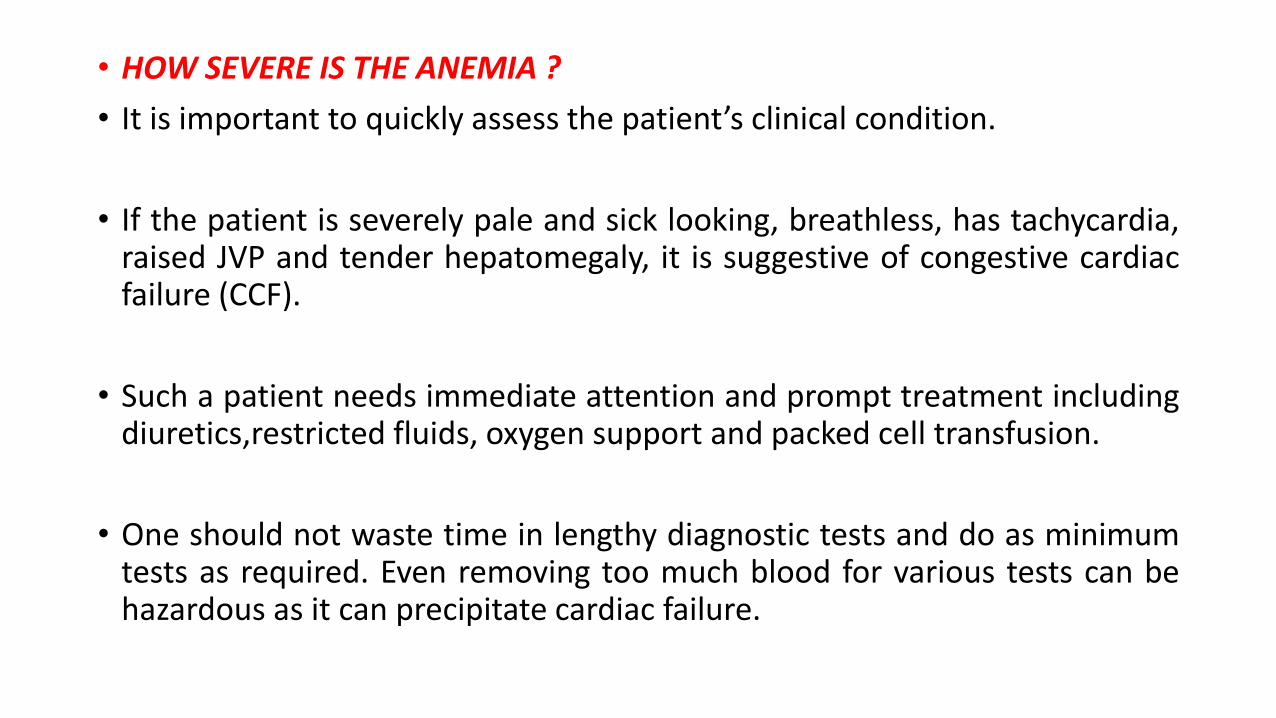
• HOW SEVERE IS THE ANEMIA ?
• It is important to quickly assess the patient’s clinical condition.
• If the patient is severely pale and sick looking, breathless, has tachycardia,raised JVP and tender hepatomegaly, it is suggestive of congestive cardiacfailure (CCF).
• Such a patient needs immediate attention and prompt treatment includingdiuretics,restricted fluids, oxygen support and packed cell transfusion.
• One should not waste time in lengthy diagnostic tests and do as minimumtests as required. Even removing too much blood for various tests can behazardous as it can precipitate cardiac failure.

• The clinical condition of the patient depends not only on the severity of anemia but also on the rate of drop of Hb.
• A child with 5 g% Hb, when it develops slowly like in iron deficiency, may be comfortable and come by walking whereas, if it deveopsacutely due to G6PD deficiency, the child may be brought in a collapsed state.

WHAT IS THE TYPE AND CAUSE OF ANEMIA
• Etiological classification, based on the disturbance of erythropoiesis.
• Morphological classification based on findings of red cell size andindices.
• Both these are not mutually exclusive and are often used together tocome to a conclusion as to the cause of anemia.

ETIOLOGICAL CLASSIFICATION OF ANEMIA
A. DECREASED EFFECTIVE PRODUCTION
• Nutritional deficiency-
e.g. deficiency of iron, folate, vitamin B12, protien, zinc, copper
• Bone marrow failure-
e.g. aplastic anemia, constitutional hypoplastic anemia, pure red
cell aplasia.
• Bone marrow infiltration-
e.g. malignancies like leukemia, lymphoma, osteopetrosis, myelofibrosis.

• Impaired erythropoietin production-
e.g. renal disease, prematurity, hypothyroidism, hypopituitarism,
chronic inflammation, protein malnutrition.
• Ineffective erythropoiesis-
e.g. Thalassemia, sideroblastic anemia,lead poisoning,
Primary dyserythropoietic anemia,
Erythropoietic protoporphyria, megaloblastic anemia.

B. INCREASED DESTRUCTION (HEMOLYTIC ANEMIA)
• Extracorpuscular causes
1. Mechanical- e.g. prosthetic valve, DIC, HUS
2. Immune- e.g. acquired immune hemolytic anemia, ABO or Rh
sensitization, mismatch transfusion.
3. Infection- e.g. malaria.
4. Sequestration- e.g. hypersplenism.
5. Complement induced, e.g. paroxysmal nocturnal hemoglobinuria

• Intracorpuscular causes (usually congenital)
1. Membrane defect—spherocytosis, stomatocytosis, elliptocytosis.
2. Enzyme defect—G6PD deficiency, PK deficiency.
3. Hemoglobin defect—Sickle cell anemia, thalassemia, HbC, HbD,
HbE disease.

• Blood loss (Hemorrhage): Acute or chronic, internal or external
1. Internal
Acute—Massive cephalhematoma, hemothorax
Chronic—Pulmonary hemosiderosis.
2. External
Acute—massive GI hemorrhage, trauma, hemoptysis
Chronic-peptic ulcer, rectal polyp, hookworm infestation

• Morphological classification of anemia
A. Microcytic, hypochromic anemia
MCV < 70 , MCH < 28 pg.
• Iron deficiency anemia.
• Anemia of chronic infection or inflammation.
• Thalassemia syndromes.
• Sideroblastic anemia.
• Lead poisoning.
• Severe protein deficiency.

B. Macrocytic anemia
• Megaloblastic anemia
1. Folate deficiency
2. Vitamin B12 deficiency
3. DNA metabolism defects like orotic aciduria

• Non-megaloblastic anemia
1. Normal newborn.
2. Reticulocytosis.
3. Aplastic anemia.
4. Liver disorders.
5. Hypothyroidism.
6. Alcoholism.
7. Down’s syndrome.

• Approach to Establishing Diagnosis
Approach to an anemic patient includes:
a. Detailed history
b. Thorough physical examination
c. Screening laboratory tests
d. Confirmatory laboratory tests.

A. AGE OF ONSET:Nutritional anemia is not seen at birth. The commonest causes of anemia in a newborn include hemolysis and hemorrhage.
Hemolysis-• ABO, Rh incompatibility.• G6PD deficiency or spherocytosis can present at birth.• Hemolysis is usually associated with icterus besides anemia.
Hemorrhage-• Huge cephalhematoma, pulmonary hemorrhage,• Intraventricular hemorrhage• Umbilical bleeding, fetoplacental or fetomaternal
hemorrhage, GI hemorrhage like in vitamin K deficiency, etc.


• At 4 months of age erythroblastopenia of infancy can occur.
• Rarely nutritional anemia can start very early, especially in preterms.
• Between 6 months to 2 years nutritional anemia and hemoglobinopathies can present as anemia.
• Fanconi’s anemia usually presents around 4 to 6 years of age.
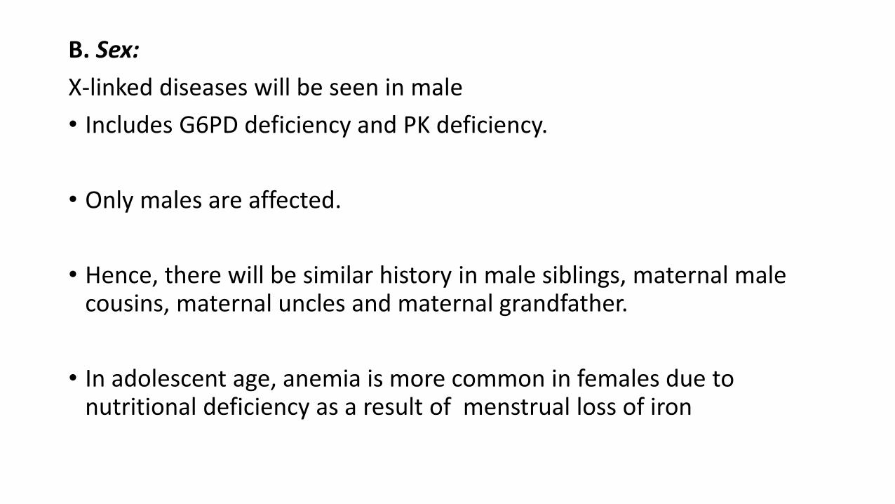
B. Sex:
X-linked diseases will be seen in male
• Includes G6PD deficiency and PK deficiency.
• Only males are affected.
• Hence, there will be similar history in male siblings, maternal male cousins, maternal uncles and maternal grandfather.
• In adolescent age, anemia is more common in females due to nutritional deficiency as a result of menstrual loss of iron

C. Community:
• G6PD deficiency is more commonly seen in Parsis, Bhanushalis and Sindhis.
• Beta-thalassemia is more common in Kutchis, Lohanas, Punjabis, Sindhis, Gujarati.
• Sickle cell disease is more common in tribals and hilly areas of Nagpur

D. Inheritance:
• All hemoglobinopathies and thalassemia syndromes are inherited in autosomal recessive manner.
• Spherocytosis is inherited as autosomal dominant condition.

E. Diet:
• Exclusive breastfeeding for 6 months, Introduction weaning food
thereafter, continuation of breast milk till 18 months, Avoidance of
animal milk in first year and balanced diet with occasional non-
vegetarian food consumption makes nutritional anemia unlikely
diagnosis.
• Iron deficiency develops where there is poor breastfeeding and
improper time and quality of weaning food, both of which are
exaggerated by bottle-feeding.

• Pica is both an effect and a cause of iron deficiency besides being seen in lead poisoning.
• Eating clay or mud (geophagia), ice (phagophagia), starch (amylophagia), paper, cloth, raw cereals, paint flakes, etc. is commonly seen in iron deficiency.
• Clay or mud can bind whatever little iron is present in food which further precipitates iron deficiency.
• Megaloblastic anemia due to folate deficiency is common in those villagers who consume a lot of goat milk.

• Drugs: Drugs can induce anemia by many ways.
• APLASTIC ANEMIA : chloramphenicol, sulpha drugs or analgesics. penicillin, alpha methyldopa or stibophen can lead to immune haemolyticanemia.
• In a patient with G6PD deficiency certain drugs like aspirin, sulpha drugs, primaquine etc can precipitate hemolysis.
• Iron deficiency anemia by chronic GI bleeding following NSAID abuse;
• Megaloblastic anemia as seen with sulpha drugs, phenytoin or folate antagonists.

• INFECTIONS AND INFESTATIONS:
• History suggestive of intrauterine infection should be elicited when
dealing with neonatal anemia especially when it is associated with
hepatosplenomegaly, IUGR, icterus and thrombocytopenia.
Hypoplastic anemia can be precipitated by hepatitis virus.
• G6PD deficiency induced hemolysis can be precipitated by many
infections and drugs used to treat such infections.
• Hemolysis could also be induced by malaria.

• Marrow suppression can occur following many viral infections, falciparum malaria, kala-azar, fulminant sepsis or drugs used in such cases.
• Nutritional anemia can be precipitated by worms due to malabsorption, nutrient deficiency and micro bleeding especially with hook worm infestations.
• Any acute infection can lead to drop in haemoglobin by 1-1.5 g% over next one week.

• Family history:
• History to be elicited in family members includes history of blood
transfusion, unexplained recurrent jaundice, gall stone removal,
splenectomy, suggests some hemolytic process.
• Similarly, history of anemia following drugs in other members will
suggest G6PD deficiency.

PHYSICAL EXAMINATION • A. Ascertain severity:
• Pulse, blood pressure and respiratory rate should be recorded.
• Look for puffiness, edema feet, sacral edema, jugulovenous
pulse, heptic tenderness, hepatojugular reflux and basal crepitations.
• All these will help to diagnose congestive cardiac failure as such
patients need urgent treatment.
• Hypertension may be seen in anemia due to renal diseases.
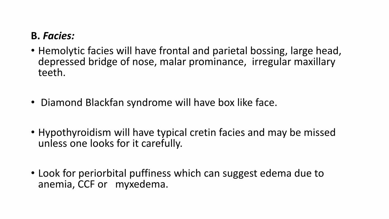
B. Facies:
• Hemolytic facies will have frontal and parietal bossing, large head, depressed bridge of nose, malar prominance, irregular maxillary teeth.
• Diamond Blackfan syndrome will have box like face.
• Hypothyroidism will have typical cretin facies and may be missed unless one looks for it carefully.
• Look for periorbital puffiness which can suggest edema due to anemia, CCF or myxedema.


• Eyes:• Fanconi’s anemia will have microcornea.
• Conjunctival vessels tortuosity is seen in sickle cell anemia and so is the presence of retinal hemorrhage or microaneurysms.
• Icterus in absence of high colored urine will suggest hemolytic anemiawith indirect hyperbilirubinemia.
• Osteopetrosis patients will develop blindness
• Oral cavity: Look for glossitis, angular stomatitis, bald tongue which will suggest nutritional anemia.Look for teeth abnormality for hemolytic anemia.

E. Nail changes:
• Platynychia, koilonychia, brittle nails are suggestive of iron deficiency.
Less common in children than in adults, but when present are
pathognomonic of IDA.
• Dyskeratotic nails will be seen in dyskeratosis congenita.
F. Lymphadenopathy:
Significant lymphadenopathy will suggest tuberculosis, HIV, infectious
mononucleosis,leukemia, lymphoma as the cause of anemia.


• Hepatosplenomegaly: Palpable tender liver with positive
hepatojugular reflux is suggestive of CCF.
• Significant hepatosplenomegaly will suggest tuberculosis, other viral
fever, HIV, leukemia, thalassemia, other hemoglobinopathies,
lymphoma, myelodysplastic syndrome, JCML, malaria, kala azar,
disorders as a cause of anemia.
• Isolated splenomegaly will go in favour of enteric fever, malaria, portal hypertension, lymphoma, CML, tropical splenomegaly or
hypersplenism,immune hemolytic anemia, congenital spherocytosis
as a cause of anemia.

• Bleeding manifestation:
• Presence of bleeding tendencies with petechiae, purpura will suggest thrombocytopenia, which can be seen in benign diseases like ITP.
or
In serious diseases like aplastic anemia, malignancies or marrow
infiltration.

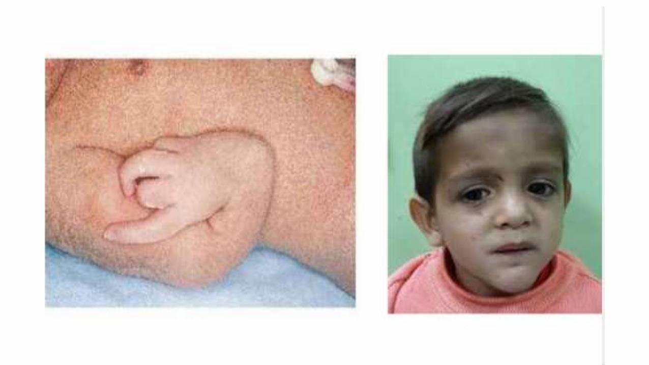
Skeletal changes:
• Patients with Fanconi’s anemia, TAR syndrome, etc. have skeletal
malformations like absent radius, absent or bifid thumb, triphalangeal
thumb, polydactyly, syndactyly, short stature,microcephaly.
• Look for associated anomalies like mental retardation, skin
hyperpigmentation,hypogonadism, renal anomalies in such cases.

Skin changes:
• Hyperpigmentation is seen in Fanconi’s anemia.
• Icterus is seen in liver diseases as well as hemolytic anemia.
• Iron deficiency can be seen in patients with carotenemia.
• Non-healing ulcers over lower limbs are seen in any chronic hemolyticanemia especially in HbS.
• Lastly localized DIC like picture with anemia and thrombocytopenia are present in patients with giant cavernous hemangioma as seen inKasabach-Merrit syndrome.

SKIN
Hyperpigmentation,
Petechia, purpura thrombocytopenia,
Jaundice
Cavernous hemangioma
Ulcers on lower extremities
Fanconi aplastic anemia.
Autoimmune hemolytic anemia with hemolytic-uremic syndromebone marrow aplasia, bone marrow
infiltration.
Hemolytic anemia, hepatitis, and aplastic anemia
Microangiopathic hemolytic anemia
S and C hemoglobinopathies,
SKULL Frontal bossing, prominence of themaxillary bones.
Congenital hemolytic anemias, thalassemia major, severe iron deficiency
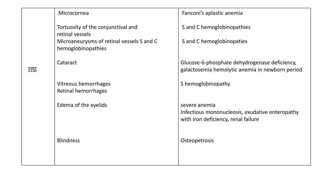
EYES
Microcornea
Tortuosity of the conjunctival andretinal vesselsMicroaneurysms of retinal vessels S and C hemoglobinopathies
Cataract
Vitreous hemorrhagesRetinal hemorrhages
Edema of the eyelids
Blindness
Fanconi’s aplastic anemia
S and C hemoglobinopathies
S and C hemoglobinopaties
Glucose-6-phosphate dehydrogenase deficiency, galactosemia hemolytic anemia in newborn period
S hemoglobinopathy
severe anemiaInfectious mononucleosis, exudative enteropathywith iron deficiency, renal failure
Osteopetrosis
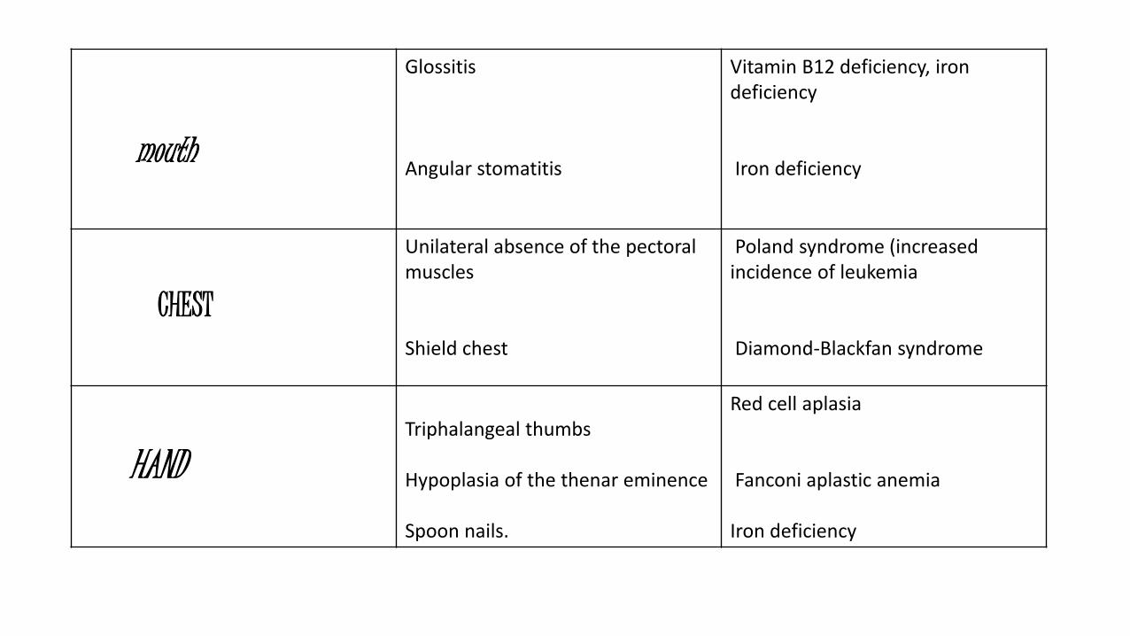
mouth
Glossitis
Angular stomatitis
Vitamin B12 deficiency, iron deficiency
Iron deficiency
CHEST
Unilateral absence of the pectoral muscles
Shield chest
Poland syndrome (increased incidence of leukemia
Diamond-Blackfan syndrome
HANDTriphalangeal thumbs
Hypoplasia of the thenar eminence
Spoon nails.
Red cell aplasia
Fanconi aplastic anemia
Iron deficiency
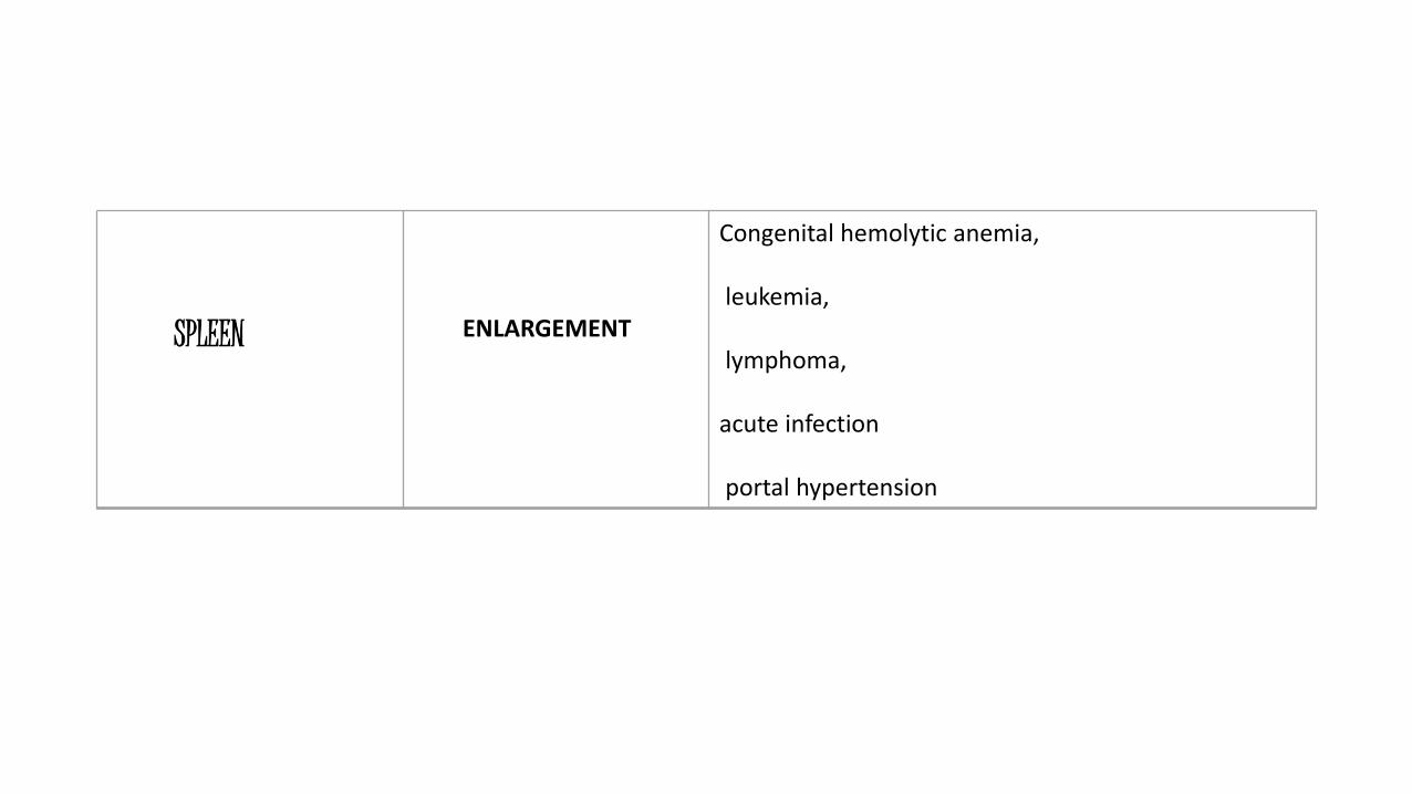
SPLEEN ENLARGEMENT
Congenital hemolytic anemia,
leukemia,
lymphoma,
acute infection
portal hypertension

• Laboratory studies often helpful in the investigation
• Usual initial studies
• Hemoglobin and hematocrit determination.
• Erythrocyte count and red cell indices, including MCV and RDW.
• Reticulocyte count.
• Study of stained blood smear.
• Leukocyte count and differential count.
• Platelet count.

Suspected iron deficiency-
• Free erythrocyte protoporphyrin.
• Serum ferritin levels.
• Stool for occult blood.
• 99mTc pertechnetate scan for Meckel’s diverticulum.
• Endoscopy (upper and lower bowel).

• Suspected vitamin B12 or folic acid deficiency
• Bone marrow.
• Serum vitamin B12 level <100pg / ml.
• Serum folate level < 3pg/ml.
• FIGLU TEST.
• Vitamin B12 absorption test (radioactive cobalt)
(Schilling test).

• Suspected hemolytic anemia-
• Evidence of red cell breakdown-
a. Blood smear.
b. Serum bilirubin level.
c. Urinary urobilinogen excretion.
d. Serum haptoglobin.
• Evidence of red cell regeneration-
a. Reticulocyte count.
b. Blood smear.
c. Skeleton radiography.

• Evidence of type of hemolytic anemia:
Corpuscular
A. Membrane-
— Blood smear.
— Osmotic fragility test
— Autohemolysis test
B. Enzymes
— Heinz-body preparation
— Enzyme assay

• C. Hemoglobin-
— Sickle test.
— Hemoglobin electrophoresis.
— Hemoglobin F determination.
— Kleihauer-Betke smear.

• Evidence of type of hemolytic anemia:
Extracorpuscular
Immune-
— Antiglobulin test
— Acid serum lysis test
— Sucrose lysis test
— Donath-Landsteiner antibody
— ANA

Approach to a child with anemia(scenario approach)
Anemia (Hb less than normal level)
- No lymph nodes
- No hepatosplenomegaly
- No petechiae or ecchymosis
----- Nutritional iron deficiency or megaloblastic
----- Pure red cell aplasia
----- Thalassemia trait
----- Lead poisoning
----- Renal disease

Anemia (Hb less than normal level)
- No lymph nodes
- No hepatosplenomegaly
- With petechiae and ecchymosis
----- Aplastic anemia
----- Bleeding disorder
----- Coagulation disorder
----- ITP
----- DIC

Anemia (Hb less than normal level)
With hepatosplenomegaly----- Thalassemia
----- Liver disorder
Infections

Anemia (Hb less than normal level)
With petechiae, lymphadenopathy and hepatosplenomegaly--- Leukamia
--- Infections
--- DIC

THANK YOU