Applied Surface Science, 325 (2015) 91-99
9
Indium tin oxide with zwitterionic interfacial design for biosensing applications in complex matrices Nadia T. Darwish, Yatimah Alias, Sook Mei Khor ∗ Department of Chemistry, Faculty of Science University of Malaya, 50603 Kuala Lumpur, Malaysia a r t i c l e i n f o Article history: Received 5 September 2014 Received in revised form 15 October 2014 Accepted 28 October 2014 Available online 5 November 2014 Keywords: Biosensor Indium tin oxide Anti-biofouling coating Zwitterionic molecules a b s t r a c t Biosensing interfaces consisting of linker molecules (COOH or NH 2 ) and charged, antifouling moieties (( SO 3− and N + (Me) 3 ) for biosensing applications were prepared for the first time by the in situ depo- sition of mixtures of aryl diazonium cations on indium tin oxide (ITO) electrodes. A linker molecule is required for the attachment of biorecognition molecules (e.g., antibodies, enzymes, DNA chains, and aptamers) close to the transducer surface. The attached molecules improve the biosensing sensitivity and also provide a short response time for analyte detection. Thus, the incorporation of a linker and antifoul- ing molecules is an important interfacial design for both affinity and enzymatic biosensors. The reductive adsorption behavior and electrochemical measurement were studied for (1) an individual compound and (2) a mixture of antifouling zwitterionic molecules together with linker molecules [combination 1: 4- sulfophenyl (SP), 4-trimethylammoniophenyl (TMAP), and 1,4-phenylenediamine (PPD); combination 2: 4-sulfophenyl (SP), 4-trimethylammoniophenyl (TMAP), and 4-aminobenzoic acid (PABA)] of aryl diazo- nium cations grafted onto an ITO electrode. The mixture ratios of SP:TMAP:PPD and SP:TMAP:PABA that provided the greatest resistance to non-specific protein adsorptions of bovine serum albumin labeled with fluorescein isothiocyanate (BSA–FITC) and cytochrome c labeled with rhodamine B isothiocyanate (RBITC–Cyt c) were determined by confocal laser scanning microscopy (CLSM). For the surface antifoul- ing study, we used 2-[2-(2-methoxyethoxy) ethoxy]acetic acid (OEG) as a standard control because of its prominent antifouling properties. Surface compositions of combinations 1 and 2 were characterized using X-ray photoelectron spectroscopy (XPS). Field-emission scanning electron microscopy (FE-SEM) was used to characterize the morphology of the grafted films to confirm the even distribution between linker and antifouling molecules grafted onto the ITO surfaces. Combination 1 (SP:TMAP:PPD) with a ratio of 0.5:1.5:0.37 exhibited the best antifouling capability with respect to resisting the nonspecific adsorption of proteins. 1. Introduction Biofouling is the accretion of proteins, cells and other biological materials onto a bare electrode surface. For fast clinical screening, immunosensors are designed to detect disease analytes such as nucleic acids, proteins or other disease biomarkers at very low concentrations from complicated sample media [1–5]. Such an accumulation of biofouling substances leads to reduced analyte diffusion and perfusion to the sensor interface, which eventually causes a decrease in sensor response and sensitivity. Polymers such as polyurethane, polyvinyl chloride, poly(dimethylsiloxane) and poly(methacrylate) have been used as antifouling agents for biosensing surfaces, where they provide selective transport ∗ Corresponding author. Tel.: +60 3 79677022x2520; fax: +60 3 9674193. E-mail address: [email protected] (S.M. Khor). of a targeted analyte to the biosensing interface while preven- ting biofouling components from reaching the electrode surface [6–10]. Although these polymers are exceptionally suitable in clinical monitoring, their functional stability is greatly affected by the biocompatibility of the biosensor materials in contact with the biological sample [11,12]. Poly(ethylene glycol) (PEG) and self-assembled monolayers with oligo(ethylene glycol) chains (OEG-SAMs) have been used to reduce the non-specific adsorption of non-desired proteins [13]. The activity of anti-biofouling coatings in complex samples, e.g., human sera, depends on several factors, including the concentra- tions and chain lengths of PEG and OEG and the temperature. At 37 ◦ C, the grafted surface can adsorb additional non-specific pro- teins. Moreover, surfaces grafted with PEG and OEG are prone to auto-oxidation in most biochemically relevant solutions for in vivo studies [6,14,15]. Zwitterionic polymers such as phospho- rylcholine, sulfobetaine, and carboxy betaine, which contain both Applied Surface Science 325 (2015) 91–99 Post-print version http://dx.doi.org/10.1016/j.apsusc.2014.10.167
Transcript of Applied Surface Science, 325 (2015) 91-99
Ia
ND
a
ARRAA
KBIAZ
1
mincadcsaf
h
ndium tin oxide with zwitterionic interfacial design for biosensingpplications in complex matrices
adia T. Darwish, Yatimah Alias, Sook Mei Khor ∗
epartment of Chemistry, Faculty of Science University of Malaya, 50603 Kuala Lumpur, Malaysia
r t i c l e i n f o
rticle history:eceived 5 September 2014eceived in revised form 15 October 2014ccepted 28 October 2014vailable online 5 November 2014
eywords:iosensor
ndium tin oxidenti-biofouling coatingwitterionic molecules
a b s t r a c t
Biosensing interfaces consisting of linker molecules (COOH or NH2) and charged, antifouling moieties(( SO3− and N+(Me)3) for biosensing applications were prepared for the first time by the in situ depo-sition of mixtures of aryl diazonium cations on indium tin oxide (ITO) electrodes. A linker moleculeis required for the attachment of biorecognition molecules (e.g., antibodies, enzymes, DNA chains, andaptamers) close to the transducer surface. The attached molecules improve the biosensing sensitivity andalso provide a short response time for analyte detection. Thus, the incorporation of a linker and antifoul-ing molecules is an important interfacial design for both affinity and enzymatic biosensors. The reductiveadsorption behavior and electrochemical measurement were studied for (1) an individual compound and(2) a mixture of antifouling zwitterionic molecules together with linker molecules [combination 1: 4-sulfophenyl (SP), 4-trimethylammoniophenyl (TMAP), and 1,4-phenylenediamine (PPD); combination 2:4-sulfophenyl (SP), 4-trimethylammoniophenyl (TMAP), and 4-aminobenzoic acid (PABA)] of aryl diazo-nium cations grafted onto an ITO electrode. The mixture ratios of SP:TMAP:PPD and SP:TMAP:PABA thatprovided the greatest resistance to non-specific protein adsorptions of bovine serum albumin labeledwith fluorescein isothiocyanate (BSA–FITC) and cytochrome c labeled with rhodamine B isothiocyanate(RBITC–Cyt c) were determined by confocal laser scanning microscopy (CLSM). For the surface antifoul-ing study, we used 2-[2-(2-methoxyethoxy) ethoxy]acetic acid (OEG) as a standard control because ofits prominent antifouling properties. Surface compositions of combinations 1 and 2 were characterized
Applied Surface Science 325 (2015) 91–99
Post-print version
using X-ray photoelectron spectroscopy (XPS). Field-emission scanning electron microscopy (FE-SEM)was used to characterize the morphology of the grafted films to confirm the even distribution betweenlinker and antifouling molecules grafted onto the ITO surfaces. Combination 1 (SP:TMAP:PPD) with aratio of 0.5:1.5:0.37 exhibited the best antifouling capability with respect to resisting the nonspecificadsorption of proteins.
. Introduction
Biofouling is the accretion of proteins, cells and other biologicalaterials onto a bare electrode surface. For fast clinical screening,
mmunosensors are designed to detect disease analytes such asucleic acids, proteins or other disease biomarkers at very lowoncentrations from complicated sample media [1–5]. Such anccumulation of biofouling substances leads to reduced analyteiffusion and perfusion to the sensor interface, which eventuallyauses a decrease in sensor response and sensitivity. Polymers
uch as polyurethane, polyvinyl chloride, poly(dimethylsiloxane)nd poly(methacrylate) have been used as antifouling agentsor biosensing surfaces, where they provide selective transport∗ Corresponding author. Tel.: +60 3 79677022x2520; fax: +60 3 9674193.E-mail address: [email protected] (S.M. Khor).
ttp://dx.doi.org/10.1016/j.apsusc.2014.10.167
of a targeted analyte to the biosensing interface while preven-ting biofouling components from reaching the electrode surface[6–10]. Although these polymers are exceptionally suitable inclinical monitoring, their functional stability is greatly affectedby the biocompatibility of the biosensor materials in contactwith the biological sample [11,12]. Poly(ethylene glycol) (PEG)and self-assembled monolayers with oligo(ethylene glycol) chains(OEG-SAMs) have been used to reduce the non-specific adsorptionof non-desired proteins [13].
The activity of anti-biofouling coatings in complex samples, e.g.,human sera, depends on several factors, including the concentra-tions and chain lengths of PEG and OEG and the temperature. At37 ◦C, the grafted surface can adsorb additional non-specific pro-
teins. Moreover, surfaces grafted with PEG and OEG are proneto auto-oxidation in most biochemically relevant solutions forin vivo studies [6,14,15]. Zwitterionic polymers such as phospho-rylcholine, sulfobetaine, and carboxy betaine, which contain both
arpascigobtOtpsc
ipVoaiBosMI(shoe1ias
mipapeIataatmt(tr(nfstXootcu
nionic and cationic groups, have been used as non-fouling mate-ials. Unfortunately, grafting of the long chains of zwitterionicolymers can result in the formation of high-impedance layersnd subsequently cause losses to electrode sensitivities. However,hort-chain molecules exhibit poor stability [15–17]. Gooding ando-workers investigated zwitterionic phenyl derivative layers bear-ng charged moieties, such as SO3
− and N+(Me)3, deposited ontolassy carbon surfaces as an alternative to OEG phenyl layers andbserved that the grafted zwitterionic phenyl derivative layers offeretter chemical stability and a shorter electron transfer pathwayo the transducer surface [15]. Zwitterionic phenyl layers, such asEG alkanethiol SAMs, have been proposed as effective alternatives
o conventional antifouling molecules because of their antifoulingroperties. Such coatings do not completely passivate the electrodeurface and therefore allow Faradaic electrochemistry. Hence, theyan be utilized in biosensing applications.
Indium tin oxide (ITO) is a biosensing substrate of increasingmportance in the field of biomaterials because of its combinedroperties of transparency, conductivity and biocompatibility [18].ery few studies have examined the use of antifouling moleculesn ITO for bioassay contexts. To date, only hydrophilic polyacryliccid brushes [19] and zwitterionic polymer brushes [20] have beennvestigated as antifouling agents. However, in these studies, onlySA–FITC was used to evaluate the antifouling capability. The use ofnly a single protein may not be representative of complex media,uch as blood plasma and serum that contain a variety of proteins.oreover, a different study grafted zwitterionic polymers onto the
TO using surface-initiated atom transfer radical polymerizationSI-ATRP). Although the obtained organic coatings were consideredtable in aqueous solution, the technique was time consuming andad moderate throughput [21]. The literature contains no accountsf research related to the capability of these antifouling moi-ties after they have been modified with linker molecules, such as,4-phenylenediamine or 4-aminobenzoic acid. A linker molecule
s required for the attachment of biorecognition molecules (e.g.,ntibodies, enzymes, DNA, or aptamers) close to the transducerurface.
The incorporation of zwitterionic phenyl layers with a linkerolecule is useful for improving the selectivity of the biosens-
ng interface, which is resistant to the nonspecific adsorption ofroteins, and for providing a shorter response time for specificnalyte detection (e.g., low impedance due to a shorter electronathway). Therefore, one objective of this study was to analyze thelectrochemical deposition behavior of OEG when deposited ontoTO electrodes compared with the behaviors of a single moleculend two mixed layer coatings comprising 4-sulfophenyl (SP), 4-rimethylammoniophenyl (TMAP), 1,4-phenylenediamine (PPD),nd 4-aminobenzoic acid (BAPA): mixed layers 1 (SP:TMAP:PPD)nd mixed layers 2 (SP:TMAP:BAPA). The second objective ofhis study was to investigate the capability of an ITO surface
odified with the aforementioned molecules to repel both a nega-ively charged protein (BSA–FITC) and a positively charged proteinRBITC–Cyt c). These example proteins were used to represent pro-eins contained in real samples. ITO was modified using differentatios of antifouling molecules (SP or TMAP) to linker moleculesPPD or BAPA). We examined the antifouling properties toward theonspecific absorption of fluorescently labeled proteins using con-
ocal laser scanning microscopy (CLSM). The third objective of thistudy was to characterize the modified surface with the lowest pro-ein adsorption level to both BSA–FITC and RBITC–Cyt c by using-ray photoelectron spectroscopy (XPS) to confirm the existencef SP, TMAP, PPD or PABA moieties on the grafted ITO. The final
bjective of this study was to examine the surface morphology ofhe grafted films, where the surface distribution of SP:TMAP:PPDould be indicated by NH-gold nanoparticles and characterizedsing field-emission scanning electron microscopy (FE-SEM).2. Experimental methods
2.1. Reagents and materials
ITO coated glass slide (surface resistivity ≤7 �/sq, L × W ×thickness 406 mm × 355 mm × 0.7 mm, sheet) was purchasedfrom (Sanyo, Japan). 4-sulfophenyl, 1,4-phenylenediamine, 4-aminobenzoic acid, sodium nitrite (NaNO2), potassium chloride(KCl), potassium dihydrogen phosphate (KH2PO4), potassiumphosphate dibasic (K2HPO4), hydrochloric acid (HCl), potas-sium ferricyanide (K3Fe(CN)6), hexamine ruthenium(III) chloride(Ru(NH3)6Cl3), sodium carbonate (Na2CO3) with 99% purity,20 nm gold nanoparticles, sodium bicarbonate (NaHCO3), rho-damine B isothiocyanate, bovine serum albumin-fluoresceinisothiocyanate, cytochrome c (from bovine heart) and 2-[2-(2-methoxyethoxy)ethoxy]acetic acid (OEG) were purchased fromSigma-Aldrich (USA). 4-Trimethylammoniophenyl (TMAP) wasobtained from Acros (USA). All solutions were prepared using Milli-Q water (18 M�/ cm) (Purelab, US).
2.2. ITO electrode surface modification
ITO substrates were soaked and cleaned in an ultrasonicatorusing dichloromethane and then 99% methanol for 10 min each,followed by treatment with 0.5 M K2CO3 in a 3:1 methanol:Milli-Qwater mixture for 30 min under sonication to remove any residualorganic contaminants. The ITO substrates were then rinsed withcopious amounts of Milli-Q water, dried and stored in an nitrogen-filled container [22]. In the first experiment, the ITO electrodeswere modified with only a single compound: SP, PPD, BAPA, orTMAP. In the second experiment, the ITO electrodes were modi-fied with a combination of SP:TMAP:PPD at different molar ratios:1:1:0.37 (mix 1), 1.5:0.5:0.37 (mix 2) and 0.5:1.5:0.37 (mix 3). In thethird experiment, the ITO electrodes were modified with a combi-nation of SP:TMAP:PABA at different molar ratios: 0.8:1:0.2 (mix 1),1:1:0.1 (mix 2) and 0.5:1:0.2 (mix 3). For the control experiment,OEG was used as the antifouling molecule on the ITO slides. TheOEG was prepared by the electrodeposition of a mixed layer of 4-aminobenzoic acid (8 mM) and 1,4-phenyldiamine (2 mM) on ITOslides. The electrode interface was fabricated using carbodiimidechemistry: we linked the OEG to the COOH moieties of PPD aminegroups by incubating the modified surface in an ethanol solutioncontaining 10 mM OEG and 40 mM DCC for 6 h at room temper-ature. In all of the aforementioned experiments, in situ methodswere used in which aryl diazonium cations were electrochemicallyreduced with two times the molar amount of NaNO2 in 0.05 M HCl.The solution was purged with nitrogen gas for 20 min before thesurface modification. The electrodeposition was performed usingcyclic voltammetry at −0.6 V to 0.2 V; the electrodes were scannedfor 5 cycles at 100 mV s−1 [23–25].
2.3. Surface passivation study via electrochemical measurements
All voltammetry measurements were performed using anAutoLabIII potentiostat (Metrohm AutoLab, Netherlands) and aconventional three-electrode system comprising an ITO workingelectrode, a platinum wire as the auxiliary electrode, and Ag/AgCl(3.0 M NaCl) as the reference electrode. All potentials were reportedrelative to the Ag/AgCl reference electrode at room temperature.The surface blocking studies on the modified sensing interface wereperformed in two aqueous solutions containing different redoxprobes. [Fe(CN)6]4−/3−, [Ru(NH3)6]3+ and CV were used to probe the
integrity of the functionalized layers on the electrode surfaces. Therespective redox-active solution (1 mM) was composed of 0.05 MKCl, 0.05 M K2HPO4, and 0.05 M KH2PO4 and was adjusted to pH 7.0.ITOs modified with SP:TMAP:PPD (mix 3) or SP:TMAP:PABA (mix
3u
2
aobDotftdstcctctTb
iecswtstwflflgsmMesucwtt
2m
afletwgHbiIawR
) were characterized using XPS. The XPS spectra were analyzedsing the Casa XPS software (version 2.3.15).
.4. Conjugation of cytochrome c to rhodamine B isothiocyanate
The fluorescent dye rhodamine B was conjugated with Cyt cccording to the method reported by Gui et al. [26]. Ten milligramsf Cyt c was dissolved in 1 mL of 0.1 M sodium carbonate-icarbonate buffer (pH 9.05), and 0.25 mL of 10 mg/mL RBITC inMSO was subsequently added to obtain a fivefold molar excessf Cyt c relative to RBITC. The tube containing the mixture solu-ion was wrapped with aluminum foil and incubated in the darkor 1 h under continuous magnetic stirring. The mixture was fil-ered through a 0.22 �m pore size syringe filter (non-sterile, 13 mmiameter; Merck Millipore, Germany), and the clear conjugatedolution was subsequently transferred to an Amicon Ultra-15 cen-rifugal filter device (3 kDa MW cut-off, EMD Millipore, USA). Theapped filter device was placed in the swinging bucket rotor of aentrifuge and spun for 10 min at 4000 rpm. The separated solu-ion in the upper reservoir of the filter device contained RBITC–Cyt, which was then characterized by UV–vis and fluorescence spec-roscopy. The RBITC–Cyt c solution was stored at −20 ◦C until use.he functionalized ITO slides were stored in clean Petri dishesefore the antifouling study.
For the antifouling study, the modified ITO electrode wasmmersed in an adequate amount of either 1 mg/mL BSA–FITC (anxample of a negatively charged protein) or 2 mg/mL RBITC–Cyt
(an example of a positively charged protein); both immersionolutions were prepared using PBS buffer, pH 7.4. The Petri dishesere kept in darkness for 1 h, and the slides were subsequently
ransferred to another Petri dish that contained only PBS. The ITOlides were soaked for 7 min to remove weakly adsorbed pro-eins from the surfaces of the slides. Afterward, the slides wereashed with Milli-Q water to remove any remaining residues. Theuorescent-labeled-protein-adsorbed surfaces were dried underowing nitrogen. The samples were mounted with 50% aqueouslycerol solution and covered with 22 × 60 mm2 microscope coverlides (Superior Marienfeld, Germany). The dried samples wereounted face-down onto a cover glass (22 × 60 mm2, No. 1, Paularienfeld, GmbH & Co. KG, Germany) with 50% aqueous glyc-
rol solution as the mounting agent. For each type of modifiedurface, three replicates were tested. Five images were capturednder 10× magnification for survey studies. Fifteen images wereaptured under 63× magnification, and their mean gray valuesere calculated to determine the fluorescence of the adsorbed pro-
eins. The mean gray values were compared with different surfaceypes.
.5. Surface antifouling study via confocal laser fluorescenticroscope imaging
BSA–FITC and RBITC–Cyt c proteins adsorbed onto function-lized electrode surfaces were measured using a confocal laseruorescence microscope (Leica Microsystems Pty, Ltd., USA)quipped with an argon laser (at 29% power) and operated inhe xyz scan mode (step size 0.04 �m). Images (1024 × 1024 px2)ere captured at 8-bit resolution using the Las AF software (Ori-
in). The imaging was carried out using two objective lenses: anCX PL APO CS 10.0 × 0.40 Dry UV and an HCX PL APO lambdalue 63.0 × 1.40 OIL UV. The gray value represents the fluorescence
ntensity. Captured TIF-format images were further analyzed usingmageJ (IJ-1.48g. jar) software to determine the mean gray valuefter background subtraction. For reference, the emission band-idth of BSA–FITC ranges from 506 nm to 575 nm. In the case ofBITC–Cyt c, the bandwidth ranges from 524 nm to 594 nm.
2.6. ITO surface characterization via XPS analysis
Spectra were obtained using XPS (AXIS Ultra DLD spectrome-ter with a hemispherical analyzer, a multichannel detector and amonochromatic Al-K� source; 1486.6 eV). Spectra were accumu-lated at a take-off angle of 90◦ with a 0.9 mm2 spot size at a pressureof less than 1 × 10⁻⁸ mbar. Survey scans (0–1000 eV) were carriedout at a 1.0 eV step size, 100 ms dwell time, and 100 eV analyzerpass energy. High-resolution scans (S2p, C1s, N1s) were performedusing a 0.1 eV step size, 100 ms dwell time, and 20 eV pass energy.Binding energies of elements were corrected with reference to theC1s peak of graphitic carbon (284.4 eV).
2.7. ITO surface characterization via FE-SEM analysis
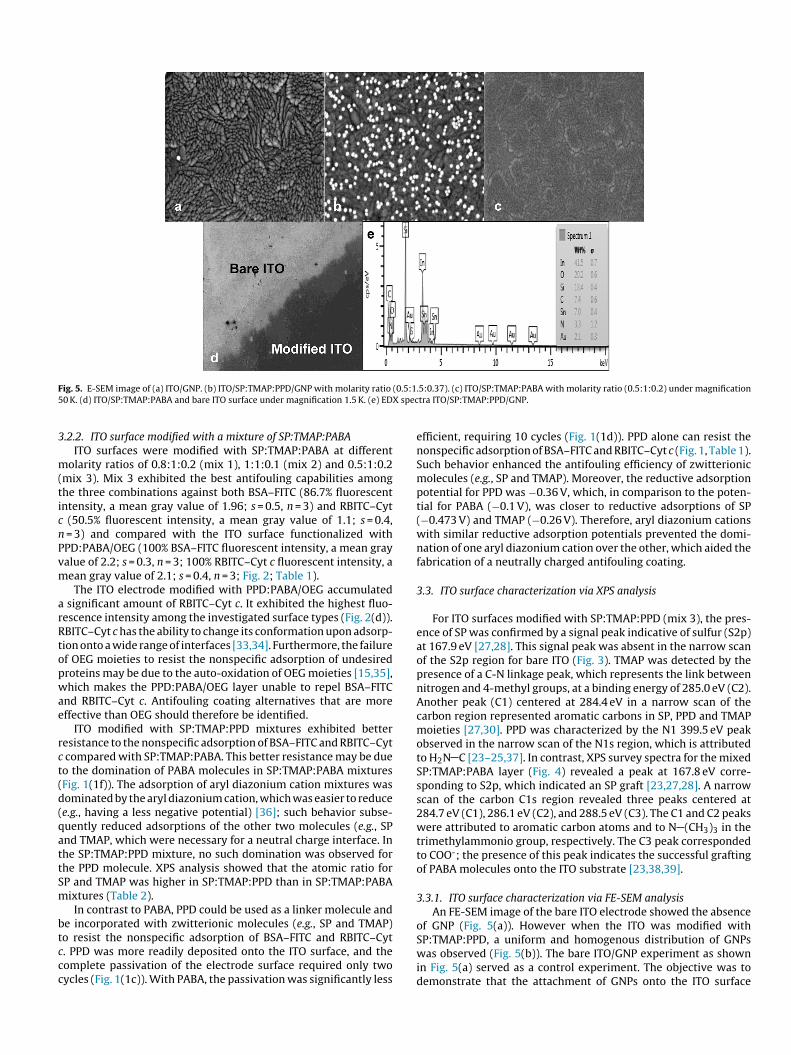
Four different types of surfaces were morphologically charac-terized using a Hitachi SU8000 field-emission scanning electronmicroscope (Tokyo, Japan): (1) bare ITO, (2) ITO/gold nanoparticles(GNPs), (3) ITO/SP:TMAP:PPD/GNPs, and (4) ITO/SP:TMAP:PABA.The slides were tested at slow scan speeds less than 2 kV and at50,000× magnification. The work destination was 2.1 mm at anaccelerating voltage of 2 kV. GNPs were used to indicate the pres-ence of PPD terminal amine groups. GNPs were immobilized byimmersing ITO/SP:TMAP:PPD and bare ITO in a GNP solution for3 h at room temperature [24]. Energy-dispersive X-ray (EDX) wasused to characterize the grafted GNPs.
3. Results and discussion
3.1. Reductive adsorption and electrochemical measurement ofindividual aryl diazonium cations and their mixtures
The reductive adsorption peaks of SP (10 mM) and PABA(10 mM) appeared at −0.473 V and −0.10 V, respectively. A cyclicvoltammogram of SP diazonium cation solution exhibited anirreversible cathodic peak located at −0.47 V (Fig. 1(1a)), similarto the CV results previously reported for glassy carbon surfaces[27,28]. A typical cyclic voltammogram of PABA diazonium cationsolution showed a single irreversible cathodic peak at −0.10 V(Fig. 1(1d)). The cyclic voltammograms for bare ITO in 1 mM[Fe(CN)6]4−/3− showed a pair of well-defined redox peaks. Elec-trode surfaces modified with aryl diazonium cations, SP and PABAshowed good passivation toward the penetration of negativelycharged redox-active species when the modified surface was testedin [Fe(CN)6]4−/3−. However, these surfaces showed weak pas-sivation properties toward the penetration of positively chargedredox active species when the surfaces were tested in 1 mM[Ru(NH3)]6
3+. This weak passivation was likely a consequence ofthe negatively charged layer that formed on the ITO electrode sur-face, which repelled [Fe(CN)6]4−/3− but allowed the penetration of[Ru(NH3)]6
3+ from the solution to the ITO electrode surface. Anopposite effect was observed when the ITO was modified withTMAP (Fig. 1(1b)). This positively charged aryl diazonium cationwith a reductive adsorption peak at −0.26 V allowed [Fe(CN)6]4−/3−
to penetrate through the layer but prevented [Ru(NH3)6]3+ fromdiffusing closer to the ITO electrode surface. The modification ofthe ITO surface with TMAP aryl diazonium cation required a highconcentration (10 mM) and up to 10 CV scan cycles to passivate theITO electrode surface. The difficulty in forming a close-packed layerat a low concentration (1 mM) may be a consequence of the sterichindrance caused by three methyl groups of TMAP, which hinders
the possibility of a densely packed structure between the TMAPmolecules [29]. The electrodeposition of PPD(5 mM; Fig. 1(1c)) indi-cated a densely packed surface coverage even after two cycles, andthe reductive adsorption peak was observed at –0.36 V. An efficient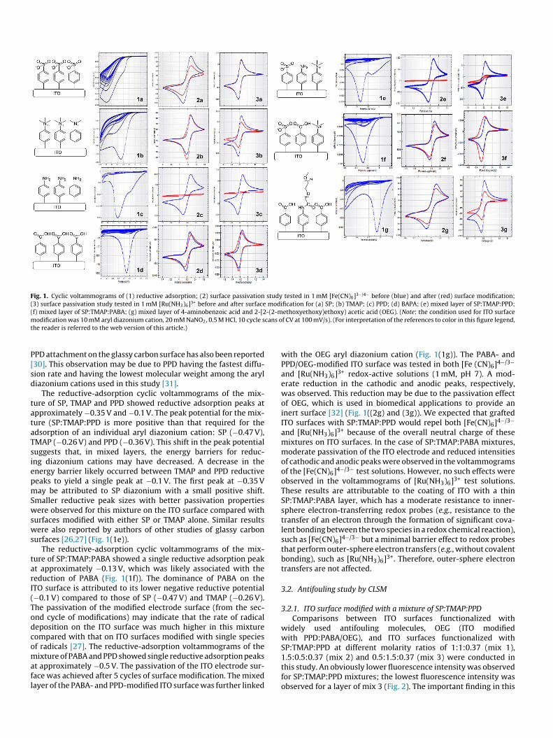
Fig. 1. Cyclic voltammograms of (1) reductive adsorption; (2) surface passivation study tested in 1 mM [Fe(CN)6]3−/4− before (blue) and after (red) surface modification;(3) surface passivation study tested in 1 mM [Ru(NH3)6]3+ before and after surface modification for (a) SP; (b) TMAP; (c) PPD; (d) BAPA; (e) mixed layer of SP:TMAP:PPD;(f) mixed layer of SP:TMAP:PABA; (g) mixed layer of 4-aminobenzoic acid and 2-[2-(2-methoxyethoxy)ethoxy) acetic acid (OEG). (Note: the condition used for ITO surfacem cans ot
P[sd
tataTsiepmSwsws
tarI(Todcomafl
odification was 10 mM aryl diazonium cation, 20 mM NaNO2, 0.5 M HCl, 10 cycle she reader is referred to the web version of this article.)
PD attachment on the glassy carbon surface has also been reported30]. This observation may be due to PPD having the fastest diffu-ion rate and having the lowest molecular weight among the aryliazonium cations used in this study [31].
The reductive-adsorption cyclic voltammograms of the mix-ure of SP, TMAP and PPD showed reductive adsorption peaks atpproximately −0.35 V and −0.1 V. The peak potential for the mix-ure (SP:TMAP:PPD is more positive than that required for thedsorption of an individual aryl diazonium cation: SP (−0.47 V),MAP (−0.26 V) and PPD (−0.36 V). This shift in the peak potentialuggests that, in mixed layers, the energy barriers for reduc-ng diazonium cations may have decreased. A decrease in thenergy barrier likely occurred between TMAP and PPD reductiveeaks to yield a single peak at −0.1 V. The first peak at −0.35 Vay be attributed to SP diazonium with a small positive shift.
maller reductive peak sizes with better passivation propertiesere observed for this mixture on the ITO surface compared with
urfaces modified with either SP or TMAP alone. Similar resultsere also reported by authors of other studies of glassy carbon
urfaces [26,27] (Fig. 1(1e)).The reductive-adsorption cyclic voltammograms of the mix-
ure of SP:TMAP:PABA showed a single reductive adsorption peakt approximately −0.13 V, which was likely associated with theeduction of PABA (Fig. 1(1f)). The dominance of PABA on theTO surface is attributed to its lower negative reductive potential−0.1 V) compared to those of SP (−0.47 V) and TMAP (−0.26 V).he passivation of the modified electrode surface (from the sec-nd cycle of modifications) may indicate that the rate of radicaleposition on the ITO surface was much higher in this mixtureompared with that on ITO surfaces modified with single speciesf radicals [27]. The reductive-adsorption voltammograms of the
ixture of PABA and PPD showed single reductive adsorption peakst approximately −0.5 V. The passivation of the ITO electrode sur-ace was achieved after 5 cycles of surface modification. The mixedayer of the PABA- and PPD-modified ITO surface was further linked
f CV at 100 mV/s). (For interpretation of the references to color in this figure legend,
with the OEG aryl diazonium cation (Fig. 1(1g)). The PABA- andPPD/OEG-modified ITO surface was tested in both [Fe (CN)6]4−/3−
and [Ru(NH3)6]3+ redox-active solutions (1 mM, pH 7). A mod-erate reduction in the cathodic and anodic peaks, respectively,was observed. This reduction may be due to the passivation effectof OEG, which is used in biomedical applications to provide aninert surface [32] (Fig. 1((2g) and (3g)). We expected that graftedITO surfaces with SP:TMAP:PPD would repel both [Fe(CN)6]4−/3−
and [Ru(NH3)6]3+ because of the overall neutral charge of thesemixtures on ITO surfaces. In the case of SP:TMAP:PABA mixtures,moderate passivation of the ITO electrode and reduced intensitiesof cathodic and anodic peaks were observed in the voltammogramsof the [Fe(CN)6]4−/3− test solutions. However, no such effects wereobserved in the voltammograms of [Ru(NH3)6]3+ test solutions.These results are attributable to the coating of ITO with a thinSP:TMAP:PABA layer, which has a moderate resistance to inner-sphere electron-transferring redox probes (e.g., resistance to thetransfer of an electron through the formation of significant cova-lent bonding between the two species in a redox chemical reaction),such as [Fe(CN)6]4−/3− but a minimal barrier effect to redox probesthat perform outer-sphere electron transfers (e.g., without covalentbonding), such as [Ru(NH3)6]3+. Therefore, outer-sphere electrontransfers are not affected.
3.2. Antifouling study by CLSM
3.2.1. ITO surface modified with a mixture of SP:TMAP:PPDComparisons between ITO surfaces functionalized with
widely used antifouling molecules, OEG (ITO modifiedwith PPD:PABA/OEG), and ITO surfaces functionalized withSP:TMAP:PPD at different molarity ratios of 1:1:0.37 (mix 1),
1.5:0.5:0.37 (mix 2) and 0.5:1.5:0.37 (mix 3) were conducted inthis study. An obviously lower fluorescence intensity was observedfor SP:TMAP:PPD mixtures; the lowest fluorescence intensity wasobserved for a layer of mix 3 (Fig. 2). The important finding in this
F from (a) FITC–BSA and (b) RBITC–Cyt c adsorbed on different surfaces. (c) Comparison ofi RBITC–Cyt c adsorbed on different surfaces.
scbott
owHactw(attRscw(
BmI
Table 1The percentage of BSA–FITC and RBITC–Cyt c adsorption at different surfaces repre-sented by the normalized fluorescence intensities relative to ITO electrode modifiedwith PPD:PABA/OEG.
Surface type The percentage of thefluorescent intensityfor adsorbed BSA–FITC
The percentage of thefluorescent intensity foradsorbed RBITC–Cyt c
Bare ITO 35.68 87.89ITO/PPD 109.54 16.45ITO/SP:TMAP:PPD with
molarity ratio of1:1:0.37 (mix 1)
75.94 16.10
ITO/SP:TMAP:PPD withmolarity ratio of1.5:0.5:0.37 (mix 2)
95.62 15.86
ITO/SP:TMAP:PPD withmolarity ratio of0.5:1.5:0.37 (mix 3)
63.33 6.99
ITO/PABA 175.64 42.68ITO/SP:TMAP:PABA
with molarity ratio of0.8:1:0.2 (mix 1)
132.14 64.82
ITO/SP:TMAP:PABAwith molarity ratio of1:1:0.1 (mix 2)
104.58 68.45
ig. 2. Confocal laser scanning microscopy (CLSM) image under 63× magnificationntensity of FITC–BSA adsorbed on different surfaces. (d) Comparison of intensity of
tudy was that the ITO surface modified with SP:TMAP:PPD wasapable of repelling both BSA–FITC and RBITC–Cyt c, as indicatedy the low fluorescence intensities. (Note that percentages of flu-rescent adsorptions for different ITO surfaces were calculated onhe basis of the mean gray value, where all values were normalizedo the ITO electrode modified with PPD:PABA/OEG.)
The fluorescence intensity percentage for adsorbed BSA–FITCn SP:TMAP:PPD-modified ITO surfaces for mixtures 1, 2 and 3ere lower than that for ITO functionalized with PPD:PABA/OEG.owever, mix 3 exhibited the greatest resistance to BSA–FITCdsorption at 63% (a mean gray value of 1.4, s = 0.7 and n = 3)ompared with the 100% absorption of BSA–FITC in the con-rol experiment (a mean gray value of 2.2, s = 0.3 and n = 3),hich involved an ITO surface functionalized with PPD:PABA/OEG
Table 1). The ITO modified with SP:TMAP:PPD mixtures 1, 2 or 3lso exhibited excellent resistance to nonspecific protein adsorp-ion. In this study, mixture 3 exhibited the best resistance becausehe fluorescence intensity percentage for the amount of adsorbedBITC–Cyt c on mix 3 was only 6.9% (a mean gray value of 0.15,
= 0.05 and n = 3) compared with the 100% absorption of RBITC–Cyt of the control experiment (a mean gray value 2.1, s = 0.4 and n = 3),hich involved functionalized ITO modified with PPD:PABA/OEG
Fig. 2).
Interestingly, the bare ITO exhibited the best capability to repelSA–FITC, with a fluorescence intensity percentage of 35.6% and aean gray value of only 0.81 (s = 0.2, n = 3). In contrast, the bare
TO exhibited a high fluorescence intensity of 1.9 (s = 0.3, n = 3)
ITO/SP:TMAP:PABAwith molarity ratio of0.5:1:0.2 (mix 3)
86.72 50.52
ITO/PPD:PABA/OEG 100.0 100.0
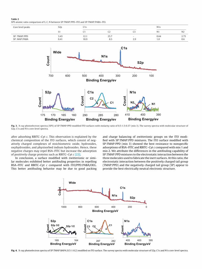
Table 2XPS atomic ratio comparison of S, C, N between SP:TMAP:PPD–ITO and SP:TMAP:PABA–ITO.
Core level peaks S2p C1s N1s
S1 C1 C2 C3 N1 N2
SP: TMAP:PPD 3.43 12.1 25.7 – 0.64 2.75SP: MAP:PABA 0.41 18.9 8.5 2.9 1.0 0.6
F ith mS
acaono
lBT
F
ig. 3. X-ray photoelectron spectra (XPS) of SP:TMAP:PPD modified ITO surfaces w2p, C1s and N1s core-level spectra.
fter adsorbing RBITC–Cyt c. This observation is explained by thehemical composition of the ITO surfaces, which consist of neg-tively charged complexes of stoichiometric oxide, hydroxides,xyhydroxides, and physisorbed indium hydroxides. Hence, theseegative charges may repel BSA–FITC but increase the adsorptionf positively charge proteins such as RBITC–Cyt c [22].
In conclusion, a surface modified with zwitterionic or simi-
ar molecules exhibited better antifouling properties in repellingSA–FITC and RBITC–Cyt c compared with ITO/PPD:PABA/OEG.his better antifouling behavior may be due to good packingig. 4. X-ray photoelectron spectra of SP:TMAP:BAPA (0.5:1:0.2) modified on ITO surface.
olarity ratio of 0.5:1.5:0.37 (mix 3). The survey spectra with molecular structure of
and charge balancing of zwitterionic groups on the ITO modi-fied with SP:TMAP:PPD mixtures. The ITO surface modified withSP:TMAP:PPD (mix 3) showed the best resistance to nonspecificadsorptions of BSA–FITC and RBITC–Cyt c compared with mix 1 andmix 2. We attribute the differences in the antifouling capability ofSP:TMAP:PPD mixtures to the electrostatic interaction between thethree molecules used to fabricate the inert surfaces. At this ratio, the
electrostatic interaction between the positively charged tail group(TMAP:PPD) and the negatively charged tail group (SP) appear toprovide the best electrically neutral electronic structure.The survey spectra with molecular structure of S2p, C1s and N1s core-level spectra.
F (0.5:15 X spec
3
m(ticnPvm
arRtopwae
rct(d(qattSm
btccc
ig. 5. E-SEM image of (a) ITO/GNP. (b) ITO/SP:TMAP:PPD/GNP with molarity ratio
0 K. (d) ITO/SP:TMAP:PABA and bare ITO surface under magnification 1.5 K. (e) ED
.2.2. ITO surface modified with a mixture of SP:TMAP:PABAITO surfaces were modified with SP:TMAP:PABA at different
olarity ratios of 0.8:1:0.2 (mix 1), 1:1:0.1 (mix 2) and 0.5:1:0.2mix 3). Mix 3 exhibited the best antifouling capabilities amonghe three combinations against both BSA–FITC (86.7% fluorescentntensity, a mean gray value of 1.96; s = 0.5, n = 3) and RBITC–Cyt
(50.5% fluorescent intensity, a mean gray value of 1.1; s = 0.4, = 3) and compared with the ITO surface functionalized withPD:PABA/OEG (100% BSA–FITC fluorescent intensity, a mean grayalue of 2.2; s = 0.3, n = 3; 100% RBITC–Cyt c fluorescent intensity, aean gray value of 2.1; s = 0.4, n = 3; Fig. 2; Table 1).The ITO electrode modified with PPD:PABA/OEG accumulated
significant amount of RBITC–Cyt c. It exhibited the highest fluo-escence intensity among the investigated surface types (Fig. 2(d)).BITC–Cyt c has the ability to change its conformation upon adsorp-ion onto a wide range of interfaces [33,34]. Furthermore, the failuref OEG moieties to resist the nonspecific adsorption of undesiredroteins may be due to the auto-oxidation of OEG moieties [15,35],hich makes the PPD:PABA/OEG layer unable to repel BSA–FITC
nd RBITC–Cyt c. Antifouling coating alternatives that are moreffective than OEG should therefore be identified.
ITO modified with SP:TMAP:PPD mixtures exhibited betteresistance to the nonspecific adsorption of BSA–FITC and RBITC–Cyt
compared with SP:TMAP:PABA. This better resistance may be dueo the domination of PABA molecules in SP:TMAP:PABA mixturesFig. 1(1f)). The adsorption of aryl diazonium cation mixtures wasominated by the aryl diazonium cation, which was easier to reducee.g., having a less negative potential) [36]; such behavior subse-uently reduced adsorptions of the other two molecules (e.g., SPnd TMAP, which were necessary for a neutral charge interface. Inhe SP:TMAP:PPD mixture, no such domination was observed forhe PPD molecule. XPS analysis showed that the atomic ratio forP and TMAP was higher in SP:TMAP:PPD than in SP:TMAP:PABAixtures (Table 2).In contrast to PABA, PPD could be used as a linker molecule and
e incorporated with zwitterionic molecules (e.g., SP and TMAP)
o resist the nonspecific adsorption of BSA–FITC and RBITC–Cyt. PPD was more readily deposited onto the ITO surface, and theomplete passivation of the electrode surface required only twoycles (Fig. 1(1c)). With PABA, the passivation was significantly less.5:0.37). (c) ITO/SP:TMAP:PABA with molarity ratio (0.5:1:0.2) under magnificationtra ITO/SP:TMAP:PPD/GNP.
efficient, requiring 10 cycles (Fig. 1(1d)). PPD alone can resist thenonspecific adsorption of BSA–FITC and RBITC–Cyt c (Fig. 1, Table 1).Such behavior enhanced the antifouling efficiency of zwitterionicmolecules (e.g., SP and TMAP). Moreover, the reductive adsorptionpotential for PPD was −0.36 V, which, in comparison to the poten-tial for PABA (−0.1 V), was closer to reductive adsorptions of SP(−0.473 V) and TMAP (−0.26 V). Therefore, aryl diazonium cationswith similar reductive adsorption potentials prevented the domi-nation of one aryl diazonium cation over the other, which aided thefabrication of a neutrally charged antifouling coating.
3.3. ITO surface characterization via XPS analysis
For ITO surfaces modified with SP:TMAP:PPD (mix 3), the pres-ence of SP was confirmed by a signal peak indicative of sulfur (S2p)at 167.9 eV [27,28]. This signal peak was absent in the narrow scanof the S2p region for bare ITO (Fig. 3). TMAP was detected by thepresence of a C-N linkage peak, which represents the link betweennitrogen and 4-methyl groups, at a binding energy of 285.0 eV (C2).Another peak (C1) centered at 284.4 eV in a narrow scan of thecarbon region represented aromatic carbons in SP, PPD and TMAPmoieties [27,30]. PPD was characterized by the N1 399.5 eV peakobserved in the narrow scan of the N1s region, which is attributedto H2N C [23–25,37]. In contrast, XPS survey spectra for the mixedSP:TMAP:PABA layer (Fig. 4) revealed a peak at 167.8 eV corre-sponding to S2p, which indicated an SP graft [23,27,28]. A narrowscan of the carbon C1s region revealed three peaks centered at284.7 eV (C1), 286.1 eV (C2), and 288.5 eV (C3). The C1 and C2 peakswere attributed to aromatic carbon atoms and to N (CH3)3 in thetrimethylammonio group, respectively. The C3 peak correspondedto COO-; the presence of this peak indicates the successful graftingof PABA molecules onto the ITO substrate [23,38,39].
3.3.1. ITO surface characterization via FE-SEM analysisAn FE-SEM image of the bare ITO electrode showed the absence
of GNP (Fig. 5(a)). However when the ITO was modified with
SP:TMAP:PPD, a uniform and homogenous distribution of GNPswas observed (Fig. 5(b)). The bare ITO/GNP experiment as shownin Fig. 5(a) served as a control experiment. The objective was todemonstrate that the attachment of GNPs onto the ITO surface
otaotSmmaIi(ta
4
mdtoanl0bwfSsasasctulfpm
A
RdHIMP
R
[
[
[
[
[
[
[
[
[
[
[
[
[
[
[
[
[
[
[
[
[
[
nly formed with the present of PPD. The EDX results confirmedhe deposition of GNPs onto modified ITO electrode surfaces at antomic ratio of 2.11 (Fig. 5(e)). The linkage of amines in the mixturef SP:TMAP:PPD to GNPs was more likely to occur by dehydrogena-ion from NH-GNP because of its stronger covalent bond [24,40].uch observations confirmed the homogenous grafting of the linkerolecule (PPD) onto the ITO surface, unlike the SP:TMAP:PABAixture, which was dominated with PABA molecule (XPS results
re shown in Table 2). The image of an SP:TMAP:PABA-modifiedTO electrode showed a thin, smooth and featureless morphologyndicates that no GNPs could be deposited onto this type of surfaceFig. 5(c)). An obvious difference was noticed in images betweenhe bare and SP:TMAP:PABA modified ITO surfaces which capturedt 1500× magnification (Fig. 5(d)).
. Conclusions
A biosensing interface consisting of antifouling and linkerolecules based on in-situ-generated aryl diazonium cations was
escribed. The differences in electrochemical behaviors among allypes of modified surfaces (either with the individual compoundr a mixed layer of aryl diazonium cations) were investigatednd evaluated. An antifouling study using confocal laser scan-ing microscopy showed that ITO surfaces coated with mixed
ayers of SP:TMAP:PPD 0.5:1.5:0.37 (mix 3) or SP:TMAP:PABA.5:1:0.2 (mix 3) exhibited better antifouling capabilities againstoth BSA–FITC and RBITC–Cyt c compared to ITO surfaces coatedith PPD:PABA/OEG. However, PPD:PABA/OEG-coated ITO sur-
aces showed lower amounts of BSA–FITC adsorbed compared toP:TMAP:PABA-coated ITO surfaces. The mixture of SP:TMAP:PPDhowed better resistance to nonspecific adsorptions of BSA–FITCnd RBITC–Cyt c compared to SP:TMAP:PABA. Analyses of XPSurvey spectra further confirmed the deposition of SP:TMAP:PPDnd SP:TMAP:PABA onto ITO surfaces. FE-SEM and EDX analyseshowed dense and homogenous distributions of GNPs, which indi-ated a good distribution of linker and antifouling molecules onhe ITO sensing interfaces. This study highlights the prospectivese of aryl-diazonium-cation-derived zwitterionic compounds and
inker molecules for the easy fabrication of ITO biosensing inter-aces. These interfaces can repel undesirable anionic and cationicroteins and detect target analytes in highly complicated biologicalatrices, such as blood, serum, and urine.
cknowledgments
This work was financially supported by the University of Malayaesearch Grant (UMRG) (RG159-12SUS, RP012C-14SUS), the Fun-amental Research Grant Scheme (FRGS) from the Ministry ofigher Education of Malaysia (MOHE) (FP014-2013A), a High
mpact Research Grant from the Ministry of Higher Education ofalaysia (HIR-MoHE F000004-21001), and University of Malaya
ostgraduate Research Grant (PG120-2012B).
eferences
[1] D. Atias, Y. Liebes, V. Chalifa-Caspi, L. Bremand, L. Lobel, R.S. Marks, P. Dus-sart, Chemiluminescent optical fiber immunosensor for the detection of IgMantibody to dengue virus in humans, Sens. Actuators, B: Chem. 140 (2009)206–215.
[2] I.T. Cavalcanti, B.V.M. Silva, N.G. Peres, P. Moura, M.D.P.T. Sotomayor, M.I.F.Guedes, R.F. Dutra, A disposable chitosan-modified carbon fiber electrode fordengue virus envelope protein detection, Talanta 91 (2012) 41–46.
[3] W.H. Chen, I.H. Hsu, Y.C. Sun, Y.K. Wang, T.K. Wu, Immunocapture couples with
matrix-assisted laser desorption/ionization time-of-flight mass spectrometryfor rapid detection of Type 1 dengue virus, J. Chromatogr. A 1288 (2013) 21–27.[4] A.C.M.S. Dias, S.L.R. Gomes-Filho, M.M.S. Silva, R.F. Dutra, A sensor tip basedon carbon nanotube-ink printed electrode for the dengue virus NS1 protein,Biosens. Bioelectron. 44 (2013) 216–221.
[
[5] M.J.A. Shiddiky, P.H. Kithva, D. Kozak, M. Trau, An electrochemical immunosen-sor to minimize the nonspecific adsorption and to improve sensitivity of proteinassays in human serum, Biosens. Bioelectron. 38 (2012) 132–137.
[6] P. Akkahat, S. Kiatkamjornwong, i.S. Yusa, V.P. Hoven, Y. Iwasaki, Develop-ment of a novel antifouling platform for biosensing probe immobilizationfrom methacryloyloxyethyl phosphorylcholine-containing copolymer brushes,Langmuir 28 (2012) 5872–5881.
[7] S. Salomon, T. Leichle, D. Dezest, F. Seichepine, S. Guillon, C. Thibault, C. Vieu,L. Nicu, Arrays of nanoelectromechanical biosensors functionalized by micro-contact printing, Nanotechnology 23 (2012) 495501.
[8] M. Gabriel, D. Strand, C.-F. Vahl, Cell adhesive and antifouling polyvinyl chloridesurfaces via wet chemical modification, Artif. Organs 36 (2012) 839–844.
[9] N. Wang, K. Burugapalli, W. Song, J. Halls, F. Moussy, A. Ray, Y. Zheng, Electro-spun fibro-porous polyurethane coatings for implantable glucose biosensors,Biomaterials 34 (2013) 888–901.
10] K.H. Chae, Y.M. Jang, Y.H. Kim, O.-J. Sohn, J.I. Rhee, Anti-fouling epoxy coatingsfor optical biosensor application based on phosphorylcholine, Sens. Actuators,B: Chem. 124 (2007) 153–160.
11] S. Brahim, D. Narinesingh, A. Guiseppi-Elie, Bio-smart hydrogels: co-joinedmolecular recognition and signal transduction in biosensor fabrication anddrug delivery, Biosens. Bioelectron. 17 (2002) 973–981.
12] S. Zhang, G. Wright, Y. Yang, Materials and techniques for electrochemicalbiosensor design and construction, Biosens. Bioelectron. 15 (2000) 273–282.
13] Q. Yu, Y. Zhang, H. Wang, J. Brash, H. Chen, Anti-fouling bioactive surfaces, ActaBiomater. 7 (2011) 1550–1557.
14] P.-Y.J. Yeh, J.N. Kizhakkedathu, J.D. Madden, M. Chiao, Electric field andvibration-assisted nanomolecule desorption and anti-biofouling for biosensorapplications, Colloids Surf., B: Biointerfaces 59 (2007) 67–73.
15] A.L. Gui, E. Luais, J.R. Peterson, J.J. Gooding, Zwitterionic phenyl layers: finally,stable, anti-biofouling coatings that do not passivate electrodes, ACS Appl.Mater. Interfaces 5 (2013) 4827–4835.
16] J.E. Gittens, T.J. Smith, R. Suleiman, R. Akid, Current and emerging environment-friendly systems for fouling control in the marine environment, Biotechnol.Adv. 31 (2013) 1738–1753.
17] Q. Shi, Y. Su, W. Chen, J. Peng, L. Nie, L. Zhang, Z. Jiang, Grafting short-chainamino acids onto membrane surfaces to resist protein fouling, J. Membr. Sci.366 (2011) 398–404.
18] S.S. Shah, M.C. Howland, L.-J. Chen, J. Silangcruz, S.V. Verkhoturov, E.A. Schweik-ert, A.N. Parikh, A. Revzin, Micropatterning of proteins and mammalian cells onindium tin oxide, ACS Appl. Mater. Interfaces 1 (2009) 2592–2601.
19] M.J.A. Shiddiky, P.H. Kithva, R. Sakandar, M. Trau, Femtomolar detection of acancer biomarker protein in serum with ultralow background current by anodicstripping voltammetry, Chem. Commun. 48 (2012) 6411–6413.
20] Y. Li, M. Giesbers, M. Gerth, H. Zuilhof, Generic top-functionalization ofpatterned antifouling zwitterionic polymers on indium tin oxide, Langmuir 28(2012) 12509–12517.
21] P. Liu, Z. Su, Surface-initiated atom transfer radical polymerization (SI-ATRP)of styrene from chitosan particles, Mater. Lett. 60 (2006) 1137–1139.
22] M. Chockalingam, N. Darwish, G. Le Saux, J.J. Gooding, Importance of the indiumtin oxide substrate on the quality of self-assembled monolayers formed fromorganophosphonic acids, Langmuir 27 (2011) 2545–2552.
23] G. Liu, M. Chockalingham, S.M. Khor, A.L. Gui, J.J. Gooding, A comparativestudy of the modification of gold and glassy carbon surfaces with mixed lay-ers of in situ generated aryl diazonium compounds, Electroanalysis 22 (2009)918–926.
24] G. Liu, E. Luais, J.J. Gooding, The fabrication of stable gold nanoparticle-modifiedinterfaces for electrochemistry, Langmuir 27 (2011) 4176–4183.
25] S. Eissa, C. Tlili, L. L’Hocine, M. Zourob, Electrochemical immunosensor forthe milk allergen �-lactoglobulin based on electrografting of organic film ongraphene modified screen-printed carbon electrodes, Biosens. Bioelectron. 38(2012) 308–313.
26] A.L. Gui, H.M. Yau, D.S. Thomas, M. Chockalingam, J.B. Harper, J.J. Gooding,Using supramolecular binding motifs to provide precise control over the ratioand distribution of species in multiple component films grafted on surfaces:demonstration using electrochemical assembly from aryl diazonium salts,Langmuir 29 (2013) 4772–4781.
27] N. Vila, D. Belanger, Mixtures of functionalized aromatic groups generated fromdiazonium chemistry as templates towards bimetallic species supported oncarbon electrode surfaces, Electrochim. Acta 85 (2012) 538–547.
28] N. Vila, M.V. Brussel, M. DAmours, J. Marwan, C. Buess-Herman, D. Belanger,Metallic and bimetallic Cu/Pt species supported on carbon surfaces bymeans of substituted phenyl groups, J. Electroanal. Chem. 609 (2007)85–93.
29] C. Combellas, F. Kanoufi, J. Pinson, F.I. Podvorica, Sterically hindered diazoniumsalts for the grafting of a monolayer on metals, J. Am. Chem. Soc. 130 (2008)8576–8577.
30] J. Lyskawa, D. Belanger, Direct modification of a gold electrode withaminophenyl groups by electrochemical reduction of in situ generatedaminophenyl monodiazonium cations, Chem. Mater. 18 (2006) 4755–4763.
31] R. Polsky, J.C. Harper, D.R. Wheeler, S.M. Dirk, D.C. Arango, S.M. Brozik,Electrically addressable diazonium-functionalized antibodies for multianalyte
electrochemical sensor applications, Biosens. Bioelectron. 23 (2008) 757–764.32] W.-P. Hu, L.-Y. Huang, T.-C. Kuo, W.-W. Hu, Y. Chang, C.-S. Chen, H.-C.Chen, W.-Y. Chen, Optimization of DNA-directed immobilization on mixedoligo(ethylene glycol) monolayers for immunodetection, Anal. Biochem. 423(2012) 26–35.

[
[
[
[
[
[
[
33] X. Chen, R. Ferrigno, J. Yang, G.M. Whitesides, Redox Properties of cytochromec adsorbed on self-assembled monolayers: a probe for protein conformationand orientation, Langmuir 18 (2002) 7009–7015.
34] D. Hobara, S.-i. Imabayashi, T. Kakiuchi, Preferential adsorption of horse heartcytochrome c on nanometer-scale domains of a phase-separated binary self-assembled monolayer of 3-mercaptopropionic acid and 1-hexadecanethiol onAu(1 1 1), Nano Lett. 2 (2002) 1021–1025.
35] R.E. Holmlin, X. Chen, R.G. Chapman, S. Takayama, G.M. Whitesides, Zwitteri-
onic SAMs that resist nonspecific adsorption of protein from aqueous buffer,Langmuir 17 (2001) 2841–2850.36] C. Louault, M. D’Amours, D. Bélanger, The electrochemical grafting of amixture of substituted phenyl groups at a glassy carbon electrode surface,ChemPhysChem 9 (2008) 1164–1170.
[
37] I. Losito, C. Malitesta, I. De Bari, C.-D. Calvano, X-ray photoelectron spectroscopycharacterization of poly(2, 3-diaminophenazine) films electrosynthesised onplatinum, Thin Solid Films 473 (2005) 104–113.
38] D.-J. Chung, S.-H. Oh, S. Komathi, A.I. Gopalan, K.-P. Lee, S.-H. Choi, One-stepmodification of various electrode surfaces using diazonium salt compoundsand the application of this technology to electrochemical DNA (E-DNA) sensors,Electrochim. Acta 76 (2012) 394–403.
39] M. Yin, Y. Yuan, C. Liu, J. Wang, Development of mussel adhesive polypeptide
mimics coating for in-situ inducing re-endothelialization of intravascular stentdevices, Biomaterials 30 (2009) 2764–2773.40] G. Liu, J. Liu, T.P. Davis, J.J. Gooding, Electrochemical impedance immunosensorbased on gold nanoparticles and aryl diazonium salt functionalized gold elec-trodes for the detection of antibody, Biosens. Bioelectron. 26 (2011) 3660–3665.