Applied anatomy of the submental island flap and its ...population for applied anatomy of the...
6
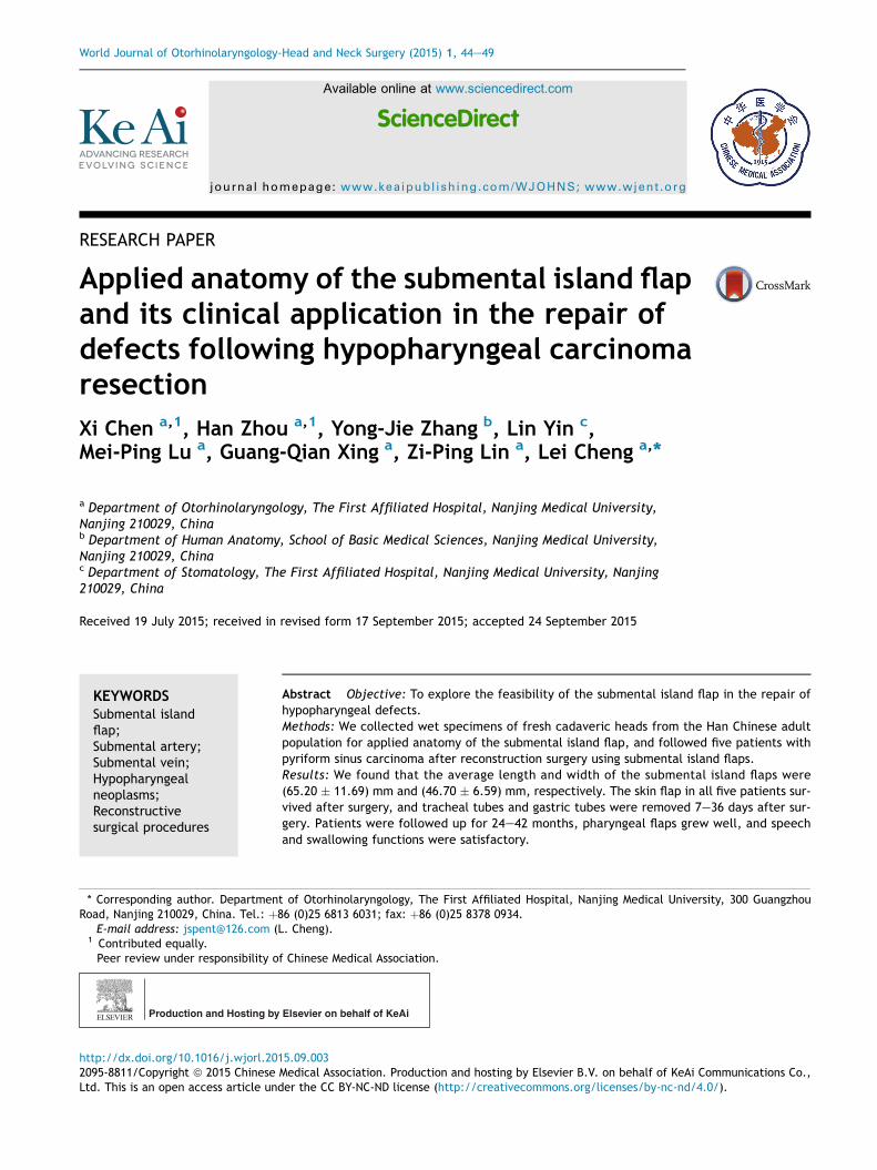
RESEARCH PAPER Applied anatomy of the submental island flap and its clinical application in the repair of defects following hypopharyngeal carcinoma resection Xi Chen a,1 , Han Zhou a,1 , Yong-Jie Zhang b , Lin Yin c , Mei-Ping Lu a , Guang-Qian Xing a , Zi-Ping Lin a , Lei Cheng a, * a Department of Otorhinolaryngology, The First Affiliated Hospital, Nanjing Medical University, Nanjing 210029, China b Department of Human Anatomy, School of Basic Medical Sciences, Nanjing Medical University, Nanjing 210029, China c Department of Stomatology, The First Affiliated Hospital, Nanjing Medical University, Nanjing 210029, China Received 19 July 2015; received in revised form 17 September 2015; accepted 24 September 2015 KEYWORDS Submental island flap; Submental artery; Submental vein; Hypopharyngeal neoplasms; Reconstructive surgical procedures Abstract Objective: To explore the feasibility of the submental island flap in the repair of hypopharyngeal defects. Methods: We collected wet specimens of fresh cadaveric heads from the Han Chinese adult population for applied anatomy of the submental island flap, and followed five patients with pyriform sinus carcinoma after reconstruction surgery using submental island flaps. Results: We found that the average length and width of the submental island flaps were (65.20 11.69) mm and (46.70 6.59) mm, respectively. The skin flap in all five patients sur- vived after surgery, and tracheal tubes and gastric tubes were removed 7e36 days after sur- gery. Patients were followed up for 24e42 months, pharyngeal flaps grew well, and speech and swallowing functions were satisfactory. * Corresponding author. Department of Otorhinolaryngology, The First Affiliated Hospital, Nanjing Medical University, 300 Guangzhou Road, Nanjing 210029, China. Tel.: þ86 (0)25 6813 6031; fax: þ86 (0)25 8378 0934. E-mail address: [email protected] (L. Cheng). 1 Contributed equally. Peer review under responsibility of Chinese Medical Association. Production and Hosting by Elsevier on behalf of KeAi http://dx.doi.org/10.1016/j.wjorl.2015.09.003 2095-8811/Copyright ª 2015 Chinese Medical Association. Production and hosting by Elsevier B.V. on behalf of KeAi Communications Co., Ltd. This is an open access article under the CC BY-NC-ND license (http://creativecommons.org/licenses/by-nc-nd/4.0/). Available online at www.sciencedirect.com ScienceDirect journal homepage: www.keaipublishing.com/WJOHNS; www.wjent.org World Journal of Otorhinolaryngology-Head and Neck Surgery (2015) 1, 44e49
Transcript of Applied anatomy of the submental island flap and its ...population for applied anatomy of the...
World Journal of Otorhinolaryngology-Head and Neck Surgery (2015) 1, 44e49
Available online at www.sciencedirect.com
ScienceDirect
journal homepage: www.keaipubl ishing.com/WJOHNS; www.wjent .org
RESEARCH PAPER
Applied anatomy of the submental island flapand its clinical application in the repair ofdefects following hypopharyngeal carcinomaresection
Xi Chen a,1, Han Zhou a,1, Yong-Jie Zhang b, Lin Yin c,Mei-Ping Lu a, Guang-Qian Xing a, Zi-Ping Lin a, Lei Cheng a,*
a Department of Otorhinolaryngology, The First Affiliated Hospital, Nanjing Medical University,Nanjing 210029, Chinab Department of Human Anatomy, School of Basic Medical Sciences, Nanjing Medical University,Nanjing 210029, Chinac Department of Stomatology, The First Affiliated Hospital, Nanjing Medical University, Nanjing210029, China
Received 19 July 2015; received in revised form 17 September 2015; accepted 24 September 2015
KEYWORDSSubmental islandflap;Submental artery;Submental vein;Hypopharyngealneoplasms;Reconstructivesurgical procedures
* Corresponding author. DepartmenRoad, Nanjing 210029, China. Tel.: þ
E-mail address: [email protected] (1 Contributed equally.Peer review under responsibility o
Production and Hosting by
http://dx.doi.org/10.1016/j.wjorl.202095-8811/Copyright ª 2015 ChineseLtd. This is an open access article un
Abstract Objective: To explore the feasibility of the submental island flap in the repair ofhypopharyngeal defects.Methods: We collected wet specimens of fresh cadaveric heads from the Han Chinese adultpopulation for applied anatomy of the submental island flap, and followed five patients withpyriform sinus carcinoma after reconstruction surgery using submental island flaps.Results: We found that the average length and width of the submental island flaps were(65.20 � 11.69) mm and (46.70 � 6.59) mm, respectively. The skin flap in all five patients sur-vived after surgery, and tracheal tubes and gastric tubes were removed 7e36 days after sur-gery. Patients were followed up for 24e42 months, pharyngeal flaps grew well, and speechand swallowing functions were satisfactory.
t of Otorhinolaryngology, The First Affiliated Hospital, Nanjing Medical University, 300 Guangzhou86 (0)25 6813 6031; fax: þ86 (0)25 8378 0934.L. Cheng).
f Chinese Medical Association.
Elsevier on behalf of KeAi
15.09.003Medical Association. Production and hosting by Elsevier B.V. on behalf of KeAi Communications Co.,der the CC BY-NC-ND license (http://creativecommons.org/licenses/by-nc-nd/4.0/).
Applied anatomy of the submental island flap 45
Conclusion: The submental island flap is a preferred material for the repair of hypopharyngealdefects after hypopharyngeal carcinoma resection, because of good blood supply, easy har-vesting, and high survival rate.Copyright ª 2015 Chinese Medical Association. Production and hosting by Elsevier B.V. onbehalf of KeAi Communications Co., Ltd. This is an open access article under the CC BY-NC-ND license (http://creativecommons.org/licenses/by-nc-nd/4.0/).
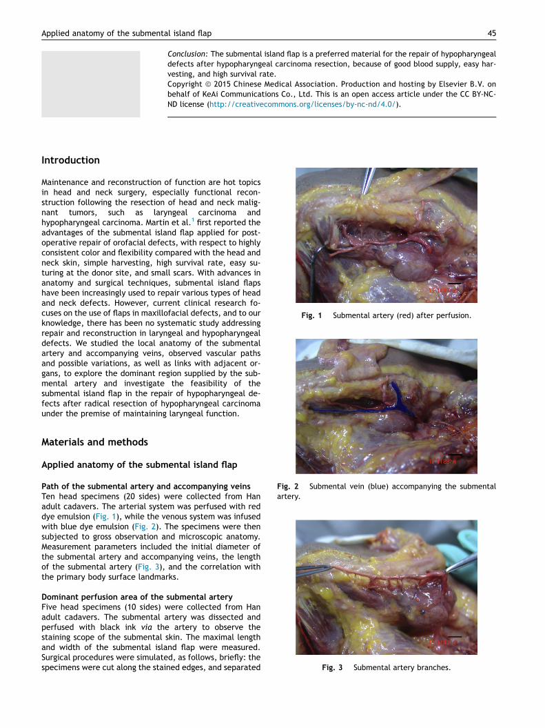
Fig. 1 Submental artery (red) after perfusion.
Introduction
Maintenance and reconstruction of function are hot topicsin head and neck surgery, especially functional recon-struction following the resection of head and neck malig-nant tumors, such as laryngeal carcinoma andhypopharyngeal carcinoma. Martin et al.1 first reported theadvantages of the submental island flap applied for post-operative repair of orofacial defects, with respect to highlyconsistent color and flexibility compared with the head andneck skin, simple harvesting, high survival rate, easy su-turing at the donor site, and small scars. With advances inanatomy and surgical techniques, submental island flapshave been increasingly used to repair various types of headand neck defects. However, current clinical research fo-cuses on the use of flaps in maxillofacial defects, and to ourknowledge, there has been no systematic study addressingrepair and reconstruction in laryngeal and hypopharyngealdefects. We studied the local anatomy of the submentalartery and accompanying veins, observed vascular pathsand possible variations, as well as links with adjacent or-gans, to explore the dominant region supplied by the sub-mental artery and investigate the feasibility of thesubmental island flap in the repair of hypopharyngeal de-fects after radical resection of hypopharyngeal carcinomaunder the premise of maintaining laryngeal function.
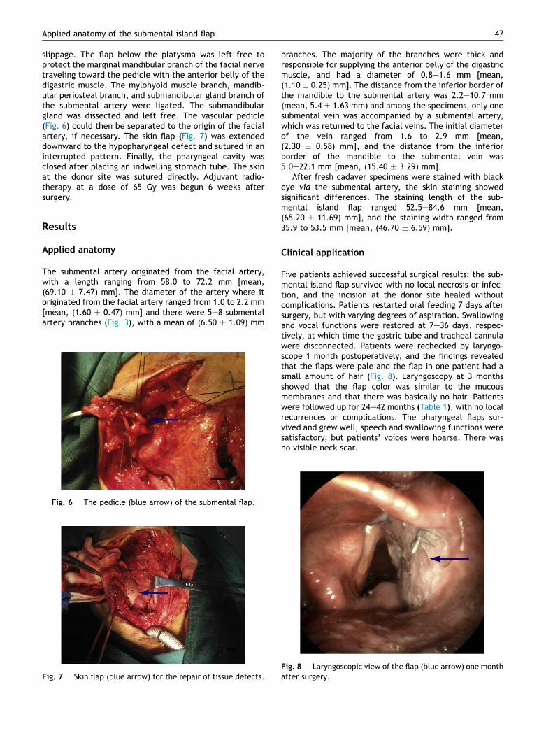
Fig. 2 Submental vein (blue) accompanying the submentalartery.
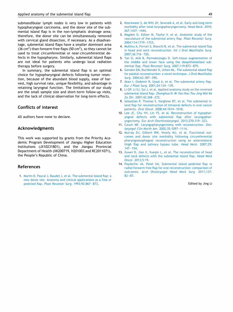
Fig. 3 Submental artery branches.
Materials and methods
Applied anatomy of the submental island flap
Path of the submental artery and accompanying veinsTen head specimens (20 sides) were collected from Hanadult cadavers. The arterial system was perfused with reddye emulsion (Fig. 1), while the venous system was infusedwith blue dye emulsion (Fig. 2). The specimens were thensubjected to gross observation and microscopic anatomy.Measurement parameters included the initial diameter ofthe submental artery and accompanying veins, the lengthof the submental artery (Fig. 3), and the correlation withthe primary body surface landmarks.
Dominant perfusion area of the submental arteryFive head specimens (10 sides) were collected from Hanadult cadavers. The submental artery was dissected andperfused with black ink via the artery to observe thestaining scope of the submental skin. The maximal lengthand width of the submental island flap were measured.Surgical procedures were simulated, as follows, briefly: thespecimens were cut along the stained edges, and separated

46 X. Chen et al.
from the beginning of the submental artery to the externalcarotid artery, then the maximal length of the vascularpedicle was measured, and the anatomic range of the flapcovered was assessed.
Fig. 4 Design of the submental island flap.
Fig. 5 Tissue defects (blue arrow) after resection of thehypopharyngeal lesion.
Clinical application of the submental island flap inthe repair of hypopharyngeal defects
Patient demographics and clinical informationFive patients (Table 1) were males aged 49e70 years, with amean of 61.2 years. Three cases were over 60 years of age,and the tumor originated in the lateral wall of the unilat-eral pyriform fossa, involving the ipsilateral half of thelarynx. The vocal cords were fixed and the ipsilateral pyr-iform tip was uninvolved; no abnormalities were found byesophageal lipiodol radiography. The preoperative pathol-ogy report suggested T3N1M0 stage squamous cell carci-noma. One 53-year-old patient was diagnosed as havingT4N1M0 stage squamous cell carcinoma of the lateral wall ofthe pyriform fossa unilaterally, affecting the cervicalesophagus. In these four patients, lymph nodes(diameter < 3 cm) were palpable in the ipsilateral neck andnone received other treatment before surgery. Anotherpatient (aged 49 years) underwent unilateral pyriform sinuscarcinoma resection and local mucosal repair for anasto-motic stenosis. All of the involved patients received chestX-ray and abdominal ultrasound, and the findings revealedno metastases. Surgery was performed between February2011 and August 2012, with written informed consent ob-tained from all patients. The research protocol wasapproved by our institutional review board.
Surgical procedureAll patients underwent unilateral or bilateral selective neckdissection (regions II, III, IV, V) under general anesthesiafollowing prophylactic tracheotomy. Three cases with afixed ipsilateral vocal cord underwent laryngofission, withvertical hemilaryngectomy and complete ipsilateral pyri-form fossa resection 2 cm away from the carcinoma. Onepatient with cervical esophageal invasion underwent theremoval of both the pyriform sinus unilaterally, and thecervical esophageal lateral wall (approximately 2 cm),leaving the larynx intact. Intraoperative pathology consul-tation revealed negative margins.
Based on the result of pinch test and the lesion of theprimary tumor, the incisions of the submental island flapwere designed and marked (Fig. 4). An appropriately-sizedsubmental island flap was selected for the repair, whichwas determined by the scope of the tissue defects (Fig. 5).
Table 1 Clinical information of cases undergoing hypopharynge
No. Gender Age(years)
TNM stage Flap size(cm � cm)
Flap surv
1 male 49 e 7 � 3 Survived2 male 70 T3N1M0 7 � 4 Survived3 male 70 T3N1M0 7 � 5 Survived4 male 53 T4N1M0 8 � 5 Survived5 male 61 T3N1M0 7 � 5 Survived
The flap size ranged from 7 cm � 4 cme8 cm � 5 cm. Inanother case, anastomotic stenosis was observed followingpyriform sinus carcinoma resection, which was repairedusing a submental island flap (7 cm � 3 cm) trimmed with alingual contour.
When the flap was produced, the facial artery on theaffected side was dissected free to the submental arterybifurcation, then the distal end of the facial artery wasligated. The flap was then revised accordingly, parallelwith the inferior border of the mandible and the free edgeof the skin flap. The skin, subcutaneous tissue, and pla-tysma were then dissected, and the skin and platysmawere sutured in an interrupted pattern to prevent skin flap
al defect repair using a submental island flap.
ival Recovery time ofdeglutition (days)
Operativecomplications
Follow-uptime (months)
7 None 4210 None 3936 None 3615 None 3214 None 24
Applied anatomy of the submental island flap 47
slippage. The flap below the platysma was left free toprotect the marginal mandibular branch of the facial nervetraveling toward the pedicle with the anterior belly of thedigastric muscle. The mylohyoid muscle branch, mandib-ular periosteal branch, and submandibular gland branch ofthe submental artery were ligated. The submandibulargland was dissected and left free. The vascular pedicle(Fig. 6) could then be separated to the origin of the facialartery, if necessary. The skin flap (Fig. 7) was extendeddownward to the hypopharyngeal defect and sutured in aninterrupted pattern. Finally, the pharyngeal cavity wasclosed after placing an indwelling stomach tube. The skinat the donor site was sutured directly. Adjuvant radio-therapy at a dose of 65 Gy was begun 6 weeks aftersurgery.
Results
Applied anatomy
The submental artery originated from the facial artery,with a length ranging from 58.0 to 72.2 mm [mean,(69.10 � 7.47) mm]. The diameter of the artery where itoriginated from the facial artery ranged from 1.0 to 2.2 mm[mean, (1.60 � 0.47) mm] and there were 5e8 submentalartery branches (Fig. 3), with a mean of (6.50 � 1.09) mm
Fig. 6 The pedicle (blue arrow) of the submental flap.
Fig. 7 Skin flap (blue arrow) for the repair of tissue defects.
branches. The majority of the branches were thick andresponsible for supplying the anterior belly of the digastricmuscle, and had a diameter of 0.8e1.6 mm [mean,(1.10 � 0.25) mm]. The distance from the inferior border ofthe mandible to the submental artery was 2.2e10.7 mm(mean, 5.4 � 1.63 mm) and among the specimens, only onesubmental vein was accompanied by a submental artery,which was returned to the facial veins. The initial diameterof the vein ranged from 1.6 to 2.9 mm [mean,(2.30 � 0.58) mm], and the distance from the inferiorborder of the mandible to the submental vein was5.0e22.1 mm [mean, (15.40 � 3.29) mm].
After fresh cadaver specimens were stained with blackdye via the submental artery, the skin staining showedsignificant differences. The staining length of the sub-mental island flap ranged 52.5e84.6 mm [mean,(65.20 � 11.69) mm], and the staining width ranged from35.9 to 53.5 mm [mean, (46.70 � 6.59) mm].
Clinical application
Five patients achieved successful surgical results: the sub-mental island flap survived with no local necrosis or infec-tion, and the incision at the donor site healed withoutcomplications. Patients restarted oral feeding 7 days aftersurgery, but with varying degrees of aspiration. Swallowingand vocal functions were restored at 7e36 days, respec-tively, at which time the gastric tube and tracheal cannulawere disconnected. Patients were rechecked by laryngo-scope 1 month postoperatively, and the findings revealedthat the flaps were pale and the flap in one patient had asmall amount of hair (Fig. 8). Laryngoscopy at 3 monthsshowed that the flap color was similar to the mucousmembranes and that there was basically no hair. Patientswere followed up for 24e42 months (Table 1), with no localrecurrences or complications. The pharyngeal flaps sur-vived and grew well, speech and swallowing functions weresatisfactory, but patients’ voices were hoarse. There wasno visible neck scar.
Fig. 8 Laryngoscopic view of the flap (blue arrow) one monthafter surgery.

48 X. Chen et al.
Discussion
Because of the hidden location of the hypopharynx, earlyhypopharyngeal carcinoma has no characteristic symptomsand is difficult to diagnose. The carcinoma may havealready affected the cervical esophagus when symptomsare noted, and the 5-year survival rate is only 15%e47%.2 Itis reported that wide resection of the hypopharynx andcervical esophagus, together with postoperative chemo-radiation, could prolong 5-year survival rates. However,resection also produces speech and swallowing dysfunction,seriously affecting patients’ quality of life. Therefore,adequate repair of tissue defects after hypopharyngealresection is crucial, because it requires both morphologicalrestoration and functional reconstruction. The challengesfacing head and neck surgeons treating these patients arerepairing the laryngeal and hypopharyngeal defects aftersurgery, and restoring swallowing and speech function.
Martin et al.1 first proposed the use of a submental is-land flap in the reconstruction of orofacial defects in 1993.Submental island flap is an axial skin flap pedicled on thesubmental artery and vein. The submental artery is a con-stant branch of the facial artery3 with a length of approx-imately 6e7 cm and evidence4,5 has shown that, from itsorigin in the facial artery, the submental artery travelsforward between the two lobes of the submaxillary gland orbetween the submandibular gland and the mandible,entering the deep (70%) or superficial layer (30%) of theanterior belly of the digastric muscle, then piercing thedeep fascia and entering the skin. The initial diameter ofthe submental artery is 1.0e2.0 mm (mean, 1.7 mm), and itelicits several branches to supply the lower lip, subman-dibular gland, platysma, the anterior belly of the digastricmuscle, as well as 1e4 perforating branches. The terminalbranches coincide with the contralateral branches, andsupply the upper neck skin. The venous diameter of thesubmental island flap vein ranges from 1.0 to 2.9 mm(mean, 2.2 mm). The submental venous system is oftenclassified into two groups: 1) tight accompanying: the veinsaccompany the submental artery with reflux into the facialvein; 2) non-tight accompanying: the veins are located su-perficially, and share 2e3 communicating branches withtight accompanying veins, then flow into the anterior ju-gular vein. Our anatomical findings showed that the sub-mental artery provided the blood supply to the submentalisland flap, and it was located in a constant position. Theaverage length was 69.1 mm; and the average diameter ofthe arteries originating from the facial artery was 1.6 mm,with 6.5 branches on average, and 5.4 mm away from theinferior border of the mandible. When a submental veinaccompanied the submental artery, it returned to the facialveins, and had an average diameter of 2.3 mm, and anaverage distance from the inferior border of the mandibleof 15.4 mm. Our findings were consistent with previousstudies.
The size of the submental island flap varies greatly and ismaximized at 18 cm � 7 cm, with a vascular pedicle lengthof up to 8 cm.6 If dissected to its origin in the facial artery,the pedicle can be extended 1e2 cm, reaching the canthusand the zygomatic arch, which provides the possibility ofrepairing defects in the upper face and the lower neck.7
The present study showed that the length and width ofthe stained submental island flap were 65.2 mm and46.7 mm, respectively, and the maximum size was84.6 mm � 53.5 mm, which differed from previously re-ported flap sizes.8 The difference largely depends on thepopulation race and the embolism of terminal branches ofthe submental artery and vein, which may affect thetraveling of dyes. A larger number of cadaver specimens isneeded for further observation and discussion.
Sebastian et al.9 described a method of harvesting sub-mental island flaps by isolating from the free edge of theflap toward the pedicle. We modified this method by firstdissecting the facial artery and vein to the submental ar-tery and vein, and cutting the skin flap after ensuringarterial supply and venous return. This modification canensure the survival of the flap. Our anatomical resultsshowed that there were many thick submental arterybranches involving the anterior belly of the digastric mus-cle, with an average diameter of 1.1 mm; therefore, werecommend removing the anterior belly of the digastricmuscle when cutting the flaps to effectively guarantee flapblood supply.
Restoration of speech and swallowing after hypophar-yngeal surgery is currently a crucial challenge for head andneck surgeons. Lee et al.10 achieved success repairinghypopharyngeal defects using a submental island flap in sixcases, but all patients underwent total laryngectomy,which seriously affected the patients’ quality of life. In ourstudy, the size of the skin flap was adjusted according tointraoperative observation, ranging from7 cm � 3 cme8 cm � 5 cm, the skin incision at the donorsite was successfully sutured, and the scars were con-cealed. We speculate that laryngeal function can be pre-served in the majority of T3, and even in T4 pyriform sinuscarcinomas. The patients in our study completely orpartially retained their laryngeal function, which signifi-cantly improved their quality of life after surgery. Althoughthere is a risk of aspiration, tracheal tubes and nasogastricfeeding tubes can be successfully removed as patientsgradually recover swallowing function.
Previous reports11e13 summarized the criteria forrepairing hypopharyngeal and cervical esophageal defects:1) ensure adequate safe margins; 2) achieve first-stagereconstruction; 3) small scar at donor site; 4) high successrate; 5) short operation time; 6) low incidence of stenosisand pharyngeal fistula; and 7) physicians experienced intreating complications. Free flaps have been consideredthe preferred method for repairing tissue defects afterresection of hypopharyngeal tumors. With advances inmicrovascular surgical techniques, this flap has becomewidely used. Recently, Paydarfar et al.14 compared sub-mental island pedicled flaps and radial forearm free flapsand found that there was no significant difference betweenthe two flaps in the restoration of swallowing and speechfunctions as well as in local recurrence rate after the sur-gical repair of oral defects, but that the submental islandflap effectively shortened operation time and post-operative hospital stay. The submental island flap has a richblood supply, survives well, and produces an invisible scar;while a forearm free flap produces visible scars at the donorsite, which then require free skin grafts. Another consid-eration is that the metastasis rate of submental and
Applied anatomy of the submental island flap 49
submandibular lymph nodes is very low in patients withhypopharyngeal carcinoma, and the donor site of the sub-mental island flap is in the non-lymphatic drainage area;therefore, the donor site can be simultaneously removedwith cervical gland dissection, if necessary. As a disadvan-tage, submental island flaps have a smaller dominant area(36 cm2) than forearm free flaps (50 cm2), so they cannot beused to treat circumferential or near-circumferential de-fects in the hypopharynx. Similarly, submental island flapsare not ideal for patients who undergo local radiationtherapy before surgery.
In summary, the submental island flap is an optimalchoice for hypopharyngeal defects following tumor resec-tion, because of the abundant blood supply, ease of har-vest, high survival rate, unique flexibility, and advantage inretaining laryngeal function. The limitations of our studyare the small sample size and short-term follow-up visits,and the lack of clinical observation for long-term effects.
Conflicts of interest
All authors have none to declare.
Acknowledgments
This work was supported by grants from the Priority Aca-demic Program Development of Jiangsu Higher EducationInstitutions (JX10231801), and the Jiangsu ProvincialDepartment of Health (XK200719, H201003 and RC2011071),the People’s Republic of China.
References
1. Martin D, Pascal J, Baudet J, et al. The submental island flap: anew donor site. Anatomy and clinical applications as a free orpedicled flap. Plast Reconstr Surg. 1993;92:867e873.
2. Keereweer S, de Wilt JH, Sewnaik A, et al. Early and long-termmorbidity after total laryngopharyngectomy. Head Neck. 2010;267:1437e1444.
3. Magden O, Edizer M, Tayfur V, et al. Anatomic study of thevasculature of the submental artery flap. Plast Reconstr Surg.2004;114:1719e1723.
4. Multinu A, Ferrari S, Bianchi B, et al. The submental island flapin head and neck reconstruction. Int J Oral Maxillofac Surg.2007;36:716e720.
5. Tan O, Atik B, Parmaksizoglu D. Soft-tissue augmentation ofthe middle and lower face using the deepithelialized sub-mental flap. Plast Reconstr Surg. 2007;119:873e879.
6. Genden EM, Buchbinder D, Urken ML. The submental island flapfor palatal reconstruction: a novel technique. J Oral MaxillofacSurg. 2004;62:387e390.
7. Akan I, Ozdemir R, Uysal A, et al. The submental artery flap.Eur J Plast Surg. 2001;24:134e139.
8. Li GP, Li XJ, Sui J, et al. Applied anatomy study on the reversedsubmental island flap. Zhonghua Er Bi Yan Hou Tou Jing Wai KeZa Zhi. 2007;42:268e272.
9. Sebastian P, Thomas S, Varghese BT, et al. The submental is-land flap for reconstruction of intraoral defects in oral cancerpatients. Oral Oncol. 2008;44:1014e1018.
10. Lee JC, Chu YH, Lin YS, et al. Reconstruction of hypophar-yngeal defects with submental flap after laryngophar-yngectomy. Eur Arch Otorhinolaryngol. 2013;270:319e323.
11. Couch ME. Laryngopharyngectomy with reconstruction. Oto-laryngol Clin North Am. 2002;35:1097e1114.
12. Murray DJ, Gilbert RW, Vesely MJ, et al. Functional out-comes and donor site morbidity following circumferentialpharyngoesophageal reconstruction using an anterolateralthigh flap and salivary bypass tube. Head Neck. 2007;29:147e154.
13. Xuwei D, Jian X, Xueqin L, et al. The reconstruction of headand neck defects with the submental island flap. Head NeckOncol. 2013;5:19.
14. Paydarfar JA, Patel UA. Submental island pedicled flap vsradial forearm free flap for oral reconstruction: comparison ofoutcomes. Arch Otolaryngol Head Neck Surg. 2011;137:82e87.
Edited by Jing Li