Application of Ultrasound Spectral Analysis for Choroidal ... › fileadmin › user_upload ›...
17
Baltic J. Modern Computing, Vol. 3 (2015), No. 1, 55-71 Application of Ultrasound Spectral Analysis for Choroidal Melanomas Before and After Treatment Analysis Jurgis TRUMPAITIS 1,2 , Rytis JURKONIS 3 , Ieva SAKALAUSKAITĖ 1 , Alvydas PAUNKSNIS 1,4 1 Department of Ophthalmology, Lithuanian University of Health Sciences, Medicine Academy, Kaunas, Lithuania, 2 Department of Ophthalmology, Neuroscience Institute, Ophthalmology laboratory, Kaunas, Lithuania, 3 Institute for Biomedical Engineering, Kaunas University of Technology, Kaunas, Lithuania, 4 JSC “Stratelus”, Vilnius, Lithuania [email protected], [email protected], [email protected], [email protected] Abstract. Choroidal Melanoma (CM) is the most common primary malignancy of the eye. The overall incidence is approximately 5–7 cases per million per year. In this paper the new technique for tumours tissue structure evaluation using ultrasound spectral analysis is presented. Based on the obtained results, it can be said that radio frequency (RF) ultrasound signals parameters at the healthy tissue area and the area with the intraocular tumour – melanoma before and after treatment statistically are significantly different. This study has shown that the lower amplitude, lower spectral intercept, high spectral slope and high momentary bandwidth are typical for choroidal melanoma if compared with healthy tissues and the lower momentary bandwidth are typical for choroidal melanoma after treatment if compared with melanoma before treatment. Application of spectral analysis using non-invasive ultrasound expert system, provides the new opportunities in early diagnosis, differentiation of tumours, evaluation of the treatment effectiveness. Keywords: ultrasound, intraocular tumours, choroidal melanoma, spectral analysis, radiofrequency signals, brachytherapy. 1. Introduction 1.1 Choroidal melanoma diagnosis and prevalence Uveal melanoma (UM) is the most common primary intraocular malignancy in adults. The overall incidence is approximately 5–7 cases per million per year, and climbs to more than 20 cases per million per year by the age of 70 (Egan et al. 1988; Singh and Topham, 2003). It is more common in the Caucasian population, especially those with blue/grey iris. The survival rates at 5, 10 and 15 years are 65%, 50% and 45% respectively (Singh and Topham, 2003). Ninety-two percent of cases of UM arise in the choroid while the remainder arise in the iris (3%) and ciliary body (5%) (Ramasamy et
Transcript of Application of Ultrasound Spectral Analysis for Choroidal ... › fileadmin › user_upload ›...
Baltic J. Modern Computing, Vol. 3 (2015), No. 1, 55-71
Application of Ultrasound Spectral Analysis for
Choroidal Melanomas Before and After Treatment
Analysis
Jurgis TRUMPAITIS
1,2, Rytis JURKONIS
3,
Ieva SAKALAUSKAITĖ1, Alvydas PAUNKSNIS
1,4
1 Department of Ophthalmology, Lithuanian University of Health Sciences, Medicine Academy,
Kaunas, Lithuania, 2 Department of Ophthalmology, Neuroscience Institute, Ophthalmology laboratory, Kaunas,
Lithuania, 3 Institute for Biomedical Engineering, Kaunas University of Technology, Kaunas, Lithuania,
4 JSC “Stratelus”, Vilnius, Lithuania
[email protected], [email protected]
Abstract. Choroidal Melanoma (CM) is the most common primary malignancy of the eye. The
overall incidence is approximately 5–7 cases per million per year. In this paper the new technique
for tumours tissue structure evaluation using ultrasound spectral analysis is presented. Based on
the obtained results, it can be said that radio frequency (RF) ultrasound signals parameters at the
healthy tissue area and the area with the intraocular tumour – melanoma before and after
treatment statistically are significantly different. This study has shown that the lower amplitude,
lower spectral intercept, high spectral slope and high momentary bandwidth are typical for
choroidal melanoma if compared with healthy tissues and the lower momentary bandwidth are
typical for choroidal melanoma after treatment if compared with melanoma before treatment.
Application of spectral analysis using non-invasive ultrasound expert system, provides the new
opportunities in early diagnosis, differentiation of tumours, evaluation of the treatment
effectiveness.
Keywords: ultrasound, intraocular tumours, choroidal melanoma, spectral analysis,
radiofrequency signals, brachytherapy.
1. Introduction
1.1 Choroidal melanoma diagnosis and prevalence
Uveal melanoma (UM) is the most common primary intraocular malignancy in
adults. The overall incidence is approximately 5–7 cases per million per year, and climbs
to more than 20 cases per million per year by the age of 70 (Egan et al. 1988; Singh and
Topham, 2003). It is more common in the Caucasian population, especially those with
blue/grey iris. The survival rates at 5, 10 and 15 years are 65%, 50% and 45%
respectively (Singh and Topham, 2003). Ninety-two percent of cases of UM arise in the
choroid while the remainder arise in the iris (3%) and ciliary body (5%) (Ramasamy et
56 Trumpaitis, Jurkonis, Sakalauskaitė and Paunksnis
al., 2014). Choroidal melanomas have the worst prognosis, while iris melanomas have
the most favourable. The latter may be due to earlier presentation, as iris melanomas
may be detected by the patient externally. Unfortunately, choroidal melanomas are
usually detected late, when the patient is symptomatic with decreased visual acuity. This
may be due to the involvement of the macula, or when complicated by secondary retinal
detachment.
Choroidal Melanoma (CM) is the most common primary malignancy of the eye
(Bell and Wilson, 2004). It metastasizes most likely into the liver, followed by
pulmonary, bone or even skin metastasis, whereas local spread may occur through the
overlying Bruch membrane, into the subretinal space or towards the orbit (Lorigan et al.,
1991).
Uveal melanomas account for approximately 98% of all ocular melanomas. More
than 90% of intraocular melanomas arise in the choroid, with about 3–4% developing in
the iris and the remainder in the ciliary body (Damato and Coupland, 2012a). In
Caucasians, uveal melanomas have an incidence of approximately 7 per million per year
(Singh et al., 2005). Presentation peaks at the age of sixty years and is rare before
adulthood. Males and females are affected in equal numbers, although iris melanomas
tend to be slightly more common in women whereas choroidal melanomas are more
common in men (Damato and Coupland, 2012a). Risk factors for uveal melanoma
include: light skin, blue eyes, tendency to cutaneous naevi, congenital ocular
melanocytosis, uveal melanocytoma and neurofibromatosis. The role of sunlight is
uncertain but it is noteworthy that most iris melanomas occur inferiorly, where there is
less protection from the upper eyelid (Damato and Coupland, 2012b).
Choroidal melanomas initially form a dome-shaped tumour. There is an overlying
retinal pigment epitheliopathy, with the multilayering of retinal pigment epithelial cells,
lipofuscin accumulation, drusen and retinal pigment epithelial detachment. In
Caucasians, most choroidal melanomas are amelanotic or only lightly pigmented and it
is the retinal pigment epithelial (RPE) proliferation that gives choroidal melanomas their
dark colour. This RPE dysfunction causes retinal degeneration and atrophy, inducing
symptoms such as metamorphopsia, blurring, visual field loss and photopsia (Damato
and Coupland, 2012b).
Choroidal melanomas are routinely classified as small (<10 mm in diameter and <3
mm in height), medium (10 to 15 mm in diameter and3 to 5 mm in height), or large (>15
mm in diameter and >5 mm in height) (Zimmerman et al., 1980; Shields et al., 1991).
Multiple studies, including the Collaborative Ocular Melanoma Study (COMS, 1998),
have specified tumor size as the strongest indicator of metastasis and therefore survival
(Mooy and De Jong, 1996). Ten-year survival rates for uveal melanomas have been
published as 81.2% for small melanomas, 60.0% for medium melanomas, and 34.8% for
large uveal melanomas (McLean et al., 1980). The COMS (1998) additionally identified
older patient age as a baseline covariate that affected the prognosis for survival.
Ocular melanomas metastasize hematogenously primarily to the liver but also to
lung, bone, kidney, and brain (Zakka et al., 1980; Mooy and De Jong, 1996). Zakka et al.
(1980) reported only 75% of ocular melanomas had metastasized at the time of death
compared with 96% of nonocular melanomas. Although ocular melanomas are not as
aggressive as cutaneous melanomas, they carry a substantial risk of metastasis. In a long-
term study of patients treated for uveal melanoma by enucleation in Finland between
1962 and 1981, 61% of patients eventually died as a result of the disease (Kujala et al.,
2003). Mortality related to uveal melanoma was 31% by 5 years, 45% by 15 years, 49%
by 25 years, and 52% by 35 years. Among patients who died of uveal melanoma, 62%,
Application of Ultrasound Spectral Analysis for Choroidal Melanomas Analysis 57
90%, 98%, and 100% died within 5, 15, 25, and 35 years, respectively. Once metastasis
occurs, survival is less than 7 months (Kath et al., 1993). Therefore, it is important to
identify cancerous lesions and begin treatment before metastasis occurs (Bell and
Wilson, 2004).
Differential diagnosis of eye tumors is one of the most important problems in
ophthalmology. Eye tumors, especially melanoma are very dangerous due to metastasis
to other organs. Malignant tumors of the eye makes 0.2 percent of all malignant tumors
diagnosed and is every a direct cause of death for every third patient (Paunksnis et al.,
2008).
Early detection of these lesions and accurate diagnosis leads to effective assistance.
There are several pathologies similar to malignant tumors: choroidal naevus,
hemangioma, subretinal hemorrhage, exudative macular degeneration, haemophthalmus.
Early diagnosis and tissue characterization is crucial in order to apply the proper
treatment, to predict course of disease and to prevent from patient's death (Paunksnis et
al., 2008)
Ultrasonic methods stand out in ophthalmology with great informative value among
the noninvasive methods. This is one of the main diagnostic methods for establishing the
diagnosis of intraocular tumors (Paunksnis et al., 2008).
During the past two decades, improvements in the resolution and reliability of
instrumentation and techniques have allowed A-scan and B-scan ultrasonography to join
direct visualization, fluorescein angiography, and radioactive phosphorus testing as
primary diagnostic modalities in the evaluation of intraocular neoplasms. Because
overlying opacities have no effect on the examining ultrasonographic frequencies,
ultrasound has the unique ability to depict the nature of intraocular pathology when the
media are optically opaque (Goldberg and Hodes, 1977).
In the last decade, ultrasound probes with higher frequency and higher resolution
have been commercialized, such as the 20 MHz probe used for posterior pole studies.
Frequency is important in ultrasound because it is directly related to resolution and
inversely related to penetration of ultrasonographic images. Moreover, the ultrasonic
wavelength is inversely related to the frequency. Thus, the 75 μm of ultrasound at 20
MHz is half the wavelength of a 10MHz transducer and, therefore, is theoretically
capable of providing a 2-fold improvement in resolution. This technique may be better
for imaging choroidal lesions, such as a UM and SCMT, because it provides a more
accurate visual of the epiescleral space, resulting in more reproducible lesion height
measurements (Coleman et al., 2004).
UH is easier to detect by 20 MHz than by 10 MHz ultrasonography. This
ultrasonographic sign appears to be correlated with the height of the tumor. Thus, we
believe UH estimation by 20MHz ultrasonography could be used as a significant
predictive factor for SCMT growth (Piñeiro-Ces et al., 2014).
Independently, pioneering researchers converged on three important clinical
applications of highfrequency B-mode imaging: these were in ophthalmology (Pavlin et
al. 1990), dermatology (Hoffmann et al., 1990) and intravascular ultrasound (Nissen et
al. 1990). Skin and intravascular applications began in the 20-MHz range, and
ophthalmic applications centred in the 40-MHz to 60-MHz range. In each case, unique
instrumentation was created to address the specific needs of the applications. The
ultrasonic visualisation of living tissue at microscopic resolution is referred to in the
literature as “ultrasound backscatter microscopy” or “ultrasound biomicroscopy” (Lizzi
and Coleman, 2004).
58 Trumpaitis, Jurkonis, Sakalauskaitė and Paunksnis
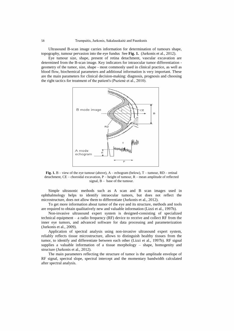
Ultrasound B-scan image carries information for determination of tumours shape,
topography, tumour pervasion into the eye fundus See Fig. 1. (Jurkonis et al., 2012).
Eye tumour size, shape, present of retina detachment, vascular excavation are
determined from the B-scan image. Key indicators for intraocular tumor differentiation -
geometry of the tumor, size, shape - most commonly used in clinical practice, as well as
blood flow, biochemical parameters and additional information is very important. These
are the main parameters for clinical decision-making: diagnosis, prognosis and choosing
the right tactics for treatment of the patient's (Puzienė et al., 2010).
Fig. 1. B – view of the eye tumour (above), A – echogram (below), T – tumour, RD – retinal
detachment, CE – choroidal excavation, P – height of tumour, R – mean amplitude of reflected
signal, B – base of the tumour.
Simple ultrasonic methods such as A scan and B scan images used in
ophthalmology helps to identify intraocular tumors, but does not reflect the
microstructure, does not allow them to differentiate (Jurkonis et al., 2012).
To get more information about tumor of the eye and its structure, methods and tools
are required to obtain qualitatively new and valuable information (Lizzi et al., 1997b).
Non-invasive ultrasound expert system is designed-consisting of specialized
technical equipment – a radio frequency (RF) device to receive and collect RF from the
inner eye tumors, and advanced software for data processing and parameterization
(Jurkonis et al., 2009).
Application of spectral analysis using non-invasive ultrasound expert system,
reliably reflects tissue microstructure, allows to distinguish healthy tissues from the
tumor, to identify and differentiate between each other (Lizzi et al., 1997b). RF signal
supplies a valuable information of a tissue morphology – shape, homogenity and
structure (Jurkonis et al., 2012).
The main parameters reflecting the structure of tumor is the amplitude envelope of
RF signal, spectral slope, spectral intercept and the momentary bandwidth calculated
after spectral analysis.
Application of Ultrasound Spectral Analysis for Choroidal Melanomas Analysis 59
1.2 Choroidal melanoma treatment
Various treatment modalities for uveal melanoma have been described in the
literature. In recent years, eye-saving procedures have become more widespread, and
these include photocoagulation, transpupillary thermotherapy, radiotherapy, local
resection, chemotherapy, immunotherapy, and stereotactic radiosurgery (Damato, 2004;
Damato and Jones, 2005) Surgical treatment may be associated with postoperative pain,
infection, insufficient resection, and secondary eye loss, and it has no superiority over
GKS in terms of recurrence (Shields et al., 1991; Damato and Jones, 2005).
Chemotherapy and immunotherapy have no curative role; the main objective of
treatment in uveal melanoma is to achieve local disease control, and no medical
treatment alone can assure this outcome (Damato, 2004).
Of these, photocoagulation and transpupillary thermotherapy are reserved for the
management of selected cases with very small tumors (Shields et al., 1996, 1998).
For posterior uveal melanomas, radiotherapy remains the most widely used method
of treatment (Shields et al., 1993). Enucleation is indicated for advanced melanomas that
occupy most intraocular structures, that have caused severe secondary glaucoma, and
that have invaded the optic nerve (Ryan et al., 1989). The principal objective of the
treatment is to reduce the risk of death from metastasis and to conserve as much vision
as possible while minimizing the risk of enucleation (Toktas et al., 2010).
The goal of all forms of radiation therapy is to destroy the reproductive integrity of a
tumour. Ionizing radiation damages the DNA of the malignant cells, which may be
misrepaired and also may disrupt the integrity of the chromosome. The effects of this
damage become manifest during mitosis, at which point the cells cannot successfully
replicate. The timing would depend on the proliferation kinetics of the cellular
constituents and it will be longer in tumours with slow turnover, including ocular
melanoma. Other consequences of irradiation include changes in growth factors and
signal transduction pathways, apoptosis, and the regulation of the cell cycle (Gragoudas,
2006).
Histopathologic studies demonstrated that PBT causes degenerative changes in the
tumour (necrosis, fibrosis, balloon cells), decreases the ability of tumour cells to
reproduce (fewermitotic figures), and damages blood supply (Saornil et al., 1992).
However, it was shown that post irradiation, most tumours shrink but do not disappear
(Boudinet et al., 2007). In 2002, Kaiserman et al. (2002) described echographic changes
of tumour thickness and internal reflectivity in uveal melanoma treated by
brachytherapy. In our study, we analyze the same dynamics in uveal melanomas treated
by PBT (Mosci et al., 2014).
Brachytherapy is a frequently used therapeutic option for the treatment of uveal
melanomas. Of the modalities listed here, plaque brachytherapy is the only invasive
method utilizing radioactivity hat delivers very high dose radiation at the tissue level
(Straatsma et al., 1988). Although a high control rate is achieved with plaque
brachytherapy, this technique is associated with a high complication rate due to the high
doses of radiation the tumor periphery is exposed to (Shields et al., 1990).
Treatment of choroidal melanoma with brachytherapy – suturing a radiation source
to the eye – was first reported by Moore in 1930. These early studies by Moore with
radon seeds paved the way for the use of other forms of radiation at clinical centers
around the world, including cobalt-60 (Brady et al., 1982), ruthenium-106 (Lommatzsch
60 Trumpaitis, Jurkonis, Sakalauskaitė and Paunksnis
and Kirsch, 1988), gold-198 (Moura et al., 1985), iodine-125 (Packer et al., 1992) and
palladium-103 (Overgaard et al., 1986). Iodine-125 is currently the most commonly
used isotope in brachytherapy for choroidal melanoma in the USA, whereas ruthenium-
106 and palladium-103 continue to be popular at some centers. Brachytherapy offers an
alternative to enucleation in the treatment of choroidal melanoma, allowing globe-
salvaging with the possibility of maintaining useful vision (Houston et al.).
Although treatment protocols and selection of tumors for plaque treatment must be
individualized and differ among clinical centers, generally acceptable indications for
plaque brachytherapy include: (1) selected small choroidal melanomas exhibiting growth
or malignant transformation; (2) medium-sized choroidal and ciliary body melanomas in
eyes with visual potential; (3) large melanomas with dimensions up to 16 mm in
diameter and 8–10 mm in thickness; (4) larger melanomas, especially in monocular
patients. However, despite success in sparing enucleation, radiation has profound effects
on the surrounding retina and optic nerve, with vision limited by the resulting radiation
retinopathy and optic neuropathy (Wen and McCannel, 2009).
The optimal tumoricidal radiation dose for uveal melanoma remains unclear (Trott,
1991), but doses range between 50 and 100 Gy, with doses less than 50 Gy being
associated with significant treatment failures (Trott, 1991). Kindy-Degnan et al. (1989)
used helium ion radiation and reported on tumor apex dosages ranging between 50 and
80 Gy, showing that regardless of whether 50, 60, 70, or 80 Gy was used, there were no
differences in tumor regression, survival, complications, or visual outcomes (Houston et
al.).
Brachytherapy is the application of radiation from isotopes over very short distances
in contact with a surface. The radiation is distributed over a short distance from a surface
or within the target tissue. The isotopes are divided into two broad categories: (1) those
that emit γ-rays or X-rays; (2) those that emit β-particles (electrons) (Houston et al.).
Plaque brachytherapy and enucleation were shown to have similar rates of
melanoma-specific deaths. In addition, small, large, and juxtapapillary tumors have been
successfully treated with plaque brachytherapy. Despite eye preservation, visual
morbidity is high secondary to radiation-related complications, including radiation
maculopathy and optic neuropathy. Alternative radioisotopes have been investigated to
minimize treatment-related effects without significant reductions in visual loss (Houston
et al.).
Besides the advantages of being non-invasive and easier for the patient to tolerate,
radiosurgery provides a single day treatment that can be completed within a few hours
(Fakiris et al., 2013). No other technique offers the same convenience for the patient.
Although previous studies have shown GKS to be a minimally invasive, eye-saving
treatment modality for uveal melanomas (Fakiris et al., 2013) secondary enucleation is
still common with this procedure. Tumor volume and tumor location are thought to be
major determinants of intraocular complications after GKS. Egan et al. (1988) proposed
that tumors of the ciliary body and tumors larger than 8 mm in height are more likely to
require secondary enucleation after treatment (Toktas et al., 2010).
2. Objectives of the study
1. Collect ultrasound images and RF signals for comparative evaluation between
healthy eye tissues and tissues with intraocular tumors: choriodal melanoma before and
after brachyterapy treatment.
Application of Ultrasound Spectral Analysis for Choroidal Melanomas Analysis 61
2. The aim of this research is to compare parameters of ultrasound signals
backscattered from healthy ocular tissues with choroidal melanoma before and after
brachyterapy treatment using spectrum analysis of frequency-dependent backscattered
radiofrequency (RF).
3. Methods and data
The study involved 70 people from 18 to 80 years attending in Hospital of
Lithuanian University of Health Sciences Kaunas Eye Clinics consultation diagnostic
department with diagnosed intraocular eye tumor – choroidal melanoma before
treatment – 35 cases, choroidal melanoma after treatment – 35 cases and 70 cases with
healthy eyes.
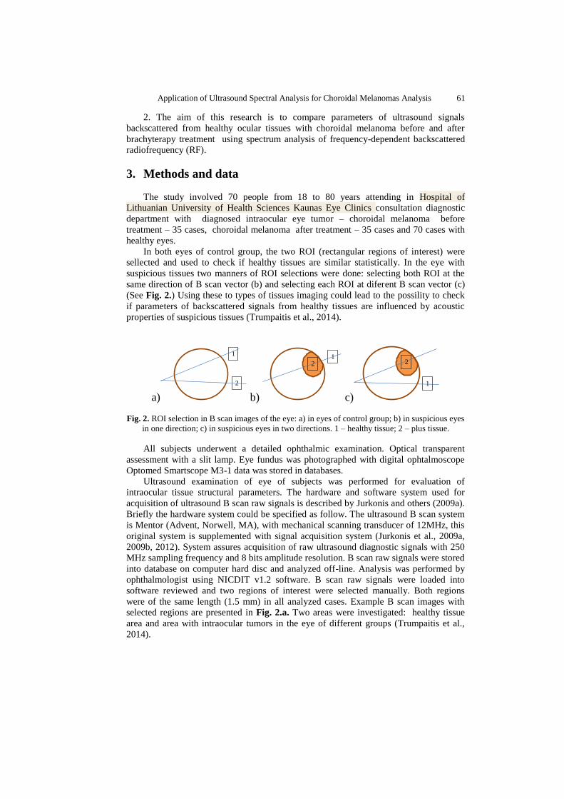
In both eyes of control group, the two ROI (rectangular regions of interest) were
sellected and used to check if healthy tissues are similar statistically. In the eye with
suspicious tissues two manners of ROI selections were done: selecting both ROI at the
same direction of B scan vector (b) and selecting each ROI at diferent B scan vector (c)
(See Fig. 2.) Using these to types of tissues imaging could lead to the possility to check
if parameters of backscattered signals from healthy tissues are influenced by acoustic
properties of suspicious tissues (Trumpaitis et al., 2014).
a)
1
2
b)
1
2
c)
1
2
Fig. 2. ROI selection in B scan images of the eye: a) in eyes of control group; b) in suspicious eyes
in one direction; c) in suspicious eyes in two directions. 1 – healthy tissue; 2 – plus tissue.
All subjects underwent a detailed ophthalmic examination. Optical transparent
assessment with a slit lamp. Eye fundus was photographed with digital ophtalmoscope
Optomed Smartscope M3-1 data was stored in databases.
Ultrasound examination of eye of subjects was performed for evaluation of
intraocular tissue structural parameters. The hardware and software system used for
acquisition of ultrasound B scan raw signals is described by Jurkonis and others (2009a).
Briefly the hardware system could be specified as follow. The ultrasound B scan system
is Mentor (Advent, Norwell, MA), with mechanical scanning transducer of 12MHz, this
original system is supplemented with signal acquisition system (Jurkonis et al., 2009a,
2009b, 2012). System assures acquisition of raw ultrasound diagnostic signals with 250
MHz sampling frequency and 8 bits amplitude resolution. B scan raw signals were stored
into database on computer hard disc and analyzed off-line. Analysis was performed by
ophthalmologist using NICDIT v1.2 software. B scan raw signals were loaded into
software reviewed and two regions of interest were selected manually. Both regions
were of the same length (1.5 mm) in all analyzed cases. Example B scan images with
selected regions are presented in Fig. 2.a. Two areas were investigated: healthy tissue
area and area with intraocular tumors in the eye of different groups (Trumpaitis et al.,
2014).
62 Trumpaitis, Jurkonis, Sakalauskaitė and Paunksnis
a) b) c)
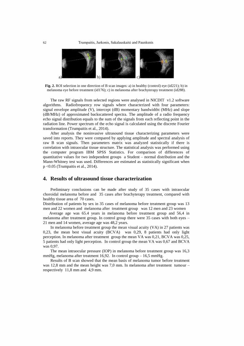
Fig. 2. ROI selection in one direction of B scan images: a) in healthy (control) eye (id221); b) in
melanoma eye before treatment (id176); c) in melanoma after brachyterapy treatment (id288).
The raw RF signals from selected regions were analysed in NICDIT v1.2 software
algorithms. Radiofrequency row signals where characterized with four parameters:
signal envelope amplitude (V), intercept (dB) momentary bandwidtht (MHz) and slope
(dB/MHz) of approximated backscattered spectra. The amplitude of a radio frequency
echo signal distribution equals to the sum of the signals from each reflecting point in the
radiation line. Power spectrum of the echo signal is calculated using the discrete Fourier
transformation (Trumpaitis et al., 2014).
After analysis the noninvasive ultrasound tissue characterizing parameters were
saved into reports. They were compared by applying amplitude and spectral analysis of
raw B scan signals. Then parameters matrix was analyzed statistically if there is
correlation with intraocular tissue structure. The statistical analysis was performed using
the computer program IBM SPSS Statistics. For comparison of differences of
quantitative values for two independent groups a Student – normal distribution and the
Mann-Whitney test was used. Differences are estimated as statistically significant when
p <0.05 (Trumpaitis et al., 2014).
4. Results of ultrasound tissue characterization
Preliminary conclusions can be made after study of 35 cases with intraocular
choroidal melanoma before and 35 cases after brachyterapy treatment, compared with
healthy tissue area of 70 cases.
Distribution of patients by sex in 35 cases of melanoma before treatment group was 13
men and 22 women and melanoma after treatment group was 12 men and 23 women
Average age was 65.4 years in melanoma before treatment group and 56,4 in
melanoma after treatment group. In control group there were 35 cases with both eyes –
21 men and 14 women, average age was 48,2 years.
In melanoma before treatment group the mean visual acuity (VA) in 27 patients was
0,23, the mean best visual acuity (BCVA) was 0,29, 8 patients had only light
perception. In melanoma after treatment group the mean VA was 0,21, BCVA was 0,25,
5 patients had only light perception. In control group the mean VA was 0,67 and BCVA
was 0,97.
The mean intraocular pressure (IOP) in melanoma before treatment group was 16,3
mmHg, melanoma after treatment 16,92. In control group – 16,5 mmHg.
Results of B scan showed that the mean basis of melanoma tumor before treatment
was 12,8 mm and the mean height was 7,0 mm. In melanoma after treatment tumour –
respectively 11,8 mm and 4,9 mm.
Application of Ultrasound Spectral Analysis for Choroidal Melanomas Analysis 63
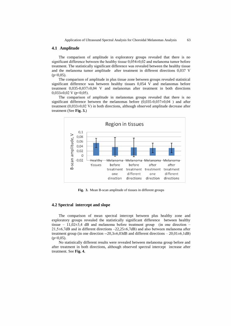
4.1 Amplitude
The comparison of amplitude in exploratory groups revealed that there is no
significant difference between the healthy tissue 0,054±0,02 and melanoma tumor before
treatment. The statistically significant difference was revealed between the healthy tissue
and the melanoma tumor amplitude after treatment in different directions 0,037 V
(p<0,05).
The comparison of amplitude in plus tissue zone between groups revealed statistical
significant difference was between healthy tissues 0,054 V and melanomas before
treatment 0,035-0,037±0,04 V and melanomas after treatment in both directions
0,033±0,02 V (p<0,05).
The comparison of amplitude in melanomas groups revealed that there is no
significant difference between the melanomas before (0,035-0,037±0,04 ) and after
treatment (0,033±0,02 V) in both directions, although observed amplitude decrease after
treatment (See Fig. 3.)
Fig. 3. Mean B-scan amplitude of tissues in different groups
4.2 Spectral intercept and slope
The comparison of mean spectral intercept between plus healthy zone and
exploratory groups revealed the statistically significant difference between healthy
tissue – 11,02±3,4 dB and melanoma before treatment group (in one direction –
21,5±6,7dB and in different directions –22,25±6,7dB) and also between melanoma after
treatment group (in one direction --20,3±6,03dB and different directions – 20,01±6,1dB)
(p<0,05).
No statistically different results were revealed between melanoma group before and
after treatment in both directions, although observed spectral intercept increase after
treatment. See Fig. 4.
64 Trumpaitis, Jurkonis, Sakalauskaitė and Paunksnis
Fig. 4. Mean spectral intercept of tissues in different groups
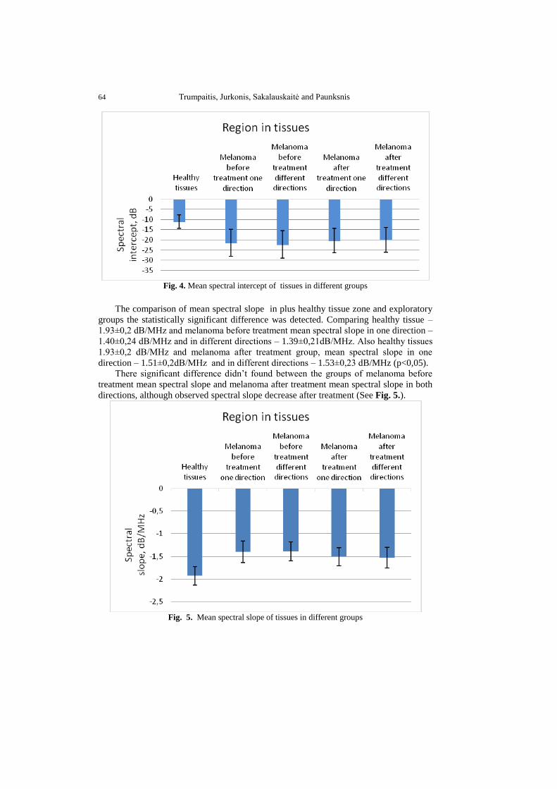
The comparison of mean spectral slope in plus healthy tissue zone and exploratory
groups the statistically significant difference was detected. Comparing healthy tissue –
1.93±0,2 dB/MHz and melanoma before treatment mean spectral slope in one direction –
1.40±0,24 dB/MHz and in different directions – 1.39±0,21dB/MHz. Also healthy tissues
1.93±0,2 dB/MHz and melanoma after treatment group, mean spectral slope in one
direction – 1.51±0,2dB/MHz and in different directions – 1.53±0,23 dB/MHz (p<0,05).
There significant difference didn’t found between the groups of melanoma before
treatment mean spectral slope and melanoma after treatment mean spectral slope in both
directions, although observed spectral slope decrease after treatment (See Fig. 5.).
Fig. 5. Mean spectral slope of tissues in different groups
Application of Ultrasound Spectral Analysis for Choroidal Melanomas Analysis 65
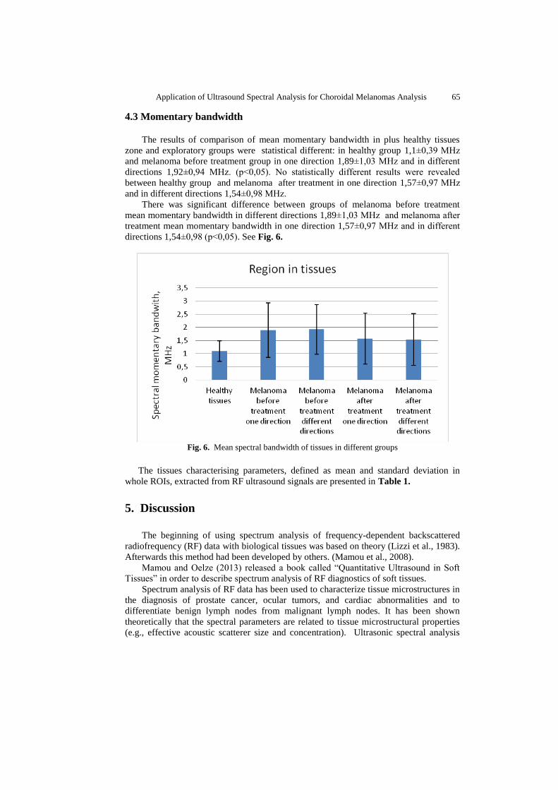
4.3 Momentary bandwidth
The results of comparison of mean momentary bandwidth in plus healthy tissues
zone and exploratory groups were statistical different: in healthy group 1,1±0,39 MHz
and melanoma before treatment group in one direction 1,89±1,03 MHz and in different
directions 1,92±0,94 MHz. (p<0,05). No statistically different results were revealed
between healthy group and melanoma after treatment in one direction 1,57±0,97 MHz
and in different directions 1,54±0,98 MHz.
There was significant difference between groups of melanoma before treatment
mean momentary bandwidth in different directions 1,89±1,03 MHz and melanoma after
treatment mean momentary bandwidth in one direction 1,57±0,97 MHz and in different
directions 1,54±0,98 (p<0,05). See Fig. 6.
Fig. 6. Mean spectral bandwidth of tissues in different groups
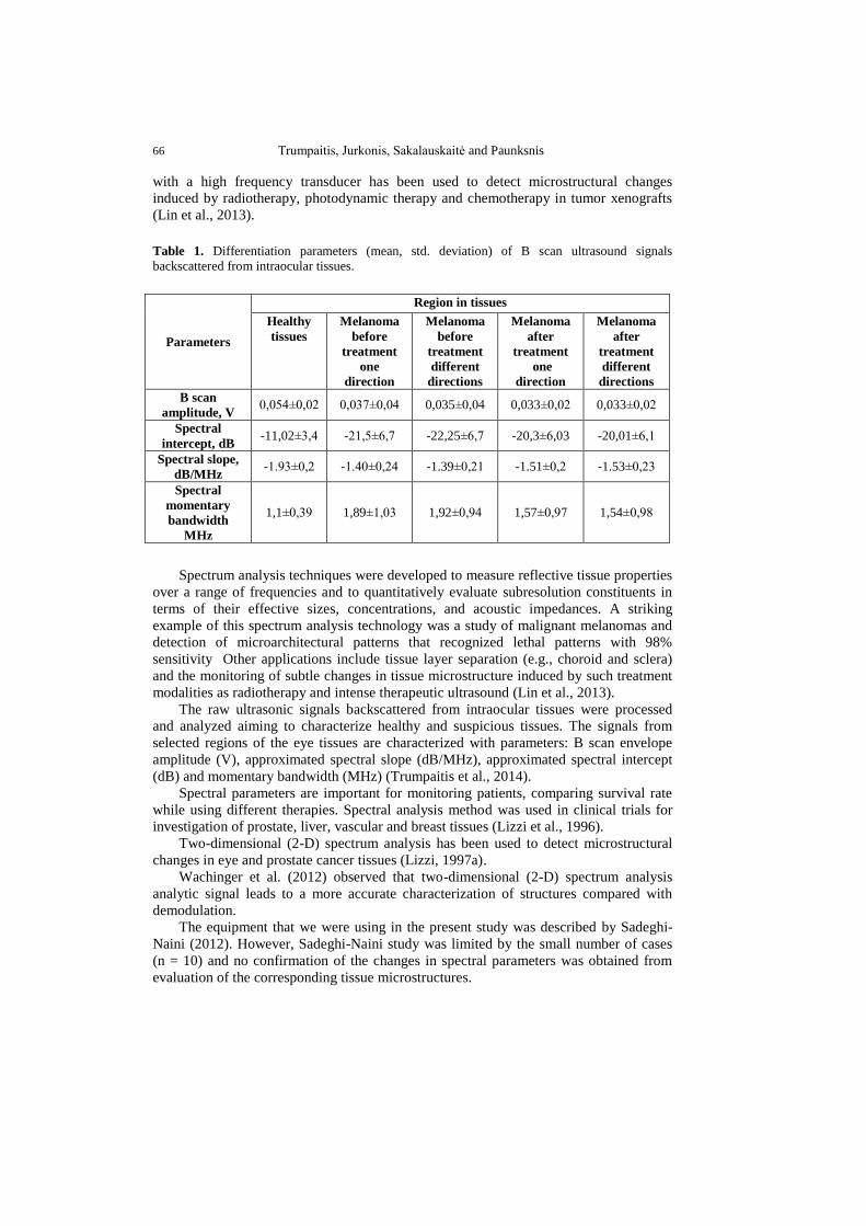
The tissues characterising parameters, defined as mean and standard deviation in
whole ROIs, extracted from RF ultrasound signals are presented in Table 1.
5. Discussion
The beginning of using spectrum analysis of frequency-dependent backscattered
radiofrequency (RF) data with biological tissues was based on theory (Lizzi et al., 1983).
Afterwards this method had been developed by others. (Mamou et al., 2008).
Mamou and Oelze (2013) released a book called “Quantitative Ultrasound in Soft
Tissues” in order to describe spectrum analysis of RF diagnostics of soft tissues.
Spectrum analysis of RF data has been used to characterize tissue microstructures in
the diagnosis of prostate cancer, ocular tumors, and cardiac abnormalities and to
differentiate benign lymph nodes from malignant lymph nodes. It has been shown
theoretically that the spectral parameters are related to tissue microstructural properties
(e.g., effective acoustic scatterer size and concentration). Ultrasonic spectral analysis
66 Trumpaitis, Jurkonis, Sakalauskaitė and Paunksnis
with a high frequency transducer has been used to detect microstructural changes
induced by radiotherapy, photodynamic therapy and chemotherapy in tumor xenografts
(Lin et al., 2013).
Table 1. Differentiation parameters (mean, std. deviation) of B scan ultrasound signals
backscattered from intraocular tissues.
Parameters
Region in tissues
Healthy
tissues
Melanoma
before
treatment
one
direction
Melanoma
before
treatment
different
directions
Melanoma
after
treatment
one
direction
Melanoma
after
treatment
different
directions
B scan
amplitude, V 0,054±0,02 0,037±0,04 0,035±0,04 0,033±0,02 0,033±0,02
Spectral
intercept, dB -11,02±3,4 -21,5±6,7 -22,25±6,7 -20,3±6,03 -20,01±6,1
Spectral slope,
dB/MHz -1.93±0,2 -1.40±0,24 -1.39±0,21 -1.51±0,2 -1.53±0,23
Spectral
momentary
bandwidth
MHz
1,1±0,39 1,89±1,03 1,92±0,94 1,57±0,97 1,54±0,98
Spectrum analysis techniques were developed to measure reflective tissue properties
over a range of frequencies and to quantitatively evaluate subresolution constituents in
terms of their effective sizes, concentrations, and acoustic impedances. A striking
example of this spectrum analysis technology was a study of malignant melanomas and
detection of microarchitectural patterns that recognized lethal patterns with 98%
sensitivity Other applications include tissue layer separation (e.g., choroid and sclera)
and the monitoring of subtle changes in tissue microstructure induced by such treatment
modalities as radiotherapy and intense therapeutic ultrasound (Lin et al., 2013).
The raw ultrasonic signals backscattered from intraocular tissues were processed
and analyzed aiming to characterize healthy and suspicious tissues. The signals from
selected regions of the eye tissues are characterized with parameters: B scan envelope
amplitude (V), approximated spectral slope (dB/MHz), approximated spectral intercept
(dB) and momentary bandwidth (MHz) (Trumpaitis et al., 2014).
Spectral parameters are important for monitoring patients, comparing survival rate
while using different therapies. Spectral analysis method was used in clinical trials for
investigation of prostate, liver, vascular and breast tissues (Lizzi et al., 1996).
Two-dimensional (2-D) spectrum analysis has been used to detect microstructural
changes in eye and prostate cancer tissues (Lizzi, 1997a).
Wachinger et al. (2012) observed that two-dimensional (2-D) spectrum analysis
analytic signal leads to a more accurate characterization of structures compared with
demodulation.
The equipment that we were using in the present study was described by Sadeghi-
Naini (2012). However, Sadeghi-Naini study was limited by the small number of cases
(n = 10) and no confirmation of the changes in spectral parameters was obtained from
evaluation of the corresponding tissue microstructures.
Application of Ultrasound Spectral Analysis for Choroidal Melanomas Analysis 67
Mortality rate of melanoma significantly depends on the histological structure
(Coleman et al., 1983). Non-invasive ultrasound system which measures spectral
analysis parameters (intercept, amplitude and slope) helps to differentiate histological
structure of the tumor (Coleman et al., 1983).
Our study has shown that the lower amplitude, lower spectral intercept, high
spectral slope and high momentary bandwidth are typical for choroidal melanoma if
compared with healthy tissues. This allows us to distinguish healthy tissues from the
abnormal tumor tissues and also to identify choroidal melanoma. This study has shown
that the lower momentary bandwidth are typical for choroidal melanoma after treatment
if compared with melanoma before treatment (p<0,05).
Spectral analysis is useful to assess the efficiency of treatment, but requires a longer
observation time.
6. Conclusions
The obtained results show that there is statistically significant difference between
the RF ultrasound signal parameters (amplitude envelope, spectrum slope, spectrum
intercept and momentary bandwidth) in the healthy tissue area and in the area with the
intraocular tumor – melanoma before and after treatment (p<0,05).
This study has shown that the lower momentary bandwidth are typical for choroidal
melanoma after treatment if compared with melanoma before treatment. It was statistical
significant difference (p<0,05).
We also noticed some differences in the RF parameters before melanoma treatment
and after treatment but they were not statistically significant (p>0,05): amplitude
decrease after treatment, spectral intercept increase after treatment and spectral slope
decrease after treatment.
Results of B scan showed that the mean basis and mean height of melanoma tumor
after treatment was significantly lower 11,8 mm and 4,9 mm if compared with 12,8 mm
and 7,0 mm before treatment.
Non-invasive ultrasound expert system provides the new opportunities in early
diagnosis, differentiation of tumors, observation of the process, evaluation of the
treatment effectiveness as well as prognosis of survival. Spectral analysis is useful to
assess the efficiency of treatment, but requires a longer observation time.
References
Aironi, V.D., Chougule, S.R., Singh, J. (2007). Choroidal melanoma: B-scan spectrum. Indian
Journal of Radiology and Imaging. 17, 8–10.
Bell, D.J., Wilson, M.W. (2004). Choroidal melanoma: natural history and management options.
Cancer Control: Journal of the Moffitt Cancer Center. 11(5), 296–303.
Boudinet, M., Berges, O., LeHuerou, J.Y., Lumbroso-LeRouic, L., Desjardins, L., Laugier, P.
(2007). Quantitative echography in the follow-up of patients treated with proton-beam
irradiation for primary choroidal melanomas. Ultrasound in Medicine and Biology. 33,
1046–1056.
Brady, L.W., Shields, J.A., Augsburger, J.J., Day, J.L. (1982). Malignant intraocular tumors.
Cancer. 49, 578–585.
68 Trumpaitis, Jurkonis, Sakalauskaitė and Paunksnis
Castro, J.R., Char, D.H., Petti, P.L., Daftari, I.K., Quivey, J.M., Singh, R.P., Blakely, E.A.,
Phillips, T.L. (1997). 15 Years experience with helium ionradiotherapy for uveal melanoma.
International Journal of Radiation Oncology Biology Physics. 39, 989–996.
Coleman, D.J., Lizzi, F.L., Silverman, R.H,, Rondeau, M.J., Smith, M.E., Torpey, J.H. (1983).
Acoustic biopsy as a means for characterization of intraocular tumors. American Academy
of Ophthalmology, Acta: XXIV International Congress of Ophthalmology, edited by Paul
Henkind, MD, J.B. Lippincott Company, Philadelphia, PA, 115–118.
Coleman, D.J., Silverman, R.H., Chabi, A., Rondeau, M.J., Shung, K.K., Cannata, J., Lincoff, H.
(2004) High-resolution ultrasonic imaging of the posterior segment. Ophthalmology. 111,
1344–1351.
Damato, B. (2004). Developments in the management of uveal melanoma. Clinical and
Experimental Ophthalmology. 32, 639–647.
Damato, B., Jones, A.G. (2005). Uveal melanoma: resection techniques. Ophthalmology Clinics of
North America. 18, 119–128.
Damato, B.E., Coupland, S.E. (2012a). Differences in uveal melanomas between men and women
from the British Isles. Eye (London). 26, 292–299.
Damato, B.E., Coupland S.E. (2012b). Ocular melanoma. Saudi Journal of Ophthalmology. 26(2),
137–144.
Egan, K.M., Seddon, J.M., Glynn, R.J., Gragoudas, E.S., Albert, D.M. (1988). Epidemiologic
aspects of uveal melanoma. Survey of Ophthalmology. 32, 239–251.
Fakiris, A.J., Lo, S.S., Henderson, M.A., Witt, T.C., Worth, R.M., Danis, R.P., Des Rosiers, P.M.,
Timmerman, R.D. (2013). Gamma-knife-based stereotactic radiosurgery for uveal
melanoma. Stereotactic and Functional Neurosurgery. 85, 106–112.
Finger, P.T., Berson, A., Ng, T., Szechter A. (2002). Palladium-103 plaque radiotherapy for
choroidal melanoma: an 11-year study. International Journal of Radiation Oncology Biology
Physic. 54, 1438–1445.
Goldberg, M.F., Hodes, B.L. (1977). Ultrasonographic diagnosis of choroidal malignant
melanoma. Survey of Ophthalmology. 22, 29–40.
Gragoudas, E.S. (2006). Proton beam irradiation of uveal melanomas: the first 30 years. The
Weisenfeld Lecture. Investigative Ophthalmology & Visual Science. 47(11), 4666–4673.
Hoffmann, K., el Gammal S,, Matthes, U., Altmeyer, P. (1989). Digital 20 mhz sonography of the
skin in preoperative diagnosis. Z Hautkr. 64, 851–858.
Houston, S.K., Boldt, H. C., Markoe, A.M., Murrayresearch, T.G. Brachytherapy for Choroidal
Melanoma. Chapter 145. Section 2. 2275–2289.
Jurkonis, R., Daukantas, S., Janušauskas, A., Lukoševičius, A., Marozas, V., Jegelevičius, D.
(2009a). Synthesis of parametric map from raw ultrasound B-Scan data. Electronics and
Electrical Engineering. 6(94), 109–112.
Jurkonis, R., Jegelevicius, D., Marozas, V. (2009b). Virtual Instrument for Ultrasound Signal
Sampling and Formation of B scan Image of Eye. Biomedical Engineering Conference
“Virtual Instruments in biomedicine 2009“. Klaipėda University, Klaipėda, 15 May, 2009,
55–60.
Jurkonis, R., Trumpaitis, J., Šarkūnaitė, D., Paunksnis, A. (2012). Application of ultrasound
radiofrequency analysis for intraocular tissues differentiation. Biomedical Engineering
Conference 2012.
Kaiserman, I., Anteby, I., Chowers, I., Blumenthal, E.Z., Kliers, I., Pe'er, J. (2002). Changes in
ultrasound findings in posterior uveal melanoma after Ruthenium 106 brachytherapy.
Ophthalmology. 109(6), 1137–1141.
Kath, R., Hayungs, J., Bornfeld, N., Sauerwein, W., Höffken, K., Seeber, S. (1993) Prognosis and
treatment of disseminated uveal melanoma. Cancer. 72, 2219–2223.
Kindy-Degnan, N.A., Char, D.H., Castro, J.R., Kroll, S., Stone, R.D., Quivey, J.M., Phillips, T.L.,
Irvine, A.R. (1989). Effect of various doses of radiation for uveal melanoma on regression,
visual acuity, complications, and survival. American Journal of Ophthalmology. 107, 114–
120.
Application of Ultrasound Spectral Analysis for Choroidal Melanomas Analysis 69
Kujala, E., Makitie, T., Kivelä, T. (2003) Very long-term prognosis of patients with malignant
uveal melanoma. Investigative Ophthalmology & Visual Science. 44, 4651–4659.
Lin, C., Cao, L., Wang, J., Zheng, W., Chen, Y., Feng, Z., Li, A., Zhou, J. (2013). Ultrasonic
spectrum analysis for in vivo characterization of tumor microstructural changes in the
evaluation of tumor response to chemotherapy using diagnostic ultrasound. BMC Cancer.
13, 302.
Lizzi, F.L., Greenebaum, M., Feleppa, E.J., Elbaum, M., Coleman, D.J. (1983). Theoretical
framework for spectrum analysis in ultrasonic tissue characterization. The Journal of the
Acoustical Society of America. 73(4), 1366–1373.
Lizzi, F.L., Astor, M., Liu, T., Deng, C., Coleman, D.J., Silverman, R.H. (1996). Ultrasonic
Spectrum Analysis for Tissue Assays and Therapy Evaluation. Cornell University Medical
College, 1300 York Avenue, New York, NY, 1002.
Lizzi, F.L. (1997a). Ultrasonic scatterer-property images of the eye and prostate. In Proceedings of
the IEEE Ultrasonics Symposium. Piscataway, NJ, USA.
Lizzi, F.L., Astor, M., Liu T., Deng, C., Coleman D.J., Silverman R.H. (1997b). Ultrasonic
spectrum analysis for tissue assays and therapy evaluation. International Journal of Imaging
Systems and Technology. 8, 3–10.
Lizzi, F.L., Coleman, J. (2004). History of Ophthalmic Ultrasound 2004 by the American Institute
of Ultrasound in Medicine. Journal of Ultrasound in Medicine. 23, 1255–1266.
Lommatzsch, P.K., Kirsch, I.H. (1988). 106Ru/106Rh plaque radiotherapy for malignant
melanomas of the choroid. With follow–up results more than 5 years. Documenta
Ophthalmologica. 68, 225–238.
Lorigan, J.G., Wallace, S., Mavligit, G.M. (1991). The prevalence and location of metastases from
ocular melanoma: imaging study in 110 patients. American Journal of Roentgenology.
157(6), 1279–1281
Mamou J. and Oelze, M.L. (2013). Quantitative Ultrasound Methods in Soft Tissues. Springer,
New York, USA, 3–20.
Mamou, J., Ketterling, J.A., Silverman, R.H. (2008). Chirp-coded excitation imaging with a high-
frequency ultrasound annular array. Ultrasonics, Ferroelectrics, and Frequency Control,
IEEE. 55(2), 508–513.
McLean, I.W., Foster, W.D., Zimmerman, L.E., Martin, D.G. (1980). Inferred natural history of
uveal melanoma. Investigative Ophthalmology & Visual Science. 19, 760–770.
Modorati, G., Miserocchi, E., Galli, L., Picozzi, P., Rama, P. (2009). Gamma knife radiosurgery
for uveal melanoma: 12 years of experience. British Journal of Ophthalmology. 93, 40–44.
Mooy, C.M., De Jong, P.T. (1996). Prognostic parameters in uveal melanoma: a review. Survey of
Ophthalmology. 41, 215–228.
Moore, R.F. (1930). Choroidal sarcoma treated by the intraocular insertion of radon seeds. British
Journal of Ophthalmology. 14, 145–152.
Mosci, C., Lanza, F.B., Mosci, S., Barla, A. (2014). Quantitative echography in primary uveal
melanoma treated by proton beam therapy. Canadian Journal of Ophthalmology. 49(1), 60–
65.
Moura, R.A., McPherson, A.R., Easley, J. (1985). Malignant melanoma of the choroid: treatment
with episcleral 198Au plaque and xenon-arc photocoagulation. Annals of Ophthalmology.
17, 114–125.
Nissen, S., Grines, C., Gurley, J. Sublett, K., Haynie, D., Diaz, C., Booth, D.C., DeMaria, A.N.
(1990). Application of a new phased-array ultrasound imaging catheter in the assessment of
vascular dimension: In vivo comparison to cineangiography. Circulation. 81, 660–666.
Overgaard, J., Overgaard, M., Hansen, P.V., von der Maase, H. (1986). Some factors of
importance in the radiation treatment of malignant melanoma. Radiotherapy and Oncology.
5, 183–192.
Packer, S., Stoller, S., Lesser, M.L, Mandel, F.S. Finger, P.T. (1992). Long-term results of iodine
125 irradiation of uveal melanoma. Ophthalmology. 99, 767–73; discussion 774.
Pavlin, C.J., Sherar, M.D., Foster, F.S. (1990). Subsurface imaging of the eye by ultrasound
biomicroscopy. Ophthalmology. 97, 244–250.
70 Trumpaitis, Jurkonis, Sakalauskaitė and Paunksnis
Paunksnis, A., Barzdžiukas, V., Kažys, R., Raišutis, R., Lukoševičius, A., Paunksnis, M.,
Janušauskas, A., Marozas, V., Jegelevičius, D., Daukantas, S., Kopsala S., Kurapkienė S.,
Kriaučiūnienė L., Jurkonis R. (2008). A non-invasive expert system for diagnosis of
intraocular tumours: the system concept. Ultragarsas (Ultrasound). 63(4), 66–72.
Puzienė, V., Jegelevičius, D., Lukoševičius, A., Jurkonis, R., Paunksnis, A., Barzdžiukas, V.
(2010). Progress towards noninvasive intraocular tumor diagnostics. Ultragarsas
(Ultrasound). 65(2), 47–51.
Piñeiro-Ces, A., Rodríguez Alvarez, M.J., Santiago, M., Bande, M., Pardo, M., Capeáns, C.,
Blanco, M.J. (2014). Detecting ultrasonographic hollowness in small choroidal melanocytic
tumors using 10 MHz and 20 MHz ultrasonography: a comparative study. Graefe's Archive
for Clinical and Experimental Ophthalmology. 252(12), 2005–2011.
Ramasamy, P., Murphy C.C., Clynes, M., Horgan, N., Moriarty, P., Tiernan, D., Beatty S.,
Kennedy, S., Paula, M. (2014). Proteomics in uveal melanoma. Experimental Eye Research.
118, 1–12.
Ryan, S.J., Schachat, A.P., Murphy, R.P. (1989). Retina. 1st Edition, Vol. 2. St. Louis: Mosby, p.
170.
Sadeghi-Naini, A., Falou, O., Hudson, J.M., Kolios, M.C., Czarnota, G.J. (2012). Conventional
frequency ultrasonic biomarkers of cancer treatment response in vivo. Translational
oncology. 6(3), 234–243.
Saornil, M.A., Egan, K.M., Gragoudas, E.S., Seddon, J.M., Walsh, S.M., Albert, D.M. (1992).
Histopathology of proton beam-irradiated vs enucleated uveal melanomas. Archives of
ophthalmology. 110(8), 1112–1118.
Shields, C.L., Shields, J.A., Karlsson, U., Menduke, H., Brady, L.W. (1990). Enucleation after
plaque radiotherapy for posterior uveal melanoma. Ophthalmology. 97, 1665–1970.
Shields, J.A., Shields, C.L., Donoso, L.A. (1991). Management of posterior uveal melanomas.
Survey of Ophthalmology. 36, 161–95.
Shields, J.A., Shields, C.L., De Potter, P., Cu-Unjieng, A., Hernandez C., Brady, L.W. (1993).
Plaque radiotherapy for uveal melanoma. International Ophthalmology Clinics. 33, 129–35.
Shields, C.L., Shields, J.A., DePotter, P., Kheterpal, S. (1996). Transpupillary thermotherapy in
the management of choroidal melanoma. Ophthalmology. 103, 1642–1650.
Shields, C.L., Shields, J.A., Cater, J., Lois, N., Edelstein, C., Gündüz, K., Mercado, G. (1998).
Transpupillary thermotherapy for choroidal melanoma: tumor control and visual results in
100 consecutive cases. Ophthalmology. 105, 581–590.
Singh, A.D., Topham, A., (2003). Survival rates with uveal melanoma in the United States: 1973–
1997. Ophthalmology. 110, 962–965.
Singh, A.D., Bergman, L., Seregard, S. (2005). Uveal melanoma: epidemiologic aspects.
Ophthalmology Clinics of North America. 18(1), 75–84.
Straatsma, B.R., Fine, S.L., Earle, J.D., Hawkins, B.S., Diener-West, M., McLaughlin, J.A.
(1988). Enucleation versus plaque irradiation for choroidal melanoma. Ophthalmology. 95,
1000–10004.
The Collaborative Ocular Melanoma Study (COMS) randomized trial of pre-enucleation radiation
of large choroidal melanoma II: initial mortality findings. (1998). COMS report No. 10.
American Journal of Ophthalmology. 125, 779–796.
Toktas, Z.O., Bicer, A., Demirci, G., Pazarli, H., Abacioglu, U., Peker, S., Kilic, T. (2010).
Gamma knife stereotactic radiosurgery yields good long-term outcomes for low-volume
uveal melanomas without intraocular complications. Journal of Clinical Neuroscience.
17(4), 441–445.
Trott, K.R. (1991). The optimal radiation dose per fraction for the treatment of malignant
melanomas. International Journal of Radiation Oncology Biology Physics. 20, 905–907.
Trumpaitis, J., Jurkonis, R., Imbrasienė, D. Grizickaitė, A., Paunksnis A. (2014). Application of
ultrasound spectral analysis for intraocular tissues differentiation. Journal of
Vibroengineering. 16(7), 3586.
Wachinger, C., Klein, T., Navab, N. (2012). The 2D analytic signal for envelope detection and
feature extraction on ultrasound images. Medical Image Analysis. 16(6), 1073–1084.
Application of Ultrasound Spectral Analysis for Choroidal Melanomas Analysis 71
Wen, J.C., McCannel, T.A. (2009). Treatment of radiation retinopathy following plaque
brachytherapy for choroidal melanoma. Current Opinion in Ophthalmology. 20, 200–204.
Zakka, K.A., Foos, R.Y., Omphroy, C.A., Straatsma, B.R. (1980). Malignant melanoma: analysis
of an autopsy population. Ophthalmology. 87, 549–556.
Zimmerman, L.E., McLean, I.W., Foster, W.D. (1980). Statistical analysis of follow-up data
concerning uveal melanomas, and the influence of enucleation. Ophthalmology. 87, 557–
564.
Authors’ information
Jurgis Trumpaitis received M.D. degree in Lithuanian University of Health Sciences
Kaunas, Lithuania, in 2006. He finished ophthalmology residency in 2010. Now he
works at Hospital of Lithuanian University of Health Sciences Kaunas Clinics as a
doctor of ophthalmology. He is a doctoral student at the final year. He is junior
researcher in department of Ophthalmology, Neuroscience Institute, Ophthalmology
laboratory, Kaunas, Lithuania. His current research interests include analysis of
ultrasonic tissue characterization of intraocular tumours.
Rytis Jurkonis received PhD degree in Electrical and Electronics Engineering from
Kaunas University of Technology, Lithuania, in 2000. Now he works at Biomedical
Engineering Institute of Kaunas University of Technology. His current research interests
include analysis of raw ultrasonic signals and ultrasonic tissue characterization.
Ieva Sakalauskaitė currently is a third year medicine student in Lithuanian University
of Health Sciences. She is interested in ophthalmology and ultrasonography.
Alvydas Paunksnis, Dr. Habil. Since 1997 Professor at Ophthalmology Clinics of
Lithuanian University of Health Sciences, Kaunas. Ophthalmologist since 1970. Chief
Researcher of Lithuanian University of Health Sciences Kaunas since 2008. Research
interests: imaging, recognition of images in ophthalmology, ultrasonic characterization
of eye tissues, e-health, telemedicine.
Received February 11, 2015, revised March 16, 2015, accepted March 19, 2015


![OPEN ACCESS Case Report Congenital Choroidal Nevus in a ...choroidal nevus) [10]; likewise, the nevus is characterized by having a high internal reflectivity, unlike the melanoma that](https://static.fdocuments.us/doc/165x107/5ea21f6a6c088018070115eb/open-access-case-report-congenital-choroidal-nevus-in-a-choroidal-nevus-10.jpg)