
Application of Tomosynthesis for Novel Coronavirus Pneumonia
6
R/F 1 COVID-19 Extra Issue (2020) Wu Zhengguang, M.D. (Chief Physician, Department of Radiology, Guangdong Second Provincial People’s Hospital) contributed an article on thorax tomosynthesis by SONIALVISION G4 TM for the treatment of patients with COVID-19 pneumonia. This article presents the report given by Wu Zhengguang, M.D.. 1. Introduction Guangdong Second Provincial People’s Hospital (Fig. 1) was founded in 1947 and is located on the south bank of the Pearl River, at the central area of the beautiful Yangcheng District—Guangzhou Xincheng City. We have overcome the long and hard time of 73 years with both wind and rain. It is becoming more and more magnificent, like the water flowing in the Pearl River, and merging with rivers along the way. At present, our hospital has 64 clinical departments and 59 specialized departments, with 2300 beds (will be 4000 beds near in future) and more than 2500 medical professionals, and 9 clinical departments have become important departments in Guangdong Province. There are more than 6000 outpatients per day and the hospital earns an annual income of RMB 2.1 billion. It is a general hospital that integrates medical, emergency, prevention, rehabilitation, education and scientific research, and is a designated health insurance facility in Guangzhou City, Guangdong Province. As the WHO International Emergency Medical Team and the nation’s first provincial emergency hospital, our hospital has become a pioneer in the development of sanitary emergency medical services. As the only emergency medical team in the Chinese government, the Emergency Medical Response Team was dispatched to Malaysia on behalf of the government, successfully completing the fourth disaster relief exercise mission of the ASEAN Regional Forum and received high praise from the National Health Commission. In our hospital, SONIALVISION G4 manufactured by Shimadzu Corporation was installed in October 2017, and its clinical use was officially started in January 2018. This SONIALVISION G4 has the latest tomosynthesis and slot radiography (long view image) functions in addition to the conventional digestive examination function, and it has been fully utilized in our department for two and a half years (Table 1). Table 1 Clinical cases using SONIALVISION G4 Examination GI series Tomosynthesis chest Tomosynthesis bone and joint SLOT radiography Spine and leg Average number of cases per day 5 20 3 2 Average number of cases per month 100 500 75 50 We have been researching and studying pneumonia and pulmonary emphysema for a long time. We believe that tomosynthesis is highly accurate in chest examinations compared with DR examinations and is more convenient than CT examinations. ● Usability and convenience: Patients can be examined while standing or recumbent (Fig. 2). Application of Tomosynthesis for Novel Coronavirus Pneumonia Chief Physician, Department of Radiology, Guangdong Second Provincial People’s Hospital, Guangzhou, China Vice Chairman, Imaging Expert Committee, China Medical Association Wu Zhengguang Wu Zhengguang, M.D. Fig.1 View of the hospital
Transcript of Application of Tomosynthesis for Novel Coronavirus Pneumonia
R/F
1 COVID-19 Extra Issue (2020)
Wu Zhengguang, M.D. (Chief Physician, Department of Radiology, Guangdong Second Provincial People’s Hospital) contributed an article on thorax tomosynthesis by SONIALVISION G4TM for the treatment of patients with COVID-19 pneumonia. This article presents the report given by Wu Zhengguang, M.D..
1. Introduction
Guangdong Second Provincial People’s Hospital (Fig. 1) was founded in 1947 and is located on the south bank of the Pearl River, at the central area of the beautiful Yangcheng District—Guangzhou Xincheng City. We have overcome the long and hard time of 73 years with both wind and rain. It is becoming more and more magnificent, like the water flowing in the Pearl River, and merging with rivers along the way.At present, our hospital has 64 clinical departments and 59 specialized departments, with 2300 beds (will be 4000 beds near in future) and more than 2500 medical professionals, and 9 clinical departments have become important departments in Guangdong Province. There are more than 6000 outpatients per day and the hospital earns an annual income of RMB 2.1 billion. It is a general hospital that integrates medical, emergency, prevention, rehabilitation,
education and scientific research, and is a designated heal th insurance fac i l i ty in Guangzhou Ci ty, Guangdong Province.As the WHO International Emergency Medical Team and the nation’s first provincial emergency hospital, our hospital has become a pioneer in the development of sanitary emergency medical services. As the only emergency medical team in the Chinese government, the Emergency Medical Response Team was dispatched to Malaysia on behalf of the government, successfully completing the fourth disaster relief exercise mission of the ASEAN Regional Forum and received high praise from the National Health Commission.In our hospital, SONIALVISION G4 manufactured by Shimadzu Corporation was installed in October 2017, and its clinical use was officially started in January 2018. This SONIALVISION G4 has the latest tomosynthesis and slot radiography (long view image) functions in addition to the conventional digestive examination function, and it has been fully utilized in our department for two and a half years (Table 1).
Table 1 Clinical cases using SONIALVISION G4
Examination
GI series
Tomosynthesis chest
Tomosynthesis
bone and joint
SLOT
radiography Spine and leg
Average number of cases per day 5 20 3 2
Average number of cases per month 100 500 75 50
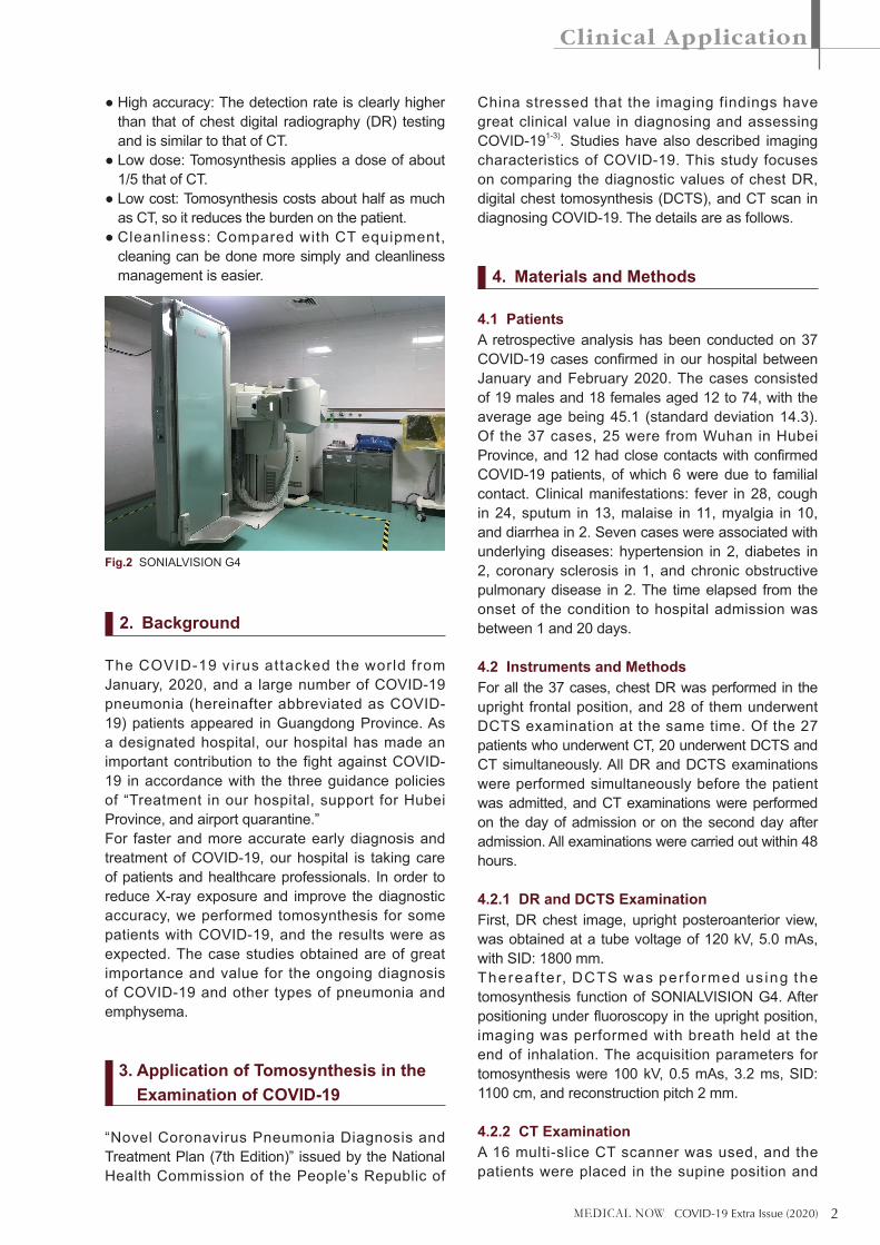
We have been researching and studying pneumonia and pulmonary emphysema for a long time. We believe that tomosynthesis is highly accurate in chest examinations compared with DR examinations and is more convenient than CT examinations.● Usabil ity and convenience: Patients can be
examined while standing or recumbent (Fig. 2).
Application of Tomosynthesis for Novel Coronavirus Pneumonia
Chief Physician, Department of Radiology,Guangdong Second Provincial People’s Hospital, Guangzhou, ChinaVice Chairman, Imaging Expert Committee, China Medical AssociationWu Zhengguang
Wu Zhengguang, M.D.
Fig.1 View of the hospital
2COVID-19 Extra Issue (2020)
● High accuracy: The detection rate is clearly higher than that of chest digital radiography (DR) testing and is similar to that of CT.
● Low dose: Tomosynthesis applies a dose of about 1/5 that of CT.
● Low cost: Tomosynthesis costs about half as much as CT, so it reduces the burden on the patient.
● Cleanliness: Compared with CT equipment, cleaning can be done more simply and cleanliness management is easier.
2. Background
The COVID-19 virus attacked the world from January, 2020, and a large number of COVID-19 pneumonia (hereinafter abbreviated as COVID-19) patients appeared in Guangdong Province. As a designated hospital, our hospital has made an important contribution to the fight against COVID-19 in accordance with the three guidance policies of “Treatment in our hospital, support for Hubei Province, and airport quarantine.”For faster and more accurate early diagnosis and treatment of COVID-19, our hospital is taking care of patients and healthcare professionals. In order to reduce X-ray exposure and improve the diagnostic accuracy, we performed tomosynthesis for some patients with COVID-19, and the results were as expected. The case studies obtained are of great importance and value for the ongoing diagnosis of COVID-19 and other types of pneumonia and emphysema.
3. Application of Tomosynthesis in the Examination of COVID-19
“Novel Coronavirus Pneumonia Diagnosis and Treatment Plan (7th Edition)” issued by the National Health Commission of the People’s Republic of
China stressed that the imaging findings have great clinical value in diagnosing and assessing COVID-191-3). Studies have also described imaging characteristics of COVID-19. This study focuses on comparing the diagnostic values of chest DR, digital chest tomosynthesis (DCTS), and CT scan in diagnosing COVID-19. The details are as follows.
4. Materials and Methods
4.1 PatientsA retrospective analysis has been conducted on 37 COVID-19 cases confirmed in our hospital between January and February 2020. The cases consisted of 19 males and 18 females aged 12 to 74, with the average age being 45.1 (standard deviation 14.3). Of the 37 cases, 25 were from Wuhan in Hubei Province, and 12 had close contacts with confirmed COVID-19 patients, of which 6 were due to familial contact. Clinical manifestations: fever in 28, cough in 24, sputum in 13, malaise in 11, myalgia in 10, and diarrhea in 2. Seven cases were associated with underlying diseases: hypertension in 2, diabetes in 2, coronary sclerosis in 1, and chronic obstructive pulmonary disease in 2. The time elapsed from the onset of the condition to hospital admission was between 1 and 20 days.
4.2 Instruments and MethodsFor all the 37 cases, chest DR was performed in the upright frontal position, and 28 of them underwent DCTS examination at the same time. Of the 27 patients who underwent CT, 20 underwent DCTS and CT simultaneously. All DR and DCTS examinations were performed simultaneously before the patient was admitted, and CT examinations were performed on the day of admission or on the second day after admission. All examinations were carried out within 48 hours.
4.2.1 DR and DCTS ExaminationFirst, DR chest image, upright posteroanterior view, was obtained at a tube voltage of 120 kV, 5.0 mAs, with SID: 1800 mm.Thereaf ter, DCTS was per formed us ing the tomosynthesis function of SONIALVISION G4. After positioning under fluoroscopy in the upright position, imaging was performed with breath held at the end of inhalation. The acquisition parameters for tomosynthesis were 100 kV, 0.5 mAs, 3.2 ms, SID: 1100 cm, and reconstruction pitch 2 mm.
4.2.2 CT ExaminationA 16 multi-slice CT scanner was used, and the patients were placed in the supine position and
Fig.2 SONIALVISION G4
3 COVID-19 Extra Issue (2020)
scanned with breath held at the end of inhalation. The scanning range was from the apex of lung to the lower section of posterior costophrenic angle. Scanning parameters: 120 kV, 250 mAs, field of view was 250 mm × 250 mm, the scanning and reconstruction matrix was 512 × 512, the scanning layer thickness was 5 mm, the reconstruction layer thickness was 1 to 2 mm, and the reconstruction interval was 0.6 mm. Image observation: Pulmonary window (window width of 1,500 to 2,000 HU, window level of -700 to -500 HU), mediastinal window (window width of 250 to 300 HU, window level of 30 to 55 HU). Informed consent has been obtained from all patients.
4.3 Image Post-ProcessingChest DR, DCTS, and CT images were transmitted to a PACS workstation in DICOM format and were evaluated by 2 senior doctors to confirm distribution and density difference of lesion, internal structure, and edge clarity. The ground-glass opacity, infiltrates, nodular shadow, small blood vessel thickening, bronchiolectasis, interlobular septa thickening, reversed-halo sign (RHS), and involvement of pulmonary lobes were observed.
4.4 Statistical MethodsSPSS 22.0 statistical software was used to process the data, and Fisher’s exact probability method was used to test the display of image signs with different examination methods. A result of P < 0.05 was considered to be a statistically significant difference.
5. Results
Of the 37 thoracic upright frontal DR images, 10 cases showed patchy ground-glass opacities, distributed mainly in the periphery of the lung fields (Fig. 3a), of which 1 case showed patchy infiltrates in both lungs. In the other 27 cases, no definite intrapulmonary lesion was detected on chest DR images. Of the 28 DCTS images, 21 cases showed multiple snowflake ground-glass density foci distributed mainly around the periphery of both lung fields, 7 cases of which showed small vessel thickening, bronchiolectasis, and a nondescript thickening of the bronchiole walls (Fig. 3b, c). Two cases had patchy pulmonary infiltrates, 2 cases had ground-glass opacities (Fig. 4b, c), and 2 cases had a reversed-halo sign (Fig. 5b, c). Among the 27 CT scans, 24 cases had ground-glass opacities in one or both lungs, including 19 cases with small vessel thickening, 13 cases with bronchiolectasis, 15 cases with interlobular thickening (Fig. 6), 2 cases with reversed-halo sign, and 2 cases with infiltrative and
nodular opacities (Table 2).For visualization of intrapulmonary ground-glass opacities, there was a salient statistical difference between DR and DCTS (P < 0.05), therefore DCTS was superior to DR in delineating intrapulmonary ground-glass opacities. There was no salient statistical difference between DCTS and CT scan (P > 0.05). This indicated that there is no significant difference between DCTS and CT scan in the delineation ability of ground-glass opacity in the lung.
6. Discussion
COVID-19 is a sudden acute infectious disease of the respi ratory system. Imaging p lays an irreplaceable role in detecting lesions, understanding the scope of lesions, comprehensively evaluating the severity of lesions, and establishing the standard for hospital discharge.“Novel Coronavirus Pneumonia Diagnosis and Treatment Plan (1st to 7th Editions)” published by the National Health Commission (of the People’s Republic of China) suggests that imaging findings have important clinical value in the diagnosis and evaluation of COVID-191-3). The DR is a two-dimensional image with a lot of overlapping tissues, making it difficult to delineate fine structures. Therefore, it can be used only for the initial screening of COVID-19. The DCTS provides multiple coronary tomographic images, avoids the anatomic overlap that is a problem in chest DR imaging, and provides a clearer image of the lesion structure4). The CT scans are fast, high-resolution volume data of the chest can be obtained, and a variety of image post-processing can be performed. Therefore, the ability to detect intrapulmonary lesions and fine structures of COVID-19 is very high.
Table 2 Fisher’s exact probability test results
Category
Ground-glass
opacity
Infiltrative shadow
Nodular shadow
Microvascular
thickening
Bronchiolectasis
Interlobular septal thickening
Reversed halo
sign
DR(n = 37) 10 1 0 0 0 0 0
DCTS(n = 28) 21 2 2 7 7 0 2
CT(n = 27) 24 2 2 19 13 15 2
Fisher’s exact test with DR and DCTS: P < 0.05Fisher’s exact test with DCTS and CT: P > 0.05

4COVID-19 Extra Issue (2020)
Fig.3 Male, 53 years old, having fever, cough for 1 week, body temperature of 38.0 °C. He returned to Guangzhou from Hubei Xianning on January 20, 2020. The PCR test result was a positive on the 28th.
a) On January 24, DR chest radiograph showed bilateral diffuse distributed pulmonary shadows in lower predominance.
b) and c) On January 24, tomosynthesis (DCTS) showed multiple flack-shaped ground-glass density shadows in the bilateral pulmonary, with thickened small blood vessels.
b) c)a)
a) Chest DR image on January 29 showed no clear lesion.
b), c) (c) is a partial enlargement): DCTS showed nodular ground-glass density shadow of the left upper lung. The patchy ground-glass density shadow of the lower right lung was distributed around the bronchial vascular tree.
Fig.4 Female, 31 years old, having fever for 1 day, pharyngeal discomfort. Arrived in Wuhan on January 18, had fever of 37.9 °C on January 22, and returned to Guangzhou on January 23. Her parents were hospitalized with COVID-19 on January 28. The PCR test result was positive on January 29.
b) c)a)
a) DR chest image on January 28 showed a flaky shadow in the left lower lung with uneven density.
b), c) (c) is a reverse partial enlargement): On January 28, DCTS showed ground-glass density shadow in the left lower lung, with low center density and reversed-halo sign (RHS).
Fig.5 Female, 36 years old, flew from Wuhan to the Philippines on January 20, 2020. After returning to Guangzhou on January 28, her body temperature was 39.5 °C, without cough or sputum. The PCR test result was positive on 31 January.
b) c)a)

5 COVID-19 Extra Issue (2020)
6.1 The Diagnostic Value of DR for COVID-19Chest DR examination is one of the commonly used chest imaging methods and is used as a clinical examination technique for COVID-19. However, there are relatively many overlapping of tissues in the chest DR image, and in the early stage of COVID-19, there is little change in the lesion density and structure, thus, it is difficult for DR to detect it, and there are few findings. Therefore, DR can only be used for bedside examination and efficacy assessment of patients with severe COVID-19. In the 37 cases in this study, only 10 cases were able to detect intrapulmonary patchy opacities on chest DR imaging, indicating that the ability of chest DR imaging to detect lesions is low.
6.2 Diagnostic Value of Chest CT for COVID-19CT is transverse tomographic imaging and can process the multiplanar images such as coronal view and sagittal view. CT greatly reduces the influence of the partial volume effect. CT examination is fast and can be completed in the time of only one breath. The density and spatial resolution of images are better compared with DCTS. Therefore, “Novel Coronavirus Pneumonia Diagnosis and Treatment Plan (7th Edition),” published by the National Health Commission, suggests that the CT scan has important clinical value for diagnosis and disease state evaluation in COVID-19. In this study, among the 27 CT scans, 24 cases were positive except for 3 cases whose lesions were not found in the first chest CT scan. Compared with DCTS, CT scan detected more intrapulmonary ground-glass opacities (24 patients compared with 21 for DCTS), as well as more microstructures such as intralesional small vessel thickening, bronchiolectasis, and interlobular septal thickening. However, Fisher’s exact test showed that DCTS and CT had no statistical difference in their ability to detect ground-glass opacities in COVID-19 (P > 0.05).
6.3 Diagnostic Value of DCTS for COVID-19The DCTS technology is a new imaging technology which combines modern computer image reconstruction theory with conventional X-ray tomography technology. DCTS collects projection images of tissues and organs at different angles at low X-ray doses and uses the filtered backprojection method to obtain digital images of any coronal section. Compared with conventional chest DR images, DCTS can avoid the influence of overlapping tissues of different densities on the delineation of fine structures in the lung, and enhance the ability to detect fine lesions in the lung.Although DCTS can obtain serial multislices of the coronal portion of the chest, artificial factors such as the influence of partial volume and the patient’s respiration may reduce the ability to visualize the fine structure of lung tissue in areas of dense tissue, such as the ribs and near the mediastinum, and may miss lesions5). On the other hand, DCTS equipment is easy to install, the imaging method is simple, and the exposure dose to the patient can be reduced. DCTS and chest DR can also be performed with the same equipment, without the need to purchase an additional specialized system. Therefore, DCTS imaging can be performed immediately after chest DR imaging for cases requiring further diagnosis.In recent years, a number of domestic and international studies have reported the important role of DCTS in the detection and follow-up of thoracic nodular lesions and in the detection and evaluation of lung cancer. In addition, with the evolution and expansion of the scope of application of DCTS technology, roles in the cervical, mammary, abdominal and skeletal systems are becoming increasingly important6). Of the 28 DCTS examinations, 21 cases showed patchy ground-glass opacities in both lungs, 7 small vessel thickening, 7 bronchiolectasis, 2 reversed-halo signs, 2 infiltrates, and 2 nodular opacities in the ground-glass opacities. The DCTS has higher detectivity
a)-c) CT scan on February 1 showed different ground-glass density shadows in both lungs, especially in the middle and lower lungs. The large lesions in the subpleural area of the right lung were in the shape of a lunate arch, and air bronchogram and dissepiment thickening are shown (arrow). Thickening of interlobular septa in small subpleural lesions of lower left lung.
Fig.6 Male, 53 years old, had a fever for 6 days and cold sweat, with a temperature of 37.0 °C to 37.5 °C. No cough, sputum, chest pain or chest tightness was found. The condition worsened as the body temperature rose to 38 °C with breathing difficulty, coughing, yellow expectoration. The PCR test result was weakly positive. On January 19, he had a meal at the same table as a fevered patient from Wuhan and had a medical history of diabetes and coronary stent implantation.
a)

6COVID-19 Extra Issue (2020)
than standard chest DR images in ground glass opacity (P < 0.05).
6.4 Comparison of Clinical Applications of Different Imaging Methods
The early image opinion of COVID-19 is formed main ly f rom the ground-glass opaci ty of low concentration. In common DR images, lesions are difficult to detect at an early stage because of tissue overlap and low resolution. As the disease progresses, the density of the lesions gradually increases to become infiltrative7,8). Therefore, thoracic DR can only be used as a means of reexamination and evaluation of therapeutic efficacy during clinical treatment in patients with severe COVID-19.DCTS is an imaging technique for observing a specific slice structure of tissue. The literature9)
reports that DCTS is much better than DR in detecting ground-glass nodules in the lung. The results of this study are basically consistent with those reported in the literature. Although DCTS can detect lesions from the coronal plane, its image reconstruction is affected by ribs, heart beat, respiratory motion of the examinee, etc., and there is still a certain difference in the delineation of the intrapulmonary lesion compared with a CT image.The study compared imaging results of patients who underwent DCTS and CT. Statistical analysis indicated that DCTS and CT showed no significant statistical difference in diagnostic ability in detecting ground-glass density lesions in COVID-19, and DCTS has comparable diagnostic ability to CT for COVID-19, suggesting that DCTS can be used as a diagnostic method for COVID-19. Our study had several limitations. The size of the patient group was relatively small, most of which were mild or common, the homogeneity of pulmonary inflammatory symptoms, and the slow onset of pulmonary inflammatory lesions in some patients.In summary, the diagnostic efficacy of DCTS for lesions of COVID-19 was comparable to that of CT and was much superior to that of DR. Both DCTS and CT can be used as a diagnostic method for early diagnosis of COVID-19. DCTS can be used as an important adjunct to CT scan and as a method of evaluation of the effect of COVID-19 therapies. DR can be used as a diagnosis and evaluation method of therapy effect of severe COVID-19.
7. Summary
Since its introduction in 2018, the SONIALVISION G4 mul t i - func t ion f la t pane l X- ray mach ine manufactured by Shimadzu Corporation has been easy to operate and has excellent image quality.
Various examinations such as GI examination, thoracic tomosynthesis, bone and joint tomosynthesis, spine/leg long view imaging, etc. can be carried out by SONIALVISION G4, and it has received favorable evaluation from each diagnosis and treatment department of our hospital. Thoracic tomosynthesis in our hospital is clearly superior to conventional DR imaging in detecting intrapulmonary ground-glass lesions. In particular, in the visualization of ground-glass lung lesions in patients with COVID-19, there was no significant difference compared with CT scan. We also apply it to the visualization of osteoarticular lesions. Not only can tomosynthesis images depict hidden lesions or minute fractures in complex areas that cannot be visualized on DR images, they can also be applied to evaluate the therapeutic effect after artificial joint replacement, avoiding the influence of metal artifacts on diagnosis unlike CT and MRI. The image quality of the whole spine and whole legs obtained by slot radiography are satisfactory for clinical use.Finally, the SONIALVISION G4 manufactured by Shimadzu Corporation has many excellent functions, has good image quality, and can meet the clinical needs of general hospitals. It is expected that new functions useful for clinical diagnosis such as bone density measurement function and curved surface tomography function will be further developed in the future. In software development, I would like them to work on the function of AI recognition for images and the function of multi slice image processing after thoracic tomosynthesis.
References1) The National Health Commission of the People’s Republic of China. “Novel
Coronavirus Pneumonia Diagnosis and Treatment Plan (7th Edition) [EB/OL].” (2020-03-04) [2020-03-15].
2) Zhu N, Zhang D, Wang W, Li X, Yang B, Song J, et al. A COVID-19 from Patients with Pneumonia in China, 2019. N Engl J Med 2020; 382 (8): 727-733.
3) Wu J T, Leung K, Leung G M. Nowcasting and forecasting the potential domestic and international spread of the 2019 nCoV outbreak originating in Wuhan, China: a modeling study. Lancet 2020; 395 (10225): 689-697.
4) Dobbins J T, Mcadams H P, Godfrey D J, Li C M. Digital tomosynthesis of the chest. J Thorac Imaging 2008; 23 (2): 86-92.
5) Li Min, Zheng Lihua, Yao Xiangxiong, et al. “Analysis and Evaluation about Diagnostic Imaging Effect of Digital Tomosynthesis [J].” Chinese Medical Equipment Journal. 2016, 37 (4):120-122.
6) Ferrari A, Bertolaccini L, Solli P, Di Salvia P O, Scaradozzi D. Digital chest tomosynthesis: the 2017 updated review of an emerging application. Annals of Translational Medicine 2018; 6 (5): 91.
7) Xu X, Chen P, Wang J, Feng J, Zhou H, Li X, et al. Evolution of the COVID-19 from the singing Wuhan outbreak and modeling of its spike protein for risk of human transmission. Sci China Life Sci 2020; 63 (3): 457-460.
8) Shi H, Han X, Jiang N, Cao Y, Alwalid O, Gu J, et al. Radiological findings from 81 patients with COVID-19 pneumonia in Wuhan, China: a descriptive study. The Lancet Infectious Diseases 2020; 20 (4): 425-434.
9) Li Guowei, Zhao Shizhu. “Comparison of the Detecting Ability of Digital Tomosynthesis and General Chest Radiography for Non-Calcified Nodules in Lung [J].” Chinese Journal of Practical Medicine. 2013, 40 (2):59-61.
10) Wu Zhengguang, Du Yushan, Li Guohong, Zhou Qijian, Wang Song. Multimodal imaging performance and clinical application of COVID-19. Chinese Zhongxi Medical Image Miscellaneous 2020, 18 (3):235-238.
b) c)


