
Application Guide for Multimode Readers - UC Santa … of its intensity is absorbed by the liquid's...
56
Application Guide for Multimode Readers Your easiest way to optimal results! d LanthaScr ertified + RANSCREENER T RANSCREENER T www.bellbrooklabs.com Red FI validated R RANSCREENER T R T RANSCREENER T RANSCREENER R T www.bellbrooklabs.com Far Red FP validated
Transcript of Application Guide for Multimode Readers - UC Santa … of its intensity is absorbed by the liquid's...

Application Guide for Multimode ReadersYour easiest way to optimal results!
dLanthaScr erti� ed+ RANSCREENERT RANSCREENERT
www.be l l b rook l abs . com
Red FI validated
R
RANSCREENERTR
TRANSCREENERT RANSCREENERR
T
www.be l l b rook l abs . com
Far Red FP validated

2
Technology OrientationTechnology Orientation 2Table of content (alphabetical) 3
ABS – Absorbance 4DNA / RNA quantification (Abs) 6MTT / MTS assays 8BCA, Modified Lowry and Bradford assays – protein quantification 10
FI – Fluorescence Intensity 12PicoGreen® and RiboGreen® DNA / RNA quantification 14Resazurin assay 16GFP (green fluorescent protein) 18
TRF – Time-Resolved Fluorescence 20DELFIA® – dissociation-enhanced lanthanide fluorescent immunoassay 22
FRET – Fluorescence Resonance Energy Transfer 24GeneBLAzer and Tango™ GPCR Assay System 26
TR-FRET – Time-Resolved Fluorescence Resonance Energy Transfer 28HTRF® – Homogeneous Time-Resolved Fluorescence 30Adapta® Universal Kinase Assay and Substrates 32LanthaScreen™ Kinase Activity Assays 34
FP – Fluorescence Polarization 36PolarScreen™ 38Transcreener® 40
Alpha – Amplified Luminescent Proximity Homogeneous Assay 42AlphaScreen / AlphaLISA 44
LUMI-Luminescence 46BioThema ATP Kit 48Dual-Luciferase® Reporter Assay (DLR™) 50BRET (Bioluminescence Resonance Energy Transfer) 52
Technical terms 54Homogeneous, Ratiometric, G-Factor , Z' (Prime) 54
Appendix 55
Tecan’s multimode readers are symbolized by a hummingbird, the embodiment of speed, precision, and color. Like the plumage of the hummingbird, our monochromators cover the entire spectrum of the visible light. Hummingbirds can hover in mid-air to target flowers even smaller than a well of a 96-well plate, mirroring the precision of instrument measurements. The hummingbird’s heart rate, which can reach as high as 1,260 beats per minute to support the rapid beating of its wings of up to 80 times per second, represents the exceptional speed and throughput rate of Tecan’s multimode readers.

3Learn more at www.tecan.com
Table of content (alphabetical)ABS – Absorbance 4Adapta® Universal Kinase Assay and Substrates 32Alpha – Amplified Luminescent Proximity Homogeneous Assay 42AlphaScreen / AlphaLISA 44Appendix 55BCA, Modified Lowry and Bradford assays – Protein quantification 10BioThema ATP Kit 48BRET (Bioluminescence Resonance Energy Transfer) 52DELFIA® – Dissociation-Enhanced Lanthanide Fluorescent Immunoassay 22DNA / RNA quantification (Abs) 6Dual-Luciferase® Reporter Assay (DLR™) 50FI – Fluorescence Intensity 12FP – Fluorescence Polarization 36FRET – Fluorescence Resonance Energy Transfer 24GeneBLAzer and Tango™ GPCR Assay System 26G-Factor 54GFP (Green Fluorescent Protein) 18Homogeneous 54HTRF® – Homogeneous Time-Resolved Fluorescence 30LanthaScreen™ Kinase Activity Assays 34LUMI-Luminescence 46MTT / MTS assays 8PicoGreen® and RiboGreen® DNA / RNA quantification 14PolarScreen™ 38Ratiometric 54Resazurin assay 16Table of content (alphabetical) 3Technology Orientation 2Technical terms 54Transcreener® 40TRF – Time-Resolved Fluorescence 20TR-FRET – Time-Resolved Fluorescence Resonance Energy Transfer 28Z' (Prime) 54

4
ABS – AbsorbanceLight is absorbed by the sample
Major applications • DNA / RNA quantification (Abs)• MTT / MTS assays• BCA, Modified Lowry and Bradford assays –
Protein quantification
TechnologyWhen light shines through a turbid or colored liquid, some of its intensity is absorbed by the liquid's molecules or particles (Figure 1). The amount of light that penetrates the sample and reaches the detector is called the transmittance (T), and the light absorbed by the sample is called the absorbance (A, Abs), or optical density (OD)1.
OD values commonly correspond to a 1 cm path length, which is the width of standard cuvettes (Figure 1). Note that absorbance is a logarithmic function of the transmittance (Table 1), as defined by the Beer-Lambert law2:
A = -log10(I1/I0)
The absorbance spectrum is a function of the molecule; light of different wavelengths can be absorbed differently depending on the sample, as shown in the absorbance spectrum in Figure 2. For this reason, the absorbance is always stated together with the wavelength, for example OD600.
l0 L l1
Figure 1: Schematic representation of an absorbance measurement performed in a cuvette. l0, l1: Intensity of light before (l0) and after (l1) passing the cuvette with length L.
Figure 2: An absorbance spectrum shows the extent of light absorption at any specific wavelength.
Table 1: The relationship between absorbance and transmittance values. An absorbance value of 3 means that only 0.1 % of the light is able to pass through the sample. Most multimode readers can only read samples up to an OD of 4.
1 Bioanalytik. Von F. Lottspeich. Spektrum, Heidelberg, 19982 Beer (1852) "Bestimmung der Absorption des rothen Lichts in farbigen Flüssigkeiten" (Determination of the absorption of red light in colored liquids),
Annalen der Physik und Chemie, vol. 86, pp. 78–88.
Absorbance Transmittance [%]
0 1001 10
2 1
3 0.1
300 400 500
Rela
tive
Inte
nsity
Wavelength (nm)
Absorption

5Learn more at www.tecan.com
Compatible readers
SupportTecan Application Notes • Automated solution for monitoring growth of Staphylococcus aureus • Explore the world in tiny drops • Cell Proliferation and Cell Viability Analysis in in vitro Systems-Cell Culture Methods on Tecan’s Infinite® 200 • NanoQuant Plate – Low Volume DNA Quantification for Affymetrix® GeneChip • pION Assay - Development of PAMPA model for skin penetration of drugs • RNA quantification: Sorted Mouse Keratinocyte Stem Cells at Karolinska institute
Features Infinite M200 PRO Infinite F200 PRO Infinite F500 Infinite M1000 PROReader design Monochromator Filter Filter MonochromatorConfiguration Quad4 Monochromators™,
2 excitation and 2 emission
monochromators for high performance, high
flexibility and accurate data acquisition
Up to 4 programmable filter pairs per slide that are
easily ejected and exchanged through the front of the instrument
Up to 6 filter pairs per slide which are individually
moveable, allowing any combination of Ex and Em
filters; easy filter change, ID chip
Premium Quad4 Monochromators,
2 excitation and 2 emission
monochromators allow for stray light reduction up to a
factor of 107
Capability scanning Yes No No YesFastest read time 20 sec (96) 20 sec (96) 14 sec (96) 11 sec (96)Plate format 6- to 384-well plates
NanoQuant PlateCuvette port
6- to 384-well platesNanoQuant Plate
6- to 1,536-well platesNanoQuant Plate
6- to 1,536-well platesNanoQuant Plate
Temp. control Ambient +5 °C to 42 °C Ambient +5 °C to 42 °C Ambient +4 °C to 42 °C Ambient +4 °C to 42 °CShaking Linear, orbital with variable
amplitudesLinear, orbital with variable
amplitudesLinear, orbital with variable
amplitudesLinear, orbital, double orbital with variable
amplitudesInjectors 2 2 2 2Gas Control Module CO2 and O2 CO2 and O2 - -Stacker Connect: stacks for
30 or 50 platesConnect: stacks for
30 or 50 platesConnect: stacks for
30 or 50 platesOn-board stacker: stacks
for 30 or 50 platesBarcode reader Yes Yes Yes YesCuvette port Yes No No No
Typcal values: Absorbance Infinite M200 PRO Infinite F200 PRO Infinite F500 Infinite M1000 PRO
Detector UV silicon photodiode UV silicon photodiode UV silicon photodiode UV silicon photodiode, 4-channel parallel reading
Light source UV xenon flash lamp UV xenon flash lamp High energy xenon flash lamp
High energy xenon flash lamp
Wavelength selection Monochromator Filter Filter MonochromatorWavelength range 230 - 1000 nm 230 - 1000 nm 230 - 1000 nm 230 - 1000 nmDynamic range 0 - 4 OD 0 - 4 OD 0 - 4 OD 0 - 4 ODBandwith <5 nm for λ ≤315 nm;
<9 nm for λ >315Filter dependent Filter dependent Fixed, 5 nm
Accuracy 0 - 2 OD: ≤ ± (1 % + 10 mOD)2 - 3 OD: ≤ ± 2.5 %
0 - 2 OD: ≤ ± (1 % + 10 mOD)2 - 3 OD: ≤ ± 2.5 %
0 - 2 OD: < ±0.8 % + 8 mOD 0 - 3 OD: ≤ ± (1 % + 6 mOD)
Precision < 0.2 % @ 260 nm Filter dependent Filter dependent 0 - 3 OD: ≤ ± (0.5 % + 5 mOD)
Linearity R2 = 0.999 (0 - 2 OD) R2 = 0.999 (0 - 2 OD) R2 = 0.999 (0 - 2 OD) 0 - 3 OD: R2 ≥0.999Baseline flatness ±10 mOD (1 sigma) ±10 mOD (1 sigma) ±10 mOD (1 sigma) ±10 mOD (1 sigma)Stray light Reduction by
a factor of 106Reduction by a factor of 107

6
DNA / RNA quantification (Abs)
DNA / RNA quantification based on absorbance
Typical detection limits for absorbance based DNA quantification are within the ng/μl range.
AlternativeLifeTechnologies' PicoGreen and RiboGreen quantification assays provide a broader dynamic range.
Sample protocol for DNA measurements1. Blank the reader with the same buffer used to dilute the DNA2. Take an appropriate volume of sample (NanoQuant Plate: 2μl,
cuvettes: volume depends on the min. / max. filling volume)3. Measure OD260 and OD280
4. If the OD260 value is greater than two, dilute samples5. To calculate the concentration, multiply the OD260
by the concentration factor shown in Table 2 and your dilution factor, if applicable
6. Determine the purity by dividing the value for OD260 by the value for OD280
Assay overview
Technology Absorbance
DNA and RNA can be quantified based on absorbance at 260 nm, which is in the UV range and not visible to the human eye (Figure 1). Tecan's multimode readers provide cuvette ports for DNA and RNA measurement (quartz or UV-transparent cuvettes must be used). Alternatively, Tecan’s patented NanoQuant Plate™ (Figure 2) is ideal for smaller volumes (2 μl), higher throughput (16 samples at once), and more economical DNA / RNA quantification. 1 OD260 corresponds to different concentrations, depending on the type of nucleic acid being quantified (Table 1).
The ratio of absorption at 260 nm vs 280 nm is commonly used to assess DNA contamination of protein solutions, since proteins – in particular, the aromatic amino acids – absorb light at 280 nm3,4. It is generally acknowledged that pure DNA has a ratio of 2, and RNA, 1.85. Generally the A260/A230 ratio also provides valuable information about the nucleic acid purity6.
Figure 1: DNA absorbance spectum.
3 a b c d e Sambrook and Russell (2001). Molecular Cloning: A Laboratory Manual (3rd ed.). Cold Spring Harbor Laboratory Press. ISBN 978-0-87969-577-4.4 (Sambrook and Russell cites the original paper: Warburg, O. and Christian W. (1942). "Isolierung und Kristallisation des Gärungsferments Enolase". Biochem. Z. 310: 384–421. )5 Glasel, J.A. (1995) Validity of Nucleic Acid Purities Monitored by A260/A280 Absorbance Ratios, Biotechniques 18:62-636 http://www.qiagen.com/literature/benchguide/default.aspx
0.001
2.349
200 300 400 500
OD
Wavelength [nm]
Figure 2: Tecan NanoQuant Plate™
Table 1: Nucleic acid concentrations at OD260 = 1 and neutral pH
1OD260 Concentration (μg/ml)
dsDNA 50ssDNA 33
RNA 40
Oligonucleotides 20–30
ABS - Absorbance

7Learn more at www.tecan.com
SupportLink • Qiagen general DNA/RNA purification protocols: http://www.qiagen.com/literature/benchguide/default.aspxTecan Application Notes • RNA quantification: Sorted Mouse Keratinocyte Stem Cells at Karolinska institute
• Low Volume DNA and RNA Quantification – NanoQuant • NanoQuant Plate – Low Volume DNA Quantification for Affymetrix® GeneChip • Explore the world in tiny drops
Reader Infinite M200 PRO Infinite F200 PRO Infinite F500 Infinite M1000 PROAssay DNA concentration DNA concentration DNA concentration DNA concentrationWavelength 260 260 260 260Bandwith 5 nm 5 nm 5 nm 5 nmFlashes 25 25 10 10Settle time 0 ms 0 ms 0 ms 0 msShaking - - -
Assay Purity check Purity check Purity check Purity checkWavelength 260/280 260/280 260/280 260/280Bandwith 5 nm 5 nm 5 nm 5 nmFlashes 25 25 10 10Settle time 0 ms 0 ms 0 ms 0 msShaking 0 sec. 0 sec. 0 sec. 0 sec.
Instrument parameters
8
The MTS assay is an improved version of the MTT assay. Its reagents are reduced more efficiently within the cell than MTT, and the resulting product is water-soluble and less cytotoxic than the insoluble formazan used in the MTT assay. This makes ita one-step (homogeneous) assay, with the convenience of adding the reagent directly to the cell culture without the intermittent steps required in the MTT assay.
However, when MTS is used in a homogeneous way the assay becomes susceptible to colorimetric interference, as traces of colored compounds may remain in the microplate10.
ProviderMTT, MTS and similar reagents are available from various chemistry distributors, or as the CellTiter 96® AQueous One Solution Cell Proliferation Assay (Promega)11.
AlternativesPrestoBlue® Cell Viability Reagent12, a new resazurin-based development from Invitrogen that offers a much shorter incubation time. Besides that, also fluorescence-based or time-resolved fluorescence assays are available.
Assay overview
TechnologyAbsorbance
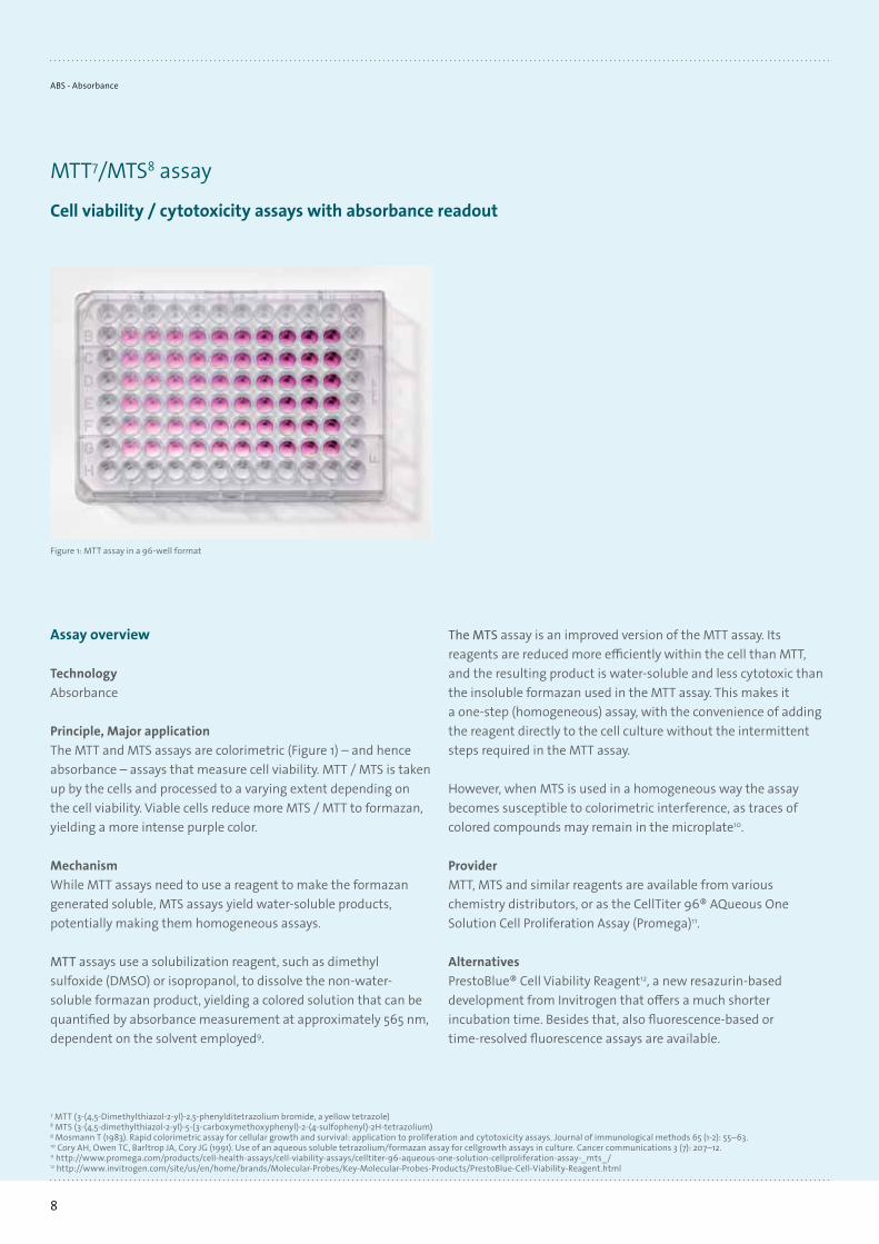
Principle, Major applicationThe MTT and MTS assays are colorimetric (Figure 1) – and hence absorbance – assays that measure cell viability. MTT / MTS is taken up by the cells and processed to a varying extent depending on the cell viability. Viable cells reduce more MTS / MTT to formazan, yielding a more intense purple color.
MechanismWhile MTT assays need to use a reagent to make the formazan generated soluble, MTS assays yield water-soluble products, potentially making them homogeneous assays.
MTT assays use a solubilization reagent, such as dimethyl sulfoxide (DMSO) or isopropanol, to dissolve the non-water-soluble formazan product, yielding a colored solution that can be quantified by absorbance measurement at approximately 565 nm, dependent on the solvent employed9.
Figure 1: MTT assay in a 96-well format
7 MTT (3-(4,5-Dimethylthiazol-2-yl)-2,5-phenylditetrazolium bromide, a yellow tetrazole)8 MTS (3-(4,5-dimethylthiazol-2-yl)-5-(3-carboxymethoxyphenyl)-2-(4-sulfophenyl)-2H-tetrazolium)9 Mosmann T (1983). Rapid colorimetric assay for cellular growth and survival: application to proliferation and cytotoxicity assays. Journal of immunological methods 65 (1-2): 55–63.10 Cory AH, Owen TC, Barltrop JA, Cory JG (1991). Use of an aqueous soluble tetrazolium/formazan assay for cellgrowth assays in culture. Cancer communications 3 (7): 207–12.11 http://www.promega.com/products/cell-health-assays/cell-viability-assays/celltiter-96-aqueous-one-solution-cellproliferation-assay-_mts_/12 http://www.invitrogen.com/site/us/en/home/brands/Molecular-Probes/Key-Molecular-Probes-Products/PrestoBlue-Cell-Viability-Reagent.html
MTT7/MTS8 assay
Cell viability / cytotoxicity assays with absorbance readout
ABS - Absorbance

9Learn more at www.tecan.com
SupportLink • Promega – CellTiter 96 AQueous One Solution Cell Proliferation Assay:
http://www.promega.com/products/cell-health-assays/cell-viabilityassays/celltiter-96-aqueous-one-solution-cell-proliferation-assay-_mts_/
• PrestoBlue® Cell Viability Reagent: http://www.invitrogen.com/site/us/en/home/brands/Molecular-Probes/Key-Molecular-Probes-Products/PrestoBlue-Cell-Viability-Reagent.html
Tecan Application Notes • Cell Proliferation and Cell Viability Analysis in in vitro Systems- Cell Culture Methods on Tecan´s Infinite® 200 • LIVE/DEAD Viability / Cytotoxicity assay • Detection of Calcein-AM and Hoechst 33342
Reader Infinite M200 PRO Infinite F200 PRO Infinite F500 Infinite M1000 PROAssay MTT assay
CellTiter 96 AQueous OneMTT assay
CellTiter 96 AQueous OneMTT assay
CellTiter 96 AQueous OneMTT assay
CellTiter 96 AQueous OneWavelength 565 nm 565 nm 565 nm 565 nmBandwith 9 nm 10 nm 10 nm 10 nmFlashes 25 25 10 10Settle time 0 ms 0 ms 0 ms 0 msShaking 180 sec; 1 mm
amplitude; orbital180 sec; 1 mm
amplitude; orbital180 sec; 1 mm
amplitude; orbital180 sec; 1 mm
amplitude; orbital
Instrument parameters

10
watersoluble chelate that can be measured at its absorption maximum of 562 nm. The linear working range for BSA is 20 to 2000 μg/ml14.The Bradford Protein Assay (BioRad) is based on the Coomassie® Brilliant Blue G-250 dye which binds to basic and aromatic amino acid residues, particularly arginine. This induces a shift of the dye’s absorbance maximum from 465 nm to 595 nm. The Bradford assay can be performed as a microassay procedure, with a linearity range of 125 to 1,000 μg/ml BSA15.In the Modified Lowry Protein Assay (Thermo Scientific Pierce), the protein reacts with cupric sulfate and tartrate in an alkaline solution, which results in formation of a tetradentate copper-protein complex, reducing the Folin-Ciocalteu Reagent. Theabsorbance of the blue, water-soluble product can be measured at 750 nm. The assay – tested with BSA protein16 – exhibits good linearity in the range of 1 to 1500 μg/ml.
AlternativesPotential alternatives for protein quantification range from absorbance-based methods using the protein extinction coefficient17 to fluorescence based assays like NanoOrange® to even dedicated18 devices.
Assay overview
TechnologyAbsorbance
Principle, Major applicationAll three assays are designed to determine the protein concentration of a sample. For detection, a liquid reagent needs to be added to the samples. This reagent interacts with the proteins, leading to a visible color change (Figure 1) that is directly proportional to the protein concentration. Absolute concentrations are calculated using a standard curve.
ProviderVarious companies have established their own assays for this purpose. The main differences between the various assays are the dynamic range and the measurement wavelength.
MechanismThe BCA™ Protein Assay (Thermo Scientific Pierce) uses bicinchoninic acid (BCA) for colorimetric quantification of total protein in a sample13. The method is based on the reduction of Cu2+ to Cu+ by proteins in an alkaline medium to form a colored
Figure 1: Bradford Protein Assay measured in cuvettes, showing increasing protein concentrations.
13 Smith, P.K., et al.: Measurement of protein using bicinchoic acid. Anal Biochem., 150, 76-85, 198514 http://www.piercenet.com/browse.cfm?fldID=0202010115 http://www.bio-rad.com/webroot/web/pdf/lsr/literature/4110065A.pdf
16 http://www.piercenet.com/browse.cfm?fldID=0202010317 http://web.expasy.org/protparam/protparam-doc.html18 http://www.millipore.com/techpublications/tech1/an2222en
BCA, Modified Lowry and Bradford assays – Protein quantification
Protein quantification assays with absorbance readout
ABS - Absorbance

11Learn more at www.tecan.com
Reader Infinite M200 PRO Infinite F200 PRO Infinite F500 Infinite M1000 PROAssay BCA assay BCA assay BCA assay BCA assayWavelength 565 nm 562 nm 562 nm 565 nmBandwith 9 nm 10 nm 10 nm 9 nmFlashes 25 25 10 10Settle time 0 ms 0 ms 0 ms 0 msShaking 0 sec. 0 sec. 0 sec. 0 sec.
Assay Modified Lowry Modified Lowry Modified Lowry Modified LowryWavelength 750 nm 750 nm 750 nm 750 nmBandwith 9 nm 10 nm 10 nm 9 nmFlashes 25 25 10 10Settle time 0 ms 0 ms 0 ms 0 msShaking 0 sec. 0 sec. 0 sec. 0 sec.
Assay Bradford Bradford Bradford BradfordWavelength 595 nm 590 nm 590 nm 595 nmBandwith 9 nm 10 nm 10 nm 9 nmFlashes 25 25 10 10Settle time 0 ms 0 ms 0 ms 0 msShaking 0 sec. 0 sec. 0 sec. 0 sec.
Instrument parameters
SupportLinks • BCA assay: http://www.piercenet.com/browse.cfm?fldID=02020103 • Modified Lowry assay: http://www.piercenet.com/browse.cfm?fldID=02020103 • Bradford assay homepage: http://www.bio-rad.com/prd/en/US/adirect/biorad?ts=1&cmd=BRCatgProductD
etail&vertical=LSR&catID=d4d4169a-12e8-4819-8b3e-ccab019c6e13 • NanoOrange: http://products.invitrogen.com/ivgn/product/N6666Tecan Application Notes • Protein Quantification in Small-Volume Samples • Protein quantification: BCA™, Modified Lowry and Bradford assays • Protein quantification on Infinite™ 200 with injectors

12
FI – Fluorescence IntensityLight is absorbed and released (emitted)
Major applications• PicoGreen® and RiboGreen® DNA / RNA quantification• Resazurin assay• GFP (Green Fluorescent Protein)
TechnologyFluorescence describes a molecule's ability to emit (release) previously absorbed light (Figure 1). The emission occurs almost instantly (within 1 nano second) and, according to the laws of physics, the emitted light will always have a higher wavelength and hence a lower energy. A fluorescence spectrum consists of an absorption (excitation) and emission spectrum (Figure 2). Fluorescence labels (fluorophores) can be attached to any available biomolecule and used to answer quantitative, as well as qualitative, questions. For example, ‘does the sample contain the fluorophore?’ (qualitative), and ‘how much of the fluorophore is in the sample?’ (quantitative). Signals are quantified as Relative Fluorescence Units [RFU].
V=3V=2V=1V=0
S1Vibrational relaxation Emission
light
Abso
rptio
n FluorescenceV=3V=2V=1V=0
S0
Excitationlight
Figure 1: Jablonski diagram drawn inside a fluorescence molecule. S = electronic state, V =vibrational level. After photon absorption (= excitation), the molecule adopts a state of higher energy S1 (= excited state) including several vibrationally excited substates. By vibrational relaxation, the molecule relaxes to the lowest excited S1 state (black arrow). From this state the molecule relaxes into the vibrational states of S0 by emitting light.
Figure 2: While the excitation spectrum describes how efficient it is to excite the fluorophore at a specific wavelength, the emission spectrum describes how efficient it is to detect the emitted light at any given wavelength. The Stokes shift describes the distance between the excitation and emission maximum, and is given in nanometers (nm).
300 400 500 600 700Re
lativ
e Fl
uroe
scen
ce U
nits
[RFU
s]Wavelength (nm)
Absorption(Excitation)
StokesShift
FluorescenceEmission
SpectralOverlap
Excitation and Emission Spectral Profiles
SupportLink • General explanation of fluorescence: http://www.olympusmicro.com/primer/lightandcolor/
fluorointroduction.htmlTecan Application Notes • Tweaking fluorescence scans • Optimizing the acquisition of 3D fluorescence spectra • Analyzing biological drug effects in 3D • Human TNF-α ELISA using Sword™ Peroxidase Reagents • Peroxidase detection using Sword™ Peroxidase Reagents • Human IL-6 chemiluminescent ELISA using Sword™ Peroxidase Reagents • Human C-reactive protein ELISA using Sword™ Peroxidase Reagents • Fluorescence-Based DNA Quantification in Small Volume Samples • DNA and RNA quantification: fast and simple with PicoGreen® dsDNA and RiboGreen®
RNA quantification reagents
13Learn more at www.tecan.com
Compatible readers
Features Infinite M200 PRO Infinite F200 PRO Infinite F500 Infinite M1000 PROReader design Monochromator Filter Filter MonochromatorConfiguration Quad4 Monochromators™,
2 excitation and 2 emission monochromators for high
performance, high flexibility and accurate
data acquisition
Up to 4 programmable filter pairs per slide that are
easily ejected and exchanged through the front of the instrument
Up to 6 filter pairs per slide which are individually
moveable, allowing any combination of Ex and Em
filters; easy filter change, ID chip
Premium Quad4 Monochro-mators, 2 excitation and 2
emission monochromators allow for stray light reduc-
tion up to a factor of 107
Transcreener FI Yes Yes Yes YesCapability – scanning Yes No No YesCapability – scanning 3D No No No YesDichroic mirrors - Dichroic mirror, optimized
for TRF measurements (Eu, Tb, Sm)
Multiple dichroic mirrors -
Z-adjustment Automatic - Automatic AutomaticPlate format 6- to 384-well plates
NanoQuant Plate6- to 384-well plates
NanoQuant Plate6- to 1,536-well plates
NanoQuant Plate6- to 1,536-well plates
NanoQuant PlateTemperature control Ambient +5 °C to 42 °C Ambient +5 °C to 42 °C Ambient +4 °C to 42 °C Ambient +4 °C to 42 °CShaking Linear, orbital with variable
amplitudesLinear, orbital with variable
amplitudesLinear, orbital with variable
amplitudesLinear, orbital, double orbital with variable
amplitudesInjectors 2 2 2 2Stacker Connect: stacks for
30 or 50 platesConnect: stacks for
30 or 50 platesConnect: stacks for
30 or 50 platesOn-board stacker: stacks
for 30 or 50 platesBarcode reader Yes Yes Yes Yes
Typcal values: Fluorescence intensity (FI) Infinite M200 PRO Infinite F200 PRO Infinite F500 Infinite M1000 PRO
Capability top Yes Yes Yes YesCapability bottom Yes Yes Yes YesLight source UV xenon flash lamp UV xenon flash lamp High energy xenon flash
lampHigh energy xenon flash
lampDetector PMT, optionally UV and red-
sensitive PMT, optionally UV and red-
sensitiveExtended wavelength (UV
and far-red)Low dark current PMT
Extended wavelength (UV and far-red)
Low dark current PMTWavelength range Standard:
Ex: 230 - 600 nmEm: 330 - 600 nm
Optional:Ex: 230 - 850 nmEm: 280 - 850 nm
Standard:Ex: 230 - 600 nmEm: 330 - 600 nm
Optional:Ex: 230 - 850 nmEm: 280 - 850 nm
Ex: 230 - 900 nmEm: 280 - 900 nm
Ex: 230 - 850 nmEm: 280 - 850 nm
Sensitivity – top 170 amol / well(1.7pM; 384-well plate)
85 amol / well(0.85pM, 384-well plate)
10 amol / well fluorescein (Greiner® 384-well low
volume black plate; 10 μl), 1pM
25 amol / well fluorescein (Greiner 384-well low
volume black plate; 10 μl), 2.5pM
Sensitivity – bottom 1.2 fmol / well(6pM; 96-well plate)
0.7 fmol / well(3.5pM; 96-well plate)
0.4 fmol / well fluorescein (Greiner
96-well SensoPlate™; 200 μl), 2pM
0.6 fmol / well fluorescein (Greiner
96-well SensoPlate; 200 μl), 3pM
Wavelength accuracy < ±2 nm for λ >315 nm;< ±1 nm for λ ≤315 nm
Filter dependent Filter dependent ≤300 nm: ±0.5 nm;>300 nm: ±1 nm
Wavelength reproducibility < ±1 nm for λ >315 nm;< ±0.5 nm for λ ≤315 nm
Filter dependent Filter dependent ≤300 nm: ±0.5 nm;>300 nm: ±1 nm
Bandwidth Ex: <5 nm for λ ≤315 nm and <9 nm for λ >315
Em: <10 nm for λ ≤315 nm and <20 nm
for λ >315
Filter dependent Filter dependent Adjustable ≤300 nm: 2.5 - 10 nm;
>300 nm: 5 - 20 nm
Wavelength selection Monochromator Filter Filter Monochromator

14
MechanismBoth assays are easy to use; simply add the dye to the sample, wait five minutes, and then read.
AlternativesIf sensitivity is not a major issue, it is possible to perform DNA quantification using absorbance at 260 nm.
Assay overview
TechnologyFluorescence Intensity
Assay design and providerLifeTechnologies' PicoGreen19 and RiboGreen20 (Figure 1) quantification assays use a fluorescence approach to determine DNA and RNA concentrations. Using the Quant-iT PicoGreen dsDNA Assay Kit, you can selectively detect as little as 25 pg/mlof dsDNA in the presence of ssDNA, RNA, and free nucleotides. The assay is linear over three orders of magnitude, and has little sequence dependence, allowing you to accurately measure DNA from many sources.
RiboGreen RNA reagent is one of the most sensitive detection dyes for the quantification of RNA in solution, offering linear fluorescence detection in the range of 1 to 200 ng/ml of RNA.
19 http://products.invitrogen.com/ivgn/product/P758920 http://products.invitrogen.com/ivgn/product/R11490
PicoGreen® and RiboGreen® DNA / RNA quantification
High sensitivity, fluorescence-based DNA / RNA quantification
Figure 1: Green DNA
FI - Fluorescence Intensity

15Learn more at www.tecan.com
SupportLinks • Quant-iT™ RiboGreen RNA Assay Kit: http://products.invitrogen.com/ivgn/product/R11490 • Quant-iT PicoGreen dsDNA Assay Kit: http://products.invitrogen.com/ivgn/product/P7589Tecan Application Notes • Fluorescence-Based DNA Quantification in Small Volume Samples • DNA and RNA quantification: fast and simple with PicoGreen® dsDNA and RiboGreen®
RNA quantification reagents • PicoGreen® assay measured in NanoQuant Plate™ • Impact of Extended Adjustable Monochromator Bandwidth in Fluorescence Based Application Technologies • Comparison of two different detection techniques for DNA
Reader Infinite M200 PRO Infinite F200 PRO Infinite F500 Infinite M1000 PROAssay PicoGreen, RiboGreen PicoGreen, RiboGreen PicoGreen, RiboGreen PicoGreen, RiboGreenMode Top Top Top TopExcitation wavelength 485 nm (9 nm) 485 nm (20 nm) 485 nm (20 nm) 485 nm (9 nm)Emission wavelength 535 nm (20 nm) 535 nm (25 nm) 535 nm (25 nm) 535 nm (20 nm)Lag time 0 μs 0 μs 0 μs 0 μsIntegration time 20 μs 20 μs 20 μs 20 μsFlashes 25 25 10 10Mirror automatic automatic automatic automaticGain optimal optimal optimal optimalZ-position automatic / automatic automaticSettle time 0 ms 0 ms 0 ms 0 ms
Instrument parameters

16
21 http://www.invitrogen.com/site/us/en/home/brands/Molecular-Probes/Key-Molecular-Probes-Products/alamarBlue-Rapid-and-Accurate-Cell-Health-Indicator.html22 http://www.promega.com/products/cell-health-assays/cell-viability-assays/celltiter_blue-cell-viability-assay/23 O’Brien, J.; Wilson, I.; Orton, T.; Pognan, F. Investigation of the Alamar Blue (resazurin) fluorescent dye for the assessment of mammalian cell cytotoxicity. Eur. J. Biochem. 2000, 267,5421–5426.24 http://www.invitrogen.com/site/us/en/home/brands/Molecular-Probes/Key-Molecular-Probes-Products/PrestoBlue-Cell-Viability-Reagent.html
Resazurin assay
A Fluorescence Intensity-based (bacterial / cell) proliferation assay
3889MC
Viable Cell
Resazurin ResorufinEmits fluorescence at 590nm
N
O
O–O O
N
O–O O
ReductionReactions
Figure 1: Viability dependet conversion of Resazurin to Resorufin (Promega)
MechanismResazurin is a redox indicator that can be added directly to cells. Viable cells convert the dark blue, oxidized form of the dye (resazurin) into a red, fluorescent reduced form called resorufin (Ex: 570 nm; Em: 590 nm). The amount of fluorescence (or absorbance) is proportional to the number of living cells, and corresponds to the cell’s metabolic activity. Damaged and non-viable cells have lower innate metabolic activity, and therefore generate a proportionally lower signal than healthy cells. The system is specific for cell viability as non-viable cells rapidly lose metabolic capacity and do not reduce resazurin. Consequently, a fluorescent signal23 is not generated.
AlternativesPrestoBlue Cell Viability Reagent24, a new development from Invitrogen that offers much shorter incubation times. The absorbance based MTT/MTS assay.
Assay overview
TechnologyFluorescence Intensity
Major applicationCell viability assays
ProviderResazurin was initially used for bacterial studies, but is now also available for eukaryotic cell-based applications under brand names such as the alamarBlue® assay21 (Life Technologies) and CellTiter-Blue® Cell Viability Assay22 (Promega).
FI - Fluorescence Intensity
17Learn more at www.tecan.com
SupportLinks • Promega CellTiter-Blue Cell Viability Assay: http://www.promega.com/products/cell-health-assays/cell-
viability-assays/celltiter_blue-cell-viability-assay/ • Invitrogen alamarBlue assay: http://www.invitrogen.com/site/us/en/home/brands/Molecular-Probes/Key-
Molecular-Probes-Products/alamarBlue-Rapid-and-Accurate-Cell-Health-Indicator.html
• PrestoBlue Cell Viability Reagent: http://www.invitrogen.com/site/us/en/home/brands/Molecular-Probes/Key-Molecular-Probes-Products/PrestoBlue-Cell-Viability-Reagent.html
Tecan Application Notes • Cell Proliferation and Cell Viability Analysis in in vitro Systems- Cell Culture Methods on Tecan´s Infinite® 200 • LIVE/DEAD Viability / Cytotoxicity assay • Detection of Calcein-AM and Hoechst 33342
Instrument parameters
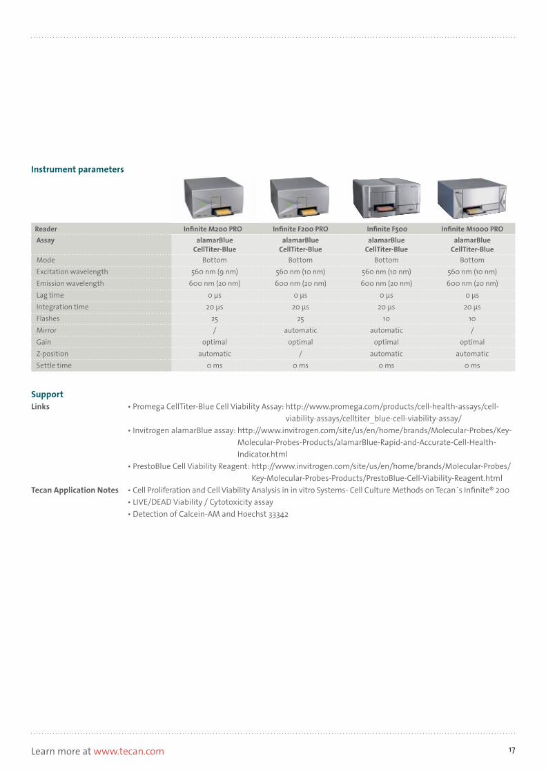
Reader Infinite M200 PRO Infinite F200 PRO Infinite F500 Infinite M1000 PROAssay alamarBlue
CellTiter-BluealamarBlue
CellTiter-BluealamarBlue
CellTiter-BluealamarBlue
CellTiter-BlueMode Bottom Bottom Bottom BottomExcitation wavelength 560 nm (9 nm) 560 nm (10 nm) 560 nm (10 nm) 560 nm (10 nm)Emission wavelength 600 nm (20 nm) 600 nm (20 nm) 600 nm (20 nm) 600 nm (20 nm)Lag time 0 μs 0 μs 0 μs 0 μsIntegration time 20 μs 20 μs 20 μs 20 μsFlashes 25 25 10 10Mirror / automatic automatic /Gain optimal optimal optimal optimalZ-position automatic / automatic automaticSettle time 0 ms 0 ms 0 ms 0 ms

18
GFP (Green Fluorescent Protein)
Fluorescent protein frequently used as an expression / activation reporter
Figure 2: GFP-transfected eukaryotic cellsFigure 1: Protein structure of GFP
Format, providerDue to multiple engineering efforts, an almost unlimited number of mutants exist, resulting in a large bandwidth of excitation and emission values. Some of these variants are commercially available, while others are published and therefore not protected. Consequently, only a selection of measurement parameters can be given, since the wavelength depends on the mutant type of the protein.
AlternativesTechnology-wise, there are a lot of alternative fluorescent proteins available, such as CFP (Cyan) and YFP (yellow). From an assay perspective, the alternative selected depends on the application. For gene expression studies, DLR® (Dual luciferase reporter assay) or GeneBLAzer® assays may be suitable. For FRET / BRETstudies, fluorescent labels might be an alternative.
Assay overview
TechnologyFluorescence Intensity
PrincipleGFP (Figure 1) is a protein derived from a jellyfish which has the ability to emit light in the green wavelength range and can be detected using standard FI measurements.
Major applicationsGFP can be used in multiple different of ways, for example as a BRET / FRET partner in binding studies, or for gene activation, where it is often fused / cloned to a gene of interest and co–expressed once the gene is activated (Figure 2). Commonly, it is differentiated between constitutive (permanent) and temporary expression. Constitutive expression is mostly used to monitor growth or proliferation of cells or bacteria, while temporary expression is used for gene activation studies.
FI - Fluorescence Intensity

19Learn more at www.tecan.com
SupportLink • http://www.olympusfluoview.com/java/excitationefficiency/index.htmlTecan Application Notes • Analyzing biological drug effects in 3D • Analyzing biological processes • Improved Detection of Green Fluorescent Protein (GFP) in the Infinite® 200 PRO • Detection of Green Fluorescent Protein (eGFP)
Instrument parameters
Reader Infinite M200 PRO Infinite F200 PRO Infinite F500 Infinite M1000 PROAssay GFP GFP GFP GFPMode Bottom Bottom Bottom BottomExcitation wavelength 483 (9) nm 485 (20) nm 485 (20) nm 485 (20) nmEmission wavelength 535 (20) nm 535 (25) nm 535 (25) nm 535 (20) nmLag time 0 μs 0 μs 0 μs 0 μsIntegration time 20 μs 20 μs 20 μs 20 μsFlashes 25 25 10 10Mirror / automatic automatic /Gain optimal optimal optimal optimalZ-position - - - -Settle time 0 ms 0 ms 0 ms 0 ms

20
25 Lakowicz, J. R. (1999). Principles of Fluorescence Spectroscopy. Kluwer Academic / Plenum Publishers
Figure 1: Schematic drawing of a time resolved emission spectrum
Lanthanide Chelate Fluorescence
Rela
tive
fluor
esce
nce
units
[RFU
]
Major applications• DELFIA® – Dissociation-Enhanced Lanthanide Fluorescent
Immunoassay
TechnologyTRF is similar to standard fluorescence, except that the light is emitted for a much longer period of time (Figure 1). The advantage of this is that the signal can be measured once all the background fluorescence (noise) has subsided, increasing the signal to noise ratio, and hence the sensitivity. Only lanthanides – also called rareearth metals – are capable of this kind of fluorescence25.
From all existing lanthanides Europium (Eu), Samarium (Sm), Terbium (Tb) and Dysprosium (Dy) are the most used ones in life science.
In most cases, it is possible to substitute fluorescence applications with TRF to achieve higher sensitivity and / or lower background noise.
TRF – Time-Resolved FluorescenceLight is absorbed and emitted for a relatively long period of time

21Learn more at www.tecan.com
Compatible readers
SupportTecan Application Note • TRF Sensitivity
Features Infinite M200 PRO Infinite F200 PRO Infinite F500 Infinite M1000 PROReader design Monochromator Filter Filter MonochromatorConfiguration Quad4 Monochromators™,
2 excitation and 2 emission monochromators for high
performance, high flexibility and accurate
data acquisition
Up to 4 programmable filter pairs per slide that are
easily ejected and exchanged through the front of the instrument
Up to 6 filter pairs per slide which are individually
moveable, allowing any combination of Ex and Em
filters; easy filter change, ID chip
Premium Quad4 Monochro-mators, 2 excitation and 2
emission monochromators allow for stray light reduc-
tion up to a factor of 107
Capability Yes No No YesCapability – TR-FRET No No No YesDichroic mirrors - Dichroic mirror, optimized
for TRF measurements (Eu, Tb, Sm)
Multiple dichroic mirrors -
Z-adjustment Automatic - Automatic AutomaticPlate format 6- to 384-well plates
NanoQuant Plate6- to 384-well plates
NanoQuant Plate6- to 1,536-well plates
NanoQuant Plate6- to 1,536-well plates
NanoQuant PlateTemperature control Ambient +5 °C to 42 °C Ambient +5 °C to 42 °C Ambient +4 °C to 42 °C Ambient +4 °C to 42 °CShaking Linear, orbital with variable
amplitudesLinear, orbital with variable
amplitudesLinear, orbital with variable
amplitudesLinear, orbital, double orbital with variable
amplitudesInjectors 2 2 2 2Stacker Connect: stacks for
30 or 50 platesConnect: stacks for
30 or 50 platesConnect: stacks for
30 or 50 platesOn-board stacker: stacks
for 30 or 50 platesBarcode reader Yes Yes Yes Yes
Typcal values: Time-Resolved fluorescence (TRF) Infinite M200 PRO Infinite F200 PRO Infinite F500 Infinite M1000 PRO
Light source UV xenon flash lamp UV xenon flash lamp High energy xenon flash lamp
High energy xenon flash lamp
Detector PMT, optionally UV and red-sensitive
PMT, optionally UV and red-sensitive
Extended wavelength (UV and far-red)
Low dark current PMT
Extended wavelength (UV and far-red)
Low dark current PMTWavelength range Standard:
Ex: 230 - 600 nmEm: 330 - 600 nm
Optional:Ex: 230 - 850 nmEm: 280 - 850 nm
Standard:Ex: 230 - 600 nmEm: 330 - 600 nm
Optional:Ex: 230 - 850 nmEm: 280 - 850 nm
Ex: 230 - 900 nmEm: 280 - 900 nm
Ex: 230 - 850 nmEm: 280 - 850 nm
Sensitivity 90 amol / well(0.9 pM; 384-well plate)
europium
2.8 amol / well(28fM; 384-well plate)
europium
0.5 amol / well europium(Greiner 384-well low volume white plate;
10 μl), 50fM europium
1.5 amol / well europium(Greiner 384-well low volume white plate;
10 μl), 150fM europiumBandwidth Ex: <5 nm for λ ≤315 nm and
<9 nm for λ >315Em: <10 nm for λ ≤315 nm
and <20 nm for λ >315
Filter dependent Filter dependent Adjustable ≤300 nm: 2.5 - 10 nm;
>300 nm: 5 - 20 nm
Wavelength selection Monochromator Filter Filter Monochromator

22
26 http://www.perkinelmer.com/Catalog/Category/ID/delfia%20trf%20assays%20and%20reagents27 Enzyme linked Immunosorbent assay
DELFIA® – Dissociation-Enhanced Lanthanide Fluorescent Immunoassay26
TRF-based alternative to absorbance-based ELISA
Figure 1: Schematic principle of DELFIA
Enhance 340 nm
617 nm
EuEu
Eu
AlternativesAs a common application, it is used as an alternative approach to the well-established, absorbance- or fluorescence-based ELISA27.
MechanismThe biomolecule (antibody, DNA probe, etc.) used for detection is labeled with one of the lanthanide chelates. Assays are performed in an endpoint manner and only need to be read once, when all pipetting steps are complete. All steps are performed according to a standard ELISA protocol. Instead of a substrate an enhancementsolution is added, that disconnects the chelate lanthanide-chelate complex from the antibody to increase the signal intensity.
Assay overview
TechnologyTRF – Time-Resolved Fluorescence
Principle, ProviderPerkinElmer offers the most common lanthanide chelates, including Europium (Eu), Samarium (Sm), Terbium (Tb) and Dysprosium (Dy), under the brand name DELFIA, making DELFIA a technology rather than a single assay.
Major applicationsIn addition to the self-labeling kits, which allow users to label almost any biomolecule with the lanthanide chelates, PerkinElmer offers pre-coupled antibodies and DNA probes. DELFIA is also available as a ready-to-go assay for cytotoxicity or cell proliferation studies. Other major applications include: receptor-ligand binding, enzyme assays, protein-protein and protein-DNA interaction studies.
EuEu
TRF – Time-Resolved Fluorescence

23Learn more at www.tecan.com
SupportLinks • DELFIA web page: http://www.perkinelmer.com/Catalog/Category/ID/delfia%20trf%20assays%20
and%20reagents • DELFIA knowledge base: http://perkinelmerreagents.onconfluence.com/pages/
viewpage.action?pageId=328586Tecan Application Note • TRF Sensitivity
Instrument parameters
Reader Infinite M200 PRO Infinite F200 PRO Infinite F500 Infinite M1000 PROAssay DELFIA DELFIA DELFIA DELFIAMode FI Top FI Top FI Top FI TopExcitation wavelength Eu: 340(9) nm Eu: 340(35)nm Eu: 340(35)nm Eu: 345(20)nmEmission wavelength Eu: 617(20) nm Eu: 612(10) nm Eu: 612(10) nm Eu: 617(20) nmLag time 200 μs 200 μs 200 μs 200 μsIntegration time 400 μs 400 μs 400 μs 400 μsFlashes 25 25 10 10 (100 Hz)Mirror / automatic automatic /Gain automatic automatic automatic automaticZ-position automatic / automatic automaticSettle time 0 ms 0 ms 0 ms 0 ms

24
28 Lakowicz, J. R. (1999). Principles of Fluorescence Spectroscopy. Kluwer Academic / Plenum Publishers
3500
20
40
60
80
100
400 450 500 550 600 700650No
rmal
ized
Inte
nsity
Wavelenght (Nanometers)
CFP DsRFP
EmissionSpectra
AbsorptionSpectra
SpectralOverlapRegion
Donor-Acceptor Spectral Overlap region
Figure 2: FRET is possible because the emission spectrum of CFP and the excitation spectrum of DsRFP overlap between 450 and 600 nm.
Figure 1: Schematic principle of FRET. Light emitted from the green molecule is used to excite the red molecule.
TechnologyAs the name implies, FRET involves energy transfer between two fluorescent molecules (Figure 1). However, there are some specific requirements for this transfer to take place. Firstly, the emission spectrum of the donor fluorophore and the excitation spectrum of the acceptor fluorophore need to overlap (Figure 2), as theemission light of the donor fluorophore is used to excite the acceptor fluorophore. Secondly, the distance between the two fluorophores – the Förster radius – should be less than 10 nm (Figure 3)28.
One way in which FRET is used is to determine if two biomolecules are in close proximity. In this case, both biomolecules must be labeled with fluorophores and then combined. After an incubation period, the assay is performed. Samples are excited at the donor excitation wavelength, and measured at the donor and acceptor emission wavelength. To compensate for well-to-well variation, for example from pipetting errors, the ratio of both values is calculated (ratiometric assay). If donor and acceptor are in close proximity, FRET will take place, otherwise only the emission signal of the donor is measurable.
Major applications• GeneBLAzer and Tango™ GPCR Assay System
FRET – Fluorescence Resonance Energy TransferLight is absorbed, transferred to another fluorophore and then emitted
Energy transfer
distance
light emitted light for excitation
0
25
50
75
100
0 2 4 6 8 10Distance (r, in Nanometers)
Förster DistanceR0
50 PercentTransferEfficiency
Distance and Energy Transfer Efficiency
Ener
gy T
rans
fer E
ffici
ency
(Per
cent
)
Figure 3: Förster radius – the distance where the FRET signal intensity is reduced to 50 %.
SupportTecan Application Notes • GeneBLAzer® Assay on Tecan’s
Infinite® F200 for cell-based screening – A FRET-technology for gene reporter assays
• Inducing hypoxia inside Tecan’s Infinite® 200 PRO multimode reader

25Learn more at www.tecan.com
Compatible readers
Features Infinite M200 PRO Infinite F200 PRO Infinite F500 Infinite M1000 PROReader design Monochromator Filter Filter MonochromatorConfiguration Quad4 Monochromators™,
2 excitation and 2 emission monochromators for high
performance, high flexibility and accurate
data acquisition
Up to 4 programmable filter pairs per slide that are
easily ejected and exchanged through the front of the instrument
Up to 6 filter pairs per slide which are individually
moveable, allowing any combination of Ex and Em
filters; easy filter change, ID chip
Premium Quad4 Monochro-mators, 2 excitation and 2
emission monochromators allow for stray light reduc-
tion up to a factor of 107
Capability – scanning Yes No No YesCapability – scanning 3D No No No YesDichroic mirrors - Dichroic mirror, optimized
for TRF measurements (Eu, Tb, Sm)
Multiple dichroic mirrors -
Z-adjustment Automatic - Automatic AutomaticPlate format 6- to 384-well plates
NanoQuant Plate6- to 384-well plates
NanoQuant Plate6- to 1,536-well plates
NanoQuant Plate6- to 1,536-well plates
NanoQuant PlateTemperature control Ambient +5 °C to 42 °C Ambient +5 °C to 42 °C Ambient +4 °C to 42 °C Ambient +4 °C to 42 °CShaking Linear, orbital with variable
amplitudesLinear, orbital with variable
amplitudesLinear, orbital with variable
amplitudesLinear, orbital, double orbital with variable
amplitudesInjectors 2 2 2 2Stacker Connect: stacks for
30 or 50 platesConnect: stacks for
30 or 50 platesConnect: stacks for
30 or 50 platesOn-board stacker: stacks
for 30 or 50 platesBarcode reader Yes Yes Yes Yes
Typcal values: Fluorescence intensity (FI) Infinite M200 PRO Infinite F200 PRO Infinite F500 Infinite M1000 PRO
Capability top Yes Yes Yes YesCapability bottom Yes Yes Yes YesLight source UV xenon flash lamp UV xenon flash lamp High energy xenon flash
lampHigh energy xenon flash
lampDetector PMT, optionally UV and red-
sensitive PMT, optionally UV and red-
sensitiveExtended wavelength (UV
and far-red)Low dark current PMT
Extended wavelength (UV and far-red)
Low dark current PMTWavelength range Standard:
Ex: 230 - 600 nmEm: 330 - 600 nm
Optional:Ex: 230 - 850 nmEm: 280 - 850 nm
Standard:Ex: 230 - 600 nmEm: 330 - 600 nm
Optional:Ex: 230 - 850 nmEm: 280 - 850 nm
Ex: 230 - 900 nmEm: 280 - 900 nm
Ex: 230 - 850 nmEm: 280 - 850 nm
Sensitivity – top 170 amol / well(1.7pM; 384-well plate)
85 amol / well(0.85pM, 384-well plate)
10 amol / well fluorescein (Greiner® 384-well low
volume black plate; 10 μl), 1pM
25 amol / well fluorescein (Greiner 384-well low
volume black plate; 10 μl), 2.5pM
Sensitivity – bottom 1.2 fmol / well(6pM; 96-well plate)
0.7 fmol / well(3.5pM; 96-well plate)
0.4 fmol / well fluorescein (Greiner
96-well SensoPlate™; 200 μl), 2pM
0.6 fmol / well fluorescein (Greiner
96-well SensoPlate; 200 μl), 3pM
Wavelength accuracy < ±2 nm for λ >315 nm;< ±1 nm for λ ≤315 nm
Filter dependent Filter dependent ≤300 nm: ±0.5 nm;>300 nm: ±1 nm
Wavelength reproducibility < ±1 nm for λ >315 nm;< ±0.5 nm for λ ≤315 nm
Filter dependent Filter dependent ≤300 nm: ±0.5 nm;>300 nm: ±1 nm
Bandwidth Ex: <5 nm for λ ≤315 nm and <9 nm for λ >315
Em: <10 nm for λ ≤315 nm and <20 nm
for λ >315
Filter dependent Filter dependent Adjustable ≤300 nm: 2.5 - 10 nm;
>300 nm: 5 - 20 nm
Wavelength selection Monochromator Filter Filter Monochromator

26
29 G-Protein Coupled Receptors: important group of cell surface receptors for cellural signalling30 http://www.invitrogen.com/site/us/en/home/Products-and-Services/Applications/Drug-Discovery/Target-and-Lead-Identification-and-Validation/g-protein_coupled_html/
GPCR-Cell-Based-Assays/GeneBLAzer-Theory.html31 β-lactamase is an enzyme that can cleave specific substrates
GeneBLAzer® and Tango™ GPCR29 Assay System30
Gene activator assay with FRET readout
MechanismAs shown in Figure 1, after the transfection, cells are loaded with an engineered fluorescent substrate which is an assembly of two fluorophores: coumarin and fluorescein. If the target gen is inactive, BLA is not expressed and the substrate molecule is not cleaved. In this state, excitation of the coumarin results in FRET to the fluorescein moiety and emission of green light.However, in the presence of BLA expression, the substrate is cleaved, causing the separation of the fluorophores, and FRET cannot occur. This results in the emission of a blue fluorescence signal from coumarin. Assays are commonly measured over several hours, or even days. During this time period the plate can either be shuttled between the incubator and the reader, or a temperature and gas controlled multimode reader such as the Infinite 200 PRO may be used.
AlternativesVarious fluorescence (e.g. GFP) or luminescence (e.g. DLR™) reporter gen assays.
Assay overview
TechnologyFluorescence Resonance Energy Transfer (FRET), ratiometric
Major application, PrincipleLife Technologies’ GeneBLAzer assays are designed to monitor the activation of genes, including surface and intracellular reporters, a wide range of signal transduction pathways, ion channels and other transporters. The basis for the GeneBLAzer assay are cell lines possessing a β-lactamase31 (BLA) gene under the control of a promotor which is downstream of the monitored target protein.
Provider The β-lactamase-transfected cell lines can either be purchased from Life Technologies or self-transfected. Tango cell lines are also based on the GeneBLAzer technology, but are designed exclusively for GPCR activation assays.
FRET - Fluorescence Resonance Energy Transfer
Figure 1: Principle of the GeneBLAzer assay. If the substrate is cleaved, the FRET signal is disrupted.

27Learn more at www.tecan.com
SupportLinks •GeneBLAzer homepage: http://www.invitrogen.com/site/us/en/home/Products-and-Services/Applications/
Drug-Discovery/Target-and-Lead-Identification-and-Validation/g-protein_coupled_html/GPCR-Cell-Based-Assays/GeneBLAzer-Theory.html?CID=flgeneblazer
• Video - TANGO cell line: http://media.invitrogen.com.edgesuite.net/stand_alone/Tango-GPCR-Cell-Lines.html • LiveBLAzer manual: http://tools.invitrogen.com/content/sfs/manuals/liveblazer_FRETBGLoadingKit_man.pdfTecan Application Notes • GeneBLAzer® Assay on Tecan’s Infinite® F200 for cell-based screening –
A FRET-technology for gene reporter assays • Inducing hypoxia inside Tecan’s Infinite® 200 PRO multimode reader
Instrument parameters
Reader Infinite M200 PRO Infinite F200 PRO Infinite F500 Infinite M1000 PROAssay GeneBLAzer GeneBLAzer GeneBLAzer GeneBLAzerMeasurement Donor Donor Donor DonorMode FI bottom FI bottom FI bottom FI bottomExcitation wavelength 409(9) nm 415(20) nm 415(20) nm 409(12) nmEmission wavelength 460(20) nm 460(20) nm 460(20) nm 460(12) nmLag time 0 μs 0 μs 0 μs 0 μsIntegration time 40 μs 40 μs 40 μs 40 μsFlashes 10 25 10 10Mirror automatic automatic automatic automaticGain optimal optimal optimal optimalZ-position calculated / calculated calculatedSettle time 0 ms 0 ms 0 ms 0 msAssay GeneBLAzer GeneBLAzer GeneBLAzer GeneBLAzerMeasurement Acceptor Acceptor Acceptor AcceptorMode FI bottom FI bottom FI bottom FI bottomExcitation wavelength 409(9) nm 415(20) nm 415(20) nm 409(12) nmEmission wavelength 530(20) nm 535(25) nm 535(25) nm 530(12) nmLag time 0 μs 0 μs 0 μs 0 μsIntegration time 40 μs 40 μs 40 μs 40 μsFlashes 10 (100 Hz) 25 10 10 (100 Hz)Mirror automatic automatic automatic automaticGain optimal optimal optimal optimalZ-position calculated / calculated calculatedSettle time 0 ms 0 ms 0 ms 0 ms
28
TR-FRET – Time-Resolved Fluorescence Resonance Energy TransferFRET with a longer lifetime and hence a lower background
Assay designTR-FRET assays are commonly designed to detect whether molecules are in close proximity. This can be exploited to determine if, for example, a protein, peptide substrate, small molecule, phosphorylation, or acetylation is present, or if binding has occurred, for example a receptor-ligand interaction. A major limitation of this technology is that the maximum distance between the donor and the acceptor molecule cannot exceed 10 nm32.
Major applications• HTRF® – Homogeneous Time-Resolved Fluorescence• Adapta® Universal Kinase Assay and Substrates• LanthaScreen™ Kinase Activity Assays
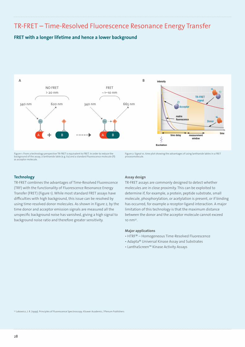
TechnologyTR-FRET combines the advantages of Time-Resolved Fluorescence (TRF) with the functionality of Fluorescence Resonance Energy Transfer (FRET) (Figure 1). While most standard FRET assays have difficulties with high background, this issue can be resolved by using time-resolved donor molecules. As shown in Figure 2, by the time donor and acceptor emission signals are measured all the unspecific background noise has vanished, giving a high signal to background noise ratio and therefore greater sensitivity.
FRET~ 1–10 nm
NO FRET> 20 nm
340 nm 620 nm
A B
Eu fl
340 nm 665 nm
A
Eu
B
fl
Figure 1: From a technology perspective TR-FRET is equivalent to FRET. In order to reduce the background of the assay, a lanthanide lable (e.g. Eu) and a standard fluorescence molecule (fl) as acceptor molecule.
Figure 2: Signal vs. time plot showing the advantages of using lanthanide lables in a FRET processmolecule.
32 Lakowicz, J. R. (1999). Principles of Fluorescence Spectroscopy. Kluwer Academic / Plenum Publishers
A
TR-FRETsignal
Acceptor
Donor
B

29Learn more at www.tecan.com
Compatible readersCompatible readers
SupportTecan Application Notes • Implementation of Tag-lite™ Technology (Cisbio bioassays) on Tecan’s Infinite® F500 Multimode Reader • GeneBLAzer® Assay on Tecan’s Infinite® F200 for cell-based screening –
A FRET-technology for gene reporter assays • LanthaScreen® TR-FRET Assay • Development of a functional assay (HTRF®, Cisbio) to detect cAMP concentration after activation
of 5-HT1A receptors • HTRF® (Cisbio) Human Interleukin beta (IL1β) assay • HTRF® Homogenous TR-FRET Assay • Homogeneous time-resolved fluorescence (HTRF®) in Tecan’s Infinite® F500 filter-based multimode reader
(White Paper)
Features Infinite F200 PRO Infinite F500 Infinite M1000 PROReader design Filter Filter MonochromatorConfiguration Up to 4 programmable filter pairs per
slide that are easily ejected and exchanged through the front of the
instrument
Up to 6 filter pairs per slide which are individually moveable, allowing any
combination of Ex and Em filters; easy filter change, ID chip
Premium Quad4 Monochromators, 2 excitation and 2 emission monochro-mators allow for stray light reduction
up to a factor of 107
HTRF Yes Yes YesLanthaScreen Yes Yes YesTranscreener TR-FRET No Yes YesCapability No No YesCapability – TR-FRET No No YesDichroic mirrors Dichroic mirror, optimized for TRF
measurements (Eu, Tb, Sm)Multiple dichroic mirrors -
Z-adjustment - Automatic AutomaticPlate format 6- to 384-well plates
NanoQuant Plate6- to 1,536-well plates
NanoQuant Plate6- to 1,536-well plates
NanoQuant PlateTemperature control Ambient +5 °C to 42 °C Ambient +4 °C to 42 °C Ambient +4 °C to 42 °CShaking Linear, orbital with variable
amplitudesLinear, orbital with variable
amplitudesLinear, orbital, double orbital with
variable amplitudes
Injectors 2 2 2Stacker Connect: stacks for
30 or 50 platesConnect: stacks for
30 or 50 platesOn-board stacker: stacks
for 30 or 50 platesBarcode reader Yes Yes Yes
Typcal values: Time-Resolved fluorescence (TRF) Infinite F200 PRO Infinite F500 Infinite M1000 PRO
Light source UV xenon flash lamp High energy xenon flash lamp High energy xenon flash lampDetector PMT, optionally UV
and red-sensitiveExtended wavelength
(UV and far-red)Low dark current PMT
Extended wavelength (UV and far-red)
Low dark current PMTWavelength range Standard:
Ex: 230 - 600 nmEm: 330 - 600 nm
Optional:Ex: 230 - 850 nmEm: 280 - 850 nm
Ex: 230 - 900 nmEm: 280 - 900 nm
Ex: 230 - 850 nmEm: 280 - 850 nm
Sensitivity 2.8 amol / well (28fM; 384-well plate)europium
0.5 amol / well europium(Greiner 384-well low volume
white plate; 10 μl), 50fM europium
1.5 amol / well europium(Greiner 384-well low volume
white plate; 10 μl), 150fM europiumBandwidth Filter dependent Filter dependent Adjustable ≤300 nm:
2.5 - 10 nm; >300 nm: 5 - 20 nmWavelength selection Filter Filter Monochromator

30
HTRF® – Homogeneous Time-Resolved Fluorescence33
TR-FRET-based assay platform
MechanismHTRF is based on Eu3+ / Tb2+ cryptate donors and XL665 or d2 acceptors, which can be coupled to almost any biomolecule desired, including proteins, peptides, DNA and small molecules. The technology is based on no wash assay procedure detecting proximity events between donor and acceptor dyes.The assay detection is obtained upon dispensing acceptor and donor conjugates to the sample to be assessed (e.g. enzymatic reaction mixture, cell lysate, or supernatant). No washing steps are required (homogeneous assay), and detection is performed after the completion of incubation, by measuring both specific donor and acceptor fluororescences (Figure 1). To compensate for well-to-well variation, the ratio of both values is calculated (hence ratiometric assay). Donor fluorescence will always be detected and used as an internal control, while an emission signal from the acceptor is only detected if both biomolecules are in close proximity and FRET occurs33.
Assay overview
TechnologyTR-FRET – Time-Resolved Fluorescence Resonance Energy Transfer, ratiometric
ProviderHTRF is Cisbio's TR-FRET-based assay platform, which provides a broad range of solutions.
Format, major applicationsBiomolecules for detection can either be self-labeled or purchased pre-labeled. Additionally, ready-to-go assays and pre-coupled antibodies are available for major targets, including GPCRs, with second messengers and binding assays, kinases, epigenetic enzymes, protein-protein interactions and biomarkers.
Figure 1: Mechanistic principle of the HTRF technology and fluorescence spectrum
33 http://www.htrf.com
wavelenght (nm) wavelenght (nm)
Fluo
resc
ence
inte
nsity
Fluo
resc
ence
inte
nsity
Donor/acceptor proximity −> FRETCryptate emission measured at 620 nmAcceptor emission measured at 665 nm
Distant donor/acceptor −> NO FRETCryptate emission measured at 620 nm
TR-FRET - Time Resolved Fluorescence Resonance Energy Transfer
31Learn more at www.tecan.com
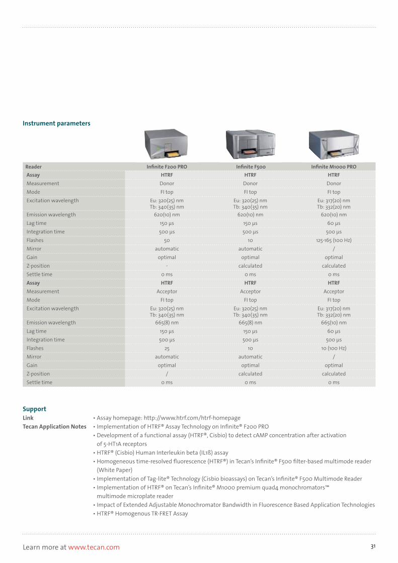
SupportLink • Assay homepage: http://www.htrf.com/htrf-homepageTecan Application Notes • Implementation of HTRF® Assay Technology on Infinite® F200 PRO • Development of a functional assay (HTRF®, Cisbio) to detect cAMP concentration after activation
of 5-HT1A receptors • HTRF® (Cisbio) Human Interleukin beta (IL1ß) assay • Homogeneous time-resolved fluorescence (HTRF®) in Tecan’s Infinite® F500 filter-based multimode reader
(White Paper) • Implementation of Tag-lite® Technology (Cisbio bioassays) on Tecan’s Infinite® F500 Multimode Reader • Implementation of HTRF® on Tecan’s Infinite® M1000 premium quad4 monochromators™
multimode microplate reader • Impact of Extended Adjustable Monochromator Bandwidth in Fluorescence Based Application Technologies • HTRF® Homogenous TR-FRET Assay
Instrument parameters
Reader Infinite F200 PRO Infinite F500 Infinite M1000 PROAssay HTRF HTRF HTRFMeasurement Donor Donor DonorMode FI top FI top FI topExcitation wavelength Eu: 320(25) nm
Tb: 340(35) nmEu: 320(25) nmTb: 340(35) nm
Eu: 317(20) nmTb: 332(20) nm
Emission wavelength 620(10) nm 620(10) nm 620(10) nmLag time 150 μs 150 μs 60 μsIntegration time 500 μs 500 μs 500 μsFlashes 50 10 125-165 (100 Hz)Mirror automatic automatic /Gain optimal optimal optimalZ-position - calculated calculatedSettle time 0 ms 0 ms 0 msAssay HTRF HTRF HTRFMeasurement Acceptor Acceptor AcceptorMode FI top FI top FI topExcitation wavelength Eu: 320(25) nm
Tb: 340(35) nmEu: 320(25) nmTb: 340(35) nm
Eu: 317(20) nmTb: 332(20) nm
Emission wavelength 665(8) nm 665(8) nm 665(10) nmLag time 150 μs 150 μs 60 μsIntegration time 500 μs 500 μs 500 μsFlashes 25 10 10 (100 Hz)Mirror automatic automatic /Gain optimal optimal optimalZ-position / calculated calculatedSettle time 0 ms 0 ms 0 ms

32
Adapta® Universal Kinase Assay and Substrates
Life Technologies' version of the ADP detection assay
MechanismThe ADP-specific antibody and the tracer are added to the sample. In an inhibited reaction (Figure 1), the monitored kinase produces no ADP and only the added, tracer-bound ADP molecule binds to the antibody, causing a high FRET signal.Active kinases convert ATP to ADP. The free ADP competes with the tracer-bound ADP to bind to the antibody, resulting in a low FRET signal. Hence, the signal intensity is indirectly proportional to the activity of the kinase.
Assay overview
TechnologyTime-Resolved Fluorescence Resonance Energy Transfer (TR-FRET), ratiometric
Principle, provider, major applicationLife Technologies' Adapta Universal Kinase Assay Kit is a homogeneous, fluorescence-based immunoassay for measuring the activity of ADP-producing enzymes, mainly kinases. Additionally, the Adapta assay is available for a selectionof lipid- and peptide-based substrates. Life Technologies supplies europium-coupled antibody specific for ADP.
TR-FRET - Time Resolved Fluorescence Resonance Energy Transfer
Figure 1: Schematic principle of the adapta assay

33Learn more at www.tecan.com
SupportLink • Assay link: www.invitrogen.com/adapta
Instrument parameters
Reader Infinite F200 PRO Infinite F500 Infinite M1000 PROAssay Adapta Adapta AdaptaMeasurement Donor Donor DonorMode FI top FI top FI topExcitation wavelength 340(35) nm 340(35) nm 317(20) nmEmission wavelength 620(10) nm 620(10) nm 620(12) nmLag time 100 μs 100 μs 100 μsIntegration time 200 μs 200 μs 200 μsFlashes 10 10 20 (100 Hz)Mirror automatic automatic /Gain optimal optimal optimalZ-position calculated calculated calculatedSettle time 0 ms 0 ms 0 msAssay Adapta Adapta AdaptaMeasurement Acceptor Acceptor AcceptorMode FI top FI top FI topExcitation wavelength 340(35) nm 340(35) nm 317(20) nmEmission wavelength 665(8) nm 665(8) nm 665(12) nmLag time 100 μs 100 μs 100 μsIntegration time 200 μs 200 μs 200 μsFlashes 10 10 20 (100 Hz)Mirror automatic automatic /Gain optimal optimal optimalZ-position calculated calculated 0 msSettle time 0 ms 0 ms 0 ms

34
LanthaScreen™ Kinase Activity Assays34
Kinase activity assay with TR-FRET readout
MechanismKinase and fluorescein-labeled substrates are incubated to enable phosphorylation. After incubation, a terbium-labeled antibody is added to the reaction (Figure 1).
Scenario 1 – kinase is activeThe substrate was phosphorylated, allowing the phospho-specific antibody to bind. The fluorescein and terbium labels are now in close proximity, resulting in a high FRET signal.
Scenario 2 – kinase is inactiveNo phosphorylation occurred, and therefore the antibody could not bind to the substrate. FRET cannot occur. The final result is a dimensionless number that is calculated as the ratio of the acceptor (fluorescein) signal to the donor (terbium) signal.
Assay overview
TechnologyTime-Resolved Fluorescence Resonance Energy Transfer (TR-FRET), ratiometric
Principle, Provider, Major applicationsLanthaScreen is a kinase activity assay sold by Life Technologies. Kinases are important cellular enzymes, and their major function is to add phosphate groups to peptide substrates. For researchers, it is important to know how active kinases are in the presence of certain inhibitors. LanthaScreen quantifies kinase activity bymeasuring the amount of phosphorylated substrate.
FormatLife Technologies supplies a broad panel of fluorescein-labeled substrates and the corresponding lanthanide-labeled antibody specifically for the detection of phosphorylated substrates.
Figure 1: Schematic principle of the LanthaScreen assay
34 www.invitrogen.com/lanthascreen
TR-FRET - Time Resolved Fluorescence Resonance Energy Transfer
dLanthaScr erti� ed+

35Learn more at www.tecan.com
SupportLink • Assay link: www.invitrogen.com/lantha screenTecan Application Note • LanthaScreen® TR-FRET Assay-Implementation on Tecan’s Infinite® F200 Multimode Reader • Implementation of LanthaScreen® Technology on Tecan’s Infinite® F200 multimode microplate reader.
In cooperation with Invitrogen Corporation • LanthaScreen® TR-FRET Assay (F500)
Instrument parameters
Reader Infinite F200 PRO Infinite F500 Infinite M1000 PROAssay LanthaScreen LanthaScreen LanthaScreenMeasurement Donor Donor DonorMode FI top FI top FI topExcitation wavelength 340(35) nm 340(35) nm 332(20) nmEmission wavelength 495(10) nm 495(10) nm 485(20) nmLag time 100 μs 100 μs 100 μsIntegration time 200 μs 200 μs 200 μsFlashes 25 10 50 (100 Hz)Mirror automatic automatic /Gain optimal optimal optimalZ-position - calculated calculatedSettle time 0 ms 0 ms 0 msAssay LanthaScreen LanthaScreen LanthaScreenMeasurement Acceptor Acceptor AcceptorMode FI top FI top FI topExcitation wavelength 340(35) nm 340(35) nm 332(20) nmEmission wavelength 520(10) nm 520(10) nm 515(20) nmLag time 100 μs 100 μs 100 μsIntegration time 200 μs 200 μs 200 μsFlashes 25 10 50 (100 Hz)Mirror automatic automatic /Gain optimal optimal optimalZ-position - calculated calculatedSettle time 0 ms 0 ms 0 ms

36
FP – Fluorescence PolarizationBinding assay for biomolecules
Assay designA major application of FP is the detection of molecular interactions. FP assays require interaction partners to be different sizes, and the smaller molecule to be labeled with a fluorophore. Commercial assays commonly provide these labeled partners or substrates. The final result is a ratio of the polarization values, measured before and after addition of the suspected interaction partner. The polarization is calculated using the equation given below, measuring the intensity of emitted light in perpendicular and parallel planes.
Changes in polarization give information about the creation of interactions and their strength. A higher mP (milli-polarization) value represents a stronger interaction between the twomolecules.
Major applications• PolarScreen™• Transcreener®
TechnologyFluorescence anisotropy is colloquially referred to as Fluorescence Polarization. When exciting a fluorophore with polarized light, the emitted light will also be polarized in the same direction. Rotational movements of the excited molecule destroy this correlation. The extent of polarization remaining depends on the size of the molecules measured: the bigger the molecules, the slower they rotate and the higher the conservation of the original polarization. Other influences include solvent viscosity, temperature and the lifetime of the excited state.35
The following metaphor is an easy way to explain FP. Imagine a little child playing in a field. While it is free, it can twist and turn as much as it wants in any direction. Once it is ‘attached’ to its mother’s hand, the movements will slow down and get direction. This comparison can be used to show how FP detects molecular interactions (Figure 1). The little child represents the smaller of the interaction partners, and the mother the larger one. A fluorescent probe is attached to the small molecule to observe the turning and twisting movements. As long as there is no interaction between the small and the large molecule, the rotation of the fluorophore is fast and the emitted light depolarized. Once it binds to a larger interaction partner, its movements will slow down and the emitted light will conserve the polarization of the excitation light.
Figure 1: Schematic reprenstation of Fluorescence Polarization
35 Lakowicz, J. R. (1999). Principles of Fluorescence Spectroscopy. Kluwer Academic / Plenum Publishers
Protein
Fluorescence Polarization
Interaction
Peptide
Fast tumblingDepolarized emission
Fluorophore
Protein
Slow tumblingPolarized emission
Equation 1: Calculation of the polarization value. I|| = light parallel to the polarization plane. I = light perpendicular to the polarization plane
mP = 1000 xI|| – I
I|| + I

37Learn more at www.tecan.com
Compatible readers
SupportTecan Application Notes • Transcreener® ADP2 Fluorescence Polarization assay • PolarScreen™ glucocorticoid receptor competitor assay, green/red • Homogeneous ADP Detection Using Transcreener® Technology on Tecan’s Infinite® F500 and
Infinite M1000 Multimode Microplate Readers • The Predictor™ hERG Fluorescence Polarization Assay • PolarScreen™ Far Red Tyrosine Kinase Assay • Screening of Vitamin D Receptor ligands with PolarScreen Red™ (Invitrogen) • PolarScreen Red™ (Invitrogen) Glucocorticoid Receptor Assay
Features Infinite F200 PRO Infinite F500 Infinite M1000 PROReader design Filter Filter MonochromatorConfiguration Up to 4 programmable filter pairs per
slide that are easily ejected and exchanged through the front of the
instrument
Up to 6 filter pairs per slide which are individually moveable, allowing any
combination of Ex and Em filters; easy filter change, ID chip
Premium Quad4 Monochromators, 2 excitation and 2 emission monochro-mators allow for stray light reduction
up to a factor of 107
Transcreener FP No Yes YesZ-adjustment - Automatic AutomaticTemperature control Ambient +5 °C to 42 °C Ambient +4 °C to 42 °C Ambient +4 °C to 42 °CShaking Linear, orbital with variable
amplitudesLinear, orbital with variable
amplitudesLinear, orbital, double orbital with
variable amplitudes
Injectors 2 2 2
Typcal values: Fluorescence Polarization (FP)
Infinite F200 PRO Infinite F500 Infinite M1000 PRO
Capability Yes Yes YesLight source Flash lamp Flash lamp 4 Different light-emitting diodes
(LED) (470, 530, 590, 635 nm)Detector PMT, optionally UV and redsensitive Extended wavelength
(UV and far-red)Low dark current PMT
Extended wavelength (UV and far-red)
Low dark current PMTPolarizer Filter Filter Liquid crystalWavelength range Standard:
Ex 300 - 600 nm, Em 330 - 600 nmOptional: Em 330 - 850 nm
300 - 750 / 330 - 750 Ex (LED): 470, 530, 590, 635 nmEm (mono): 280 - 850 nm
Sensitivity <4 mP standard deviation@ 1nM fluorescein
<2 mP standard deviation @ 1nM fluorescin
< 2 mP standard deviation @ 1nM fluorescein
Wavelength selection Excitation: LED; Emission: Monochromator

38
PolarScreen™
FP-based kinase activity assay; FP-equivalent to LanthaScreen
Active kinases will phosphorylate the non-tracer bound substrate, which then competes with the tracer-bound substrate for binding to the antibody, resulting in a high FP signal. Hence, the FP signal is inversely proportional to the amount of phosphorylated substrate.
FormatFluorescence labels are available for green (fluorescein), red (rhodamine derivative) or far-red detection. Red fluorescence readouts help to reduce artifacts from autofluorescence and scattered light.
Assay overview
TechnologyFluorescence Polarization (FP)
Provider, Major applicationPolarScreen36 is Life Technologies’ version of a FP-based kinase activity assay. Life Technologies offers a panel of phospho-specific antibodies which detect peptide substrates when phosphorylated by protein kinases.
MechanismThe target kinase is incubated with a dedicated, unlabeled substrate (Figure 1). Antibody – specific for the phosphorylated- phosphorylation site of the substrate – and additionally, tracer-bound substrate are added to the sample. If the unlabeled substrate’s phosphorylation site remains unphosphorylated, for example due to an inactive enzyme, the antibody will only bind to the added, tracer-bound substrate, causing a low FP signal.
Figure 1: Schematic principle of the PolarScreen assay.
36 http://tools.invitrogen.com/content.cfm?pageid=10935
FP - Fluorescence Polarization

39Learn more at www.tecan.com
SupportLink • Assay link: http://tools.invitrogen.com/content.fm?pageid=10935Tecan Application Notes • PolarScreen™ glucocorticoid receptor competitor assay, green/red • PolarScreen™ Far Red Tyrosine Kinase Assay • PolarScreen Red™ (Invitrogen) Glucocorticoid Receptor Assay
Instrument parameters
Reader Infinite F200 PRO Infinite F500 Infinite M1000 PROAssay PolarScreen PolarScreen PolarScreenExcitation wavelength Green: 485 nm
Red: 535 (25) nmFar Red: 610 (20) nm
Green: 485 nmRed: 535 (25) nm
Far Red: 610 (20) nm
Green: 470 nmRed: 530 nm
Far Red: 590nmEmission wavelength Green: 535 (25) nm
Red: 590 (20) nmFar Red: 670 (40) nm
Green: 535 (25) nmRed: 590 (20) nm
Far Red: 670 (40) nm
Green: 530 (20) nmRed: 590 (20) nm
Far Red: 680 (20) nmFlashes 10 10 20Mirror / automatic automaticG-factor calculated calculated calculatedGain optimal optimal optimalZ-position / calculated calculatedSettle time 0 ms 0 ms 0 ms

40
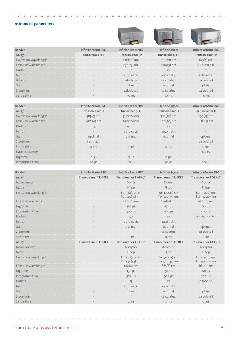
Transcreener®
Nucleotide (ADP, GDP, etc.) detection assay
MechanismThe mechanism is the same for all Transcreener assays. The anti-body is preloaded with the corresponding nucleotide, which is conjugated to a tracer molecule. All assays use a far red tracer that minimizes compound interference. For example, in the ADP² FP assay the detection mixture comprises of Alexa 633 ADP and a highly selective ADP monoclonal antibody. The Transcreener ADP2 FP assay measures the progress of any enzyme that produces ADP by displacing the tracer by ADP thereby causing a decrease in fluorescence polarization. (Figure 2)
Assay overview
TechnologyFluorescence polarization (FP) Fluorescence intensity (FI)Time-resolved fluorescence resonance energy transfer (TR-FRET)
Principle, providerBellbrook's Transcreener assays37 are designed to detect various mono- and dinucleotides using FP, TR-FRET or FI detection mode. Four assays (Table 1) cover thousands of target enzymes, including any kinase, ATPase or GTPase. Transcreener is a universal assay method that can be used across entire families of nucleotide-dependent enzymes. All assays are based on different antibodies that show a high affinity for one specific nucleotide (Figure 1).
Figure 1: Transcreener targets
37 http://www.bellbrooklabs.com/transcreener_hts_assays.html
AMP/GMP
GDPUDP
Ligases (DNA, Amino Acid, Protein)
Methyltransferases
Phosphodiesterases
Sialytransferases
ADP
Glycosyltransferases
FucosyltransferasesGTPases (Small G Proteins)
GAPs
Kinases (Protein, Lipid, CHO)
Helicases Carboxylases
ATPases (HSPs, Nucleotidases)
RANSCREENERT RANSCREENERT
www.be l l b rook l abs . com
Red FI validated
R
RANSCREENERTR
TRANSCREENERT RANSCREENERR
T
www.be l l b rook l abs . com
Far Red FP validated
Figure 2: Transcreener principle
Table 1: Alternative readouts
Assay Readout
Transcreener ADP2 Assays
FP, FI, TR-FRET
Transcreener AMP/GMP Assay
FP, FI
Transcreener GDP Assays
FP
Transcreener UDP Assays
FP
ADP ADP ADP
SupportLink • Assay homepage: http://www.bellbrooklabs.com/transcreener_hts_assays.htmlTecan Application Notes • Transcreener® ADP2 Fluorescence Polarization assay • Homogeneous ADP Detection Using Transcreener® Technology on Tecan’s Infinite® F500 and Infinite M1000
Multimode Microplate Readers
FP - Fluorescence Polarization
41Learn more at www.tecan.com
Instrument parameters
Reader Infinite M200 PRO Infinite F200 PRO Infinite F500 Infinite M1000 PROAssay Transcreener FP Transcreener FP Transcreener FP Transcreener FPExcitation wavelength - 610(20) nm 610(20) nm 635(5) nmEmission wavelength - 670(25) nm 670(25) nm 680(20) nmFlashes - 10 10 10Mirror - automatic automatic automaticG-factor - calculated calculated calculatedGain - optimal optimal optimalZ-position - calculated calculated calculatedSettle time - 50 ms 50 ms 50 ms
Reader Infinite M200 PRO Infinite F200 PRO Infinite F500 Infinite M1000 PROAssay Transcreener FI Transcreener FI Transcreener FI Transcreener FIExcitation wavelength 585(9) nm 580(20) nm 580(20) nm 590(15) nmEmission wavelength 627(20) nm 620(20) nm 620(20) nm 625(15) nmFlashes 25 25-100 10 10Mirror - automatic automatic -Gain optimal optimal optimal optimalZ-position optimzed - - calculatedSettle time 0 ms 0 ms 0 ms 0 msFlash frequency 100 HzLag time 0 μs 0 μs 0 μs -Integration time 20 μs 20 μs 20 μs 20 μs
Reader Infinite M200 PRO Infinite F200 PRO Infinite F500 Infinite M1000 PROAssay Transcreener TR-FRET Transcreener TR-FRET Transcreener TR-FRET Transcreener TR-FRETMeasurement - Donor Donor DonorMode - FI top FI top FI topExcitation wavelength - Eu: 320(25) nm
Tb: 340(35) nmEu: 320(25) nmTb: 340(35) nm
Eu: 317(20) nmTb: 332(20) nm
Emission wavelength - 620(10) nm 620(10) nm 620(10) nmLag time - 150 μs 150 μs 60 μsIntegration time - 500 μs 500 μs 500 μsFlashes - 50 10 125-165 (100 Hz)Mirror - automatic automatic /Gain - optimal optimal optimalZ-position - - calculated calculatedSettle time - 0 ms 0 ms 0 msAssay Transcreener TR-FRET Transcreener TR-FRET Transcreener TR-FRET Transcreener TR-FRETMeasurement - Acceptor Acceptor AcceptorMode - FI top FI top FI topExcitation wavelength - Eu: 320(25) nm
Tb: 340(35) nmEu: 320(25) nmTb: 340(35) nm
Eu: 317(20) nmTb: 332(20) nm
Emission wavelength - 665(8) nm 665(8) nm 665(10) nmLag time - 150 μs 150 μs 60 μsIntegration time - 500 μs 500 μs 500 μsFlashes - 25 10 10 (100 Hz)Mirror - automatic automatic /Gain - optimal optimal optimalZ-position - / calculated calculatedSettle time - 0 ms 0 ms 0 ms

42
Alpha – Amplified Luminescent Proximity Homogeneous AssayFRET equivalent with reduced distance limitations and amplification
When the donor bead is excited, it will convert ambient oxygen in its close environment (200 nm) into reactive oxygen molecule. If a binding event occurs, oxygen radicals will eventually reach the acceptor bead and cause an emission signal at a lower wavelength compared to the excitation wavelength.
The Alpha technology’s versatility offers the possibility to assay many biological targets, including enzymes, receptor-ligand interactions, low affinity interactions, second messenger levels, DNA, RNA, proteins, protein-protein interactions, peptides, sugars and small molecules38.
Major applications• AlphaScreen / AlphaLISA
TechnologyMechanismAlpha38 is a technology invented by PerkinElmer. The principle is similar to (TR-)FRET, since the Alpha technology also relies on the interaction of an acceptor and a donor to yield a signal (Figure 1). However, instead of using simple fluorophores, chemically reactive beads are used, and the chemistry that produces the signal is also different. The main advantage of AlphaScreen® and AlphaLISA® is that the distance between the interaction partners can be up to 200 nm, compared to 10 nm for (TR-)FRET. Furthermore, there is an amplification effect which increases the sensitivity of the assay. Background is reduced because the emission wavelength is lower than the excitation wavelength.
Assay designThe assay requires a donor bead, an acceptor bead, and a tagged antibody. The acceptor bead – coupled to a target-specific antibody – and the tagged antibody to capture the donor bead, are added to the sample and incubated to allow the components to bind to their target molecule. Finally, the donor bead is added, which binds to the tagged antibody.
Figure 1: Schematic function of the Alpha technology demonstrated by AlphaLISA® assay.
38 http://www.perkinelmer.com/catalog/category/id/alphatech

43Learn more at www.tecan.com
Features Infinite F200 PRO Infinite M1000 PROTemperature control Ambient +5 °C to 42 °C Ambient +4 °C to 42 °CShaking Linear, orbital with variable amplitudes Linear, orbital, double orbital with variable
amplitudesInjectors 2 2Gas Control Module Yes NoBarcode reader Yes Yes
Typcal values: Amplified Luminescent Proximity Homogeneous Assay (Alpha) Infinite F200 PRO Infinite M1000 PRO
Light source Xenon flash lamp High power laser diode (680 nm) for high-end Alpha assays
Detector Dedicated PMT for Alpha andfluorescence measurements
Low noise photon counting PCT (lumi PMT)
Sensitivity ≤50 ng/ml Omnibead ≤100 amol / well bio-LCK-P≤2.5 ng/ml Omnibeads
Uniformity ≤ 5 % CV ≤ 3 % CVZ’-Value ≥ 0.8 ≥ 0.9Reading speed <11 min (384-well plate) <2min (384-well plate)Temperature correction No YesFilters Optimized fluorescence filter Optimized luminescence filter
Alpha – Amplified Luminescent Proximity Homogeneous AssayFRET equivalent with reduced distance limitations and amplification
Compatible readers
SupportLinks • Assay homepage: http://www.perkinelmer.com/catalog/category/id/alphatech • Alpha knowledge base: http://perkinelmerreagents.onconfluence.com/pages/viewpage.
action?pageId=328672 • Nature advertisement: http://www.nature.com/nmeth/journal/v5/n12/pdf/nmeth.f.230.pdfTecan Application Notes • Implementation of AlphaLISA® technology in the Infinite® M1000 PRO • New Infinite® M1000 PRO with AlphaScreen® module • Implementation of AlphaScreen® technology in the Infinite® M1000 PRO • Implementation of AlphaScreen® technology on the Infinite® F200 PRO • Implementation of AlphaLISA® technology on the Infinite® F200 PRO

44
0
20
60
40
80
100
120
500 520 540 560 580 600 620 640 660 680 700
Wavelength (nm)
AlphaScreen EmissionAlphaLISA Emission
Minimal inner filter effectsin plasma/serum
Rela
tive
Fluo
resc
ence
uni
ts (%
)
Figure 1: Emission spectra of TAR (AlphaScreen) and europium (AlphaLISA) acceptor beads.
39 http://www.perkinelmer.com/catalog/category/id/alphatech
SupportLinks • Tecan homepage: http://www.tecan.com/alphascreen • Assay homepage: http://www.perkinelmer.com/catalog/category/id/alphatech • Alpha knowledge base: http://perkinelmerreagents.onconfluence.com/pages/viewpage.
action?pageId=328672 • PE recomended settings: http://perkinelmerreagents.onconfluence.com/pages/viewpage.action?pageId=32
8672#AlphaLISAAlphaScreennowashassays-InstrumentoptionssettingsTecan Application Notes • Implementation of AlphaLISA® technology in the Infinite® M1000 PRO • New Infinite® M1000 PRO with AlphaScreen® module • Implementation of AlphaScreen® technology in the Infinite® M1000 PRO • Implementation of AlphaScreen® technology on the Infinite® F200 PRO • Implementation of AlphaLISA® technology on the Infinite® F200 PRO
PrincipleAlphaLISA is a development targeting laboratories working with crude blood samples, as the autofluorescence of heme group overlaps with the emission peak (Figure 1) of the AlphaScreen acceptor. The main difference between AlphaLISA and AlphaScreen is that the AlphaLISA emission peak (europium emission) of theacceptor bead is smaller than that of the AlphaScreen assay (rubrene emission).
MechanismFor a closer description of the assay mechanism, please refer to the Alpha Technology section.
Assay overview
TechnologyAlpha Technology
Provider, FormatAlphaScreen and AlphaLISA are PerkinElmer’s Alpha Technology39 assay / reagent platforms. PerkinElmer supplies donor beads coupled to streptavidin, and blank acceptor beads for self-labeling. Additionally, acceptor beads can be purchased precoupledto antibodies specific for a broad range of targets.
AlphaScreen / AlphaLISA
Alpha Technology assay platforms
Alpha – Amplified Luminescent Proximity Homogeneous Assay
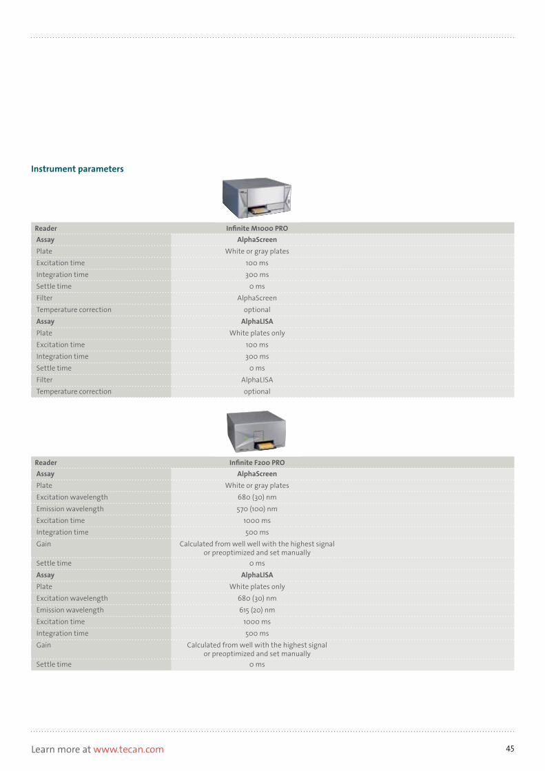
45Learn more at www.tecan.com
Instrument parameters
Reader Infinite M1000 PROAssay AlphaScreenPlate White or gray platesExcitation time 100 msIntegration time 300 msSettle time 0 msFilter AlphaScreenTemperature correction optionalAssay AlphaLISAPlate White plates onlyExcitation time 100 msIntegration time 300 msSettle time 0 msFilter AlphaLISATemperature correction optional
Reader Infinite F200 PROAssay AlphaScreenPlate White or gray platesExcitation wavelength 680 (30) nmEmission wavelength 570 (100) nmExcitation time 1000 msIntegration time 500 msGain Calculated from well well with the highest signal
or preoptimized and set manuallySettle time 0 msAssay AlphaLISAPlate White plates onlyExcitation wavelength 680 (30) nmEmission wavelength 615 (20) nmExcitation time 1000 msIntegration time 500 msGain Calculated from well with the highest signal
or preoptimized and set manuallySettle time 0 ms

46
LUMI-LuminescenceLight is emitted from the sample
Various forms of luminescence can be differentiated.1. Glow luminescence, which generates stable and measurable
light up to several hours, for example the BioThema ATP assay2. Flash luminescence, which is characterized by rapid, but short-
lived, light generation, for example DLR and Aequorin (injectors are required for flash luminescence)
3. Multicolor luminescence, such as BRET1/2/3/e and Chroma-Glow™
Major applications• Dual-Luciferase® Reporter Assay (DLR™)• Bio Thema ATP detection kit• BRET (Bioluminescence Resonance Energy Transfer)
TechnologyLuminescence is widely known as a reaction that causes the release of light. It can be caused by chemical reactions, electrical energy, subatomic motion, or stress on a crystal. For molecular biology, bioluminescence is the most important of the various luminescence reactions. At the core of this technology is the luciferase enzyme. Luciferases (Firefly, Renilla) (Figure 1) convert a substrate into an excited state (Figure 2). When returning to the ground state, a photon (light) is released (emitted).
One striking difference between fluorescence and luminescence is that luminescence requires no excitation light. This reduces the background to almost zero, resulting in outstanding sensitivity.
Luciferase
LuciferinATP, Mg2+
OxyluciferinAMP, CO2
OHHOHO COOH N
N SN S
S
hv
NS
Figure 1: Firefly Figure 2: Substrate cleavage by luciferase

47Learn more at www.tecan.com
LUMI-LuminescenceLight is emitted from the sample
SupportTecan Application Notes• Dual-Luciferase® Reporter Gene Assay • Gaseous luminescence standards on
Infinite® multimode readers• Peroxidase detection using SwordTM Peroxidase Reagents• Human IL-6 chemiluminescent ELISA using
Sword™ Peroxidase Reagents
• Emission spectra of luminescent signals• An Optimized Mouse IL-6 ELISA using Sword Diagnostics
Peroxidase Reagents (Infinite® M200)• Luminescence sensitivity• Mycoplasm detection with the MycoAlert™ assay system
Compatible readers
Features Infinite M200 PRO Infinite F200 PRO Infinite F500 Infinite M1000 PROReader design Monochromator Filter Filter MonochromatorConfiguration Quad4 Monochromators™,
2 excitation and 2 emission monochromators for high
performance, high flexibility and accurate
data acquisition
Up to 4 programmable filter pairs per slide that are
easily ejected and exchanged through the front of the instrument
Up to 6 filter pairs per slide which are individually
moveable, allowing any combination of Ex and Em
filters; easy filter change, ID chip
Premium Quad4 Monochro-mators, 2 excitation and 2
emission monochromators allow for stray light reduc-
tion up to a factor of 107
DLReady™ Yes Yes Yes YesCapability Yes Yes Yes YesPlate format 6- to 384-well plates
NanoQuant Plate6- to 384-well plates
NanoQuant Plate6- to 1,536-well plates
NanoQuant Plate6- to 1,536-well plates
NanoQuant PlateTemperature control Ambient +5 °C to 42 °C Ambient +5 °C to 42 °C Ambient +4 °C to 42 °C Ambient +4 °C to 42 °CInjectors 2 2 2 2Stacker Connect: stacks for
30 or 50 platesConnect: stacks for
30 or 50 platesConnect: stacks for
30 or 50 platesOn-board stacker: stacks
for 30 or 50 platesBarcode reader Yes Yes Yes Yes
Typcal values: Luminescence (LUMI) Infinite M200 PRO Infinite F200 PRO Infinite F500 Infinite M1000 PRO
Capability – glow Yes Yes Yes YesCapability – flash Yes (with injectors) Yes (with injectors) Yes (with injectors) Yes (with injectors)Capability – scanning No No No YesCapability – multicolor (BRET)
Yes Yes Yes Yes
Detector Photon counting system with low dark current PMT
Photon counting system with low dark current PMT
Photon counting system with low darkcurrent PMT
Photon counting low dark current PMT
Wavelength range 380 - 600 nm 380 - 600 nm 380 - 600 nm 380 - 600 nmSensitivity (flash) 12 amol ATP / well
(218fM; 384-well plate) – 384 / 55 μl
12 amol ATP / well(218fM; 384-well plate) –
384 / 55 μl
12 amol ATP / well(218 fM; 384-well plate) –
384 / 55 μl
12 amol ATP / well(218 fM; 384-well plate) –
384 / 55 μlSensitivity (glow) 225 amol ATP / well
(9pM; low volume 384-well plate)
225 amol ATP / well(9 pM; low volume
384-well plate)
225 amol ATP / well(9 pM; low volume
384-well plate)
225 amol ATP / well(9 pM; low volume
384-well plate)Dynamic range 9 decades (OD2) 9 decades (OD2) 8 decades (OD1) 9 decades (OD2)

48
42 http://biothema.se/products/kits /
Bio Thema ATP detection kit42
ATP detection kit based on luminescence
MechanismThe luciferase and substrate are added to the sample. If ATP is present, the luciferase converts the substrate and light is released (Figure 1). If no ATP is present, no light is released. The more ATP is present in the sample the stronger is the signal. In a last step the signal is quantified by comparing it to a standard ATP curve.
Major applicationsMajor applications include ATP detection, Cell proliferation, Cytotoxicity, Enzymatic monitoring.
Assay overview
TechnologyLuminescence (Glow)
Principle, providerATP (Adenosine-Tri-Phosphate) is a molecule for short time energy storage and required for almost any reaction in living organisms. Biothema's ATP detection kit measure and quantifies the level of ATP and correlates it to the activity of an enzyme or cells.
FormatThe kit consists of a luciferase, the substrate and an ATP standard to quantify the signal of the sample.
luciferin oxyluciferin
ATP ADP
light
Luciferase
Figure 1: Schematic principle of luminescence caused by a luciferase based on the conversion of ATP.
LUMI-Luminescence

49Learn more at www.tecan.com
SupportLinks • Assay homepage: http://biothema.se/products/kits/Tecan Application Notes • Luminescence sensitivity
Instrument parameters
Reader Infinite M200 PRO Infinite F200 PRO Infinite F500 Infinite M1000 PROAssay Bio Thema ATP Bio Thema ATP Bio Thema ATP Bio Thema ATPIntegration time 10,000 ms integration 10,000 ms integration 10,000 ms integration 10,000 ms integrationAttenuation automatic automatic automatic automaticSettle time - 0 ms 0 ms 0 ms

50
40 http://www.promega.com/products/reporter-assays-and-transfection/reporterassays/dual_luciferase-reporter-assay-system/41 Houskeeper genes are proteins or enzymes that are constitutionally expressed in most cells as for instance the DNA polymerase
Dual-Luciferase® Reporter Assay (DLR™)40
Luminescence-based normalizable gene activator assay
MechanismCells need to be transfected with both luciferase reporter genes (Figure 1). The firefly luciferase reporter is measured first by adding Luciferase Assay Reagent II (LAR II) to generate a ‘glow-type’ luminescent signal. After quantifying the firefly luminescence, the reaction is quenched, and the Renilla luciferase reaction simultaneously initiated, by adding Stop & Glo® Reagent to the tube. The Stop & Glo Reagent produces a ‘glow-type’ signal from the Renilla luciferase, which decays slowly over the course of the measurement.
Assays are commonly measured over several hours, or even days. During this time period, the plate can either be shuttled between the incubator and the reader, or a temperature and gas controlled multimode reader such as the Infinite 200 PRO may be used.
Assay overview
TechnologyLuminescence (flash)
Purpose, provider, major applicationPromega's DLR assay measures gene activation / expression using a luminescence-based readout. One particular challenge of gene activator assays is quantification of the result. The DLR assay resolves this issue by normalizing the output using two luciferases. While one luciferase measures the expression of the gene of interest, the second luciferase measures the expression of a housekeeping gene41, which is used to normalize the signal. Promega has two series of firefly and Renilla luciferase vectors – pGL4 and pRL – designed for use with the DLR assay systems.
luc mRNA
+
luc
RE
Clone RE upstream ofthe firefly luciferase (luc) gene.
1. Determine promoter/response element (RE) of interest. The amount of firefly luciferase is dependent on experimentalcontrol of the responseelement of interest.
Add luciferase detectionreagent and measure firefly luciferase activity.
Transfect constructinto cells.
RE Gene of Interest
4.
3.
2.
6614
MA
Figure 1: DLR assay mechanism
LUMI-Luminescence

51Learn more at www.tecan.com
SupportLinks • Assay homepage: http://www.promega.com/products/reporter-assays-and-transfection/reporter-assays/
dual_luciferasereporter-assay-system/Tecan Application Notes • Dual-Luciferase® Reporter Gene Assay
Instrument parameters
Reader Infinite M200 PRO Infinite F200 PRO Infinite F500 Infinite M1000 PROAssay DLR DLR DLR DLRWell Well wise Well wise Well wise Well wiseInjector A 100 μl;
refill for every injection100 μl;
refill for every injection100 μl;
refill for every injection100 μl;
refill for every injectionWait 3 sec 3 sec 3 sec 3 secMeasure 10,000 ms integration 10,000 ms integration 10,000 ms integration 10,000 ms integrationAttenuation automatic automatic automatic automaticInjector B 100 μl;
refill for every injection100 μl;
refill for every injection100 μl;
refill for every injection100 μl;
refill for every injectionWait 3 sec 3 sec 3 sec 3 secMeasure 10,000 ms integration 10,000 ms integration 10,000 ms integration 10,000 ms integrationAttenuation automatic automatic automatic automaticSettle time 0 ms 0 ms 0 ms 0 ms
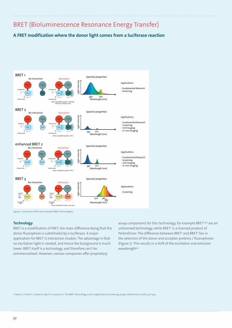
52
42 Bacart J, Corbel C, Jockers R, Bach S, Couturier C: The BRET technology and its application to screening assays. Biotechnol J 2008, 3:311-324.
BRET (Bioluminescence Resonance Energy Transfer)A FRET modification where the donor light comes from a luciferase reaction
assay components for this technology, for example BRET1/3/e are an unlicensed technology, while BRET2 is a licensed product of PerkinElmer. The difference between BRET1 and BRET2 lies in the selection of the donor and acceptor proteins / fluorophores (Figure 1). This results in a shift of the excitation and emission wavelength42.
TechnologyBRET is a modification of FRET, the main difference being that the donor fluorophore is substituted by a luciferase. A major application for BRET is interaction studies. The advantage is that no excitation light is needed, and hence the background is muchlower. BRET itself is a technology, and therefore can't be commercialized. However, various companies offer proprietary
Luciferine+ ATP + O2+Mg2+
Oxyluciferin+AMP
Luciferine+ ATP + O2+Mg2+
Oxyluciferin+AMP
BRET 3
Wavelength (nm)
Ligh
t int
ensi
ty
500 565583
Spectral properties
Protein Y
Protein X
mn 565
Interaction
Protein Y
Protein X
mn 565
No interaction
Applications:
- Screening
Firefly Firefly
mn 385
DsRedDsRed
Other compatible acceptors: Cy3, Cy3.5
GFP2
Deep Blue C+
O2
Coelenteramide
Coelenterazine+
O2
Coelenteramide
Rluc
BRET 2
Protein Y
Protein X
No interaction
mn 593 mn 593
ProteinY
Interaction
ProteinX
mn 015 395 510Wavelength (nm)
Ligh
t int
ensi
ty
Spectral properties
Applications:
- Fundamental Research- Screening- Cell Imaging- in vivo imaging
Rluc GFP2
Other compatible acceptor: GFP10
enhanced BRET 2
Protein Y
Protein X
No interaction
Deep Blue C+
O2
Coelenteramidemn 593 395 510
Wavelength (nm)
Ligh
t int
ensi
ty
Spectral propertiesInteraction
mn 593
Coelenterazine+
O2
Coelenteramide
ProteinY
ProteinX
mn 015
Applications:
- Fundamental Research- Screening- Cell Imaging- in vivo imaging
Rluc8Rluc8 GFP2GFP2
Other compatible acceptor: GFP10
EYFP EYFP
BRET 1
Protein Y
Protein X
No interaction
Coelenterazine+
O2
Coelenteramide
Rluc
mn 084
Coelenterazine+
O2
Coelenteramide
ProteinY
Interaction
ProteinX
mn 035mn 084 480 530Wavelength (nm)
Ligh
t int
ensi
tySpectral properties
Applications:
- Fundamental Research- Screening
Rluc
Other compatible acceptors: YFPtopaz,YFPcitrine, YFPVenus, YPet
Coelenterazine+
O2
Coelenteramide
Coelenterazine+
O2
Coelenteramide
QD-BRET
480 605Wavelength (nm)
Ligh
t int
ensi
ty
Spectral propertiesNo interaction
Rlucor
Rluc8
mn 084
6-His
QDOT coo-coo-
coo-coo-
coo-
coo-
coo-
coo-
Interaction
Rlucor
Rluc8
mn 084
6-His
mn 506Ni
++
QD coo-coo-
coo-coo-
coo-
coo-
coo-
coo-Ni
++ Ni
+
+Ni
+
+
Applications:
- Screening- in vivo imaging
Other compatible acceptors: Other QDs
Figure 1: Summary of the most relevant BRET technologies.

53Learn more at www.tecan.com
SupportTecan Application Notes • Bioluminescence Resonance Energy Transfer (BRET) • Optimization of BRET2™ measurement parameters
Compatible readers
Features Infinite M200 PRO Infinite F200 PRO Infinite F500 Infinite M1000 PROReader design Monochromator Filter Filter MonochromatorConfiguration Quad4 Monochromators™,
2 excitation and 2 emission monochromators for high
performance, high flexibility and accurate
data acquisition
Up to 4 programmable filter pairs per slide that are
easily ejected and exchanged through the front of the instrument
Up to 6 filter pairs per slide which are individually
moveable, allowing any combination of Ex and Em
filters; easy filter change, ID chip
Premium Quad4 Monochro-mators, 2 excitation and 2
emission monochromators allow for stray light reduc-
tion up to a factor of 107
DLReady™ Yes Yes Yes YesCapability Yes Yes Yes YesPlate format 6- to 384-well plates
NanoQuant Plate6- to 384-well plates
NanoQuant Plate6- to 1,536-well plates
NanoQuant Plate6- to 1,536-well plates
NanoQuant PlateTemperature control Ambient +5 °C to 42 °C Ambient +5 °C to 42 °C Ambient +4 °C to 42 °C Ambient +4 °C to 42 °CInjectors 2 2 2 2Stacker Connect: stacks for
30 or 50 platesConnect: stacks for
30 or 50 platesConnect: stacks for
30 or 50 platesOn-board stacker: stacks
for 30 or 50 platesBarcode reader Yes Yes Yes Yes
Typcal values: Luminescence (LUMI) Infinite M200 PRO Infinite F200 PRO Infinite F500 Infinite M1000 PRO
Capability – glow Yes Yes Yes YesCapability – flash Yes (with injectors) Yes (with injectors) Yes (with injectors) Yes (with injectors)Capability – scanning No No No YesCapability – multicolor (BRET)
Yes Yes Yes Yes
Detector Photon counting system with low dark current PMT
Photon counting system with low dark current PMT
Photon counting system with low darkcurrent PMT
Photon counting low dark current PMT
Wavelength range 380 - 600 nm 380 - 600 nm 380 - 600 nm 380 - 600 nmSensitivity (flash) 12 amol ATP / well
(218fM; 384-well plate) – 384 / 55 μl
12 amol ATP / well(218fM; 384-well plate) –
384 / 55 μl
12 amol ATP / well(218 fM; 384-well plate) –
384 / 55 μl
12 amol ATP / well(218 fM; 384-well plate) –
384 / 55 μlSensitivity (glow) 225 amol ATP / well
(9pM; low volume 384-well plate)
225 amol ATP / well(9 pM; low volume
384-well plate)
225 amol ATP / well(9 pM; low volume
384-well plate)
225 amol ATP / well(9 pM; low volume
384-well plate)Dynamic range 9 decades (OD2) 9 decades (OD2) 8 decades (OD1) 9 decades (OD2)
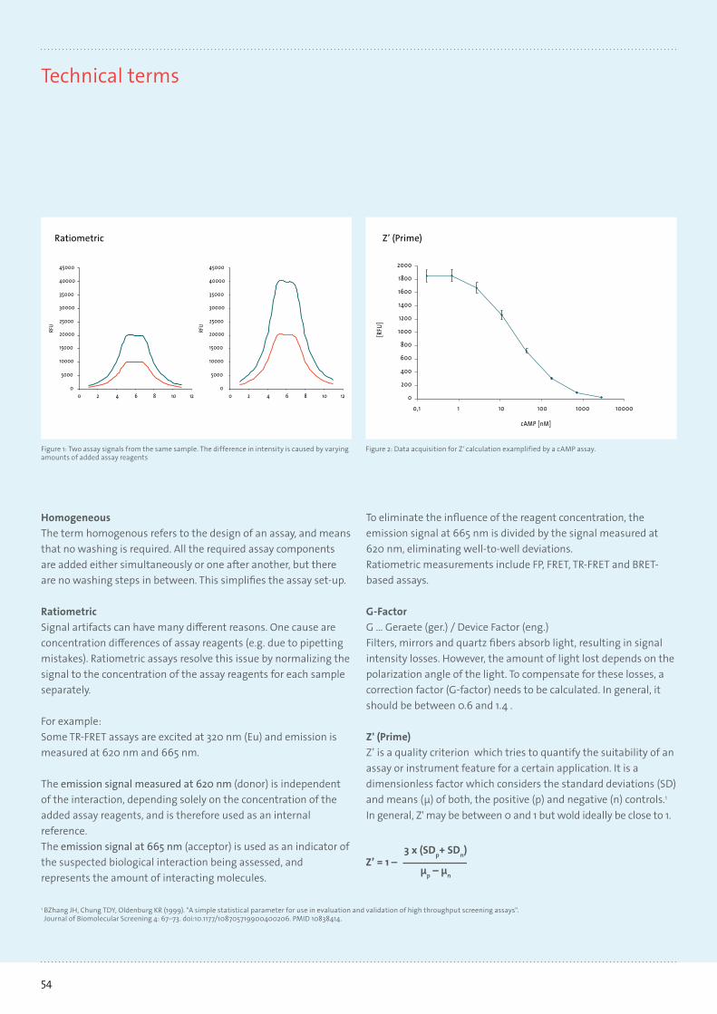
54
Technical terms
HomogeneousThe term homogenous refers to the design of an assay, and means that no washing is required. All the required assay components are added either simultaneously or one after another, but there are no washing steps in between. This simplifies the assay set-up.
RatiometricSignal artifacts can have many different reasons. One cause are concentration differences of assay reagents (e.g. due to pipetting mistakes). Ratiometric assays resolve this issue by normalizing the signal to the concentration of the assay reagents for each sample separately.
For example:Some TR-FRET assays are excited at 320 nm (Eu) and emission is measured at 620 nm and 665 nm.
The emission signal measured at 620 nm (donor) is independent of the interaction, depending solely on the concentration of the added assay reagents, and is therefore used as an internal reference.The emission signal at 665 nm (acceptor) is used as an indicator of the suspected biological interaction being assessed, and represents the amount of interacting molecules.
To eliminate the influence of the reagent concentration, the emission signal at 665 nm is divided by the signal measured at 620 nm, eliminating well-to-well deviations.Ratiometric measurements include FP, FRET, TR-FRET and BRET-based assays.
G-Factor G … Geraete (ger.) / Device Factor (eng.)Filters, mirrors and quartz fibers absorb light, resulting in signal intensity losses. However, the amount of light lost depends on the polarization angle of the light. To compensate for these losses, a correction factor (G-factor) needs to be calculated. In general, it should be between 0.6 and 1.4 .
Z' (Prime)Z’ is a quality criterion which tries to quantify the suitability of an assay or instrument feature for a certain application. It is a dimensionless factor which considers the standard deviations (SD) and means (μ) of both, the positive (p) and negative (n) controls.1 In general, Z' may be between 0 and 1 but wold ideally be close to 1.
00 2 4 6 8 10 12 0 2 4 6 8 10 12
5000
10000
15000
20000
25000
35000
40000
45000
RFU
30000
0
5000
10000
15000
20000
25000
35000
40000
45000
RFU
30000
cAMP [nM]
[RFU
]
2000
1800
1600
1400
1200
1000
800
600
400
200
00,1 1 10 100 1000 10000
Figure 1: Two assay signals from the same sample. The difference in intensity is caused by varying amounts of added assay reagents
Figure 2: Data acquisition for Z' calculation examplified by a cAMP assay.
Z’ (Prime)Ratiometric
1 BZhang JH, Chung TDY, Oldenburg KR (1999). "A simple statistical parameter for use in evaluation and validation of high throughput screening assays". Journal of Biomolecular Screening 4: 67–73. doi:10.1177/108705719900400206. PMID 10838414.
Z’ = 1 –3 x (SDp+ SDn)
µp – µn

55Learn more at www.tecan.com
Appendix
Reader-Assay Compatibility, validations (V) and certifications (C)
Infinite M200 PRO Infinite F200 PRO Infinite F500 Infinite M1000 PRO
Technology Reader capability ABS x x x x
Reader capability FI x x x x
Reader capability TRF x x x x
Reader capability FRET x x x x
Reader capability TR-FRET x x x
Reader capability FP x x x
Reader capability ALPHA x x
Reader capability LUMI x x x x
Binding studies BRET LUMI x x x x
HTRF® TR-FRET x white plates x (C) x white plates
Delfia® TRF x x x x
Omnia® FI x x x (V) x (V)
Predictor™ hERG FP x x (V) x (V)
Gene expression GeneBLAzer™ FRET x (V) x (V) x (V) x (V)
DLR™ LUMI x (C) x (C) x (C) x (C)
Signal transduction Z-Lyte® FRET x x x (V) x (V)
LanthaScreen™ TR-FRET x (C) x (C) x (C)
PolarScreen™ FP x x (V) x (V)
Adapta® TR-FRET x x (V) x (V)
Transcreener ADP2 FP x x (C) x (C)
Transcreener ADP2 FI x (C) x (C) x (C) x (C)
Transcreener ADP2 TR-FRET x x (V) x (V)
AuthorThomas Hengstl · Application Specialist/Sales & MarketingT +43 6246 8933 108 · F +43 6246 8933 6108 · [email protected]

Tecan Group Ltd. makes every effort to include accurate and up-to-date information within this publication, however, it is possible that omissions or errors might have occurred. Tecan Group Ltd. cannot, therefore, make any representations or warranties, expressed or implied, as to the accuracy or completeness of the information provided in this publication. Changes in this publication can be made at any time without notice. All mentioned trademarks are protected by law. For technical details and detailed procedures of the specifications provided in this document please contact your Tecan representative. This brochure may contain reference to applications and products which are not available in all markets. Please check with your local sales representative.
All pictures used in this document are copyright protected. Any use requires Tecan’s prior written approval.
Tecan and Infinite are registered trademarks and NanoQuant Plate and Quad4 Monochromators are trademarks of Tecan Group Ltd. Männedorf, Switzerland.
Windows and Excel are registered trademarks of Microsoft Corporation in the United States and other countries. HTRF is a registered trademark of Cisbio bioassays, France. The fluorescence ratio associated with the HTRF readout is a correction method developed by Cisbio international and covered by the US patent 5,527,684 and its foreign equivalents, for which Cisbio international has granted a license to Tecan Group Ltd. Its application is strictly limited to the use of HTRF reagents and technology, excluding any other TR-FRET technologies. Transcreener HTS Assay Platform is a patented technology of BellBrook Labs, LLC USA. Transcreener is a registered trademark of BellBrook Labs, LLC USA. CellTiter 96 AQueous One Solution Cell Proliferation Assay, Dual-Luciferase Reporter Assay (DLR) and ENLITEN are registered trademarks and DLReady is a trademark of Promega Corporation, USA. Adapta, AlamarBlue, GeneBLAzer, Omnia, PicoGreen, PrestoBlue, Ribogreen, NanoOrange and Z’-LYTE are registered trademarks and LanthaScreen, LiveBLAzer, PolarScreen, Predictor and Tango are trademarks of Invitrogen Corporation Carlsbad, USA. BCA Protein Assay is a trademark of the Thermo Scientific Pierce cooperation. AlphaLISA, AlphaScreen and Delfia are registered trademarks of PerkinElmer, USA.
© 2013, Tecan Trading AG, Switzerland, all rights reserved. For disclaimer and trademarks please visit www.tecan.com
www.tecan.com 3978
23 V
1.0, 0
2-20
13, 3
0084
114
Australia +61 3 9647 4111 Austria +43 62 46 89 33 Belgium +32 15 42 13 19 China +86 21 2206 3206 Denmark +45 70 23 44 50 France +33 4 72 76 04 80 Germany +49 79 51 94 170 Italy +39 02 92 44 790 Japan +81 44 556 73 11 Netherlands +31 18 34 48 174 Singapore +65 644 41 886 Spain +34 93 490 01 74 Sweden +46 31 75 44 000 Switzerland +41 44 922 89 22 UK +44 118 9300 300 USA +1 919 361 5200 Other countries +41 44 922 8125
Tecan – Who we areTecan is a leading global provider of life science laboratory instruments for the biopharmaceuticals, forensics, clinical diagnostics and academic sectors, specializing in the development and production of automation and detection solutions, including microplate readers, microarray products and washers.
Founded in Switzerland in 1980, Tecan has manufacturing, research and development sites in both North America and Europe, and maintains a sales and service network in 52 countries. To date, Tecan has distributed approximately 20,000 microplate readers worldwide, and is committed to continuous technological improvements and compliance with the highest global quality standards.


![(b) (c) · 2018. 12. 10. · Surface Evolver [6] soft are tak-ing in to accoun t the liquid's surface tension lv and the liquid-solid con tact energy. Due to the small scale of the](https://static.fdocuments.us/doc/165x107/60a60fbc40a4d55b026335dd/b-c-2018-12-10-surface-evolver-6-soft-are-tak-ing-in-to-accoun-t-the.jpg)