Applicability of LIVE/DEAD BacLight Stain with - Aaqr.org
13
Aerosol and Air Quality Research, 13: 1755–1767, 2013 Copyright © Taiwan Association for Aerosol Research ISSN: 1680-8584 print / 2071-1409 online doi: 10.4209/aaqr.2012.10.0293 Applicability of LIVE/DEAD BacLight Stain with Glutaraldehyde Fixation for the Measurement of Bacterial Cell Concentration and Viability in the Air Kotaro Murata, Daizhou Zhang * Faculty of Environmental and Symbiotic Sciences, Prefectural University of Kumamoto, 3-1-100 Tsukide, Kumamoto 862- 8502, Japan ABSTRACT Data on the concentration and viability of microorganisms in the atmosphere provide crucial information with regard to their dispersion, roles in environmental changes, and effects on ecosystems. In this study, we investigated the application of the LIVE/DEAD ® BacLight™ Bacterial Viability Kit (BacLight stain) with regard to the enumeration of viable and non-viable bacterial cells in the air, with the 4',6-diamidino-2-phenylindole (DAPI) stain used as the control of total cell counts. Two cultured bacterial strains isolated from an air sample, Bacillus subtilis and Micrococcus sp., were used in laboratory experiments. The results of BacLight staining agreed well with those of DAPI staining in detecting total cell counts, and the detection efficiency of Bacillus subtilis was 76–112%. Bacterial viability (number ratio of viable cells to total cells) showed consistency among repeated experiments with the same sample replicates, indicating the high confidence of counting viable cells, as well as total cells with the stain. Fixation of samples with glutaraldehyde prevented fluorescence bleaching at the exposure to fluorescence and increased detection accuracy. Application of the BacLight stain with and without fixation to air samples that were collected with a bio-sampler at an urban site proved the effectiveness of this approach in determining the cell concentration and viability of airborne bacteria at a 1-hour time resolution. A combination of BacLight staining and glutaraldehyde fixation treatment, in parallel with experiments without this treatment in field measurements, is proposed to ensure the detection accuracy. The method described here is able to measure the concentration and viability of bacterial cells in the air, and their variation with weather conditions. Keywords: Bacterial abundance; Viability; Epifluorescent microscopy; BacLight stain; Bio-sampler. INTRODUCTION Bioaerosols are referred to as airborne particles originating from biological origins, including viruses, bacteria, fungi, pollen, plant debris and their derivatives, and constitute a major fraction of atmospheric aerosols (Hurst et al., 2002; Jaenicke, 2005; Xu et al., 2011). They are released from surface waters, soil, and plants by wind erosion, water splash and wave action. Their size ranges from tens of nanometers to hundreds of micrometers (Hurst et al., 2002). Recent studies demonstrate that airborne bacteria, as a major component of bioaerosols, play an important role in the Earth environment (Griffin et al., 2001; Prospero et al., 2005; Iwasaka et al., 2009; Womack et al., 2010; Morris et al., 2011). In addition to their contribution to the bio-materials such as protein and fat in atmospheric aerosols, airborne bacteria can function as efficient nuclei for ice cloud formation and affect the * Corresponding author. Tel.:81-96-3216712; Fax: 81-96-3846765 E-mail address: [email protected] hydrological processes and radiation transfer in the atmosphere, i.e., their climate effects (Sun and Ariya, 2006; Christner et al., 2008; Morris et al., 2011). Dispersion of bacteria with airflows links the bacterial communities in geographically isolated regions (Kellogg and Griffin, 2006) and blurs the distinction of closely related species in distant areas (Fenchel and Finlay, 2004). In order to elucidate the diversity and geography of bacteria in the atmosphere and their environmental effects, one needs to know their concentration and viable status in space and time under different weather conditions. For example, the cell number concentration of total bacteria is crucial to ice cloud formation because both live and dead cells and even cell fragments can act as nuclei for icing (Christner et al., 2008; Möhler et al., 2008). Bacterial viability is primary information to assess the ecological effects because settling of live bacteria to the surface potentially alters the indigenous microbial composition of the deposition regions such as seawater (Maki et al., 2011) and lake water (Hervàs et al., 2009). Unfortunately, very little is known about the total cell concentration and the viability of airborne bacteria in the atmosphere because of the lack of appropriate approaches (Womack et al., 2010). Describing
Transcript of Applicability of LIVE/DEAD BacLight Stain with - Aaqr.org
Aerosol and Air Quality Research, 13: 1755–1767, 2013 Copyright © Taiwan Association for Aerosol Research ISSN: 1680-8584 print / 2071-1409 online doi: 10.4209/aaqr.2012.10.0293
Applicability of LIVE/DEAD BacLight Stain with Glutaraldehyde Fixation for the Measurement of Bacterial Cell Concentration and Viability in the Air Kotaro Murata, Daizhou Zhang* Faculty of Environmental and Symbiotic Sciences, Prefectural University of Kumamoto, 3-1-100 Tsukide, Kumamoto 862-8502, Japan ABSTRACT
Data on the concentration and viability of microorganisms in the atmosphere provide crucial information with regard to their dispersion, roles in environmental changes, and effects on ecosystems. In this study, we investigated the application of the LIVE/DEAD® BacLight™ Bacterial Viability Kit (BacLight stain) with regard to the enumeration of viable and non-viable bacterial cells in the air, with the 4',6-diamidino-2-phenylindole (DAPI) stain used as the control of total cell counts. Two cultured bacterial strains isolated from an air sample, Bacillus subtilis and Micrococcus sp., were used in laboratory experiments. The results of BacLight staining agreed well with those of DAPI staining in detecting total cell counts, and the detection efficiency of Bacillus subtilis was 76–112%. Bacterial viability (number ratio of viable cells to total cells) showed consistency among repeated experiments with the same sample replicates, indicating the high confidence of counting viable cells, as well as total cells with the stain. Fixation of samples with glutaraldehyde prevented fluorescence bleaching at the exposure to fluorescence and increased detection accuracy. Application of the BacLight stain with and without fixation to air samples that were collected with a bio-sampler at an urban site proved the effectiveness of this approach in determining the cell concentration and viability of airborne bacteria at a 1-hour time resolution. A combination of BacLight staining and glutaraldehyde fixation treatment, in parallel with experiments without this treatment in field measurements, is proposed to ensure the detection accuracy. The method described here is able to measure the concentration and viability of bacterial cells in the air, and their variation with weather conditions. Keywords: Bacterial abundance; Viability; Epifluorescent microscopy; BacLight stain; Bio-sampler. INTRODUCTION
Bioaerosols are referred to as airborne particles originating from biological origins, including viruses, bacteria, fungi, pollen, plant debris and their derivatives, and constitute a major fraction of atmospheric aerosols (Hurst et al., 2002; Jaenicke, 2005; Xu et al., 2011). They are released from surface waters, soil, and plants by wind erosion, water splash and wave action. Their size ranges from tens of nanometers to hundreds of micrometers (Hurst et al., 2002). Recent studies demonstrate that airborne bacteria, as a major component of bioaerosols, play an important role in the Earth environment (Griffin et al., 2001; Prospero et al., 2005; Iwasaka et al., 2009; Womack et al., 2010; Morris et al., 2011). In addition to their contribution to the bio-materials such as protein and fat in atmospheric aerosols, airborne bacteria can function as efficient nuclei for ice cloud formation and affect the * Corresponding author. Tel.:81-96-3216712; Fax: 81-96-3846765 E-mail address: [email protected]
hydrological processes and radiation transfer in the atmosphere, i.e., their climate effects (Sun and Ariya, 2006; Christner et al., 2008; Morris et al., 2011). Dispersion of bacteria with airflows links the bacterial communities in geographically isolated regions (Kellogg and Griffin, 2006) and blurs the distinction of closely related species in distant areas (Fenchel and Finlay, 2004).
In order to elucidate the diversity and geography of bacteria in the atmosphere and their environmental effects, one needs to know their concentration and viable status in space and time under different weather conditions. For example, the cell number concentration of total bacteria is crucial to ice cloud formation because both live and dead cells and even cell fragments can act as nuclei for icing (Christner et al., 2008; Möhler et al., 2008). Bacterial viability is primary information to assess the ecological effects because settling of live bacteria to the surface potentially alters the indigenous microbial composition of the deposition regions such as seawater (Maki et al., 2011) and lake water (Hervàs et al., 2009). Unfortunately, very little is known about the total cell concentration and the viability of airborne bacteria in the atmosphere because of the lack of appropriate approaches (Womack et al., 2010). Describing
Murata and Zhang, Aerosol and Air Quality Research, 13: 1755–1767, 2013 1756
the cell concentrations of live and dead bacteria and their variation in space and time is still a great challenge toward understanding their impacts on atmospheric physics and chemistry and on the development and evolution of ecosystems (Morris et al., 2011).
Airborne bacteria are usually detected with culture-based approaches. Their abundance is estimated by counting bacterial colonies forming on a nutrient agar plate after incubation for hours or days (Hurst et al., 2002). This method is an easy and convenient approach and suitable for routine measurements because of its low cost and high time resolution. The disadvantage of this approach is that only culturable bacteria can be detected and investigated with the colony-forming unit on the culture media rather than the abundance of bacteria. It is well known that only a small percentage of bacteria in a natural environment (usually less than 1–10%) can grow on selected nutrient media, and most bacteria are “viable but non-culturable” or dead (Roszak and Colwell, 1987; Amann et al., 1995; Lighthart, 2000; Maier et al., 2000).
A number of culture-independent approaches have been developed to pursue the abundance of bacteria in the air. The metagenomic technique is a powerful one, which is being widely used to identify airborne bacteria (Kakikawa et al., 2008; Maki et al., 2008; Maki et al., 2010; Nishimura et al., 2010). Genes of bacteria are excellent candidates for phylogenetic analysis and a small subunit of rRNA of bacteria can offer sufficient information on the bacterial composition. Application of quantitative polymerase chain reaction (qPCR) can obtain quantitative information of target genes or target organism in air samples (Peccia and Hernandez, 2006). Recently, EMA-qPCR (ethidium monoazide qPCR) was introduced to measure viable bacteria in samples by eliminating DNA of dead cells from the analysis (Rudi et al., 2005; Georgakopoulos et al., 2009). These metagenomic techniques are very efficient approaches to identify bacteria in samples but not good at the quantification of bacteria. They identify the integral abundance of bacteria in DNA amounts rather than cell counts (Peccia and Hernandez, 2006; Georgakopoulos et al., 2009; Després et al., 2012). Bacterial number concentration can only be obtained via a transfer from DNA amounts to cell number by assuming a relation between DNA amounts and cell number (Kakikawa et al., 2008).
Enumeration with fluorescent staining is a traditional technique to detect the abundance of bacteria in the air. So far the most frequently used fluorescent dye for bacterial enumeration in air samples is 4',6-diamidino-2-phenylindole (DAPI). This dye binds the DNA of bacterial cells and makes microbes appear blue under fluorescence. It has been used to measure the abundance of airborne bacteria in various environments, such as indoor air (Li and Huang, 2006), outdoor air (Chi and Li, 2007; Rodríguez de Evgrafov et al., 2010; Li et al., 2011), and high elevation sites (Bowers et al., 2009; Bowers et al., 2012; Xia et al., 2013). The cell concentration of bacteria can be obtained with this stain but the viable status of the bacteria is not available because it dyes all bacterial cells the same color. Chi and Li (2007) measured the viability of airborne bacteria (the number
ratio of viable bacterial cells to total bacterial cells) by measuring total and non-viable cells with different dyes and obtained the viability by subtracting non-viable cell counts from total cell counts. A counter, the Ultraviolet Aerodynamic Particle Sizer (UV-APS) which is a fluorescence-based instrument, has been developed to count the real-time number of live bioaerosol particles in the air. Its results include not only live bacteria but also other bioaerosol particles, and it cannot measure the total concentration including live and dead ones in parallel (Hairston et al., 1997; Brosseau et al., 2000; Huffman et al., 2010; Xu et al., 2011; Després et al., 2012). Recently, Park et al. (2012) developed a method based on ultraviolet and visible spectroscopy and succeeded in measuring rapidly the viability of bioaerosols in laboratory-prepared samples.
Despite the development of new powerful methods, there are few data that can be used for the assessment of the effects of airborne bacteria on the environment. Primary modeling studies suggested bioaerosols were of minor importance to global ice nuclei concentrations and precipitation processes (Hoose et al., 2010). However, recent field studies of cloud and precipitation in the western United States showed the substantial role of dust and bioaerosols in the mid-level ice cloud formation where precipitation processes were initiated (Creamean et al., 2013). A large discrepancy between modeling and observation remains because of the lack of observational data that allow parameterization of bacteria-associated processes in the air. Therefore, an accessible method is needed to easily gather the data of bacterial abundance and viability in the air. Developing a reliable method would permit interdisciplinary collaboration for wide observation campaigns of airborne bacteria as well as to provide reference data for a verification of traditional methods or other new technologies.
Hara and Zhang (2012) challenged measuring the concentration of viable and non-viable bacterial cells in atmospheric dust samples with the LIVE/DEAD® BacLight™ Bacterial Viability Kit and found the effectiveness of the kit at a high sampling-time resolution by coupling with an efficient sampler. The fluorescent staining dye, LIVE/DEAD® BacLight™ Bacterial Viability Kit (Invitrogen™, Molecular Probes Inc., OR, USA) was developed to assay the viability of bacteria. This stain is able to distinguish between live and dead bacteria by dying them different colors. Although there is still argument on how to define dead bacteria (Hannig et al., 2010), this stain can offer the viability of bacteria as well as the bacterial abundance and has been used to study bacteria in water (Boulos et al., 1999), soil (Janssen et al., 2002), cloud water, and precipitation (Bauer et al., 2002).
In this study, the applicability of the BacLight stain to enumerate viable and non-viable bacteria in the air, where bacterial concentration is remarkably lower than in water and soil, was carefully examined, and the improvement with fixation treatment of samples was furthered with laboratory experiments, the application to atmospheric bioaerosol samples was tested, and a method suitable for the detection of the abundance and viability of bacteria in the air is offered. METHOD
Murata and Zhang, Aerosol and Air Quality Research, 13: 1755–1767, 2013 1757
LABORATORY TEST Bacterial Samples
Two cultured bacterial strains, Bacillus subtilis (hereafter described as B. subtilis; accession number JN092588 from DDBJ/EMBL/GenBank databases) and Micrococcus sp. (accession number GU073283), were adopted in laboratory experiments. Both strains were isolated from the culture of a sample collected for airborne bacteria detection at 1000 m altitude over the sea area close to Goto Island (126.9°E, 31.3°N), Nagasaki, Japan during an aircraft measurement on 11 December, 2010. B. subtilis and Micrococcus sp. are both gram-positive bacteria and mainly inhabit soil (Madigan et al., 2012). Bacilli are spore-forming bacteria that are more resistant against environmental stress than other nonspore-forming ones (Roszak and Colwell, 1987). Similar bacterial strains are frequently present as the dominant species among culturable bacterial fraction in the atmosphere (Fang et al., 2007) and have been well detected in surface and elevated air in East Asia with cultural method and DNA analysis (Harrison et al., 2005; Fang et al., 2007; Hua et al., 2007; Maki et al., 2008; Lee et al., 2009; Yukimura et al., 2009; Maki et al., 2010; Smith et al., 2010; Womack et al., 2010; Kobayashi et al., 2011).
For each experiment of bacterial fluorescent staining and enumeration, a set of B. subtilis or Micrococcus sp. replicates was prepared from one strain. To prepare one set of samples, a strain of B. subtilis or Micrococcus sp. was inoculated into 3 mL of Bacto™ Tryptic Soy Broth (Becton Dickinson and Company, NJ, USA) and incubated at 30°C for 12–20 hours in a shaking incubator at 125 rev/min (OD595 ≈ 2.0). Then 100 μL of incubated vegetative cells were transferred into 10 mL of 0.9% saline (pre-filtered through 0.2 μm pore filters), which was further separated into several parts for subsequent staining. Fluorescent Staining
The nucleic acid binding fluorescent dye, LIVE/DEAD® BacLight™ Bacterial Viability Kit L13152 (hereafter BacLight stain; Invitrogen™, Molecular Probes Inc., OR, USA) was used to stain samples. BacLight stain is composed of two dyes: SYTO 9 and propidium iodide (PI). SYTO 9 penetrates all bacterial membranes (intact and injured) and labels bacterial cells green. PI can only penetrate injured bacterial membranes and labels the bacterial cells red while diminishing the green stained by SYTO 9. We consider bacterial cells with injured membranes to be non-viable (dead) cells and those uninjured to be viable (live) cells following the development of this stain. Thus, in this study, bacterial cells green under fluorescence were identified as viable ones and those red as non-viable ones. The concentration of SYTO 9 in the BacLight stain solution was 6 μM and that of PI was 30 μM. The amount of the dye solution applied to each sample for staining was 1/100 of the sample volume.
For comparison and quality control, the traditional dye 4',6-diamidino-2-phenylindole (DAPI; Dojindo Molecular Technologies Inc., Japan) was used to stain bacterial cells in samples for enumeration. DAPI binds AT-rich region of
nucleic acid and stains all bacteria blue under fluorescence. The concentration of DAPI in the stock solution was 50 μg/mL and the dye solution applied to each sample was 1/100 of the sample volume.
The staining was conducted under complete dark conditions at 4°C. Staining time for each sample was 15 minutes. After the staining of a sample, it was filtered through a 0.2 μm-pore-size black polycarbonate membrane filter (ADVANTEC®, Toyo Roshi Kaisha Ltd., Japan). Then, the filter was placed on a glass slide. 10 μL of immersion oil (TypeFF, Cargille-Sacher Laboratories, Inc., NJ, USA) was dropped before it was covered with a cover slide. Observation and Enumeration of Bacteria
Stained samples were viewed and photographed by using an epifluorescence microscope (Eclipse 80i, Nikon Corp., Japan) equipped with a 100 W mercury lamp. Filters of 450–490 nm excitation and 520 nm emission were used for the enumeration of BacLight-stained samples and filters of 360–370 nm excitation and 400 nm emission were used for the enumeration of DAPI-stained samples.
The experiments were carried out in 3 rounds. One BacLight-stained sample and one DAPI-stained sample of B. subitilis were subjected to the enumeration in each experimental round. For each sample, 3 pictures were taken in different fields under the microscope. Bacterial cells were counted from 3 random fields (area of each field: 30 μm × 30 μm = 900 μm2) in each picture and bacterial abundance in the sample was estimated from the 9 fields. Fluorescence Bleaching and Fixation with Glutarladehyde
The color of stained samples actually faded gradually after exposure to fluorescence under the microscope, resulting in the fluorescence bleaching of stained bacteria. Pictures of stained bacteria had to be taken quickly. Delay of picture taking or an extension of exposure of a stained sample to fluorescence would cause a large uncertainty in its enumeration. We found that color fading still happened even though we used the neutral density filters of the microscope to reduce the fluorescence irradiation to samples and made an effort to photograph samples within 30 seconds of exposure.
In order to test if the fading could be minimized with fixation agents, some samples of B. subtilis and Micrococcus sp. were treated with glutaraldehyde (25% solution, Wako Pure Chemical Industries, Ltd., Japan) before staining. Glutaraldehyde is a 5-carbon dialdehyde that displays potent bactericidal, fungicidal, mycobactericidal, sporicidal, and virucidal activity (Russell, 1994). It can strongly fix microorganisms or other biological cells by linking amino groups in the cell membranes and is widely used as a fixation agent in both laboratory studies and field observations (Gorman et al., 1980; Kepner and Pratt, 1994; Tietjen and Wetzel, 2003). In this study, the fixation was conducted with 1% (v/v) glutaraldehyde in dark at 4°C for 30 minutes according to previous studies (Kepner and Pratt, 1994).
Fixed samples, and also samples without the fixation treatment, were stained with the BacLight stain or the DAPI stain and were subjected to the subsequent epifluorescence
Murata and Zhang, Aerosol and Air Quality Research, 13: 1755–1767, 2013 1758
microscopic observation. B. subtilis samples were also subjected to enumeration as described above. The experiments of staining the fixed and unfixed samples with BacLight stain and DAPI stain were carried out in three rounds. To investigate the fluorescence bleaching due to the exposure and the improvement by the fixation, results from fixed and unfixed samples at the beginning of exposure and after 1-minute exposure were compared. This is because 1 minute is the common required time limitation of stained bacteria exposure to the fluorescence in microscopic enumerating. FIELD TEST
The staining coupled with and without glutaraldehyde fixation treatment was tested to quantify bacteria in the open air. Six samples were collected on a balcony of a building in the Prefectural University of Kumamoto, Japan (32°48'N, 130°45'E; 20 m above the ground) between 8 and 26 January, 2012. A BioSampler® (SKC Inc., PA, USA) which combined impingement into a liquid with centrifugal motion was used to collect airborne bacterial samples. The sampler with the liquid impingement can recover culturable cells of bacteria and fungi as much as 10× greater than usual membrane filtration methods (Griffin et al., 2011). The Biosampler is more efficient than impingers in collecting air-borne bacteria because it suppresses bacterial re-aerosolization by swirling the collection liquid medium. Its collection efficiency for particles of 0.3, 0.5, 1.0, and 2.0 μm were 79, 89, 96 and 100%, respectively (Willeke et al., 1998; Fabian et al., 2005). The inside of the BioSampler was coated with dimethyl polysiloxane (L-25, Fuji systems Corp., Japan) in order to minimize the adherence of microbial cells onto the glass surface. Before sample collection, the BioSampler was washed with particle-free water (pre-filtered through a 0.025 μm pore-size filter) and heat-sterilized at 180°C for 2 hours.
For the collection of each sample, 20 mL particle-free phosphate buffered saline (PBS, pH = 7.4) that had been autoclaved at 121°C for 20 minutes was used to collect airborne bacteria. PBS could prevent possible damage of bacterial cells due to osmotic pressure change during sample collection. The sampling flow rate was 12.5 ± 0.6 L/min and the sampling time was 60 minutes. To ensure the collection efficiency, the PBS was checked and loss was compensated every 20 minutes. After the sampling, the PBS was transferred to a centrifuge tube and adjusted to 25 mL with particle-free water. Then the sample was separated into three equal portions. Two were treated with the glutaraldehyde fixation and then stained by the BacLight stain and the DAPI stain, respectively. One was directly stained by the BacLight stain. Blank samples were prepared and enumerated with the same procedures in three experiments, except that the PBS was not used for sampling. Bacterial counts in the blank samples were found 1–2 orders smaller than in the air samples.
In previous tests with airborne bacteria samples, we found that many bacteria were hardly identified with microscope photographs because of their small size. For
this reason, bacteria in the samples of this study were counted directly from the microscope fields. Bacterial counting was conducted in 20 random 100 μm × 100 μm fields for each sample. The bacterial concentration (C) in the air was calculated with the counts from the 20 fields,
viable non viable mediatotal
field filtered air
N N S VC
S V V
(1)
viable mediaviable
field filtered air
N S VC
S V V
(2)
non viable medianon viable
field filtered air
N S VC
S V V
(3)
according to where N is the number of bacterial cells in each field, S is the area of the filter, Vmedia is the volume of PBS, Sfield is the area of each microscopic field, Vfiltered is the volume of filtered PBS, and Vair is the volume of sample air. Counting viable bacteria in each field was accomplished as soon as possible (all less than 30 seconds), in an effort to minimize the probable uncertainties due to bacterial bleaching. RESULTS LABORATORY TEST
In this section, we first compare the results of B. subtilis and Micrococcus sp. samples stained with BacLight stain and DAPI stain. Then we report the influence of the exposure of stained samples to fluorescence on the color of bacterial cells. Finally, results of the application of the fixation agent compared with those without fixation are described. BacLight Staining and Comparison with DAPI Staining
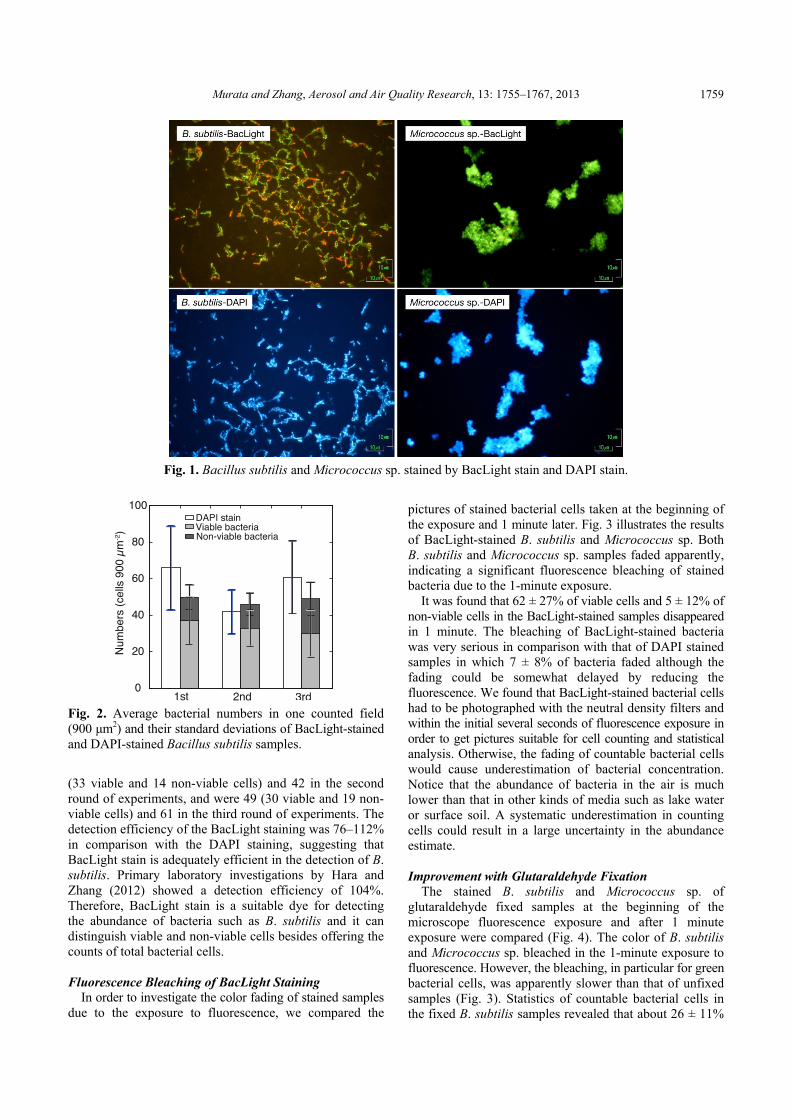
Fig. 1 illustrates fluorescence microscopy photographs of B. subtilis and Micrococcus sp. samples after the BacLight staining and the DAPI staining. There were a number of green cells in addition to red ones in the BacLight staining pictures, indicating the presence of viable bacterial cells. In the DAPI staining pictures, all bacterial cells were blue. DAPI staining is unable to distinguish viable and non-viable cells,and the results from this staining are the total cell counts.
Fig. 2 shows the statistics of bacterial cell detection of B. subtilis samples. Micrococcus sp. samples were not available for cell counting because they were too aggregated to enumerate. Some cells of B. subtilis were also aggregated on the filter. Bacterial cells that could be identified by their rod shape in the aggregates were enumerated. But if there were cells overlapped by others, they could not be seen and counted under the microscope. In the first round of experiments, there were about 50 (37 viable and 13 non-viable cells on average) B. subtilis cells in one field of the BacLight-stained sample and there were about 66 cells in one field of the DAPI-stained sample. The figures were 47
Murata and Zhang, Aerosol and Air Quality Research, 13: 1755–1767, 2013 1759
Fig. 1. Bacillus subtilis and Micrococcus sp. stained by BacLight stain and DAPI stain.
Fig. 2. Average bacterial numbers in one counted field (900 μm2) and their standard deviations of BacLight-stained and DAPI-stained Bacillus subtilis samples.
(33 viable and 14 non-viable cells) and 42 in the second round of experiments, and were 49 (30 viable and 19 non-viable cells) and 61 in the third round of experiments. The detection efficiency of the BacLight staining was 76–112% in comparison with the DAPI staining, suggesting that BacLight stain is adequately efficient in the detection of B. subtilis. Primary laboratory investigations by Hara and Zhang (2012) showed a detection efficiency of 104%. Therefore, BacLight stain is a suitable dye for detecting the abundance of bacteria such as B. subtilis and it can distinguish viable and non-viable cells besides offering the counts of total bacterial cells. Fluorescence Bleaching of BacLight Staining
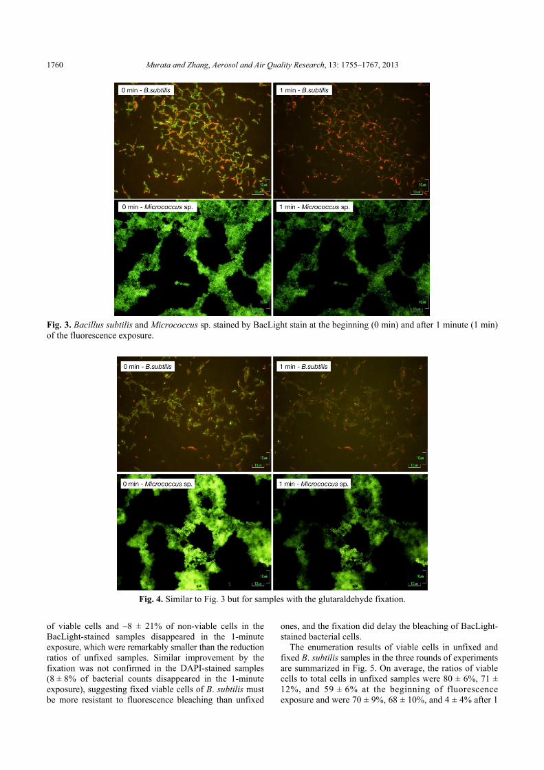
In order to investigate the color fading of stained samples due to the exposure to fluorescence, we compared the
pictures of stained bacterial cells taken at the beginning of the exposure and 1 minute later. Fig. 3 illustrates the results of BacLight-stained B. subtilis and Micrococcus sp. Both B. subtilis and Micrococcus sp. samples faded apparently, indicating a significant fluorescence bleaching of stained bacteria due to the 1-minute exposure.
It was found that 62 ± 27% of viable cells and 5 ± 12% of non-viable cells in the BacLight-stained samples disappeared in 1 minute. The bleaching of BacLight-stained bacteria was very serious in comparison with that of DAPI stained samples in which 7 ± 8% of bacteria faded although the fading could be somewhat delayed by reducing the fluorescence. We found that BacLight-stained bacterial cells had to be photographed with the neutral density filters and within the initial several seconds of fluorescence exposure in order to get pictures suitable for cell counting and statistical analysis. Otherwise, the fading of countable bacterial cells would cause underestimation of bacterial concentration. Notice that the abundance of bacteria in the air is much lower than that in other kinds of media such as lake water or surface soil. A systematic underestimation in counting cells could result in a large uncertainty in the abundance estimate. Improvement with Glutaraldehyde Fixation
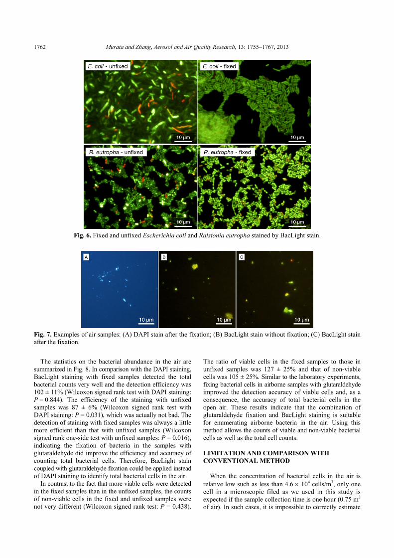
The stained B. subtilis and Micrococcus sp. of glutaraldehyde fixed samples at the beginning of the microscope fluorescence exposure and after 1 minute exposure were compared (Fig. 4). The color of B. subtilis and Micrococcus sp. bleached in the 1-minute exposure to fluorescence. However, the bleaching, in particular for green bacterial cells, was apparently slower than that of unfixed samples (Fig. 3). Statistics of countable bacterial cells in the fixed B. subtilis samples revealed that about 26 ± 11%
Murata and Zhang, Aerosol and Air Quality Research, 13: 1755–1767, 2013 1760
Fig. 3. Bacillus subtilis and Micrococcus sp. stained by BacLight stain at the beginning (0 min) and after 1 minute (1 min) of the fluorescence exposure.
Fig. 4. Similar to Fig. 3 but for samples with the glutaraldehyde fixation.
of viable cells and –8 ± 21% of non-viable cells in the BacLight-stained samples disappeared in the 1-minute exposure, which were remarkably smaller than the reduction ratios of unfixed samples. Similar improvement by the fixation was not confirmed in the DAPI-stained samples (8 ± 8% of bacterial counts disappeared in the 1-minute exposure), suggesting fixed viable cells of B. subtilis must be more resistant to fluorescence bleaching than unfixed
ones, and the fixation did delay the bleaching of BacLight-stained bacterial cells.
The enumeration results of viable cells in unfixed and fixed B. subtilis samples in the three rounds of experiments are summarized in Fig. 5. On average, the ratios of viable cells to total cells in unfixed samples were 80 ± 6%, 71 ± 12%, and 59 ± 6% at the beginning of fluorescence exposure and were 70 ± 9%, 68 ± 10%, and 4 ± 4% after 1

Murata and Zhang, Aerosol and Air Quality Research, 13: 1755–1767, 2013 1761
Fig. 5. Ratios of the viable cells to the total cells in each field (900 µm2) of Bacillus subtilis samples with and without glutaraldehyde fixation at the beginning (0 min) and after 1 minute (1 min) of the fluorescence exposure.
minute. In contrast, the ratios in fixed samples were 88 ± 6%, 78 ± 7%, and 94 ± 6% at the beginning of exposure and were 82 ± 8%, 73 ± 9%, and 85 ± 7% after 1 minute Although viable cell counts in fixed samples also decreased due to the fluorescence exposure, the reduction relative to the total cells on average, 7%, was much smaller than that in unfixed samples, 23%. These results indicate that the enumeration accuracy of B. subtilis treated with the fixation was better than that without the fixation. Fixation with glutaraldehyde can benefit the observation and enumeration of BacLight-stained B. subtilis cells with epifluorescence microscopy.
The fixation was expected to have protected viable cells from environmental stresses that might injure the cells during the experiments such as osmotic pressure change in filtration and dye washing. Glutaraldehyde is a chemical disinfectant and displays antimicrobial activity. Fixation with this agent can strengthen the membranes of bacterial cells. For gram-positive bacteria such as B. subtilis, glutaraldehyde can react with the peptidoglycan and produce cross-links in cell walls, resulting in a strengthening and sealing effect on the walls (Gorman et al., 1980). This mechanism should have worked on the B. subtilis and strengthened the bacteria against the osmotic change and/or the filtration pressure.
It must be noted that the antimicrobial action of glutaraldehyde differs according to bacteria. Fixation of gram-negative bacteria with glutaraldehyde could cause partial sealing or contraction of the outer layers of the cell envelope and destruct the cells (Gorman et al., 1980). It was reported that the counted viable cells of Escherichia coli (E. coli) were reduced due to fixation with 5% glutaraldehyde in BacLight staining (Boulos et al., 1999). However, in the same study, Citrobacter freundii was not affected by glutaraldehyde, although it also belonged to the group of gram-negative bacteria. In order to test glutaraldehyde-
induced effects on viable cells of gram-negative bacteria in BacLight stain, we applied 1% glutaraldehyde fixation to E. coli JM109 and Ralstonia eutropha JCM 11282T using the same procedures as to B. subtilis and Micrococcus sp. in the laboratory experiments. Fig. 6 shows the examples of the stained E. coli and R. eutropha at the beginning of fluorescence exposure. Fixed bacteria in the microscopic field were obviously clearer in shape than those in unfixed samples. Many bacterial cells in the unfixed samples were ambiguous and uncountable. Moreover, there were approximately 20–30 non-viable cells (red cells) in one field (approximately 3000 μm2) of unfixed samples, while few were found in fixed samples, in particular in fixed E. coli samples. These results indicate that 1% glutaraldehyde fixation is also applicable to some gram-negative bacteria for the improvement of enumeration accuracy in the case of BacLight stain.
Gram-positive bacteria were generally found in the atmosphere by culturing methods, whereas gram-negative bacteria were primarily found by culture-independent methods (Després et al., 2012). According to the present results, investigation of airborne bacterial abundance with BacLight staining and enumeration is expected to benefit from glutaraldehyde fixation. The fixation can increase the accuracy of cell counts of viable and total bacteria, such as B. subtilis to the extent of our experiments. However, we cannot deny the probable presence of cases that there are substantial bacteria in the air whose cell membranes could be severely damaged by the fixation agent within a short time and viable ones would be identified as non-viable ones. For the sake of detection accuracy of counting viable and non-viable cells in the air, we suggest using the combination of BacLight stain and glutaraldehyde fixation treatment in parallel with experiments without the treatment in field measurements. The confidence of results on bacterial abundance and viability can be adequately ensured with this combination.
FIELD TEST
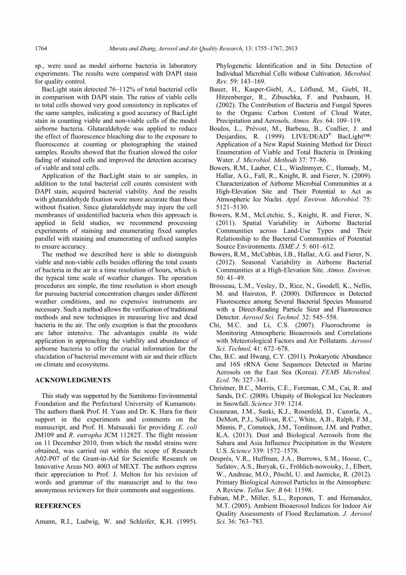
Fig. 7 shows examples of the pictures of unfixed and fixed open-air samples stained with BacLight stain and fixed samples with DAPI stain. The samples actually consisted of not only bacteria but also other particulate matters such as mineral particles, soot and droplets. Those particles were distinguishable from bacterial cells according to their irregular and aggregate morphology and fluorescent color. For example, mineral particles usually in irregular shape looked yellow by BacLight stain and greenish yellow or white by DAPI stain (Hara et al., 2011; Hara and Zhang, 2012). In contrast, bacteria were usually spherical and had a size close to or smaller than 1 μm in diameter, which was consistent with the reported size of bacteria in surface soil and sea-water (Roszak and Colwell, 1987). Some particles in Fig. 7 were dust particles and soot particles. In the atmosphere, there are particles in spherical shape with a size close to 1 μm besides bacteria. They are mainly droplets of salt solution or soot spheres and do not contain fluorescent components (Seinfeld and Pandis, 1998).
Murata and Zhang, Aerosol and Air Quality Research, 13: 1755–1767, 2013 1762
Fig. 6. Fixed and unfixed Escherichia coli and Ralstonia eutropha stained by BacLight stain.
Fig. 7. Examples of air samples: (A) DAPI stain after the fixation; (B) BacLight stain without fixation; (C) BacLight stain after the fixation.
The statistics on the bacterial abundance in the air are summarized in Fig. 8. In comparison with the DAPI staining, BacLight staining with fixed samples detected the total bacterial counts very well and the detection efficiency was 102 ± 11% (Wilcoxon signed rank test with DAPI staining: P = 0.844). The efficiency of the staining with unfixed samples was 87 ± 6% (Wilcoxon signed rank test with DAPI staining: P = 0.031), which was actually not bad. The detection of staining with fixed samples was always a little more efficient than that with unfixed samples (Wilcoxon signed rank one-side test with unfixed samples: P = 0.016), indicating the fixation of bacteria in the samples with glutaraldehyde did improve the efficiency and accuracy of counting total bacterial cells. Therefore, BacLight stain coupled with glutaraldehyde fixation could be applied instead of DAPI staining to identify total bacterial cells in the air.
In contrast to the fact that more viable cells were detected in the fixed samples than in the unfixed samples, the counts of non-viable cells in the fixed and unfixed samples were not very different (Wilcoxon signed rank test: P = 0.438).
The ratio of viable cells in the fixed samples to those in unfixed samples was 127 ± 25% and that of non-viable cells was 105 ± 25%. Similar to the laboratory experiments, fixing bacterial cells in airborne samples with glutaraldehyde improved the detection accuracy of viable cells and, as a consequence, the accuracy of total bacterial cells in the open air. These results indicate that the combination of glutaraldehyde fixation and BacLight staining is suitable for enumerating airborne bacteria in the air. Using this method allows the counts of viable and non-viable bacterial cells as well as the total cell counts.
LIMITATION AND COMPARISON WITH CONVENTIONAL METHOD
When the concentration of bacterial cells in the air is relative low such as less than 4.6 104 cells/m3, only one cell in a microscopic filed as we used in this study is expected if the sample collection time is one hour (0.75 m3 of air). In such cases, it is impossible to correctly estimate

Murata and Zhang, Aerosol and Air Quality Research, 13: 1755–1767, 2013 1763
Fig. 8. Average bacterial abundances in the air and standard deviations identified with the DAPI staining and BacLight staining from the six air samples (marked by A–F).
the bacterial cell concentration in the air with the samples. One hour samples are only suitable to cases when the bacterial concentration is more than 105 cells/m3. Sample collection time needs to be extended if the anticipated bacterial concentration in the air is lower than 105 cells/m3 in order to get confidential results. According to published literature in which airborne bacteria were studied with microscopic enumeration and qPCR, the concentration of bacterial cells in surface air is usually about 104–105 cells/m3 or more except for extreme clean areas such as alpine, remote marine and elevated air (Tong, 1999; Tong and Lighthart, 2000; Bauer et al., 2002; Harrison et al., 2005; Chi and Li, 2007; Bowers et al., 2009; Bowers et al., 2011; Cho and Hwang, 2011; Bowers et al., 2012; Hara and Zhang, 2012; Yamaguchi et al., 2012; Xia et al., 2013). In addition, there might be BacLight stain-resistant bacteria in the air. In this regard, the bacteria detected with the BacLight stain should be considered as the low bounds of bacterial concentration in the air.
Table 1 shows the comparison of the BacLight stain with conventional DNA-based methods and DAPI stain in the measurements of airborne bioaerosols. DNA-based methods including qPCR can be used in principle to accurately identify and quantify bioaerosols in the air in terms of DNA and its contents. DAPI staining can detect total DNA-containing bioaerosols. BacLight staining can quantify viable and non-viable bacteria in cell number. The methods with fluorescent stain can get the cell concentration in the air but cannot obtain the information on bacterial composition and community.
Recently, instruments and fluorescence sensors, such as waveband integrated bioaerosol sensors (WIBS) and ultraviolet aerodynamic particle sizer (UV-APS), were developed to monitor the real time in situ concentration of bioaerosols in the air. WIBS measures the real time concentration of total bioaerosols (Kaye et al., 2005; Toprak and Schnaiter, 2013) and UV-APS measures the real time number concentration of live bioaerosols in the air (Prenni et al., 2009; Huffman et al., 2010, 2012). Both enable the information on the number size distribution of the bioaerosols. These instruments are much more efficient than methods with fluorescent stain to demonstrate the evolution of bioaerosols in the air. However, the particles measured by the instruments include not only biological ones but also those whose components (e.g., the organic compounds of polycyclic aromatic hydrocarbons) display fluorescence (Huffman et al., 2010). Moreover, results from these instruments in field studies lack data qualification by comparisons with other approaches, which make the uncertainties in the data unknown. In this regard, the BacLight stain method we propose in this study actually supplies an available way for the inter-comparison of results from these different kinds of approaches, which will largely benefit the studies of bioaerosols in the air. SUMMARY
In this study, BacLight stain was tested to enumerate the
viable and non-viable cells of airborne bacteria. Two gram-positive bacterial strains, B. subtilis and Micrococcus
Table 1. Comparison of the BacLight stain and conventional methods (DNA-based and DAPI staining) in the measurement of bioaerosols.
Bacteria Other bioaerosols Remarks
DNA-based method Bulk DNA concentration Bulk DNA concentration DNA of viable ones available with EMA-PCR
DAPI staining Total cells Total particles Stain all DNA-containing particles
BacLight staining Viable and nonviable No data Usually effective for gram-positive bacteria
Murata and Zhang, Aerosol and Air Quality Research, 13: 1755–1767, 2013 1764
sp., were used as model airborne bacteria in laboratory experiments. The results were compared with DAPI stain for quality control.
BacLight stain detected 76–112% of total bacterial cells in comparison with DAPI stain. The ratios of viable cells to total cells showed very good consistency in replicates of the same samples, indicating a good accuracy of BacLight stain in counting viable and non-viable cells of the model airborne bacteria. Glutaraldehyde was applied to reduce the effect of fluorescence bleaching due to the exposure to fluorescence at counting or photographing the stained samples. Results showed that the fixation slowed the color fading of stained cells and improved the detection accuracy of viable and total cells.
Application of the BacLight stain to air samples, in addition to the total bacterial cell counts consistent with DAPI stain, acquired bacterial viability. And the results with glutaraldehyde fixation were more accurate than those without fixation. Since glutaraldehyde may injure the cell membranes of unidentified bacteria when this approach is applied in field studies, we recommend processing experiments of staining and enumerating fixed samples parallel with staining and enumerating of unfixed samples to ensure accuracy.
The method we described here is able to distinguish viable and non-viable cells besides offering the total counts of bacteria in the air in a time resolution of hours, which is the typical time scale of weather changes. The operation procedures are simple, the time resolution is short enough for pursuing bacterial concentration changes under different weather conditions, and no expensive instruments are necessary. Such a method allows the verification of traditional methods and new techniques in measuring live and dead bacteria in the air. The only exception is that the procedures are labor intensive. The advantages enable its wide application in approaching the viability and abundance of airborne bacteria to offer the crucial information for the elucidation of bacterial movement with air and their effects on climate and ecosystems. ACKNOWLEDGMENTS
This study was supported by the Sumitomo Environmental Foundation and the Prefectural University of Kumamoto. The authors thank Prof. H. Yuan and Dr. K. Hara for their support in the experiments and comments on the manuscript, and Prof. H. Matsusaki for providing E. coli JM109 and R. eutropha JCM 11282T. The flight mission on 11 December 2010, from which the model strains were obtained, was carried out within the scope of Research A02-P07 of the Grant-in-Aid for Scientific Research on Innovative Areas NO. 4003 of MEXT. The authors express their appreciation to Prof. J. Melton for his revision of words and grammar of the manuscript and to the two anonymous reviewers for their comments and suggestions. REFERENCES Amann, R.I., Ludwig, W. and Schleifer, K.H. (1995).
Phylogenetic Identification and in Situ Detection of Individual Microbial Cells without Cultivation. Microbiol. Rev. 59: 143–169.
Bauer, H., Kasper-Giebl, A., Löflund, M., Giebl, H., Hitzenberger, R., Zibuschka, F. and Puxbaum, H. (2002). The Contribution of Bacteria and Fungal Spores to the Organic Carbon Content of Cloud Water, Precipitation and Aerosols. Atmos. Res. 64: 109–119.
Boulos, L., Prévost, M., Barbeau, B., Coallier, J. and Desjardins, R. (1999). LIVE/DEAD® BacLight™: Application of a New Rapid Staining Method for Direct Enumeration of Viable and Total Bacteria in Drinking Water. J. Microbiol. Methods 37: 77–86.
Bowers, R.M., Lauber, C.L., Wiedinmyer, C., Hamady, M., Hallar, A.G., Fall, R., Knight, R. and Fierer, N. (2009). Characterization of Airborne Microbial Communities at a High-Elevation Site and Their Potential to Act as Atmospheric Ice Nuclei. Appl. Environ. Microbiol. 75: 5121–5130.
Bowers, R.M., McLetchie, S., Knight, R. and Fierer, N. (2011). Spatial Variability in Airborne Bacterial Communities across Land-Use Types and Their Relationship to the Bacterial Communities of Potential Source Environments. ISME J. 5: 601–612.
Bowers, R.M., McCubbin, I.B., Hallar, A.G. and Fierer, N. (2012). Seasonal Variability in Airborne Bacterial Communities at a High-Elevation Site. Atmos. Environ. 50: 41–49.
Brosseau, L.M., Vesley, D., Rice, N., Goodell, K., Nellis, M. and Hairston, P. (2000). Differences in Detected Fluorescence among Several Bacterial Species Measured with a Direct-Reading Particle Sizer and Fluorescence Detector. Aerosol Sci. Technol. 32: 545–558.
Chi, M.C. and Li, C.S. (2007). Fluorochrome in Monitoring Atmospheric Bioaerosols and Correlations with Meteorological Factors and Air Pollutants. Aerosol Sci. Technol. 41: 672–678.
Cho, B.C. and Hwang, C.Y. (2011). Prokaryotic Abundance and 16S rRNA Gene Sequences Detected in Marine Aerosols on the East Sea (Korea). FEMS Microbiol. Ecol. 76: 327–341.
Christner, B.C., Morris, C.E., Foreman, C.M., Cai, R. and Sands, D.C. (2008). Ubiquity of Biological Ice Nucleators in Snowfall. Science 319: 1214.
Creamean, J.M., Suski, K.J., Rosenfeld, D., Cazorla, A., DeMott, P.J., Sullivan, R.C., White, A.B., Ralph, F.M., Minnis, P., Comstock, J.M., Tomlinson, J.M. and Prather, K.A. (2013). Dust and Biological Aerosols from the Sahara and Asia Influence Precipitation in the Western U.S. Science 339: 1572–1578.
Després, V.R., Huffman, J.A., Burrows, S.M., Hoose, C., Safatov, A.S., Buryak, G., Fröhlich-nowoisky, J., Elbert, W., Andreae, M.O., Pöschl, U. and Jaenicke, R. (2012). Primary Biological Aerosol Particles in the Atmosphere: A Review. Tellus Ser. B 64: 11598.
Fabian, M.P., Miller, S.L., Reponen, T. and Hernandez, M.T. (2005). Ambient Bioaerosol Indices for Indoor Air Quality Assessments of Flood Reclamation. J. Aerosol Sci. 36: 763–783.

Murata and Zhang, Aerosol and Air Quality Research, 13: 1755–1767, 2013 1765
Fang, Z., Ouyang, Z., Zheng, H., Wang, X. and Hu, L. (2007). Culturable Airborne Bacteria in Outdoor Environments in Beijing, China. Microb. Ecol. 54: 487–496.
Fenchel, T. and Finlay, B.J. (2004). The Ubiquity of Small Species: Patterns of Local and Global Diversity. Bioscience 54: 777–784.
Georgakopoulos, D.G., Després, V., Fröhlich-Nowoisky, J., Psenner, R., Ariya, P.A., Pósfai, M., Ahern, H.E., Moffett, B.F. and Hill, T.C.J. (2009). Microbiology and Atmospheric Processes: Biological, Physical and Chemical Characterization of Aerosol Particles. Biogeosciences 6: 721–737.
Gorman, S.P., Scott, E.M. and Russell, A.D. (1980). Antimicrobial Activity, Uses and Mechanism of Action of Glutaraldehyde. J. Appl. Bacteriol. 48: 161–190.
Griffin, D., Garrison, V., Herman, J. and Shinn, E. (2001). African Desert Dust in the Caribbean Atmosphere: Microbiology and Public Health. Aerobiologia 17: 203–213.
Griffin, D.W., Gonzalez, C., Teigell, N., Petrosky, T., Northup, D.E. and Lyles, M. (2011). Observations on the Use of Membrane Filtration and Liquid Impingement to Collect Airborne Microorganisms in Various Atmospheric Environments. Aerobiologia 27: 25–35.
Hairston, P.P., Ho, J. and Quant, F.R. (1997). Design of an Instrument for Real-Time Detection of Bioaerosols Using Simultaneous Measurement of Particle Aerodynamic Size and Intrinsic Fluorescence. J. Aerosol Sci. 28: 471–482.
Hannig, C., Follo, M., Hellwig, E. and Al-Ahmad, A. (2010). Visualization of Adherent Micro-Organisms Using Different Techniques. J. Med. Microbiol. 59: 1–7.
Hara, K., Zhang, D., Yamada, M., Matsusaki, H. and Arizono, K. (2011). A Detection of Airborne Particles Carrying Viable Bacteria in an Urban Atmosphere of Japan. Asian J. Atmos. Environ. 5–3: 152–156.
Hara, K. and Zhang, D. (2012). Bacterial Abundance and Viability in Long-Range Transported Dust. Atmos. Environ. 47: 20–25.
Harrison, R.M., Jones, A.M., Biggins, P.D.E., Pomeroy, N., Cox, C.S., Kidd, S.P., Hobman, J.L., Brown, N.L. and Beswick, A. (2005). Climate Factors Influencing Bacterial Count in Background Air Samples. Int. J. Biometeorol. 49: 167–178.
Hervàs, A., Camarero, L., Reche, I. and Casamayor, E.O. (2009). Viability and Potential for Immigration of Airborne Bacteria from Africa That Reach High Mountain Lakes in Europe. Environ. Microbiol. 11: 1612–1623.
Hoose, C., Kristjánsson, J.E. and Burrows, S.M. (2010). How Important Is Biological Ice Nucleation in Clouds on a Global Scale? Environ. Res. Lett. 5: 024009.
Hua, N.P., Kobayashi, F., Iwasaka, Y., Shi, G.Y. and Naganuma, T. (2007). Detailed Identification of Desert-Originated Bacteria Carried by Asian Dust Storms to Japan. Aerobiologia 23: 291–298.
Huffman, J.A., Treutlein, B. and Pöschl, U. (2010). Fluorescent Biological Aerosol Particle Concentrations and Size Distributions Measured with an Ultraviolet Aerodynamic Particle Sizer (UV-APS) in Central Europe.
Atmos. Chem. Phys. 10: 3215–3233. Huffman, J.A., Sinha, B., Garland, R.M., Snee-Pollmann,
A., Gunthe, S.S., Artaxo, P., Martin, S.T., Andreae, M.O. and Pöschl, U. (2012). Size Distributions and Temporal Variations of Biological Aerosol Particles in the Amazon Rainforest Characterized by Microscopy and Real-Time UV-APS Fluorescence Techniques during AMAZE-08. Atmos. Chem. Phys. 12: 11997–12019.
Hurst, C.J., Crawfird, R.L., Knudsen, G.R., McInerney, M.J. and Stetzembach, L.D. (2002). Manual of Environmental Microbiology (Second Edition), ASM Press, Washington.
Iwasaka, Y., Shi, G.Y., Yamada, M., Kobayashi, F., Kakikawa, M., Maki, T., Naganuma, T., Chen, B., Tobo, Y. and Hong, C.S. (2009). Mixture of Kosa (Asian Dust) and Bioaerosols Detected in the Atmosphere over the Kosa Particles Source Regions with Balloon-Borne Measurements: Possibility of Long-Range Transport. Air Qual. Atmos. Health 2: 29–38.
Jaenicke, R. (2005). Abundance of Cellular Material and Proteins in the Atmosphere. Science 308: 73.
Janssen, P.H., Yates, P.S., Grinton, B.E., Taylor, P.M. and Sait, M. (2002). Improved Culturability of Soil Bacteria and Isolation in Pure Culture of Novel Members of the Divisions Acidobacteria, Actinobacteria, Proteobacteria, and Verrucomicrobia. Appl. Environ. Microbiol. 68: 2391–2396.
Kakikawa, M., Kobayashi, F., Maki, T., Yamada, M., Higashi, T., Chen, B., Shi, G., Hong, C., Tobo, Y. and Iwasaka, Y. (2008). Dustborne Microorganisms in the Atmosphere over an Asian Dust Source Region, Dunhuang. Air Qual. Atmos. Health 1: 195–202.
Kaye, P., Stanley, W.R., Hirst, E., Foot, E.V., Baxter, K.L. and Barrington, S.J. (2005). Single Particle Multichannel Bio-Aerosol Fluorescence Sensor. Opt. Express 13: 3583–3593.
Kellogg, C.A. and Griffin, D.W. (2006). Aerobiology and the Global Transport of Desert Dust. Trends Ecol. Evol. 21: 638–644.
Kepner, R.L., Jr. and Pratt, J.R. (1994). Use of Fluorochromes for Direct Enumeration of Total Bacteria in Environmental Samples: Past and Present. Microbiol. Rev. 58: 603–615.
Kobayashi, F., Morosawa, S., Maki, T., Kakikawa, M., Yamada, M., Tobo, Y., Hon, C.S., Matsuki, A. and Iwasaka, Y. (2011). Atmospheric Bioaerosol, Bacillus sp., at an Altitude of 3,500 m over the Noto Peninsula: Direct Sampling via Aircraft. Asian J. Atmos. Environ. 5–3: 164–171.
Lee, S., Choi, B., Yi, S.M. and Ko, G.P. (2009). Characterization of Microbial Community during Asian Dust Events in Korea. Sci. Total Environ. 407: 5308–5314.
Li, C.S. and Huang, T.Y. (2006). Fluorochrome in Monitoring Indoor Bioaerosols. Aerosol Sci. Technol. 40: 237–241.
Li, M., Qi, J., Zhang, H., Huang, S., Li, L. and Gao, D. (2011). Concentration and Size Distribution of Bioaerosols in an Outdoor Environment in the Qingdao Coastal

Murata and Zhang, Aerosol and Air Quality Research, 13: 1755–1767, 2013 1766
Region. Sci. Total Environ. 409: 3812–3819. Lighthart, B. (2000). Mini-Review of the Concentration
Variations Found In the Alfresco Atmospheric Bacterial Populations. Aerobiologia 16: 7–16.
Madigan, M., Martinko, J., Stahl, D. and Clark, D. (2012). Block Biology of Microorganisms (13 ed.), Peason Education, Inc., San Francisco.
Maier, M.R., Pepper, L.I. and Gerba, P.C. (2000). Environmental Microbiology, Academic Press, London.
Maki, T., Susuki, S., Kobayashi, F., Kakikawa, M., Yamada, M., Higashi, T., Chen, B., Shi, G., Hong, C., Tobo, Y., Hasegawa, H., Ueda, K. and Iwasaka, Y. (2008). Phylogenetic Diversity and Vertical Distribution of a Halobacterial Community in the Atmosphere of an Asian Dust (KOSA) Source Region, Dunhuang City. Air Qual. Atmos. Health 1: 81–89.
Maki, T., Susuki, S., Kobayashi, F., Kakikawa, M., Tobo, Y., Yamada, M., Higashi, T., Matsuki, A., Hong, C., Hasegawa, H. and Iwasaka, Y. (2010). Phylogenetic Analysis of Atmospheric Halotolerant Bacterial Communities at High Altitude in an Asian Dust (KOSA) Arrival Region, Suzu City. Sci. Total Environ. 408: 4556–4562.
Maki, T., Ishikawa, A., Kobayashi, F., Kakikawa, M., Aoki, K., Matsunaga, T., Hasegawa, H. and Iwasaka, Y. (2011). Effects of Asian Dust (KOSA) Deposition Event on Bacterial and Microbial Communities in the Pacific Ocean. Asian J. Atmos. Environ. 5–3: 157–163.
Möhler, O., Georgakopoulos, D.G., Morris, C.E., Benz, S., Ebert, V., Hunsmann, S., Saathoff, H., Schnaiter, M. and Wagner, R. (2008). Heterogeneous Ice Nucleation Activity of Bacteria: New Laboratory Experiments at Simulated Cloud Conditions. Biogeosciences 5: 1425–1435.
Morris, C.E., Sands, D.C., Bardin, M., Jaenicke, R., Vogel, B., Leyronas, C., Ariya, P.A. and Psenner, R. (2011). Microbiology and Atmospheric Processes: Research Challenges Concerning the Impact of Airborne Micro-Organisms on the Atmosphere and Climate. Biogeosciences 8: 17–25.
Nishimura, Y., Kenzaka, T., Sueyoshi, A., Li, P., Fujiyama, H., Baba, T., Yamaguchi, N. and Nasu, M. (2010). Similarity of Bacterial Community Structure between Asian Dust and Its Sources Determined by rRNA Gene-Targeted Approaches. Microbes Environ. 25: 22–27.
Park, C.W., Yoon, K.Y., Byeon, J.H., Kim, K. and Hwang, J. (2012). Development of Rapid Assessment Method to Determine Bacterial Viability Based on Ultraviolet and Visible (UV-Vis) Spectroscopy Analysis Including Application to Bioaerosols. Aerosol Air Qual. Res. 12: 108–117.
Peccia, J. and Hernandez, M. (2006). Incorporating Polymerase Chain Reaction-Based Identification, Population Characterization, and Quantification of Microorganisms into Aerosol Science: A Review. Atmos. Environ. 40: 3941–3961.
Prenni, A.J., Petters, M.D., Kreidenweis, S.M., Heald, C.L., Martin, S.T., Artaxo, P., Garland, R.M., Wollny, A.G. and Pöschl U. (2009). Relative Roles of Biogenic
Emissions and Saharan Dust as Ice Nuclei in the Amazon Basin. Nat. Geosci. 2: 402–405.
Prospero, J., Blades, E., Mathison, G. and Naidu, R. (2005). Interhemispheric Transport of Viable Fungi and Bacteria from Africa to the Caribbean with Soil Dust. Aerobiologia 21: 1–19.
Rodríguez de Evgrafov, M., Walker, J.J., Pace, N.R. and Hernandez, M.T. (2010). Molecular Source Tracking of Bioaerosols in the Quarantined Katrina Flood Zone. Aerosol Sci. Technol. 44: 230–239.
Roszak, D.B. and Colwell, R.R. (1987). Survival Strategies of Bacteria in the Natural Environment. Microbiol Rev. 51: 365–379.
Rudi, K., Moen, B., Drømtorp, S.M. and Holck, A.L. (2005). Use of Ethidium Monoazide and PCR in Combination for Quantification of Viable and Dead Cells in Complex Samples. Appl. Environ. Microbiol. 71: 1018–1024.
Russell, A.D. (1994). Glutaraldehyde: Current Status and Uses. Infect. Control Hosp. Epidemiol. 15: 724–733.
Seinfeld, J.H. and Pandis, S.N. (1998). Atmospheric Chemistry and Physics, John Wiley, Hoboken, NJ.
Smith, D.J., Griffin, D.W. and Schuerger, A.C. (2010). Stratospheric Microbiology at 20 km over the Pacific Ocean. Aerobiologia 26: 35–46.
Sun, J. and Ariya, P.A. (2006). Atmospheric Organic and Bio-Aerosols as Cloud Condensation Nuclei (CCN): A Review. Atmos. Environ. 40: 795–820.
Tietjen, T.E. and Wetzel, R.G. (2003). Seasonal and Spatial Distribution of Bacterial Biomass and the Percentage of Viable Cells in a Reservoir of Alabama. J. Plankton Res. 25: 1521–1534.
Tong, Y. (1999). Diurnal Distribution of Total and Culturable Atmospheric Bacteria at a Rural Site. Aerosol Sci. Technol. 30: 246–254.
Tong, Y. and Lighthart, B. (2000). The Annual Bacterial Particle Concentration and Size Distribution in the Ambient Atmosphere in a Rural Area of the Willamette Valley, Oregon. Aerosol Sci. Technol. 32: 393–403.
Toprak, E. and Schnaiter, M. (2013). Fluorescent Biological Aerosol Particles Measured with the Waveband Integrated Bioaerosol Sensor WIBS-4: Laboratory Tests Combined with a One Year Field Study. Atmos. Chem. Phys. 13: 225–243.
Willeke, K., Lin, X. and Grinshpun, S.A. (1998). Improved Aerosol Collection by Combined Impaction and Centrifugal Motion. Aerosol Sci. Technol. 28: 439–456.
Womack, A.M., Bohannan, B.J.M. and Green, J.L. (2010). Biodiversity and Biogeography of the Atmosphere. Philos. Trans. R. Soc. London, Ser. B 365: 3645–3653.
Xia, Y., Conen, F. and Alewell, C. (2013). Total Bacterial Number Concentration in Free Tropospheric Air above the Alps. Aerobiologia 29: 153–159.
Xu, Z., Wu, Y., Shen, F., Chen, Q., Tan, M. and Yao, M. (2011). Bioaerosol Science, Technology, and Engineering: Past, Present, and Future. Aerosol Sci. Technol. 45: 1337–1349.
Yamaguchi, N., Ichijo, T., Sakotani, A., Baba, T. and Nasu, M. (2012). Global Dispersion of Bacterial Cells

Murata and Zhang, Aerosol and Air Quality Research, 13: 1755–1767, 2013 1767
on Asian Dust. Sci. Rep. 2: 525. Yukimura, K., Nakai, R., Kohshima, S., Uetake, J., Kanda,
H. and Naganuma, T. (2009). Spore-Forming Halophilic Bacteria Isolated from Arctic Terrains: Implications for Long-Range Transportation of Microorganisms. Polar
Sci. 3: 163–169.
Received for review, November 23, 2012 Accepted, April 18, 2013