APPENDIX C Achilles Tendinopathy: Details of Articles on...
24
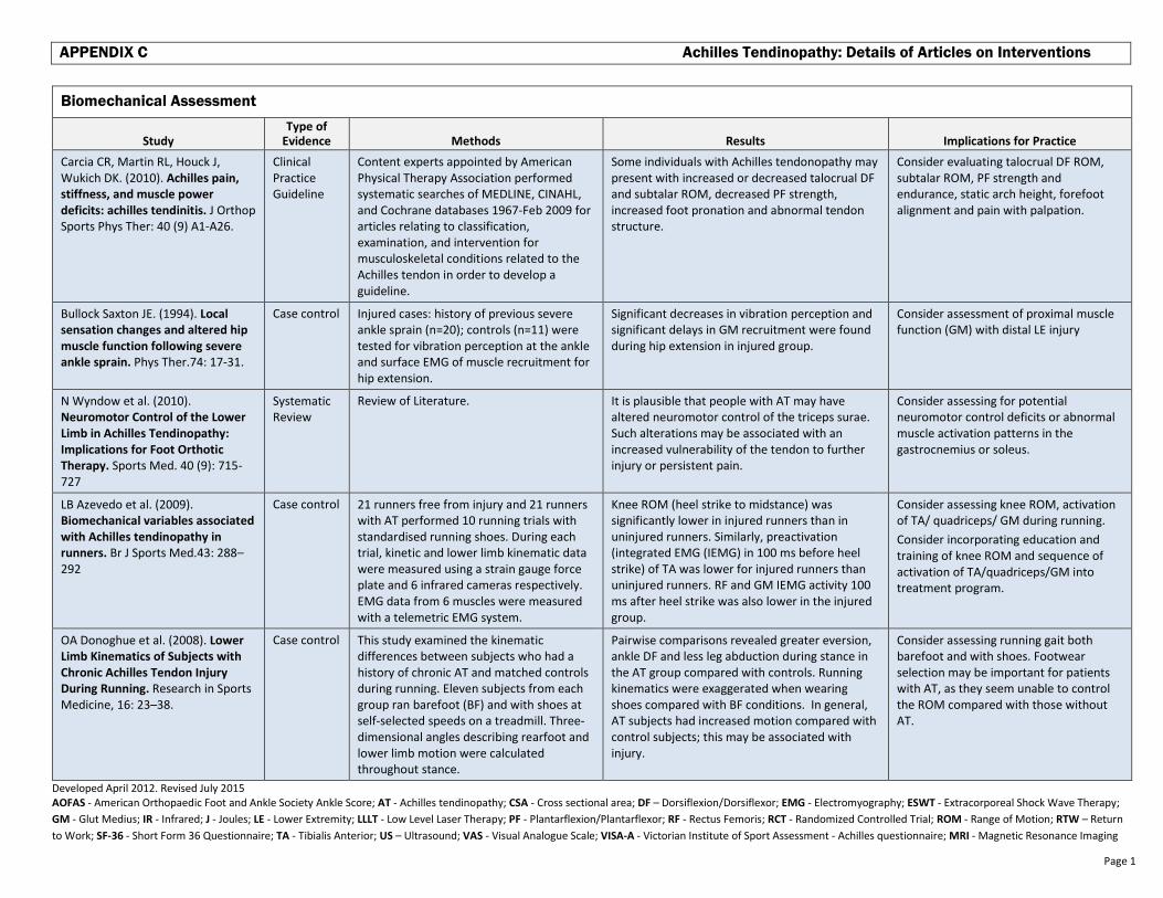
Developed April 2012. Revised July 2015 AOFAS - American Orthopaedic Foot and Ankle Society Ankle Score; AT - Achilles tendinopathy; CSA - Cross sectional area; DF – Dorsiflexion/Dorsiflexor; EMG - Electromyography; ESWT - Extracorporeal Shock Wave Therapy; GM - Glut Medius; IR - Infrared; J - Joules; LE - Lower Extremity; LLLT - Low Level Laser Therapy; PF - Plantarflexion/Plantarflexor; RF - Rectus Femoris; RCT - Randomized Controlled Trial; ROM - Range of Motion; RTW – Return to Work; SF-36 - Short Form 36 Questionnaire; TA - Tibialis Anterior; US – Ultrasound; VAS - Visual Analogue Scale; VISA-A - Victorian Institute of Sport Assessment - Achilles questionnaire; MRI - Magnetic Resonance Imaging Page 1 APPENDIX C Achilles Tendinopathy: Details of Articles on Interventions Biomechanical Assessment Study Type of Evidence Methods Results Implications for Practice Carcia CR, Martin RL, Houck J, Wukich DK. (2010). Achilles pain, stiffness, and muscle power deficits: achilles tendinitis. J Orthop Sports Phys Ther: 40 (9) A1-A26. Clinical Practice Guideline Content experts appointed by American Physical Therapy Association performed systematic searches of MEDLINE, CINAHL, and Cochrane databases 1967-Feb 2009 for articles relating to classification, examination, and intervention for musculoskeletal conditions related to the Achilles tendon in order to develop a guideline. Some individuals with Achilles tendonopathy may present with increased or decreased talocrual DF and subtalar ROM, decreased PF strength, increased foot pronation and abnormal tendon structure. Consider evaluating talocrual DF ROM, subtalar ROM, PF strength and endurance, static arch height, forefoot alignment and pain with palpation. Bullock Saxton JE. (1994). Local sensation changes and altered hip muscle function following severe ankle sprain. Phys Ther.74: 17-31. Case control Injured cases: history of previous severe ankle sprain (n=20); controls (n=11) were tested for vibration perception at the ankle and surface EMG of muscle recruitment for hip extension. Significant decreases in vibration perception and significant delays in GM recruitment were found during hip extension in injured group. Consider assessment of proximal muscle function (GM) with distal LE injury N Wyndow et al. (2010). Neuromotor Control of the Lower Limb in Achilles Tendinopathy: Implications for Foot Orthotic Therapy. Sports Med. 40 (9): 715- 727 Systematic Review Review of Literature. It is plausible that people with AT may have altered neuromotor control of the triceps surae. Such alterations may be associated with an increased vulnerability of the tendon to further injury or persistent pain. Consider assessing for potential neuromotor control deficits or abnormal muscle activation patterns in the gastrocnemius or soleus. LB Azevedo et al. (2009). Biomechanical variables associated with Achilles tendinopathy in runners. Br J Sports Med.43: 288– 292 Case control 21 runners free from injury and 21 runners with AT performed 10 running trials with standardised running shoes. During each trial, kinetic and lower limb kinematic data were measured using a strain gauge force plate and 6 infrared cameras respectively. EMG data from 6 muscles were measured with a telemetric EMG system. Knee ROM (heel strike to midstance) was significantly lower in injured runners than in uninjured runners. Similarly, preactivation (integrated EMG (IEMG) in 100 ms before heel strike) of TA was lower for injured runners than uninjured runners. RF and GM IEMG activity 100 ms after heel strike was also lower in the injured group. Consider assessing knee ROM, activation of TA/ quadriceps/ GM during running. Consider incorporating education and training of knee ROM and sequence of activation of TA/quadriceps/GM into treatment program. OA Donoghue et al. (2008). Lower Limb Kinematics of Subjects with Chronic Achilles Tendon Injury During Running. Research in Sports Medicine, 16: 23–38. Case control This study examined the kinematic differences between subjects who had a history of chronic AT and matched controls during running. Eleven subjects from each group ran barefoot (BF) and with shoes at self-selected speeds on a treadmill. Three- dimensional angles describing rearfoot and lower limb motion were calculated throughout stance. Pairwise comparisons revealed greater eversion, ankle DF and less leg abduction during stance in the AT group compared with controls. Running kinematics were exaggerated when wearing shoes compared with BF conditions. In general, AT subjects had increased motion compared with control subjects; this may be associated with injury. Consider assessing running gait both barefoot and with shoes. Footwear selection may be important for patients with AT, as they seem unable to control the ROM compared with those without AT.
Transcript of APPENDIX C Achilles Tendinopathy: Details of Articles on...
Developed April 2012. Revised July 2015 AOFAS - American Orthopaedic Foot and Ankle Society Ankle Score; AT - Achilles tendinopathy; CSA - Cross sectional area; DF – Dorsiflexion/Dorsiflexor; EMG - Electromyography; ESWT - Extracorporeal Shock Wave Therapy;
GM - Glut Medius; IR - Infrared; J - Joules; LE - Lower Extremity; LLLT - Low Level Laser Therapy; PF - Plantarflexion/Plantarflexor; RF - Rectus Femoris; RCT - Randomized Controlled Trial; ROM - Range of Motion; RTW – Return
to Work; SF-36 - Short Form 36 Questionnaire; TA - Tibialis Anterior; US – Ultrasound; VAS - Visual Analogue Scale; VISA-A - Victorian Institute of Sport Assessment - Achilles questionnaire; MRI - Magnetic Resonance Imaging
Page 1
APPENDIX C Achilles Tendinopathy: Details of Articles on Interventions
Biomechanical Assessment
Study
Type of
Evidence Methods Results Implications for Practice
Carcia CR, Martin RL, Houck J, Wukich DK. (2010). Achilles pain,
stiffness, and muscle power
deficits: achilles tendinitis. J Orthop Sports Phys Ther: 40 (9) A1-A26.
Clinical Practice Guideline
Content experts appointed by American Physical Therapy Association performed systematic searches of MEDLINE, CINAHL, and Cochrane databases 1967-Feb 2009 for articles relating to classification, examination, and intervention for musculoskeletal conditions related to the Achilles tendon in order to develop a guideline.
Some individuals with Achilles tendonopathy may present with increased or decreased talocrual DF and subtalar ROM, decreased PF strength, increased foot pronation and abnormal tendon structure.
Consider evaluating talocrual DF ROM, subtalar ROM, PF strength and endurance, static arch height, forefoot alignment and pain with palpation.
Bullock Saxton JE. (1994). Local
sensation changes and altered hip
muscle function following severe
ankle sprain. Phys Ther.74: 17-31.
Case control Injured cases: history of previous severe ankle sprain (n=20); controls (n=11) were tested for vibration perception at the ankle and surface EMG of muscle recruitment for hip extension.
Significant decreases in vibration perception and significant delays in GM recruitment were found during hip extension in injured group.
Consider assessment of proximal muscle function (GM) with distal LE injury
N Wyndow et al. (2010). Neuromotor Control of the Lower
Limb in Achilles Tendinopathy:
Implications for Foot Orthotic
Therapy. Sports Med. 40 (9): 715-727
Systematic Review
Review of Literature. It is plausible that people with AT may have altered neuromotor control of the triceps surae. Such alterations may be associated with an increased vulnerability of the tendon to further injury or persistent pain.
Consider assessing for potential neuromotor control deficits or abnormal muscle activation patterns in the gastrocnemius or soleus.
LB Azevedo et al. (2009). Biomechanical variables associated
with Achilles tendinopathy in
runners. Br J Sports Med.43: 288–292
Case control
21 runners free from injury and 21 runners with AT performed 10 running trials with standardised running shoes. During each trial, kinetic and lower limb kinematic data were measured using a strain gauge force plate and 6 infrared cameras respectively. EMG data from 6 muscles were measured with a telemetric EMG system.
Knee ROM (heel strike to midstance) was significantly lower in injured runners than in uninjured runners. Similarly, preactivation (integrated EMG (IEMG) in 100 ms before heel strike) of TA was lower for injured runners than uninjured runners. RF and GM IEMG activity 100 ms after heel strike was also lower in the injured group.
Consider assessing knee ROM, activation of TA/ quadriceps/ GM during running.
Consider incorporating education and training of knee ROM and sequence of activation of TA/quadriceps/GM into treatment program.
OA Donoghue et al. (2008). Lower
Limb Kinematics of Subjects with
Chronic Achilles Tendon Injury
During Running. Research in Sports Medicine, 16: 23–38.
Case control This study examined the kinematic differences between subjects who had a history of chronic AT and matched controls during running. Eleven subjects from each group ran barefoot (BF) and with shoes at self-selected speeds on a treadmill. Three-dimensional angles describing rearfoot and lower limb motion were calculated throughout stance.
Pairwise comparisons revealed greater eversion, ankle DF and less leg abduction during stance in the AT group compared with controls. Running kinematics were exaggerated when wearing shoes compared with BF conditions. In general, AT subjects had increased motion compared with control subjects; this may be associated with injury.
Consider assessing running gait both barefoot and with shoes. Footwear selection may be important for patients with AT, as they seem unable to control the ROM compared with those without AT.
Developed April 2012. Revised July 2015 AOFAS - American Orthopaedic Foot and Ankle Society Ankle Score; AT - Achilles tendinopathy; CSA - Cross sectional area; DF – Dorsiflexion/Dorsiflexor; EMG - Electromyography; ESWT - Extracorporeal Shock Wave Therapy;
GM - Glut Medius; IR - Infrared; J - Joules; LE - Lower Extremity; LLLT - Low Level Laser Therapy; PF - Plantarflexion/Plantarflexor; RF - Rectus Femoris; RCT - Randomized Controlled Trial; ROM - Range of Motion; RTW – Return
to Work; SF-36 - Short Form 36 Questionnaire; TA - Tibialis Anterior; US – Ultrasound; VAS - Visual Analogue Scale; VISA-A - Victorian Institute of Sport Assessment - Achilles questionnaire; MRI - Magnetic Resonance Imaging
Page 2
Study
Type of
Evidence Methods Results Implications for Practice
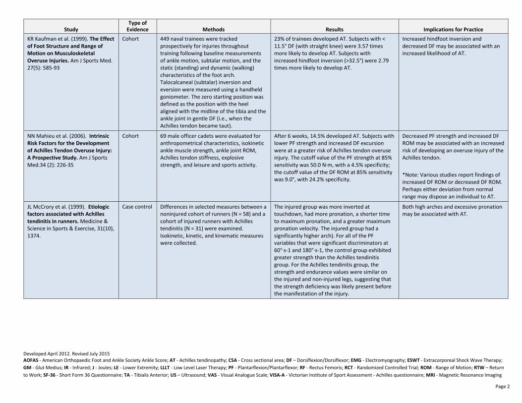
KR Kaufman et al. (1999). The Effect
of Foot Structure and Range of
Motion on Musculoskeletal
Overuse Injuries. Am J Sports Med. 27(5): 585-93
Cohort 449 naval trainees were tracked prospectively for injuries throughout training following baseline measurements of ankle motion, subtalar motion, and the static (standing) and dynamic (walking) characteristics of the foot arch. Talocalcaneal (subtalar) inversion and eversion were measured using a handheld goniometer. The zero starting position was defined as the position with the heel aligned with the midline of the tibia and the ankle joint in gentle DF (i.e., when the Achilles tendon became taut).
23% of trainees developed AT. Subjects with < 11.5° DF (with straight knee) were 3.57 times more likely to develop AT. Subjects with increased hindfoot inversion (>32.5°) were 2.79 times more likely to develop AT.
Increased hindfoot inversion and decreased DF may be associated with an increased likelihood of AT.
NN Mahieu et al. (2006). Intrinsic
Risk Factors for the Development
of Achilles Tendon Overuse Injury:
A Prospective Study. Am J Sports Med.34 (2): 226-35
Cohort 69 male officer cadets were evaluated for anthropometrical characteristics, isokinetic ankle muscle strength, ankle joint ROM, Achilles tendon stiffness, explosive strength, and leisure and sports activity.
After 6 weeks, 14.5% developed AT. Subjects with lower PF strength and increased DF excursion were at a greater risk of Achilles tendon overuse injury. The cutoff value of the PF strength at 85% sensitivity was 50.0 N·m, with a 4.5% specificity; the cutoff value of the DF ROM at 85% sensitivity was 9.0°, with 24.2% specificity.
Decreased PF strength and increased DF ROM may be associated with an increased risk of developing an overuse injury of the Achilles tendon.
*Note: Various studies report findings of increased DF ROM or decreased DF ROM. Perhaps either deviation from normal range may dispose an individual to AT.
JL McCrory et al. (1999). Etiologic
factors associated with Achilles
tendinitis in runners. Medicine & Science in Sports & Exercise, 31(10), 1374.
Case control Differences in selected measures between a noninjured cohort of runners (N = 58) and a cohort of injured runners with Achilles tendinitis (N = 31) were examined. Isokinetic, kinetic, and kinematic measures were collected.
The injured group was more inverted at touchdown, had more pronation, a shorter time to maximum pronation, and a greater maximum pronation velocity. The injured group had a significantly higher arch). For all of the PF variables that were significant discriminators at 60°·s-1 and 180°·s-1, the control group exhibited greater strength than the Achilles tendinitis group. For the Achilles tendinitis group, the strength and endurance values were similar on the injured and non-injured legs, suggesting that the strength deficiency was likely present before the manifestation of the injury.
Both high arches and excessive pronation may be associated with AT.
Developed April 2012. Revised July 2015 AOFAS - American Orthopaedic Foot and Ankle Society Ankle Score; AT - Achilles tendinopathy; CSA - Cross sectional area; DF – Dorsiflexion/Dorsiflexor; EMG - Electromyography; ESWT - Extracorporeal Shock Wave Therapy;
GM - Glut Medius; IR - Infrared; J - Joules; LE - Lower Extremity; LLLT - Low Level Laser Therapy; PF - Plantarflexion/Plantarflexor; RF - Rectus Femoris; RCT - Randomized Controlled Trial; ROM - Range of Motion; RTW – Return
to Work; SF-36 - Short Form 36 Questionnaire; TA - Tibialis Anterior; US – Ultrasound; VAS - Visual Analogue Scale; VISA-A - Victorian Institute of Sport Assessment - Achilles questionnaire; MRI - Magnetic Resonance Imaging
Page 3
Study
Type of
Evidence Methods Results Implications for Practice
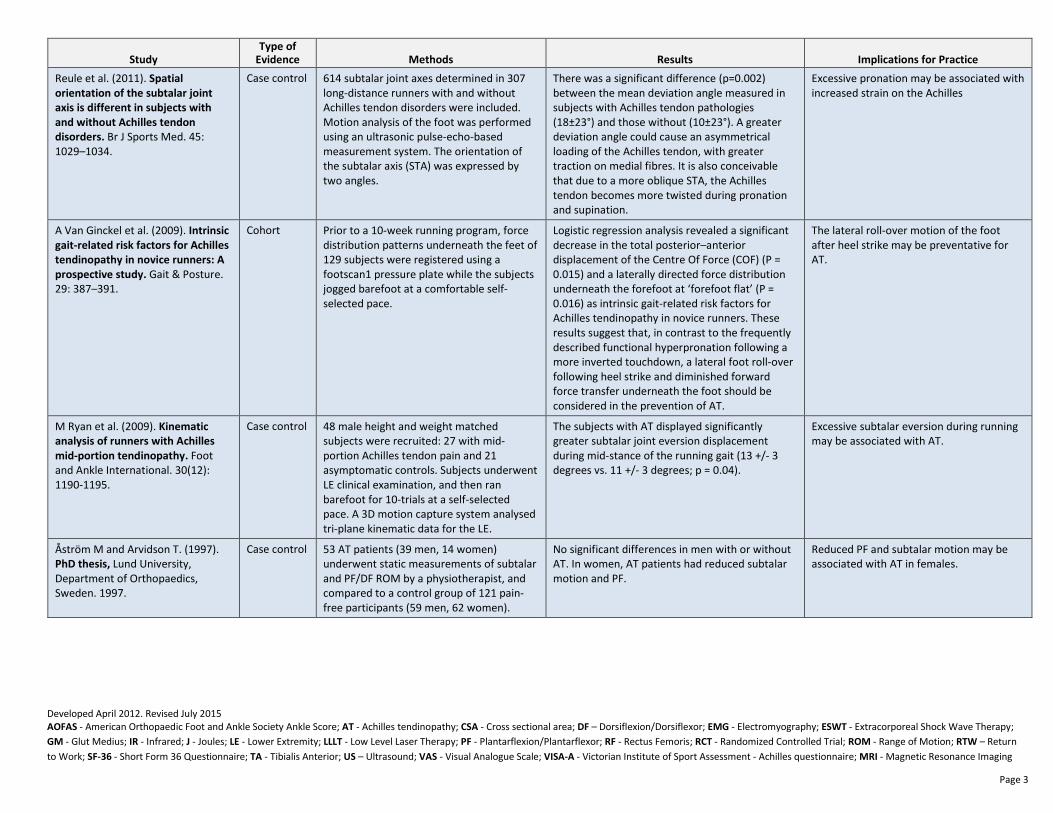
Reule et al. (2011). Spatial
orientation of the subtalar joint
axis is different in subjects with
and without Achilles tendon
disorders. Br J Sports Med. 45: 1029–1034.
Case control 614 subtalar joint axes determined in 307 long-distance runners with and without Achilles tendon disorders were included. Motion analysis of the foot was performed using an ultrasonic pulse-echo-based measurement system. The orientation of the subtalar axis (STA) was expressed by two angles.
There was a significant difference (p=0.002) between the mean deviation angle measured in subjects with Achilles tendon pathologies (18±23°) and those without (10±23°). A greater deviation angle could cause an asymmetrical loading of the Achilles tendon, with greater traction on medial fibres. It is also conceivable that due to a more oblique STA, the Achilles tendon becomes more twisted during pronation and supination.
Excessive pronation may be associated with increased strain on the Achilles
A Van Ginckel et al. (2009). Intrinsic
gait-related risk factors for Achilles
tendinopathy in novice runners: A
prospective study. Gait & Posture. 29: 387–391.
Cohort Prior to a 10-week running program, force distribution patterns underneath the feet of 129 subjects were registered using a footscan1 pressure plate while the subjects jogged barefoot at a comfortable self-selected pace.
Logistic regression analysis revealed a significant decrease in the total posterior–anterior displacement of the Centre Of Force (COF) (P = 0.015) and a laterally directed force distribution underneath the forefoot at ‘forefoot flat’ (P = 0.016) as intrinsic gait-related risk factors for Achilles tendinopathy in novice runners. These results suggest that, in contrast to the frequently described functional hyperpronation following a more inverted touchdown, a lateral foot roll-over following heel strike and diminished forward force transfer underneath the foot should be considered in the prevention of AT.
The lateral roll-over motion of the foot after heel strike may be preventative for AT.
M Ryan et al. (2009). Kinematic
analysis of runners with Achilles
mid-portion tendinopathy. Foot and Ankle International. 30(12): 1190-1195.
Case control 48 male height and weight matched subjects were recruited: 27 with mid-portion Achilles tendon pain and 21 asymptomatic controls. Subjects underwent LE clinical examination, and then ran barefoot for 10-trials at a self-selected pace. A 3D motion capture system analysed tri-plane kinematic data for the LE.
The subjects with AT displayed significantly greater subtalar joint eversion displacement during mid-stance of the running gait (13 +/- 3 degrees vs. 11 +/- 3 degrees; p = 0.04).
Excessive subtalar eversion during running may be associated with AT.
Åström M and Arvidson T. (1997). PhD thesis, Lund University, Department of Orthopaedics, Sweden. 1997.
Case control 53 AT patients (39 men, 14 women) underwent static measurements of subtalar and PF/DF ROM by a physiotherapist, and compared to a control group of 121 pain-free participants (59 men, 62 women).
No significant differences in men with or without AT. In women, AT patients had reduced subtalar motion and PF.
Reduced PF and subtalar motion may be associated with AT in females.
Developed April 2012. Revised July 2015 AOFAS - American Orthopaedic Foot and Ankle Society Ankle Score; AT - Achilles tendinopathy; CSA - Cross sectional area; DF – Dorsiflexion/Dorsiflexor; EMG - Electromyography; ESWT - Extracorporeal Shock Wave Therapy;
GM - Glut Medius; IR - Infrared; J - Joules; LE - Lower Extremity; LLLT - Low Level Laser Therapy; PF - Plantarflexion/Plantarflexor; RF - Rectus Femoris; RCT - Randomized Controlled Trial; ROM - Range of Motion; RTW – Return
to Work; SF-36 - Short Form 36 Questionnaire; TA - Tibialis Anterior; US – Ultrasound; VAS - Visual Analogue Scale; VISA-A - Victorian Institute of Sport Assessment - Achilles questionnaire; MRI - Magnetic Resonance Imaging
Page 4
Study
Type of
Evidence Methods Results Implications for Practice
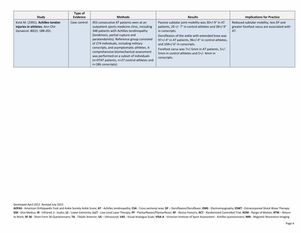
Kvist M. (1991). Achilles tendon
injuries in athletes. Ann Chir Gynaecol. 80(2): 188-201.
Case control 455 consecutive AT patients seen at an outpatient sports medicine clinic, including 348 patients with Achilles tendinopathy (tendinosis, partial rupture and paratendonitis). Reference group consisted of 274 individuals, including military conscripts, and asymptomatic athletes. A comprehensive biomechanical assessment was performed on a subset of individuals (n=97AT patients, n=27 control athletes and n=186 conscripts).
Passive subtalar joint mobility was 30+/-9° in AT patients, 29 +/- 7° in control athletes and 38+/-9° in conscripts.
Dorsiflexion of the ankle with extended knee was 97+/-4° in AT patients, 96+/-4° in control athletes, and 104+/-6° in conscripts.
Forefoot varus was 7+/-5mm in AT patients, 5+/-5mm in control athletes and 5+/- 4mm in conscripts.
Reduced subtalar mobility, less DF and greater forefoot varus are associated with AT.

Developed April 2012. Revised July 2015 AOFAS - American Orthopaedic Foot and Ankle Society Ankle Score; AT - Achilles tendinopathy; CSA - Cross sectional area; DF – Dorsiflexion/Dorsiflexor; EMG - Electromyography; ESWT - Extracorporeal Shock Wave Therapy;
GM - Glut Medius; IR - Infrared; J - Joules; LE - Lower Extremity; LLLT - Low Level Laser Therapy; PF - Plantarflexion/Plantarflexor; RF - Rectus Femoris; RCT - Randomized Controlled Trial; ROM - Range of Motion; RTW – Return
to Work; SF-36 - Short Form 36 Questionnaire; TA - Tibialis Anterior; US – Ultrasound; VAS - Visual Analogue Scale; VISA-A - Victorian Institute of Sport Assessment - Achilles questionnaire; MRI - Magnetic Resonance Imaging
Page 5
Manual Therapy
Study
Type of
Evidence Methods Results Implications for Practice
Carcia CR, Martin RL, Houck J, Wukich DK. (2010). Achilles pain,
stiffness, and muscle power
deficits: achilles tendinitis. J Orthop Sports Phys Ther. 40 (9): A1-A26.
Clinical Practice Guidelines
Content experts appointed by American Physical Therapy Association performed systematic searches of MEDLINE, CINAHL, and Cochrane Database 1967-Feb 2009 for articles relating to classification, examination, and intervention for musculoskeletal conditions related to the Achilles tendon to develop guidelines.
A single case study was found using soft tissue mobilization techniques. Recommendations are based on this case study as well as on the clinical experience of the guideline development team.
Soft tissue mobilization can be used to reduce pain and improve mobility and function in AT (Recommendation based on expert opinion).
Christenson RE (2007). Effectiveness of specific soft tissue
mobilizations for the management
of Achilles tendinosis: Single case
study-Experimental design. Manual Therapy. 12: 63-71.
Case study Single Case study ABA design used to evaluate the effectiveness of a protocol of accessory and combined specific soft tissue mobilizations (SSTMs) in a 39 year old female with 5 year history of Achilles tendonosis. 18 weeks of treatment and 12 weeks follow up. STTMs: applied perpendicular to the neutral tendon in the direction of restriction (medial or lateral) to the point of marked tissue resistance and the onset of mild to moderate pain. This can be progressed to applying mobilization during isometric holds of the tricep surae and finally dynamically during plantar flexion against mild resistance.
All outcomes improved:
VISA-A questionnaire: 100% at completion of treatment and follow up;
Visual Analogue scale: 0 for pain.
Although outcomes using soft tissue mobilization for AT were favorable, the potential clinical impact is limited by the single case study design.
Woodman RM, Pare L. (1982). Evaluation and treatment of soft
tissue lesions of the ankle and
forefoot using a Cyriax approach. Phys Ther. 62 (8): 1144-47.
Case Study Single case: 16 year old girl, ballet dancer with 3 month history of “Achilles tendonitis, tenosynovitis of tibialis posterior and peroneal tendons;” treatment included rest, steroid injections, and deep frictions for 20-30 minutes 3x week for 2 weeks.
After 14 sessions pain had improved and patient returned to ballet with some modifications to avoid end range PF. No further follow up was done.
The single case design and limited follow up limit the potential clinical impact of this study.
Brosseau L, Casimiro L, Milne S. et al. (2002). Deep transverse friction
massage for treating tendinitis. Cochrane Databse Syst Rev. 4.
Systematic Review
MEDLINE, EMBASE, HealthSTAR, Sport Discus, CINAHL, the Cochrane Controlled Trials Register, PEDro, were searched up to June 2002. Reference lists were also scanned for additional studies. Data extracted and methodological quality was assessed.
Only 2 studies were found: First, one study on Iliotibial Band Syndrome in runners found no statistical difference in pain after four treatments with DTFM; there was a clinically important difference in pain with running. The second study on extensor carpi radialis tendonitis showed no improvement in pain or function after 9 sessions of DTFM.
No conclusions can be made on the effectiveness of Deep Tendon Friction Massage (DTFM) for treatment of tendinitis.

Developed April 2012. Revised July 2015 AOFAS - American Orthopaedic Foot and Ankle Society Ankle Score; AT - Achilles tendinopathy; CSA - Cross sectional area; DF – Dorsiflexion/Dorsiflexor; EMG - Electromyography; ESWT - Extracorporeal Shock Wave Therapy;
GM - Glut Medius; IR - Infrared; J - Joules; LE - Lower Extremity; LLLT - Low Level Laser Therapy; PF - Plantarflexion/Plantarflexor; RF - Rectus Femoris; RCT - Randomized Controlled Trial; ROM - Range of Motion; RTW – Return
to Work; SF-36 - Short Form 36 Questionnaire; TA - Tibialis Anterior; US – Ultrasound; VAS - Visual Analogue Scale; VISA-A - Victorian Institute of Sport Assessment - Achilles questionnaire; MRI - Magnetic Resonance Imaging
Page 6
Study
Type of
Evidence Methods Results Implications for Practice
Imai, K., Ikoma, K., Chen, Q., Zhao, C., An, K.-N., & Gay, R. E. (2015). Biomechanical and Histological
Effects of Augmented Soft Tissue
Mobilization Therapy on Achilles
Tendinopathy in a Rabbit Model. Journal of Manipulative and
Physiological Therapeutics, 38(2), 112-118. doi: http://dx.doi.org/10.1016/j.jmpt.2014.12.003
Animal Study
Both Achilles tendons of 12 rabbits were injected with collagenase to induce injury simulating AT. One side received augmented soft tissue mobilization (ASTM) while the other received no treatment. ASTM was performed on the Achilles tendon on post-operative days 21, 24, 28, 31, 35 and 38. 10 days after treatment tendons were examined with dynamic viscoelasticity and light microscopy.
CSA in treated tendons were greater than in controls. Storage modulus was lower in treated tendons, but elasticity was not significantly increased. Microscopy of the control tendons showed wavy tendon fibers with well-stained type III collagen, both of which were not evident in the treated tendons.
Rabbit tendons treated with ASTM had superior biomechanical function than the no treatment tendons. This implies that ASTM may be effective in the treatment of chronic Achilles tendinopathy in patients. Clinical impact is limited by fact that this is an animal study.
Voorn R. (1998). Case report: can
sacroiliac joint dysfunction cause
chronic Achilles tendonitis? JOSPT. 27(6): 436-443.
Case Study 29 year old pole jumper with one year history of Achilles pain. Failed local conservative management. Entire LE was evaluated.
Assessment revealed: right sacroiliac dysfunction (posterior rotation right innominate and soft tissue irritation, altered gait patterns with running and walking, right functional leg length shortening and external rotation.)
Treatment included: manipulation/manual therapy of sacroiliac joint including prone thrust, supine leg thrust, and muscle energy techniques; heel raises, double and single leg hops, pulley exercises for back and hip strengthening.
Athlete resumed training 9 weeks after LE assessment.
Consider a comprehensive assessment of the LE and treatment of proximal regions of the body for management of AT. This includes the sacroiliac joint, lumbar spine, and hips.

Developed April 2012. Revised July 2015 AOFAS - American Orthopaedic Foot and Ankle Society Ankle Score; AT - Achilles tendinopathy; CSA - Cross sectional area; DF – Dorsiflexion/Dorsiflexor; EMG - Electromyography; ESWT - Extracorporeal Shock Wave Therapy;
GM - Glut Medius; IR - Infrared; J - Joules; LE - Lower Extremity; LLLT - Low Level Laser Therapy; PF - Plantarflexion/Plantarflexor; RF - Rectus Femoris; RCT - Randomized Controlled Trial; ROM - Range of Motion; RTW – Return
to Work; SF-36 - Short Form 36 Questionnaire; TA - Tibialis Anterior; US – Ultrasound; VAS - Visual Analogue Scale; VISA-A - Victorian Institute of Sport Assessment - Achilles questionnaire; MRI - Magnetic Resonance Imaging
Page 7
Exercise
Study
Type of
Evidence Methods Results Implications for Practice
Beyer, R., Kongsgaard, M., Hougs Kjaer, B., Ohlenschlaeger, T., Kjaer, M., & Magnusson, S. P. (2015). Heavy Slow Resistance Versus
Eccentric Training as Treatment for
Achilles Tendinopathy: A
Randomized Controlled Trial. Am J
Sports Med. Doi: 10.1177/0363546515584760
RCT n=58 patients with chronic mid-portion AT were randomized to either eccentric training (ECC) or heavy slow resistance training (HSR) for 12 weeks. VISA-A, pain, tendon swelling, tendon neovascularization, patient satisfaction were measured at 0, 12 and 52 weeks.
Both treatments gave positive and equally good results that were long-lasting. HSR was associated with greater patient satisfaction than ECC at 12 weeks, but not after 52 weeks.
Heavy slow resistance training for 12 weeks is equally as effective as eccentric training for improving clinical severity and pain of chronic mid-portion AT.
Gaerdin A, Movin T, Svensson L, Shalabi A. (2010). The long-term
clinical and MRI results following
eccentric calf muscle training in
chronic Achilles tendinosis. Skeletal Radiol. 39(5): 435-42.
Observational study
n=24 with a median duration of 18 months. Evaluated before and after 3 months of daily eccentric exs. 4 patients did not conform but the 20 remaining patients did the 4.2 year follow-up.
Decreased pain, improved performance and decreased intratendinous signal when compared to index exam and after 3 months of eccentric training. At 4.2 year follow-up the improvements were greater despite no further active treatment. This may indicate a good long-term prognosis for AT patients.
Eccentric loading of the calf muscles with knee straight and bent 3 x 15 twice per day resulted in short and long term changes in pain, performance and tissue health via imaging.
Grigg NL, Stevenson NJ, Wearing SC, Smeathers JE (2010). Incidental
walking activity is sufficient to
induce time-dependent
conditioning of the Achilles
tendon. Gait Posture. 31(1): 64-7.
Observational study
This study investigated the effect of incidental daily walking on Achilles tendon diametral strain.
Short repetitive loads are sufficient to induce time-dependent conditioning of the Achilles tendon.
Even short duration daily walking can condition the Achilles tendon.
Grigg NL, Wearing SC, Smeathers JE. (2011). Achilles Tendinopathy has
an Aberrant Strain Response to
Eccentric Exercise. Med Sci Sports Exerc. 8. [Epub ahead of print].
Observational study
Sonograms of AT prior to, immediately after and 24 hours after eccentric exs. Tendon thickness, echogenicity, AP strain. Study n=11, control=9 (no tendinopathy).
All tendons decreased in thickness right after eccentrics. Lower AP strain in symptomatic tendons. Pre-exs thickness restored in 24 hours. AT is a bilateral or systemic process. Structural changes of AT alter fluid movement within the tendon matrix which may disrupt remodeling.
This provides evidence that there may be a bilateral process involved with AT. Although the response to exs in this study was thought to be aberrant, there are generally positive results with eccentric exs.
Grigg NL, Wearing SC, Smeathers JE. (2009). Eccentric calf muscle
exercise produces a greater acute
reduction in Achilles tendon
thickness than concentric exercise.
Br J Sports Med. 43(4): 280-3.
Observational study
11 healthy male adults. Isolated eccentrics on one leg and concentric on the other with body weight + 20%. Sagittal sonograms prior, immediately and 3, 6, 12 and 245 hours later.
Eccentric loading invoked a greater reduction in Achilles tendon thickness immediately after exs but appears to recover fully in the similar time frame to concentric loading.
It is unclear as to whether the results were due to a potentially greater load with eccentric vs. concentric exercise.
Developed April 2012. Revised July 2015 AOFAS - American Orthopaedic Foot and Ankle Society Ankle Score; AT - Achilles tendinopathy; CSA - Cross sectional area; DF – Dorsiflexion/Dorsiflexor; EMG - Electromyography; ESWT - Extracorporeal Shock Wave Therapy;
GM - Glut Medius; IR - Infrared; J - Joules; LE - Lower Extremity; LLLT - Low Level Laser Therapy; PF - Plantarflexion/Plantarflexor; RF - Rectus Femoris; RCT - Randomized Controlled Trial; ROM - Range of Motion; RTW – Return
to Work; SF-36 - Short Form 36 Questionnaire; TA - Tibialis Anterior; US – Ultrasound; VAS - Visual Analogue Scale; VISA-A - Victorian Institute of Sport Assessment - Achilles questionnaire; MRI - Magnetic Resonance Imaging
Page 8
Study
Type of
Evidence Methods Results Implications for Practice
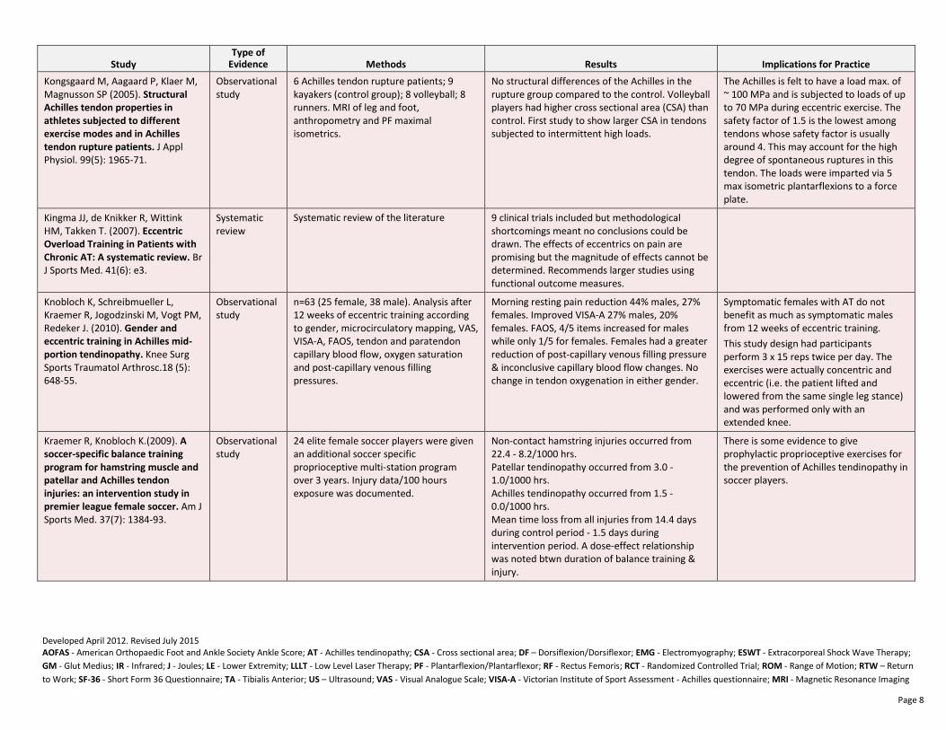
Kongsgaard M, Aagaard P, Klaer M, Magnusson SP (2005). Structural
Achilles tendon properties in
athletes subjected to different
exercise modes and in Achilles
tendon rupture patients. J Appl Physiol. 99(5): 1965-71.
Observational study
6 Achilles tendon rupture patients; 9 kayakers (control group); 8 volleyball; 8 runners. MRI of leg and foot, anthropometry and PF maximal isometrics.
No structural differences of the Achilles in the rupture group compared to the control. Volleyball players had higher cross sectional area (CSA) than control. First study to show larger CSA in tendons subjected to intermittent high loads.
The Achilles is felt to have a load max. of ~ 100 MPa and is subjected to loads of up to 70 MPa during eccentric exercise. The safety factor of 1.5 is the lowest among tendons whose safety factor is usually around 4. This may account for the high degree of spontaneous ruptures in this tendon. The loads were imparted via 5 max isometric plantarflexions to a force plate.
Kingma JJ, de Knikker R, Wittink HM, Takken T. (2007). Eccentric
Overload Training in Patients with
Chronic AT: A systematic review. Br J Sports Med. 41(6): e3.
Systematic review
Systematic review of the literature 9 clinical trials included but methodological shortcomings meant no conclusions could be drawn. The effects of eccentrics on pain are promising but the magnitude of effects cannot be determined. Recommends larger studies using functional outcome measures.
Knobloch K, Schreibmueller L, Kraemer R, Jogodzinski M, Vogt PM, Redeker J. (2010). Gender and
eccentric training in Achilles mid-
portion tendinopathy. Knee Surg Sports Traumatol Arthrosc.18 (5): 648-55.
Observational study
n=63 (25 female, 38 male). Analysis after 12 weeks of eccentric training according to gender, microcirculatory mapping, VAS, VISA-A, FAOS, tendon and paratendon capillary blood flow, oxygen saturation and post-capillary venous filling pressures.
Morning resting pain reduction 44% males, 27% females. Improved VISA-A 27% males, 20% females. FAOS, 4/5 items increased for males while only 1/5 for females. Females had a greater reduction of post-capillary venous filling pressure & inconclusive capillary blood flow changes. No change in tendon oxygenation in either gender.
Symptomatic females with AT do not benefit as much as symptomatic males from 12 weeks of eccentric training.
This study design had participants perform 3 x 15 reps twice per day. The exercises were actually concentric and eccentric (i.e. the patient lifted and lowered from the same single leg stance) and was performed only with an extended knee.
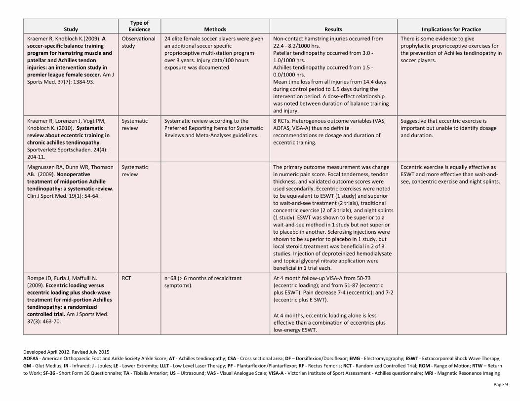
Kraemer R, Knobloch K.(2009). A
soccer-specific balance training
program for hamstring muscle and
patellar and Achilles tendon
injuries: an intervention study in
premier league female soccer. Am J Sports Med. 37(7): 1384-93.
Observational study
24 elite female soccer players were given an additional soccer specific proprioceptive multi-station program over 3 years. Injury data/100 hours exposure was documented.
Non-contact hamstring injuries occurred from 22.4 - 8.2/1000 hrs. Patellar tendinopathy occurred from 3.0 - 1.0/1000 hrs. Achilles tendinopathy occurred from 1.5 - 0.0/1000 hrs. Mean time loss from all injuries from 14.4 days during control period - 1.5 days during intervention period. A dose-effect relationship was noted btwn duration of balance training & injury.
There is some evidence to give prophylactic proprioceptive exercises for the prevention of Achilles tendinopathy in soccer players.
Developed April 2012. Revised July 2015 AOFAS - American Orthopaedic Foot and Ankle Society Ankle Score; AT - Achilles tendinopathy; CSA - Cross sectional area; DF – Dorsiflexion/Dorsiflexor; EMG - Electromyography; ESWT - Extracorporeal Shock Wave Therapy;
GM - Glut Medius; IR - Infrared; J - Joules; LE - Lower Extremity; LLLT - Low Level Laser Therapy; PF - Plantarflexion/Plantarflexor; RF - Rectus Femoris; RCT - Randomized Controlled Trial; ROM - Range of Motion; RTW – Return
to Work; SF-36 - Short Form 36 Questionnaire; TA - Tibialis Anterior; US – Ultrasound; VAS - Visual Analogue Scale; VISA-A - Victorian Institute of Sport Assessment - Achilles questionnaire; MRI - Magnetic Resonance Imaging
Page 9
Study
Type of
Evidence Methods Results Implications for Practice
Kraemer R, Knobloch K.(2009). A
soccer-specific balance training
program for hamstring muscle and
patellar and Achilles tendon
injuries: an intervention study in
premier league female soccer. Am J Sports Med. 37(7): 1384-93.
Observational study
24 elite female soccer players were given an additional soccer specific proprioceptive multi-station program over 3 years. Injury data/100 hours exposure was documented.
Non-contact hamstring injuries occurred from 22.4 - 8.2/1000 hrs. Patellar tendinopathy occurred from 3.0 - 1.0/1000 hrs. Achilles tendinopathy occurred from 1.5 - 0.0/1000 hrs. Mean time loss from all injuries from 14.4 days during control period to 1.5 days during the intervention period. A dose-effect relationship was noted between duration of balance training and injury.
There is some evidence to give prophylactic proprioceptive exercises for the prevention of Achilles tendinopathy in soccer players.
Kraemer R, Lorenzen J, Vogt PM, Knobloch K. (2010). Systematic
review about eccentric training in
chronic achilles tendinopathy. Sportverletz Sportschaden. 24(4): 204-11.
Systematic review
Systematic review according to the Preferred Reporting Items for Systematic Reviews and Meta-Analyses guidelines.
8 RCTs. Heterogenous outcome variables (VAS, AOFAS, VISA-A) thus no definite recommendations re dosage and duration of eccentric training.
Suggestive that eccentric exercise is important but unable to identify dosage and duration.
Magnussen RA, Dunn WR, Thomson AB. (2009). Nonoperative
treatment of midportion Achille
tendinopathy: a systematic review.
Clin J Sport Med. 19(1): 54-64.
Systematic review
The primary outcome measurement was change in numeric pain score. Focal tenderness, tendon thickness, and validated outcome scores were used secondarily. Eccentric exercises were noted to be equivalent to ESWT (1 study) and superior to wait-and-see treatment (2 trials), traditional concentric exercise (2 of 3 trials), and night splints (1 study). ESWT was shown to be superior to a wait-and-see method in 1 study but not superior to placebo in another. Sclerosing injections were shown to be superior to placebo in 1 study, but local steroid treatment was beneficial in 2 of 3 studies. Injection of deproteinized hemodialysate and topical glyceryl nitrate application were beneficial in 1 trial each.
Eccentric exercise is equally effective as ESWT and more effective than wait-and-see, concentric exercise and night splints.
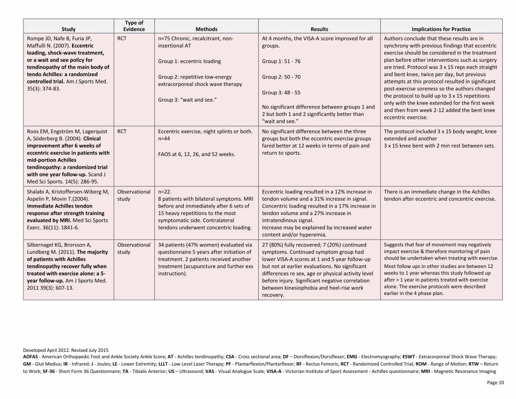
Rompe JD, Furia J, Maffulli N. (2009). Eccentric loading versus
eccentric loading plus shock-wave
treatment for mid-portion Achilles
tendinopathy: a randomized
controlled trial. Am J Sports Med. 37(3): 463-70.
RCT n=68 (> 6 months of recalcitrant symptoms).
At 4 month follow-up VISA-A from 50-73 (eccentric loading); and from 51-87 (eccentric plus ESWT). Pain decrease 7-4 (eccentric); and 7-2 (eccentric plus E SWT).
At 4 months, eccentric loading alone is less effective than a combination of eccentrics plus low-energy ESWT.
Developed April 2012. Revised July 2015 AOFAS - American Orthopaedic Foot and Ankle Society Ankle Score; AT - Achilles tendinopathy; CSA - Cross sectional area; DF – Dorsiflexion/Dorsiflexor; EMG - Electromyography; ESWT - Extracorporeal Shock Wave Therapy;
GM - Glut Medius; IR - Infrared; J - Joules; LE - Lower Extremity; LLLT - Low Level Laser Therapy; PF - Plantarflexion/Plantarflexor; RF - Rectus Femoris; RCT - Randomized Controlled Trial; ROM - Range of Motion; RTW – Return
to Work; SF-36 - Short Form 36 Questionnaire; TA - Tibialis Anterior; US – Ultrasound; VAS - Visual Analogue Scale; VISA-A - Victorian Institute of Sport Assessment - Achilles questionnaire; MRI - Magnetic Resonance Imaging
Page 10
Study
Type of
Evidence Methods Results Implications for Practice
Rompe JD, Nafe B, Furia JP, Maffulli N. (2007). Eccentric
loading, shock-wave treatment,
or a wait and see policy for
tendinopathy of the main body of
tendo Achilles: a randomized
controlled trial. Am J Sports Med. 35(3): 374-83.
RCT n=75 Chronic, recalcitrant, non-insertional AT
Group 1: eccentric loading
Group 2: repetitive low-energy extracorporeal shock wave therapy
Group 3: “wait and see.”
At 4 months, the VISA-A score improved for all groups.
Group 1: 51 - 76
Group 2: 50 - 70
Group 3: 48 - 55 No significant difference between groups 1 and 2 but both 1 and 2 significantly better than “wait and see.”
Authors conclude that these results are in synchrony with previous findings that eccentric exercise should be considered in the treatment plan before other interventions such as surgery are tried. Protocol was 3 x 15 reps each straight and bent knee, twice per day, but previous attempts at this protocol resulted in significant post-exercise soreness so the authors changed the protocol to build up to 3 x 15 repetitions only with the knee extended for the first week and then from week 2-12 added the bent knee eccentric exercise.
Roos EM, Engström M, Lagerquist A, Söderberg B. (2004). Clinical
improvement after 6 weeks of
eccentric exercise in patients with
mid-portion Achilles
tendinopathy: a randomized trial
with one year follow-up. Scand J Med Sci Sports. 14(5): 286-95.
RCT Eccentric exercise, night splints or both. n=44
FAOS at 6, 12, 26, and 52 weeks.
No significant difference between the three groups but both the eccentric exercise groups fared better at 12 weeks in terms of pain and return to sports.
The protocol included 3 x 15 body weight, knee extended and another 3 x 15 knee bent with 2 min rest between sets.
Shalabi A, Kristoffersen-Wiberg M, Aspelin P, Movin T.(2004). Immediate Achilles tendon
response after strength training
evaluated by MRI. Med Sci Sports Exerc. 36(11): 1841-6.
Observational study
n=22. 8 patients with bilateral symptoms. MRI before and immediately after 6 sets of 15 heavy repetitions to the most symptomatic side. Contralateral tendons underwent concentric loading.
Eccentric loading resulted in a 12% increase in tendon volume and a 31% increase in signal. Concentric loading resulted in a 17% increase in tendon volume and a 27% increase in intratendinous signal. Increase may be explained by increased water content and/or hyperemia.
There is an immediate change in the Achilles tendon after eccentric and concentric exercise.
Silbernagel KG, Brorsson A, Lundberg M. (2011). The majority
of patients with Achilles
tendinopathy recover fully when
treated with exercise alone: a 5-
year follow-up. Am J Sports Med. 2011 39(3): 607-13.
Observational study
34 patients (47% women) evaluated via questionnaire 5 years after initiation of treatment. 2 patients received another treatment (acupuncture and further exs instruction).
27 (80%) fully recovered; 7 (20%) continued symptoms. Continued symptom group had lower VISA-A scores at 1 and 5 year follow-up but not at earlier evaluations. No significant differences re sex, age or physical activity level before injury. Significant negative correlation between kinesiophobia and heel-rise work recovery.
Suggests that fear of movement may negatively impact exercise & therefore monitoring of pain should be undertaken when treating with exercise.
Most follow ups in other studies are between 12 weeks to 1 year whereas this study followed up after > 1 year in patients treated with exercise alone. The exercise protocols were described earlier in the 4 phase plan.
Developed April 2012. Revised July 2015 AOFAS - American Orthopaedic Foot and Ankle Society Ankle Score; AT - Achilles tendinopathy; CSA - Cross sectional area; DF – Dorsiflexion/Dorsiflexor; EMG - Electromyography; ESWT - Extracorporeal Shock Wave Therapy;
GM - Glut Medius; IR - Infrared; J - Joules; LE - Lower Extremity; LLLT - Low Level Laser Therapy; PF - Plantarflexion/Plantarflexor; RF - Rectus Femoris; RCT - Randomized Controlled Trial; ROM - Range of Motion; RTW – Return
to Work; SF-36 - Short Form 36 Questionnaire; TA - Tibialis Anterior; US – Ultrasound; VAS - Visual Analogue Scale; VISA-A - Victorian Institute of Sport Assessment - Achilles questionnaire; MRI - Magnetic Resonance Imaging
Page 11
Study
Type of
Evidence Methods Results Implications for Practice
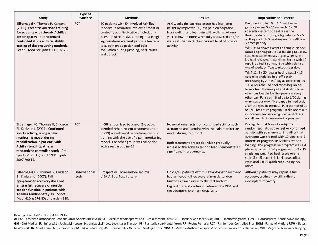
Silbernagel K, Thomee P, Karlson J. (2001). Eccentric overload training
for patients with chronic Achilles
tendinopathy - a randomized
controlled study with reliability
testing of the evaluating methods.
Scand J Med Sci Sports. 11: 197-206.
RCT 40 patients with 54 involved Achilles tendons randomized into experiment or control group. Evaluations included: a questionnaire, ROM, jumping test (single leg countermovement jump), a toe raise test, pain on palpation and pain evaluation during jumping, heel raises and at rest.
At 6 weeks the exercise group had less jump height by improved PF, less pain on palpation, less swelling and less pain with walking. At one year follow-up more were fully recovered and/or were satisfied with their current level of physical activity.
Program included: Wk 1: Stretches to gastroc/soleus 5 x 30 sec each; 3 x 20 concentric eccentric heel raises toe flexion/extension. Single leg balance. 5 x 5m walking on hells & walking on toes. All done 3 times per day.
Wk 2-3: As above except add single leg heel raises beginning at 3 x 5 & building to 3 x 15. Eccentric calf exercises began when single leg heel raises were painfree. Began with 10 reps & added 2 per day. Stretching done at end of workout. Two workouts per day.
Wk 4-12: 2 x 20 regular heel raises. 3 x 15 eccentric single leg heel off a stair (increasing by 2 reps / day as tolerated). 20-100 quick rebound heel raises beginning from 2 feet. Balance gait and stretch done every day but the loading program every other day. Pain permitted up to 5/10 during exercises but only if it stopped immediately after the specific exercise. Pain permitted up to 5/10 for entire program if it did not result in soreness next morning. Pain & stiffness not allowed to increase during program.
Silbernagel KG, Thomee R, Eriksson BI, Karlsson J. (2007). Continued
sports activity, using a pain-
monitoring model during
rehabilitation in patients with
Achilles tendinopathy: a
randomized controlled study. Am J Sports Med. 35(6): 897-906. Epub 2007 Feb 16.
RCT n=38 randomized to one of 2 groups. Identical rehab except treatment group (n=19) was allowed to continue exercise training with the use of a pain monitoring model. The other group was called the active rest group (n=19).
No negative effects from continued activity such as running and jumping with the pain monitoring model during treatment.
Both treatment protocols (which gradually increased the Achilles tendon load) demonstrated significant improvements.
During the first 6 weeks subjects randomized into active rest or continued activity with pain monitoring. After that everyone was trained with 12 weeks to 6 months of progressive Achilles tendon loading. The progressive program was a 4 phase approach that progressed to 3 x 15 single leg weighted heel raises over a stair, 3 x 15 eccentric heel raises off a stair, and 3 x 20 quick rebounding heel raises.
Silbernagel KG, Thomee R, Eriksson BI, Karlsson J.(2007). Full
symptomatic recovery does not
ensure full recovery of muscle
tendon function in patients with
Achilles tendinopathy. Br J Sports Med. 41(4): 276-80; discussion 280.
Observational study
Prospective, non-randomized trial VISA-A-S vs. Test battery.
Only 4/16 patients with full symptomatic recovery had achieved full recovery of muscle tendon function as measured by the test battery.
Highest correlation found between the VISA and the counter-movement drop jump.
Although patients may report a full recovery, testing may still indicate incomplete recovery.
Developed April 2012. Revised July 2015 AOFAS - American Orthopaedic Foot and Ankle Society Ankle Score; AT - Achilles tendinopathy; CSA - Cross sectional area; DF – Dorsiflexion/Dorsiflexor; EMG - Electromyography; ESWT - Extracorporeal Shock Wave Therapy;
GM - Glut Medius; IR - Infrared; J - Joules; LE - Lower Extremity; LLLT - Low Level Laser Therapy; PF - Plantarflexion/Plantarflexor; RF - Rectus Femoris; RCT - Randomized Controlled Trial; ROM - Range of Motion; RTW – Return
to Work; SF-36 - Short Form 36 Questionnaire; TA - Tibialis Anterior; US – Ultrasound; VAS - Visual Analogue Scale; VISA-A - Victorian Institute of Sport Assessment - Achilles questionnaire; MRI - Magnetic Resonance Imaging
Page 12
Study
Type of
Evidence Methods Results Implications for Practice
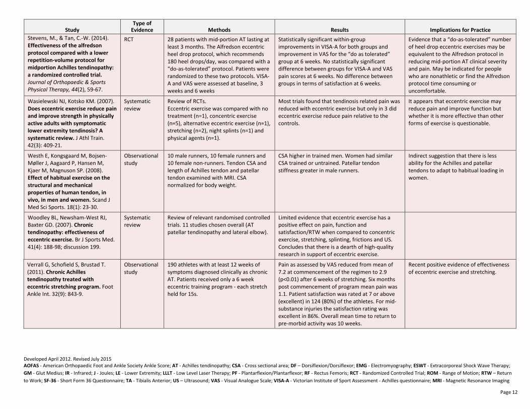
Stevens, M., & Tan, C.-W. (2014). Effectiveness of the alfredson
protocol compared with a lower
repetition-volume protocol for
midportion Achilles tendinopathy:
a randomized controlled trial. Journal of Orthopaedic & Sports
Physical Therapy, 44(2), 59-67.
RCT 28 patients with mid-portion AT lasting at least 3 months. The Alfredson eccentric heel drop protocol, which recommends 180 heel drops/day, was compared with a “do-as-tolerated” protocol. Patients were randomized to these two protocols. VISA-A and VAS were assessed at baseline, 3 weeks and 6 weeks
Statistically significant within-group improvements in VISA-A for both groups and improvement in VAS for the “do as tolerated” group at 6 weeks. No statistically significant difference between groups for VISA-A and VAS pain scores at 6 weeks. No difference between groups in terms of satisfaction at 6 weeks.
Evidence that a “do-as-tolerated” number of heel drop eccentric exercises may be equivalent to the Alfredson protocol in reducing mid-portion AT clinical severity and pain. May be indicated for people who are nonathletic or find the Alfredson protocol time consuming or uncomfortable.
Wasielewski NJ, Kotsko KM. (2007). Does eccentric exercise reduce pain
and improve strength in physically
active adults with symptomatic
lower extremity tendinosis? A
systematic review. J Athl Train. 42(3): 409-21.
Systematic review
Review of RCTs. Eccentric exercise was compared with no treatment (n=1), concentric exercise (n=5), alternative eccentric exercise (n=1), stretching (n=2), night splints (n=1) and physical agents (n=1).
Most trials found that tendinosis related pain was reduced with eccentric exercise but only in 3 did eccentric exercise reduce pain relative to the controls.
It appears that eccentric exercise may reduce pain and improve function but whether it is more effective than other forms of exercise is questionable.
Westh E, Kongsgaard M, Bojsen-Møller J, Aagaard P, Hansen M, Kjaer M, Magnuson SP. (2008). Effect of habitual exercise on the
structural and mechanical
properties of human tendon, in
vivo, in men and women. Scand J Med Sci Sports. 18(1): 23-30.
Observational study
10 male runners, 10 female runners and 10 female non-runners. Tendon CSA and length of Achilles tendon and patellar tendon examined with MRI. CSA normalized for body weight.
CSA higher in trained men. Women had similar CSA trained or untrained. Patellar tendon stiffness greater in male runners.
Indirect suggestion that there is less ability for the Achilles and patellar tendons to adapt to habitual loading in women.
Woodley BL, Newsham-West RJ, Baxter GD. (2007). Chronic
tendinopathy: effectiveness of
eccentric exercise. Br J Sports Med. 41(4): 188-98; discussion 199.
Systematic review
Review of relevant randomised controlled trials. 11 studies chosen overall (AT patellar tendinopathy and lateral elbow).
Limited evidence that eccentric exercise has a positive effect on pain, function and satisfaction/RTW when compared to concentric exercise, stretching, splinting, frictions and US. Concludes that there is a dearth of high-quality research in support of eccentric exercise.
Verrall G, Schofield S, Brustad T. (2011). Chronic Achilles
tendinopathy treated with
eccentric stretching program. Foot Ankle Int. 32(9): 843-9.
Observational study
190 athletes with at least 12 weeks of symptoms diagnosed clinically as chronic AT. Patients received only a 6 week eccentric training program - each stretch held for 15s.
Pain as assessed by VAS reduced from mean of 7.2 at commencement of the regimen to 2.9 (p<0.01) after 6 weeks of stretching. Six months post commencement of program mean pain was 1.1. Patient satisfaction was rated at 7 or above (excellent) in 124 (80%) of the athletes. For mid-substance injuries the satisfaction rating was excellent in 86%. Overall mean time to return to pre-morbid activity was 10 weeks.
Recent positive evidence of effectiveness of eccentric exercise and stretching.
Developed April 2012. Revised July 2015 AOFAS - American Orthopaedic Foot and Ankle Society Ankle Score; AT - Achilles tendinopathy; CSA - Cross sectional area; DF – Dorsiflexion/Dorsiflexor; EMG - Electromyography; ESWT - Extracorporeal Shock Wave Therapy;
GM - Glut Medius; IR - Infrared; J - Joules; LE - Lower Extremity; LLLT - Low Level Laser Therapy; PF - Plantarflexion/Plantarflexor; RF - Rectus Femoris; RCT - Randomized Controlled Trial; ROM - Range of Motion; RTW – Return
to Work; SF-36 - Short Form 36 Questionnaire; TA - Tibialis Anterior; US – Ultrasound; VAS - Visual Analogue Scale; VISA-A - Victorian Institute of Sport Assessment - Achilles questionnaire; MRI - Magnetic Resonance Imaging
Page 13
Study
Type of
Evidence Methods Results Implications for Practice
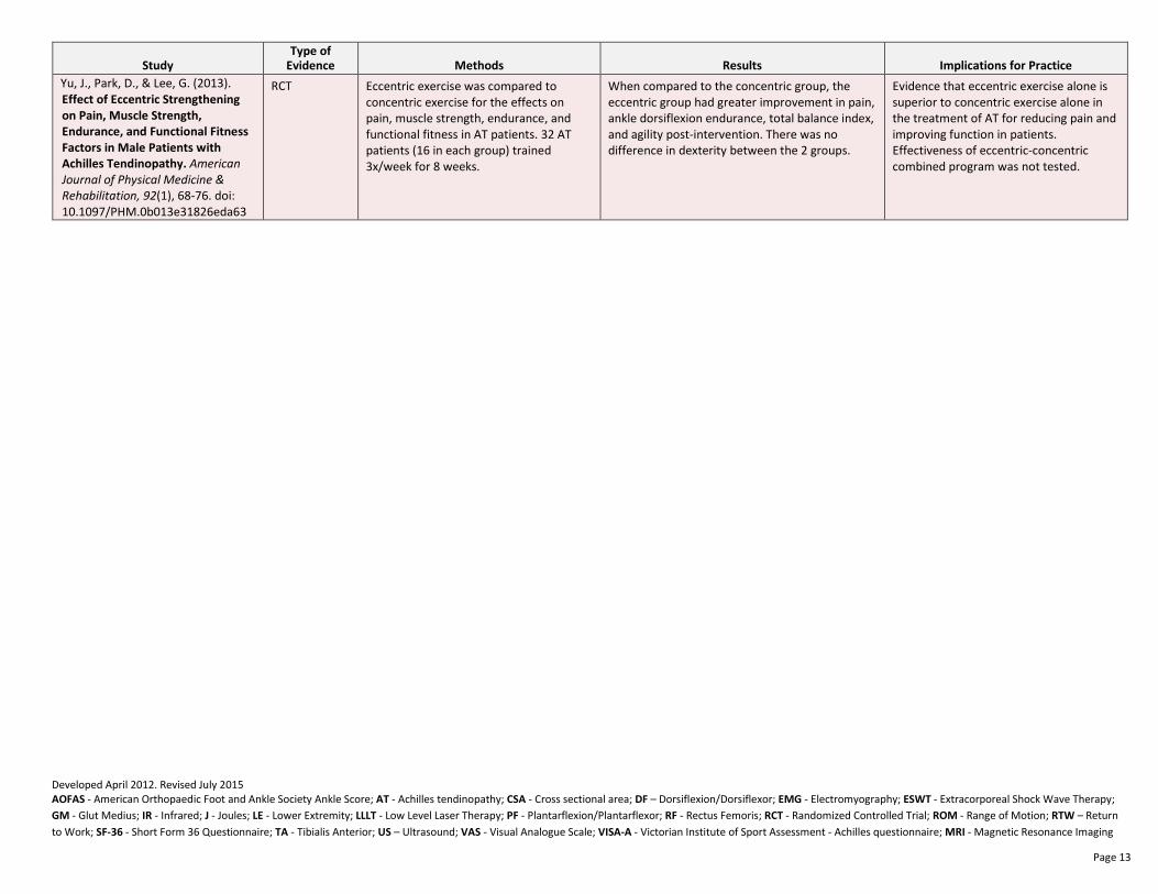
Yu, J., Park, D., & Lee, G. (2013). Effect of Eccentric Strengthening
on Pain, Muscle Strength,
Endurance, and Functional Fitness
Factors in Male Patients with
Achilles Tendinopathy. American
Journal of Physical Medicine &
Rehabilitation, 92(1), 68-76. doi: 10.1097/PHM.0b013e31826eda63
RCT Eccentric exercise was compared to concentric exercise for the effects on pain, muscle strength, endurance, and functional fitness in AT patients. 32 AT patients (16 in each group) trained 3x/week for 8 weeks.
When compared to the concentric group, the eccentric group had greater improvement in pain, ankle dorsiflexion endurance, total balance index, and agility post-intervention. There was no difference in dexterity between the 2 groups.
Evidence that eccentric exercise alone is superior to concentric exercise alone in the treatment of AT for reducing pain and improving function in patients. Effectiveness of eccentric-concentric combined program was not tested.
Developed April 2012. Revised July 2015 AOFAS - American Orthopaedic Foot and Ankle Society Ankle Score; AT - Achilles tendinopathy; CSA - Cross sectional area; DF – Dorsiflexion/Dorsiflexor; EMG - Electromyography; ESWT - Extracorporeal Shock Wave Therapy;
GM - Glut Medius; IR - Infrared; J - Joules; LE - Lower Extremity; LLLT - Low Level Laser Therapy; PF - Plantarflexion/Plantarflexor; RF - Rectus Femoris; RCT - Randomized Controlled Trial; ROM - Range of Motion; RTW – Return
to Work; SF-36 - Short Form 36 Questionnaire; TA - Tibialis Anterior; US – Ultrasound; VAS - Visual Analogue Scale; VISA-A - Victorian Institute of Sport Assessment - Achilles questionnaire; MRI - Magnetic Resonance Imaging
Page 14
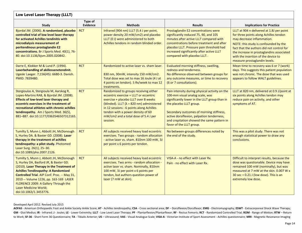
Low Level Laser Therapy (LLLT)
Study
Type of
Evidence Methods Results Implications for Practice
Bjordal JM. (2006). A randomised, placebo
controlled trial of low level laser therapy
for activated Achilles tendinitis with
microdialysis measurement of
peritendinous prostaglandin E2
concentrations. Br J Sports Med. 40(1), 76-80. doi:10.1136/bjsm.2005.020842.
RCT Infrared (904 nm) LLLT (5.4 J per point, power density 20 mW/cm2) and placebo LLLT (0 J) were administered to both Achilles tendons in random blinded order.
Prostaglandin E2 concentrations were significantly reduced 75, 90, and 105 minutes after active LLLT compared with concentrations before treatment and after placebo LLLT. Pressure pain threshold had increased significantly after active LLLT compared with placebo LLLT.
LLLT at 904 n delivered at 1.8J per point for three points along Achilles tendon may decrease inflammation.
NOTE: this study is confounded by the fact that the authors did not control for the increase in prostaglandins associated with the insertion of the device to measure prostaglandin levels.
Darre E, Klokker M & Lund P. (1994). Laserbehandling af akillessenetendinit. Ugeskr Laeger. 7;156(45): 6680-3. Danish. PMID: 7839480.
RCT Randomized to active laser vs. sham laser. 830 nm, 30mW, intensity 150 mW/cm2. Total dose was set to max 16 Joule (4 J at 4 points on tendon). 5 Rx/week to max 12 treatments.
Evaluated morning stiffness, swelling, redness and tenderness. No difference observed between groups for any outcome measures, or time to recovery (6 or 7 consultations).
Mean time to recovery was 6 or 7 (work) days. This suggests this patient population was not chronic. The dose that was used appears to follow WALT guidelines.
Stergioulas A, Stergioula M, Aarskog R, Lopes-Martins RAB, & Bjordal JM. (2008). Effects of low-level laser therapy and
eccentric exercises in the treatment of
recreational athletes with chronic achilles
tendinopathy. Am J Sports Med. 36(5), 881–887. doi:10.1177/0363546507312165.
RCT Randomized to groups receiving either eccentric exercise + LLLT or eccentric exercise + placebo LLLT over 8 weeks (blinded). LLLT (λ = 820 nm) administered in 12 sessions - 6 points along Achilles tendon with a power density of 60 mW/cm2 and a total dose of 5.4 J per session.
Pain intensity during physical activity on the 100-mm visual analog scale, was significantly lower in the LLLT group than in the placebo LLLT group. Secondary outcomes of morning stiffness, active dorsiflexion, palpation tenderness, and crepitation showed the same pattern in favor of the LLLT group.
LLLT at 820 nm, delivered at 0.9 J/point at six points along Achilles tendon may reduce pain on activity, and other symptoms of AT.
Tumilty S, Munn J, Abbott JH, McDonough S, Hurley DA. & Baxter GD. (2008). Laser
therapy in the treatment of achilles
tendinopathy: a pilot study. Photomed Laser Surg. 26(1), 25–30. doi:10.1089/pho.2007.2126.
RCT All subjects received heavy load eccentric exercises. Two groups - random allocation - active laser vs. sham. 810nm 100 mW, 3J per point x 6 points per tendon.
No between-groups differences noted by the end of the study.
This was a pilot study. There was not enough statistical power to draw any conclusions.
Tumilty S, Munn J, Abbott JH, McDonough S, Hurley DA, Basford JR, & Baxter GD. (2010). Laser Therapy in the Treatment of
Achilles Tendinopathy: A Randomised
Controlled Trial. AIP Conf. Proc. -- May 31, 2010 -- Volume 1226, pp. 163-169LASER FLORENCE 2009: A Gallery Through the Laser Medicine World; doi:10.1063/1.3453776.
RCT All subjects received heavy load eccentric exercises. Two arms - random allocation - active laser vs. sham. Nominally, 810nm 100 mW, 3J per point x 6 points per tendon, but authors question power of laser (7 mW at skin).
VISA-A - no effect with Laser Rx.
Pain - no effect with Laser Rx.
Difficult to interpret results, because the dose was questionable. Device may have remained 100 mW (nominally), but was measured at 7 mW at the skin. 0.007 W x 30 sec = 0.21 J (low dose). This is an extremely low dose.
Developed April 2012. Revised July 2015 AOFAS - American Orthopaedic Foot and Ankle Society Ankle Score; AT - Achilles tendinopathy; CSA - Cross sectional area; DF – Dorsiflexion/Dorsiflexor; EMG - Electromyography; ESWT - Extracorporeal Shock Wave Therapy;
GM - Glut Medius; IR - Infrared; J - Joules; LE - Lower Extremity; LLLT - Low Level Laser Therapy; PF - Plantarflexion/Plantarflexor; RF - Rectus Femoris; RCT - Randomized Controlled Trial; ROM - Range of Motion; RTW – Return
to Work; SF-36 - Short Form 36 Questionnaire; TA - Tibialis Anterior; US – Ultrasound; VAS - Visual Analogue Scale; VISA-A - Victorian Institute of Sport Assessment - Achilles questionnaire; MRI - Magnetic Resonance Imaging
Page 15
Study
Type of
Evidence Methods Results Implications for Practice
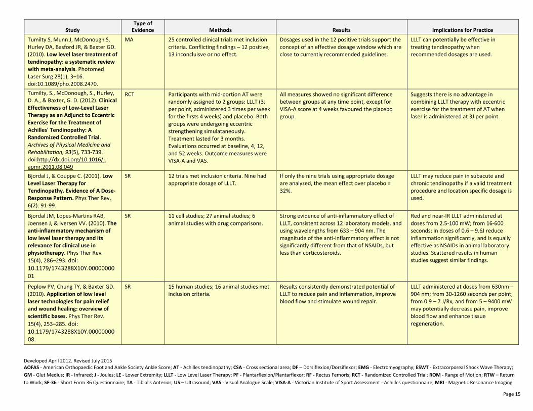
Tumilty S, Munn J, McDonough S, Hurley DA, Basford JR, & Baxter GD. (2010). Low level laser treatment of
tendinopathy: a systematic review
with meta-analysis. Photomed Laser Surg 28(1), 3–16. doi:10.1089/pho.2008.2470.
MA 25 controlled clinical trials met inclusion criteria. Conflicting findings – 12 positive, 13 inconcluisve or no effect.
Dosages used in the 12 positive trials support the concept of an effective dosage window which are close to currently recommended guidelines.
LLLT can potentially be effective in treating tendinopathy when recommended dosages are used.
Tumilty, S., McDonough, S., Hurley, D. A., & Baxter, G. D. (2012). Clinical
Effectiveness of Low-Level Laser
Therapy as an Adjunct to Eccentric
Exercise for the Treatment of
Achilles' Tendinopathy: A
Randomized Controlled Trial.
Archives of Physical Medicine and
Rehabilitation, 93(5), 733-739. doi:http://dx.doi.org/10.1016/j. apmr.2011.08.049
RCT Participants with mid-portion AT were randomly assigned to 2 groups: LLLT (3J per point, administered 3 times per week for the firsts 4 weeks) and placebo. Both groups were undergoing eccentric strengthening simulataneously. Treatment lasted for 3 months. Evaluations occurred at baseline, 4, 12, and 52 weeks. Outcome measures were VISA-A and VAS.
All measures showed no significant difference between groups at any time point, except for VISA-A score at 4 weeks favoured the placebo group.
Suggests there is no advantage in combining LLLT therapy with eccentric exercise for the treatment of AT when laser is administered at 3J per point.
Bjordal J, & Couppe C. (2001). Low
Level Laser Therapy for
Tendinopathy. Evidence of A Dose-
Response Pattern. Phys Ther Rev, 6(2): 91-99.
SR 12 trials met inclusion criteria. Nine had appropriate dosage of LLLT.
If only the nine trials using appropriate dosage are analyzed, the mean effect over placebo = 32%.
LLLT may reduce pain in subacute and chronic tendinopathy if a valid treatment procedure and location specific dosage is used.
Bjordal JM, Lopes-Martins RAB, Joensen J, & Iversen VV. (2010). The
anti-inflammatory mechanism of
low level laser therapy and its
relevance for clinical use in
physiotherapy. Phys Ther Rev. 15(4), 286–293. doi: 10.1179/1743288X10Y.0000000001
SR 11 cell studies; 27 animal studies; 6 animal studies with drug comparisons.
Strong evidence of anti-inflammatory effect of LLLT, consistent across 12 laboratory models, and using wavelengths from 633 – 904 nm. The magnitude of the anti-inflammatory effect is not significantly different from that of NSAIDs, but less than corticosteroids.
Red and near-IR LLLT administered at doses from 2.5-100 mW; from 16-600 seconds; in doses of 0.6 – 9.6J reduce inflammation significantly, and is equally effective as NSAIDs in animal laboratory studies. Scattered results in human studies suggest similar findings.
Peplow PV, Chung TY, & Baxter GD. (2010). Application of low level
laser technologies for pain relief
and wound healing: overview of
scientific bases. Phys Ther Rev. 15(4), 253–285. doi: 10.1179/1743288X10Y.0000000008.
SR 15 human studies; 16 animal studies met inclusion criteria.
Results consistently demonstrated potential of LLLT to reduce pain and inflammation, improve blood flow and stimulate wound repair.
LLLT administered at doses from 630nm – 904 nm; from 30-1260 seconds per point; from 0.9 – 7 J/Rx; and from 5 – 9400 mW may potentially decrease pain, improve blood flow and enhance tissue regeneration.
Developed April 2012. Revised July 2015 AOFAS - American Orthopaedic Foot and Ankle Society Ankle Score; AT - Achilles tendinopathy; CSA - Cross sectional area; DF – Dorsiflexion/Dorsiflexor; EMG - Electromyography; ESWT - Extracorporeal Shock Wave Therapy;
GM - Glut Medius; IR - Infrared; J - Joules; LE - Lower Extremity; LLLT - Low Level Laser Therapy; PF - Plantarflexion/Plantarflexor; RF - Rectus Femoris; RCT - Randomized Controlled Trial; ROM - Range of Motion; RTW – Return
to Work; SF-36 - Short Form 36 Questionnaire; TA - Tibialis Anterior; US – Ultrasound; VAS - Visual Analogue Scale; VISA-A - Victorian Institute of Sport Assessment - Achilles questionnaire; MRI - Magnetic Resonance Imaging
Page 16
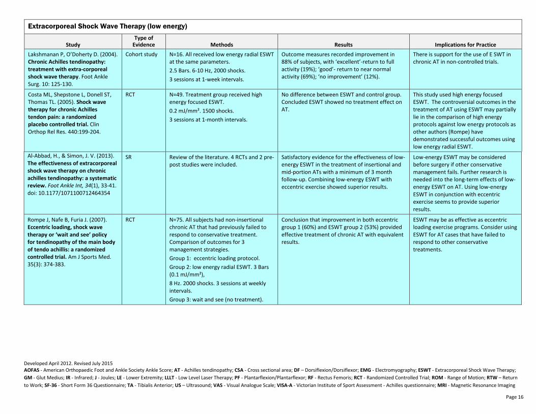
Extracorporeal Shock Wave Therapy (low energy)
Study
Type of
Evidence Methods Results Implications for Practice
Lakshmanan P, O’Doherty D. (2004). Chronic Achilles tendinopathy:
treatment with extra-corporeal
shock wave therapy. Foot Ankle Surg. 10: 125-130.
Cohort study N=16. All received low energy radial ESWT at the same parameters.
2.5 Bars. 6-10 Hz, 2000 shocks.
3 sessions at 1-week intervals.
Outcome measures recorded improvement in 88% of subjects, with ‘excellent’-return to full activity (19%); ‘good’- return to near normal activity (69%); ‘no improvement’ (12%).
There is support for the use of E SWT in chronic AT in non-controlled trials.
Costa ML, Shepstone L, Donell ST, Thomas TL. (2005). Shock wave
therapy for chronic Achilles
tendon pain: a randomized
placebo controlled trial. Clin Orthop Rel Res. 440:199-204.
RCT N=49. Treatment group received high energy focused ESWT.
0.2 mJ/mm². 1500 shocks.
3 sessions at 1-month intervals.
No difference between ESWT and control group. Concluded ESWT showed no treatment effect on AT.
This study used high energy focused ESWT. The controversial outcomes in the treatment of AT using ESWT may partially lie in the comparison of high energy protocols against low energy protocols as other authors (Rompe) have demonstrated successful outcomes using low energy radial ESWT.
Al-Abbad, H., & Simon, J. V. (2013). The effectiveness of extracorporeal
shock wave therapy on chronic
achilles tendinopathy: a systematic
review. Foot Ankle Int, 34(1), 33-41. doi: 10.1177/1071100712464354
SR Review of the literature. 4 RCTs and 2 pre-post studies were included.
Satisfactory evidence for the effectiveness of low-energy ESWT in the treatment of insertional and mid-portion ATs with a minimum of 3 month follow-up. Combining low-energy ESWT with eccentric exercise showed superior results.
Low-energy ESWT may be considered before surgery if other conservative management fails. Further research is needed into the long-term effects of low-energy ESWT on AT. Using low-energy ESWT in conjunction with eccentric exercise seems to provide superior results.
Rompe J, Nafe B, Furia J. (2007). Eccentric loading, shock wave
therapy or ‘wait and see’ policy
for tendinopathy of the main body
of tendo achillis: a randomized
controlled trial. Am J Sports Med.
35(3): 374-383.
RCT N=75. All subjects had non-insertional chronic AT that had previously failed to respond to conservative treatment. Comparison of outcomes for 3 management strategies.
Group 1: eccentric loading protocol.
Group 2: low energy radial ESWT. 3 Bars (0.1 mJ/mm²),
8 Hz. 2000 shocks. 3 sessions at weekly intervals.
Group 3: wait and see (no treatment).
Conclusion that improvement in both eccentric group 1 (60%) and ESWT group 2 (53%) provided effective treatment of chronic AT with equivalent results.
ESWT may be as effective as eccentric loading exercise programs. Consider using ESWT for AT cases that have failed to respond to other conservative treatments.
Developed April 2012. Revised July 2015 AOFAS - American Orthopaedic Foot and Ankle Society Ankle Score; AT - Achilles tendinopathy; CSA - Cross sectional area; DF – Dorsiflexion/Dorsiflexor; EMG - Electromyography; ESWT - Extracorporeal Shock Wave Therapy;
GM - Glut Medius; IR - Infrared; J - Joules; LE - Lower Extremity; LLLT - Low Level Laser Therapy; PF - Plantarflexion/Plantarflexor; RF - Rectus Femoris; RCT - Randomized Controlled Trial; ROM - Range of Motion; RTW – Return
to Work; SF-36 - Short Form 36 Questionnaire; TA - Tibialis Anterior; US – Ultrasound; VAS - Visual Analogue Scale; VISA-A - Victorian Institute of Sport Assessment - Achilles questionnaire; MRI - Magnetic Resonance Imaging
Page 17
Study
Type of
Evidence Methods Results Implications for Practice
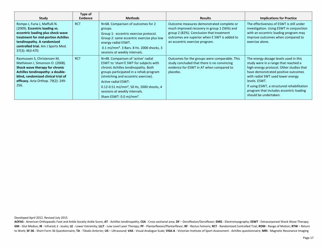
Rompe J, Furia J, Maffulli N. (2009). Eccentric loading vs.
eccentric loading plus shock wave
treatment for mid-portion Achilles
tendinopathy. A randomized
controlled trial. Am J Sports Med. 37(3): 463-470.
RCT N=68. Comparison of outcomes for 2 groups.
Group 1: eccentric exercise protocol. Group 2: same eccentric exercise plus low energy radial ESWT.
0.1 mJ/mm². 3 Bars. 8 Hz. 2000 shocks, 3 sessions at weekly intervals.
Outcome measures demonstrated complete or much improved recovery in group 1 (56%) and group 2 (82%). Conclusion that treatment outcomes are superior when E SWT is added to an eccentric exercise program.
The effectiveness of ESWT is still under investigation. Using ESWT in conjunction with an eccentric loading program may improve outcomes when compared to exercise alone.
Rasmussen S, Christensen M, Mathiesen I, Simonson O. (2008). Shock wave therapy for chronic
Achilles tendinopathy: a double-
blind, randomized clinical trial of
efficacy. Acta Orthop. 79(2): 249-256.
RCT N=48. Comparison of ‘active’ radial ESWT to ‘sham’E SWT for subjects with chronic Achilles tendinopathy. Both groups participated in a rehab program (stretching and eccentric exercise).
Active radial ESWT:
0.12-0.51 mJ/mm², 50 Hz, 2000 shocks, 4 sessions at weekly intervals.
Sham ESWT: 0.0 mJ/mm².
Outcomes for the groups were comparable. This study concluded that there is no convincing evidence for ESWT in AT when compared to placebo.
The energy dosage levels used in this study were in a range that reached a high-energy protocol. Other studies that have demonstrated positive outcomes with radial SWT used lower energy levels. ESWT.
If using ESWT, a structured rehabilitation program that includes eccentric loading should be undertaken.
Developed April 2012. Revised July 2015 AOFAS - American Orthopaedic Foot and Ankle Society Ankle Score; AT - Achilles tendinopathy; CSA - Cross sectional area; DF – Dorsiflexion/Dorsiflexor; EMG - Electromyography; ESWT - Extracorporeal Shock Wave Therapy;
GM - Glut Medius; IR - Infrared; J - Joules; LE - Lower Extremity; LLLT - Low Level Laser Therapy; PF - Plantarflexion/Plantarflexor; RF - Rectus Femoris; RCT - Randomized Controlled Trial; ROM - Range of Motion; RTW – Return
to Work; SF-36 - Short Form 36 Questionnaire; TA - Tibialis Anterior; US – Ultrasound; VAS - Visual Analogue Scale; VISA-A - Victorian Institute of Sport Assessment - Achilles questionnaire; MRI - Magnetic Resonance Imaging
Page 18
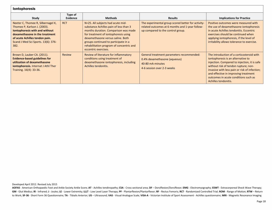
Iontophoresis
Study
Type of
Evidence Methods Results Implications for Practice
Neeter C, Thomee R, Silbernagel K, Thomee P, Karlson J. (2003). Iontophoresis with and without
dexamethasone in the treatment
of acute Achilles tendon pain. Scand J Med Sci Sports. 13(6): 376-382.
RCT N=25. All subjects had acute mid-substance Achilles pain of less than 3 months duration. Comparison was made for treatment of iontophoresis using dexamethasone versus saline. Both groups continued to participate in a rehabilitation program of concentric and eccentric exercises.
The experimental group scored better for activity-related outcomes at 6 months and 1-year follow-up compared to the control group.
Positive outcomes were measured with the use of dexamethasone iontophoresis in acute Achilles tendonitis. Eccentric exercises should be continued when applying iontophoresis, if the level of irritability allows tolerance to exercise.
Brown D, Lauber CA. (2011). Evidence-based guidelines for
utilization of dexamethasone
iontophoresis. Internat J Athl Ther Training. 16(4): 33-36.
Review Review of literature for inflammatory conditions using treatment of dexamethasone iontophoresis, including Achilles tendonitis.
General treatment parameters recommended:
0.4% dexamethasone (aqueous)
40-80 mA-minutes
4-6 session over 2-3 weeks
The introduction of a corticosteroid with iontophoresis is an alternative to injection. Compared to injection, it is safe without risk of tendon rupture; non-invasive with less pain or risk of infection; and effective in improving treatment outcomes in acute conditions such as Achilles tendonitis.
Developed April 2012. Revised July 2015 AOFAS - American Orthopaedic Foot and Ankle Society Ankle Score; AT - Achilles tendinopathy; CSA - Cross sectional area; DF – Dorsiflexion/Dorsiflexor; EMG - Electromyography; ESWT - Extracorporeal Shock Wave Therapy;
GM - Glut Medius; IR - Infrared; J - Joules; LE - Lower Extremity; LLLT - Low Level Laser Therapy; PF - Plantarflexion/Plantarflexor; RF - Rectus Femoris; RCT - Randomized Controlled Trial; ROM - Range of Motion; RTW – Return
to Work; SF-36 - Short Form 36 Questionnaire; TA - Tibialis Anterior; US – Ultrasound; VAS - Visual Analogue Scale; VISA-A - Victorian Institute of Sport Assessment - Achilles questionnaire; MRI - Magnetic Resonance Imaging
Page 19
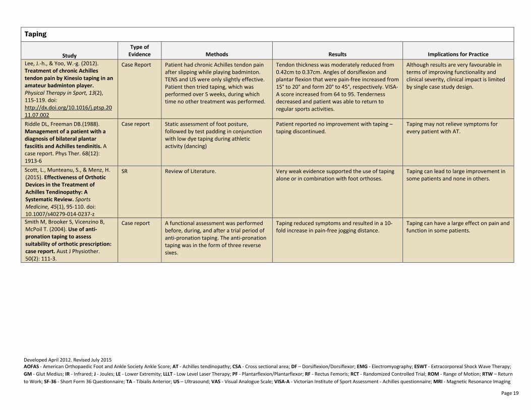
Taping
Study
Type of
Evidence Methods Results Implications for Practice
Lee, J.-h., & Yoo, W.-g. (2012). Treatment of chronic Achilles
tendon pain by Kinesio taping in an
amateur badminton player. Physical Therapy in Sport, 13(2), 115-119. doi: http://dx.doi.org/10.1016/j.ptsp.2011.07.002
Case Report Patient had chronic Achilles tendon pain after slipping while playing badminton. TENS and US were only slightly effective. Patient then tried taping, which was performed over 5 weeks, during which time no other treatment was performed.
Tendon thickness was moderately reduced from 0.42cm to 0.37cm. Angles of dorsiflexion and plantar flexion that were pain-free increased from 15° to 20° and form 20° to 45°, respectively. VISA-A score increased from 64 to 95. Tenderness decreased and patient was able to return to regular sports activities.
Although results are very favourable in terms of improving functionality and clinical severity, clinical impact is limited by single case study design.
Riddle DL, Freeman DB.(1988). Management of a patient with a
diagnosis of bilateral plantar
fasciitis and Achilles tendinitis. A case report. Phys Ther. 68(12): 1913-6
Case report Static assessment of foot posture, followed by test padding in conjunction with low dye taping during athletic activity (dancing)
Patient reported no improvement with taping – taping discontinued.
Taping may not relieve symptoms for every patient with AT.
Scott, L., Munteanu, S., & Menz, H. (2015). Effectiveness of Orthotic
Devices in the Treatment of
Achilles Tendinopathy: A
Systematic Review. Sports
Medicine, 45(1), 95-110. doi: 10.1007/s40279-014-0237-z
SR Review of Literature. Very weak evidence supported the use of taping alone or in combination with foot orthoses.
Taping can lead to large improvement in some patients and none in others.
Smith M, Brooker S, Vicenzino B, McPoil T. (2004). Use of anti-
pronation taping to assess
suitability of orthotic prescription:
case report. Aust J Physiother. 50(2): 111-3.
Case report A functional assessment was performed before, during, and after a trial period of anti-pronation taping. The anti-pronation taping was in the form of three reverse sixes.
Taping reduced symptoms and resulted in a 10-fold increase in pain-free jogging distance.
Taping can have a large effect on pain and function in some patients.
Developed April 2012. Revised July 2015 AOFAS - American Orthopaedic Foot and Ankle Society Ankle Score; AT - Achilles tendinopathy; CSA - Cross sectional area; DF – Dorsiflexion/Dorsiflexor; EMG - Electromyography; ESWT - Extracorporeal Shock Wave Therapy;
GM - Glut Medius; IR - Infrared; J - Joules; LE - Lower Extremity; LLLT - Low Level Laser Therapy; PF - Plantarflexion/Plantarflexor; RF - Rectus Femoris; RCT - Randomized Controlled Trial; ROM - Range of Motion; RTW – Return
to Work; SF-36 - Short Form 36 Questionnaire; TA - Tibialis Anterior; US – Ultrasound; VAS - Visual Analogue Scale; VISA-A - Victorian Institute of Sport Assessment - Achilles questionnaire; MRI - Magnetic Resonance Imaging
Page 20
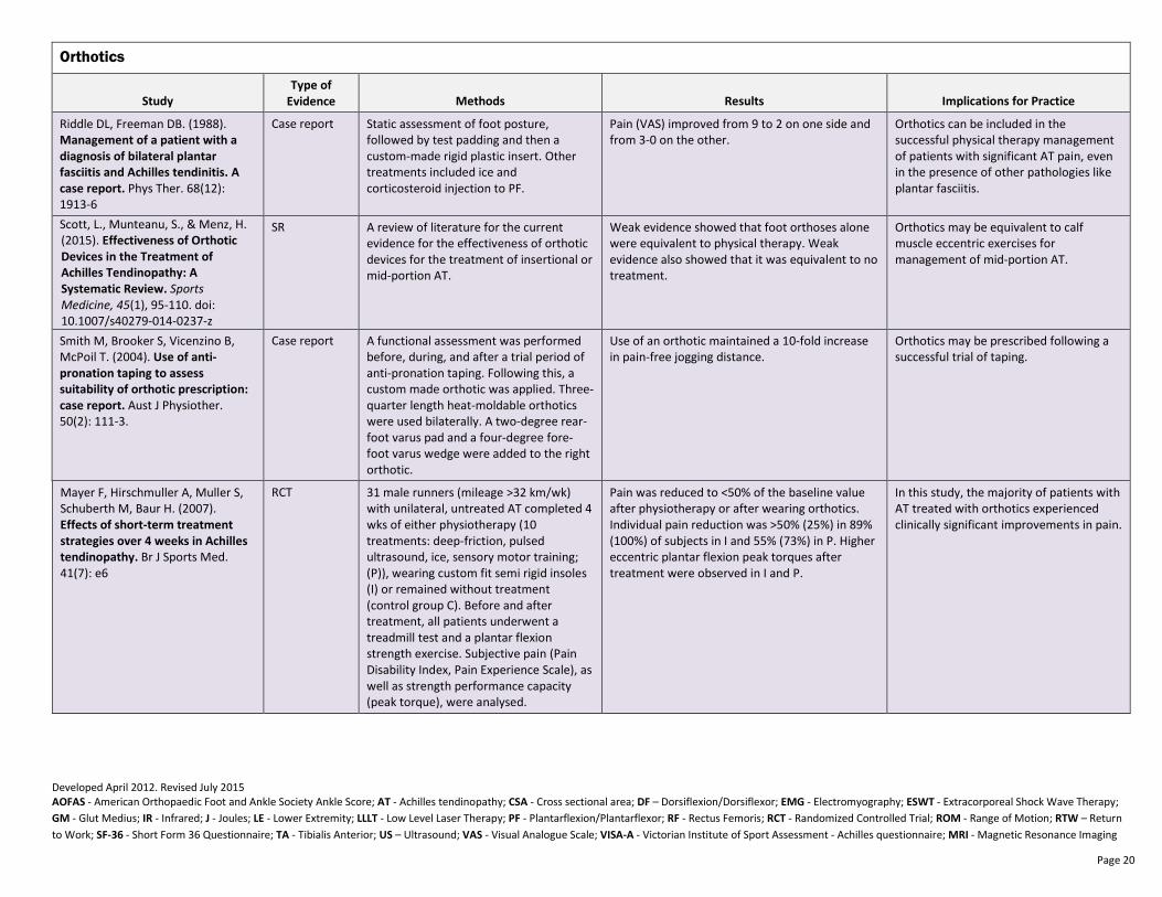
Orthotics
Study Type of
Evidence Methods Results Implications for Practice
Riddle DL, Freeman DB. (1988). Management of a patient with a
diagnosis of bilateral plantar
fasciitis and Achilles tendinitis. A
case report. Phys Ther. 68(12): 1913-6
Case report Static assessment of foot posture, followed by test padding and then a custom-made rigid plastic insert. Other treatments included ice and corticosteroid injection to PF.
Pain (VAS) improved from 9 to 2 on one side and from 3-0 on the other.
Orthotics can be included in the successful physical therapy management of patients with significant AT pain, even in the presence of other pathologies like plantar fasciitis.
Scott, L., Munteanu, S., & Menz, H. (2015). Effectiveness of Orthotic
Devices in the Treatment of
Achilles Tendinopathy: A
Systematic Review. Sports
Medicine, 45(1), 95-110. doi: 10.1007/s40279-014-0237-z
SR A review of literature for the current evidence for the effectiveness of orthotic devices for the treatment of insertional or mid-portion AT.
Weak evidence showed that foot orthoses alone were equivalent to physical therapy. Weak evidence also showed that it was equivalent to no treatment.
Orthotics may be equivalent to calf muscle eccentric exercises for management of mid-portion AT.
Smith M, Brooker S, Vicenzino B, McPoil T. (2004). Use of anti-
pronation taping to assess
suitability of orthotic prescription:
case report. Aust J Physiother. 50(2): 111-3.
Case report A functional assessment was performed before, during, and after a trial period of anti-pronation taping. Following this, a custom made orthotic was applied. Three-quarter length heat-moldable orthotics were used bilaterally. A two-degree rear-foot varus pad and a four-degree fore-foot varus wedge were added to the right orthotic.
Use of an orthotic maintained a 10-fold increase in pain-free jogging distance.
Orthotics may be prescribed following a successful trial of taping.
Mayer F, Hirschmuller A, Muller S, Schuberth M, Baur H. (2007). Effects of short-term treatment
strategies over 4 weeks in Achilles
tendinopathy. Br J Sports Med. 41(7): e6
RCT 31 male runners (mileage >32 km/wk) with unilateral, untreated AT completed 4 wks of either physiotherapy (10 treatments: deep-friction, pulsed ultrasound, ice, sensory motor training; (P)), wearing custom fit semi rigid insoles (I) or remained without treatment (control group C). Before and after treatment, all patients underwent a treadmill test and a plantar flexion strength exercise. Subjective pain (Pain Disability Index, Pain Experience Scale), as well as strength performance capacity (peak torque), were analysed.
Pain was reduced to <50% of the baseline value after physiotherapy or after wearing orthotics. Individual pain reduction was >50% (25%) in 89% (100%) of subjects in I and 55% (73%) in P. Higher eccentric plantar flexion peak torques after treatment were observed in I and P.
In this study, the majority of patients with AT treated with orthotics experienced clinically significant improvements in pain.
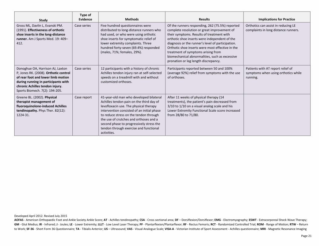
Developed April 2012. Revised July 2015 AOFAS - American Orthopaedic Foot and Ankle Society Ankle Score; AT - Achilles tendinopathy; CSA - Cross sectional area; DF – Dorsiflexion/Dorsiflexor; EMG - Electromyography; ESWT - Extracorporeal Shock Wave Therapy;
GM - Glut Medius; IR - Infrared; J - Joules; LE - Lower Extremity; LLLT - Low Level Laser Therapy; PF - Plantarflexion/Plantarflexor; RF - Rectus Femoris; RCT - Randomized Controlled Trial; ROM - Range of Motion; RTW – Return
to Work; SF-36 - Short Form 36 Questionnaire; TA - Tibialis Anterior; US – Ultrasound; VAS - Visual Analogue Scale; VISA-A - Victorian Institute of Sport Assessment - Achilles questionnaire; MRI - Magnetic Resonance Imaging
Page 21
Study
Type of
Evidence Methods Results Implications for Practice
Gross ML, Davlin L, Evanski PM. (1991). Effectiveness of orthotic
shoe inserts in the long-distance
runner. Am J Sports Med. 19: 409–412.
Case series Five hundred questionnaires were distributed to long-distance runners who had used, or who were using orthotic shoe inserts for symptomatic relief of lower extremity complaints. Three hundred forty-seven (69.4%) responded (males, 71%; females, 29%).
Of the runners responding, 262 (75.5%) reported complete resolution or great improvement of their symptoms. Results of treatment with orthotic shoe inserts were independent of the diagnosis or the runner's level of participation. Orthotic shoe inserts were most effective in the treatment of symptoms arising from biomechanical abnormalities, such as excessive pronation or leg length discrepancy.
Orthotics can assist in reducing LE complaints in long distance runners.
Donoghue OA, Harrison AJ, Laxton P, Jones RK. (2008). Orthotic control
of rear foot and lower limb motion
during running in participants with
chronic Achilles tendon injury. Sports Biomech. 7(2): 194-205.
Case series 12 participants with a history of chronic Achilles tendon injury ran at self-selected speeds on a treadmill with and without customized orthoses.
Participants reported between 50 and 100% (average 92%) relief from symptoms with the use of orthoses.
Patients with AT report relief of symptoms when using orthotics while running.
Greene BL. (2002). Physical
therapist management of
fluoroquinolone-induced Achilles
tendinopathy. Phys Ther. 82(12): 1224-31.
Case report 41-year-old man who developed bilateral Achilles tendon pain on the third day of levofloxacin use. The physical therapy intervention consisted of an initial phase to reduce stress on the tendon through the use of crutches and orthoses and a second phase to progressively stress the tendon through exercise and functional activities.
After 11 weeks of physical therapy (14 treatments), the patient's pain decreased from 3/10 to 1/10 on a visual analog scale and his Lower Extremity Functional Scale score increased from 28/80 to 71/80.
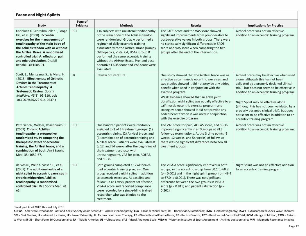
Developed April 2012. Revised July 2015 AOFAS - American Orthopaedic Foot and Ankle Society Ankle Score; AT - Achilles tendinopathy; CSA - Cross sectional area; DF – Dorsiflexion/Dorsiflexor; EMG - Electromyography; ESWT - Extracorporeal Shock Wave Therapy;
GM - Glut Medius; IR - Infrared; J - Joules; LE - Lower Extremity; LLLT - Low Level Laser Therapy; PF - Plantarflexion/Plantarflexor; RF - Rectus Femoris; RCT - Randomized Controlled Trial; ROM - Range of Motion; RTW – Return
to Work; SF-36 - Short Form 36 Questionnaire; TA - Tibialis Anterior; US – Ultrasound; VAS - Visual Analogue Scale; VISA-A - Victorian Institute of Sport Assessment - Achilles questionnaire; MRI - Magnetic Resonance Imaging
Page 22
Brace and Night Splints
Study
Type of
Evidence Methods Results Implications for Practice
Knobloch K, Schreibmueller L, Longo UG, et al. (2008). Eccentric
exercises for the management of
tendinopathy of the main body of
the Achilles tendon with or without
the AirHeel Brace. A randomized
controlled trial. A: effects on pain
and microcirculation. Disabil Rehabil. 30:1685-91.
RCT 116 subjects with unilateral tendinopathy of the main body of the Achilles tendon were randomized; Group A performed a regimen of daily eccentric training associated with the AirHeel Brace (Donjoy Orthopedics, Vista, CA, USA). Group B performed the same eccentric training without the AirHeel Brace. Pre- and post-operative FAOS score and VAS score were evaluated.
The FAOS score and the VAS score showed significant improvements from pre-operative to post-operative values in both groups. There were no statistically significant differences in FAOS score and VAS score when comparing the two groups after the end of the intervention.
Airheel brace was not an effective addition to an eccentric training program.
Scott, L., Munteanu, S., & Menz, H. (2015). Effectiveness of Orthotic
Devices in the Treatment of
Achilles Tendinopathy: A
Systematic Review. Sports
Medicine, 45(1), 95-110. doi: 10.1007/s40279-014-0237-z
SR Review of Literature. One study showed that the AirHeel brace was as effective as calf muscle eccentric exercises, and two studies showed it did not provide any added benefit when used in conjunction with the exercise program.
Weak evidence showed that an ankle joint dorsiflexion night splint was equally effective to a calf muscle eccentric exercise program, and strong evidence showed it did not provide any added benefit when it was used in conjunction with the exercise program.
Airheel brace may be effective when used alone (although this has not been validated by a properly designed clinical trial), but does not seem to be effective in addition to an eccentric training program.
Night Splint may be effective alone (although this has not been validated by a properly designed clinical trial), but does not seem to be effective in addition to an eccentric training program.
Petersen W, Welp R, Rosenbaum D. (2007). Chronic Achilles
tendinopathy: a prospective
randomized study comparing the
therapeutic effect of eccentric
training, the AirHeel brace, and a
combination of both. Am J Sports Med. 35: 1659-67.
RCT One hundred patients were randomly assigned to 1 of 3 treatment groups: (1) eccentric training, (2) AirHeel brace, and (3) combination of eccentric training and AirHeel brace. Patients were evaluated at 6, 12, and 54 weeks after the beginning of the treatment protocol with ultrasonography, VAS for pain, AOFAS, and SF-36.
The VAS score for pain, AOFAS score, and SF-36 improved significantly in all 3 groups at all 3 follow-up examinations. At the 3 time points (6 weeks, 12 weeks, and 54 weeks) of follow-up, there was no significant difference between all 3 treatment groups.
Airheel brace was not an effective addition to an eccentric training program.
de Vos RJ, Weir A, Visser RJ, et al. (2007). The additional value of a
night splint to eccentric exercises in
chronic midportion Achilles
tendinopathy: a randomised
controlled trial. Br J Sports Med. 41: e5.
RCT Both groups completed a 12wk heavy-load eccentric training program. One group received a night splint in addition to eccentric exercises. At baseline and follow-up at 12wks, patient satisfaction, VISA-A score and reported compliance were recorded by a single-blind trained researcher who was blinded to the treatment.
The VISA-A score significantly improved in both groups; in the eccentric group from 50.1 to 68.8 (p = 0.001) and in the night splint group from 49.4 to 67.0 (p<0.001). There was no significant difference between the two groups in VISA-A score (p = 0.815) and patient satisfaction (p = 0.261).
Night splint was not an effective addition to an eccentric training program.
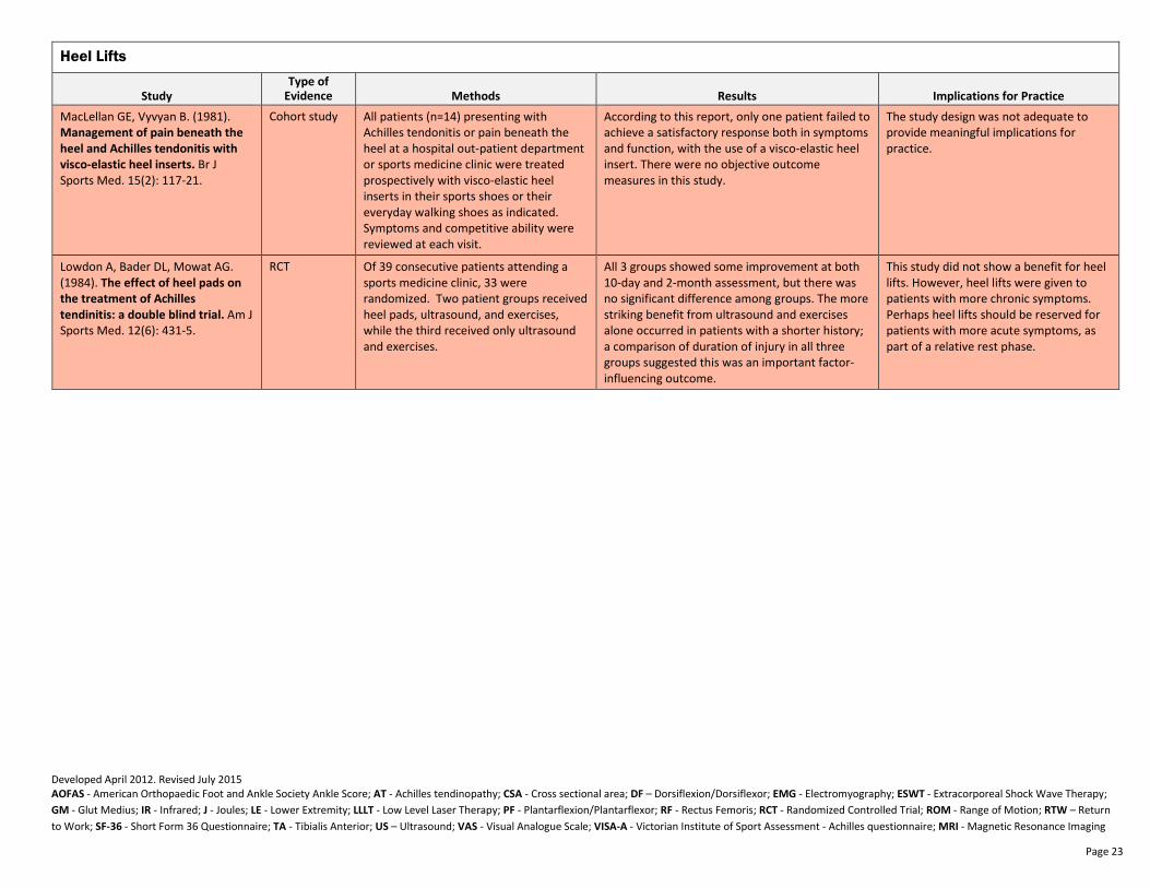
Developed April 2012. Revised July 2015 AOFAS - American Orthopaedic Foot and Ankle Society Ankle Score; AT - Achilles tendinopathy; CSA - Cross sectional area; DF – Dorsiflexion/Dorsiflexor; EMG - Electromyography; ESWT - Extracorporeal Shock Wave Therapy;
GM - Glut Medius; IR - Infrared; J - Joules; LE - Lower Extremity; LLLT - Low Level Laser Therapy; PF - Plantarflexion/Plantarflexor; RF - Rectus Femoris; RCT - Randomized Controlled Trial; ROM - Range of Motion; RTW – Return
to Work; SF-36 - Short Form 36 Questionnaire; TA - Tibialis Anterior; US – Ultrasound; VAS - Visual Analogue Scale; VISA-A - Victorian Institute of Sport Assessment - Achilles questionnaire; MRI - Magnetic Resonance Imaging
Page 23
Heel Lifts
Study
Type of
Evidence Methods Results Implications for Practice
MacLellan GE, Vyvyan B. (1981). Management of pain beneath the
heel and Achilles tendonitis with
visco-elastic heel inserts. Br J Sports Med. 15(2): 117-21.
Cohort study All patients (n=14) presenting with Achilles tendonitis or pain beneath the heel at a hospital out-patient department or sports medicine clinic were treated prospectively with visco-elastic heel inserts in their sports shoes or their everyday walking shoes as indicated. Symptoms and competitive ability were reviewed at each visit.
According to this report, only one patient failed to achieve a satisfactory response both in symptoms and function, with the use of a visco-elastic heel insert. There were no objective outcome measures in this study.
The study design was not adequate to provide meaningful implications for practice.
Lowdon A, Bader DL, Mowat AG. (1984). The effect of heel pads on
the treatment of Achilles
tendinitis: a double blind trial. Am J Sports Med. 12(6): 431-5.
RCT Of 39 consecutive patients attending a sports medicine clinic, 33 were randomized. Two patient groups received heel pads, ultrasound, and exercises, while the third received only ultrasound and exercises.
All 3 groups showed some improvement at both 10-day and 2-month assessment, but there was no significant difference among groups. The more striking benefit from ultrasound and exercises alone occurred in patients with a shorter history; a comparison of duration of injury in all three groups suggested this was an important factor-influencing outcome.
This study did not show a benefit for heel lifts. However, heel lifts were given to patients with more chronic symptoms. Perhaps heel lifts should be reserved for patients with more acute symptoms, as part of a relative rest phase.
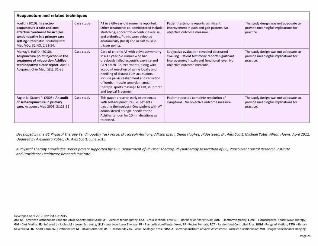
Developed April 2012. Revised July 2015 AOFAS - American Orthopaedic Foot and Ankle Society Ankle Score; AT - Achilles tendinopathy; CSA - Cross sectional area; DF – Dorsiflexion/Dorsiflexor; EMG - Electromyography; ESWT - Extracorporeal Shock Wave Therapy;
GM - Glut Medius; IR - Infrared; J - Joules; LE - Lower Extremity; LLLT - Low Level Laser Therapy; PF - Plantarflexion/Plantarflexor; RF - Rectus Femoris; RCT - Randomized Controlled Trial; ROM - Range of Motion; RTW – Return
to Work; SF-36 - Short Form 36 Questionnaire; TA - Tibialis Anterior; US – Ultrasound; VAS - Visual Analogue Scale; VISA-A - Victorian Institute of Sport Assessment - Achilles questionnaire; MRI - Magnetic Resonance Imaging
Page 24
Acupuncture and related techniques
Foell J. (2010). Is electro-
acupuncture a safe and cost-
effective treatment for Achilles
tendonopathy in a primary care
setting? InternatMusculoskeletal Med VOL. 32 NO. 2 51-54.
Case study AT in a 68-year-old runner is reported. Other treatments co-administered include stretching, concentric-eccentric exercise, and orthotics. Points were selected anatomically (local) and in calf muscle trigger points.
Patient testimony reports significant improvement in pain and gait pattern. No objective outcome measure.
The study design was not adequate to provide meaningful implications for practice.
Murray I, Hall D. (2010). Acupuncture point injection in the
treatment of midportion Achilles
tendinopathy: a case report. Aust J Acupunct Chin Med; 5(1): 31-35.
Case study Case of chronic AT with pelvic asymmetry in a 42 year-old runner who had previously failed eccentric exercise and GTN patch. Co-treatments, along with acupoint injection of saline locally and needling of distant TCM acupuoints, include pelvic realignment and reduction of lumbar muscle tone via manual therapy, sports massage to calf, ibuprofen and topical Traumeel.
Subjective evaluation revealed decreased swelling. Patient testimony reports significant improvement in pain and functional level. No objective outcome measure.
The study design was not adequate to provide meaningful implications for practice.
Fagan N, Staten P. (2003). An audit
of self-acupuncture in primary
care. Acupunct Med 2003: 21:28-31
Case study This paper presents early experiences with self-acupuncture (i.e. patients treating themselves). One patient with AT administered a single needle to the Achilles tendon for 10min durations as tolerated.
Patient reported complete resolution of symptoms. No objective outcome measure.
The study design was not adequate to provide meaningful implications for practice.
Developed by the BC Physical Therapy Tendinopathy Task Force: Dr. Joseph Anthony, Allison Ezzat, Diana Hughes, JR Justesen, Dr. Alex Scott, Michael Yates, Alison Hoens. April 2012.
Updated by Alexandra Kobza, Dr. Alex Scott. June 2015.
A Physical Therapy Knowledge Broker project supported by: UBC Department of Physical Therapy, Physiotherapy Association of BC, Vancouver Coastal Research Institute
and Providence Healthcare Research Institute.