Appendix A: Nuclear Medicine Studies Covered in This Book978-3-319-26704-3/1.pdf · Tc-99m-MAG3...
21
251 © Springer International Publishing Switzerland 2016 W.C. Klingensmith III, The Mathematics and Biology of the Biodistribution of Radiopharmaceuticals-A Clinical Perspective, DOI 10.1007/978-3-319-26704-3 Appendix A: Nuclear Medicine Studies Covered in This Book Appendix A lists the 39 nuclear medicine studies that are included in this book and, in particular, that are discussed in a systematic fashion in Part III, Quantitative Evaluation in Nuclear Medicine Studies. The list is a subset of the 49 diagnostic nuclear medicine studies found in the Nuclear Medicine Procedure Manual [1]. Nine infrequently performed nuclear medicine studies have been omitted, and the Thyroid Imaging Study with I-123 and the Thyroid Uptake Measurement with I-123 have been combined into one study. Cardiovascular System Cardiac Gated Blood Pool Study at Rest (Tc-99m-red blood cells) Lymphoscintigraphy (Tc-99m-sulfur colloid) Myocardial Perfusion Study (N-13-ammonia) Myocardial Perfusion Study (Rb-82 as rubidium chloride) Myocardial Perfusion Study (Tc-99m-sestamibi, Tc-99m-tetrofosmin) Myocardial Perfusion and Viability Study (Tl-201-thallous chloride) Myocardial Viability Study (F-18-fluorodeoxyglucose) Central Nervous System Brain Death Angiography Study (Tc-99m-DTPA) Brain Glucose Metabolism Study (F-18-fluorodeoxyglucose) Brain Perfusion Study (Tc-99m-HM-PAO, Tc-99m-ECD) Cisternography (In-111-DTPA) Striatal Dopamine Transporter Study (I-123-ioflupane [DaTscan]) Ventricular Shunt Study (Tc-99m-DTPA) Endocrine System Neuroectodermal / Norepinephrine Imaging (I-123-MIBG, I-131-MIBG) Parathyroid Study (I-123 as sodium iodide and Tc-99m-sestamibi) Thyroid Imaging and Uptake Study (I-123 as sodium iodide) Thyroid Metastases Study (I-123 as sodium iodide)
Transcript of Appendix A: Nuclear Medicine Studies Covered in This Book978-3-319-26704-3/1.pdf · Tc-99m-MAG3...
251© Springer International Publishing Switzerland 2016W.C. Klingensmith III, The Mathematics and Biology of the Biodistribution of Radiopharmaceuticals-A Clinical Perspective, DOI 10.1007/978-3-319-26704-3
Appendix A: Nuclear Medicine Studies Covered in This Book
Appendix A lists the 39 nuclear medicine studies that are included in this book and, in particular, that are discussed in a systematic fashion in Part III, Quantitative Evaluation in Nuclear Medicine Studies. The list is a subset of the 49 diagnostic nuclear medicine studies found in the Nuclear Medicine Procedure Manual [1]. Nine infrequently performed nuclear medicine studies have been omitted, and the Thyroid Imaging Study with I-123 and the Thyroid Uptake Measurement with I-123 have been combined into one study.
Cardiovascular System Cardiac Gated Blood Pool Study at Rest (Tc-99m-red blood cells) Lymphoscintigraphy (Tc-99m-sulfur colloid) Myocardial Perfusion Study (N-13-ammonia) Myocardial Perfusion Study (Rb-82 as rubidium chloride) Myocardial Perfusion Study (Tc-99m-sestamibi, Tc-99m-tetrofosmin) Myocardial Perfusion and Viability Study (Tl-201-thallous chloride) Myocardial Viability Study (F-18-fl uorodeoxyglucose)
Central Nervous System Brain Death Angiography Study (Tc-99m-DTPA) Brain Glucose Metabolism Study (F-18-fl uorodeoxyglucose) Brain Perfusion Study (Tc-99m-HM-PAO, Tc-99m-ECD) Cisternography (In-111-DTPA) Striatal Dopamine Transporter Study (I-123-iofl upane [DaTscan]) Ventricular Shunt Study (Tc-99m-DTPA)
Endocrine System Neuroectodermal / Norepinephrine Imaging (I-123-MIBG, I-131-MIBG) Parathyroid Study (I-123 as sodium iodide and Tc-99m-sestamibi) Thyroid Imaging and Uptake Study (I-123 as sodium iodide) Thyroid Metastases Study (I-123 as sodium iodide)

252
Gastrointestinal System Esophageal Motility Study (Tc-99m-sulfur colloid) Gastric Emptying Study (Tc-99m-sulfur colloid/In-111-DTPA) Gastrointestinal Bleeding Study (Tc-99m-red blood cells) Hepatic Artery Perfusion Study (Tc-99m-MAA) Hepatic Hemangioma Study (Tc-99m-red blood cells) Hepatobiliary Study (Tc-99m-trimethylbromo-IDA) Liver-Spleen Study (Tc-99m-sulfur colloid) Meckel’s Diverticulum Study (Tc-99m-pertechnetate)
Genitourinary System Cystogram – Direct (Tc-99m-DTPA, Tc-99m-sulfur colloid) Renal Glomerular Filtration Study (Tc-99m-DTPA) Renal Tubular Function Study (Tc-99m-DMSA) Renal Tubular Excretion Study (Tc-99m-MAG3)
Infection Imaging White Blood Cell Activation Study (F-18-fl uorodeoxyglucose) White Blood Cell Migration Study (In-111-white blood cells, Tc-99m-white
blood cells)
Pulmonary System Lung Aerosol Ventilation Study (Tc-99m-DTPA) Lung Perfusion Study (Tc-99m-macroaggregated albumin) Lung Ventilation Study (Xe-133 gas)
Skeletal System Bone Mineral Study (F-18 as sodium fl uoride) Bone Mineral Study (Tc-99m-methylene diphosphonate, Tc-99m-hydroxy
methylene diphosphonate)
Tumor Imaging B-Cell Lymphoma Imaging Study (In-111-ibritumomab tiuxetan [Zevalin]) Neuroendocrine Tumor – Somatostatin Receptor Study (In-111-pentetreotide) Tumor Glucose Metabolism Study (F-18-fl uorodeoxyglucose)
Reference
1. Klingensmith WC, Eshima D, Goddard J. Nuclear medicine procedure manual 2012–14, 10th ed. Englewood: Wick Publishing; 2012.
Appendix A: Nuclear Medicine Studies Covered in This Book

253
Appendix B: Radiopharmaceuticals and Their Associated Studies
Appendix B lists all of the radiopharmaceuticals covered in this book, which in turn constitutes all of the commonly used radiopharmaceuticals. In addition, beneath each radiopharmaceutical is a sublist of the nuclear medicine studies in which the radiopharmaceutical is used.
F-18 as Sodium Fluoride Bone Mineral Study
F-18-Fluorodeoxyglucose (F-18-FDG) Brain Metabolism Study Myocardial Viability Study White Blood Cell Activation Study Tumor Glucose Metabolism Study
I-123 as Sodium Iodine Parathyroid Study Thyroid Imaging and Uptake Study
I-123-Iodobenzylguanidine (I-123-MIBG) Neuroectodermal/Norepinephrine Study
I-123-Iofl upane [DaTscan ® ] Striatal Dopamine Transporter Study
I-131 as Sodium Iodine Thyroid Metastases Study
In-111-DTPA Cisternography
In-111-Ibritumomab Tiuxetan [Zevalin ® ] B-Cell Lymphoma Imaging Study

254
In-111-Oxyquinoline White Blood Cell Migration Study
In-111-Pentetreotide Neuroendocrine Tumor – Somatostatin Receptor Study
N-13-Ammonia Myocardial Perfusion Study
Rb-82 as Rubidium Chloride Myocardial Perfusion Study
Tc-99m-Dimercaptosuccinic Acid (Tc-99m-DMSA) Renal Tubular Function Study
Tc-99m-DTPA Brain Death Angiography Study Cystogram – Direct Lung Aerosol Ventilation Study Renal Glomerular Filtration Study Ventricular Shunt Study
Tc-99m-Ethylene-Cysteine-Dimer (Tc-99m-ECD) Brain Perfusion Study
Tc-99m-Exametazine (Tc-99m-HMPAO) Brain Perfusion Study White Blood Cell Migration Study
Tc-99m-Iminodiacetates (Tc-99m-trimethylbromo-iminodiacetic acid) Hepatobiliary Study
Tc-99m-Macroaggregated Albumin Hepatic Artery Perfusion Study Lung Perfusion Study
Tc-99m-MAG3 (Tc-99m-mercaptoacetyltriglycine) Renal Tubular Excretion Study
Tc-99m-(Hydroxy)Methylenediphosphonate (Tc-99m-MDP/HMDP) Bone Mineral Study
Tc-99m-Pertechnetate Meckel’s Diverticulum Study
Appendix B: Radiopharmaceuticals and Their Associated Studies

255
Tc-99m-Red Blood Cells Cardiac Gated Blood Pool Study Gastrointestinal Bleeding Study Hemangioma Study
Tc-99m-Sestamibi Myocardial Perfusion Study Parathyroid Study
Tc-99m-Sulfur Colloid Gastric Emptying Study Liver-Spleen Study Lymphoscintigraphy
Tl-201 as Thallous Chloride Myocardial Perfusion and Viability Study
Xe-133-Gas Lung Ventilation Study
Appendix B: Radiopharmaceuticals and Their Associated Studies
257
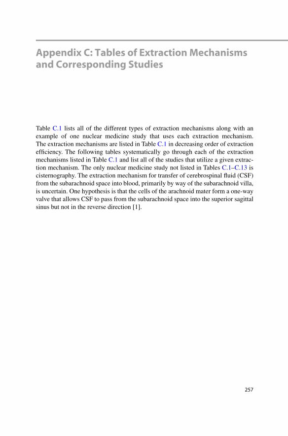
Appendix C: Tables of Extraction Mechanisms and Corresponding Studies
Table C.1 lists all of the different types of extraction mechanisms along with an example of one nuclear medicine study that uses each extraction mechanism. The extraction mechanisms are listed in Table C.1 in decreasing order of extraction effi ciency. The following tables systematically go through each of the extraction mechanisms listed in Table C.1 and list all of the studies that utilize a given extrac-tion mechanism. The only nuclear medicine study not listed in Tables C.1 – C.13 is cisternography. The extraction mechanism for transfer of cerebrospinal fl uid (CSF) from the subarachnoid space into blood, primarily by way of the subarachnoid villa, is uncertain. One hypothesis is that the cells of the arachnoid mater form a one-way valve that allows CSF to pass from the subarachnoid space into the superior sagittal sinus but not in the reverse direction [1].

258
Tab
le C
.1
All
radi
opha
rmac
eutic
al e
xtra
ctio
n m
echa
nism
s fr
om b
lood
Ext
ract
ion
mec
hani
sm
cate
gory
E
xam
ple
stud
y R
adio
phar
mac
eutic
al
Spec
ifi c
extr
actio
n m
echa
nism
E
xtra
ctio
n ef
fi cie
ncy
Satu
rabl
e
Em
boliz
atio
n Pu
lmon
ary
Perf
usio
n St
udy
Tc-
99m
-mac
roag
greg
ated
al
bum
in
Mic
roem
boliz
atio
n 10
0 %
N
o
Phag
ocyt
osis
L
iver
-Spl
een
Stud
y T
c-99
m-s
ulfu
r co
lloid
Ph
agoc
ytos
is b
y si
nuso
idal
m
acro
phag
es
Hig
h N
o
Pass
ive
diff
usio
n th
roug
h ce
ll w
all
Myo
card
ial P
erfu
sion
N
-13-
amm
onia
L
ipop
hilic
ity
Hig
h N
o
Che
mis
orpt
ion
Bon
e M
iner
al S
tudy
T
c-99
m-M
DP/
HM
DP
Adh
eren
ce o
f PO
4 tra
cers
to
surf
ace
of th
e bo
ne
Mod
erat
e N
o
Ion
exch
ange
B
one
Min
eral
Stu
dy
F-18
as
sodi
um fl
uori
de
Exc
hang
e of
F − f
or O
H − in
hy
drox
yapa
tite
Mod
erat
e N
o
Filtr
atio
n R
enal
Glo
mer
ular
Fi
ltrat
ion
Stud
y T
c-99
m-D
TPA
Pa
ssiv
e fi l
trat
ion
base
d on
si
ze
20 %
N
o
Faci
litat
ed tr
ansp
ort
Tum
or G
luco
se
Met
abol
ism
Stu
dy
F18-
fl uor
odeo
xygl
ucos
e G
LU
T g
luco
se tr
ansp
orte
rs
Mod
erat
e M
aybe
Act
ive
tran
spor
t – c
lass
of
mol
ecul
es
Hep
atob
iliar
y St
udy
Tc-
99m
-tri
met
hylb
rom
o-ID
A
Org
anic
ion
tran
spor
t sys
tem
M
oder
ate
Yes
Act
ive
tran
spor
t –
rela
tivel
y sp
ecifi
c T
hyro
id I
mag
ing
Stud
y I-
123
Na +
/I − s
ympo
rter
(N
IS)
prot
ein
Mod
erat
e Y
es
Cel
l mig
ratio
n W
hite
Blo
od C
ell
Mig
ratio
n St
udy
Tc-
99m
-whi
te b
lood
cel
ls
Mig
ratio
n se
cond
ary
to
chem
otax
is
Low
N
o
Rec
epto
r bi
ndin
g (n
o en
ergy
inpu
t)
Som
atos
tatin
Rec
epto
r St
udy
In-1
11-p
ente
trio
tide
“Loc
k an
d ke
y” fi
t L
ow
Yes
Epi
tope
bin
ding
B
-Cel
l Lym
phom
a Im
agin
g St
udy
In-1
11-i
britu
mom
ab ti
uxet
an
[Zev
alin
] “L
ock
and
key”
fi t
Low
Y
es
Appendix C: Tables of Extraction Mechanisms and Corresponding Studies

259
Table C.2 Radiopharmaceutical extraction mechanisms: intravascular microembolization
Study Radiopharmaceutical Size Specifi c extraction mechanism
Extraction effi ciency Saturable
Gastrointestinal system
Hepatic Artery Perfusion Study
Tc-99m- macroaggregated albumin
10–80 μm Microembolization into arterioles
Near 100 %
No
Pulmonary system
Pulmonary Perfusion Study
Tc-99m- macroaggregated albumin
10–80 μm Microembolization into arterioles
Near 100 %
No
Table C.3 Radiopharmaceutical extraction mechanisms: fi ltration
Study Radiopharmaceutical Size Specifi c extraction mechanism
Extraction effi ciency Saturable
Genitourinary system
Renal Glomerular Filtration Study
Tc-99m-DTPA 482.31 Renal glomerular fi ltration
20 % No
Table C.4 Radiopharmaceutical extraction mechanisms: phagocytosis
Study Radiopharmaceutical Size
Specifi c extraction mechanism
Extraction effi ciency Saturable
Cardiovascular system
Lymphangiography Filtered Tc-99m- sulfur colloid
0.04–0.22 μm
Phagocytosis by sinusoidal macrophages
High No
Gastrointestinal system
Liver-Spleen Study Tc-99m-sulfur colloid
0.4–0.8 μm
Phagocytosis by sinusoidal macrophages
High No
Table C.5 Radiopharmaceutical extraction mechanisms: chemisorption
Study Radiopharmaceutical Size Extraction mechanism
Extraction effi ciency Saturable
Skeletal system
Bone Mineral Study
Tc-99m-MDP/HMDP 274.92 Adherence of PO 4 tracers to surface of the bone
Moderate No
Appendix C: Tables of Extraction Mechanisms and Corresponding Studies

260
Table C.6 Radiopharmaceutical extraction mechanisms: ion exchange
Study Radiopharmaceutical Size Specifi c extraction mechanism
Extraction effi ciency Saturable
Skeletal system
Bone Mineral Study
F-18 as sodium fl uoride
19.00 Exchange of F − for OH − in hydroxyapatite
Moderate No
Table C.7 Radiopharmaceutical extraction mechanisms: passive diffusion
Study Radiopharmaceutical Size Extraction mechanism
Extraction effi ciency Saturable
Cardiovascular system
Myocardial Perfusion Study
N-13-ammonia 17.0 Lipophilicity High No
Myocardial Perfusion Study
Tc-99m-sestamibi 777.7 Lipophilicity Moderate, about 38 %
No
Central nervous system
Brain Perfusion Study
Tc-99m-HMPAO 384.3 Lipophilicity Moderate No
Brain Perfusion Study
Tc-99m-ECD 313.5 Lipophilicity Moderate No
Endocrine system
Parathyroid Study
Tc-99m-sestamibi 777.7 Lipophilicity Moderate, about 38 %??
No
Table C.8 Radiopharmaceutical extraction mechanisms: white blood cell migration
Study Radiopharmaceutical Size Extraction mechanism
Extraction effi ciency Saturable
Infection imaging
White Blood Cell Migration Study
Tc-99m-white blood cells
12–15 μm diameter
Migration secondary to chemotaxis
Low No
Appendix C: Tables of Extraction Mechanisms and Corresponding Studies

261
Table C.9 Radiopharmaceutical extraction mechanisms: facilitative transport
Study Radiopharmaceutical Size Extraction mechanism
Extraction effi ciency Saturable
Cardiovascular system
Myocardial Viability Study
F18-fl uorodeoxyglucose 166.17 GLUT glucose transporters
Low–moderate
Maybe
Central nervous system
Brain Glucose Metabolism Study
F18-fl uorodeoxyglucose 166.17 GLUT glucose transporters
Low–moderate
Maybe
Infection imaging
White Blood Cell Activation Study
F18-fl uorodeoxyglucose 166.17 GLUT glucose transporters
Low–moderate
Maybe
Tumor imaging
Tumor Glucose Metabolism Study
F18-fl uorodeoxyglucose 166.17 GLUT glucose transporters
Low–moderate
Maybe
Appendix C: Tables of Extraction Mechanisms and Corresponding Studies

262
Tab
le C
.10
R
adio
phar
mac
eutic
al e
xtra
ctio
n m
echa
nism
s: a
ctiv
e tr
ansp
ort –
cla
ss o
f m
olec
ules
Stud
y R
adio
phar
mac
eutic
al
Size
E
xtra
ctio
n m
echa
nism
E
xtra
ctio
n ef
fi cie
ncy
Satu
rabl
e
Car
diov
ascu
lar
syst
em
Myo
card
ial P
erfu
sion
St
udy
Rb-
82-r
ubid
ium
chl
orid
e 85
.47
Na +
/K + p
ump
Hig
h N
o
Myo
card
ial P
erfu
sion
and
V
iabi
lity
Stud
y T
l-20
1-th
allo
us c
hlor
ide
204.
38
Na +
/K + p
ump
Hig
h N
o
Cen
tral
ner
vous
sys
tem
Stri
atal
Dop
amin
e T
rans
port
er S
tudy
I-
123-
iofl u
pane
[D
aTsc
an®]
407.
24
Dop
amin
e tr
ansp
orte
r re
cept
ors
Mod
erat
e Y
es
End
ocri
ne s
yste
m
Neu
roec
tode
rmal
Im
agin
g st
udy
I-12
3-M
IBG
19
3.17
N
orep
inep
hrin
e m
onoa
min
e ne
urot
rans
mitt
ers
Mild
Y
es
Thy
roid
Im
agin
g St
udy
I-12
3 12
2.90
N
a + /I
− s
ympo
rter
(N
IS)
prot
ein
Mod
erat
e Y
es
Thy
roid
Met
asta
sis
I-12
3, I
-131
12
2.90
N
a + /I
− s
ympo
rter
(N
IS)
prot
ein
Mod
erat
e Y
es
Thy
roid
Upt
ake
Mea
sure
men
t I-
123
122.
90
Na +
/I − s
ympo
rter
(N
IS)
prot
ein
Mod
erat
e Y
es
Gas
troi
ntes
tina
l sys
tem
Hep
atob
iliar
y St
udy
Tc-
99m
- tri
met
hylb
rom
o- ID
A
328.
28
Org
anic
ion
tran
spor
t sys
tem
M
oder
ate
Yes
Mec
kel’s
Div
ertic
ulum
St
udy
Tc-
99m
- per
tech
neta
te
162.
91
H + /K
+ A
TPa
se p
roto
n pu
mp
Mod
erat
e N
o
Gen
itou
rina
ry s
yste
m
Ren
al T
ubul
ar F
unct
ion
Tc-
99m
-DM
SA
281.
13
Poss
ibly
fi ltr
atio
n an
d tu
bula
r re
abso
rptio
n 5
%
No
Ren
al T
ubul
ar S
ecre
tion
Stud
y T
c-99
m-M
AG
3 24
2.19
O
rgan
ic a
nion
tran
spor
ter
1 ~6
5 %
N
o
Appendix C: Tables of Extraction Mechanisms and Corresponding Studies
263
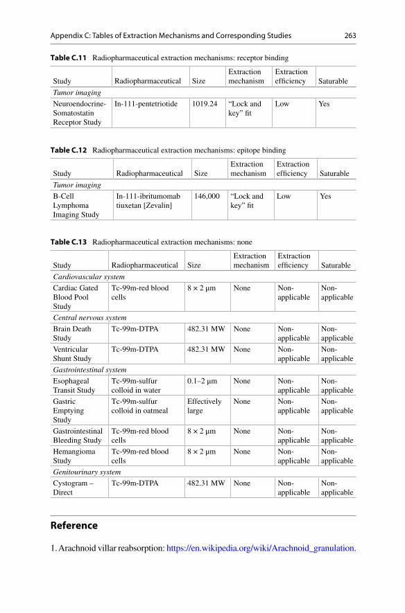
Table C.11 Radiopharmaceutical extraction mechanisms: receptor binding
Study Radiopharmaceutical Size Extraction mechanism
Extraction effi ciency Saturable
Tumor imaging
Neuroendocrine- Somatostatin Receptor Study
In-111-pentetriotide 1019.24 “Lock and key” fi t
Low Yes
Table C.12 Radiopharmaceutical extraction mechanisms: epitope binding
Study Radiopharmaceutical Size Extraction mechanism
Extraction effi ciency Saturable
Tumor imaging
B-Cell Lymphoma Imaging Study
In-111-ibritumomab tiuxetan [Zevalin]
146,000 “Lock and key” fi t
Low Yes
Table C.13 Radiopharmaceutical extraction mechanisms: none
Study Radiopharmaceutical Size Extraction mechanism
Extraction effi ciency Saturable
Cardiovascular system
Cardiac Gated Blood Pool Study
Tc-99m-red blood cells
8 × 2 μm None Non- applicable
Non- applicable
Central nervous system
Brain Death Study
Tc-99m-DTPA 482.31 MW None Non- applicable
Non- applicable
Ventricular Shunt Study
Tc-99m-DTPA 482.31 MW None Non- applicable
Non- applicable
Gastrointestinal system
Esophageal Transit Study
Tc-99m-sulfur colloid in water
0.1–2 μm None Non- applicable
Non- applicable
Gastric Emptying Study
Tc-99m-sulfur colloid in oatmeal
Effectively large
None Non- applicable
Non- applicable
Gastrointestinal Bleeding Study
Tc-99m-red blood cells
8 × 2 μm None Non- applicable
Non- applicable
Hemangioma Study
Tc-99m-red blood cells
8 × 2 μm None Non- applicable
Non- applicable
Genitourinary system
Cystogram – Direct
Tc-99m-DTPA 482.31 MW None Non- applicable
Non- applicable
Reference
1. Arachnoid villar reabsorption: https://en.wikipedia.org/wiki/Arachnoid_granulation .
Appendix C: Tables of Extraction Mechanisms and Corresponding Studies

265
Appendix D: Distribution of Cardiac Output in the Body at Rest
The success of both diagnostic and therapeutic procedures is fundamentally depen-dent on the optimal biodistribution of radiopharmaceuticals. The biodistribution of most, but not all, radiopharmaceuticals depends primarily on clearance of radio-pharmaceuticals from the blood into organs, tissues, or lesions. In turn, clearance of radiopharmaceuticals depends on four factors: (1) blood fl ow, (2) extraction effi -ciency, (3) the presence of unusual amounts of competing substances, and (4) the amount of radiopharmaceutical available to be cleared from the blood from the time of injection to the end of image acquisition or therapeutic irradiation (see Chap. 4 : Evaluation of Clearance).
Table D.1 below lists the amount of cardiac output that passes to each of the major organs of the body. In the case of many diagnostic nuclear medicine studies, the relevant blood fl ow is to a whole organ, e.g., Renal Tubular Secretion Study with Tc-99m-MAG3 and Myocardial Perfusion Study with Tc-99m-sestamibi. On the other hand, there are a number of studies in which the radiopharmaceutical localizes or clears into lesions rather whole organs, e.g., Tumor Glucose Metabolism Study with F-18-fl uorodeoxyglucose and White Blood Cell Activation Study with F-18- fl uorodeoxyglucose. In these studies the increased clearance of radiopharmaceuti-cal in the lesions is often due, at least in part, to increased blood fl ow, but the amount of increase is hard to determine.
Table D.1 Distribution of blood in the body at rest
Organ Percent of cardiac output (%)
Flow/organ (L/min)
Flow/100 g (mL/min-100 g)
Lungs 100 5.0 –
Brain 14 0.70 55
Heart 4 0.20 70
Liver and GI tract 27 1.35 100
Kidneys 20 1.00 400
Skeletal muscle 21 1.05 5
Skin 5 0.25 10
Bone and others 9 0.45 3
Adapted from Pearson Education, Inc., 2013

267© Springer International Publishing Switzerland 2016W.C. Klingensmith III, The Mathematics and Biology of the Biodistribution of Radiopharmaceuticals-A Clinical Perspective, DOI 10.1007/978-3-319-26704-3
A Arteriovenous malformation (AVM) , 42T, 45, 231
lung AVM , 45T Attenuation correction , 24T, 90T, 147, 150,
152, 153, 197 PET , 21 quantitative measurement of relative
function , 91–94 in relative measurements , 22T, 31, 90T,
91–94, 214, 220 SPECT , 21, 91, 94, 153 tomography , 22T, 24T, 101T
Aunt Minnie phenomenon , 16–17, 29
B Background correction , 21, 103, 141, 147,
150, 152, 153, 158, 164, 193, 197, 214, 220, 225, 235, 246
ROI placement , 142F quantitation, absolute , 103, 106F quantitation, relative , 95–96 tomography , 95
B-Cell Lymphoma Imaging Study (In-111-ibritumomab tiuxetan [Zevalin®]) , 239–250
chemical structure, In-111-ibritumomab tiuxetan , 242F
extraction mechanism , 240–242, 244, 245
protocol summary diagram , 241F, 247F Biodistribution of radiopharmaceuticals , vii,
ix, 3, 20, 29–32, 37–43, 48, 129, 136, 210, 265
Blood concentration of radiopharmaceutical embolus , 42T, 43
Blood fl ow . See also First circulation time-activity curves
clearance as proxy for , 35T, 167F clearance equation , viii, 38, 40T, 44,
45, 116, 118 coronary blood fl ow , 33, 36F, 145, 146F, 152 defi nition , 61–62 vs. perfusion , 62, 145, 146, 150, 151, 153, 166
Body surface area (BSA) , 31–32 creatinine clearance , 114–117 general equation for need to normalize for
BSA , 113–119, 115T GLOFIL® Study (I-125-iothalamate) ,
120–122 gram based (SUV) clearance , 119–120 organ based clearance , 117–119
Bolus shape , 68F, 76, 78F Bone Mineral Study (F-18 as sodium fl uoride) ,
235–236 extraction mechanism , 235, 236F protocol summary diagram , 236
Bone Mineral Study (Tc-99m-MDP) , 237–238 chemical structure, Tc-99m-MDP , 237F extraction mechanism , 237 protocol summary diagram , 238F
Brain Death Study (Tc-99m-DTPA) , 161–162 blood fl ow, fi rst circulation , 61–62 chemical structure, Tc-99-DTPA , 162F extraction mechanism , 161 protocol summary diagram , 162F
Brain Glucose Metabolism Study (F-18-fl uorodeoxyglucose) , 162–165
chemical structure, F-18-fl uorodeoxyglucose , 162, 163F
extraction mechanism , 162, 163F, 164 protocol summary diagram , 165F
Index
Note: Page numbers with an F refer to a fi gure; page numbers with a T refer to a table.

268
Brain Perfusion Study (Tc-99m-HMPAO) , 166–167
chemical structure, Tc-99m-HMPAO , 166, 166F
extraction mechanism , 166, 167F protocol summary diagram , 167F
C Cardiac Gated Blood Pool Study (Tc-99m-red
blood cells) , 139–142 background subtraction ROI placement ,
142F central volume principle , 57T, 58 leading edge transit time , 20, 21, 20T,
58, 141 left ventricular ejection fraction: normal
values , 141–142, 143T left ventricular failure , 21 protocol summary diagram , 141 pulmonary transit time: normal values ,
140–141 Central volume principle , 51–59
clinical applications of , 56–59, 57T, 58F conceptual analysis of , 52–54, 53F, 54F delta function , 52, 53F diagram of , 52F frequency distribution of transit times ,
55, 56F instantaneous/impulse injection ,
52, 53F, 55 laminar fl ow , 52, 53F, 55 leading edge transit time , 57T, 58–59 mathematical analysis , 52, 55–56 mean transit time , 51–59, 54F plug fl ow , 52, 53F, 54F studies that involve , 57T variants of , 56, 57T
Cisternography (In-111-DTPA) , 167–170 central volume principle , 57T, 58 chemical structure, In-111-DTPA ,
167, 168F CSF rhinorrhea , 113, 114T, 124, 170 CSF rhinorrhea equation , 124, 170 extraction mechanism , 168, 169F leading edge transit time , 20–21, 58 leading edge transit time: normal , 57T pledget to plasma ratio: normal , 124, 170 protocol summary diagram , 169F
Clearance clearance rate , 39, 40T, 43, 45, 114, 115,
117, 120, 122, 132, 158, 219 of creatinine , 43, 44, 114–117, 115T defi nition , 33, 38, 103 importance of, 33, 103
proxy for blood fl ow , 35T, 147F, 150F, 152F, 154F, 192, 193F
vs. perfusion , 33, 36F Clearance equation
amount vs. rate , 43 analysis of , 40T clinical applications , 45–48, 45T conceptual use 47 conditions that affect , 42T evaluation of biodistribution of
radiopharmaceuticals , 38–43 mathematical analysis , 38–45 non-saturable extraction mechanism ,
39, 42T relative clearance , 39–40 saturable extraction mechanism , 39, 41T simplifi ed relative clearance
equation , 40 Clinical examples, conditions & correlation , 45T
acute tubular necrosis (ATN) , 217–218, 218F
CT intravenous contrast material , 45T, 46 glomerulonephritis, chronic , 86 Graves disease , 5, 5F, 45T, 46 hyperglycemia, 42T,-47 infection , 45, 45T injection embolus , 45T, 46, 47F low iodine diet , 42T, 45T, 46 lung AVM , 45T lung cancer , 45T, 46, 48F, 165F, 247F left ventricular failure , 21 subacute viral thyroiditis , 45T, 46F, 46 tuberculoma , viii
Compartmental analysis biodistribution of radiopharmaceuticals ,
30, 37 limitations of , 30, 37
Competing substances clearance equation , 38–39 overall extraction effi ciency , 39
Convolution analysis , 75–88 blood fl ow , 75, 76T, 77, 84, 85F central volume principle , 84 clinical applications , 84–88 conceptual analysis , 76–83 excretory pathway , 76T, 77, 84 frequency distribution of transit times ,
79F, 80F mathematical analysis , 83–84 tabular analysis , 81T, 82T
Coronary blood fl ow , 33, 36F, 145, 146F, 152 Creatinine clearance test , 114–117
compared to nuclear medicine clearance , 43
equation , 43–44, 116–117
Index

269
Cross talk , ix, 30 CT contrast material
competes with radioiodine , 41, 42T Cystogram-Direct (Tc-99m-DTPA) ,
123–124, 203–204 chemical structure , 204, 204F extraction mechanism , 203–205, 208 protocol summary diagram , 204F, 207F residual bladder volume , 123, 123F,
203–204 residual bladder volume equation , 123
D de Hevesy, George
radiotracer principle , 4, 4T
E Einstein, Albert
comment on quantitation , 17 Embolus
from injection site , 42T Esophageal Transit Study (Tc-99m-sulfur
colloid in water) , 187–188 extraction mechanism , 187 geometric mean , 188 protocol summary diagram , 188F quantitation of transit time: normal range ,
187–188, 188F Extraction effi ciency (EE) . See also Extraction
mechanism clearance equation , 38–42, 40T, 44–47 conditions that affect , 42T net extraction effi ciency , 38, 40 overall extraction effi ciency , 39, 40T, 44 saturation of , 38–39, 129
Extraction mechanism . See also Extraction effi ciency (EE)
clearance equation , 38, 39, 41, 41T, 42T, 44
net extraction effi ciency , 38, 40 overall extraction effi ciency , 39, 44 saturation of , 38, 39, 41, 135 studies by extraction mechanism
(Appendix C) , 257, 258-263T types of extraction mechanisms ,
131T, 257, 258T Eye-brain complex , 16–18, 16F, 89, 249
digital vs. visual , 18–19, 19F image distortion , 17–18
Abraham Lincoln , 17, 18F CAPTCHA , 17, 18F
Watson, James D., complexity of the brain , 17
F F-18 as sodium fl uoride
Bone Mineral Study , 235–236, 252, 253 F-18-fl uorodeoxyglucose (FDG)
Brain Glucose Metabolism Study , 34T, 41T, 162–165, 251, 261T
myocardial uptake , 155, 156F, 157F Myocardial Viability Study , 22T, 34T, 41T,
90T, 154–158, 261T Preface , viii suboptimal tracer can be optimal
radiopharmaceutical , 10–11, 11F Tumor Glucose Metabolism Study , 24T,
35T, 41T, 100, 100T, 101T, 107, 115T, 119, 131T, 244–249, 252, 253, 258T, 261T, 265
White Blood Cell Activation Study , 35T, 42T, 223–226, 252, 253, 261, 265
First circulation time-activity curves , 61–73 blood fl ow , 61–73 clinical experience with , 72 CT fi rst circulation time-indicator curves ,
61, 72 derivation of equation for regional blood
fl ow , 62–70 fi rst moment time , 67 indices of , 71–72, 70F, 72F interpretation of , 70F MR fi rst circulation time-indicator
curves , 61 studies with , 62T
G Galileo Galilei
importance of quantitation , 15 Gamma function
fi rst circulation time-activity curves , 63F recirculation , 63F
Gastric Emptying Study (Tc-99m-sulfur colloid in instant oatmeal) , 40, 93, 95, 189–190, 252, 263T
extraction mechanism (Tc-99m-sulfur colloid in instant oatmeal) , 189
Gastric Emptying Study Worksheet , 97–98 geometric mean , 93–94 protocol summary diagram , 190F quantitation of gastric emptying: normal
ranges , 191T Gastrointestinal Bleeding Study (Tc-99m-RBCs) ,
37T, 40, 190–191, 252, 255, 263 extraction mechanism, Tc-99m-RBCs , 191 protocol summary diagram , 192F
Geometric mean , 22T, 90T, 91, 93–94, 188, 189, 193, 220
Index
270
Geometry of fi eld of view , 91 GLOFIL® (I-125-iothalamate) , 31, 113, 114T,
115T, 120–122 . See also Body surface area (BSA)
GLOFIL® Renal Filtration Clearance Study , 121T, 122
GLOFOL® equation , 122 Glucose transporter protein (GLUT)
diagram , viii, 156-157F, 163-164F, 224-225F, 245-246F
suboptimal tracer can be optimal radiopharmaceutical , 10–11, 11F
H Hepatic Artery Perfusion Study (Tc-99m-
macroaggregated albumin) , 22T, 35T, 42, 62, 90, 91, 191–194, 252, 254
extraction mechanism , 192 geometric mean , 91 protocol summary diagram , 192F, 193F quantitation of hepatic shunts:
implications , 192–194, 193F Hepatic Hemangioma Study (Tc-99m-red
blood cells) , 194, 252 extraction mechanism , 194 protocol summary diagram , 194
Hepatobiliary Study (Tc-99m-trimethylbromo-IDA) , 20T, 21, 22T, 34T, 36T, 57T, 76T, 90T, 91, 131T, 195–197
background correction , 197 chemical structure , 195F extraction mechanism , 195, 196F gallbladder ejection fraction
equation , 197 leading edge transit time , 20T, 21, 196 protocol summary diagram , 196F quantitation of gallbladder ejection
fraction: normal range , 195, 197
I I-123 as sodium iodine
Parathyroid study , 178–180 Thyroid Imaging & Uptake Study ,
180–183 Thyroid Metastases Study , 183–184
I-123-iodobenzylguanidine (I-123-MIBG) Neuroectodermal/Norepinephrine study ,
177–178 I-123-iofl upane [DaTscan®]
Striatal Dopamine Transporter Study , 12, 41T, 170–172, 262T
I-131 as sodium iodine Thyroid Metastases Study , 183–184 Image interpretation digital vs. visual ,
18-19F image distortion , 17, 18F
Image reconstruction , 25T, 30 Imaging, medical
attributes that make it useful , 5–6, 5F In-111-DTPA
Cisternography , 20T, 57T, 114T, 124, 167–170
leading edge transit time , 20T, 21 In-111-ibritumomab tiuxetan [Zevalin®]
B-Cell Lymphoma Imaging Study , 239–241, 253, 258T, 263T
In-111-oxyquinoline White Blood Cell Migration
Study , 254 In-111-pentetreotide
Neuroendocrine Tumor-Somatostatin Receptor Study,131T , 241–244, 254, 263
Infection , 13T, 33, 35T, 45, 45T, 130T, 135, 223–228, 252, 260T, 261T
tuberculoma , viii Iodine, non-radioactive . See also CT contrast
material competes with radioiodine , 42T low iodine diet , 42T
L Laminar fl ow
central volume principle , 52, 53F convolution analysis , 76, 77F, 81 fi rst circulation time-activity curves ,
63F, 65, 66, 71 leading edge transit time vs. mean transit
time , 20 vs. plug fl ow , 53F
Leading edge transit time central volume principle , 20, 21, 57T, 58,
59, 170 vs. mean transit time , 20, 30 studies that include , 20T
Liver-Spleen Study , 131T, 135, 197–199, 258, 259
extraction mechanism , 197–198, 198F
protocol summary diagram , 199F
Index

271
Lord Kelvin importance of quantitation , 15
Lung Aerosol Study (Tc-99m-DTPA) , 35–36, 229–231
clearance as proxy for ventilation , 230T extraction mechanism , 229 protocol summary diagram , 231 radiopharmaceutical size , 230F
Lung Perfusion Study (Tc-99m-macroaggregated albumin) , 230–232
clearance as proxy for blood fl ow , 232F extraction mechanism , 230–232 protocol summary diagram , 232F radiopharmaceutical size , 230, 231F
Lung Ventilation Study (Xe-133 gas) , 35–36, 37T, 230T, 232–233
clearance as proxy for ventilation , 230T extraction mechanism , 232 protocol summary diagram , 233
Lymphoscintigraphy (fi ltered Tc-99m-sulfur colloid) , 35, 37T, 40, 132, 142–145, 251, 255
extraction mechanism , 143, 144F overview , 144F protocol summary diagram , 145
M Mean transit time . See also Central volume
principle vs. leading edge transit time , 20, 30
Meckel’s Diverticulum Study (Tc-99m-pertechnetate) , 199–200, 252, 254
chemical structure, Tc-99m-pertechnetate , 200F
extraction mechanism , 199 protocol summary diagram , 200F
Modulation transfer function , 30 Molecular biology
PET , 7–8, 8F, 8T Myocardial Perfusion Study (N-13-ammonia) ,
22T, 34T, 35T, 90T, 143T, 145–148 clearance vs. blood fl ow , 146F extraction mechanism , 145, 146F left ventricular ejection fraction: normal
values , 143T, 147 protocol summary diagram , 147F quantitative measurement: myocardial
perfusion (clearance) , 147, 148F Myocardial Perfusion Study (Rb-82 chloride) ,
22T, 35T, 42T, 90T, 148–150
clearance vs. blood fl ow , 146F extraction mechanism , 149, 149F left ventricular ejection fraction: normal
values , 143T, 148, 150 protocol summary diagram , 150F quantitative measurement: myocardial
perfusion (clearance) , 150 Myocardial Perfusion Study
(Tc-99m-sestamibi) , 150–152 chemical structure, Tc-99m-sestamibi ,
151F clearance vs. blood fl ow , 146F extraction mechanism , 151 left ventricular ejection fraction: normal
values , 143T, 152 protocol summary diagram , 152F quantitative measurement:
myocardial perfusion (clearance) , 152
Myocardial Perfusion & Viability Study (Tl-201-thallous chloride) , 34T, 153–154, 251, 262T
clearance vs. blood fl ow , 146F extraction mechanism , 153 left ventricular ejection fraction: normal
values , 143T, 154 protocol summary diagram , 154F quantitative measurement: myocardial
perfusion (clearance) , 154 Myocardial uptake (clearance)
proxy for blood fl ow , 36F viability marker , 36F
Myocardial Viability Study (F-18 fl uorodeoxyglucose) , 34T, 41T, 154–158, 251
chemical structure, F-18 fl uorodeoxyglucose , 155F
extraction mechanism , 155, 156F, 157F
left ventricular ejection fraction: normal values , 143T, 158
protocol summary diagram , 158F quantitative measurement: myocardial
viability (clearance) , 158
N N-13-ammonia
Myocardial Perfusion Study , 22T, 34T, 35T, 42T, 90T, 143T, 145–148, 251, 254, 260T
myocardial uptake , 36F
Index

272
Necrosis blood fl ow , 42T
Neuroectodermal/Norepinephrine Study (I-123-MIBG) , 177–178
chemical structure, I-123-MIBG , 178F extraction mechanism , 177, 178F protocol summary diagram , 178F
Neuroendocrine Tumor-Somatostatin Receptor Study (In-111-pentetreotide [Octreotide®]) , 241–244
chemical structure, In-111-pentetreotide , 241–242, 242F, 243F
extraction mechanism , 242, 243T protocol summary diagram , 243F
Nuclear medicine anatomy vs. function , 6F Nobel Prizes , 4T place within the realm of science , 3–4, 4F role in diagnostic imaging , 5, 6F scientifi c discoveries underlying nuclear
medicine , 4, 4T scope of practice , 12–13, 13T why it can image function , 5–6, 6T, 6F
Nuclear Medicine Procedure Manual , 32, 33, 40, 129, 251
Nuclear medicine studies covered in this book (Appendix A) , 251–252
O O-15 water
ideal blood fl ow tracer , 36F
P Parathyroid Study (I-123 &
Tc-99m-sestamibi) , 178–180 chemical structure, Tc-99m-sestamibi , 179F extraction mechanism , 179 protocol summary diagram , 180F
Perfusion defi nition , 62 proxy for blood fl ow , 35, 147F, 192, 193F vs. blood fl ow , 62, 145, 151, 153, 166
Plug fl ow , 52, 53F, 54F, 65, 77F Pool of atoms, molecules, or substances ,
38–39 actual , 39–40 clearance equation , 38–39 normal , 39–40
Positron emission tomography (PET) conferred resolution , 9–10, 10F detection resolution , 9–10, 10F imaging component of molecular
medicine , 11–12, 12F
importance in nuclear medicine , 6–7, 7F molecular biology vs. physiology , 7, 8F, 8T most powerful imaging machine , 9, 9F spatial resolution, types of , 8–10, 10F
Protein interaction map , 11–12, 12F Protocol design diagrams
defi nition of symbols , 135, 136F
Q Quantitation . See also Aunt Minnie phenomenon
Albert Einstein , 17 as a consultant , 26 Galileo Galilei , 15 Lord Kelvin , 15 quantitative vs. visual image
interpretation , 15 radiologist with a ruler… , 17 Rhind mathematical papyrus , 15
Quantitation of function: absolute measurements
attenuation correction , 101T, 106 background correction , 101T, 106F overview , 23, 99–101 renal clearance , 103–107, 104F, 105F renal tubular clearance (% Uptake)
worksheet , 109–110 studies with , 24T, 100T SUV equation , 108 thyroid uptake equation , 103 thyroid uptake worksheet , 111 tumor clearance , 107–109 vs. relative measurements , 99
Quantitation of function: relative measurements
attenuation correction , 92–94, 92F, 93F background correction , 95–96, 95F, 96F comparison to normal range , 94 gastric emptying worksheet , 97–98 geometric mean , 93–94 overview , 91 relative function equation , 96–97 studies with , 22T vs. absolute measurement , 89
R Radioactive decay , 30, 97 Radiopharmaceuticals
importance of an optimal radiopharmaceutical , 130, 132
radiopharmaceuticals and their use in clinical studies (Appendix B) , 253–255
suboptimal tracer can be an optimal radiopharmaceutical , 10–11, 11F
Index

273
Radiotracer principle George de Hevesy , 4T
Rb-82 as rubidium chloride Myocardial Perfusion study , 22T, 34T,
35T, 90T, 251, 254 myocardial uptake , 36F, 146F
Region of interest (ROI) generating numbers , 19 quality control , 20
Regional blood fl ow , 61–73 . See also First circulation time-activity curves
Appendix D , 265T Renal Glomerular Filtration Study
(Tc-99m-DTPA) , 204–207 chemical structure,
Tc-99m-DTPA , 205 compared to creatinine clearance ,
43–45 extraction mechanism , 204, 205F, 206F leading edge transit time , 20T, 21, 58, 59 protocol summary diagram , 207F quantitation ( See Renal Tubular Excretion
Study (Tc-99m-MAG3)) Renal Tubular Excretion Study
(Tc-99m-MAG3), 42F, 62F, 84, 90, 100, 101F, 115F, 252, 254 208–219
acute tubular necrosis (ATN) , 217–218, 218F
blood fl ow , 212T, 213, 213F chemical structure, Tc-99m-MAG3 ,
208, 208F clearance , 212T, 213–214, 213F clinical considerations ,
217–219, 217T, 219T convolution analysis , 84–88, 85F, 87F excretory system , 212T, 214F, 216, 217F extraction mechanism , 208, 209F fi rst circulation time-activity curves , 70F leading edge transit time , 20T, 21 nephron unit theory , 217 parenchymal transit , 212T, 214–216, 214F,
215F, 216F protocol summary diagram , 210F, 211F quantitation of functional parameters:
normal ranges , 215T, 215–216 quantitation, general , 215T, 215–216 Tc-99m-MAG3 Renal Worksheet , 221
Renal Tubular Function Study (Tc-99m-DMSA) , 22, 34, 90, 219–220
chemical structure, Tc-99m-DMSA , 220F extraction mechanism , 219 protocol summary diagram , 220F quantitation of relative clearance:
normal , 220
S Standard uptake value (SUV)
errors in , 23–26, 25T, 248–249, 248T mathematical analysis of , 107–109 quantitation, absolute , 100T, 101T
Stewart-Hamilton equation , 64–65, 76 cardiac output , 64 regional blood fl ow , 64
Striatal Dopamine Transporter Study (I-123-iofl upane [DaTscan®]) , 12, 41, 170–172
chemical structure, I-123-iofl upane , 171F extraction mechanism , 171, 171F protocol summary diagram , 172F
Subramanian, Manny importance of a good
radiopharmaceutical , 130
T Tc-99m-dimercaptosuccinic acid
(Tc-99m-DMSA) Renal Tubular Function Study ,
219–220 Tc-99m-DTPA
Brain Death Study , 37T, 61, 75, 161–162, 263T
clearance of , 35, 35T Cystogram-Direct , 203 leading edge transit time , 20T, 21 Lung Aerosol Ventilation Study ,
35, 40, 229, 252, 254 Renal Glomerular Filtration Study ,
20, 21, 22T, 24T, 34T, 36T, 42T, 43, 57T, 59, 62T, 75, 76T, 90, 96, 100, 101T, 115, 117, 131T, 204–207
Ventricular Shunt Study , 172 Tc-99m-examatazine (Tc-99m-HMPAO)
Brain Perfusion Study , 34T, 35T, 42T, 62, 166–167
White Blood Cell Study , 226–228
Tc-99m-trimethylbromo-iminodiacetic acid Hepatobiliary Study , 21, 22T, 34T, 36T,
57T, 75, 76, 76T, 90, 91, 131T, 195–197, 252, 254, 258, 263
Tc-99m-macroaggregated albumin (Tc-99m-MAA)
Hepatic Artery Perfusion Study , 22T, 35T, 62, 90T, 191–194, 252, 254
Tc-99m-mercaptoacetyltriglycine (Tc-99m-MAG3)
protein binding , 38 Renal Tubular Excretion Study , 208
Index

274
Tc-99m-methylenediphophonate (Tc-99m-MDP)
Bone Mineral Study , 237–238, 252, 254, 259T
Tc-99m-pertechnetate Meckel’s Diverticulum Study ,
199–200, 252, 254 Tc-99m-red blood cells (Tc-99m-RBCs)
Cardiac Gated Blood Pool Study , 20T, 21, 22T, 37T, 40, 57T, 58, 90T, 91, 132, 139–142, 251, 255
blood fl ow , 61 Gastrointestinal Bleeding Study ,
37T, 40, 190–191, 252, 255 Hepatic Hemangioma Study , 190, 194, 252 leading edge transit time , 20T, 21
Tc-99m-sestamibi Myocardial Perfusion Study , 42T, 89, 90T,
94, 143T, 150–152, 251, 255 myocardial uptake , 36F Parathyroid Study , 178–180, 251, 255
Tc-99m-sulfur colloid clearance of , 35 Gastric Emptying Study , 22T, 40, 57T,
90T, 93, 95, 189–190, 252, 255 Liver-Spleen Study , 34, 42T, 131T, 135,
197–199, 252, 255 Lymphoscintigraphy , 35, 37T, 40, 132,
142–145, 252, 255 Thyroid Imaging & Uptake Study (I-123) ,
180–183 6 & 24 h uptakes: normal ranges , 183 extraction mechanism , 180–181, 181F protocol summary diagram , 182F Thyroid Uptake Worksheet , 182F
Thyroid Metastases Study (I-123, I-131) , 34, 183–184. . See also Thyroid Imaging & Uptake Study (I-123)
extraction mechanism , 183, 181F protocol summary diagram , 184F
Thyroid probe , 19 Thyroid Uptake Measurement (I-123) .
See also Thyroid Imaging & Uptake Study (I-123)
absolute measurement , 102–103, 102F attenuation correction , 1–2 protocol summary diagram , 182 thyroid uptake equation thyroid uptake worksheet , 111, 185
Tl-201 as thallous chloride Myocardial Perfusion & Viability Study ,
22T, 42T, 153–154 myocardial uptake , 36
Tumor Glucose Metabolism Study (F-18-fl uorodeoxyglucose) , 24T, 31, 35T, 41T, 100T, 101T, 107–109, 244–249, 252, 253, 258T, 261T, 265
chemical structure, F-18-fl uorodeoxyglucose , 244F
extraction mechanism , 244, 245F, 246F, 247F
protocol summary diagram , 247F SUV equation , 246 SUV errors , 248–249, 248T SUV measurement , 246–247
V Ventricular Shunt Study (Tc-99m-DTPA) ,
22, 56, 57T, 90T, 91, 172–173, 251, 254, 263T
central volume principle , 56, 57F, 173F chemical structure, Tc-99m-DTPA , 162 extraction mechanism , 172 protocol summary diagram , 173F reservoir washout time: normal range ,
173, 57F
W Wagner, Henry N. Jr.
dedication , v the defi nition of disease is molecular , 11
Watson, James D. complexity of the brain , 17
White Blood Cell Activation Study (F-18-fl uorodeoxyglucose) , 223–226
chemical structure, F-18-fl uorodeoxyglucose , 223F
extraction mechanism , 223–225, 224F, 225F
protocol summary diagram , 226F White Blood Cell Migration Study , 35T, 42T,
131T, 223, 226–228, 252, 258F, 260 chemical structure,
Tc-99m-HMPAO , 226F extraction mechanism , 226–227, 227F protocol summary diagram , 227F
X Xe-133 gas
clearance of , 35 Lung Ventilation Study , 35, 37, 230,
232–233
Index


![[XLS]images.fedex.comimages.fedex.com/us/servicealerts/update/FedEx_Service... · Web view1040 3073 7032 10028 15940 2839 5340 20138 26704 6045 19805 20635 1041 3076 7033 10029 15946](https://static.fdocuments.us/doc/165x107/5aeca6687f8b9a90318e8d73/xls-view1040-3073-7032-10028-15940-2839-5340-20138-26704-6045-19805-20635-1041.jpg)