Appendix 2 of Section 7 of ICAR Guidelines: Digital Dermatitis … · 2020-03-30 · Holzhauer M,...
30
Appendix 2 of Section 7 of ICAR Guidelines - Digital Dermatitis Associated Claw Horn Lesions Section 7 – Digital Dermatitis Associated Claw Horn Lesions Version March, 2020
Transcript of Appendix 2 of Section 7 of ICAR Guidelines: Digital Dermatitis … · 2020-03-30 · Holzhauer M,...

Appendix 2 of Section 7 of ICAR Guidelines - Digital Dermatitis Associated Claw Horn Lesions Section 7 – Digital Dermatitis Associated Claw Horn Lesions Version March, 2020

ICAR Claw Health Atlas – Appendix 2Digital Dermatitis-Associated Claw Horn Lesions
by J. Kofler, A. Fiedler, N. Charfeddine, N. Capion,
T. Fjeldaas, G. Cramer, N.J. Bell, K.E. Müller, A.-M. Christen,
G. Thomas, B. Heringstad, K.F. Stock, M. Holzhauer,
J.M. Nieto, C. Egger-Danner and D. Döpfer
January 2020
2
Title of the series: ICAR Technical Series
Title of the issue: ICAR Claw Health Atlas – Appendix 2: Digital Dermatitis-associated Claw Horn Lesions
Editors: ICAR Working Group on Functional Traits (ICAR FT WG) and international claw health experts
Coordination of work: Christa Egger-Danner
Citation: Kofler J., Fiedler A., Charfeddine N., Capion N., Fjeldaas T., Cramer G., Bell N.J., Müller K.E., Christen A.-M., Thomas G., Heringstad B., Stock K.F., Holzhauer M., Nieto J.M., Egger-Danner C., Döpfer D. (2020): ICAR Claw Health Atlas – Appendix 2: Digital Dermatitis-associated Claw Horn Lesions. http://www.icar.org/Documents/ICAR_Claw_Health_Atlas_Appendix2_DD-associated Claw Horn Lesions.pdf
Authors and contributorsAustria:
Christa Egger-Danner, Zucht Data EDV-Dienstleistungen GmbH, Vienna; Johann Kofler, University Clinic for Ruminants, University of Veterinary Medicine, Vienna
Canada: Anne-Marie Christen, Valacta, Québec
Denmark: Nynne Capion, Department of Large Animal Sciences, University of Copenhagen, Copenhagen
France: Gilles Thomas, Institut de l´Élevage, Paris
Publishing Information

3
Germany: Andrea Fiedler, Hoof Health Practice Drs. Fiedler, Grimm & Kröger, Munich; Kerstin Müller, Veterinary Medicine Faculty, Freie Universität, Berlin; Kathrin Friederike Stock, VIT, Vereinigte Informationssysteme Tierhaltung w.V., Verden
Norway: Bjorg Heringstad, Dept. of Animal and Aquacultural Sciences, Norwegian University of Life Sciences, Ås; Terje Fjeldaas, Norwegian University of Life Sciences, Oslo
Spain: Noureddine Charfeddine, Conafe, Madrid; Jose Maria Nieto, Claw Health Care Service SERAGRO, A Coruña
The Netherlands: Menno Holzhauer, GD Animal Health, Deventer
United Kingdom: Nick J Bell, Wimborne, Dorset
United States of America: Dörte Döpfer, Food Animal Production Medicine, School of Vet. Medicine, Univ. of Wisconsin, Madison; Gerard Cramer, College of Veterinary Medicine, University of Minnesota, St. Paul
Publisher: ICAR, Via Savoia 78, Scala A, Int. 3, 00191, Rome, Italy; Tel: +39 06 85 237 1; Email: [email protected]
Copyright by: ICAR, Via Savoia 78, Scala A, Int. 3, 00191, Rome, Italy
ISSN: 92-95014-14-6
ISBN: 92-95014-22-7
Edition: First edition, January 2020

4
Table of contents
Definition of digital dermatitis-associated claw horn lesions 5
Overview of DD-associated claw horn lesions 7
List of image contributors 29
© Kofler, AUT © Kofler, AUT
5
Digital dermatitis-associated claw horn lesionsDD-associated bovine claw horn lesions, characterized by penetration of the horn capsule and associated with white line infection and sole ulcers etc. are frequently encountered in dairy herds endemically infected by digital dermatitis (DD) (Blowey 2011, Evans et al. 2011, Holzhauer and Pijl 2011). In herds with endemic DD-infection, the exposed corium of the wall and/or the sole is also infected with Treponema spp. because they are present everywhere in the environment (Evans et al. 2011).
This disease responds poorly to standard DD treatment (Evans et al. 2011) or standard treatments of white line infection or sole ulcer (without a secondary infection with DD-associated Treponema spp.), therefore cows affected commonly have these lesions for several months, and sometimes for more than a year. Therefore historically these lesions have been called 'non-healing' claw horn lesions (Blowey 2011, Evans et al. 2011).
In view of recent studies reporting successful treatment of these 'non-healing' lesions, it is proposed that the nomenclature is changed to DD-associated white line disease (DD-WLD), DD-associated sole ulcers (DD-SU), DD-associated bulb ulcers (DD-BU), DD-associated toe ulcers (DD-TU), DD-associated toe necrosis (DD-TN) and DD-associated horn fissures (DD-HF) (Kofler et al. 2015).
Most of these large DD-associated claw horn lesions are chronic, persist for several months and represent a serious welfare issue for the affected cattle. Appropriate and effective treatment for these lesions consists of the complete removal of all loose horn around the lesion and thinning out the horn margins around the lesion using a hoof knife, followed by surgical debridement of the infected corium layer using local anesthesia. In all these cases, a block must be applied to the partner claw, and a protecting bandage (protection of the wound from immediate new contamination) to the foot. Systemic administration of NSAIDs for three days and weekly bandage changes until the complete lesion is covered by a new horn layer (this takes approximately 2 - 8 weeks depending on the size of the corium lesion) have been reported to be very effective (Nouri et al. 2013; Kofler et al. 2015). Claw amputation has never been indicated in such cases (Nouri et al. 2013; Kofler et al. 2015).
In long-standing cases of such DD-associated claw horn lesions (several months and up to more than a year), the surgical removal of the infected corium by a veterinarian using a local anesthesia is mandatory to prevent the DD-associated Treponema spp. in the infected corium completely inhibiting horn cell production.
DD-associated toe ulcers (DD-TU),or DD-associated toe ulcers (DD-SU),
6An alternative therapeutic approach in early and therefore small DD-associated claw horn lesions consists of removal of all loose horn around the lesion, thinning out the horn margins around the lesion using a hoof knife (therapeutic hoof trimming), followed by topical application of salicylic acid paste or powder (using licensed products only), and again blocking the partner claw and application of a bandage followed by regular changes of the bandage (Holzhauer & Pijl 2001, Fiedler & Maierl 2015). By the first bandage change one week later, all the dried salicylic acid must be completely removed from the lesion, the wound cleaned, and the local treatment continued using salicylic acid or an antimicrobial spray application, depending on the current wound healing status. Again, the lesion must be covered with a bandage. Bandage changes must be performed until the lesion is covered completely by new horn.
Definition of DD-associated claw horn lesions
Including DD-associated white line infection or abscess (DD-WLA), DD-associated sole ulcer (DD-SU), DD-associated bulb ulcer (DD-BU), DD-associated toe ulcer (DD-TU), DD-associated toe necrosis (DD-TN) and DD-associated axial or abaxial or dorsal horn fissure (DD-HF). Claw horn lesions with an exposed corium of various extents in cows with a present endemic DD infection showing a secondary infection of DD-associated Treponema spp., and having a history of several months and up to more than one year without healing. The exposed corium in these claw horn lesions may show the same characteristic surface morphology and the same pungent odour as is common in M2 lesions on digital skin.
ReferencesBlowey RW (2011): Non-healing hoof lesions in dairy cows. Vet Rec 169, 534.
Evans NJ, Blowey RW, Timofte D et al. (2011): Association between bovine digital dermatitis treponemes and a range of 'non-healing' bovine hoof disorders. Vet Rec 168, 214-217.
Fiedler A, Maierl J (2015): A case study: treatment of 'non-healing' bovine hoof horn lesions. Proceedings of 18th Int. Symposium & 10th Int. Conference on Lameness in Ruminants, Valdivia, Chile, p. 138.
Holzhauer M, Pijl R (2011): Non-healing white line lesion-advanced experience. Proceedings of 16th Symposium & 8th Conference on Lameness in Ruminants, Rotorua, NZ, p. 149.
Kofler J, Glonegger-Reichert J, Dietrich J, Sykora S, Tichy A, Brandt S (2015): A simple surgical treatment for digital dermatitis-associated white line lesions and sole ulcers. Vet J 204, 229-231.
Nouri M, Ashrafi-Helan J (2013): Observations on healing process of wall ulcers with concurrent digital dermatitis in 52 cattle: gross and light microscopic pathology. Animal Vet Sci 1 (6), 60-65.
DD-associated toe ulcers (DD-TU),or DD-associated toe ulcers (DD-SU),

7
DD-associated white line abscess (DD-WLA)
Highly increased heel height (left) caused by non-weight bearing of the plantar sole area for prolonged periods. The same claw after hoof trimming and the removal of loose horn (right).
© Kofler, AUT © Kofler, AUT

8
Long-standing DD-associated white-line infection (left) and long-standing DD-WLA after the removal of some loose wall horn (right).
DD-associated white line abscess (DD-WLA)
© Kofler, AUT © Kofler, AUT

9
Some loose horn around the lesion had been removed (left), after surgical removal of all the loose horn and the infected corium layer using local anaesthesia, the lesion appears clean and free of infected tissue (right).
DD-associated white line abscess (DD-WLA)
© Kofler, AUT © Kofler, AUT

10
DD-infected corium before the removal of dirt (left) and after cleaning and removal of all loose horn on the wall and the bulb of the heel and thinning out the horn margins (right) using local anaesthesia.
DD-associated white line abscess (DD-WLA)
© Kofler, AUT © Kofler, AUT

11
DD-WLA after the removal of all loose horn (left); the abaxial wall still has to be removed (right) and a block applied to the partner claw.
DD-associated white line abscess (DD-WLA)
© SERAGRO, SPA © SERAGRO, SPA

12
DD-infected corium after the removal of all the loose horn and careful thinning out of the horn margins using local anaesthesia.
DD-associated white line abscess (DD-WLA)
© Jaroch, POL
© Fiedler, GER

13
DD-associated sole ulcers after an inadequate therapeutic attempt; the circles indicate the exposed corium of the sole.
DD-associated sole ulcer (DD-SU)
© Kofler, AUT © Kofler, AUT
14
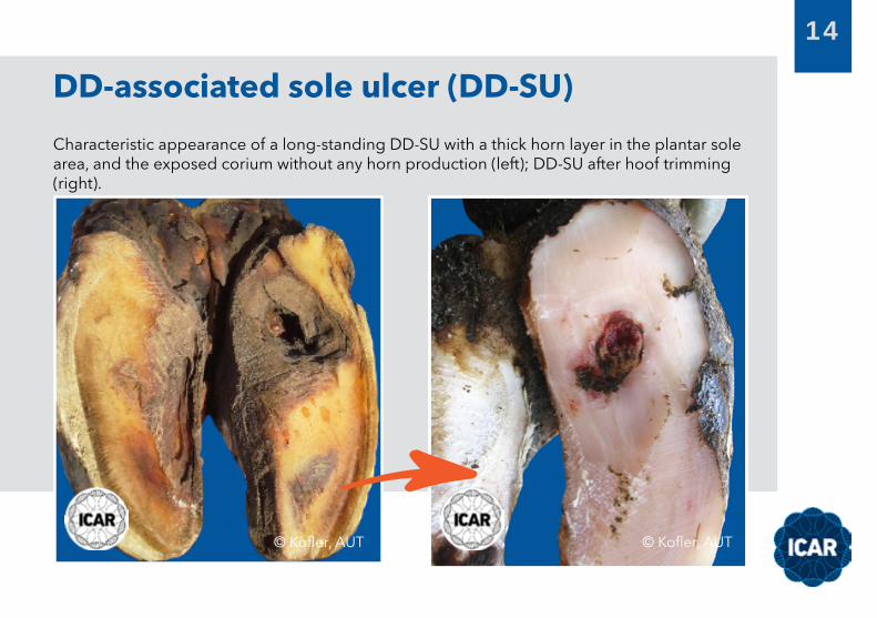
Characteristic appearance of a long-standing DD-SU with a thick horn layer in the plantar sole area, and the exposed corium without any horn production (left); DD-SU after hoof trimming (right).
DD-associated sole ulcer (DD-SU)
© Kofler, AUT © Kofler, AUT

15
© Fjeldaas, NOR
DD-SU after the complete removal of all loose horn using local anaesthesia.
DD-associated sole ulcer (DD-SU)
© ISAP, SPA © Fiedler, GER

16
Small (left) and a large long-standing DD-SU (right) with extended pododermatitis involving the corium of the bulbs of the heel after the removal of loose horn using local anaesthesia.
DD-associated sole ulcer (DD-SU)
© SERAGRO, SPA © Capion, DEN

17
Long-standing DD-BU and white-line infection after the removal of loose horn around the exposed corium using local anaesthesia.
DD-associated bulb ulcer (DD-BU)
© Capion, DEN

18
Small (left) and a large long-standing DD-BU after the removal of all loose horn using local anaesthesia.
DD-associated bulb ulcer (DD-BU)
© Fiedler, GER © Fiedler, GER

19
DD-BU after exposure and removal of the double sole.
DD-associated bulb ulcer (DD-BU)
© SERAGRO, SPA
20
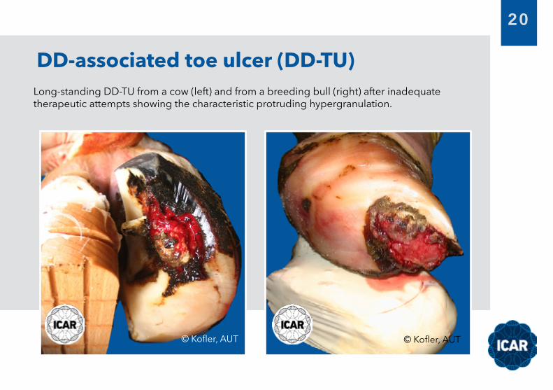
Long-standing DD-TU from a cow (left) and from a breeding bull (right) after inadequate therapeutic attempts showing the characteristic protruding hypergranulation.
DD-associated toe ulcer (DD-TU)
© Kofler, AUT © Kofler, AUT

21
DD-infected corium at the toe with hypergranulation before cleaning and removing the dirt (left), and after therapeutic hoof trimming and surgical debridement of the infected corium layer using local anaesthesia (right).
DD-associated toe ulcer (DD-TU)
© Kofler, AUT © Kofler, AUT

22
DD-TN after the removal of some loose horn.
DD-associated toe necrosis (DD-TN)
© Fiedler, GER © Kofler, AUT

23
DD-associated toe necrosis (DD-TN)
Long-standing DD-TN (approximately 18 months in the claw of the left image) after the removal of loose horn.
© Lutz, FRA © ISAP, SPA
24
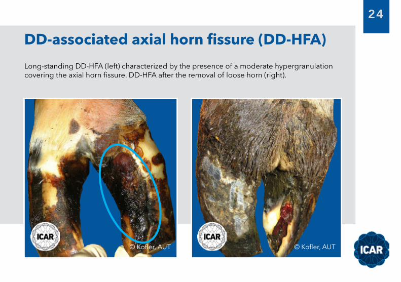
Long-standing DD-HFA (left) characterized by the presence of a moderate hypergranulation covering the axial horn fissure. DD-HFA after the removal of loose horn (right).
DD-associated axial horn fissure (DD-HFA)
© Kofler, AUT © Kofler, AUT

25
Long-standing DD-HFA (left) characterized by the presence of severe hypergranulation covering the fissure. Long-standing DD-HFA after the removal of loose horn (right).
DD-associated axial horn fissure (DD-HFA)
© Kofler, AUT
© Kofler, AUT
26
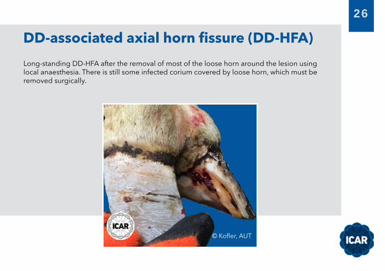
Long-standing DD-HFA after the removal of most of the loose horn around the lesion using local anaesthesia. There is still some infected corium covered by loose horn, which must be removed surgically.
DD-associated axial horn fissure (DD-HFA)
© Kofler, AUT

27
Long-standing DD-HFA characterized by the presence of severe hypergranulation tissue covering the axial fissure. Note that the hypergranulation originates from the axial wall (fissure).
DD-associated axial horn fissure (DD-HFA)
© Kofler, AUT © Kofler, AUT
28
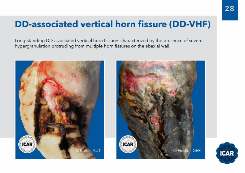
Long-standing DD-associated vertical horn fissures characterized by the presence of severe hypergranulation protruding from multiple horn fissures on the abaxial wall.
DD-associated vertical horn fissure (DD-VHF)
© Kofler, AUT © Fiedler. GER
29
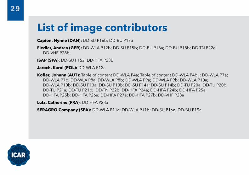
Capion, Nynne (DAN): DD-SU P16b; DD-BU P17a
Fiedler, Andrea (GER): DD-WLA P12b; DD-SU P15b; DD-BU P18a; DD-BU P18b; DD-TN P22a; DD-VHF P28b
ISAP (SPA): DD-SU P15a; DD-HFA P23b
Jaroch, Karol (POL): DD-WLA P12a
Kofler, Johann (AUT): Table of content DD-WLA P4a; Table of content DD-WLA P4b; ; DD-WLA P7a; DD-WLA P7b; DD-WLA P8a; DD-WLA P8b; DD-WLA P9a; DD-WLA P9b; DD-WLA P10a; DD-WLA P10b; DD-SU P13a; DD-SU P13b; DD-SU P14a; DD-SU P14b; DD-TU P20a; DD-TU P20b; DD-TU P21a; DD-TU P21b; DD-TN P22b; DD-HFA P24a; DD-HFA P24b; DD-HFA P25a; DD-HFA P25b; DD-HFA P26a; DD-HFA P27a; DD-HFA P27b; DD-VHF P28a
Lutz, Catherine (FRA): DD-HFA P23a
SERAGRO Company (SPA): DD-WLA P11a; DD-WLA P11b; DD-SU P16a; DD-BU P19a
List of image contributors