Apoptotic mechanisms in the neonatal brain following ... · Apoptotic mechanisms in the neonatal...
73
Doctoral thesis for the Degree of Doctor of Philosophy, Faculty of Medicine Apoptotic mechanisms in the neonatal brain following hypoxia-ischemia Ylva Carlsson Göteborg 2011 Perinatal Center Department of Obstetrics and Gynecology The Institute of Clinical Sciences The Sahlgrenska Academy University of Gothenburg Sweden
Transcript of Apoptotic mechanisms in the neonatal brain following ... · Apoptotic mechanisms in the neonatal...
Doctoral thesis for the Degree of Doctor of Philosophy, Faculty of Medicine
Apoptotic mechanisms in the neonatal
brain following hypoxia-ischemia
Ylva Carlsson
Göteborg 2011
Perinatal Center Department of Obstetrics and Gynecology
The Institute of Clinical Sciences The Sahlgrenska Academy University of Gothenburg
Sweden

Ylva Carlsson
Research education for a doctoral degree corresponds to studies that amount to 240 higher education credits (hecs). For the award of a doctoral degree, it is required that an approved disputation (defending a scientific thesis) has been held and that the course requirements for postgraduate courses (a minimum of 30 hecs) are achieved. A doctoral degree awarded within medical science is denoted medicine doktor.
A thesis at the Sahlgrenska Academy shall as far as possible be written as a composite thesis, but in certain cases may take the form of a monograph. A composition thesis consists of a self-contained text summarising the research (“the frame”) based on 2-4 part papers. The frame consists of a summary of the doctoral project and results obtained, as well as a more detailed description of background, questions, results and significance. The frame can advantageously be written as a review article. (Postgraduate studies at the Sahlgrenska Academy: general regulations)
Cover illustration by Ylva Carlsson and Cecilia Larsson
Printed by Ineko, Sweden 2011
Ylva Carlsson, 2011
ISBN; 978-91-628-8334-8
2

Apoptotic mechanisms in the neonatal brain following hypoxia-ischemia
3
Abstract
Apoptotic mechanisms in the neonatal brain following hypoxia-ischemia
Ylva Carlsson
Perinatal Center, Department of Obstetrics and Gynecology, Institute of Clinical Sciences, The Sahlgrenska Academy, University of Gothenburg, Box 432, 405 30 Göteborg, Sweden
Introduction: Neonatal encephalopathy is often perinatally acquired and caused by hypoxia-ischemia (HI). Brain injury develops with a delay, over 12-48 hours, after the insult. Hypothermia, an established neuroprotective treatment, saves 1 infant in 9 from neurological deficits suggesting that there is room for further improvement. HI leads to cell death through multiple pathways, including apoptosis. The aim of this thesis was to investigate different apoptotic pathways and to explore possible apoptotic targets for future pharmacological treatment after perinatal brain injury. We investigated (I) the involvement of caspase-2 alone, (II) and in combination with hypothermia, (III) the role of c-Jun N-terminal kinase (JNK), and (IV) Cyclophilin D (CypD), a regulator of the mitochondrial membrane permeability transition pore..
Materials and methods: Wild type (WT) C57BL/6 and transgenic mice with gene deletion of caspase-2 (I, II) and CypD (IV) were used in the ibotenate (excitotoxic)-model (I), and/or Rice-Vannucci´s HI-model (I-IV) at postnatal day 5 (I) or 9 (I-IV). The mixed lineage kinase inhibitor CEP-1347 was used to explore the role of JNK after neonatal HI (III).
Results: Caspase-2-deficient mice demonstrated less gray and white matter injury after both neonatal HI and an excitotoxic insult (I). Hypothermia provided additional protection in caspase-2 deficient mice (II). CEP-1347 was neuroprotective in the immature brain, by reducing apoptosis and attenuating microgliosis (III). CypD gene deficiency enhanced HI injury and Bax inhibitory peptide (BIP) reduced injury in the immature brain, whereas CypD deletion protected and BIP had no effect on brain damage in the mature mouse brain. Apoptosis was more pronounced in the immature CypD deficient mice than in WT controls, while adults showed minimal apoptotic activation.
Conclusion: Apoptosis has a more prominent role in the immature brain and different pathways leading to cell death after HI are at play in the immature as compared to the adult brain. This suggests that different pharmacological interventions are required in the immature and the mature brain. We suggest that caspase-2 as well as Bax dependent mitochondrial permeabilization are important neuroprotective targets in neonatal HI brain injury.
Key words: hypoxia-ischemia, brain, caspase-2, cyclophilin D, MLK, neonatal

Ylva Carlsson
Svensk sammanfattning
Syrebrist i samband med förlossningen kan leda till obotliga hjärnskador och resultera i livslånga handikapp, till exempel cerebral pares, epilepsi och utvecklingsstörning. Tidigare forskning har visat att hjärnskadan ofta utvecklas timmar till dagar efter perioden med syrebrist. Kliniska studier har visat att hjärnskador och risken för neurologiska resttillstånd minskas genom kylbehandling. Att sådan behandling inte bara är effektiv i djurexperimentella studier utan även kliniskt innebär ett stort genombrott, men kylbehandling räddar endast ett barn av nio från att utveckla hjärnskador och för tidigt födda barn kan inte kylbehandlas. Det övergripande målet med forskningen är att hitta en behandlingsstrategi som kan användas i kombination med kylbehandling, men också ensamt till de barn där kylbehandling inte kan/får ges.
Efter syrebrist finns många olika mekanismer och vägar som leder till celldöd och hjärnskada. Genom att specifikt påverka utvalda mekanismer som är viktiga i den nyfödda hjärnan och som aktiveras vid skada, och inte de som fysiologiskt är kritiska för hjärnans utveckling, hoppas vi kunna finna nya strategier som skyddar hjärnan. Den i avhandlingen beskrivna forskningen har fokuserat på att experimentellt kartlägga och påverka de vägar som leder till programmerad celldöd, så kallad apoptos, och på de enzymer som styr celldöden. Fokus har även legat på mitokondrierna, cellernas kraftverk, som tycks bestämma om cellen skall dö genom programmerad celldöd eller överleva.
Två modeller har använts, dels hypoxi-ischemi (HI), dvs. syrebrist i kombination med minskat blodflöde, samt ibotenat-modellen där hjärnan exponeras för en signalsubstans, som i normala fall förmedlar nervsignalerna, men som i för stor mängd kan orsaka hjärnskador. Skadorna som utvecklas i dessa modeller efterliknar skadan väl hos både för tidigt födda och fullgångna barn utsatta för syrebrist i samband med förlossningen. Vi har experimentellt kartlagt vilken roll enzymet caspas-2 spelar vid utvecklingen av hjärnskador hos barn utsatta för syrebrist. Detta har vi gjort genom att slå ut caspas-2, antingen genetiskt genom att ta bort genen i möss (s.k. knock-out teknik), eller genom att blockera syntesen av enzymet med hjälp av siRNA. Caspas-2 är inte nämnvärt involverat i den fysiologiska apoptotiska processen i hjärnan, men caspas-2 finns i hög grad i den omogna hjärnan och nivån sjunker med stigande ålder. Både efter HI och i ibotenat-modellen innebär minskningen av caspas-2 minskade hjärnskador.
4

Apoptotic mechanisms in the neonatal brain following hypoxia-ischemia
5
Skyddet ökade ytterligare om kylbehandling adderades. Vi har dessutom funnit att kylbehandling är effektivt i en HI modell på nyfödd mus. Gängse behandling idag för syrebrist efter en förlossning är kylbehandling, vilket gör att varje möjlig ny terapi i framtiden troligen kommer att kombineras med kylning. Denna experimentella modell kan således vara ett sätt att pröva olika farmakologiska terapier i kombination med kylbehandling.
I HI-modellen har vi dessutom använt substansen CEP-1347, som genomgått kliniska prövningar hos vuxna för behandling av Parkinson med nedslående resultat, men som har mycket få biverkningar. Vi fann att hos nyfödda råttor skyddar CEP-1347 hjärnan från skador efter syrebrist, vilket gör att den kan vara värd att undersöka närmare i detta sammanhang. Den påverkar inte inflammationen som uppstår i hjärnan efter HI skada, utan minskar apoptosen, genom att påverka c-Jun N-terminal kinase (JNK), ett enzym som är viktigt i den apoptotiska skadeprocessen.
I mitokondrierna finns en port, en så kallad por, varigenom apoptosen styrs. Cyclophilin D (CypD) är ett protein delaktigt i öppnandet av denna por. Återigen användes transgena möss för att klarlägga rollen för detta protein efter HI skada. Här kunde vi påvisa att hjärnskademekanismerna är olika beroende på hjärnans ålder. Där en behandling som kan skydda vuxna, i vårt fall borttagande av CypD, istället ökar skadan hos de nyfödda djuren. Istället visade sig en annan por, som regleras av proteinet Bax, vara viktig hos nyfödda. Att hämma Bax kan vara en sätt att skydda den omogna hjärnan mot skador efter syrebrist.
Sammanfattningsvis ter sig apoptotiska processer vara mycket viktiga i den omogna hjärnan, betydligt viktigare än i den vuxna hjärnan i samband med hjärnskador. Hypotesen är att en substans som hämmar caspas-2 och/eller påverkar mitokondriernas poröppning har möjlighet att bli en användbar terapi i samband med hjärnskador hos nyfödda utsatta för syrebrist. Först måste man dock närmare granska vad det är som aktiverar caspas-2 och mitokondriernas poröppning och hur man bäst kan hämma detta utan att störa andra viktiga funktioner i den växande hjärnan. Forskningen som presenteras i denna avhandling har visat att mekanismerna för hjärnskador är olika i den omogna och i den vuxna hjärnan. Det är alltså viktigt att poängtera att detta kan innebära att en del läkemedel, som utvecklas för behandling av hjärnskador hos vuxna, förmodligen inte kan användas till nyfödda barn. Slutligen är det värt att notera att det finns många möjligheter att påverka apoptosen efter HI. Det är möjligt att en kombinationsbehandling, alternativt skräddarsydd behandling kan bli aktuell i framtiden.

Ylva Carlsson
List of original papers
This thesis is based on the following papers, which will be referred to in the text by their Roman numerals:
I Genetic inhibition of caspase-2 reduces hypoxic-ischemic and excitotoxic neonatal brain injury
Carlsson Y*, Schwendimann L*, Vontell R, Rousset CI, Wang X, Lebon S, Charriaut-Marlangue C, Supramaniam V, Hagberg H**, Gressens P**, Jacotot E**.
Ann Neurol. 2011 Mar 28. doi: 10.1002/ana.22431
II Combined effect of hypothermia and caspase-2 gene deficiency on neonatal hypoxic-ischemic brain injury
Carlsson Y, Wang X, Schwendimann L, Rousset CI, Jacotot E, Gressens P, Thoresen M, Mallard C, Hagberg H.
Submitted
III Role of mixed lineage kinase inhibition in neonatal hypoxia-ischemia
Carlsson Y, Leverin AL, Hedtjärn M, Wang X, Mallard C, Hagberg H.
Dev Neurosci. 2009;31(5):420-6
IV Developmental shift of cyclophilin D contribution to hypoxic-ischemic brain injury
Wang X, Carlsson Y*, Basso E*, Zhu C, Rousset CI, Rasola A, Johansson BR, Blomgren K, Mallard C, Bernardi P, Forte MA, Hagberg H.
J Neurosci. 2009;29(8):2588-96
*Both authors contributed equally to this article; **shared senior authorship
6

Apoptotic mechanisms in the neonatal brain following hypoxia-ischemia
7
This thesis was supported by these foundations:
We are very grateful to Dr Carol Troy at Columbia University for supplying us with the caspase-2 knockout mice and to Lundbeck, Köpenhamn for supplying us with CEP-1347. This work was supported by Medical Research Council strategic award (MRC; United Kingdom, P19381, HH), University Paris 7, Medical Research Council (VR, Sweden, 2006-3396, HH; VR K2009-54X-21119-01-4, XW), ALF-LUA (Sweden, ALFGBG2863, HH), and the Sixth Framework Programme of the European Commission (STREP – Neobrain consortium, Contract Number LSHM-CT-2006-036534, HH, CM, PG and EJ), Leducq foundation and Wellcome Trust (Programme Grant WT094823MA, HH, PG), Martina and Wilhelm Lundbergs foundation (YC), SU-foundations (YC), funds from the Göteborg Medical Society (YC) and Stiftelsen Anna Brita och Bo Castegrens Minne (YC).

Ylva Carlsson
Table of content
ABSTRACT ......................................................................................................................... 3 SVENSK SAMMANFATTNING....................................................................................... 4 LIST OF ORIGINAL PAPERS ......................................................................................... 6 ABBREVIATIONS ........................................................................................................... 10 INTRODUCTION............................................................................................................. 12
Perinatal brain injury ................................................................................................... 13 Clinical background ................................................................................................... 13 Therapeutic window ................................................................................................... 13 Mechanisms – primary injury .................................................................................... 14 Mechanisms – secondary injury................................................................................. 15
Cell Death – Apoptosis and Necrosis ........................................................................... 15 Apoptotic pathways ....................................................................................................... 16
Caspases...................................................................................................................... 17 Caspase-2 .................................................................................................................... 17 Intrinsic pathway ........................................................................................................ 19 Extrinsic pathway ....................................................................................................... 19 The mitogen-activated protein kinases ...................................................................... 20 Mitochondrial permeabilization ................................................................................. 21
Inflammation ................................................................................................................. 23 Microglia..................................................................................................................... 24
Excitotoxicity ................................................................................................................. 25 Free radicals................................................................................................................... 26 Neuroprotective strategies ............................................................................................ 27 Mechanisms of hypothermia......................................................................................... 28
AIMS OF THIS THESIS .................................................................................................. 29 METHODOLOGICAL CONSIDERATIONS................................................................ 30
Genetically modified mice (I, II, IV) ............................................................................ 30 Age of brain (I-IV)......................................................................................................... 31 HI model (I-IV) .............................................................................................................. 31 Ibotenate model (I) ........................................................................................................ 33 Hypothermia treatment (II).......................................................................................... 33 siRNA (I) ........................................................................................................................ 34 Gray matter injury evaluation (I-IV) .......................................................................... 34
MAP-2 ......................................................................................................................... 34 Total tissue loss........................................................................................................... 35
8

Apoptotic mechanisms in the neonatal brain following hypoxia-ischemia
9
Neuropathological score............................................................................................. 35 Subcortical white matter injury (I-II).......................................................................... 35
NF, MBP and Olig-2 .................................................................................................. 35 Markers for apoptosis (I, III-IV).................................................................................. 36
TUNEL, Caspase-3 and AIF...................................................................................... 36 Markers for microglia (III)........................................................................................... 36
Isolectin, OX-18 and OX-42 ....................................................................................... 36 Gender (I-IV) ................................................................................................................. 37 Statistics (I-IV)............................................................................................................... 37
SUMMARY OF RESULTS .............................................................................................. 39 Involvement of Casp2 in neonatal brain injury (I, II)................................................ 39
Role of Casp2 in HI brain injury ............................................................................... 39 Role of Casp2 in the ibotenate-model of brain injury ............................................... 41 Role of Casp2 siRNA in brain injury ......................................................................... 42 Casp2 and hypothermia.............................................................................................. 42
MLK inhibition attenuates neonatal brain injury in a caspase-dependent manner (III) ................................................................................................................... 44
CEP-1347 reduces neonatal HI brain injury............................................................. 44 CEP-1347 reduces apoptosis ...................................................................................... 44 CEP-1347 effect on inflammation ............................................................................. 44
Age dependent differences in cell death pathways (IV) ............................................. 45 Effects of CypD deficiency on HI brain injury.......................................................... 45 Mitochondria characteristics in CypD-/- mice ............................................................ 45 Effect of CypD deficiency on apoptotic mechanisms after HI .................................. 45 Effect of CypD deficiency on Bax expression............................................................ 46 The role of Bax inhibition in HI brain injury ........................................................... 46
DISCUSSION..................................................................................................................... 48 Role of Casp2 in neonatal brain injury........................................................................ 48
Casp2 and hypothermia.............................................................................................. 51 Role of JNK after HI in perinatal brain injury........................................................... 54 Age different response after HI - the importance of mitochondria........................... 56
CLINICAL IMPLICATIONS AND FUTURE PERSPECTIVES ................................ 58 ACKNOWLEDGMENTS................................................................................................. 61 REFERENCES .................................................................................................................. 63

Ylva Carlsson
Abbreviations
AIF apoptosis inducing factor AMPA α-amino-3-hydroxy-5-methylisoxazole-4-propionic acid AP-1 activator protein-1 Apaf-1 apoptotic protease activating factor-1 ATF-2 activating transcription factor-2 ATP adenosine triphosphat Bax pro-apoptotic Bcl-2 associated X protein Bcl-2 b-cell lymphoma-2 BIP bax inhibiting peptide Ca2+ calcium CARD caspase recruitment domain Casp2 caspase-2 Casp2-/- homozygous casp2 knock-out Casp2+/- heterozygote casp2 mice CP cerebral palsy CR3 complement receptor 3 CsA cyclosporin A CypD cyclophilin D CypD-/- cyclophilin D knock-out Cyt C cytochrome C DIABLO direct inhibitor of apoptosis-binding protein with low pl DISC death-inducing signalling complex EAA excitotory amino acids Figure Fig. GABA gamma-aminobutyric acid GAD glutamate decarboxylase Heterozygote het HI hypoxia-ischemia or hypoxic-ischemic HIE hypoxic-ischemic encephalopathy H2O2 hydrogen peroxide IAP inhibitors of apoptosis i.c.v intracerebroventricularly i.p. intraperitoneally JNK/SAPK c-Jun N-terminal kinase or Stress-activated protein kinase IL interleukin MAP-2 microtubulus associated protein-2 MAPK mitogen-activated protein kinase MBP myelin basic protein MCP-1 monocyte chemoattractant protein-1 MHC I major histocompatibility complex I antigen MLK mixed lineage kinase MPT (mitochondrial) membrane permeability transition MRI magnetic resonance imaging NE neonatal encephalopathy
10

Apoptotic mechanisms in the neonatal brain following hypoxia-ischemia
11
NF neuronal filament NMDA N-methyl-D-aspartate NNT number needed to treat NO nitric oxide NOS nitric oxide synthase Olig-2 oligodendrocyte transcription factor-2 PCR polymerase chain reaction PIDD p53-induced protein with a death domain PND postnatal day PTP permeability transition pore PVL periventricular leukomalacia RAIDD receptor-interacting protein (RIP)-associated ICH-1/CED-3-
homologous protein with a death domain ROS reactive oxygen species SEM standard error of the mean siRNA small interfering RNA Smac second mitochondria-derived activator of caspase TNF-α tumour necrosis factor-α TUNEL terminal deoxynucleotidyl transferase-mediated dUTP nick end-
labelling WT wild-type XIAP X-linked inhibitor of apoptosis
Ylva Carlsson
Introduction
Asphyxia originates from the greek word for pulselessness. Today however, the term
asphyxia is generally used for a condition with severly deficient gas exchange
leading to hypercapnia and hypoxemia and ultimately severe lactic acidosis [Fatemi,
et al., 2009]. Asphyxia can arise during pregnancy, known as chronic asphyxia, and
can be caused by an underlying disease in the mother or the placenta. Asphyxia can
also arise during delivery, so called perinatal asphyxia, due to for example placental
abruption, rupture of the uterus, umbilical cord compression or maternal hypotension.
Neonatal encephalopathy (NE) is a clinically defined syndrome of disturbed
neurological function in the earliest days of life in the term infant, manifested by
difficulty with initiating and maintaining respiration, depression of tone and reflexes,
subnormal level of consciousness and often seizures. Also preterm babies can present
with this, but the signs are often more subtle [du Plessis and Volpe, 2002]. Several
different etiologies, such as metabolic disease, infection, drug exposure, nervous
system malformation and neonatal stroke are possible causes of neonatal
encephalopathy and should be excluded, but generally the term NE refers to hypoxic-
ischemic encephalopathy (HIE) resulting from perinatal asphyxia.
Although questioned [Nelson and Chang, 2008], som recent studies have shown that
the majority of children with neonatal encephalopathy, seizures or both, but without
specific syndromes or congenital heart defects, had evidence of perinatally acquired
insults and that the majority of cerebral palsy (CP) both in preterm and in term seem
to arise during the perinatal period [Jacobsson, et al., 2002; Cowan, et al., 2003;
Himmelmann, et al., 2010]. The overall CP prevalence in Sweden between the years
of 1999-2002 was 2.18/1000 live births [Himmelmann, et al., 2010].
Perinatal asphyxia at term often results in a dyskinetic cerebral palsy, which is
associated with multiple handicaps with a larger impairment load, such as only 16%
walking without aids, 60% being bound to a wheelchair and 50% suffering from
epilepsy, than other forms of CP and hence a larger need of aid from society
12

Apoptotic mechanisms in the neonatal brain following hypoxia-ischemia
13
[Himmelmann, et al., 2009]. The prognosis for HIE in preterm is also depressing;
Logitharajah et. al [Logitharajah, et al., 2009] rapported recently a 2-year outcome
where one third had died, nearly a quarter had developed a severe form of CP and
only one third of the infants had normal outcomes.
In the last ten years a significant increase in prevalence in children born at term and
with dyskinetic CP has been reported, in spite of decreasing neonatal mortality
[Himmelmann, et al.; Sellier, et al.]. The cause behind this is unknown.
Perinatal brain injury
Clinical background
The preterm white matter is more susceptable to hypoxic-ischemic (HI) injury,
because of the vulnerability of immature oligendrocytes prior to myelination
[Dammann, et al., 2001; Leviton and Gressens, 2007; Logitharajah, et al., 2009].
This gives rise to the pathological diagnosis periventricular leukomalacia (PVL),
which shows a reduction in myelination leading to a lack of white matter. PVL
represents the typical response of the preterm brain to an insult, either for example HI
or damage caused by cytokines associated with infection [Rennie, et al., 2007].
Children born at term with HIE injury often have cortical/subcortical lesions as well
as basal ganglia lesions [Fatemi, et al., 2009; Himmelmann, et al., 2010]. Lesions in
the basal ganglia, thalamus and the internal capsule are predictive of CP [Rutherford,
et al., 2005]. These different patterns of injury might however not be as distinctly
separated as previously thought. MRI studies have lately questioned this, showing a
high degree of basal ganglia damage also in preterm infants [Logitharajah, et al.,
2009].
Therapeutic window
HIE is not a single event but rather an evolving process [Ferriero, 2004]. MRI studies
show progression of lesion size over the first few days after injury [Cowan, et al.,
2003]. After the initial decrease in oxidative energy metabolism during HI, at least

Ylva Carlsson
partial recovery occurs [Wyatt, et al., 1989; Yager, et al., 1992; du Plessis and Volpe,
2002], before a secondary decline follows approximately 6-15 hours later. Hence,
many neurons and other cells seem to “commit” to die or survive over a period of
days to weeks [Beilharz, et al., 1995; Ferriero, 2002; Gunn and Thoresen, 2006].
Those infants who did not show even a transient recovery suffer from a very high
mortality and neurodevelopmental outcome has been shown to be closely associated
with the degree of secondary energy failure after 24 to 48 hours [Roth, et al., 1997].
This biphasic pattern creates a “window of opportunity”, where intervention might be
able to affect the outcome later in life (figure 1 (Fig.)).
Primary cell death Delayed secondary cell deathHypothermia
1 hour 6 hours 5 days
Cytotoxic mechanisms
ExcitotoxicityInflammation
Apoptosis
Hyp
oxia
-isch
emia
latent phase
Fig.1
Primary cell death Delayed secondary cell deathHypothermiaHypothermia
1 hour 6 hours 5 days
Cytotoxic mechanisms
ExcitotoxicityInflammation
ApoptosisCytotoxic mechanismsCytotoxic mechanisms
ExcitotoxicityExcitotoxicityInflammationInflammation
ApoptosisApoptosis
Hyp
oxia
-isch
emia
Hyp
oxia
-isch
emia
latent phase
Fig.1
Fig.1 Cell death Cell death occurs in a two-phased manner, where neuroprotective intervention during the latent phase, before delayed cell death starts, can be effective.
Mechanisms – primary injury
During the initial phase of HI there is rapid depletion of adenosine-triphosphate
(ATP) leading to disruption of ATP-dependent processes, such as failure of the
Na+/K+-pump and depolarization of the cell membrane [Wyatt, et al., 1989; Yager, et
al., 1992; Dirnagl, et al., 1999]. The depolarization of neurons and glia also leads to
the release of excitatory amino acids into the extracellular space. Reuptake
mechanisms, which are ATP-dependent, become compromised leading to
accumulation of glutamate to excitotoxic levels and the overactivation of N-methyl-
D-aspartate (NMDA) receptors increases intracellular calcium (Ca2+) levels
14

Apoptotic mechanisms in the neonatal brain following hypoxia-ischemia
15
[Vannucci and Hagberg, 2004]. The increased level of intracellular Ca2+ causes
influx of Ca2+ into cell mitochondria, thereby uncoupling oxidative phosphorylation,
triggering ATP hydrolysis and causes mitochondrial swelling or if only a limited
amount of mitochondria is involved evokes the release of pro-apoptotic proteins into
the cytosol [Dirnagl, et al., 1999; Puka-Sundvall, et al., 2000].
Mechanisms – secondary injury
The primary insult is often followed by at least a partial restoration of cell energy
metabolism [Wyatt, et al., 1989; Yager, et al., 1992], before a secondary decline of
energy failure follows. Exactly what triggers this secondary decline is unknown;
however this secondary phase is characterized by excessive entry of Ca2+ into cells,
induction of free radicals such as reactive oxygen species (ROS) and nitric oxide
(NO), another wave of excitotoxic amino acids (EAA) release, inflammatory
reactions and apoptosis [McRae, et al., 1995; Bona, et al., 1999; Blomgren and
Hagberg, 2006; Northington, et al., 2011b].
Cell Death – Apoptosis and Necrosis
The form of cell death depends on the severity of injury and on the brain area
involved. Necrosis predominates in more severe cases and in an earlier phase and in
the forebrain, whereas apoptosis occurs in areas with milder ischemic injury, often
days after the insult, localized at the border of the insult and are especially centered
to the thalamus and brainstem but also in other areas [Beilharz, et al., 1995; Hu, et
al., 2000; Northington, et al., 2001; Puyal, et al., 2009]. Necrosis is associated with
swelling of the cytoplasm and organelles and leakage of cytoplasmic contents into
the extracellular space leads to a secondary inflammatory response [Vannucci and
Hagberg, 2004]. Apoptosis, on the contrary, is highly regulated and an energy
requiring process whereby the cell commits to suicide [Orrenius, et al., 2003].
Several studies have shown an important role of apoptosis in HI injury in the
neonatal brain as opposed to the adult brain [Beilharz, et al., 1995; du Plessis and
Volpe, 2002; Johnston, et al., 2002; Zhu, et al., 2005; Zhu, et al., 2007b]. The

Ylva Carlsson
newborn brain is primed to respond to various insults with the activation of apoptotic
cascades due to the importance of programmed cell death in the normal development
of the central nervous system. Pro-apoptotic proteins are highly expressed in the
developing brain [Zhu, et al., 2005; Wang, et al., 2009]. In addition to necrosis and
apoptosis, neurons in rodents subjected to neonatal HI are showing morphology
intermediate between that of classic apoptosis and necrosis [Leist and Jaattela, 2001;
Northington, et al., 2007]. The nuclei of such cells have large, irregularly shaped
chromatin clumps, similar to apoptotic neurons, but the cytoplasm shows changes
similar to necrotic neurons. This morphology coincides with mitochondrial energy
failure and activation of apoptotic pathways after neonatal HI. Mitochondrial energy
failure likely prevents execution of a full apoptotic phenotype and it is presumed that
mitochondrial failure may interrupt apoptotic cascades initiated by injury to the
immature rodent brain, resulting in the hybrid phenotype of neuronal cell death
[Northington, et al., 2007; Northington, et al., 2011b]. Lately it is becoming clear that
necrotic cell death can be as controlled and programmed as caspase-dependent cell
death. This so-called necroptosis has been shown in the delayed phase of neonatal
brain injury [Northington, et al., 2011a].
Apoptotic pathways
Multiple apoptotic pathways have been shown to be involved in neonatal HI cell
death [Vannucci and Hagberg, 2004; Northington, et al., 2011b]. There are at least
two broad pathways that lead to apoptosis, an "intrinsic" and an "extrinsic" pathway.
More than 30 proteins in the family of B-cell lymphoma-2 (Bcl-2), which regulates
apoptosis, have been described. They can be divided into two groups: anti- and pro-
apoptotic members [Orrenius, et al., 2003]. In the normal state a fine balance is
maintained between the pro-apoptotic proteins such as Bak, Bid and Bax (pro-
apoptotic Bcl-2 associated X protein), with their BH-3 death domain and the anti-
apoptotic proteins such as Bcl-2 and Bcl-XL.
16

Apoptotic mechanisms in the neonatal brain following hypoxia-ischemia
17
Caspases
Cysteine-dependent aspartate-directed proteases (caspases) are a group of
intracellular proteases preserved through evolution [Siegel, 2006]. They are essential
for the execution step in apoptosis and they also mediate inflammation and apoptosis
in the neonatal brain [Johnston, et al., 2002]. In humans, as well as in mouse, 14
members have been identified so far [Riedl and Yigong, 2004]. All executioner
caspases are pro-enzymes, which can be cleaved and activated by other caspases, in a
cascade-like manner. The caspase protein contains three domains, an amino-terminal
prodomain, a large subunit (~20kDa) and a small subunit (~10kDa). Caspases can be
classified as initiator or upstream caspases, such as caspase-2, -8, -9, or as effector or
downstream caspases, such as caspase-3, -6, and -7, as defined by their place in the
cascade. A third group of caspases is involved in mediating inflammatory reactions,
for example caspase-1 activates interleukin (IL)-18 and IL-1β. Caspases are inhibited
by the inhibitor of apoptosis protein (IAP) family. X-linked inhibitor of apoptosis
(XIAP) for example inhibits caspase-3, -7 and -9. Caspases have been found to be
critically important during brain development and lacking for example caspase-3 and
caspase-9 results in severe malformation of the nervous system [Kuida, et al., 1996;
Hakem, et al., 1998; Kuida, et al., 1998; Zheng, et al., 2000]. Studies have shown that
when one caspase is being knocked out, others tend to increase compensatorily [Troy
and Salvesen, 2002].
Caspase-2
Caspase-2 (Casp2) is an initiator caspase, a key enzyme in the route to destruction.
Casp2 is developmentally regulated and can initiate mitochondrial outer membrane
permeabilization [Enoksson, et al., 2004]. Casp2 activity has been shown to increase
after HI in immature as opposed to adult mice [Wang, et al., 2009]. Casp2 plays a
role in stress-induced apoptosis and can be induced by DNA damage through
ultraviolet radiation, trophic factor withdrawal and cytokine deprivation [Robertson,
et al., 2002; Zhivotovsky and Orrenius, 2005]. Like other caspases, pro-casp2
contains a long pro-domain, closely related to the structure of caspase-9, both
containing a caspase-recruitment domain (CARD), important for its ability to induce

Ylva Carlsson
cell death [Paroni, et al., 2002]. Pro-casp2 also contains two subunits, but the
cleavage specificity is more related to the effectors caspase-3 and -7. Hence casp2 is
unique having features of both initiator and effector caspases. It can be spliced into
two different mRNAs; Caspase-2L a pro-apoptotic enzyme and Caspase-2S which
have antagonistic effects on cell death. The expression of casp2 varies with
developmental stage [Wang, et al., 2009] and is the only pro-caspase present
constitutively in the nucleus. Pro-casp2 is thought to associate with receptor-
interacting protein (RIP)-associated ICH-1/CED-3-homologous protein with a death
domain (RAIDD), and together with p53-inducible death domain-containing protein
(PIDD) forming the PIDDosome complex [Zhivotovsky and Orrenius, 2005;
Baptiste-Okoh, et al., 2008; Vakifahmetoglu-Norberg and Zhivotovsky, 2010]. Casp2
is also known to be able to dimerize by itself, hence activation do not always require
cleavage [Orrenius, et al., 2003]. Casp2 can cleave Bid, but depletion of Bid does not
stop casp2 from releasing cytochrome C (Cyt C) and Smac (second mitochondria-
derived activator of caspase) also known as DIABLO (direct inhibitor of apoptosis-
binding protein with low pl) from the mitochondria [Guo, et al., 2002]. Casp2 can in
addition cleave and interact with Bax [Kumar and Vaux, 2002; Chauvier, et al.,
2005], but is also able to induce the release of Cyt C independently [Robertson, et al.,
2004b]. Casp2 can also induce the release of apoptosis inducing factor (AIF). AIF
translocates to the nucleus and induces caspase-independent apoptosis by causing
chromatin condensation and large-scale DNA fragmentation in the nucleus [Guo, et
al., 2002]. Casp2 has also been shown to be involved in c-Jun N-terminal kinase (or
Stress-activated protein kinase; JNK/SAPK) as well as mitogen-activated protein
kinase (MAPK) p38 activation [Dirsch, et al., 2004]. Casp2 is activated prior to
Caspase-9 and -3 [Dirsch, et al., 2004]. In contrast to this picture of being a key
caspase, casp2 knock-out mice develop normally, besides excess numbers of germ
cells in the ovaries and an enhanced cell death in facial motor neurons [Bergeron, et
al., 1998; Zhivotovsky and Orrenius, 2005]. Caspase inhibition has been shown to
promote survival and functional outcome in a variety of neurological disease models
[Rideout and Stefanis, 2001; Orrenius, et al., 2003; Northington, et al., 2005].
18

Apoptotic mechanisms in the neonatal brain following hypoxia-ischemia
19
Intrinsic pathway
DNA damage, infection and the presence of free radicals can trigger the intrinsic
apoptotic pathway [Rudel, et al., 2010]. Bax and/or Bak are activated. Bax
translocates to the mitochondria, where Bak is already present in the outer
mitochondrial membrane and both can through independent oligomerization in the
outer mitochondrial membrane create a pore, through which multiple proteins
including Cyt C, Smac/Diablo, endonuclease G, Omi/HtrA2 and AIF can escape. Cyt
C activates the apoptosome complex [Kroemer and Martin, 2005; Siegel, 2006] with
apoptosis activating factor-1 (Apaf-1) and pro-caspase-9 in the presence of dATP.
Smac/DIABLO release to the cytosol contributes to apoptosis by interacting with
endogenous IAPs, thereby enhancing caspase-3 and -9 activities, as does Omi/HtrA2.
This results in the activation of pro-caspase-9, which is followed by conversion of
procaspase-3 to active caspase-3 [Benjelloun, et al., 2003]. Caspase-3 activation
results in proteolysis of essential cellular proteins, including cytoskeleton proteins
and kinases, and is required for DNA fragmentation and the morphological features
associated with apoptotic cell death [Jänicke, et al., 1998] (Fig.2).
The intrinsic pathway might also operate through caspase-independent mechanisms
such as through AIF referred to above. The translocation of AIF from the
mitochondria to the nucleus is triggered by the release of molecular signals from the
nucleus like poly(ADP-ribose) monomers. The AIF translocation to the nucleus after
HI depends on the formation of an AIF-cyclophilin A complex [Zhu, et al., 2007a] in
the cytosol. Movement of AIF into the nucleus leads to chromatinolysis and is
greater in the immature brain than in the adult [Zhu, et al., 2003] (fig.2).
Extrinsic pathway
A number of cell surface receptors respond, through cross-linking of death receptors,
to cytokine (inflammatory) stimulation, resulting in activation of cell death signalling
programs, known as the extrinsic apoptotic pathway. Among those, Fas death is one
of the most extensively studied. Fas receptors activate caspase-8, which cleaves Bid.
Bid induces the translocation, oligomerization and insertion of Bax and/or Bak into

Ylva Carlsson
the mitochondrial outer membrane leading to mitochondrial permeabilization
[Fatemi, et al., 2009; Rudel, et al., 2010]. Caspase-8 can also, through activation of
the death-inducing signalling complex (DISC) activate caspase-3 directly [Siegel,
2006]. Caspase-8 and the DISC complex is also known to be able to activate casp2
[Vakifahmetoglu-Norberg and Zhivotovsky, 2010]. HI activates Fas death receptor
signalling in the neonatal brain [Vannucci and Hagberg, 2004]. The intrinsic
apoptosis cascade can also be activated following Fas death receptor signalling and
functions to amplify Fas-mediated cell death (fig.2).
The mitogen-activated protein kinases
MAPKs are serine/threonine kinases. They regulate a diverse array of functions, such
as neuronal survival, cell growth and proliferation as well as apoptosis, all depending
on the stimuli and cell-type involved in the activation [Abe, et al., 2000; Saporito, et
al., 2002; Bogoyevitch, et al., 2004]. They are activated by MAPK kinases
(MAPKK), which can be induced by growth factors or cytokines as well as cell-
stressors. Three main pathways can be discerned: firstly the extracellular signal-
regulated protein kinases (ERKs), mainly involved in proliferation and cell survival,
secondly p38 MAPK regulating mainly inflammatory cytokines and thirdly JNK.
JNK is a mediator of stress-induced apoptosis, shown to be activated by growth
factors and cytokines. Three isoforms exist, where JNK1 is constitutively expressed
and JNK3 is stress-induced and the isoform that exists in the brain. JNK is activated
by MKK4 and MKK7, which are two MAPKs that act directly on JNK. Mixed
lineage kinase (MLK) 3 can activate MKK4/7, but MLK3 can also activate p38. An
interaction between casp2 and JNK has been observed in some cells and JNK
inhibition has been shown to partially inhibit casp2 processing [Zhivotovsky and
Orrenius, 2005], suggesting that JNK contributes to casp2 dependent apoptotic
signalling upstream of mitochondria [Dirsch, et al., 2004]. The loss of JNK1 and
JNK2 in combination has been shown to be embryonically lethal [Bogoyevitch, et al.,
2004], while JNK3-/- are protected both from adult [Kuan, et al., 2003; Bogoyevitch,
et al., 2004] and neonatal HI damage [Pirianov, et al., 2007]. JNK is activated
upstream of mitochondria and JNK inhibition reduces Cyt C release as well as
caspase-9 and -2 release and subsequent DNA fragmentation, probably via Bim, a
20
Apoptotic mechanisms in the neonatal brain following hypoxia-ischemia
21
Bcl-2 family protein [Bogoyevitch, et al., 2004; Dirsch, et al., 2004; Gao, et al.,
2005; Pirianov, et al., 2007]. JNK is activated by ROS induction and N-
acetylcysteine treatment inhibits JNK [Dirsch, et al., 2004]. JNK phosphorylates c-
jun [Pirianov, et al., 2007; Nijboer, et al., 2010], but other targets exist as well such
as activating transcription factor-2 (ATF-2). ATF-2 is expressed in neurons and
involved in neuronal migration, however over-expression results in cell death
[Pearson, et al., 2005]. Activator protein-1 (AP-1) is a transcription factor, which is a
heterodimeric protein, composed of the proteins c-Fos, c-Jun and ATF-2. AP-1 is
activated by JNK [Vexler, et al., 2006]. JNK can also cleave Bid, resulting in jBid
releasing Smac/Diablo from the mitochondria [Deng, et al., 2003].
Mitochondrial permeabilization
Mitochondrial dysfunction plays a central role in the delayed mechanisms of brain
cell injury [Hagberg, et al., 2009]. As mentioned before, mitochondrial respiration is
markedly decreased in a biphasic pattern, with an initial decrease immediately after
HI which then recovers to almost normal levels at 3 hours followed by a secondary
diminution 8-24 hours after HI [Gilland, et al., 1998]. Mitochondrial
permeabilization plays an important role as an event that marks the point of no return
in multiple pathways to cell death [Hagberg, et al., 2009]. Several apoptotic
pathways converge upon the mitochondria and can induce mitochondrial
permeabilization. In the acute phase of brain cell injury Ca2+ is accumulating
intracellularly due to lack of ATP. As a consequence mitochondria try to regulate the
intracellular environment and increase their uptake, resulting in an increase in the
permeability of the inner mitochondrial membrane, loss of membrane potential (∆Ψ),
mitochondrial swelling and rupture of the outer mitochondrial membrane referred to
as mitochondrial membrane permeability transition (MPT). Hence, an increase in
intracellular Ca2+ can cause MPT [Robertson, et al., 2004a]. The opening of the
permeability transition pore (PTP) in the inner mitochondrial membrane is crucial for
MPT. The exact composition of the PTP and its regulation is still not completely
known. Cyclophilin D (CypD), a mitochondrial member of the cyclophilin family
with enzymatic capacity and a crucial role in protein folding, is believed to be a main
regulator of MPT in the adult brain [Orrenius, et al., 2003; Tsujimoto, et al., 2006].

Ylva Carlsson
DNA damage, UV radiation, trophic factor withdrawal
Cytokine stimulationFas death receptor
Caspase-8
DISC
Nucleus
DNA fragmentation
Apaf-1
Cyt C
Caspase-9
MAPK
JNK
Bid
BimBax
Mitochondria
ANT
VDACCypD BakBax
RAIDDPIDD
p53Casp2
AIFCyclophilin A
Caspase-3
Smac/Diablo
IAP
MLK
AP-1c-jun
?
Fig.2
extrinsic intrinsic
DNA damage, UV radiation, trophic factor withdrawal
Cytokine stimulationFas death receptor
Caspase-8
DISC
Caspase-8
DISC
Nucleus
DNA fragmentation
Apaf-1
Cyt C
Caspase-9
Apaf-1
Cyt C
Caspase-9
MAPK
JNK
Bid
BimBaxBax
Mitochondria
ANT
VDACCypD BakBax
RAIDDPIDD
p53Casp2
AIFCyclophilin A
AIFCyclophilin A
Caspase-3Caspase-3
Smac/Diablo
IAP
MLK
AP-1c-jun
?
Fig.2
extrinsic intrinsic
Fig.2 Apoptosis pathway There are at least two broad pathways leading to apoptosis, an extrinsic and an intrinsic pathway. In both pathways, signalling results in the activation of a family of cysteine proteases, named caspases which act in a proteolytic cascade to dismantle and remove the dying cell.
22

Apoptotic mechanisms in the neonatal brain following hypoxia-ischemia
23
CypD resides in the mitochondrial matrix, but associates with the inner
mitochondrial membrane during the MPT. VDAC and ANT are two other proteins
thought to be part of the PTP together with CypD, however this has been questioned
lately and they might only have regulatory functions [Tsujimoto, et al., 2006; Rasola,
et al., 2010]. MPT is inhibited by Cyclosporine A (CsA), a known CypD inhibitor.
However, in the developing brain, Bax seems to play a more prominent role for
mitochondrial permeabilization primarily of the outer mitochondrial membrane
[Hagberg, et al., 2009]. HI in the neonatal brain has been shown to induce membrane
permeabilization and lead to the release of pro-apoptotic proteins, such as Cyt C,
AIF, Smac/DIABLO and caspase-9, from the mitochondrial intermembrane space
into the cytoplasm [Wang, et al., 2004; Tsujimoto, et al., 2006]. Inhibition of both
AIF and caspases are protective indicating that inhibiting the result of mitochondrial
permeabilization is neuroprotective [Zhu, et al., 2007b].
Inflammation
Fetomaternal infection is known to increase the risk of CP [Jacobsson, et al., 2002].
The inflammatory response and cytokine production, which follows an infection,
seem to render the brain more vulnerable to later HI, even when the infection is
distant from the brain [Eklind, et al., 2001]. Cytokines are a group of soluble proteins
and peptides which act at low concentrations and play an important role in cell to cell
communication and in modulating the functional activities of individual cells.
Cytokines, such as IL-6 and chemokines such as monocyte chemoattractant protein-1
(MCP-1) are ubiquitous signalling molecules that help regulate growth, development
and acute responses such as fever and inflammation [Dammann and O'Shea, 2008].
Inflammatory cytokines are associated with neonatal HIE and the cytokine level of
IL-6 and IL-8 are associated with the degree of HIE [Sävman, et al., 1998]. IL-1β and
IL-6 are elevated in newborns who have evidence of perinatal brain damage as
compared to controls [Dammann and O'Shea, 2008]. After experimental neonatal HI
an up-regulation of many inflammatory genes, including IL-6, associated with
cellular activation of microglia in the injured hemisphere occurs [Bona, et al., 1999].
IL-6 is neuroprotective after excitotoxic injury in rat hippocampus [Pizzi, et al.,
Ylva Carlsson
2004]. IL-1β triggers the recruitment of neutrophils and is increased 3-6 hours after
neonatal HI [Hagberg, et al., 1996]. It plays a role in worsening HI injury after
infection and antagonism protects the neonatal brain [Hagberg, et al., 1996; Vexler
and Yenari, 2009]. IL-1β is often used as a marker of inflammation after HI
[Hagberg, et al., 1996]. MCP-1 is a chemokine produced locally in the brain [Fox, et
al., 2005], released from microglial cells, regulating monocyte accumulation. MCP-1
increases 4 hours after neonatal HI and remains elevated until 48 hours after HI
[Ivacko, et al., 1997]. JNK is involved in the induction of MCP-1 production [Zhou,
et al., 2007]. It appears, therefore, that inflammatory processes may either potentiate
HI-induced injury or exert a neuroprotective effect, all depending on context
[Nijboer, et al., 2008; Vexler and Yenari, 2009].
Microglia
Microglial cells are resident macrophages in the brain and the main cell type
providing immunosurveillance in the brain. Microglial aggregation has been
observed in human infants after HI insults. Microglia change their phenotype
depending on activation and they are activated and seen within hours after HI in the
immature brain; coinciding with DNA degradation. They reach maximal levels after
2-3 days and the number of activated microglia remain elevated up to 14 days after
an HI insult [Beilharz, et al., 1995; McRae, et al., 1995; Bona, et al., 1999]. Although
microglia have been suggested to play an important role in removing cellular debris
and stimulating tissue regeneration it has also been suggested that microglia play a
role in inflammatory and injurious processes after HI injury [Hagberg, et al., 1996;
Hedtjarn, et al., 2002; Svedin, et al., 2007]. Microglia may contribute to secondary
brain injury through the production of pro-inflammatory cytokines, ROS, NO,
complement factors, and excitotoxic neurotransmitters [Fatemi, et al., 2009]. Overall
microglia activation is associated with increased cell loss [Barrett, et al., 2007]. It
appears that the way they are activated plays a crucial role if they are harmful or not
[Schwartz, et al., 2006]. Caspase-8 as well as caspase-3 and -7 have been shown to
be able to regulate microglia activation [Burguillos, et al., 2011].
24

Apoptotic mechanisms in the neonatal brain following hypoxia-ischemia
25
Excitotoxicity
Excitotoxicity, which refers to excessive glutamatergic activation leading to cell
injury and death, is an important mechanism of injury in the neonatal brain
[Johnston, et al., 2002; Vannucci and Hagberg, 2004]. Glutamate is the predominant
excitatory amino acid neurotransmitter in the brain. There are three major groups of
glutamate receptors within the post-synaptic membrane; NMDA, α-amino-3-
hydroxy-5-methylisoxazole-4-propionic acid (AMPA) and kainic acid. The
glutamate transporter is dependent on a sodium gradient created by Na+/K+ ATPase
that is powered by anaerobic glucose metabolism, and impaired delivery of glucose
to the brain by ischemia and/or hypoglycemia impairs glutamate removal from the
synapses. Glutamate has been shown to accumulate in the brain in asphyxiated
neonatal lambs [Hagberg, et al., 1987] as well as in cerebrospinal fluid in
asphyxiated human infants [Hagberg, et al., 1993] and this coincide with an increase
in intracellular Ca2+ and pro-apoptotic pathways via caspase-3 [Vannucci and
Hagberg, 2004]. The excitatory neuronal circuits, important for synaptic plasticity, as
well as the expression of specific glutamate receptor subtypes in excitatory synapses,
changes during development in the perinatal brain, and these changes can be related
to the changing patterns of pathology at different gestational ages [Grafe, 1994;
Johnston, 1995; Blomgren and Hagberg, 2006]. The distribution and molecular
characteristics of NMDA-type glutamate receptors appear to be an especially
important determinant of the pattern of neuronal injury in the perinatal brain
[Vannucci and Hagberg, 2004; Johnston, 2005; Kaindl, et al., 2009]. The immature
NMDA receptor channels open more easily and flux more Ca2+ than their adult
counterparts. NMDA receptors probably mediate much of the injury to neurons in
structures such as cerebral cortex, basal ganglia, hippocampus and thalamus
associated with HI injury in animal models [Johnston, 1995]. Topiramate is an
AMPA antagonist [Liu, et al., 2004] showing promising treatment effects in
combination with hypothermia. The immature brain can withstand longer periods of
energy deprivation than the adult brain because of its low energy requirement
[Barrett, et al., 2007], yet when a critical threshold of energy deprivation is reached,

Ylva Carlsson
excitotoxic injury is enhanced because of the developmentally enhanced excitatory
pathways [Johnston, 2005].
Free radicals
The neonatal brain has a high rate of oxygen consumption, high availability of iron
for the catalytic formation of free radicals and low concentration of antioxidants,
making it susceptible to damage [Blomgren and Hagberg, 2006]. Oxidative stress-
regulated release of pro-apoptotic factors from mitochondria appears to play an
important role in the immature brain [Blomgren and Hagberg, 2006]. Mature
oligodendrocytes carry increased antioxidant enzymes compared with the
oligodendrocyte precursors present in the immature brain, which may partly explain
the susceptibility of premature infants to white matter damage [Lafemina, et al.,
2006]. After HI excess Ca2+ influx via glutamate receptors leads to severe oxidative
stress and excitotoxic cell death itself also involves direct activation of neuronal
nitric oxide synthase (nNOS) and the generation of nitric oxide (NO). This leads to
mitochondrial dysfunction and increased formation of ROS, oxidative damage and
cell death [Vannucci and Hagberg, 2004; Fatemi, et al., 2009]. There is an
accumulation of hydrogen peroxide (H2O2) after HI in neonatal mice because of the
low capacity of glutathione peroxidase in the immature brain [Lafemina, et al.,
2006]. Accumulation of H2O2 is damaging to the immature brain, due to the high
levels of free iron [Vannucci and Hagberg, 2004]. Most data suggest that oxidative
stress contributes to the post-ischemic impairment of mitochondrial respiration
[Fatemi, et al., 2009] and may initiate mitochondrial permeabilization, which
eventually allows the release of mitochondrial intermembrane proteins with the
potential to execute apoptosis. Consistent with the notion that excessive NO in the
neonate may be detrimental; Ferriero et al have shown that neuronal NOS (nNOS)
knock-out mice are protected from neonatal HI-induced brain damage [Ferriero, et
al., 1996]. The free radical scavenging agent N-acetylcysteine is able to cross the
placenta and has been shown to be neuroprotective in neonatal rats [Wang, et al.,
2007; Fatemi, et al., 2009].
26

Apoptotic mechanisms in the neonatal brain following hypoxia-ischemia
27
Neuroprotective strategies
The ultimate goal for HIE treatment is improving long-term motor and cognitive
outcomes as well as decreasing mortality. The theory of a therapeutic window was
proven when treatment after HIE with hypothermia showed an effect decreasing
mortality as well as survival free of any sensorimotor disability at the 18-24 months
follow-up [Edwards, et al., 2010; Jacobs, et al., 2011]. Further supportive
information comes from MRI studies that suggested that both head cooling and total
body cooling were associated with a reduced incidence of basal ganglia/thalamic
brain lesions [Logitharajah, et al., 2009] predictive of CP [Rutherford, et al., 2005].
Moderate (32-34 ºC) and prolonged (24-72 h) therapeutic hypothermia has now
become standard of care for neonatal HI brain injury [Perlman, et al., 2010].
Hypothermia studies [Jacobs, et al., 2011] show that in the control groups
approximately 60% dies or develop major disability [Gluckman, et al., 2005;
Shankaran, et al., 2005; Azzopardi, et al., 2009]. The ICE trial, recently published,
shows an absolute risk reduction by 15% [Gunn and Thoresen, 2006; Jacobs, et al.,
2011] and the clinical hypothermia trials have a number needed to treat (NNT) of 9,
suggesting that further improvement may be feasible with add-on treatments
[Edwards, et al., 2010]. We also still lack a neuroprotective treatment option for
preterm infants, who are not eligible for hypothermia treatment. A few studies
indicate that hypothermia might be safe for at least the near-term preterm [Bennet, et
al., 2007], but studies have also shown hypothermia being risky for preterm babies
[Barrett, et al., 2007]. Hypothermia has also failed to show effect in severly affected
children [Gluckman, et al., 2005; Azzopardi, et al., 2009]. Pharmacological treatment
with both xenon, erythropoietin and topiramate have shown additional effect in
combination with hypothermia and for xenon this effect was additive, which supports
the hypothesis that neuroprotection offered by hypothermia can be further improved
[Liu, et al., 2004; Hobbs, et al., 2008; Cilio and Ferriero, 2010]. Another drawback of
hypothermia is that treatment needs to be started within 6 hours. In animal studies
certain drugs have shown a neuroprotective effect with a wider therapeutic window
[Medja, et al., 2006].

Ylva Carlsson
Mechanisms of hypothermia
Possible side-effects of hypothermia include sinusbradycardia, prolonged QT-
interval without arrythmia, pulmonary hypertension, scalp-edema in the case of
selective head-cooling and overt bleeding due to increased thrombocytopenia.
Several larger clinical trials have however reported that moderate hypothermia in the
neonatal setting is safe [Gunn and Thoresen, 2006; Edwards, et al., 2010].
Although mild to moderate, hypothermia still has a remarkable neuroprotective effect
against ischemic brain injury. This effect is likely attributed to its broad inhibitory
actions on a variety of harmful cellular processes induced by HI. Hypothermia
reduces cerebral metabolism by about 5% for every degree of temperature reduction,
which delays the onset of anoxic cell depolarization [Gunn, et al., 1997]. However
the protective effect from hypothermia is still there although controlling
depolarization, implicating that the critical effect of hypothermia lies somewhere else
[Gunn and Thoresen, 2006]. Hypothermia also seems to decrease the levels of
extracellular EAA and NO [Thoresen, et al., 1997]. Among the proposed key
mechanisms underlying hypothermic neuroprotection is the inhibition of intracellular
signalling events that initiate the cell death cascade [Edwards, et al., 1995].
Hypothermia increases neuronal survival in the basal ganglia and suppresses the
activation of caspase-3 [Barrett, et al., 2007] and has also been shown to suppress
microglial activation [Wagner, et al., 1999]. Hypothermia reduces inflammation
triggered by ischemia [Silverstein, et al., 1997], reducing the expression of TNF-α,
IL-1β and IL-18 and increases the anti-inflammatory cytokine IL-10 [Wagner, et al.,
1999; Azzopardi and Edwards, 2007]. In summary, it is likely that hypothermia
influences multiple pathways that contribute to neuroprotection.
28

Apoptotic mechanisms in the neonatal brain following hypoxia-ischemia
29
Aims of this thesis
The overall objective was to investigate different apoptotic pathways
after excitotoxic and HI perinatal brain injury and to explore possible
targets for future pharmacological treatment after HIE.
Specific objectives were
Paper I to evaluate the hypothesis that casp2 may be an operative
target during perinatal brain injury.
Paper II to explore the neuroprotective efficacy of hypothermia in
combination with casp2 gene deficiency after HI in
neonatal mice.
Paper III to evaluate the cerebroprotective potency as well as the
effects on apoptotic and inflammatory markers of a MLK-
inhibitor, CEP-1347, after HI in neonatal rats.
Paper IV to examine the contribution of MPT in immature brain
injury through genetic deletion of its positive regulator
CypD and to assess the contribution of Bax dependent
permeabilization in the immature versus the adult brain.

Ylva Carlsson
Methodological considerations
This thesis is based on studies in vivo and in vitro. Detailed
descriptions of the methods are given in each individual paper.
Additional methodological considerations are discussed in this section.
Genetically modified mice (I, II, IV) Mice are very useful as experimental animals due to their easy maintenance and short
breeding time. Genetically modified mice have led to great advances in research,
allowing researchers to explore the role of genes in normal development and
physiology as well as in disease states. The mice are inbred for at least 20
generations, making them as genetically alike as possible and hence improving the
possibilities to explore the reaction to different events or substances. The most
commonly used laboratory mouse is the C57BL/6. It is intermediately sensitive to HI
as compared to 129Sv and CD1, two other commonly used strains. The damage
induced by HI in C57BL/6 mice is increasing, when increasing the duration of HI, in
contrast to the strain 129Sv, who show approximately the same amount of injury
irrespective of HI duration [Sheldon, et al., 1998]. Mixed breeding involving
heterozygote (het) x het mating (or homocygote x het) , allows comparison of all
three genotypes (wild type (WT), het, and homozygote) within each litter, which is
an optimal design considering the litter to litter variability. In those cases when the
experimental conditions doesn’t allow the mixed litter design (e.g. hypothermia
experiments in the present study) homozygous WT are compared with homozygous
knock-out litters. This usually require a higher number of animals in each group due
to higher variability and the animals have to be back-crossed to make sure that the
WT mice are genetically alike the knock-out mice in all other respects but the gene
deleted.
30

Apoptotic mechanisms in the neonatal brain following hypoxia-ischemia
31
Age of brain (I-IV) It is difficult to translate brain developmental age from rodents to humans and the
comparison depends on the parameter being studied. Rats and mice differ from
humans as regards to rodent brain being lissencephalic rather than gyrencephalic and
the limited proportion of white matter in rodents. Romijn et al. [Romijn, et al., 1991]
compared 4 different markers for brain age in rodents and human (numerical synapse
formation, development of glutamate decarboxylase (GAD) activity, which is the key
enzyme for the synthesis of the main inhibitory neurotransmitter gamma-
aminobutyric acid (GABA), the development of choline acetyltransferase, the key
enzyme for the synthesis of neurotransmitter acetylcholine and finally development
of electrical activity) and came to the conclusion that rat postnatal day (PND) 10-14
is equivalent to the human neonate at term. Most data suggest that the maturity of the
mouse brain is very similar to the rat brain at least with respect to white matter and
examining the oligodendrocyte lineage progression PND7 rodent white matter is
similar to that of human between 30 and 36 gestational weeks and by P14 mature
oligodendrocytes are so abundant in the rat and mouse that it would be equivalent to
the full-term infant of 40 gestational weeks [Craig, et al., 2003]. The maturity of the
subventricular zone in mice at PND10 is equivalent to that of human at term [Brazel,
et al., 2004]. In summary PND9 (or 10 depending on if the birthday is counted as
zero or 1) in mice corresponds to near-term or term depending on what you are
studying, while a PND7 rodent might be considered slightly more immature
corresponding to 32-34 weeks of gestation [Hagberg, et al., 1997; Hagberg, et al.,
2002].
HI model (I-IV) The most widely used model of neonatal HI is the Rice-Vanucci model in PND7 old
rats [Rice, et al., 1981; Hagberg, et al., 1997], later modified to mice as well
[Ditelberg, et al., 1996; Hedtjarn, et al., 2002], resulting in neuronal injury in 92% of
the animals. The method to produce HI brain damage in immature rodents consists of
a permanent unilateral common carotid artery ligation followed by a period in a
hypoxic environment. Neither ligation nor hypoxia alone induces any injury due to

Ylva Carlsson
extensive collaterals in most rodent strains. During hypoxia cerebral blood flow is
reduced by 40–60% in the hemisphere ipsilateral to the ligation [Vannucci and
Hagberg, 2004] and this together with a compensatory vasodilatation of the vessels,
leads to the fact that the collaterals no longer can support the ligated hemisphere
fully, leading to partial focal brain ischemia. Cerebral blood flow is then restored to
control values immediately upon return to normoxic conditions [Vannucci and
Hagberg, 2004]. The injury is primarily seen in areas supplied by the middle cerebral
artery; cerebral cortex, hippocampus, thalamus and striatum, hence including
periventricular white matter, ipsilateral to the side of the carotid artery [Rice, et al.,
1981; Towfighi, et al., 1991; Vannucci and Hagberg, 2004]. The cerebellum and
brain stem are not damaged. Damage distribution between rats and mice seems to
differ to some extent. Mice have more damage in hippocampus and less in cerebral
cortex compared to rats [Brywe, et al., 2005] (II), which might be due to differences
in vascular anatomy. No morphological damage is detected in the contralateral
hemisphere [Grafe, 1994; Towfighi, et al., 1994] giving the advantage of using it as
an “internal” control. Studies have also shown that sham-operated animals (5 min of
anesthesia and cervical incision and suture) are comparable to naive controls,
including no differences in energy and glycolytic metabolites and apoptosis in the
brain [Grafe, 1994]. However, ligation only has in one mouse strain (CD1) produced
brain injury [Comi, et al., 2009]. This does not seem to be the case for C57BL/6, who
reacts very differently to HI in comparison to CD1 mice [Sheldon, et al., 1998], but
we are currently investigating this further.
Another major advantage with the model is that it allows inclusion of a sufficient
number of animals for dose-response evaluation of neuroprotective agents as well as
allowing long-term evaluation of brain injury and functional impairments. It also
shares several important features with birth asphyxia in the human neonate such as
the combination of hypoxia and ischemia followed by reperfusion and changes in
cellular energy metabolism after HI, however a disadvantage and the difference from
birth asphyxia in human neonates is its unilateral distribution of damage and the lack
of multi-organ dysfunction [Hagberg, et al., 1997]. Another draw-back is the great
variability in the model between animals within and between litters [Grafe, 1994;
32

Apoptotic mechanisms in the neonatal brain following hypoxia-ischemia
33
Hagberg, et al., 1997; Sheldon, et al., 1998]. The commonly used anesthetic
isoflurane has been shown to decrease injury in our model after prolonged exposure
[Chen, et al., 2011]. Hence we try to limit operation time and thus the exposure to
isoflurane to a maximum of five minutes per mouse. The 50 minute duration of
hypoxia was chosen based on titration to produce consistent moderate brain damage
and without increasing the mortality (usually <10% in most of our studies) [Hedtjarn,
et al., 2002; Hagberg, et al., 2004].
Ibotenate model (I) An intracerebral injection of ibotenate (excitotoxic alkaloid acting on NMDA
receptors) induces focal lesions in 5-day-old mice [Marret, et al., 1995]. This
produces a white matter lesion, mimicking the injury observed in preterm neonates.
The development of the lesion can be blocked by treatment with a NMDA receptor
antagonist. Neuronal cortical cell death induced by ibotenate mimics gray matter
lesions observed in some human term or near-term neonates and the sensitivity of the
developing brain to HI damage parallels sensitivity to NMDA neurotoxicity
[Johnston, 2005]. Microglia activated by ibotenate release soluble factors such as
cytokines, nitric oxide, free radicals and glutamate. Glutamate has been shown to
cause white matter damage linking inflammation to excitotoxicity [Johnston, 2005]
Hypothermia treatment (II) During hypothermia a chamber, formerly used for human neonates, was used and
each animal was placed in individual boxes, preventing them from heating each
other. Those boxes are placed in the middle of the chamber, so that all animals
should be heated to the same degree. One animal in each chamber (hypothermia as
well as normothermia) was used as sentinel for measurement of core temperature
using a rectal probe and one probe was also placed in each chamber to monitor the
environmental temperature. The temperature was controlled with these probes and a
temperature reading was obtained every second (Software Daisy lab 10.0, Physitemp
Instruments, Inc., Clifton, New Jersey). The mouse pups quickly lost temperature
during every moment of handling, hence the low starting temperature in paper II.

Ylva Carlsson
However, the drop in temperature was very short-lasting and did not differ between
the experimental groups. The pups were put, both as regards to HI as well as
hypothermia, into a pre-heated chamber set at a temperature to keep the pups at about
the correct goal temperature and as quickly as they lose temperature they are also
heated. This quick temperature loss/warming is probably due to the low body volume
of mice.
siRNA (I) Small interfering RNA molecules are short sequences of double-stranded RNA (19-
27bp in length), which suppress expression of target genes by inducing the
breakdown of the cognate mRNA and hence hinders it to be used for translation of a
protein. According to the rules that have been established to recognize on-target
effects and mitigate off-target effects of siRNA [Cullen, 2006; Jackson, et al., 2006],
low quantities of three different siRNA were used (si2a, si2b, si2c) targeting the
same gene (mouse casp2). As each siRNA has unique off-target spectrum, but the
same intended target, observing the same phenotype (neuroprotection) with multiple
individual siRNA that contain distinct seed sequences increase confidence that the
phenotype results from silencing of the intended target. In vitro 95% of Casp2 was
blocked. In vivo in the ibotenate model 50% of mRNA was blocked. However this
lower percentage is uncertain, since the penetration abilities of the siRNAs are not
known and has to be put in relation to the sample analysed for mRNA.
Gray matter injury evaluation (I-IV)
MAP-2
Microtubule associated protein -2 (MAP-2) is part of the cytoskeleton in intact
neurons (dendrites and soma) and proximal axons. In the immature neonatal brain,
loss of MAP-2 staining corresponds well to areas of brain injury and hence infarction
in gray matter. The loss in staining is gradual over 12-24 hours after HI and parallels
the secondary loss of glucose metabolism in the tissue and accompanies activation of
caspase-3 [Gilland, et al., 1998; Puka-Sundvall, et al., 2000]. Complete loss of MAP-
34
Apoptotic mechanisms in the neonatal brain following hypoxia-ischemia
35
2 occurs in dead and irreversible injured cells, but it is possible that a partial
temporary decrease can be seen in surviving cells.
Total tissue loss
The volume of tissue loss was calculated according to the Cavaliere Principle, which
states that the volume of an irregularly shaped object can be estimated from a set of
two-dimensional slices throughout the object, provided that they are parallel,
separated by a known distance and begin randomly within the object. In the formula
of V=∑Apt, V is the total volume expressed as mm3 , ∑A is the sum of the areas
measured, P is the inverse of the sections sampling fraction, and t is the section
thickness (5-9 µm) [Hedtjarn, et al., 2002]. This measures preferably the sum of
infarction and total tissue volume loss.
Neuropathological score
Neuropathological score could be used as a complement to total tissue loss, but is
also a tool to evaluate injury in each brain region. It does however not include an
evaluation of the white matter injury, which is done separately [Hagberg, et al.,
2004].
Subcortical white matter injury (I-II)
NF, MBP and Olig-2
Neurofilament (NF) is the major intermediate filament of neurons and plays an
important role in the conductivity of impulses down the axon [Miller, et al., 2002].
Myelin basic protein (MBP) is an indicator for developing oligodendrocytes and
myelin, and loss of MBP can be an indicator of disruption in the myelination process
[Kinney and Back, 1998; Back, et al., 2002]. Studies have shown that acute
oligodendroglial injury can be quantified in a sensitive manner by measurements of
MBP immunostaining, confirmed by Hedtjärn et. al [Hedtjarn, et al., 2005], as well
as oligodendrocyte transcription factor-2 (Olig-2), a marker of oligodendrocytes
throughout their lineage [Liu, et al., 2002] [Billiards, et al., 2008].

Ylva Carlsson
Markers for apoptosis (I, III-IV)
TUNEL, Caspase-3 and AIF
Terminal deoxynucleotidyl transferase-mediated dUTP nick end-labeling (TUNEL)
is a nuclear labelling used as a measure of DNA cleavage and hence as evidence for
apoptosis being involved in neuronal death. TUNEL labels random DNA
fragmentation, that could have other causes but apoptosis [West, et al., 2006].
TUNEL has however been shown to correlate well with the expression of caspase-3
and the loss of MAP-2 [Zhu, et al., 2000]. Positive TUNEL labelling begins already
at 6 hours post HI, increases with a maximum reached at 18-24 hours [Zhu, et al.,
2000].
Caspase-3 has been identified as one of the key executors of apoptotic cell death and
caspase-3 activation is required for DNA fragmentation and the morphological
features associated with apoptotic cell death [Jänicke, et al., 1998].
AIF is a marker of the caspase-independent apoptotic pathway and is translocated to
the nucleus upon activation. This leads to large-scale DNA fragmentation and
chromatin condensation [Zhu, et al., 2007b]. AIF redistribution is shown to occur in
areas with neuronal damage and displays a close correlation with TUNEL labelling
[Zhu, et al., 2003].
Markers for microglia (III)
Isolectin, OX-18 and OX-42
Microglial cells were identified by Griffonia simplicifolia isolectin-B4, which is a
glycoprotein that has high specificity to α-D-galactosyl residues which can be found
on the surface on microglial cells and on cerebral capillaries [Streit and Kreutzberg,
1987; Streit, 1990]. Isolectin stains both resting inactive and activated microglia. To
evaluate the acute microglia response OX-42 (complement receptor 3, CR3) and OX-
18 (major histocompatibility complex I antigen, MHC I) immunoreactivity was used.
According to McRae et al. [McRae, et al., 1995], the expression of OX-18 and OX-
36

Apoptotic mechanisms in the neonatal brain following hypoxia-ischemia
37
42 on microglia occurs early and these are sensitive markers of microglial activation
after HI.
Gender (I-IV) Studies show a gender difference in CP prevalence, where there is a female majority
among children born at term, while a male predominance in children born preterm
[Himmelmann, et al., 2010]. This observation is consistent with earlier data showing
that the cognitive and motor outcome of brain injury is worse in male than in female
in low birth weight infants [Johnston and Hagberg, 2007]. A gender difference has
also been shown in animal models. Sex-related differences in cell death pathways are
present in neonatal mice and rats in vivo [Zhu, et al., 2006b; Nijboer, et al., 2007].
Although no difference in injury after HI were present, the apoptotic pathways differs
between sexes [Zhu, et al., 2006b], with greater translocation of AIF from
mitochondria to nuclei in males and greater activation of caspase-3 in females. A
greater influence of the excitotoxic pathway in immature males has also been shown
[Johnston and Hagberg, 2007]. This agrees with that XY neurons are more sensitive
to oxidative stress and glutamate excitotoxicity, while XX neurons are more sensitive
to etoposide and staurosporine, agents that activate caspase-dependent apoptosis [Du,
et al., 2004]. The important conclusion to draw is that gender can influence degree of
damage, mechanism of injury and thereby the efficacy of neuroprotective agents.
Statistics (I-IV) Data was tested for normal distribution using GraphPad Prism Version 4 or 5. The
Student's t-test was used in cases of normal distribution when two experimental
groups were compared and the paired t-test was used for comparisons between
uninjured and injured hemispheres. To compare multiple groups, a two-way-
ANOVA was used, followed by Bonferroni post hoc test. Data that was not normally
distributed was analysed using non-parametric tests (Mann–Whitney U test and
Kruskall Wallis, followed by Dunn´s correction). Results are given as mean ±
standard error of the mean (SEM). Statistical differences were considered significant
when p<0.05. Poweranalysis was performed for each experiment and based on 80%

Ylva Carlsson
power, significance level 0.05 and variability in the model according to our previous
studies.
38

Apoptotic mechanisms in the neonatal brain following hypoxia-ischemia
39
Summary of Results
Involvement of Casp2 in neonatal brain injury (I, II)
Casp2 expression is high in both the neonatal mouse and rat brain with
progressive decrease from the second week of life.
Role of Casp2 in HI brain injury
Casp2-deficient 9-day-old mice (casp2-/- mice) presented a significant reduction of
infarct volume when compared to WT mice (31.6%, p=0.025; I, Fig.2A). Brain
injury in WT mice was more pronounced in hippocampus than in striatum, cerebral
cortex and thalamus (II, Fig.1B). Brain damage was significantly reduced in both
casp2 heterozygote (casp2+/-) and casp2-/- mice compared with WT mice in
hippocampus, thalamus and cerebral cortex. In the striatum brain injury was lower in
casp2-/- mice as compared to both WT and casp2+/- mice (II, Fig.1B). The decrease in
injury was not sex dependent and casp2-/- mice also presented reduced NF
degradation in the subcortical white matter (NF loss: 39% in casp2-/- versus 51% in
WT, p=0.046; I, Fig.2C).
These data was confirmed once more and with an even stronger protective effect in
casp2-/- mice, when exploring the effect of a novel caspase-9 inhibitor. The infarction
volume was reduced by 59% from 22.7 ± 3.1 in WT mice to 9.3 ± 2.2 in casp2-/- mice
(p=0.0003, Fig.3A). The area of infarction measured as lack of MAP-2
immunoreactivity, was significantly reduced in 8 out of 9 brain levels in casp2-/-, with
the largest difference at the level of hippocampus (Fig.3B).

Ylva Carlsson
Fig.3A
***
WT Casp2-/-0
10
20
30
infa
rctio
nvo
lum
e(m
m3 )
Fig.3A
******
WT Casp2-/-0
10
20
30
infa
rctio
nvo
lum
e(m
m3 )
******
******
Fig.3B
0
20
40
60
80
100
120
140
0 2 4 6 8 10level
area
of i
nfar
ctio
n(m
m2 ) WT
Casp2-/-
anterior posterior
**
**
**
**
******
******
Fig.3B
0
20
40
60
80
100
120
140
0 2 4 6 8 10level
area
of i
nfar
ctio
n(m
m2 ) WTWT
Casp2-/-Casp2-/-
anterior posterior
**
**
**
**
Fig.3C
***
WT Casp2-/-0
5
10
15
tota
l neu
ropa
thol
ogic
al s
core
(mm
3 )
Fig.3C
***
WT Casp2-/-0
5
10
15
tota
l neu
ropa
thol
ogic
al s
core
(mm
3 )
Fig.3D
striatum hippocampus thalamus cortex
WTCasp2-/-
***
***
*****
0
2
4
6ne
urop
atho
logi
cal s
core
(mm
3 )
Fig.3D
striatum hippocampus thalamus cortex
WTCasp2-/-
WTCasp2-/-
******
******
********
0
2
4
6ne
urop
atho
logi
cal s
core
(mm
3 )
Fig.3 Gray matter injury evaluation Infarction volume calculated according to the Cavalieri Principle from the area loss of MAP-2 immunoreactivity in the ipsilateral hemisphere (A). Infarction size, expressed as loss of MAP-2 immunoreactivity in the ipsilateral hemisphere, was quantified in 9 coronal sections numbered from anterior to posterior levels, significantly different for level 1 through 8. (B). Neuropathological scoring of HI brain injury in the striatum (0–6), hippocampus (0–6), thalamus (0–6) and cerebral cortex (0–4) is shown (D) as well as the total injury score (0–22) for WT and casp2-/- mice (C). A reduction of brain injury in casp2-/- mice compared with WT mice is shown. Data were analyzed with two-Way ANOVA and values are expressed as means + SEM. Results from Bonferroni post hoc test are given in the figure; ***p<0.001; **p<0.01.
The neuropathology score was substantially lower in casp2-/- mice than in WT mice
for all regions evaluated; being most marked in the thalamus (73%), followed by
cerebral cortex (58%), hippocampus (51%) and striatum (48%) (Fig.3D). The total
neuropathology score was significantly reduced (p<0.0001) in casp2-/- mice (5.8 ±
1.0) as opposed to WT mice (12.9 ±0.6) (Fig.3C). Casp2-/- animals showed, in
agreement with our previous findings a decrease in both NF staining (p=0.0073,
40

Apoptotic mechanisms in the neonatal brain following hypoxia-ischemia
41
Fig.4A), as well as MBP (p=0.0008, Fig.4B) after HI. There was a significant loss of
NF and MBP immunoreactivity in WT animals subjected to HI indicating white
matter injury affecting both axons and myelin. The deficit in NF staining was
significantly reduced, by 50%, in casp2-/- mice (9.2 ±2.4) compared with WT mice
18.3 ±2.1 and the loss of MBP was decreased by 44% in casp2-/- mice (23.1 ± 4.2)
versus WT mice 41.1 ± 2.8 (Fig.4A-B). The neuroprotection offered by casp2 gene
deficiency was not sex dependent (data not shown).
Fig.4A
**
WT Casp2-/-0
5
10
15
20
25
% N
F lo
ss
Fig.4A
**
WT Casp2-/-0
5
10
15
20
25
% N
F lo
ss
Fig.4B
***
WT Casp2-/-0
10
20
30
40
50
% M
BP
loss
Fig.4B
***
WT Casp2-/-0
10
20
30
40
50
% M
BP
loss
Fig.4 White matter injury evaluation Percentage tissue loss of NF (A) or MBP (B) staining in the ipsilateral hemisphere in relation to the contralateral hemisphere. Injury was evaluated 3 days post HI. Casp2-/- mice are protected after HI as compared to WT mice. Data were analyzed with two-Way ANOVA and values are expressed as means + SEM. Results from Bonferroni post hoc test are given in the figure; ***p<0.001; **p<0.01.
Role of Casp2 in the ibotenate-model of brain injury
Ibotenate induces focal lesions in 5-day-old mice; however homozygous casp2 gene
deficiency conferred a 41% reduction of lesion volume of cortical and a 59%
reduction in white matter injury as compared to WT animals (I, Fig.2E). Similar to
casp2-/- but with a less marked effect, casp2+/- neonates showed reduced lesion
volumes of cortical (23%) and white matter (28%) injury (I, Fig.2E).
Immunohistochemistry of casp2-/- mice, both after HI as well as after ibotenate,
showed a decrease in the number of cleaved caspase-3 positive cells as compared to
WT mice; hence caspase-3 activation appeared to be, at least in part, casp2-
dependent (I, Fig.2G-I).

Ylva Carlsson
Role of Casp2 siRNA in brain injury
In order to further evaluate the involvement of casp2 in perinatal brain damage casp2
-specific siRNA was used to target casp2. Through intraparenchymal administration
of three casp2-specific siRNA a more than 50% protection in both gray and white
matter of the neonatal brain against excitotoxic insult was achieved, whereas no
significant differences were observed between non-treated animals and non-targeting
controls (I, Fig.3A-B).
Casp2 and hypothermia
The administration of 5 hours of hypothermia directly after HI provided additional
protection in mice with casp2 gene deletion (II, Fig.3). Regional score demonstrated
that hypothermia enhanced the protective effect of casp2 gene deletion in
hippocampus and thalamus. Such an additive effect was not found in the other two
regions; striatum was mostly protected by casp2 gene deficiency, whereas the
cerebral cortex was mostly rescued by hypothermia (II, Fig.4A-D). The infarction
volume confirmed the reduction in injury after hypothermia in WT and casp2-/- mice
g.5).
Fig.5 Brain injury
as assessed by neuropathological score (p<0.0001, Fi
evaluation after hypothermia
e infarction volume was
Fig.5*
**
hypothermia normothermia0
10
20
30WTCasp2-/-
Infa
rctio
nvo
lum
e(m
m3 )
Fig.5*
****
hypothermia normothermia0
10
20
30WTCasp2-/-WTCasp2-/-
Infa
rctio
nvo
lum
e(m
m3 ) Th
significantly reduced in WT and casp2-/- mice after hypothermia (p<0.0001). Data were analyzed with two-Way ANOVA and values are expressed as means + SEM. Results from Bonferroni post hoc test are given in the figure; **p<0.01; * p≤0.05.
White matter injury, assessed on the basis of MBP and NF immunoreactivity, was
reduced by hypothermia independently of casp2 genotype (two-way ANOVA; MBP:
genotype p=0.948, temperature p<0.0001; NF: genotype p=0.764, temperature
p=0.0003, II, Fig.5). This finding was supported by the number of Olig-2 positive
42
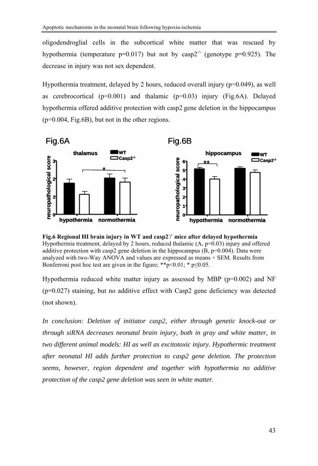
Apoptotic mechanisms in the neonatal brain following hypoxia-ischemia
43
, reduced overall injury (p=0.049), as well
as cerebrocortical (p=0.001) and thalamic (p=0.03) injury (Fig.6A). Delayed
oligodendroglial cells in the subcortical white matter that was rescued by
hypothermia (temperature p=0.017) but not by casp2-/- (genotype p=0.925). The
decrease in injury was not sex dependent.
Hypothermia treatment, delayed by 2 hours
hypothermia offered additive protection with casp2 gene deletion in the hippocampus
(p=0.004, Fig.6B), but not in the other regions.
Fig.6AFig.6A Fithalamus
neur
opat
holo
gica
l sco
re
hypothermia normothermia0
1
2
3
*
WTCasp2-/-
thalamus
neur
opat
holo
gica
l sco
re
hypothermia normothermia0
1
2
3
*
WTCasp2-/-
hippocampus
hypothermia normothermia0
1
2
3
4
5
6
neur
opat
holo
gica
l sco
re **WTCasp2-/-
g.6Bhippocampus
hypothermia normothermia0
1
2
3
4
5
6
neur
opat
holo
gica
l sco
re **WTCasp2-/-
Fig.6B
Fig.6 Regional HI brain injury in WT and casp2-/- mice after delayed hypothermia Hypothermia treatment, delayed by 2 hours, reduced thalamic (A, p=0.03) injury and offered
ditive protection with casp2 gene deletion in the hippocampus (B, p=0.004). Data were
NF
eficiency was detected
: Deletion of initiator casp2, either through genetic knock-out or
through siRNA decreases neonatal brain injury, both in gray and white matter, in
adanalyzed with two-Way ANOVA and values are expressed as means + SEM. Results fromBonferroni post hoc test are given in the figure; **p<0.01; * p≤0.05.
Hypothermia reduced white matter injury as assessed by MBP (p=0.002) and
(p=0.027) staining, but no additive effect with Casp2 gene d
(not shown).
In conclusion
two different animal models: HI as well as excitotoxic injury. Hypothermic treatment
after neonatal HI adds further protection to casp2 gene deletion. The protection
seems, however, region dependent and together with hypothermia no additive
protection of the casp2 gene deletion was seen in white matter.

Ylva Carlsson
MLK inhibition attenuates neonatal brain injury in a caspase-dependent manner (III)
CEP-1347 reduces neonatal HI brain injury
CEP-1347 is a MLK inhibitor. When CEP-1347 was administrated after HI, 6 hours
after HI and once daily until the rats were sacrificed; there was a 28% decrease in
overall tissue volume loss (p=0.019) in CEP-1347-treated as compared to vehicle-
treated animals (III, Fig.1). This injury reduction was further supported when
assessing injury through neuropathological score, where the total neuropathological
injury score was reduced by 27% in CEP-1347 treated rats (p = 0.046, III, Fig.1). The
number of isolectin-positive cells with the morphology of activated microglia, i.e.
retracted processes and rounded cell body phenotype, was counted and was shown to
be substantially increased after HI. CEP-1347, reduced the number of isolectin-
positive cells at 7 days after HI as compared to vehicle-treated animals (p=0.038, III,
Fig.1).
CEP-1347 reduces apoptosis
TUNEL staining was significantly (p=0.021) decreased in the hippocampus 24 hours
after HI in CEP-1347-treated animals and a similar tendency was found in the
cerebral cortex and striatum (p=0.066, III, Fig.2). This is consistent with a significant
(p=0.047) reduction of cells expressing the cleaved activated form of caspase-3 in the
striatum 24 hours after HI and a similar tendency is found in the cortex and
hippocampus (III, Fig.2). There was no significant difference in the number of AIF-
positive cells between CEP-1347-treated and vehicle-treated animals. Total p-JNK
immunoreactivity was significantly decreased at 4 hours (p=0.049) in CEP-1347-
treated rats, but no statistically significant difference was detected 8 hours after HI
(p=0.404). There was no significant change in c-jun levels after HI in CEP-treated
animals compared to controls.
CEP-1347 effect on inflammation
There was no significant difference in the expression of microglia markers OX-42
(CR3) and OX-18 (MHC I) immunoreactivity in CEP-1347-treated versus vehicle-
44

Apoptotic mechanisms in the neonatal brain following hypoxia-ischemia
45
treated animals at 24 hours after HI (III, Table 1). The expression of mRNA for the
pro-inflammatory cytokines IL-1β, IL-6 and MCP-1 was not different in CEP-1347-
treated versus vehicle-treated animals at 4 and 8 hours after HI (III, Table 2).
Similarly, protein expression of IL-1β and IL-6 was not different in CEP-1347-
treated versus vehicle-treated animals at 8 hours after HI (III, Table 3).
In conclusion: MLK inhibition reduces HI neonatal brain injury in a caspase-
dependent fashion, but had no detectable effect on inflammatory cytokines.
Age dependent differences in cell death pathways (IV)
Effects of CypD deficiency on HI brain injury
Analysis of brain injury in PND9 WT, CypD deficient mice (CypD-/-), and CypD het
(CypD+/-) mice revealed a more pronounced damage in CypD-/- mice (IV, Fig.1).
This is in sharp contrast to PND60 mice, where absence of the CypD-/- gene resulted
in a significant decrease in HI brain injury. No sex dependency could be discerned.
Mitochondria characteristics in CypD-/- mice
Mitochondrial morphology was not different comparing WT and CypD-/- at PND9 or
PND60 and CypD was as abundant in immature as in mature brain (IV, Fig.2A-E).
HI did not alter the expression of CypD itself in mitochondria enriched cell fractions.
Brain mitochondria from both neonatal and adult CypD-/- mice were desensitized to
Ca2+, requiring approximately twice the amount of Ca2+ to open the PTP than
mitochondria from WT animals (IV, Fig.3). Most importantly, brain mitochondria
from neonatal WT mice required far more Ca2+ to open the PTP than mitochondria
from adult mice, regardless of the presence of CypD. CsA was a more efficient
desensitizer in adult than in neonatal WT mitochondria, whereas it was ineffective in
mitochondria from CypD-/- animals.
Effect of CypD deficiency on apoptotic mechanisms after HI
Casp2, caspase-3, and -9 were markedly induced in the brain after HI at PND9,
whereas the activation and alterations were negligible at PND60 (IV, Fig.4A-C). At

Ylva Carlsson
PND9, casp2, caspase-3, and -9 were all significantly more activated in CypD-/- than
WT mice, whereas at PND60 the opposite was observed. A decrease of Cyt C was
observed in the mitochondrial fraction, matched by an increase in the cytosolic
fraction after HI at both ages. At PND9, the release of Cyt C at 24 hours after HI was
much more pronounced in CypD-/- compared with WT mice, whereas in adult mice,
the opposite was found (IV, Fig. 5A, D-E). AIF decreased at 6 and 24 hours after HI
in the mitochondria-enriched cell fractions in WT and CypD-/- mice at PND9, but the
loss was most pronounced at 24 hours after HI in CypD-/- mice (IV, Fig.5A). AIF
changes after HI in the adult brain were limited and independent of genotype. In
PND9 CypD-/- mice, post-HI changes were further confirmed by
immunohistochemistry. AIF immunoreactivity translocated from mitochondria to
nuclei after HI. Double immunostaining showed that Cyt C expression shifted from a
weak expression in the cytosol of normal control brain cells to a much stronger
staining with a diffuse cytosolic localization in cells that highly expressed the
cleaved active form of caspase-3 after HI (IV, Fig.5F).
Effect of CypD deficiency on Bax expression
In normal control brains from WT mice, a marked decrease of mitochondrial Bax
expression independent of CypD during the development from PND3 to PND60 was
found (IV, Fig.6A). After HI at PND9, translocation of Bax to mitochondria
increased at 1.5 and 3 hours in both genotypes, but the increase was more
pronounced in CypD-/- than in WT individuals at 3 hours (IV, Fig.6B). In brains of
PND60 mice, Bax expression was low and no obvious changes after HI were
detected.
The role of Bax inhibition in HI brain injury
A single injection of Bax inhibiting peptide (BIP) was administered
intracerebroventricularly (i.c.v) immediately before HI in the WT mice. BIP reduced
brain injury score by 79% and caspase-3 activation by 65% in PND9 WT mice,
whereas the corresponding effects were negligible in PND60 WT mice on both the
brain injury score as well as the caspase-3 activation (IV, Fig.6C-D). In addition, Cyt
C release was assessed in vitro by adding BH3-peptide to isolated brain mitochondria
46

Apoptotic mechanisms in the neonatal brain following hypoxia-ischemia
47
in a cytosol-like medium. Under these conditions, BH3-peptide induced Cyt C
release in both the absence and presence of exogenous full-length recombinant Bax
at PND9, whereas at PND60, BH3-peptide induced Cyt C release required the
presence of exogenous Bax, probably because the endogenous level of Bax in adult
brain mitochondria was too low. Moreover, BH3-peptide induced a more pronounced
Cyt C release in CypD-/- than in WT mice in PND9 brain mitochondria. In contrast,
BH3-induced Cyt C release was attenuated in mitochondria from CypD-/- compared
with WT mice at PND60. Bax alone did not elicit Cyt C release from isolated PND9
brain mitochondria, but exogenous Bax enhanced the effect of the BH3-peptide (IV,
Fig.7).
In conclusion: CypD-/- increases HI brain injury in neonatal mice as opposed to adult
mice, where the deletion is protective. This difference is neither due to a change in
the mitochondrial morphology nor in the components/regulators of the PTP itself.
However neonatal mitochondria are less sensitive to Ca2+, demanding more Ca2+ to
open the PTP than adult mice mitochondria. Apoptotic pathways are activated to a
higher degree in neonatal mice brain than in adult mice brain and utterly most in
CypD-/- neonatal mice. Bax expression decreases during development and is high
after HI in neonatal mice, with a more pronounced increase in CypD-/- mice as
opposed to WT mice. Inhibiting Bax leads to a decrease in injury as well as a
decrease of apoptotic markers in neonatal mice, but not in adult mice and a more
pronounced release of the apoptotic marker Cyt C was seen in neonatal CypD-/- as
compared to WT neonatal mice after adding a BH3-peptide mimicking Bax action.

Ylva Carlsson
Discussion
Perinatal brain injury leads to life-long handicaps. A way to
decrease/prevent poor outcome after NE and to increase life quality for
those who are affected would be the ultimate goal. Hypothermia
treatment proves the theory about a therapeutic window, a time-frame
where it is possible to influence the outcome and change the course of
the disease. Hypothermia improves outcome by 15%. Some children,
for example pre-term neonates, are so far not eligble for this treatment
[Barrett, et al., 2007] and the treatment has been shown not to be
effective in severly affected children, why additive or even preferbly
synergistic or alternative treatment would be valuable. The aim for this
thesis was to elucidate the mechanisms behind different apoptotic
pathways that occur during perinatal asphyxia, pathways where
manipulation would be possible and hence be used as pharmacological
targets for potential treatments.
Role of Casp2 in neonatal brain injury This thesis demonstrates a broad range of neuroprotective strategies involving casp2.
Genetic deficiency of casp2 is neuroprotective in newborn mice exposed to both HI
as well as excitotoxicity. In all experiments performed casp2 gene deletion reduced
gray matter injury by as much as 59% (Fig.3A) and white matter injury by 50%
(Fig.3B) in comparison to WT mice after HI brain injury. Using siRNA to delete
casp2 after excitotoxic injury also reduced both cortical and white matter damage in
the neonatal brain. This is in conjunction with other reports in vitro showing that
decrease or deletion of casp2 expression using casp2-/- cells [Tiwari, et al., 2011] or
casp2-specific siRNA [Troy, et al., 1997] decrease injury after oxidative stress.
Altogether, combined with our finding that casp2 mediates HI and excitotoxic
48

Apoptotic mechanisms in the neonatal brain following hypoxia-ischemia
49
neonatal brain damage and the importance of casp2 in apoptotic cell death, at least in
the immature brain, one can suggest that casp2 might be a potential target for future
neuroprotective strategies in neonates.
Casp2 is an upstream caspase, which can initiate the release of mitochondrial
constituents [Troy and Shelanski, 2003] and is known to be able to regulate diverse
cell signalling pathways, such as DNA damage [Robertson, et al., 2004a; Kumar,
2009]. In WT animals, the brain expression of casp2 is high at birth and
progressively decreases during postnatal development. Casp2 activity has been
shown to increase after HI in immature mice as opposed to adult mice [Wang, et al.,
2009]. Quite contrary to the protective effect in our neonatal setting; in a model for
adult focal cerebral ischemia casp2-/- mice have the same amount of brain injury as
WT mice [Bergeron, et al., 1998]. A non-selective caspase inhibition reduces injury
and saves neurons from apoptosis in the neonatal brain [Cheng, et al., 1998; West, et
al., 2006; Clark, et al., 2007]. A more selective inhibition would however be
desirable, since particularly some caspases seem to play a very important role in
normal brain development [Lu, et al., 2002; Gulyaeva, 2003]. Caspase-3-/- and
caspase-9-/- mice, as opposed to casp2-/- mice, have enlarged brains with an
overabundance of neurons resulting from lack of apoptosis [Kuida, et al., 1996;
Kuida, et al., 1998]. Caspase-3 is involved in the regulation of GluR1, a subunit part
of the AMPA receptor and as a consequence influences neuronal plasticity and is
able to prevent excitotoxic provoked necrosis, through cleavage of GluR1 in
conjunction with cell damage, hence preventing excess Ca2+ influx into the cell. The
modulation of targets must therefore be done carefully; the results may not always be
the expected ones [Lu, et al., 2002]. The degree of protection provided by casp2 gene
deficiency varies in the different studies that we have performed. We observed better
protection of casp2-/- mice in the study involving a novel caspase-9 inhibitor, which
may be due to that the HI brain injury was evaluated at an earlier time point in this
study, 3 days as opposed to 7 days post HI. However, already at 24 hours after HI
there is a marked decrease of glucose utilization paralleled by a loss of MAP-2
immunoreactivity suggesting that most of the injury has become irreversible at that
point [Gilland, et al., 1998]. Thus, it is unlikely that there is a marked further

Ylva Carlsson
progression of injury between 3 and 7 days post HI. This is also supported by a
neonatal hypothermia study, where no difference in injury is seen 1 week and 6
weeks post HI in neonatal rats [Bona, et al., 1998]. However, delayed injury
occurring weeks after HI is reported [Geddes, et al., 2001]. Cerebral edema peaks at
72 hours in those animals with the largest damage [Yager and Ashwal, 2009], which
could also have affected our evaluation. The variable degree of protection may also
partly relate to the Rice-Vannucci HI model, which is variable with regard to the
extent of injury within and between litters [Grafe, 1994]. Individual operation-times
and hence a difference in isoflurane exposure could possible also contribute to the
variation [Chen, et al., 2011]. Finally, it is reported that casp2-/- mouse neonates
(PND 1), develop a compensatory cell death pathway in the brain involving
Smac/Diablo. This leads to decreased inhibition of caspase-9, followed by a 3-fold
up-regulation of caspase-9, finally leading to caspase-9 dependent cell death [Troy, et
al., 2001]. Such a compensatory mechanism could of course be at play to a variable
degree also in 9-day-old mice, explaining why the protection offered by casp2 gene
deficiency varied from substantial to mild/moderate [Carlsson, et al., 2011] in the
various studies that we have completed.
In order to explore the role of casp2 in neonatal HI, there is a need for development
of a selective casp2 inhibitor. TRP601, which is a casp2, caspase-3 and -7 inhibitor,
shows promising results in the Rice-Vannucci HI model, mouse ibotenate-model and
neonatal stroke model decreasing injury without noticeable side-effects on the
animals [Chauvier, et al., 2011]. A selective casp2 inhibitor is however currently not
available. Meanwhile using transgenic mice carrying conditional (floxed) alleles
(Cre-Lox), with an inducible deletion of casp2 specifically in neurons, would be a
way to proceed, involving for example the CamKIIα promoter which is turned on at
PND3-5 [Nijboer, et al., 2011] with subsequent deletion of the casp2 gene. One
problem with non-conditional transgenic mice is that the deleted gene or inserted
gene could cause secondary alterations in other proteins over time during brain
development. This could be avoided using the Cre-Lox technology.
Studies performed in caspase-3-/- and caspase-9-/- mice show a compensatory up-
regulation of other caspases [Zheng, et al., 2000]. Although casp2-/- mice develop
50

Apoptotic mechanisms in the neonatal brain following hypoxia-ischemia
51
normally [Bergeron, et al., 1998], as mentioned above, a study published on casp2
gene deletion in sympathetic neurons indicates that casp2-/- mice develop a
compensatory pathway leading to apoptotic and necrotic cell death [Troy, et al.,
2001]. This gives a hint of the delicate balance as regards to the multiple apoptotic
pathways in a cell as well as implying that traditional knockout animals might not be
the best way to study those pathways, but instead to aim for an inducible gene
deletion. Despite up-regulated caspases, casp2-/- mice are still very well protected
after HI.
Casp2 and hypothermia
Hypothermia in combination with casp2-/- gene deletion reduces neonatal brain injury
further. Total brain injury score was reduced by 45% in hypothermic casp2-/- mice
compared with normothermic WT mice, while hypothermia alone reduced injury by
20% in WT mice and by 36% in casp2-/- mice. Brain injury measured as infarction
volume was reduced by 58% in hypothermic casp2-/- mice compared with
normothermic WT mice. Pharmacological treatment with xenon, erythropoietin and
topiramate all show additive effect in combination with hypothermia, which supports
the hypothesis that neuroprotection offered by hypothermia can be further improved
[Liu, et al., 2004; Hobbs, et al., 2008; Cilio and Ferriero, 2010]. The present work is,
however, the first example showing that hypothermic protection can be enhanced
also if combined with gene deficiency in a neonatal mouse model of HI. The present
results strengthen the concept that casp2 contributes to perinatal brain injury.
The hypothesis was that the combination of hypothermia with casp2 deletion would
be superior to any intervention alone because complementary mechanisms are
targeted. This was confirmed by immediate hypothermia, but when the hypothermia
was delayed, an additive effect was only shown in the hippocampus. The reason for
this is unclear, but it is interesting to observe that some regions were better protected
by one mode of treatment, probably related to different mechanistic profiles.
The casp2 gene deletion protected white matter from HI injury [Carlsson, et al.,
2011] (Fig.2A-B), however when hypothermia and casp2 gene deletion was
combined the casp2 gene deletion did not seem to have any effect in the white matter

Ylva Carlsson
(II). Somehow the protective mechanisms involving casp2 gene deletion and
hypothermia might interact in white matter in particular. Caspase-6 seems to be
important in mediating apoptosis in white matter and this might just reflect a
difference in the apoptotic cascade due to different mechanistic profiles in lack of
casp2 versus hypothermia induction [Park, et al., 2010]. In order to exclude the
possibility that the difference observed is due to an already present difference in
casp2-/- and WT mice; the contralateral hemisphere was used as an internal control
and also analyzed [Grafe, 1994; Towfighi, et al., 1994]. Qualitatively the staining
looks similar and when comparing the contralateral gray and white matter staining in
the mixed breeding litters (casp2-/-, casp2+/- and WT) there was no difference as
regards to gray matter injury analyzed with MAP-2 or white matter injury using NF
and MBP staining. In addition there was no difference concerning olig-2 staining in
the contralateral hemisphere. This indicates that, since all groups were treated alike,
it is only if the contralateral hemisphere staining is different in the different groups
that a bias problem would be introduced, which is obviously not the case in the
present studies.
To our knowledge, this is the first study on post HI hypothermia in a neonatal PND9
mouse model of HI. We chose to cool only 3ºC from 36ºC to 33ºC, which is slightly
less than what is used clinically (3.5ºC) and less than what is done in some previous
rat studies: 5ºC for 3-5 hours. Zhu et al. [Zhu, et al., 2006a] uses mild (34ºC)
intraischemic hypothermia in PND7 mice resulting in a complete protection in the
cerebral cortex, indicating that mice do not demand a higher degree of cooling than
rats and since mice, due to a 3-times smaller body volume, could be expected to be
more vulnerable than rats; we chose a milder treatment approach - 3ºC and 4 to 5
hours of cooling. The degree of duration of hypothermia is nevertheless something
we could consider to change in future studies. After 5 hours of hypothermia we got a
significant protection (20%) in the WT group (normothermic WT versus
hypothermic WT) by this cooling strategy, however in the delayed setting this
significant difference was lost. Normally in rats a mild insult and 5ºC for 5 hours
would give at least 50% protection [Thoresen, et al., 1996b]. Furthermore, the mice
seemed to endure the present treatment protocol well. On the other hand in the
52

Apoptotic mechanisms in the neonatal brain following hypoxia-ischemia
53
delayed setting the temperature effect was still stronger than the genotype effect with
a significant overall effect and we managed to demonstrate that under at least some
experimental conditions hypothermia and casp2 gene deficiency are additive. In
addition, in a clinical situation the cooling may not always be optimal, and maybe we
should instead increase the duration of cooling in the delayed hypothermia group.
Gunn et al. [Gunn and Thoresen, 2006] show that the duration of cooling is important
in the delayed setting.
Due to the fact that sentinel animals might experience increased stress because of the
rectal probe; these animals were only used as temperature sentinels and excluded
from the rest of the study. Even if the probe diameter was small and precautions were
taken to reduce the trauma, the stress involved affects outcome [Thoresen, et al.,
1996a]. Indeed, analyzing the sentinels exposed to HI and 5 hours of hypothermia
separately, as regards to MAP-2, NF and MBP, revealed that the damage is less
pronounced in the sentinels, which is fully in accordance with previous work.
During carotid artery ligation operations are made sequentially, which means that
some pups have a longer delay between ligation and hypoxia than others. If the delay
between ligation and hypoxia is prolonged, some studies show that injury decreases
and that the variability in the model increases. This is due to circulation being partly
restored again [Lee, et al., 2004; Taniguchi and Andreasson, 2008]. We took this into
consideration and the time from first ligation to hypoxia was in no case longer than
130 minutes, often shorter. In order to decrease this confounding effect further, the
ligation was always performed mixing casp2-/- and WT mice.
As mentioned previously we discovered that mouse pups easily lose body
temperature when they are away from the dam, but could also quickly re-warm again.
This is probably due to their small body volume, but gave us a problem in the
experimental situation. Every bit of handling meant a decrease in body temperature
and this might implicate that the difference in heating by the dam after HI could
contribute to the damage variability that we observe in this model.
Ylva Carlsson
Role of JNK after HI in perinatal brain injury CEP-1347 has passed clinical trials in adult humans as a safe and well tolerated drug
[Parkinson Study Group, 2004]. It has been tested in a clinical trial in patients
diagnosed with Parkinson at an early stage, but failed to show a decrease in disease
progress [2007]. However in a neonatal animal model CEP-1347 reduced HI brain
injury in a caspase-dependent fashion. Indeed, in another recently published article
by Nijboer et al., they use a JNK inhibitor in PND7 rats in the Rice-Vannucci model
of neonatal HI [Nijboer, et al., 2010], and show 30% reduction in injury, which is
comparable to our results.
In this study, they also demonstrate that JNK and c-jun increase 3 to 6 hours after HI
and return to baseline 12 hours after HI. TNF-α, IL-1β, IL-6 as well as several other
inflammatory cytokines are not affected by the JNK inhibitor [Nijboer, et al., 2010].
Interestingly they show a decrease in α-fodrin, a marker for necrotic cell death, and
imply that JNK inhibition decreases necrosis instead of apoptosis. However JNK has
been proven to be involved in apoptosis; where JNK inhibition reduces Cyt C release
as well as caspase-9 and -2 activation and subsequent DNA fragmentation, probably
via Bim, a Bcl-2 family protein [Bogoyevitch, et al., 2004; Dirsch, et al., 2004; Gao,
et al., 2005; Pirianov, et al., 2007]. CEP-1347 has been shown to decrease apoptotic
cell death by us and others [Mathiasen, et al., 2004], which means that the mild effect
might possibly be a reflection of cell death passing through another pathway, when
signalling via JNK is blocked or simply that the JNK inhibition is not sufficiently
complete or specific. CEP-1347 inhibits pJNK activation completely 30 min to 4
hours after insult, however resulting in only 70% reduction in cell death speaking in
favour of the theory that an alternative pathway of cell death is being activated
[Mathiasen, et al., 2004]. JNK seems to be activated mostly after oxidative stress and
the activation in stress-induced cell death seems to be highly dependent on cell type
and signalling context [Dirsch, et al., 2004]. As mentioned previously, JNK can also
cleave Bid, resulting in jBid releasing Smac/Diablo from the mitochondria [Deng, et
al., 2003] and the expression of smac/Diablo after HI in animals treated with the JNK
inhibitor by Nijboer is decreased [Nijboer, et al., 2010]. It would be interesting to
evaluate the expression of Smac/Diablo after CEP-1347 as well.
54

Apoptotic mechanisms in the neonatal brain following hypoxia-ischemia
55
In addition the decrease in p-JNK expression in our study was marginal and no
significant drop in c-jun expression was detected. The lack of effect of CEP-1347 on
c-jun in our study could be due to the fact that whole-brain tissue analysis was
performed, disguising regional differences. Further JNK activation is quite transient
and therefore difficult to detect [Pirianov, et al., 2007]. One study shows a peak of c-
jun at 2-4 hours after HI in neonatal rats, which could explain the trend in our study
at 4 hours and the lack of effect at 8 hours [Jiang, et al., 2004], but other studies show
that JNK and p-c-jun are visible at 4 and 8 hours [Dirsch, et al., 2004; Jiang, et al.,
2004; Nijboer, et al., 2010]. A way to approach this is to investigate the expression of
AP-1 as Nijboer et al. do [Nijboer, et al., 2010]. Something we should bear in mind is
that it is the phosphorylation of c-jun that is critical and not the level of the total
protein itself [Saporito, et al., 2002].
We did not observe any effect of CEP-1347 on inflammatory cytokines. We have
used the same dose as in all other studies showing significant effect involving CEP-
1347; 1mg/kg [Saporito, et al., 2002], hence the lack of effect is unlikely due to
wrong dosage. It could also be due to a lack in our methods in detecting the cytokines
as well as too few animals in each group. The results however, is fully in concurrence
with the recently published study by Nijboer as well as a study investigating the
effect of CEP-1347 in human and murine microglial cultures after LPS treatment
[Lund, et al., 2005; Nijboer, et al., 2010]. The reduction in brain injury after CEP-
1347 was also accompanied by a marked reduction of microglia at 7 days offering
additional indirect support of the cerebroprotective efficacy of CEP-1347 in this
model. Decreased microgliosis has been shown to be associated with less damage;
however a decrease in microgliosis might not always be beneficial [Colton, 2009;
Vexler and Yenari, 2009].
CEP-1347, an MLK inhibitor, attenuates injury and decreases caspase-dependent cell
death, but does not seem to have a major effect on the inflammatory response in a
neonatal rat model of HI. This is confirmed by a recent published study, which
importantly includes long-term outcome speaking in favour for JNK inhibition in a
neonatal setting after HI. The drug may have potential as a clinical neuroprotective
treatment in neonates as it is well tolerated in humans [Parkinson Study Group, 2004;

Ylva Carlsson
Parkinson Study Group, 2007], but its mechanisms of action remains to be explored
further.
Age different response after HI - the importance of mitochondria Mitochondria are the power source of the cell. Several studies show that the
mitochondria are critically involved in necrosis and execution of apoptosis and
thereby in determination of cell fate [Hagberg, et al., 2009]. Mitochondrial
permeabilization appears to be of utmost importance in the cell death pathway in
response to HI in both the immature and the adult brain. However, in what way the
permeability transition works after HI and what the pores consist of seem to be
elusive. Studies show that CypD does not seem to be part of the pore, but rather
regulates it and experiments in transgenic animals rule out that VDAC and ANT are
molecular components of the pore [Kokoszka, et al., 2004; Rasola, et al., 2010].
Instead of being part of the pore, CypD seems to mask an inhibitory site, Pi, which is
the actual permeability transition desensitizing agent. The hypothesis is thus that
CypD favours pore opening by making the Pi site on the pore not available [Rasola,
et al., 2010]. The present study showed that permeability transition pore opening
depended on brain maturation and adds to previous studies that brain damage
mechanisms depend on the age of the brain. A treatment that could protect the adult
brain, in this case the deletion of CypD, increased instead damage in the immature
brain. This difference was neither due to a change in mitochondrial morphology nor
in the assembly of the pore itself or age-dependent differences in CypD levels
[Eliseev, et al., 2007]. Adding to the age dependent differences; neonatal
mitochondria were less sensitive to Ca2+, demanding more Ca2+ to open the
permeability transition pore than adult mouse mitochondria and CypD-/- mouse
mitochondria demanded about twice as much Ca2+ as WT mice.
CypD prevented rather than enhanced mitochondrial permeabilization in the
immature brain. Our hypothesis is that CypD indirectly interacts with and inhibits
Bax dependent permeabilization of the outer mitochondrial membrane. Our data
suggest that CypD is not essential for physiological apoptosis in the early stages of
56

Apoptotic mechanisms in the neonatal brain following hypoxia-ischemia
57
development. Bax on the other hand seems crucial to development, since Bax/Bak
double-knock-out mice display defective apoptosis [Roset, et al., 2007]. Bax
expression decreases during development [Northington, et al., 2001] and is known to
translocates to mitochondria after HI in the neonatal mouse brain, with a more
pronounced increase in CypD-/- mice as opposed to WT mice. Inhibiting Bax led to a
decrease in injury as well as a decrease in apoptotic markers after HI in neonatal
mice, but not in adult mice and a more pronounced release of the apoptotic marker
Cyt C was seen in neonatal CypD-/- as compared to WT neonatal mice after adding a
BH3-peptide mimicking Bax action. BIP administered immediately before HI
reduced brain injury and caspase-3 activation. Indeed, a follow-up study
subsequently demonstrates that BIP offers a reduction in white matter injury,
reducing Cyt C and also AIF release and provides an improvement in functional
recovery 7 weeks post HI [Wang, et al., 2010]. This confirms the results from a study
using BIP in an in vitro model for neonatal excitotoxic injury, which shows a
reduction in glutamate-induced neuronal cell death, as well as a decreased number of
TUNEL-positive cells and suppression of the increase in caspase-3 and –9. In
addition BIP prevents Bax translocation to the mitochondria [Iriyama, et al., 2009].
We suggest that inhibition of Bax translocation to mitochondria blocks mitochondrial
permeabilization and could thereby be an effective strategy to protect the immature
brain from damage after asphyxia.

Ylva Carlsson
Clinical implications and future perspectives
In conclusion this thesis has shown that apoptosis seems far more important in the
immature brain as opposed to the adult brain in conjunction with HI brain damage. It
seems like we lose some of this remarkable ability of apoptosis with increasing age.
This could imply that pharmacological drugs aimed for treatment of HI in the adult
brain might not be beneficial in the immature brain and vice versa. It seems hopeful
however that we have possibilities to affect apoptosis after HI. Data from studies in
adults show that excitotoxic drugs have a short therapeutic time frame, while caspase
inhibitors have a longer ability to intervene [Dirnagl, et al., 1999]. Hence, a
substance inhibiting casp2 might be a useful therapy after perinatal asphyxia.
How casp2 is activated in the neonatal brain remains to be established in order to find
out a way to best inhibit its activation and to be able to prevent unwanted side-effects
in the developing brain. Casp2 induces mitochondrial outer membrane
permeabilization and cell death [Guo, et al., 2002]. Genotoxic cell stress, as induced
by doxorubicin and etoposide, triggers activation of casp2 in a complex involving
PIDD, whose expression is induced by p53, and RAIDD [Tinel and Tschopp, 2004].
Genetic down-regulation of PIDD is reported to reduce hippocampal brain damage
after transient global ischemia [Niizuma, et al., 2008], and inhibiting p53 after
neonatal HI reduces brain injury together with mitochondrial permeabilization
[Nijboer, et al., 2011]. Several other studies also points in the direction that PIDD is
involved in the activation of casp2 [Zhivotovsky and Orrenius, 2005; Baptiste-Okoh,
et al., 2008; Vakifahmetoglu-Norberg and Zhivotovsky, 2010]. This speaks in favour
for casp2 being important after ischemic injury and could point towards perinatal HI
brain injury triggering PIDDosome activation and consequently casp2 activation,
which in turn cleaves Bid and triggers mitochondrial permeabilization and the
intrinsic pathway of apoptosis. The next step would be to investigate brain damage in
transgenic mice with an inducible p53 deletion or to inhibit PIDD/RAIDD in the
neonatal HI model to further explore the mechanisms. PIDD has however been
58

Apoptotic mechanisms in the neonatal brain following hypoxia-ischemia
59
shown not to be essential for casp2 activation [Bouchier-Hayes, et al., 2009], which
could mean that a dual inhibition would be preferable.
Although having compensatory up-regulation of other pathways involving apoptosis,
the casp2-/- mice are well protected after HI. A way to further explore this is to
investigate if they do have less expression of smac/DIABLO, Cyt C, Bid, Bax as well
as less protein expression of caspase-3 as one would hypothesize [Guo, et al., 2002;
Kumar and Vaux, 2002; Robertson, et al., 2004a; Chauvier, et al., 2005]. Casp2 can
also induce the release of AIF [Guo, et al., 2002]. Is the protection based on the fact
that both caspase-dependent- as well as independent pathways are affected? Does
hypothermia have any influence on those caspase-independent pathways in the casp2-
/- mice? Where does the additive effect lie? In order to truly explore what casp2 really
does, one way would be to investigate both casp2 as an inducible gene deletion to
avoid any compensatory pathways evolved during development as well as using a
selective casp2 inhibitor. TRP601, a caspase-2/-3 and-7 inhibitor shows promising
results [Chauvier, et al., 2011].
The study by Nijboer et al. [Nijboer, et al., 2010] indicates that JNK inhibition would
decrease necrosis. This opens up, at least in theory, an interesting possibility of
combining JNK inhibitor affecting necrosis with casp2 inhibition mainly decreasing
apoptosis in order to enhance the protective effect . Something that speaks in favour
of this is also that an interaction between casp2 and JNK has been observed in some
cells and that JNK inhibition has been shown to partially inhibit casp2 processing
[Zhivotovsky and Orrenius, 2005], suggesting that JNK contributes to casp2
dependent apoptotic signalling upstream of mitochondria [Dirsch, et al., 2004] and a
dual treatment would strengthen the inhibition.
This thesis emphasizes the important difference between the adult and the immature
brain and puts the attention to mitochondrial permeabilization and Bax as important
targets in order to decrease neonatal brain damage. Bax has the ability to
heterodimerize with multiple Bcl-2 family members [Knudson, et al., 1995] and
Bax/Bak double-knock-out mice display defective apoptosis in multiple tissues
[Lindsten, et al., 2000] suggesting that Bax may have a central role in regulating

Ylva Carlsson
apoptosis in early development. However one concern is thus that Bax inhibition
might block apoptosis important for normal brain development as well. Further
studies are needed to rule out this concern, preferably using Bax cre-lox mice.
Hypothermia has become the standard of care after NE, saving one infant in nine
from death or neurologic disability. Many questions remain however, such as can we
provide added protection? Therapeutics targeting apoptosis may prevent delayed cell
death, but would not affect earlier necrotic or excitotoxic injury. A combination
therapy seems the choice of the future, but there is a need for better understanding
when to treat and who to treat. Preventing apoptosis also includes the risk of saving
neurons without function, or of imparing physiological mechanisms that suppress
inflammation, which might not be benefical as discussed before. Gender is something
else we need to take into consideration as well as the age of the child – preterm or
term. The recent progress in the use of MRI will surely help us decide who to treat.
Also several other treatments such as Xenon, EPO as well systemic injection of cord
blood cells seem like promising strategies in the treatment of NE. Finally, given the
number of different pathways involved in HIE, it is likely that the best outcome will
be achieved by a multi-modal therapeutic approach such as a combination of an
inhibitor of mitochondrial permeabilization with anti-apoptotic treatment and
hypothermia.
60

Apoptotic mechanisms in the neonatal brain following hypoxia-ischemia
61
Acknowledgments
I would like to express my sincere gratitude to everyone, who in one way or another has been involved in the creation of this thesis. Without your support, friendship and love I would never have been able to complete my thesis.
In particular, I would like to thank my main supervisor, Professor Henrik Hagberg, who through his support, guidance, responsiveness and mentorship has been a true inspiration for me.
Thank you to my co-supervisors, Professor Carina Mallard, who is always available to listen, give good advice and help me troubleshoot problems; and Dr. Xiaoyang Wang, who with a friendly smile and supportive words has guided me through experimental design and methodology problems.
To my co-authors, Catherine I. Rousset, Etienne Jacotot, Marianne Thoresen and Pierre Gressens, thank you for the kind and patient way you gave me such wise advice.
Anna-Lena Leverin, I would never have come this far without your excellent technical support and great teaching skills. That you do it all with a friendly smile is even more wonderful. Thank you also to Eva Cambert, Barbro Jilderos and Pernilla Svedin for your friendship and great technical assistance.
Many thanks to Cecilia Larsson for the design of the front page, Maria Forss for combining proofreading and wise advice with friendship and to Lars Stage for always coming to my aid when there was a crisis.
Thank you so much Agneta Cedefors-Blom for helping me out of trouble and for helping me through tiresome logistical problems. You are a star Anja Andersson – always friendly and helpful. Nothing is ever a problem. Your positive spirit energizes others. To Arne Larsson, thank you for making everyday life easier in the department.
To all my colleagues and friends at PMC, both former and present, senior and junior, I have had a great time here and you have all in one
Ylva Carlsson
way or another contributed to my experience. I give special hugs to Pernilla Svedin, Nina L Jansson, Christina Doverhag, Linnea Stridh, Katarina Järlestedt and Mårten Strömberg who have been here with me since the beginning of my research.
A special thanks to the head of our department, Professor Ian Milsom, and to my former and present bosses, Margareta Wennergren, Maud Ankardal, Ulla-Britt Wennerholm and Anneli Falk; for your support and encouragement to do research.
To all my colleagues and friends at the Department of Obstetrics and Gynecology, thank you for your support and friendship.
I thank my former boss Kerstin Jonsson and my mentors Yvonne Hansson and Kenneth Krohn for convincing me that Obstetrics and Gynecology is the speciality for me; and for supporting, encouraging, and having faith in me when I needed it most.
Friends and family – you truly are the most important in my life! Maria, Sophia and Karin – what would I do without you? My beloved Klara; I miss you so much and I wish you were here with me today.
Mum and Dad – you are the best! You have always been there for me and I am truly grateful for that.
Fredrik, you are the best companion, husband and friend one can have. I love our discussions. Your encouragement and support are tremendously important to me. Thank you for being there for me all these years.
Tyra and Eira – you are the miracles of my life!
62

Apoptotic mechanisms in the neonatal brain following hypoxia-ischemia
63
References
Abe J, Baines CP, Berk BC (2000): Role of mitogen-activated protein kinases in ischemia and reperfusion injury: The good and the bad. Circ Res 86:607-609.
Azzopardi D, Edwards AD (2007): Hypothermia. Semin Fetal Neonatal Med 12:303-310. Azzopardi DV, Strohm B, Edwards AD, Dyet L, Halliday HL, Juszczak E, Kapellou O,
Levene M, Marlow N, Porter E, Thoresen M, Whitelaw A, Brocklehurst P (2009): Moderate hypothermia to treat perinatal asphyxial encephalopathy. N Engl J Med 361:1349-1358.
Back SA, Luo NL, Borenstein NS, Volpe JJ, Kinney HC (2002): Arrested oligodendrocyte lineage progression during human cerebral white matter development: Dissociation between the timing of progenitor differentiation and myelinogenesis. J Neuropathol Exp Neurol 61:197-211.
Baptiste-Okoh N, Barsotti AM, Prives C (2008): A role for caspase 2 and pidd in the process of p53-mediated apoptosis. Proc Natl Acad Sci U S A 105:1937-1942.
Barrett RD, Bennet L, Davidson J, Dean JM, George S, Emerald BS, Gunn AJ (2007): Destruction and reconstruction: Hypoxia and the developing brain. Birth Defects Res C Embryo Today 81:163-176.
Beilharz EJ, Williams CE, Dragunow M, Sirimanne ES, Gluckman PD (1995): Mechanisms of delayed cell death following hypoxic-ischemic injury in the immature rat: Evidence for apoptosis during selective neuronal loss. Brain Res Mol Brain Res 29:1-14.
Benjelloun N, Joly LM, Palmier B, Plotkine M, Charriaut-Marlangue C (2003): Apoptotic mitochondrial pathway in neurones and astrocytes after neonatal hypoxia-ischaemia in the rat brain. Neuropathol Appl Neurobiol 29:350-360.
Bennet L, Roelfsema V, George S, Dean JM, Emerald BS, Gunn AJ (2007): The effect of cerebral hypothermia on white and grey matter injury induced by severe hypoxia in preterm fetal sheep. J Physiol 578:491-506.
Bergeron L, Perez GI, Macdonald G, Shi L, Sun Y, Jurisicova A, Varmuza S, Latham KE, Flaws JA, Salter JC, Hara H, Moskowitz MA, Li E, Greenberg A, Tilly JL, Yuan J (1998): Defects in regulation of apoptosis in caspase-2-deficient mice. Genes Dev 12:1304-1314.
Billiards SS, Haynes RL, Folkerth RD, Borenstein NS, Trachtenberg FL, Rowitch DH, Ligon KL, Volpe JJ, Kinney HC (2008): Myelin abnormalities without oligodendrocyte loss in periventricular leukomalacia. Brain Pathol 18:153-163.
Blomgren K, Hagberg H (2006): Free radicals, mitochondria, and hypoxia-ischemia in the developing brain. Free Radic Biol Med 40:388-397.
Bogoyevitch MA, Boehm I, Oakley A, Ketterman AJ, Barr RK (2004): Targeting the jnk mapk cascade for inhibition: Basic science and therapeutic potential. Biochim Biophys Acta 1697:89-101.
Bona E, Andersson AL, Blomgren K, Gilland E, Puka-Sundvall M, Gustafson K, Hagberg H (1999): Chemokine and inflammatory cell response to hypoxia-ischemia in immature rats. Pediatr Res 45:500-509.
Bona E, Hagberg H, Loberg EM, Bagenholm R, Thoresen M (1998): Protective effects of moderate hypothermia after neonatal hypoxia-ischemia: Short- and long-term outcome. Pediatr Res 43:738-745.

Ylva Carlsson
Bouchier-Hayes L, Oberst A, McStay GP, Connell S, Tait SW, Dillon CP, Flanagan JM, Beere HM, Green DR (2009): Characterization of cytoplasmic caspase-2 activation by induced proximity. Mol Cell 35:830-840.
Brazel CY, Rosti RT, 3rd, Boyce S, Rothstein RP, Levison SW (2004): Perinatal hypoxia/ischemia damages and depletes progenitors from the mouse subventricular zone. Dev Neurosci 26:266-274.
Brywe KG, Leverin AL, Gustavsson M, Mallard C, Granata R, Destefanis S, Volante M, Hagberg H, Ghigo E, Isgaard J (2005): Growth hormone-releasing peptide hexarelin reduces neonatal brain injury and alters akt/glycogen synthase kinase-3beta phosphorylation. Endocrinology 146:4665-4672.
Burguillos MA, Deierborg T, Kavanagh E, Persson A, Hajji N, Garcia-Quintanilla A, Cano J, Brundin P, Englund E, Venero JL, Joseph B (2011): Caspase signalling controls microglia activation and neurotoxicity. Nature 472:319-324.
Carlsson Y, Schwendimann L, Vontell R, Rousset CI, Wang X, Lebon S, Charriaut-Marlangue C, Supramaniam V, Hagberg H, Gressens P, Jacotot E (2011): Genetic inhibition of caspase-2 reduces hypoxic-ischemic and excitotoxic neonatal brain injury. Ann Neurol 2011 Mar 28. doi: 10.1002/ana.22431.
Chauvier D, Lecoeur H, Langonne A, Borgne-Sanchez A, Mariani J, Martinou JC, Rebouillat D, Jacotot E (2005): Upstream control of apoptosis by caspase-2 in serum-deprived primary neurons. Apoptosis 10:1243-1259.
Chauvier D, Renolleau S, Holifanjaniaina S, Ankri S, Bezault M, Schwendimann L, Rousset C, Casimir R, Hoebeke J, Smirnova M, Debret G, Trichet AP, Carlsson Y, Wang X, Bernard E, Hebert M, Rauzier JM, Matecki S, Lacampagne A, Rustin P, Mariani J, Hagberg H, Gressens P, Charriaut-Marlangue C, Jacotot E (2011): Targeting neonatal ischemic brain injury with a pentapeptide-based irreversible caspase inhibitor. Cell Death Dis 2:203.
Chen H, Burris M, Fajilan A, Spagnoli F, Tang J, Zhang JH (2011): Prolonged exposure to isoflurane ameliorates infarction severity in the rat pup model of neonatal hypoxia-ischemia. Transl Stroke Res 2:382-390.
Cheng Y, Deshmukh M, D'Costa A, Demaro JA, Gidday JM, Shah A, Sun Y, Jacquin MF, Johnson EM, Holtzman DM (1998): Caspase inhibitor affords neuroprotection with delayed administration in a rat model of neonatal hypoxic-ischemic brain injury. J Clin Invest 101:1992-1999.
Cilio MR, Ferriero DM (2010): Synergistic neuroprotective therapies with hypothermia. Semin Fetal Neonatal Med 15:293-298.
Clark RS, Nathaniel PD, Zhang X, Dixon CE, Alber SM, Watkins SC, Melick JA, Kochanek PM, Graham SH (2007): Boc-aspartyl(ome)-fluoromethylketone attenuates mitochondrial release of cytochrome c and delays brain tissue loss after traumatic brain injury in rats. J Cereb Blood Flow Metab 27:316-326.
Colton CA (2009): Heterogeneity of microglial activation in the innate immune response in the brain. J Neuroimmune Pharmacol 4:399-418.
Comi AM, Trescher WH, Abi-Raad R, Johnston MV, Wilson MA (2009): Impact of age and strain on ischemic brain injury and seizures after carotid ligation in immature mice. Int J Dev Neurosci 27:271-277.
Cowan F, Rutherford M, Groenendaal F, Eken P, Mercuri E, Bydder GM, Meiners LC, Dubowitz LM, de Vries LS (2003): Origin and timing of brain lesions in term infants with neonatal encephalopathy. Lancet 361:736-742.
Craig A, Ling Luo N, Beardsley DJ, Wingate-Pearse N, Walker DW, Hohimer AR, Back SA (2003): Quantitative analysis of perinatal rodent oligodendrocyte lineage progression and its correlation with human. Exp Neurol 181:231-240.
64

Apoptotic mechanisms in the neonatal brain following hypoxia-ischemia
65
Cullen BR (2006): Enhancing and confirming the specificity of rnai experiments. Nat Methods 3:677-681.
Dammann O, Hagberg H, Leviton A (2001): Is periventricular leukomalacia an axonopathy as well as an oligopathy? Pediatr Res 49:453-457.
Dammann O, O'Shea TM (2008): Cytokines and perinatal brain damage. Clin Perinatol 35:643-663.
Deng Y, Ren X, Yang L, Lin Y, Wu X (2003): A jnk-dependent pathway is required for tnfalpha-induced apoptosis. Cell 115:61-70.
Dirnagl U, Iadecola C, Moskowitz MA (1999): Pathobiology of ischaemic stroke: An integrated view. Trends Neurosci 22:391-397.
Dirsch VM, Kirschke SO, Estermeier M, Steffan B, Vollmar AM (2004): Apoptosis signaling triggered by the marine alkaloid ascididemin is routed via caspase-2 and jnk to mitochondria. Oncogene 23:1586-1593.
Ditelberg JS, Sheldon RA, Epstein CJ, Ferriero DM (1996): Brain injury after perinatal hypoxia-ischemia is exacerbated in copper/zinc superoxide dismutase transgenic mice. Pediatr Res 39:204-208.
Du L, Bayir H, Lai Y, Zhang X, Kochanek PM, Watkins SC, Graham SH, Clark RS (2004): Innate gender-based proclivity in response to cytotoxicity and programmed cell death pathway. J Biol Chem 279:38563-38570.
du Plessis AJ, Volpe JJ (2002): Perinatal brain injury in the preterm and term newborn. Curr Opin Neurol 15:151-157.
Edwards AD, Brocklehurst P, Gunn AJ, Halliday H, Juszczak E, Levene M, Strohm B, Thoresen M, Whitelaw A, Azzopardi D (2010): Neurological outcomes at 18 months of age after moderate hypothermia for perinatal hypoxic ischaemic encephalopathy: Synthesis and meta-analysis of trial data. BMJ 340:c363.
Edwards AD, Yue X, Squier MV, Thoresen M, Cady EB, Penrice J, Cooper CE, Wyatt JS, Reynolds EO, Mehmet H (1995): Specific inhibition of apoptosis after cerebral hypoxia-ischaemia by moderate post-insult hypothermia. Biochem Biophys Res Commun 217:1193-1199.
Eklind S, Mallard C, Leverin AL, Gilland E, Blomgren K, Mattsby-Baltzer I, Hagberg H (2001): Bacterial endotoxin sensitizes the immature brain to hypoxic-ischaemic injury. Eur J Neurosci 13:1101-1106.
Eliseev RA, Filippov G, Velos J, VanWinkle B, Goldman A, Rosier RN, Gunter TE (2007): Role of cyclophilin d in the resistance of brain mitochondria to the permeability transition. Neurobiol Aging 28:1532-1542.
Enoksson M, Robertson JD, Gogvadze V, Bu P, Kropotov A, Zhivotovsky B, Orrenius S (2004): Caspase-2 permeabilizes the outer mitochondrial membrane and disrupts the binding of cytochrome c to anionic phospholipids. J Biol Chem 279:49575-49578..
Fatemi A, Wilson MA, Johnston MV (2009): Hypoxic-ischemic encephalopathy in the term infant. Clin Perinatol 36:835-858.
Ferriero DM (2002): Timing is everything--delaying therapy for delayed cell death. Dev Neurosci 24:349-351.
Ferriero DM (2004): Neonatal brain injury. N Engl J Med 351:1985-1995. Ferriero DM, Holtzman DM, Black SM, Sheldon RA (1996): Neonatal mice lacking neuronal
nitric oxide synthase are less vulnerable to hypoxic-ischemic injury. Neurobiol Dis 3:64-71.
Fox C, Dingman A, Derugin N, Wendland MF, Manabat C, Ji S, Ferriero DM, Vexler ZS (2005): Minocycline confers early but transient protection in the immature brain

Ylva Carlsson
following focal cerebral ischemia-reperfusion. J Cereb Blood Flow Metab 25:1138-1149.
Gao Y, Signore AP, Yin W, Cao G, Yin XM, Sun F, Luo Y, Graham SH, Chen J (2005): Neuroprotection against focal ischemic brain injury by inhibition of c-jun n-terminal kinase and attenuation of the mitochondrial apoptosis-signaling pathway. J Cereb Blood Flow Metab 25:694-712.
Geddes R, Vannucci RC, Vannucci SJ (2001): Delayed cerebral atrophy following moderate hypoxia-ischemia in the immature rat. Dev Neurosci 23:180-185.
Gilland E, Bona E, Hagberg H (1998): Temporal changes of regional glucose use, blood flow, and microtubule-associated protein 2 immunostaining after hypoxia-ischemia in the immature rat brain. J Cereb Blood Flow Metab 18:222-228.
Gluckman PD, Wyatt JS, Azzopardi D, Ballard R, Edwards AD, Ferriero DM, Polin RA, Robertson CM, Thoresen M, Whitelaw A, Gunn AJ (2005): Selective head cooling with mild systemic hypothermia after neonatal encephalopathy: Multicentre randomised trial. Lancet 365:663-670.
Grafe MR (1994): Developmental changes in the sensitivity of the neonatal rat brain to hypoxic/ischemic injury. Brain Res 653:161-166..
Gulyaeva NV (2003): Non-apoptotic functions of caspase-3 in nervous tissue. Biochemistry (Mosc) 68:1171-1180.
Gunn AJ, Gunn TR, de Haan HH, Williams CE, Gluckman PD (1997): Dramatic neuronal rescue with prolonged selective head cooling after ischemia in fetal lambs. J Clin Invest 99:248-256.
Gunn AJ, Thoresen M (2006): Hypothermic neuroprotection. NeuroRx 3:154-169. Guo Y, Srinivasula SM, Druilhe A, Fernandes-Alnemri T, Alnemri ES (2002): Caspase-2
induces apoptosis by releasing proapoptotic proteins from mitochondria. J Biol Chem 277:13430-13437.
Hagberg H, Andersson P, Kjellmer I, Thiringer K, Thordstein M (1987): Extracellular overflow of glutamate, aspartate, gaba and taurine in the cortex and basal ganglia of fetal lambs during hypoxia-ischemia. Neurosci Lett 78:311-317.
Hagberg H, Bona E, Gilland E, Puka-Sundvall M (1997): Hypoxia-ischaemia model in the 7-day-old rat: Possibilities and shortcomings. Acta Paediatr Suppl 422:85-88.
Hagberg H, Gilland E, Bona E, Hanson LA, Hahin-Zoric M, Blennow M, Holst M, McRae A, Soder O (1996): Enhanced expression of interleukin (il)-1 and il-6 messenger rna and bioactive protein after hypoxia-ischemia in neonatal rats. Pediatr Res 40:603-609.
Hagberg H, Mallard C, Rousset CI, Xiaoyang W (2009): Apoptotic mechanisms in the immature brain: Involvement of mitochondria. J Child Neurol 24:1141-1146.
Hagberg H, Peebles D, Mallard C (2002): Models of white matter injury: Comparison of infectious, hypoxic-ischemic, and excitotoxic insults. Ment Retard Dev Disabil Res Rev 8:30-38.
Hagberg H, Thornberg E, Blennow M, Kjellmer I, Lagercrantz H, Thiringer K, Hamberger A, Sandberg M (1993): Excitatory amino acids in the cerebrospinal fluid of asphyxiated infants: Relationship to hypoxic-ischemic encephalopathy. Acta Paediatr 82:925-929.
Hagberg H, Wilson MA, Matsushita H, Zhu C, Lange M, Gustavsson M, Poitras MF, Dawson TM, Dawson VL, Northington F, Johnston MV (2004): Parp-1 gene disruption in mice preferentially protects males from perinatal brain injury. J Neurochem 90:1068-1075.
Hakem R, Hakem A, Duncan GS, Henderson JT, Woo M, Soengas MS, Elia A, de la Pompa JL, Kagi D, Khoo W, Potter J, Yoshida R, Kaufman SA, Lowe SW, Penninger JM, Mak TW (1998): Differential requirement for caspase 9 in apoptotic pathways in vivo. Cell 94:339-352.
66

Apoptotic mechanisms in the neonatal brain following hypoxia-ischemia
67
Hedtjarn M, Leverin AL, Eriksson K, Blomgren K, Mallard C, Hagberg H (2002): Interleukin-18 involvement in hypoxic-ischemic brain injury. J Neurosci 22:5910-5919.
Hedtjarn M, Mallard C, Arvidsson P, Hagberg H (2005): White matter injury in the immature brain: Role of interleukin-18. Neurosci Lett 373:16-20.
Himmelmann K, Hagberg G, Uvebrant P (2010): The changing panorama of cerebral palsy in sweden. X. Prevalence and origin in the birth-year period 1999-2002. Acta Paediatr 99:1337-1343.
Himmelmann K, McManus V, Hagberg G, Uvebrant P, Krageloh-Mann I, Cans C (2009): Dyskinetic cerebral palsy in europe: Trends in prevalence and severity. Arch Dis Child 94:921-926.
Hobbs C, Thoresen M, Tucker A, Aquilina K, Chakkarapani E, Dingley J (2008): Xenon and hypothermia combine additively, offering long-term functional and histopathologic neuroprotection after neonatal hypoxia/ischemia. Stroke 39:1307-1313.
Hu BR, Liu CL, Ouyang Y, Blomgren K, Siesjo BK (2000): Involvement of caspase-3 in cell death after hypoxia-ischemia declines during brain maturation. J Cereb Blood Flow Metab 20:1294-1300.
Iriyama T, Kamei Y, Kozuma S, Taketani Y (2009): Bax-inhibiting peptide protects glutamate-induced cerebellar granule cell death by blocking bax translocation. Neurosci Lett 451:11-15.
Ivacko J, Szaflarski J, Malinak C, Flory C, Warren JS, Silverstein FS (1997): Hypoxic-ischemic injury induces monocyte chemoattractant protein-1 expression in neonatal rat brain. J Cereb Blood Flow Metab 17:759-770.
Jackson AL, Burchard J, Leake D, Reynolds A, Schelter J, Guo J, Johnson JM, Lim L, Karpilow J, Nichols K, Marshall W, Khvorova A, Linsley PS (2006): Position-specific chemical modification of sirnas reduces "Off-target" Transcript silencing. RNA 12:1197-1205.
Jacobs SE, Morley CJ, Inder TE, Stewart MJ, Smith KR, McNamara PJ, Wright IM, Kirpalani HM, Darlow BA, Doyle LW (2011): Whole-body hypothermia for term and near-term newborns with hypoxic-ischemic encephalopathy: A randomized controlled trial. Arch Pediatr Adolesc Med 165(8):692-700.
Jacobsson B, Hagberg G, Hagberg B, Ladfors L, Niklasson A, Hagberg H (2002): Cerebral palsy in preterm infants: A population-based case-control study of antenatal and intrapartal risk factors. Acta Paediatr 91:946-951.
Jiang KW, Yang CW, Shui QX, Xia ZZ, Zhang Y (2004): [time-course of mu-calpain activation, c-fos, c-jun, hsp70 and hsp27 expression in hypoxic-ischemic neonatal rat brain]. Zhonghua Er Ke Za Zhi 42:441-445.
Johnston MV (1995): Neurotransmitters and vulnerability of the developing brain. Brain Dev 17:301-306.
Johnston MV (2005): Excitotoxicity in perinatal brain injury. Brain Pathol 15:234-240. Johnston MV, Hagberg H (2007): Sex and the pathogenesis of cerebral palsy. Dev Med Child
Neurol 49:74-78. Johnston MV, Nakajima W, Hagberg H (2002): Mechanisms of hypoxic neurodegeneration in
the developing brain. Neuroscientist 8:212-220. Jänicke RU, Sprengart ML, Wati MR, Porter AG (1998): Caspase-3 is required for DNA
fragmentation and morphological changes associated with apoptosis. J Biol Chem 273:9357-9360.
Kaindl AM, Favrais G, Gressens P (2009): Molecular mechanisms involved in injury to the preterm brain. J Child Neurol 24:1112-1118.

Ylva Carlsson
Kinney HC, Back SA (1998): Human oligodendroglial development: Relationship to periventricular leukomalacia. Semin Pediatr Neurol 5:180-189.
Knudson CM, Tung KS, Tourtellotte WG, Brown GA, Korsmeyer SJ (1995): Bax-deficient mice with lymphoid hyperplasia and male germ cell death. Science 270:96-99.
Kokoszka JE, Waymire KG, Levy SE, Sligh JE, Cai J, Jones DP, MacGregor GR, Wallace DC (2004): The adp/atp translocator is not essential for the mitochondrial permeability transition pore. Nature 427:461-465.
Kroemer G, Martin SJ (2005): Caspase-independent cell death. Nat Med 11:725-730. Kuan CY, Whitmarsh AJ, Yang DD, Liao G, Schloemer AJ, Dong C, Bao J, Banasiak KJ,
Haddad GG, Flavell RA, Davis RJ, Rakic P (2003): A critical role of neural-specific jnk3 for ischemic apoptosis. Proc Natl Acad Sci U S A 100:15184-15189.
Kuida K, Haydar TF, Kuan CY, Gu Y, Taya C, Karasuyama H, Su MS, Rakic P, Flavell RA (1998): Reduced apoptosis and cytochrome c-mediated caspase activation in mice lacking caspase 9. Cell 94:325-337.
Kuida K, Zheng TS, Na S, Kuan C, Yang D, Karasuyama H, Rakic P, Flavell RA (1996): Decreased apoptosis in the brain and premature lethality in cpp32-deficient mice. Nature 384:368-372.
Kumar S (2009): Caspase 2 in apoptosis, the DNA damage response and tumour suppression: Enigma no more? Nat Rev Cancer 9:897-903.
Kumar S, Vaux DL (2002): Apoptosis. A cinderella caspase takes center stage. Science 297:1290-1291.
Lafemina MJ, Sheldon RA, Ferriero DM (2006): Acute hypoxia-ischemia results in hydrogen peroxide accumulation in neonatal but not adult mouse brain. Pediatr Res 59:680-683..
Lee HT, Chang YC, Wang LY, Wang ST, Huang CC, Ho CJ (2004): Camp response element-binding protein activation in ligation preconditioning in neonatal brain. Ann Neurol 56:611-623.
Leist M, Jaattela M (2001): Four deaths and a funeral: From caspases to alternative mechanisms. Nat Rev Mol Cell Biol 2:589-598.
Leviton A, Gressens P (2007): Neuronal damage accompanies perinatal white-matter damage. Trends Neurosci 30:473-478.
Lindsten T, Ross AJ, King A, Zong WX, Rathmell JC, Shiels HA, Ulrich E, Waymire KG, Mahar P, Frauwirth K, Chen Y, Wei M, Eng VM, Adelman DM, Simon MC, Ma A, Golden JA, Evan G, Korsmeyer SJ, MacGregor GR, Thompson CB (2000): The combined functions of proapoptotic bcl-2 family members bak and bax are essential for normal development of multiple tissues. Mol Cell 6:1389-1399.
Liu Y, Barks JD, Xu G, Silverstein FS (2004): Topiramate extends the therapeutic window for hypothermia-mediated neuroprotection after stroke in neonatal rats. Stroke 35:1460-1465.
Liu Y, Silverstein FS, Skoff R, Barks JD (2002): Hypoxic-ischemic oligodendroglial injury in neonatal rat brain. Pediatr Res 51:25-33.
Logitharajah P, Rutherford MA, Cowan FM (2009): Hypoxic-ischemic encephalopathy in preterm infants: Antecedent factors, brain imaging, and outcome. Pediatr Res 66:222-229.
Lu C, Fu W, Salvesen GS, Mattson MP (2002): Direct cleavage of ampa receptor subunit glur1 and suppression of ampa currents by caspase-3: Implications for synaptic plasticity and excitotoxic neuronal death. Neuromolecular Med 1:69-79.
Lund S, Porzgen P, Mortensen AL, Hasseldam H, Bozyczko-Coyne D, Morath S, Hartung T, Bianchi M, Ghezzi P, Bsibsi M, Dijkstra S, Leist M (2005): Inhibition of microglial inflammation by the mlk inhibitor cep-1347. J Neurochem 92:1439-1451.
68

Apoptotic mechanisms in the neonatal brain following hypoxia-ischemia
69
Marret S, Mukendi R, Gadisseux JF, Gressens P, Evrard P (1995): Effect of ibotenate on brain development: An excitotoxic mouse model of microgyria and posthypoxic-like lesions. J Neuropathol Exp Neurol 54:358-370.
Mathiasen JR, McKenna BA, Saporito MS, Ghadge GD, Roos RP, Holskin BP, Wu ZL, Trusko SP, Connors TC, Maroney AC, Thomas BA, Thomas JC, Bozyczko-Coyne D (2004): Inhibition of mixed lineage kinase 3 attenuates mpp+-induced neurotoxicity in sh-sy5y cells. Brain Res 1003:86-97.
McRae A, Gilland E, Bona E, Hagberg H (1995): Microglia activation after neonatal hypoxic-ischemia. Brain Res Dev Brain Res 84:245-252.
Medja F, Lelievre V, Fontaine RH, Lebas F, Leroux P, Ouimet T, Saria A, Rougeot C, Dournaud P, Gressens P (2006): Thiorphan, a neutral endopeptidase inhibitor used for diarrhoea, is neuroprotective in newborn mice. Brain 129:3209-3223.
Miller CC, Ackerley S, Brownlees J, Grierson AJ, Jacobsen NJ, Thornhill P (2002): Axonal transport of neurofilaments in normal and disease states. Cell Mol Life Sci 59:323-330.
Nelson KB, Chang T (2008): Is cerebral palsy preventable? Curr Opin Neurol 21:129-135. Niizuma K, Endo H, Nito C, Myer DJ, Kim GS, Chan PH (2008): The piddosome mediates
delayed death of hippocampal ca1 neurons after transient global cerebral ischemia in rats. Proc Natl Acad Sci U S A 105:16368-16373.
Nijboer CH, Groenendaal F, Kavelaars A, Hagberg HH, van Bel F, Heijnen CJ (2007): Gender-specific neuroprotection by 2-iminobiotin after hypoxia-ischemia in the neonatal rat via a nitric oxide independent pathway. J Cereb Blood Flow Metab 27:282-292.
Nijboer CH, Heijnen CJ, Groenendaal F, May MJ, van Bel F, Kavelaars A (2008): A dual role of the nf-kappab pathway in neonatal hypoxic-ischemic brain damage. Stroke 39:2578-2586.
Nijboer CH, Heijnen CJ, van der Kooij MA, Zijlstra J, van Velthoven CT, Culmsee C, van Bel F, Hagberg H, Kavelaars A (2011): Targeting the p53 pathway to protect the neonatal ischemic brain. Ann Neurol 70:255-264.
Nijboer CH, van der Kooij MA, van Bel F, Ohl F, Heijnen CJ, Kavelaars A (2010): Inhibition of the jnk/ap-1 pathway reduces neuronal death and improves behavioral outcome after neonatal hypoxic-ischemic brain injury. Brain Behav Immun 24:812-821.
Northington FJ, Chavez-Valdez R, Graham EM, Razdan S, Gauda EB, Martin LJ (2011a): Necrostatin decreases oxidative damage, inflammation, and injury after neonatal hi. J Cereb Blood Flow Metab 31:178-189.
Northington FJ, Chavez-Valdez R, Martin LJ (2011b): Neuronal cell death in neonatal hypoxia-ischemia. Ann Neurol 69:743-758.
Northington FJ, Ferriero DM, Martin LJ (2001): Neurodegeneration in the thalamus following neonatal hypoxia-ischemia is programmed cell death. Dev Neurosci 23:186-191.
Northington FJ, Graham EM, Martin LJ (2005): Apoptosis in perinatal hypoxic-ischemic brain injury: How important is it and should it be inhibited? Brain Res Brain Res Rev 50:244-257.
Northington FJ, Zelaya ME, O'Riordan DP, Blomgren K, Flock DL, Hagberg H, Ferriero DM, Martin LJ (2007): Failure to complete apoptosis following neonatal hypoxia-ischemia manifests as "Continuum" Phenotype of cell death and occurs with multiple manifestations of mitochondrial dysfunction in rodent forebrain. Neuroscience 149:822-833.
Orrenius S, Zhivotovsky B, Nicotera P (2003): Regulation of cell death: The calcium-apoptosis link. Nat Rev Mol Cell Biol 4:552-565.

Ylva Carlsson
Park KJ, Grosso CA, Aubert I, Kaplan DR, Miller FD (2010): P75ntr-dependent, myelin-mediated axonal degeneration regulates neural connectivity in the adult brain. Nat Neurosci 2010;13(5):559-66.
Parkinson Study Group (2004): The safety and tolerability of a mixed lineage kinase inhibitor (cep-1347) in pd. Neurology 62:330-332.
Parkinson Study Group (2007): Mixed lineage kinase inhibitor cep-1347 fails to delay disability in early parkinson disease. Neurology 69:1480-1490.
Paroni G, Henderson C, Schneider C, Brancolini C (2002): Caspase-2 can trigger cytochrome c release and apoptosis from the nucleus. J Biol Chem 277:15147-15161.
Pearson AG, Curtis MA, Waldvogel HJ, Faull RL, Dragunow M (2005): Activating transcription factor 2 expression in the adult human brain: Association with both neurodegeneration and neurogenesis. Neuroscience 133:437-451.
Perlman JM, Wyllie J, Kattwinkel J, Atkins DL, Chameides L, Goldsmith JP, Guinsburg R, Hazinski MF, Morley C, Richmond S, Simon WM, Singhal N, Szyld E, Tamura M, Velaphi S (2010): Neonatal resuscitation: 2010 international consensus on cardiopulmonary resuscitation and emergency cardiovascular care science with treatment recommendations. Pediatrics 126:1319-1344.
Pirianov G, Brywe KG, Mallard C, Edwards AD, Flavell RA, Hagberg H, Mehmet H (2007): Deletion of the c-jun n-terminal kinase 3 gene protects neonatal mice against cerebral hypoxic-ischaemic injury. J Cereb Blood Flow Metab 27:1022-1032.
Pizzi M, Sarnico I, Boroni F, Benarese M, Dreano M, Garotta G, Valerio A, Spano P (2004): Prevention of neuron and oligodendrocyte degeneration by interleukin-6 (il-6) and il-6 receptor/il-6 fusion protein in organotypic hippocampal slices. Mol Cell Neurosci 25:301-311.
Puka-Sundvall M, Wallin C, Gilland E, Hallin U, Wang X, Sandberg M, Karlsson J, Blomgren K, Hagberg H (2000): Impairment of mitochondrial respiration after cerebral hypoxia-ischemia in immature rats: Relationship to activation of caspase-3 and neuronal injury. Brain Res Dev Brain Res 125:43-50.
Puyal J, Vaslin A, Mottier V, Clarke PG (2009): Postischemic treatment of neonatal cerebral ischemia should target autophagy. Ann Neurol 66:378-389.
Rasola A, Sciacovelli M, Pantic B, Bernardi P (2010): Signal transduction to the permeability transition pore. FEBS Lett 584:1989-1996.
Rennie JM, Hagmann CF, Robertson NJ (2007): Outcome after intrapartum hypoxic ischaemia at term. Semin Fetal Neonatal Med 12:398-407.
Rice JE, 3rd, Vannucci RC, Brierley JB (1981): The influence of immaturity on hypoxic-ischemic brain damage in the rat. Ann Neurol 9:131-141.
Rideout HJ, Stefanis L (2001): Caspase inhibition: A potential therapeutic strategy in neurological diseases. Histol Histopathol 16:895-908.
Riedl SJ, Yigong S (2004): Molecular mechanisms of caspase regulation during apoptosis. Nat Rev Cell Biol 2004:5(11):897-907.
Robertson CL, Bucci CJ, Fiskum G (2004a): Mitochondrial response to calcium in the developing brain. Brain Res Dev Brain Res 151:141-148.
Robertson JD, Enoksson M, Suomela M, Zhivotovsky B, Orrenius S (2002): Caspase-2 acts upstream of mitochondria to promote cytochrome c release during etoposide-induced apoptosis. J Biol Chem 277:29803-29809.
Robertson JD, Gogvadze V, Kropotov A, Vakifahmetoglu H, Zhivotovsky B, Orrenius S (2004b): Processed caspase-2 can induce mitochondria-mediated apoptosis independently of its enzymatic activity. EMBO Rep 5:643-648.
70

Apoptotic mechanisms in the neonatal brain following hypoxia-ischemia
71
Romijn HJ, Hofman MA, Gramsbergen A (1991): At what age is the developing cerebral cortex of the rat comparable to that of the full-term newborn human baby? Early Hum Dev 26:61-67.
Roset R, Ortet L, Gil-Gomez G (2007): Role of bcl-2 family members on apoptosis: What we have learned from knock-out mice. Front Biosci 12:4722-4730.
Roth SC, Baudin J, Cady E, Johal K, Townsend JP, Wyatt JS, Reynolds EO, Stewart AL (1997): Relation of deranged neonatal cerebral oxidative metabolism with neurodevelopmental outcome and head circumference at 4 years. Dev Med Child Neurol 39:718-725.
Rudel T, Kepp O, Kozjak-Pavlovic V (2010): Interactions between bacterial pathogens and mitochondrial cell death pathways. Nat Rev Microbiol 8:693-705.
Rutherford MA, Azzopardi D, Whitelaw A, Cowan F, Renowden S, Edwards AD, Thoresen M (2005): Mild hypothermia and the distribution of cerebral lesions in neonates with hypoxic-ischemic encephalopathy. Pediatrics 116:1001-1006.
Saporito MS, Hudkins RL, Maroney AC (2002): Discovery of cep-1347/kt-7515, an inhibitor of the jnk/sapk pathway for the treatment of neurodegenerative diseases. Prog Med Chem 40:23-62.
Sävman K, Blennow M, Gustafson K, Tarkowski E, Hagberg H (1998): Cytokine response in cerebrospinal fluid after birth asphyxia. Pediatr Res 43:746-751.
Schwartz M, Butovsky O, Bruck W, Hanisch UK (2006): Microglial phenotype: Is the commitment reversible? Trends Neurosci 29:68-74.
Sellier E, Surman G, Himmelmann K, Andersen G, Colver A, Krageloh-Mann I, De-la-Cruz J, Cans C Trends in prevalence of cerebral palsy in children born with a birthweight of 2,500 g or over in europe from 1980 to 1998. Eur J Epidemiol 25:635-642.
Shankaran S, Laptook AR, Ehrenkranz RA, Tyson JE, McDonald SA, Donovan EF, Fanaroff AA, Poole WK, Wright LL, Higgins RD, Finer NN, Carlo WA, Duara S, Oh W, Cotten CM, Stevenson DK, Stoll BJ, Lemons JA, Guillet R, Jobe AH (2005): Whole-body hypothermia for neonates with hypoxic-ischemic encephalopathy. N Engl J Med 353:1574-1584.
Sheldon RA, Sedik C, Ferriero DM (1998): Strain-related brain injury in neonatal mice subjected to hypoxia-ischemia. Brain Res 810:114-122.
Siegel RM (2006): Caspases at the crossroads of immune-cell life and death. Nat Rev Immunol 6:308-317.
Silverstein FS, Barks JD, Hagan P, Liu XH, Ivacko J, Szaflarski J (1997): Cytokines and perinatal brain injury. Neurochem Int 30:375-383.
Streit WJ (1990): An improved staining method for rat microglial cells using the lectin from griffonia simplicifolia (gsa i-b4). J Histochem Cytochem 38:1683-1686.
Streit WJ, Kreutzberg GW (1987): Lectin binding by resting and reactive microglia. J Neurocytol 16:249-260.
Svedin P, Hagberg H, Savman K, Zhu C, Mallard C (2007): Matrix metalloproteinase-9 gene knock-out protects the immature brain after cerebral hypoxia-ischemia. J Neurosci 27:1511-1518.
Taniguchi H, Andreasson K (2008): The hypoxic-ischemic encephalopathy model of perinatal ischemia. J Vis Exp 19(21):955 doi: 10.3791/955.
Thoresen M, Bagenholm R, Loberg EM, Apriccna F (1996a): The stress of being restrained reduces brain damage after a hypoxic-ischaemic insult in the 7-day-old rat. Neuroreport 7:481-484.

Ylva Carlsson
Thoresen M, Bagenholm R, Loberg EM, Apricena F, Kjellmer I (1996b): Posthypoxic cooling of neonatal rats provides protection against brain injury. Arch Dis Child Fetal Neonatal 74:3-9.
Thoresen M, Satas S, Puka-Sundvall M, Whitelaw A, Hallstrom A, Loberg EM, Ungerstedt U, Steen PA, Hagberg H (1997): Post-hypoxic hypothermia reduces cerebrocortical release of no and excitotoxins. Neuroreport 8:3359-3362.
Tinel A, Tschopp J (2004): The piddosome, a protein complex implicated in activation of caspase-2 in response to genotoxic stress. Science 304:843-846.
Tiwari M, Lopez-Cruzan M, Morgan WW, Herman B (2011): Loss of caspase-2-dependent apoptosis induces autophagy after mitochondrial oxidative stress in primary cultures of young adult cortical neurons. J Biol Chem 286:8493-8506.
Towfighi J, Housman C, Vannucci RC, Heitjan DF (1994): Effect of unilateral perinatal hypoxic-ischemic brain damage on the gross development of opposite cerebral hemisphere. Biol Neonate 65:108-118.
Towfighi J, Yager JY, Housman C, Vannucci RC (1991): Neuropathology of remote hypoxic-ischemic damage in the immature rat. Acta Neuropathol 81:578-587.
Troy CM, Rabacchi SA, Hohl JB, Angelastro JM, Greene LA, Shelanski ML (2001): Death in the balance: Alternative participation of the caspase-2 and -9 pathways in neuronal death induced by nerve growth factor deprivation. J Neurosci 21:5007-5016.
Troy CM, Shelanski ML (2003): Caspase-2 redux. Cell Death Differ 10:101-107. Troy CM, Stefanis L, Greene LA, Shelanski ML (1997): Nedd2 is required for apoptosis after
trophic factor withdrawal, but not superoxide dismutase (sod1) downregulation, in sympathetic neurons and pc12 cells. J Neurosci 17:1911-1918.
Tsujimoto Y, Nakagawa T, Shimizu S (2006): Mitochondrial membrane permeability transition and cell death. Biochim Biophys Acta 1757:1297-1300.
Wagner CL, Eicher DJ, Katikaneni LD, Barbosa E, Holden KR (1999): The use of hypothermia: A role in the treatment of neonatal asphyxia? Pediatr Neurol 21:429-443.
Vakifahmetoglu-Norberg H, Zhivotovsky B (2010): The unpredictable caspase-2: What can it do? Trends Cell Biol 20:150-159.
Wang X, Carlsson Y, Basso E, Zhu C, Rousset CI, Rasola A, Johansson BR, Blomgren K, Mallard C, Bernardi P, Forte MA, Hagberg H (2009): Developmental shift of cyclophilin d contribution to hypoxic-ischemic brain injury. J Neurosci 29:2588-2596.
Wang X, Han W, Du X, Zhu C, Carlsson Y, Mallard C, Jacotot E, Hagberg H (2010): Neuroprotective effect of bax-inhibiting peptide on neonatal brain injury. Stroke 41:2050-2055.
Wang X, Svedin P, Nie C, Lapatto R, Zhu C, Gustavsson M, Sandberg M, Karlsson JO, Romero R, Hagberg H, Mallard C (2007): N-acetylcysteine reduces lipopolysaccharide-sensitized hypoxic-ischemic brain injury. Ann Neurol 61:263-271.
Wang X, Zhu C, Hagberg H, Korhonen L, Sandberg M, Lindholm D, Blomgren K (2004): X-linked inhibitor of apoptosis (xiap) protein protects against caspase activation and tissue loss after neonatal hypoxia-ischemia. Neurobiol Dis 16:179-189.
Vannucci SJ, Hagberg H (2004): Hypoxia-ischemia in the immature brain. J Exp Biol 207:3149-3154.
West T, Atzeva M, Holtzman DM (2006): Caspase-3 deficiency during development increases vulnerability to hypoxic-ischemic injury through caspase-3-independent pathways. Neurobiol Dis 22:523-537.
Vexler ZS, Tang XN, Yenari MA (2006): Inflammation in adult and neonatal stroke. Clin Neurosci Res 6:293-313.
72

Apoptotic mechanisms in the neonatal brain following hypoxia-ischemia
73
Vexler ZS, Yenari MA (2009): Does inflammation after stroke affect the developing brain differently than adult brain? Dev Neurosci 31:378-393.
Wyatt JS, Edwards AD, Azzopardi D, Reynolds EO (1989): Magnetic resonance and near infrared spectroscopy for investigation of perinatal hypoxic-ischaemic brain injury. Arch Dis Child 64:953-963.
Yager JY, Ashwal S (2009): Animal models of perinatal hypoxic-ischemic brain damage. Pediatr Neurol 40:156-167.
Yager JY, Brucklacher RM, Vannucci RC (1992): Cerebral energy metabolism during hypoxia-ischemia and early recovery in immature rats. Am J Physiol 262:672-677.
Zheng TS, Hunot S, Kuida K, Momoi T, Srinivasan A, Nicholson DW, Lazebnik Y, Flavell RA (2000): Deficiency in caspase-9 or caspase-3 induces compensatory caspase activation. Nat Med 6:1241-1247.
Zhivotovsky B, Orrenius S (2005): Caspase-2 function in response to DNA damage. Biochem Biophys Res Commun 331:859-867.
Zhou Y, Ling EA, Dheen ST (2007): Dexamethasone suppresses monocyte chemoattractant protein-1 production via mitogen activated protein kinase phosphatase-1 dependent inhibition of jun n-terminal kinase and p38 mitogen-activated protein kinase in activated rat microglia. J Neurochem 102:667-678.
Zhu C, Qiu L, Wang X, Hallin U, Cande C, Kroemer G, Hagberg H, Blomgren K (2003): Involvement of apoptosis-inducing factor in neuronal death after hypoxia-ischemia in the neonatal rat brain. J Neurochem 86:306-317.
Zhu C, Wang X, Deinum J, Huang Z, Gao J, Modjtahedi N, Neagu MR, Nilsson M, Eriksson PS, Hagberg H, Luban J, Kroemer G, Blomgren K (2007a): Cyclophilin a participates in the nuclear translocation of apoptosis-inducing factor in neurons after cerebral hypoxia-ischemia. J Exp Med 204:1741-1748.
Zhu C, Wang X, Hagberg H, Blomgren K (2000): Correlation between caspase-3 activation and three different markers of DNA damage in neonatal cerebral hypoxia-ischemia. J Neurochem 75:819-829.
Zhu C, Wang X, Huang Z, Qiu L, Xu F, Vahsen N, Nilsson M, Eriksson PS, Hagberg H, Culmsee C, Plesnila N, Kroemer G, Blomgren K (2007b): Apoptosis-inducing factor is a major contributor to neuronal loss induced by neonatal cerebral hypoxia-ischemia. Cell Death Differ 14:775-784.
Zhu C, Wang X, Xu F, Bahr BA, Shibata M, Uchiyama Y, Hagberg H, Blomgren K (2005): The influence of age on apoptotic and other mechanisms of cell death after cerebral hypoxia-ischemia. Cell Death Differ 12:162-176.
Zhu C, Wang X, Xu F, Qiu L, Cheng X, Simbruner G, Blomgren K (2006a): Intraischemic mild hypothermia prevents neuronal cell death and tissue loss after neonatal cerebral hypoxia-ischemia. Eur J Neurosci 23:387-393.
Zhu C, Xu F, Wang X, Shibata M, Uchiyama Y, Blomgren K, Hagberg H (2006b): Different apoptotic mechanisms are activated in male and female brains after neonatal hypoxia-ischaemia. J Neurochem 96:1016-1027.