Aphotopolymerizedcompositehydrogelandsurgicalimplantingtoo ... for a nucleus pulposus replacement...
10
A photopolymerized composite hydrogel and surgical implanting tool for a nucleus pulposus replacement Andreas Schmocker a, b, 1 , Azadeh Khoushabi a, c, 1 , Daniela A. Frauchiger d , Benjamin Gantenbein d , Constantin Schizas e, 2 , Christophe Moser b , Pierre-Etienne Bourban c , Dominique P. Pioletti a, * a Laboratory of Biomechanical Orthopedics, Institute of Bioengineering, Ecole Polytechnique F ed erale de Lausanne (EPFL), Switzerland b Laboratory of Applied Photonics Devices, Institute of Microengineering, EPFL, Switzerland c Laboratory of Polymer and Composite Technology, Institute of Materials, EPFL, Switzerland d Tissue and Organ Mechanobiology, Institute for Surgical Technology and Biomechanics, University of Bern, Bern, Switzerland e Centre Hospitalier Universitaire Vaudois, Orthopedic Department, Lausanne, Switzerland article info Article history: Received 10 September 2015 Received in revised form 6 February 2016 Accepted 15 February 2016 Available online 21 February 2016 Keywords: Medical device poly(ethylene-glycol)dimethacrylate Minimally invasive surgery Nano-cellulose fibers Orthopedic implant Photopolymerization abstract Nucleus pulposus replacements have been subjected to highly controversial discussions over the last 40 years. Their use has not yet resulted in a positive outcome to treat herniated disc or degenerated disc disease. The main reason is that not a single implant or tissue replacement was able to withstand the loads within an intervertebral disc. Here, we report on the development of a photo-polymerizable poly(ethylene glycol)dimethacrylate nano-fibrillated cellulose composite hydrogel which was tuned according to native tissue properties. Using a customized minimally-invasive medical device to inject and photopolymerize the hydrogel insitu, samples were implanted through an incision of 1 mm into an intervertebral disc of a bovine organ model to evaluate their long-term performance. When implanted into the bovine disc model, the composite hydrogel implant was able to significantly re-establish disc height after surgery (p < 0.0025). The height was maintained after 0.5 million loading cycles (p < 0.025). The mechanical resistance of the novel composite hydrogel material combined with the minimally invasive implantation procedure into a bovine disc resulted in a promising functional orthopedic implant for the replacement of the nucleus pulposus. © 2016 Elsevier Ltd. All rights reserved. 1. Introduction The impact of low back pain on society is tremendous [1]. The economic burden was estimated to be up to 355 $ per capita and annum for direct costs and 507 $ for indirect costs [2] which results in a total of 200 billion per annum for a country such as the United States of America [3]. In its early stage, low back pain with or without sciatica is addressed with conservative treatments such as physical therapy or medication [4,5]. In a later stage, decompres- sion surgery might be warranted [6] and in the case of disc herniation a more invasive procedure such as discectomy is un- dertaken [7]. For persisting pain due to disc degeneration fusion of one or several spinal segments might become necessary [8]. The reasons for more severe surgery are mainly disc protrusion and herniation, degenerative disc disease [9] and spondylolisthesis [10]. For all these conditions, the range of motion of the spinal segment increases which leads to a segment instability and pain [11]. The ideal solution is a stabilization of the joint by reinforcing the degenerated or missing tissue of the nucleus pulposus (NP) [12] which is the core of the intervertebral disc (IVD). However, up to date, all implantation attempts with a NP replacement material have failed due to extrusion, expulsion or subsidence of the implanted material [13,14]. The required material properties have been subject of controversial discussions over the last two decades. The current, general consensus is that more mechanically resistant materials need to be developed [15e17]. What is clear, is that a material needs to be implanted in a minimally invasive manner to * Corresponding author. EPFL/STI/IBI/LBO, Station 19, 1015, Lausanne, Switzerland. E-mail address: dominique.pioletti@epfl.ch (D.P. Pioletti). 1 Equal contributions. 2 The author's actual address is: Neuro-orthopedic Spine Unit, Clinic Cecil, Lau- sanne, Switzerland. Contents lists available at ScienceDirect Biomaterials journal homepage: www.elsevier.com/locate/biomaterials http://dx.doi.org/10.1016/j.biomaterials.2016.02.015 0142-9612/© 2016 Elsevier Ltd. All rights reserved. Biomaterials 88 (2016) 110e119
Transcript of Aphotopolymerizedcompositehydrogelandsurgicalimplantingtoo ... for a nucleus pulposus replacement...

A photopolymerized composite hydrogel and surgical implanting toolfor a nucleus pulposus replacement
Andreas Schmocker a, b, 1, Azadeh Khoushabi a, c, 1, Daniela A. Frauchiger d,Benjamin Gantenbein d, Constantin Schizas e, 2, Christophe Moser b,Pierre-Etienne Bourban c, Dominique P. Pioletti a, *a Laboratory of Biomechanical Orthopedics, Institute of Bioengineering, !Ecole Polytechnique F!ed!erale de Lausanne (EPFL), Switzerlandb Laboratory of Applied Photonics Devices, Institute of Microengineering, EPFL, Switzerlandc Laboratory of Polymer and Composite Technology, Institute of Materials, EPFL, Switzerlandd Tissue and Organ Mechanobiology, Institute for Surgical Technology and Biomechanics, University of Bern, Bern, Switzerlande Centre Hospitalier Universitaire Vaudois, Orthopedic Department, Lausanne, Switzerland
a r t i c l e i n f o
Article history:Received 10 September 2015Received in revised form6 February 2016Accepted 15 February 2016Available online 21 February 2016
Keywords:Medical devicepoly(ethylene-glycol)dimethacrylateMinimally invasive surgeryNano-cellulose fibersOrthopedic implantPhotopolymerization
a b s t r a c t
Nucleus pulposus replacements have been subjected to highly controversial discussions over the last 40years. Their use has not yet resulted in a positive outcome to treat herniated disc or degenerated discdisease. The main reason is that not a single implant or tissue replacement was able to withstand theloads within an intervertebral disc. Here, we report on the development of a photo-polymerizablepoly(ethylene glycol)dimethacrylate nano-fibrillated cellulose composite hydrogel which was tunedaccording to native tissue properties. Using a customized minimally-invasive medical device to inject andphotopolymerize the hydrogel insitu, samples were implanted through an incision of 1 mm into anintervertebral disc of a bovine organ model to evaluate their long-term performance. When implantedinto the bovine disc model, the composite hydrogel implant was able to significantly re-establish discheight after surgery (p < 0.0025). The height was maintained after 0.5 million loading cycles (p < 0.025).The mechanical resistance of the novel composite hydrogel material combined with the minimallyinvasive implantation procedure into a bovine disc resulted in a promising functional orthopedic implantfor the replacement of the nucleus pulposus.
© 2016 Elsevier Ltd. All rights reserved.
1. Introduction
The impact of low back pain on society is tremendous [1]. Theeconomic burden was estimated to be up to 355 $ per capita andannum for direct costs and 507 $ for indirect costs [2] which resultsin a total of 200 billion per annum for a country such as the UnitedStates of America [3]. In its early stage, low back pain with orwithout sciatica is addressed with conservative treatments such asphysical therapy or medication [4,5]. In a later stage, decompres-sion surgery might be warranted [6] and in the case of disc
herniation a more invasive procedure such as discectomy is un-dertaken [7]. For persisting pain due to disc degeneration fusion ofone or several spinal segments might become necessary [8]. Thereasons for more severe surgery are mainly disc protrusion andherniation, degenerative disc disease [9] and spondylolisthesis [10].For all these conditions, the range of motion of the spinal segmentincreases which leads to a segment instability and pain [11].
The ideal solution is a stabilization of the joint by reinforcing thedegenerated or missing tissue of the nucleus pulposus (NP) [12]which is the core of the intervertebral disc (IVD). However, up todate, all implantation attempts with a NP replacement materialhave failed due to extrusion, expulsion or subsidence of theimplanted material [13,14]. The required material properties havebeen subject of controversial discussions over the last two decades.The current, general consensus is that more mechanically resistantmaterials need to be developed [15e17]. What is clear, is that amaterial needs to be implanted in a minimally invasive manner to
* Corresponding author. EPFL/STI/IBI/LBO, Station 19, 1015, Lausanne,Switzerland.
E-mail address: [email protected] (D.P. Pioletti).1 Equal contributions.2 The author's actual address is: Neuro-orthopedic Spine Unit, Clinic Cecil, Lau-
sanne, Switzerland.
Contents lists available at ScienceDirect
Biomaterials
journal homepage: www.elsevier .com/locate/biomateria ls
http://dx.doi.org/10.1016/j.biomaterials.2016.02.0150142-9612/© 2016 Elsevier Ltd. All rights reserved.
Biomaterials 88 (2016) 110e119
avoid damaging existing tissue [15]. It also needs to re-establishdisc height without destroying the endplate [12,16] and shouldnot extrude when the spinal segment is cyclically loaded [13,18].We propose an implant solution based on a photopolymerizablematerial. The low viscosity of the material is ideal for injecting itthrough a small capillary and for flowing into tissue interstices. Thecapillary also guides the curing light, thus providing an originalsolution for minimal intrusion and optimal control.
Photopolymerized implants first appeared more than 40 yearsago in dentistry [19] where the initiation of the photo-polymerization reaction by light was a significant advantage interms of control and integration into enamel. Recent advances inwater-based and biocompatible materials which allows photo-polymerization of volumes of several cm3, open a new avenue for insitu photopolymerized implants for orthopedics [20], oncology [21]and ophthalmology [22]. Especially, photopolymerized hydrogelsprovide promising solutions for tissue replacements [23]. However,despite the large body of work in photopolymerized hydrogels,none of them to our knowledge, has the performance required for aNP replacement, and there are no devices available for controlled,minimally invasive placement of photopolymerized hydrogels.
Hydrogels lack structural strength, and thus are not adapted tocases of high mechanical stress, such as in the IVD [24]. Conven-tional methods to improve the hydrogel stiffness, such asincreasing the cross-linker density, usually result in a loss ofstrength and water content [25e27]. Different hydrogel designshave been proposed to tackle this issue [25,28] such as slip-linkhydrogels [29] and double network hydrogels [30]. Althoughthese methods result in significant improvement of stiffness andtoughness, their long and/or sequential preparation makes in situcuring impractical. The use of a composite hydrogel is an alternativeapproach. Composite hydrogels combine the high stiffness andstrength while preserving the one-step hydrogel preparation. Thecomposite material retains the short curing time making thecomposite material suitable for in situ insertion.
Minimally invasive photopolymerization has been achieved insitu by transdermal illumination [31] and irradiation through thewalls of blood vessels [32]. To achieve the photocuring deeperwithin tissue, methods and devices need to be developed.
Photopolymerized poly(ethylene glycol)dimethacrylate(PEGDMA) has been widely investigated for biomedical applica-tions such as cell encapsulation [33], tissue engineering [34] anddrug delivery [35]. PEGDMA is highly hydrophilic and the resultinghydrogel properties are tunable by changing the polymer's mo-lecular weight and water content. Cellulose fibers showed to be apromising composite material for the reinforcement of the poly-meric matrices [36,37]. The use of cellulose fibers to reinforce thehydrogelmatrix is advantageous because cellulose is biocompatibleand its addition only slightly influences the equilibrium watercontent [38]. Recently, in a separate study [39], the authors haveshown that the hydrogel properties such as precursor viscosity,curing kinematics, swelling ratios and mechanical characteristics(elastic modulus, fracture strain/stress, energy dissipation) can betailored by changing the concentration of nano-fibrillated cellulose(NFC) and the PEGDM molecular weight. Furthermore it has beenobserved that nano-fibrillated cellulose fibers also have a positiveimpact onto the bio-optical scattering properties of a hydrogelwhich results inmore efficient and homogenous curing during lightillumination.
In this study, relative concentration of NFC and molecularweight of PEGDMA were tuned in the composite hydrogel in orderto match closely the properties of the NP native tissue. We furtherhypothesize that by injecting the liquid precursor and activating itthrough photopolymerization, the tissue integration of theimplanted material is strongly enhanced.
2. Materials and methods
A hydrogel was first tailored to match the properties of nativeNP tissue in terms of elasticity and water content. A selectedhydrogel was then evaluated against native bovine NP tissue duringspecifically designed functional tests: 1) confined compressionwasdone to avoid subsidence into the IVD endplate, 2) the hydrogel'sswelling pressure was tested to be able to re-establish disc heightand 3) an extrusion test was performed to evaluate the hydrogel'sresistance to extrusion or expulsion. Following material testing, asurgical injection, illumination and monitoring device was devel-oped and applied to implant the composite hydrogel into an ex vivobovine IVD organ culture model.
2.1. Sample preparation
2.1.1. PEGDMA synthesisPEGDMA was synthesized according to the description by Lin-
Gibson et al. [40]. Poly(ethylene glycol) with molecular weights of6 and 20 kDa and triethanolamine (99%) were purchased fromSigma Aldrich, Buchs, Switzerland. Poly(ethylene glycol) was driedby the aid of dean-stark distillation. Extra dry dichloromethane(99.8%) and diethyl ether (99.5%, extra dry over molecular sieve)were purchased from Acros, Basel, Switzerland. Dried poly(-ethylene glycol) (20 g) was dissolved in 60 ml dichloromethane.Methacrylic anhydride (303 mg) and triethanolamine (462 mg)were added to the solution and the methacrylation was carried outunder dry argon flow. After five days, the solution was precipitatedin diethyl ether, filtered and dried overnight in vacuum at roomtemperature. The H NMR spectrum revealed a 74% and 90% degreeof methacrylation for the PEGDMA 6 and 20 kDa, respectively.
2.1.2. NFC preparationCellulose pulp (bleached softwood pulp, elemental chlorine
free) with a residual chlorine content of 0.4 wt% was purchasedfrom Zellstoff Stendal, Arneburg, Germany. Cellulose pulp wasfibrillated with a high-shear homogenizer by pumping the sus-pension through two consecutive chambers with diameters of 400and 200 mm (i.e., H230Z 400 mm and H30Z 200 mm, respectively) for 12passes. The resulting NFC suspensionwas concentratedwith the aidof centrifugation (50000 rpm, 25 !C, three times during 15 min).According to cryo-SEM images the NFCs diameter was in the rangeof 2e100 nm and their length in the range of a few micrometers.
2.1.3. Composite hydrogel preparationPhosphate buffered saline (PBS, pH 7.4) was purchased from
Gibco, Basel, Switzerland and 4-(2-hydro- xyethoxy) phenyl-(2-hydroxy-2-propyl) ketone (Irgacure 2959) was purchased fromBASF, Basel, Switzerland. PEGDMA powder (10 wt%) and NFC (0.5vol%) were added to PBS. The PEGDMA was dissolved by keepingthe solution in 37 !C water bath for 15 min and the photoinitiatorIrgacure 2959 (0.1 wt%) was added to the suspension. The sus-pension was then homogenized by ultra turex (IKA T25 digital, SN25 10G, Staufen, Germany) for 20 min in a dark chamber. The ho-mogenized suspension was degassed at a pressure of 20 mbar.
In order to characterize the hydrogel properties, the precursorswere cast in plastic molds with a diameter of 8 mm and height of4 mm, covered by microscope slides and illuminated by mono-chromatic 365 nm ultra violet lamp (AxonLab, Baden, Switzerland)with an intensity of 5 mWcm"2 during 30 min.
2.1.4. Sample sterilizationThe photoinitiator solution and the PEGDMA dissolved in PBS
were passed through the 0.22 mm filter (Millex®GS, Millipore Cor-poration, Bedford, MA). NFC were autoclaved and the NFC solution
A. Schmocker et al. / Biomaterials 88 (2016) 110e119 111

was sterilized by UV light illumination during 30 min.
2.1.5. Nucleus pulposus preparationIVDs were isolated from bovine tails (age between 10 and 14
month old) obtained from a local abattoir. The endplates wereremoved surgically and the discs immediately frozen. An 8 mmsurgical biopsy punch (Stiefel, Brentford, United Kingdom) wasused to extract the frozen NPs from the IVDs. To thaw the samples,they were put in a closed plastic bag during 5e10 min to avoid anyevaporation of water.
2.2. Material characterization tests
2.2.1. Water content evaluationHydrogels and NP weights were measured after swelling in PBS
(Ws) and after 2 days of drying (Wd) in a vacuum oven (40 !C). Thewater content was then calculated from: 100 # (Ws"Wd)/Ws.
2.2.2. Unconfined compressionMechanical properties of the swollen hydrogels and NP were
characterized under displacement-controlled monotonic loading(1 mm/min) until reaching 80% compressive engineering strain ofthe sample. Measurements were performed with an Instron E3000linear mechanical testing machine (Norwood, MA, United States).As illustrated in Fig. 1a, the samples were placed in a chamber filledwith PBS during compression. A transparent substrate and a high-resolution camera were employed to measure the sample's areathroughout the test. With the measurement of the deformationarea, the stressestrain curves were calculated. The elastic moduliwere calculated as linear extrapolations of the true stressestraincurves in the linear range of 0.15e0.25 strain.
2.2.3. Confined compressionA custom-made confinement setup with a porous upper and
lower plate (Fig. 1b) was used to compress the samples. The usedrate was 10 mm/min. The porous upper and lower plates werecovered additionally with filter paper (11 mm, grade 1, Whatman,Kent, United Kingdom) in order to avoid any type of extrusionthrough the holes of the porous plate.
2.2.4. Swelling pressureTo evaluate the swelling pressure, a pre-load of 0.15 N was
applied and the corresponding displacement represented the zeroposition. The chamber (Fig.1b) was filled with PBS and the load wasmonitored while keeping the displacement constant. A preliminary48 h test showed that all the samples reach the equilibrium pres-sure after 8 h, thus all the tests were done over a period of 8 h. Theinitial load of 0.15 N was subtracted and the swelling pressurecalculated as the average pressure during the last hour of the test.
2.2.5. ExtrusionSamples were compressed at a rate of 1 mm/min until they
extruded through the hole in the lower plate (Fig. 1c). Plates withdifferent diameters of 0.5, 0.7, 1, 1.5 and 2 mm were used. Themaximally applied pressure before the sample's extrusion was re-ported as the extrusion pressure.
2.3. Implantation of the hydrogel into an organ model
2.3.1. General protocolTo test the performance of the composite hydrogel as a potential
NP replacement, we used a bovine ex vivo IVD organ culture model(age of the animals: between 10 and 14 month old), which havebeen proven to be a adequate option [41,42]. Bovine caudal discsshowed to be similar to human IVDs in terms of size, biomechanicalbehavior and biology [43,44].
Fig. 2 illustrates the protocol which was used to prepare the IVDorgan model and to evaluate the implanted photopolymerizedcomposite hydrogels. The success of a NP replacement can beevaluated by measuring the height of an IVD during cyclic loading[45,46]. To evaluate the performance of the implant, the height ofthe implant was measured at four different “states”: 1) healthystate (after disc isolation), 2) pre-operative state (degenerated disc),3) post-operative state (repaired disc after hydrogel injection andphotopolymerization) and 4) state during follow up (disc after cy-clic loading). For each state, the organ height was measured by acaliper according to the method presented in the Supplementarydata I. Before performing the measurement, a preconditioningwas performed on each sample to assure inter-state comparability(red boxes in Fig. 2). The results were all normalized to the healthydisc state.
Fig. 1. Customized compression setups: a) unconfined compression setup in which the sample's area and applied load were measured simultaneously using a transparent casingand an optical tracking system, b) confined compression setup for swelling pressure assays, c) extrusion setup for sample extrusion d) bioreactor for long-term incubation andtesting e) live IVD organ culture within the bioreactor chamber.
A. Schmocker et al. / Biomaterials 88 (2016) 110e119112

2.3.2. Organ model preparationFresh bovine tails (animal age between 10 and 14 month old)
were obtained at a local abattoir. IVDs were isolated according tothe previously described protocol [41,47]. Briefly, to create thedegenerated IVD model, papain (Sigma Aldrich, Buchs,Switzerland) was injected into the discs at a concentration of 100 U/ml. A buffer was prepared with 55 mM Na-Citrate, 150 mM NaCland 5 mM EDTA in 400 ml H2O. The pH was set to 6.0 and thevolume was adjusted to 500 ml with H2O. Before injection, thepapain solution was prepared by adding 5 mM of Cysteine-HCl andpapain (100 U/ml) to the buffer. Between 100 and 200 ml of papainsolution was injected via a 25 Gage needle (outer diameter0.51 mm) into each disc and the injection positionwas marked by asurgical thread. The injected discs were incubated (37 !C, 5% CO2)during 6 days to digest the NP and form the cavity.
2.3.3. Surgery device for injection, photoactivation and reactionmonitoring
To perform surgeries in a minimally invasive manner, a surgicaldevice with the following functionalities was developed: 1) pres-surization and injection of the liquid hydrogel, 2) illumination andphotoactivation of the hydrogel and 3) reaction monitoring of thephotopolymerization reaction.
This was achieved by building a customized probe which com-bines an injection and an illumination channel at the same time.The sample is illuminated with UVA light at 365 nm (photo-polymerization) and visible light at 532 nm (monitoring). A com-bination of fluorescence and Raman spectroscopy is used to trackthe photopolymerization reaction in situ. A more extensivedescription of the probe and the device is presented elsewhere [48].
2.3.4. Surgical procedureA 19 Gage needle (outer diameter 1.06 mm) containing the
optical fiber (600 mm core, NA of 0.22, Polymicron Technologies,Phoenix, AZ, United States) was connected to the customized probe.The area between the optical fiber and the needle was used forinjection. The composite hydrogel was injected at the same locationand needle path as the papain. The precursor was then illuminatedduring 45 min using the LED light (365 nm, Nichia, Tokyo, Japan)butt-coupled to the proximal end of the optical fiber.
2.3.5. Bioreactor loading parametersEach preconditioning (red boxes in Fig. 2) consisted of a 12 h free
swelling (FS) phase and 2 h cyclic compression (CC) phase (1 Hz,0.15 ± 0.05 MPa). During the incubation phase (day 2e8) the IVDswere left under free swelling condition. During the cycliccompression phase (day 9e15) the samples were cyclically loaded(0.2 Hz, 16 h per day at 0.15 ± 0.05 MPa and 8 h per day at0.0625 ± 0.0125 MPa).
2.3.6. HistologyHistology was done on day 16 i.e. at the end of the cyclic loading.
Each IVD was cut on the transverse plain using a specificallydesigned cutter (Supplementary data I). After removal of the end-plates by a scalpel, samples were frozen and sliced along thesagittal plain by a microtome (Leica CM3050 S, Leica Microsystems,Heerbrugg, Switzerland). Due to the high water content of thehydrogel (z90 wt%), the slice thickness was increased to 100 mm.Tissue sections were placed on a cover slip and fixed in 10%formalin solution (Sigma Aldrich, Buchs, Switzerland) during15 min. Sections were then stained with Hematoxyline & Eosin(H&E) solution and mounted with xylene-based glas™ medium(Sakura, Horgen, Switzerland). A light-transmitting microscope(Axiovert 100, Zeiss, Feldbach, Switzerland) was used for imaging.The magnification of the objective was 20x.
2.4. Statistics
All values are presented as mean (±standard deviation). Thestatistical data analysis was done using Matlab (Mathworks, NatickMA, United States). Un-paired t-tests were used to comparedifferent populations. For populations which changed over time(e.g. the organmodel), paired t-tests were used for comparison. Thepopulation size (n) was always between 3 and 5 samples. P < 0.05was considered as a significant result (denoted as *). P > 0.05 wasconsidered as a non-significant result (ns). P < 0.01 was denoted as** and p < 0.001 as ***.
3. Results
3.1. Hydrogel design and selection
The material selection was done iteratively by comparingequilibrium water content, ultimate rupture strain and elasticmodulus of native NP tissue and candidate hydrogels (Fig. 3). Thehydrogel properties were tuned by changing the cross-linker
Fig. 2. Testing protocol and predicted evolution of IVD height during degeneration, surgery and loading. Discs were isolated at day 1, papain was injected at day 2 to induce thedegeneration of the IVD over an incubation period of 6 days (day 2e8), the composite hydrogel was implanted at day 8 followed by a 6 day cyclic loading period (day 9e15) and thenevaluated using histology at day 16. All states (four red boxes) were compared between each other: healthy state (after disc isolation; disc height: h1), pre-operative state (impairedand degenerated organ; disc height: h2), post-operative state (repaired; disc height: h3) and state during follow-up (after cyclic loading; disc height: h4). Before evaluating theheight of an IVD in one state the specimen was conditioned by a 12 h free swelling (FS) and a 2 h cyclic compression phase (CC). (For interpretation of the references to colour in thisfigure legend, the reader is referred to the web version of this article.)
A. Schmocker et al. / Biomaterials 88 (2016) 110e119 113
density and concentration of NFC reinforcement in order to achievehigh deformability and strength.
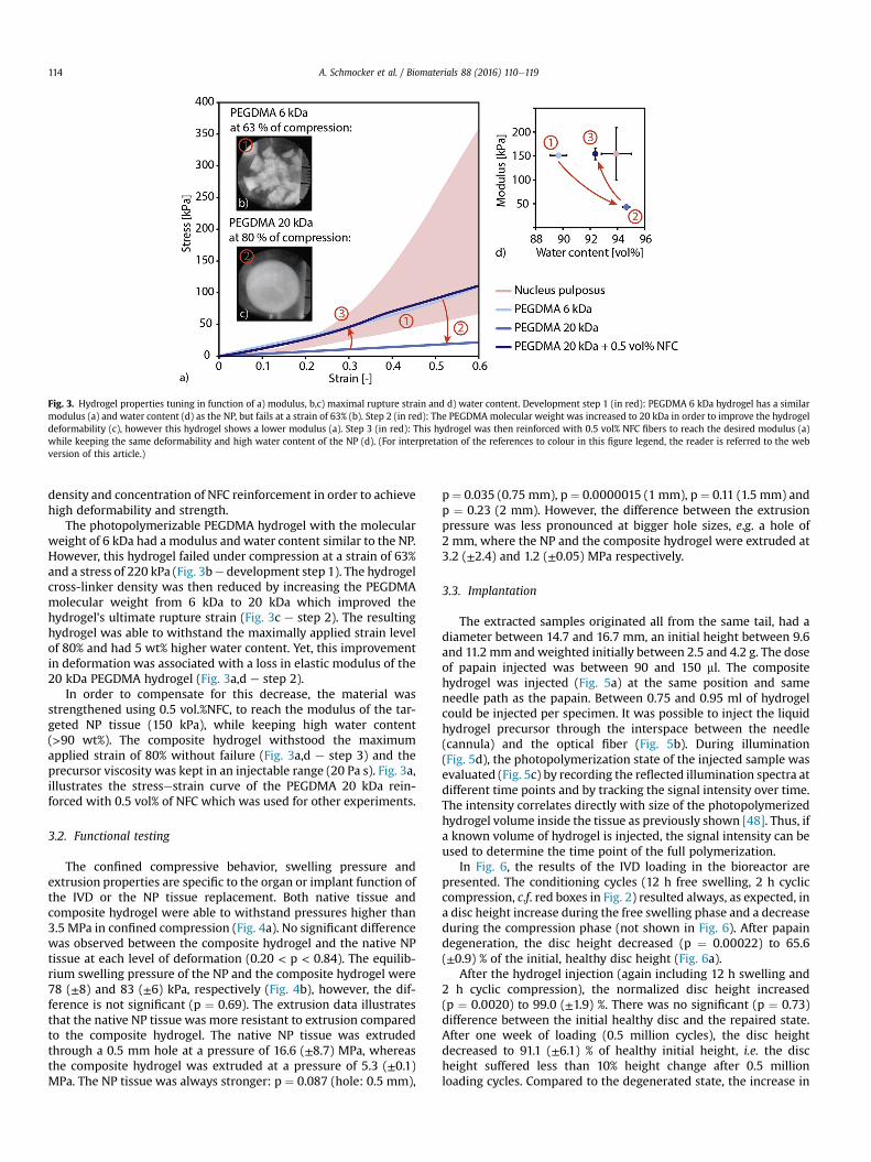
The photopolymerizable PEGDMA hydrogel with the molecularweight of 6 kDa had a modulus and water content similar to the NP.However, this hydrogel failed under compression at a strain of 63%and a stress of 220 kPa (Fig. 3be development step 1). The hydrogelcross-linker density was then reduced by increasing the PEGDMAmolecular weight from 6 kDa to 20 kDa which improved thehydrogel's ultimate rupture strain (Fig. 3c e step 2). The resultinghydrogel was able to withstand the maximally applied strain levelof 80% and had 5 wt% higher water content. Yet, this improvementin deformation was associated with a loss in elastic modulus of the20 kDa PEGDMA hydrogel (Fig. 3a,d e step 2).
In order to compensate for this decrease, the material wasstrengthened using 0.5 vol.%NFC, to reach the modulus of the tar-geted NP tissue (150 kPa), while keeping high water content(>90 wt%). The composite hydrogel withstood the maximumapplied strain of 80% without failure (Fig. 3a,d e step 3) and theprecursor viscosity was kept in an injectable range (20 Pa s). Fig. 3a,illustrates the stressestrain curve of the PEGDMA 20 kDa rein-forced with 0.5 vol% of NFC which was used for other experiments.
3.2. Functional testing
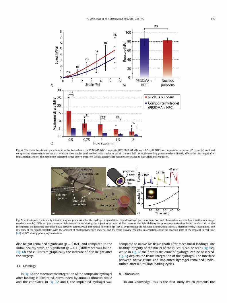
The confined compressive behavior, swelling pressure andextrusion properties are specific to the organ or implant function ofthe IVD or the NP tissue replacement. Both native tissue andcomposite hydrogel were able to withstand pressures higher than3.5 MPa in confined compression (Fig. 4a). No significant differencewas observed between the composite hydrogel and the native NPtissue at each level of deformation (0.20 < p < 0.84). The equilib-rium swelling pressure of the NP and the composite hydrogel were78 (±8) and 83 (±6) kPa, respectively (Fig. 4b), however, the dif-ference is not significant (p ¼ 0.69). The extrusion data illustratesthat the native NP tissue was more resistant to extrusion comparedto the composite hydrogel. The native NP tissue was extrudedthrough a 0.5 mm hole at a pressure of 16.6 (±8.7) MPa, whereasthe composite hydrogel was extruded at a pressure of 5.3 (±0.1)MPa. The NP tissue was always stronger: p ¼ 0.087 (hole: 0.5 mm),
p¼ 0.035 (0.75 mm), p¼ 0.0000015 (1 mm), p ¼ 0.11 (1.5 mm) andp ¼ 0.23 (2 mm). However, the difference between the extrusionpressure was less pronounced at bigger hole sizes, e.g. a hole of2 mm, where the NP and the composite hydrogel were extruded at3.2 (±2.4) and 1.2 (±0.05) MPa respectively.
3.3. Implantation
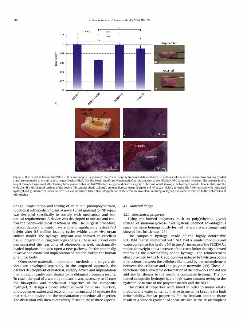
The extracted samples originated all from the same tail, had adiameter between 14.7 and 16.7 mm, an initial height between 9.6and 11.2 mm andweighted initially between 2.5 and 4.2 g. The doseof papain injected was between 90 and 150 ml. The compositehydrogel was injected (Fig. 5a) at the same position and sameneedle path as the papain. Between 0.75 and 0.95 ml of hydrogelcould be injected per specimen. It was possible to inject the liquidhydrogel precursor through the interspace between the needle(cannula) and the optical fiber (Fig. 5b). During illumination(Fig. 5d), the photopolymerization state of the injected sample wasevaluated (Fig. 5c) by recording the reflected illumination spectra atdifferent time points and by tracking the signal intensity over time.The intensity correlates directly with size of the photopolymerizedhydrogel volume inside the tissue as previously shown [48]. Thus, ifa known volume of hydrogel is injected, the signal intensity can beused to determine the time point of the full polymerization.
In Fig. 6, the results of the IVD loading in the bioreactor arepresented. The conditioning cycles (12 h free swelling, 2 h cycliccompression, c.f. red boxes in Fig. 2) resulted always, as expected, ina disc height increase during the free swelling phase and a decreaseduring the compression phase (not shown in Fig. 6). After papaindegeneration, the disc height decreased (p ¼ 0.00022) to 65.6(±0.9) % of the initial, healthy disc height (Fig. 6a).
After the hydrogel injection (again including 12 h swelling and2 h cyclic compression), the normalized disc height increased(p ¼ 0.0020) to 99.0 (±1.9) %. There was no significant (p ¼ 0.73)difference between the initial healthy disc and the repaired state.After one week of loading (0.5 million cycles), the disc heightdecreased to 91.1 (±6.1) % of healthy initial height, i.e. the discheight suffered less than 10% height change after 0.5 millionloading cycles. Compared to the degenerated state, the increase in
Fig. 3. Hydrogel properties tuning in function of a) modulus, b,c) maximal rupture strain and d) water content. Development step 1 (in red): PEGDMA 6 kDa hydrogel has a similarmodulus (a) and water content (d) as the NP, but fails at a strain of 63% (b). Step 2 (in red): The PEGDMA molecular weight was increased to 20 kDa in order to improve the hydrogeldeformability (c), however this hydrogel shows a lower modulus (a). Step 3 (in red): This hydrogel was then reinforced with 0.5 vol% NFC fibers to reach the desired modulus (a)while keeping the same deformability and high water content of the NP (d). (For interpretation of the references to colour in this figure legend, the reader is referred to the webversion of this article.)
A. Schmocker et al. / Biomaterials 88 (2016) 110e119114
disc height remained significant (p ¼ 0.021) and compared to theinitial healthy state, no significant (p ¼ 0.11) difference was found.Fig. 6b and c illustrate graphically the increase of disc height afterthe surgery.
3.4. Histology
In Fig. 6d themacroscopic integration of the composite hydrogelafter loading is illustrated, surrounded by annulus fibrosus tissueand the endplates. In Fig. 6e and f, the implanted hydrogel was
compared to native NP tissue (both after mechanical loading). Thehealthy integrity of the nuclei of the NP cells can be seen (Fig. 6e),while in Fig. 6f the fibrous structure of hydrogel can be observed.Fig. 6g depicts the tissue integration of the hydrogel. The interfacebetween native tissue and implanted hydrogel remained undis-turbed after 0.5 million loading cycles.
4. Discussion
To our knowledge, this is the first study which presents the
Fig. 4. The three functional tests done in order to evaluate the PEGDMA-NFC composite (PEGDMA 20 kDa with 0.5 vol% NFC) in comparison to native NP tissue (a) confinedcompression stressestrain curves that evaluate the samples confined behavior similar as within the real IVD tissue, (b) swelling pressure which directly affects the disc height afterimplantation and (c) the maximum tolerated stress before extrusion which assesses the sample's resistance to extrusion and expulsion.
Fig. 5. a) Customized minimally invasive surgical probe used for the hydrogel implantation. Liquid hydrogel precursor injection and illumination are combined within one singleneedle (cannula). Different joints ensure high pressurization during the injection. An optical fiber permits the light delivery for photopolymerization. b) At the distal tip of theinstrument, the hydrogel precursor flows between cannula wall and optical fiber into the IVD. c) By recording the reflected illumination spectra a signal intensity is calculated. Theintensity of the signal correlates with the amount of photopolymerized material and therefore provides valuable information about the reaction state of the implant in real time[48]. d) IVD during photopolymerization.
A. Schmocker et al. / Biomaterials 88 (2016) 110e119 115
design, implantation and testing of an in situ photopolymerized,functional orthopedic implant. A novel tunedmaterial for NP repairwas designed specifically to comply with mechanical and bio-optical requirements. A device was developed to initiate and con-trol the photoechemical reaction in situ. The surgical procedure,medical device and implant were able to significantly restore IVDheight after 0.5 million loading cycles within an ex vivo organculture model. The hydrogel implant also showed an excellenttissue integration during histology analysis. These results not onlydemonstrated the feasibility of photopolymerized, mechanicallyloaded implants, but also open a new pathway for the minimallyinvasive and controlled implantation of material within the humanor animal body.
Often novel materials, implantation methods and surgery de-vices are developed separately. In the proposed approach, theparallel development of material, surgery device and implantationmethod significantly contributed to the obtained promising results.To reach the goal of a working implant it was necessary to 1) tunethe bio-optical and mechanical properties of the compositehydrogel, 2) design a device which allowed for in situ injection,photopolymerization and reaction monitoring and 3) evaluate thematerial, the device and the implantation procedure all together.The discussion will then successively focus on these three aspects.
4.1. Material design
4.1.1. Mechanical propertiesUsing pre-formed polymers, such as poly(ethylene glycol)
instead of monomer/cross-linker systems seemed advantageoussince the more homogenously formed network was stronger andshowed less brittleness [25].
The composite hydrogel made of the highly deformablePEGDMA matrix reinforced with NFC had a similar modulus andwater content as the healthy NP tissue. An increase of the PEGDMA'smolecularweight and a decrease of the cross-linker density allowedimproving the deformability of the hydrogel. The reinforcementeffect provided by theNFC additionwas induced by hydrogen bondsinteractions between the cellulose fibrils and by the entanglementbetween the cellulose and the polymer networks [49]. These in-teractions still allowed the deformation of the networks and did notadd any brittleness to the resulting composite hydrogel. The ob-tained composite hydrogel had a high water content owing to thehydrophilic nature of the polymer matrix and the NFCs.
The material properties were tuned in order to mimic elasticmodulus and water content of native tissue while keeping the highdeformability. Similar properties for the implant and the tissueresult in a smooth gradient of shear stresses at the tissue/implant
Fig. 6. a) Disc height of bovine tail IVD (n ¼ 3) before surgery (degenerated state), after surgery (repaired state) and after 0.5 million cycles of ex vivo compressive loading (loadedstate) are compared to the initial disc height (healthy disc). The disc height significantly increased after implantation of the PEGDMA-NFC composite hydrogel. The increase in discheight remained significant after loading. b) Degenerated bovine tail IVD before surgery and c) after surgery. d) IVD cut in half showing the hydrogel, annulus fiborsus (AF) and theendplate (EP). Histological sections of the bovine IVD samples (H&E-staining): annulus fibrosus tissue (purple) and NP tissue (violet). e) Native NP, f) NP replaced with implantedhydrogel and g) interface between native tissue and implanted tissue. (For interpretation of the references to colour in this figure legend, the reader is referred to the web version ofthis article.)
A. Schmocker et al. / Biomaterials 88 (2016) 110e119116

interface which reduces the risk of extrusion or expulsion. In orderto evaluate the possible failure modes of the implant, differenttesting methods were designed. The PEGDMA-NFC compositehydrogel was tested in a confined and unconfined compressionmode. The similar behavior of the composite hydrogel and thenative tissue in confined compression demonstrated that the ma-terial interaction with water is similar to that of the NP tissue forthe range of applied stresses and testing rates.
The comparable swelling pressures of the composite hydrogeland of the native NP indicated that the implant possibly can restorethe healthy IVD height by swelling. Furthermore, the swelling leadto a continuous tissue/implant interface and ensured a homoge-nous load distribution. The extrusion test evaluated the hydrogelperformance in the harshest condition where the material wassqueezed until it ruptured completely. It could not bulge or adhereto surrounding tissue as it would do in reality. The compositehydrogel ruptured to pieces at high pressures whereas the nativetissue is irreversibly deformed, but without rupturing. Thisdifferent behavior might be due to a lack of available energydissipation within the composite hydrogel system compared to thenative NP tissue. This indicates that by adding energy dissipationroutes in the hydrogel, e.g. via ionic interactions, an increasedresistance to extrusion of the hydrogel could be expected. Theextrusion test had also one contradictory aspect. There was a sig-nificant difference between NP and hydrogel with a hole of 0.75 and1mm, but no significant difference for 0.5, 1.5 and 2mm.We do nothave a clear explanation for this result. The non-signifcant differ-ence with hole size of 0.5 mm might be due to the extremely highand non-physiological extrusion pressures (up to 25 MPa). Morework is required to understand the extrusion process which mightbe more complex and include non linear rupture mechanisms.
4.1.2. Bio-optical propertiesPhotopolymerization allows for controlled curing and also in-
creases the implant's adherence to native tissue. The photo-polymerizable curing system is highly advantageous compared toother in situ cured systems (e.g. heat reactive or chemical curing)because it provides the surgeonwith an extra degree of freedom bydecoupling the material insertion and curing. Additionally, thephotopolymerization of an injected, low-viscosity liquid enablesthe precursor to flow into tissue interstices and leads to an optimalcontact between implanted material and tissue. During illumina-tion, the penetration of liquid precursor into the microroughness ofthe surrounding tissue results in the formation of mechanicalinterlocking which improves the tissue/implant interface [50].
The illumination from an optical fiber results in a cone of light.We have previously developed aMonte Carlo model to simulate thecuring light distribution in the hydrogel [51]. It was shown that theaddition of light scattering elements such as intralipid particleschange the conic polymerization shape to a homogenous sphericalshape. However, lipid addition drastically weakened the mechan-ical properties of the hydrogel. To avoid this drawback, experi-mental data illustrated that the NFC fibers and the network theyform, also act as light scattering element resulting in acceleratedcuring [39] and more homogeneously cured volumes [51].
4.2. Surgical device design
Injection, photopolymerization or monitoring of a photo-polymer using a probe have been achieved previously, but thecombination of the three functions in one probe is novel. We haveshown that the probe can monitor the whole curing volume [48].
A potential issue of the surgical device is however the 1 mmincision which may lead later to the extrusion of implanted mate-rial. Within human IVDs pressures of up to 2.5 MPa have been
measured [52]. The extrusion results (Fig. 4c) indicate that thehydrogel can be extruded only through holes larger than 0.5 mm indiameter. In order to avoid extrusionwithin a human IVD the probediameter (19 Gage, ~1 mm) needs to be reduced to 0.5 mm. Toachieve this, a smaller optical fiber and needle would be required todo the surgery. In the current study a LED light was used for illu-mination. The LED can easily be replaced with laser illuminationand be delivered within a small diameter fiber with an externaldiameter of 125 mm. This would allow performing the surgerythrough a needle as small as 25 or 26 Gages (0.51 and 0.46 mmrespectively) which is a relevant factor for the delivery of hydrogelsas has been shown from clinics and experimentally in animalstudies [53e55].
4.3. Evaluation of minimally invasive implantation procedure
Usually the biomechanical evaluation of tissue is associatedwith high inter-sample variance and strong creep within the first2 h of loading [56]. The conditioning method (free swelling andcyclic loading e c.f. red boxes in Fig. 2) counters this effect andallowed reaching standard deviations between 0.9 and 6.1% over 15dayswhich is low in the field [57e59]. Between day 2 and 8 the discheight decreased by 1.0% and between day 9 and 15 another 7.9%.Although these changes were not significant, one could argue thatideally there should not be any decrease in disc height. However,the natural degradation of the IVD in ex vivo condition is importantand usually much higher height decreases were observed over sucha time frame for healthy discs [60]. Thus it can be considered thatthe obtained 7.9% are negligible. The histology results in Fig. 6eegfurther support these results: the in situ photopolymerized com-posite hydrogel was able to integrate well into existing tissue andthe created interface was able to resist the extensive loadingregime. This suggests that the implanted material remains at itsposition during loading contributing tomoderate loss in disc heightand low standard deviations. However, the exact adhesion orentanglement mechanisms (e.g. mechanical interlocking or cova-lent bonding) remain unclear and need to be further investigated.
From a general point of view, two options can be considered toevaluate the procedure and the implant: in vivo or in vitro animalmodels. Due to their size andmetabolism, bovine, canine and ovineIVD models show high resemblance to human IVDs [61]. Yet,degraded in vivo IVD models come along with high ethical issuesand limited options to control the loading. The ex vivomodel in thebioreactors is an attractive solution to evaluate IVD replacements.The used bovine NP tissue has similar mechanical propertiescompared to human NP [62,63]. Additionally, bovine NP tissue canbe easily obtained while human non-degenerated tissue is difficultto obtain. To obtain long-term results in vivo, an attractive optioncould be the canine model [64] as it is the only animal model with aclinical veterinary counterpart.
To demonstrate the potential for a clinical translation, it isrequired that the implant is imaged during the implantation pro-cedure. In the Supplementary data II, the water in the hydrogel wasreplaced with a water-based Iodine marker. The first picture illus-trated how the injected material can be imaged in a clinical situa-tion using X-ray tomography to inject the material and visualize itsposition after injection. The second picture demonstrated how theT2 signal during magnetic resonance imaging could be used todifferentiate between an empty and a filled cavity.
Another requirement for a clinical translation is the need to treatlater stage IVD degeneration or disc herniation (which are twodistinct pathologies) where the procedure is limited by thedegenerative state of the AF. We show that our material is able toresist extrusion through a hole of 0.5 mm up to pressures of ~5 MPaand 2mmup to pressures of ~1MPa (Fig. 4c). We also show that the
A. Schmocker et al. / Biomaterials 88 (2016) 110e119 117

material is not extruded over a cyclic loading of 0.5 million cycles inbovine intervertebral disc. Thus, the proposed implant andmaterialbear a certain potential to treat NP and AF tissue. Yet, for pathol-ogies including large AF fissures, it would be necessary to combineit with an AF sealant device. It should also be considered that thereare other solutions such as total IVD replacement [65,66], whichespecially seem to be promising when imitating the viscoelasticresponse of the IVD [67], something which was not taken into ac-count in this work.
A remaining challenge is to decrease the total illuminationcuring time, which, for this study, was chosen to be excessively long(45 min) to assure optimal tissue integration. However, wedemonstrated previously that volumes of more than 2 ml (i.e.enough for a human NP) can be photopolymerized in less than10 min using the same optical device [68]. It is also possible tochoose different photoinitiators such as Eosin Y with higher pho-topolymerization rates in the range between 10 s and 3 min [69].Moreover, the curing power in the experiment was limited to 4mWin the fiber and can easily be increased by more than one order ofmagnitude using a laser light source. In summary, the procedurecould easily be translated into clinics and seems to have a highpotential to be successful.
5. Conclusions
A composite hydrogel having similar functional properties asa native bovine NP was developed. Using a specifically designedprobe for in situ photopolymerization, the composite hydrogelwas implanted into an IVD model and was able to withstandloading without signs of damage. The composite hydrogel showedpromising performance during implantation and significantlyre-established IVD disc height from 65.6 to 99.0% and maintained iton a significantly higher level over 0.5 million loading cycles duringan ex vivo study. After 15 days of loading, a continuous, undisturbedtissue/implant interface was observed during histology.
6. Acknowledgments
Funding for this work was provided by the two SNF grants#10024003165465 and #310030_153411. We acknowledge Dr. T.Zimmermann and Dr. P. Tingaut (App. Wood Mat. Lab., EMPA,Dübendorf, Switzerland) for providing and preparing the NFC fi-bers, the group of Prof. H. Frauenrath (Lab. of Macromolec. andOrganic Mat., EPFL), specifically Dr. R. Marty, for his assistance withthe synthesis of PEGDMA, Sandra Jaccoud (Lab. of Biomech. Or-thopedics, EPFL) for help and advice related to sample preparationand sterilization, Eva Roth (Inst. for Surg. Techn. and Biomech.,University of Bern, Switzerland) for her assistancewith biochemicalassays and Jessica Dessimoz (Histology Core Facility, EPFL) for herhelp with histology. We thank Dr. Harald Bon!el, Department ofDiagnostic, Interventional and Pediatric Radiology, Inselspital,University of Bern for the MRI scanning of the bovine IVDs.
Appendix A. Supplementary data
Supplementary data related to this article can be found at http://dx.doi.org/10.1016/j.biomaterials.2016.02.015.
References
[1] D. Borenstein, Mechanical low back pain[mdash]a rheumatologist's view, Nat.Rev. Rheumatol. 9 (2013) 643e653.
[2] S. Dagenais, J. Caro, S. Haldeman, A systematic review of low back pain cost ofillness studies in the United States and internationally, Spine J. 8 (2008) 8e20.
[3] J.N. Katz, Lumbar disc disorders and low-back pain: socioeconomic factors andconsequences, J. Bone Jt. Surg. 88 (2006) 21e24.
[4] M.W. van Tulder, B.W. Koes, L.M. Bouter, Conservative treatment of acute andchronic nonspecific low back pain: a systematic review of randomizedcontrolled trials of the most common interventions, Spine 22 (1997)2128e2156.
[5] G. WADDELL, 1987 Volvo award in clinical sciences: a new clinical model forthe treatment of low-back pain, Spine 12 (1987) 632e644.
[6] A.H. McGregor, S.P.F. Hughes, The evaluation of the surgical management ofnerve root compression in patients with low back pain: part 1: the assessmentof outcome, Spine 27 (2002) 1465e1470.
[7] R. Chou, J. Baisden, E.J. Carragee, D.K. Resnick, W.O. Shaffer, J.D. Loeser, Surgeryfor low back pain: a review of the evidence for an American pain societyclinical practice guideline, Spine 34 (2009) 1094e1109.
[8] P. Fritzell, O. H€agg, P. Wessberg, A. Nordwall, S.L.S.S. Group, 2001 Volvo awardwinner in clinical studies: lumbar fusion versus nonsurgical treatment forchronic low back pain: a multicenter randomized controlled trial from theswedish lumbar spine study group, Spine 26 (2001) 2521e2532.
[9] C.M. Bono, C.K. Lee, Critical analysis of trends in fusion for degenerative discdisease over the past 20 years: influence of technique on fusion rate andclinical outcome, Spine 29 (2004) 455e463.
[10] N. Miyakoshi, E. Abe, Y. Shimada, K. Okuyama, T. Suzuki, K. Sato, Outcome ofone-level posterior lumbar interbody fusion for spondylolisthesis and post-operative intervertebral disc degeneration adjacent to the fusion, Spine 25(2000) 1837e1842.
[11] M.M. Panjabi, Clinical spinal instability and low back pain, J. Electromyogr.Kinesiol. 13 (2003) 371e379.
[12] D. Coric, P.V. Mummaneni, Nucleus replacement technologies, J. Neurosurg.Spine 8 (2008) 115e120.
[13] H. Serhan, D. Mhatre, H. Defossez, C.M. Bono, Motion-preserving technologiesfor degenerative lumbar spine: The past, present, and future horizons, SAS J. 5(2011) 75e89.
[14] M.H. Pelletier, W.R. Walsh, N. South, Nucleus replacement, Compr. Biomater.(2011) 171e190.
[15] J.C. Iatridis, S.B. Nicoll, A.J. Michalek, B.A. Walter, M.S. Gupta, Role of biome-chanics in intervertebral disc degeneration and regenerative therapies: whatneeds repairing in the disc and what are promising biomaterials for its repair?spine J. off. J. North Am. Spine Soc. 13 (2013) 243e262.
[16] N.L. Nerurkar, D.M. Elliott, R.L. Mauck, Mechanical design criteria for inter-vertebral disc tissue engineering, J. biomech. 43 (2010) 1017e1030.
[17] S. Reitmaier, U. Wolfram, A. Ignatius, H.-J. Wilke, A. Gloria, J.M. Martín-Mar-tínez, et al., Hydrogels for nucleus replacementefacing the biomechanicalchallenge, J. Mech. Behav. Biomed. Mater. 14 (2012) 67e77.
[18] G. Lewis, Nucleus pulposus replacement and regeneration/repair technolo-gies: present status and future prospects, J. Biomed. Mater. Res. Part B, Appl.biomater. 100 (2012) 1702e1720.
[19] M. Buonocore, Adhesive sealing of pits and fissures for caries prevention, withuse of ultraviolet light, J. Am. Dent. Assoc. 80 (1970) 324e328.
[20] J.A. Burdick, A.J. Peterson, K.S. Anseth, Conversion and temperature profilesduring the photoinitiated polymerization of thick orthopaedic biomaterials,Biomaterials 22 (2001) 1779e1786.
[21] J.L. Hill-West, S.M. Chowdhury, M.J. Slepian, J.A. Hubbell, Inhibition ofthrombosis and intimal thickening by in situ photopolymerization of thinhydrogel barriers, Proc. Natl. Acad. Sci. 91 (1994) 5967e5971.
[22] M.W. Grinstaff, Designing hydrogel adhesives for corneal wound repair, Bio-materials 28 (2007) 5205e5214.
[23] K.T. Nguyen, J.L. West, Photopolymerizable hydrogels for tissue engineeringapplications, Biomaterials 23 (2002) 4307e4314.
[24] K.S. Anseth, C.N. Bowman, L. Brannon-Peppas, Mechanical properties ofhydrogels and their experimental determination, Biomaterials 17 (1996)1647e1657.
[25] S. Naficy, H.R. Brown, J.M. Razal, G.M. Spinks, P.G. Whitten, Progress towardrobust polymer hydrogels, Aust. J. Chem. 64 (2011) 1007e1025.
[26] G. Lake, A. Thomas, The strength of highly elastic materials, Proc. R. Soc. Lond.Ser. A Math. Phys. Sci. 300 (1967) 108e119.
[27] K.S. Anseth, C.N. Bowman, L. Brannon-Peppas, Mechanical properties ofhydrogels and their experimental determination, Biomaterials 17 (1996)1647e1657.
[28] C.W. Peak, J.J. Wilker, G. Schmidt, A review on tough and sticky hydrogels,Colloid Polym. Sci. 291 (2013) 2031e2047.
[29] Y. Okumura, K. Ito, The polyrotaxane gel: a topological gel by figure-of-eightcross-links, Adv. Mater. 13 (2001) 485e487.
[30] J.P. Gong, Y. Katsuyama, T. Kurokawa, Y. Osada, Double-network hydrogelswith extremely high mechanical strength, Adv. Mater. 15 (2003) 1155e1158.
[31] J. Elisseeff, K. Anseth, D. Sims, W. McIntosh, M. Randolph, R. Langer, Trans-dermal photopolymerization for minimally invasive implantation, Proc. Natl.Acad. Sci. 96 (1999) 3104e3107.
[32] J.A. Hubbell, In situ material transformations in tissue engineering, MRS Bull.21 (1996) 33e35.
[33] S.J. Bryant, K.S. Anseth, Hydrogel properties influence ECM production bychondrocytes photoencapsulated in poly (ethylene glycol) hydrogels,J. Biomed. Mater. Res. 59 (2002) 63e72.
[34] S.J. Bryant, K.S. Anseth, Controlling the spatial distribution of ECM compo-nents in degradable PEG hydrogels for tissue engineering cartilage, J. Biomed.Mater. Res. Part A 64 (2003) 70e79.
[35] N. Nagai, H. Kaji, H. Onami, Y. Ishikawa, M. Nishizawa, N. Osumi, et al.,A polymeric device for controlled transscleral multi-drug delivery to the
A. Schmocker et al. / Biomaterials 88 (2016) 110e119118

posterior segment of the eye, Acta Biomater. 10 (2014) 680e687.[36] I. Sir!o, D. Plackett, Microfibrillated cellulose and new nanocomposite mate-
rials: a review, Cellulose 17 (2010) 459e494.[37] T. Zimmermann, E. P€ohler, T. Geiger, Cellulose fibrils for polymer reinforce-
ment, Adv. Eng. Mater. 6 (2004) 754e761.[38] A.C. Borges, C. Eyholzer, F. Duc, P.E. Bourban, P. Tingaut, T. Zimmermann, et al.,
Nanofibrillated cellulose composite hydrogel for the replacement of the nu-cleus pulposus, Acta Biomater. 7 (2011) 3412e3421.
[39] A. Khoushabi, A. Schmocker, C. Schizas, C. Moser, D. Pioletti, J.-A. Manson, etal., The combined roles of NFC fibres and PEGDM molecular weight on photo-polymerization, swelling behaviour and mechanical properties of compositehydrogels, Compos. Sci. Technol. 119 (2015) 93e99.
[40] S. Lin-Gibson, S. Bencherif, J.A. Cooper, S.J. Wetzel, J.M. Antonucci, B.M. Vogel,et al., Synthesis and characterization of peg dimethacrylates and theirhydrogels, Biomacromolecules 5 (2004) 1280e1287.
[41] S.C.W. Chan, A. Bürki, H.M. Bon!el, L.M. Benneker, B. Gantenbein-Ritter, Papain-induced in vitro disc degeneration model for the study of injectable nucleuspulposus therapy, Spine J. off. J. North Am. Spine Soc. 13 (2013) 273e283.
[42] B. Gantenbein, S. Illien-Jünger, S.C. Chan, J. Walser, L. Haglund, S.J. Ferguson, etal., Organ culture bioreactorseplatforms to study human intervertebral discdegeneration and regenerative therapy, Curr. stem cell Res. Ther. 10 (2015)339.
[43] J.C. Beckstein, S. Sen, T.P. Schaer, E.J. Vresilovic, D.M. Elliott, Comparison ofanimal discs used in disc research to human lumbar disc: axial compressionmechanics and glycosaminoglycan content, Spine 33 (2008) E166eE173.
[44] C.N. Demers, J. Antoniou, F. Mwale, Value and limitations of using the bovinetail as a model for the human lumbar spine, Spine 29 (2004) 2793e2799.
[45] C.L. Korecki, J.J. Costi, J.C. Iatridis, Needle puncture injury affects intervertebraldisc mechanics and biology in an organ culture model, Spine 33 (2008)235e241.
[46] S. Reitmaier, L. Kreja, K. Gruchenberg, B. Kanter, J. Silva-Correia, J. Oliveira, etal., In vivo biofunctional evaluation of hydrogels for disc regeneration, Eur.Spine J. 23 (2014) 19e26.
[47] C.W.C. Samantha, G.-R. Benjamin, Preparation of intact bovine tail interver-tebral discs for organ culture, J. Vis. Exp. (60) (2012).
[48] A. Schmocker, A. Khoushabi, C. Schizas, P.-E. Bourban, D. Pioletti, C. Moser,Miniature probe for the delivery and monitoring of a photopolymerizablematerial, J. Biomed. Opt. 20 (12) (2015).
[49] M.-P. Lowys, J. Desbrieres, M. Rinaudo, Rheological characterization of cellu-losic microfibril suspensions. Role of polymeric additives, Food Hydrocoll. 15(2001) 25e32.
[50] J. Elisseeff, K. Anseth, D. Sims, W. McIntosh, M. Randolph, R. Langer, Trans-dermal photopolymerization for minimally invasive implantation, Proc. Natl.Acad. Sci. 96 (1999) 3104e3107.
[51] A. Schmocker, A. Khoushabi, C. Schizas, P.-E. Bourban, D.P. Pioletti, C. Moser,Photopolymerizable hydrogels for implants: MONTE-Carlo modeling andexperimental in vitro validation, J. Biomed. Opt. 19 (2014) 35004.
[52] H.J. Wilke, P. Neef, M. Caimi, T. Hoogland, L.E. Claes, New in vivo measure-ments of pressures in the intervertebral disc in daily life, Spine 24 (1999)755e762.
[53] D.M. Elliott, C.S. Yerramalli, J.C. Beckstein, J.I. Boxberger, W. Johannessen,E.J. Vresilovic, The effect of relative needle diameter in puncture and shaminjection animal models of degeneration, Spine 33 (2008) 588e596.
[54] J.-L. Wang, Y.-C. Tsai, Y.-H. Wang, The leakage pathway and effect of needlegauge on degree of disc injury post anular puncture: a comparative studyusing aged human and adolescent porcine discs, Spine 32 (2007) 1809e1815.
[55] E.J. Carragee, A.S. Don, E.L. Hurwitz, J.M. Cuellar, J. Carrino, R. Herzog, 2009ISSLS prize winner: does discography cause accelerated progression ofdegeneration changes in the lumbar disc: a ten-year matched cohort study,Spine 34 (2009) 2338e2345.
[56] M.A. Adams, D.W. McMillan, T.P. Green, P. Dolan, Sustained loading generatesstress concentrations in lumbar intervertebral discs, Spine 21 (1996)434e438.
[57] R.D. Bowles, H.H. Gebhard, R. Hartl, L.J. Bonassar, Tissue-engineered inter-vertebral discs produce new matrix, maintain disc height, and restorebiomechanical function to the rodent spine, Proc. Natl. Acad. Sci. 108 (2011).
[58] N. Bergknut, La Smolders, L.H. Koole, G. Voorhout, R.E. Hagman, A.-S. Lagerstedt, et al., The performance of a hydrogel nucleus pulposus pros-thesis in an ex vivo canine model, Biomaterials 31 (2010) 6782e6788.
[59] K. Wuertz, K. Godburn, J.J. MacLean, A. Barbir, J. Stinnett Donnelly,P.J. Roughley, et al., In vivo remodeling of intervertebral discs in response toshort-and long-term dynamic compression, J. Orthop. Res. 27 (2009)1235e1242.
[60] C. Korecki, J. MacLean, J. Iatridis, Characterization of an in vitro intervertebraldisc organ culture system, Eur. Spine J. 16 (2007) 1029e1037.
[61] M. Alini, S.M. Eisenstein, K. Ito, C. Little, Masuda K. Kettler aA, et al., Are animalmodels useful for studying human disc disorders/degeneration? Eur. Spine J.off. Publ. Eur. Spine Soc. Eur. Spinal Deform. Soc. Eur. Sect. Cerv. Spine Res. Soc.17 (2008) 2e19.
[62] W. Johannessen, D.M. Elliott, Effects of degeneration on the biphasic materialproperties of human nucleus pulposus in confined compression, Spine 30(2005).
[63] D. P!eri!e, D. Korda, J.C. Iatridis, Confined compression experiments on bovinenucleus pulposus and annulus fibrosus: sensitivity of the experiment in thedetermination of compressive modulus and hydraulic permeability,J. biomech. 38 (2005) 2164e2171.
[64] N. Bergknut, J.P. Rutges, H.-J.C. Kranenburg, L.A. Smolders, R. Hagman, H.-J. Smidt, et al., The dog as an animal model for intervertebral disc degenera-tion? Spine 37 (2012) 351e358.
[65] A. Gloria, F. Causa, R. De Santis, P.A. Netti, L. Ambrosio, Dynamic-mechanicalproperties of a novel composite intervertebral disc prosthesis, J. Mater. Sci.Mater. Med. 18 (2007) 2159e2165.
[66] A. Gloria, R. De Santis, L. Ambrosio, F. Causa, K.E. Tanner, A multi-componentfiber-reinforced PHEMA-based hydrogel/HAPEXTM device for customizedintervertebral disc prosthesis, J. Biomater. Appl. 25 (2011) 795e810.
[67] J. Silva-Correia, A. Gloria, M.B. Oliveira, J.F. Mano, J.M. Oliveira, L. Ambrosio, etal., Rheological and mechanical properties of acellular and cell-laden meth-acrylated gellan gum hydrogels, J. Biomed. Mater. Res. Part A 101 (2013)3438e3446.
[68] A.M. Schmocker, A. Khoushabi, B. Gantenbein-Ritter, S. Chan, H.M. Bon!el, P.-E. Bourban, et al., Minimally invasive photopolymerization in intervertebraldisc tissue cavities, in: A. Wax, V. Backman (Eds.), SPIE BiOS: InternationalSociety for Optics and Photonics, 2014, p. 895206.
[69] Y. An, Ja Hubbell, Intraarterial protein delivery via intimally-adherent bilayerhydrogels, J. Control. Release 64 (2000) 205e215.
A. Schmocker et al. / Biomaterials 88 (2016) 110e119 119


![The protective effects of PI3K/Akt pathway on human nucleus pulposus … · 2020. 1. 28. · nucleus pulposus cells and nucleus pulposus progenitor cells [14]. Previous studies have](https://static.fdocuments.us/doc/165x107/60b265dd0d8b8040e758b496/the-protective-effects-of-pi3kakt-pathway-on-human-nucleus-pulposus-2020-1-28.jpg)