
Aph
25
Dr.Tarig Mahmoud Ahmed MD SUDAN HAIL UNIVERSITY KSA
-
Upload
tariggally -
Category
Health & Medicine
-
view
159 -
download
2
Transcript of Aph

Dr.Tarig Mahmoud Ahmed
MD SUDAN
HAIL UNIVERSITY KSA

Antepartum haemorrhage (APH) is any
bleeding occurring in the antenatal
period after 24 weeks to delivery of the
baby..
It complicates 2–5 per cent of
pregnancies.
At term, APH can be difficult to
distinguish from a ‘show’ which is the
release of the cervical mucus in the
early stages of labour.

CAUSES:
Placental causes:
Placental abruption
Placenta praevia
Vasa praevia
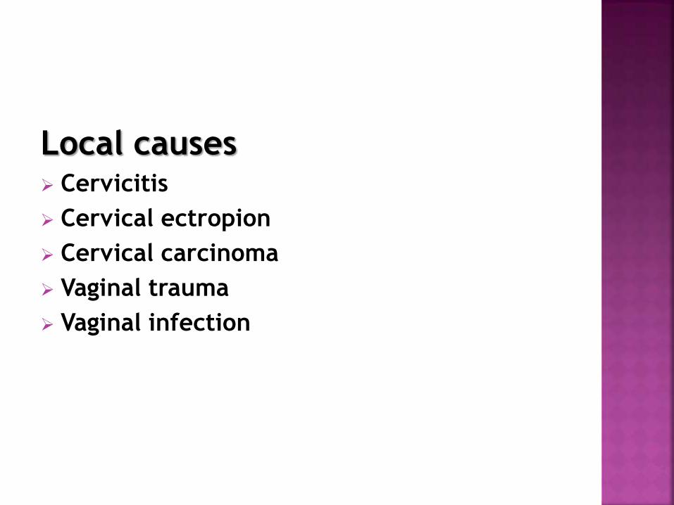
Local causes Cervicitis
Cervical ectropion
Cervical carcinoma
Vaginal trauma
Vaginal infection

Placenta praevia is defined as a
placenta that has implanted into the
lower segment of the uterus.
It is now classified as either major, in
which the placenta is covering the
internal cervical os, or minor, when the
placenta is sited within the lower
segment of the uterus, but does not
cover the cervical os.


Multiple gestation
Previous Caesarean section
Uterine structural anomaly
Assisted conception
Multiparous

The incidence in the UK is approximately 5
per 1000 and is increasing due to the rising
Caesarean section rate and increasing
maternal age.
In women who have had a previous
caesearean section, there is a risk of
placenta implants into, and thus invades,
into the previous scar ‘morbidly adherent
placenta’.

morbidly adherent placenta are three types:
1. Placenta accreta.Placenta is abnormally adherent to the
uterine wall.
2. Placenta increta.Placenta is abnormally invading into the
uterine wall.
3. Placenta percreta.Placenta is invading through the uterine
wall.

Diagnosis:
recurrent painless bleeding in the 3rd
trimester.
On abdominal palpation, the uterus will
be soft and non-tender and the
presenting part will be high.
ultrasoundscans will demonstrate the
abnormal location of the placenta.
A digital examination is contraindicated
as this can precipitate bleeding.

Management:
resuscitated using approach of ABC.
If the bleeding is minor and the fetus
uncompromised, the patient should be
admitted for observation for at least 24
hrs.
Women with major placenta praevia
who have had recurrent bleeding should
be admitted as inpatients from 34
weeks till Caesarean section at 37–38
weeks .

Cases of minor placenta praevia can be
considered for a vaginal delivery if the
placenta is a minimum of 2 cm away from
the cervical os.
There is risk of serious maternal haemo -
rrhage, either as APH or during Caesarean
section when the placental bed may not
contract, or due to morbid adherence.
Time of elective delivery when reaching
37–38 weeks.
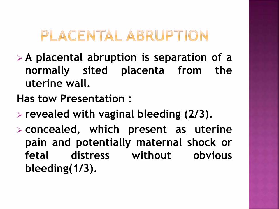
A placental abruption is separation of a
normally sited placenta from the
uterine wall.
Has tow Presentation :
revealed with vaginal bleeding (2/3).
concealed, which present as uterine
pain and potentially maternal shock or
fetal distress without obvious
bleeding(1/3).

Risk factors for placental abruption:
Hypertension
Smoking
Trauma to abdomen
Cocaine use
Anticoagulant therapy
Polyhydramnios and multiple gestation
FGR
High parity
sudden decompression of the uterus (e.g.after rupture of the membrane inpolyhydramnios).
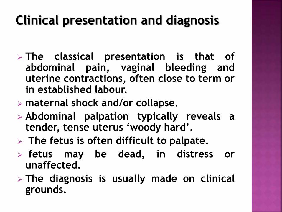
Clinical presentation and diagnosis
The classical presentation is that ofabdominal pain, vaginal bleeding anduterine contractions, often close to term orin established labour.
maternal shock and/or collapse.
Abdominal palpation typically reveals atender, tense uterus ‘woody hard’.
The fetus is often difficult to palpate.
fetus may be dead, in distress orunaffected.
The diagnosis is usually made on clinicalgrounds.

Hypovolaemic shock
Disseminated intravascular coagulation
(DIC)
Acute renal failure
Fetomaternal haemorrhage (important for
mothers who are rhesus negative)
Perinatal mortality
FGR (When abruption is chronic or recurrent)

Management:
resuscitated using approach of ABC.
2 14-gauge intravenous lines .
Full blood count and clotting studies.
Test for renal function and liver
function tests.
Cross-match at least 6 units of blood.
Fluid resuscitation intravenously.
Foley catheter into the bladder and
fluid balance chart.

In very severe cases, the fetus will be dead
and vaginal delivery can be accelerated by
artificial rupture of the membranes.
If the fetus is alive, delivery without
compromising the mother’s resuscitation is
urgent and this will usually be by Caesarean
section.

Placenta praevia Vs Placental abruption
pain
abruption - constant
placenta praevia - painless
obstetric shock
abruption - the actual amount of
bleeding may be far in excess of
vaginal loss
placenta praevia - obsetric shock in
proportion to amount of vaginal loss

uterus
abruption - uterus is tender and tense
placenta praevia - uterus is non-tender
fetus
abruption - normal presentation and
lie
placenta praevia - may have abnormal
presentation and/ or lie

fetal heart
abruption - fetal heart distressed/absent
placenta praevia - in general, fetal heart
normal
associated problems:
abruption - may be a complication of pre-
eclampsia, may cause DIC.
placenta praevia - small antepartum
haemorrhage may occur before larger
bleed

Vasa praevia is rupture of fetal
vessels running within the
membranes, often near to the
cervical os and damaged when the
membranes rupture.
it is catastrophic for the fetus as it
is fetal blood that is lost

placenta praevia.
a velamentous placental insertion.
multiple pregnancy.

Management: When vasa previa ruptured
cardiotocograph will rapidly become
abnormal with a fetal tachycardia,followed
by deep deceleration.
If the baby is still alive once the diagnosis
is suspected, the immediate action is
delivery by emergency Caesarean section

Thank you