A&P Chapter 7 - Amazon Simple Storage Services3.amazonaws.com/scschoolfiles/760/ap_chapter_7.pdf ·...
25
1 The Skeletal System Chapter 7 The Skeletal System • Parts of the skeletal system • Bones • Joints • Cartilages • Ligaments • Divided into 2 divisions • Axial skeleton • Appendicular skeleton Functions of Bones • Supports the body • Protects soft organs • Movement • Storage of minerals and fats • Blood cell formation -
Transcript of A&P Chapter 7 - Amazon Simple Storage Services3.amazonaws.com/scschoolfiles/760/ap_chapter_7.pdf ·...

1
The Skeletal
System
Chapter 7
The Skeletal System
• Parts of the skeletal system
• Bones
• Joints
• Cartilages
• Ligaments
• Divided into 2 divisions
• Axial skeleton
• Appendicular skeleton
Functions of Bones
• Supports the body
• Protects soft organs
• Movement
• Storage of minerals and fats
• Blood cell formation -

2
Bones of the Human Body
• The skeleton has bones
• 2 basic types of bone tissue
• Compact bone
• Solid - strength
• Spongy bone
• Made of trabeculae – small needle-like pieces of bone
• Many open spaces
Compact & Spongy Bone
7-4
Classification of Bones• 4 types of bones – long, short, flat & irregular,
• Long bones
• Typically longer than wide
• Have a shaft with heads at both ends
• Examples:
• Short bones
• Generally cube-shape
• Contain mostly spongy bone
• Examples:

3
Classification of Bones• Flat bones
• Thin and flattened
• Usually curved
• Thin layers of compact bone around spongy bone
• Examples: Skull, ribs, sternum, scapula
• Irregular bones
• Irregular shape
• Example:
Slide 5.4cCopyright © 2003 Pearson Education, Inc. publishing as Benjamin Cummings
Figure 5.1
Gross Anatomy of a Long Bone
• Diaphysis
• Shaft
• Composed of mostly compact bone
• Epiphysis
• Ends of bone
• Composed mostly of spongy bone
• Proximal & distal ends Figure 5.2a

4
Structures of a Long Bone• Periosteum
• Outside covering of the diaphysis
• Fibrous connective tissue
• Sharpey’s fibers
• Secures periosteum to underlying bone
• Arteries
• Supply bone cells with nutrients
• Endosteum
• Inner layer
Structures of a Long Bone
• Articular cartilage
• Covers external
surface of the epiphyses
• Decreases friction at joint surfaces
• Epiphyseal plate –
growth plate
Figure 5.2a
Structures of a Long Bone
• Medullary cavity
• Cavity of the shaft
• Contains yellow marrow (fat)
Figure 5.2a
5
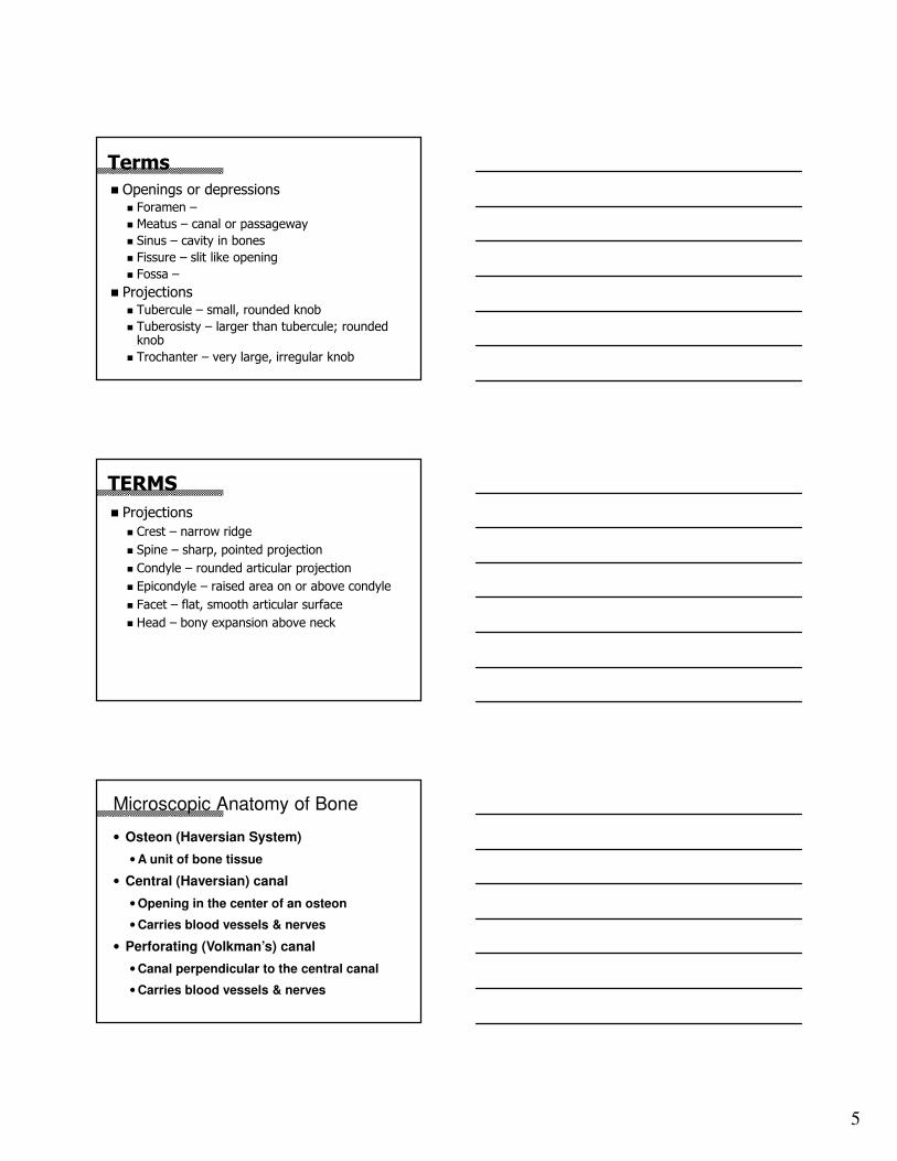
Terms
� Openings or depressions � Foramen –
� Meatus – canal or passageway
� Sinus – cavity in bones
� Fissure – slit like opening
� Fossa –
� Projections � Tubercule – small, rounded knob
� Tuberosisty – larger than tubercule; rounded knob
� Trochanter – very large, irregular knob
TERMS
� Projections
� Crest – narrow ridge
� Spine – sharp, pointed projection
� Condyle – rounded articular projection
� Epicondyle – raised area on or above condyle
� Facet – flat, smooth articular surface
� Head – bony expansion above neck
Microscopic Anatomy of Bone
• Osteon (Haversian System)
• A unit of bone tissue
• Central (Haversian) canal
• Opening in the center of an osteon
• Carries blood vessels & nerves
• Perforating (Volkman’s) canal
• Canal perpendicular to the central canal
• Carries blood vessels & nerves

6
Microscopic Anatomy of Bone• Osteocytes –
• Lacunae
• Cavities containing bone cells; arranged in concentric rings
• Lamellae
• -
• Canaliculi
• Tiny canals that radiate from the central canal to lacunae
• Form a transport system
Bone Growth
• Epiphyseal plates allow for growth of long bone during childhood
•New cartilage is continuously formed
•Older cartilage becomes ossified
•Bone replaces cartilage

7
Arrows
indicate
growth plates
are still
present.
Epiphyseal
plates usually
calcify
between the
ages of 17-25.
Types of Bone Cells
• Osteocytes
• Mature bone cells
• Osteoblasts
• Bone-forming cells
• Osteoclasts
• Bone-destroying cells
• Break down bone for remodeling & release of Ca
Skeleton
• Divided into 2 divisions
• Axial skeleton
• Appendicular skeleton

8
The Axial Skeleton
• Forms the longitudinal part of the body
• Divided into 3 parts
• Skull
• Vertebral column
• Thorax
The Axial Skeleton
The Skull
• 2 sets of bones
• Cranium
• Facial bones
• Bones are joined by sutures
• Only the mandible is attached by a freely
movable joint

9
The Skull
Skull
Frontal (1)
• forehead
• frontal sinuses
• supraorbital foramen
• coronal suture
SkullParietal (2)
• side & roof of cranium
• sagittal suture
• coronal suture
• lambdoidal suture
• squamosal suture

10
SkullTemporal (2)
• side of skull
• below parietal
• floor of cranium
• squamosal suture
• external acoustic meatus
• mastoid process
• styloid process
• zygomatic process
Mastoid
Styloid
Zygomatic
Process
SkullOccipital (1)
• back of skull
• foramen magnum
• occipital condyles
• external occipital
protuberence
• lambdoidal suture
SkullSphenoid (1)
• base of cranium
• sides of skull
• butterfly bone
• sella turcica Sella turcica
Sphenoid

11
Skull
Ethmoid (1)
• roof and walls of nasal cavity
• cribiform plates
• perpendicular plate
• ethmoidal sinuses
Facial SkeletonMaxillary (2)
• upper jaw
• anterior roof of mouth
• house upper teeth
• palatine suture
Facial SkeletonPalatine (2)
• L-shaped bones behind maxillae
• posterior section of hard palate; forms
soft palate

12
Facial SkeletonZygomatic (2)
• prominences of cheeks
• lateral walls & base of orbits
Facial SkeletonLacrimal (2)
• medial walls of orbits
• groove from orbit to
nasal cavity carries tears to
nasal cavity
Nasal (2)
• bridge of nose
Facial SkeletonVomer (1)
• inferior portion of nasal cavity
• joins w/ethmoid to form nasal septum
13
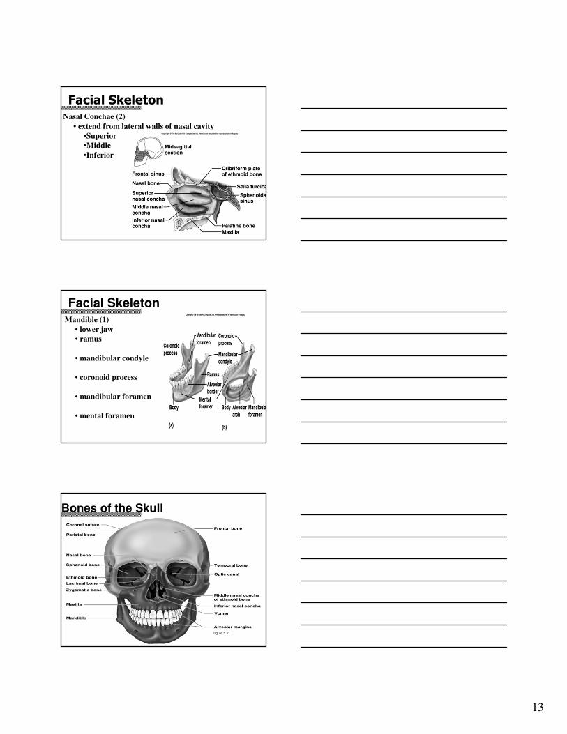
Facial SkeletonNasal Conchae (2)
• extend from lateral walls of nasal cavity
•Superior
•Middle
•Inferior
Facial SkeletonMandible (1)
• lower jaw
• ramus
• mandibular condyle
• coronoid process
• mandibular foramen
• mental foramen
Bones of the Skull
Figure 5.11

14
Human Skull, Superior View
Figure 5.8
Human Skull, Inferior View
Figure 5.9
Sinuses• Cavities w/in cranium
• Functions
• Lighten the skull
• Give resonance & amplification to voice
Figure 5.10

15
The Hyoid Bone
• only bone that does
not articulate
w/another bone
• Base for the tongue
The Vertebral Column
• Vertebrae
separated by intervertebral discs
• Each vertebrae is
given a name & #
according to its location
Figure 5.14
Structure of a Vertebrae
Figure 5.16
• Body - large flat area;
supports wt. of body
• Vertebral foramen – opening
for spinal cord
• Spinous process – posterior
projection
• Transverse process – lateral
projections on either side
• Pedicle – upper extension of
body
• Lamina – arch of vertebrae

16
3 Types of Vertebrae• Cervical - first 7 vertebrae of neck
• Only vertebrae w/ transverse foramen
• Only vertebrae w/ bifed spinous process
• Atlas –
• Axis – 2nd vertebrae with dens; pivot joint that allows head to
rotate
• Thoracic – vertebrae
•Long angled spinous processes
• Lumbar – in lower back
• Thickest body
• Spinous process are shorter & thicker
Cervical Vertebrae
Thoracic Vertebrae
Lumbar Vertebrae

17
Sacrum and Coccyx
� Sacrum – 5 fused vertebrae; join with/hip bones
� Coccyx – 4 fused vertebrae; forms tailbone
The Thorax
• Protects
major organs
Figure 5.19a
• Made up of
3 parts
• Sternum
• Ribs
• Thoracic vertebrae
Thorax
� Ribs – 12 pair
� True ribs – attached to sternum (1-7)
� False ribs – attached to cartilage of 7th
� Floating ribs – not attached to any structure
� Sternum – connected to ribs by cartilage
� Manubrium – superior region of sternum
� Body – main part
� Xiphoid – inferior end; used in landmarking for CPR

18
The Appendicular Skeleton
• Limbs (appendages)
• Pectoral girdle (shoulder)
• Pelvic girdle
Pectoral Girdle
• Composed of 2 bones
• Clavicle – collarbone; connects to sternum
& scapula
• Scapula – shoulder blade; connects to
humerus & clavicle
Shoulder

19
Bones of the Shoulder Girdle
Bones of the Shoulder Girdle
Bones of the Upper Limb
• Humerus -long bone
forming
upper arm

20
Bones of the Upper Limb
• The forearm has
2 bones
•Ulna –inner
bone
•Radius – bone
inline w/thumb
Bones of the Upper Limb• The hand
•Carpals – wrist
•Metacarpals – palm
•Phalanges – fingers
• Distal
• Middle
• Proximal
So Long Top Part Here Comes The Thumb
� S – scaphoid
� L – lunate
� T – triquetrum
� P – pisiform
� H – hamate
� C – capitate
� T – trapezoid
� T – trapezium
Carpal Bones

21
Bones of the Pelvic Girdle
• Coxal - hip bones
• Composed of three pair of fused bones
• Ilium
• Ischium
• Pubic bone
• The wt. of the upper body rests on the pelvis
• Protects several organs
• Reproductive organs
• Bladder
• Part of the large intestine
Illum� Form most of hip
� iliac crest – upper edge
� iliac fossa – depression under crest
� greater sciatic notch – large indentation for nerves
� sacroiliac joint – joins w/sacrum
� anterior superior spine – front of iliac crest
� posterior superior spine – back of iliac crest

22
Ishium
� Sit down bone
� ischial tuberosity – bony area that supports wt when sitting
� lesser sciatic notch – indentation for nerves
� ischial spine – points toward pelvic cavity

23
Pubis
� Most anterior bone
� Obturator foramen – largest opening in body; blood & nerves pass to thigh
� Acetabulum – articulation of femur

24
Bones of the Lower Limbs
• Femur- upper
thigh bone
• Articulates with
acetabulum
• Largest bone
Bones of the Lower Limbs
Slide
5.40b
• 2 bones of
lower leg
• Tibia - medial
• Fibula – lateral
Bones of the Lower Limbs
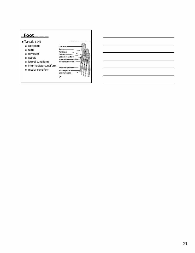
• The foot
•Talus –
•Metatarsals
– sole
•Phalanges –
•Calcaneus - Figure 5.25
25
Foot
� Tarsals (14)
� calcaneus
� talus
� navicular
� cuboid
� lateral cuneiform
� intermediate cuneiform
� medial cuneiform