Antitumor activity of some metal complexes: Effect on hepatic DNA, RNA, and protein synthesis in...
14
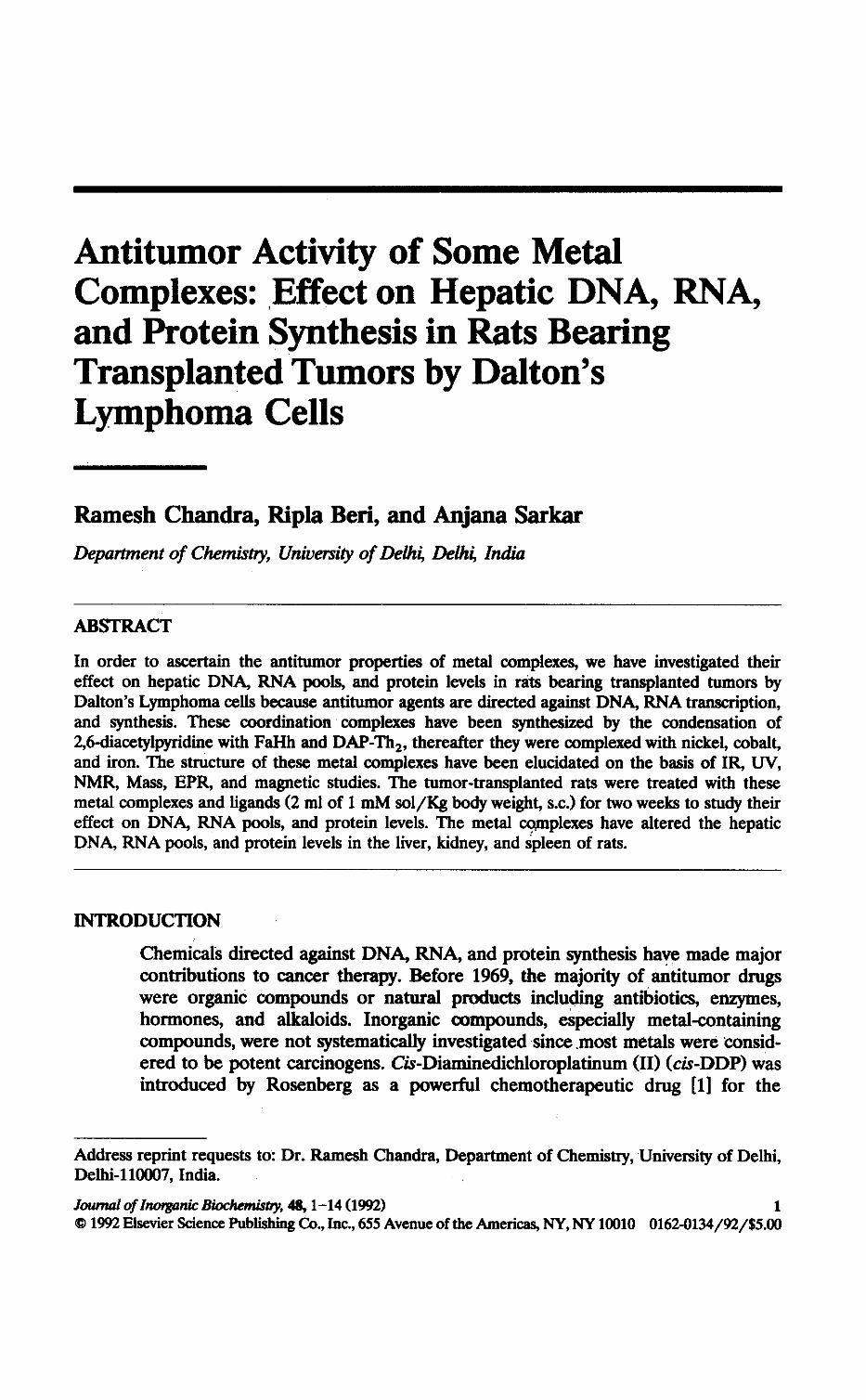
AntitumorActivityofSomeMetal Complexes :EffectonHepaticDNA,RNA, andProteinSynthesisinRatsBearing TransplantedTumorsbyDalton's LymphomaCells RameshChandra,RiplaBeri,andAnjanaSarkar DepartmentofChemistry,UniversityofDelhi,Delhi,India ABSTRACT Inordertoascertaintheantitumorpropertiesofmetalcomplexes,wehaveinvestigatedtheir effectonhepaticDNA,RNApools,andproteinlevelsinratsbearingtransplantedtumorsby Dalton'sLymphomacellsbecauseantitumoragentsaredirectedagainstDNA,RNAtranscription, andsynthesis .Thesecoordinationcomplexeshavebeensynthesizedbythecondensationof 2,6-diacetylpyridinewithFaHhandDAP-Th 2 , thereaftertheywerecomplexedwithnickel,cobalt, andiron .ThestructureofthesemetalcomplexeshavebeenelucidatedonthebasisofIR,UV, NMR,Mass,EPR,andmagneticstudies .Thetumor-transplantedratsweretreatedwiththese metalcomplexesandligands(2mlof1mMsol/Kgbodyweight, s .c.) fortwoweekstostudytheir effectonDNA,RNApools,andproteinlevels .Themetalcomplexeshavealteredthehepatic DNA,RNApools,andproteinlevelsintheliver,kidney,andspleenofrats . INTRODUCTION ChemicalsdirectedagainstDNA,RNA,andproteinsynthesishavemademajor contributionstocancertherapy .Before1969,themajorityofantitumordrugs wereorganiccompoundsornaturalproductsincludingantibiotics,enzymes, hormones,andalkaloids.Inorganiccompounds,especiallymetal-containing compounds,werenotsystematicallyinvestigatedsince .mostmetalswereconsid- eredtobepotentcarcinogens .Cis-Diaminedichloroplatinum(II) (cis-DDP) was introducedbyRosenbergasapowerfulchemotherapeuticdrug[1]forthe Addressreprintrequeststo :Dr .RameshChandra,DepartmentofChemistry,UniversityofDelhi, Delhi-110007,India . JournalofInorganicBiochemisdy, 48,1-14(1992) 1 01992ElsevierSciencePublishingCo .,Inc .,655AvenueoftheAmericas,NY,NY10010 0162-0134/92/$5 .00
-
Upload
ramesh-chandra -
Category
Documents
-
view
212 -
download
0
Transcript of Antitumor activity of some metal complexes: Effect on hepatic DNA, RNA, and protein synthesis in...
Antitumor Activity of Some MetalComplexes: Effect on Hepatic DNA, RNA,and Protein Synthesis in Rats BearingTransplanted Tumors by Dalton'sLymphoma Cells
Ramesh Chandra, Ripla Beri, and Anjana SarkarDepartment of Chemistry, University of Delhi, Delhi, India
ABSTRACT
In order to ascertain the antitumor properties of metal complexes, we have investigated theireffect on hepatic DNA, RNA pools, and protein levels in rats bearing transplanted tumors byDalton's Lymphoma cells because antitumor agents are directed against DNA, RNA transcription,and synthesis. These coordination complexes have been synthesized by the condensation of2,6-diacetylpyridine with FaHh and DAP-Th 2, thereafter they were complexed with nickel, cobalt,and iron. The structure of these metal complexes have been elucidated on the basis of IR, UV,NMR, Mass, EPR, and magnetic studies . The tumor-transplanted rats were treated with thesemetal complexes and ligands (2 ml of 1 mM sol/Kg body weight, s.c.) for two weeks to study theireffect on DNA, RNA pools, and protein levels . The metal complexes have altered the hepaticDNA, RNA pools, and protein levels in the liver, kidney, and spleen of rats .
INTRODUCTION
Chemicals directed against DNA, RNA, and protein synthesis have made majorcontributions to cancer therapy . Before 1969, the majority of antitumor drugswere organic compounds or natural products including antibiotics, enzymes,hormones, and alkaloids. Inorganic compounds, especially metal-containingcompounds, were not systematically investigated since .most metals were consid-ered to be potent carcinogens. Cis-Diaminedichloroplatinum (II) (cis-DDP) wasintroduced by Rosenberg as a powerful chemotherapeutic drug [1] for the
Address reprint requests to: Dr. Ramesh Chandra, Department of Chemistry, University of Delhi,Delhi-110007, India.
Journal of Inorganic Biochemisdy, 48,1-14 (1992)
10 1992 Elsevier Science Publishing Co., Inc., 655 Avenue of the Americas, NY, NY 10010 0162-0134/92/$5 .00
2 R. Chandra et alL
treatment of certain human cancers [2] and malignancies [3]. This carbon freecoordination compound, first synthesized more than a century ago, has becomethe first member of a new class of metal-based antitumor drugs . Numerousstudies suggest that the curvative effect of the metal complex can be attributedto the attack of cellular DNA and to the formation of several types of adductswith DNA bases. Several results also suggest that the antitumor activity ofcis-DDP is related to the intrastrand adducts [4-6] .
Most anticancer drugs in use today are directed against DNA transcriptionand synthesis [7]. Mithramycin is an antitumor antibiotic which is active againsta variety of experimental and human tumors [8]. Its antitumor activity is due tothe inhibition of DNA-directed RNA-synthesis, an effect which is achieved bybinding to double-stranded DNA [9] . This interaction with DNA requiresdivalent metal ions [10, 11] and the presence of guanine bases [12] . Recently,novel drugs with antimitochondrial properties were investigated for their antitu-mor activity because they appear to exploit the biochemical and structuralabnormalities of tumor mitochondria [13, 14]. Gossypol is one of a class ofcompounds that selectively accumulates within tumor mitochondria and subse-quently uncouples electron transport from oxidative phosphorylation [15, 16] .Although platinum complexes have been most intensively studied, research hasbeen extended to practically all metals .
Increasing interest in purely inorganic compounds, coordination complexes,and organometallic compounds [17-29], which are now extensively and systemat-ically screened as antitumor agents, have led us to study the in vivo antitumoractivity of these coordination metal complexes of macrocyclic ligands . The aimof the present investigation is to illustrate possibilities and problem in elucidat-ing biochemical defects at the level of mitochondria, and discuss the possibilityof therapeutic use of these metal complexes of macrocyclic ligands, whichresemble biological systems. This antitumor activity of these complexes has beenexhibited on the basis of hepatic DNA, RNA pools, and total protein levels inthe liver, kidney, and spleen of rats bearing transplanted tumors by Dalton'sLymphoma cells.
MATERIALS AND METHODS
Chemicals
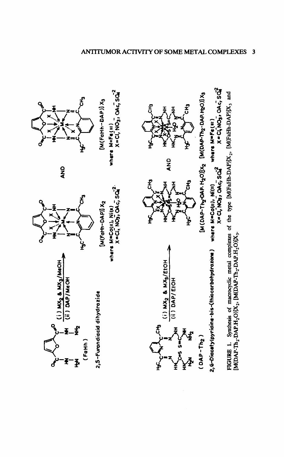
All chemicals used in the present investigation were of analytical grade andpurchased from Sigma Chemical Co. (MO, U.S.), Pharmacia (Sweden), E. Merck(Germany), and Fine Chemicals (India). Ligands (FaHh and DAP-Th 2 ) andmacrocyclic metal complexes of cobalt (Co+ 2 ), nickel (Ni +2 ), and iron (Fe")were synthesized in the laboratory [30] and characterized on the basis of spectraland thermal studies (Fig . 1).
Treatment of Animals
Female Wistar strain rats (110-150 g) bearing transplanted tumors were usedthroughout this study. Rats were housed individually in separate cages withraised wire mesh floors and were randomly allocated in fourteen differentgroups (four animals in each group) ; they all had equal body weights. Tumorswere transplanted in healthy rats by inoculation of Dalton's Lymphoma cells(2 x 10' cells), which became observable after 4-6 days . Animals with tumor
COW
CIIM MX2 & MX3/MSOH
NH(i
i)I
I
DAP/MeOH
H2N
NH2
(Faith)
2,5-Furandiacid dihydrazide
H3C
,CH3
I
1
(i)
MX2
& MX
3/Et
OH
HN~N
N%
(ii
)DA
P/ E
tOH
MCos Ss
HC
KiN
A(DAP-Th2)
0
[M(FaHh-DAP)] X2
wher
e M=
Co(u
), N
ita)
X=Cl,, N03, OA c, Sd?'
AND
O44h
-
00
IX
XHN~
ti~fNH
I .~.1
IA
X
II ~CH3
[M(FaHh-DAP)] X3
wher
e M=
Fs (
m)
X=Cl, NO3, OAc, S04 2
,CH3
r
a
SIR :
AND
"
tto
WNH
WC`
3C
•CH3
[M (DAP-Th
2-OAP.
H20)
]X2
[M(D
AP-;%-DAP. H2O)] X3
2,6-
Diacstylpyridine-bis-(thiocarbohydrazone
) where M=CoW), Ni(u)
when
MuF
e(ai
)X= Cl, NO j, OAc, SG42
XaCc NO3, OA
C-1S0
FIGURE
1.Sy
nthe
sis
of macrocyclic metal com
plex
es o
f th
e ty
pe [
M(Fa
Hh-D
AP)]
X2,
[M(
FaHh
-DAP
)]X
3an
d[M
(DAP
-The-DAP
.H20)]
X2, [M(DAP-Th
e-DAP.H
20)]X3
.
w
4 R Chandra et al.
growth were injected subcutaneously with 2.0 ml of 1 mM sol/Kg b .w. for twoweeks, of each metal complex (Co+ 2 , Ni + 2 , Fe + 3 ) of the two ligands as follows:
Control animals bearing transplanted tumors were injected with normal saline.Animals in all groups were allowed free access to laboratory rat chowfed anddrinking water ad labitum .
Tissue Preparation
Animals were fasted for 24 hrs after final administration of ligands and metalcomplexes before sacrifice . Animals were then anesthetized with ether andsacrificed by decapitation . Mitochondria and microsomes were prepared byhomogenizing the tissues (liver, kidney, and spleen) in three volumes of 0 .1 Mpotassium phosphate buffer, pH 7.4, containing 0.25 M sucrose as describedpreviously [31], after in situ perfusion of tissues with ice-cold saline .
DNA/RNA Determination
DNA and RNA were extracted from hepatic tissues by the method of Munro[32] .
Assays
DNA was assayed by the method of Burton [33]. RNA was estimated by themethod of Schneider [34] . Protein concentration in cellular homogenates andsubcellular fractions of liver, kidney, and spleen was estimated using crystallinebovine serum albumin as a standard, by the method of Lowry et al. [35] .
RESULTS
Only a single healthy cancer cell is required to initiate the entire process ofneoplasia, including unrestricted growth, metastasis, invasion, compression ofnontumor tissues, and other pathological manifestations of the disease . Thefailure to find specific differences between tumor cells and other cells indicatesa pressing need for improved methods for separation and identification of thenuclear and nucleolar components of cancer cells, i.e., DNA, RNA, nuclearenzymes, and proteins, in order to determine their functional significance .
Effect of Administration of Ligands and Their Metal Complexes on Body Weights
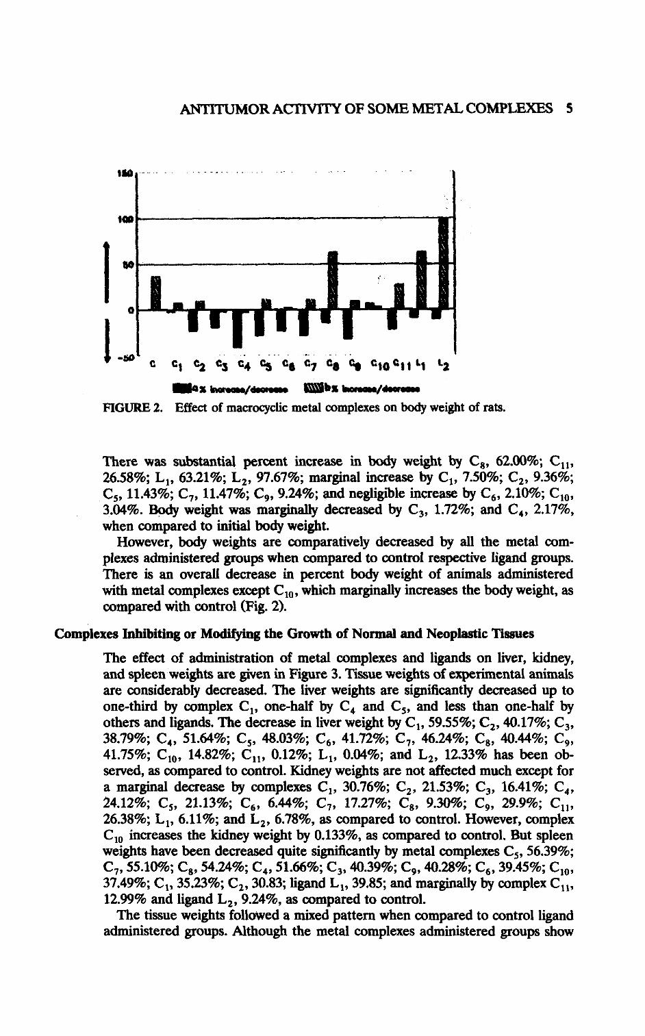
The alteration in body weight and percentage increase/decrease of s .c. adminin-stration of metal complexes and ligands is given in Figure 2 . Administration ofmetal complexes decreased body weight, as compared to initial body weight .
Group Treatment Group TreatmentC ControlC1 [Fe(FaHh-DAP)10 3 C7 [Fe(DAP-The-DAP.H20)]C13C2 [Co(FaHh-DAP)]C12 C8 ICo(DAP-11 2-DAP.H20)]C12C3 [Ni(FaHh-DAP)]S04 C9 [Co(DAP-The-DAP.H20)](OAc)2C4 IFe(FaHh-DAP)]2(S04)3 C10 [Ni(DAP-Th e-DAP.H20)$04C5 [Co(FaHh-DAP)](OAc)2 Cl, [Co(DAP-n2-DAP.H20)XNO3)2C6 [Co(FaHh-DAP)](N0 3)2 L2 DAP-Th2 (2,6-diacetylpyridine-
bis-thiocarbohydrazone)L1 FaHh (Furandiaciddihydrazide)
ANTITUMOR ACTIVITY OF SOME METAL COMPLEXES 5
110
140
C1 C2 C3 C4 %S C6 C7 es C6 C10'2 1 14rax wwemq/ .a Mb* bean»/M~
FIGURE 2. Effect of macrocyclic metal complexes on body weight of rats .
There was substantial percent increase in body weight by C8 , 62.00% ; C11,26.58%; L 1, 63.21% ; L 2, 97.67%; marginal increase by C,, 7.50%; C2 , 9.36% ;C5, 11.43%; C„ 11.47%; C9 , 9.24%; and negligible increase by C 6 , 2.10%; C10 ,3.04% . Body weight was marginally decreased by C 3, 1 .72%; and C4 , 2.17%,when compared to initial body weight .
However, body weights are comparatively decreased by all the metal com-plexes administered groups when compared to control respective ligand groups .There is an overall decrease in percent body weight of animals administeredwith metal complexes except C 10 , which marginally increases the body weight, ascompared with control (Fig. 2) .
Complexes Inhibiting or Modifying the Growth of Normal and Neoplastic Tissues
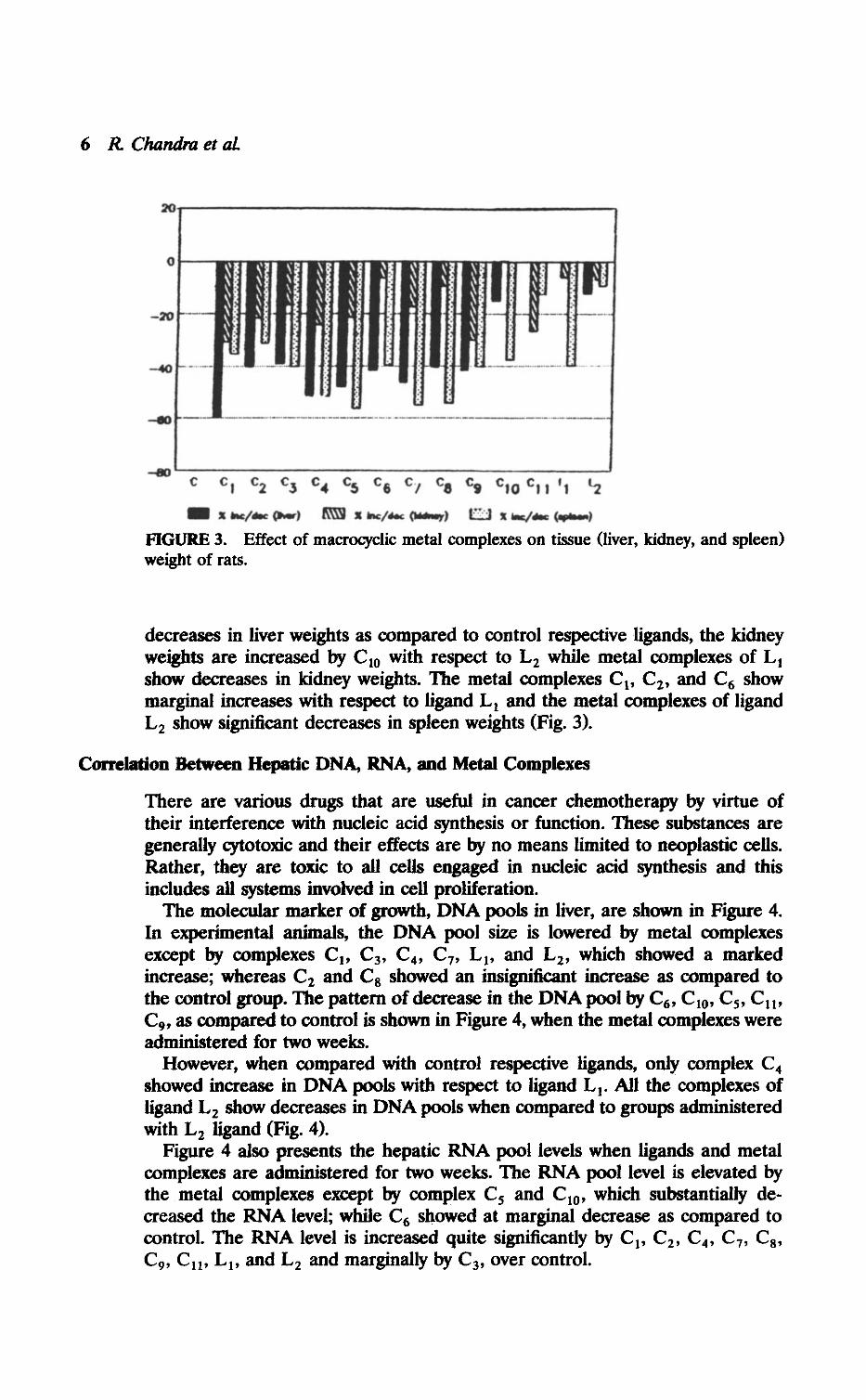
The effect of administration of metal complexes and ligands on liver, kidney,and spleen weights are given in Figure 3 . Tissue weights of experimental animalsare considerably decreased . The liver weights are significantly decreased up toone-third by complex C 1 , one-half by C 4 and C5, and less than one-half byothers and ligands. The decrease in liver weight by C1, 59.55%; C2 , 40.17%; C 3 ,38.79%; C4, 51.64%; C5, 48.03%; C6 , 41.72%; C 7, 46.24%; C8 , 40.44%; C9 ,41.75%; C 10, 14.82%; C11 , 0.12%; L1, 0.04%; and L 2 , 12.33% has been ob-served, as compared to control . Kidney weights are not affected much except fora marginal decrease by complexes C 1 , 30.76%; C2 , 21.53%; C3 , 16.41%; C4 ,24.12%; C5, 21.13%; C6 , 6.44%; C7, 17.27%; C 8, 9.30%; C9, 29.9% ; C11,26.38% ; L 1 , 6.11%; and L2 , 6.78%, as compared to control. However, complexC 10 increases the kidney weight by 0 .133%, as compared to control. But spleenweights have been decreased quite significantly by metal complexes C 5 , 56.39% ;C7, 55.10%; C8, 54.24%; C 4, 51.66%; C 3 , 40.39% ; C9 , 40.28%; C6 , 39.45% ; C,0,37.49%; C1, 35.23%; C2 , 30.83 ; ligand L 1, 39.85; and marginally by complex C 11 ,12.99% and ligand L 2 , 9.24%, as compared to control .
The tissue weights followed a mixed pattern when compared to control ligandadministered groups. Although the metal complexes administered groups show
6 R Chandra et al.
20
0
-20
-40
-40
c c ) c2 c3 c4 CS ce cY ca c9 CIO C) ) 't 12
01 z he/rr OWW) & i MC/Aw omw) Ci z r./dw 4P604
FIGURE 3. Effect of macrocyclic metal complexes on tissue (liver, kidney, and spleen)weight of rats .
decreases in liver weights as compared to control respective ligands, the kidneyweights are increased by C 10 with respect to L 2 while metal complexes of L 1show decreases in kidney weights . The metal complexes C 1, C2, and C6 showmarginal increases with respect to ligand L 1 and the metal complexes of ligandL2 show significant decreases in spleen weights (Fig. 3) .
Correlation Between Hepatic DNA, RNA, and Metal Complexes
There are various drugs that are useful in cancer chemotherapy by virtue oftheir interference with nucleic acid synthesis or function . These substances aregenerally cytotoxic and their effects are by no means limited to neoplastic cells.Rather, they are toxic to all cells engaged in nucleic acid synthesis and thisincludes all systems involved in cell proliferation .
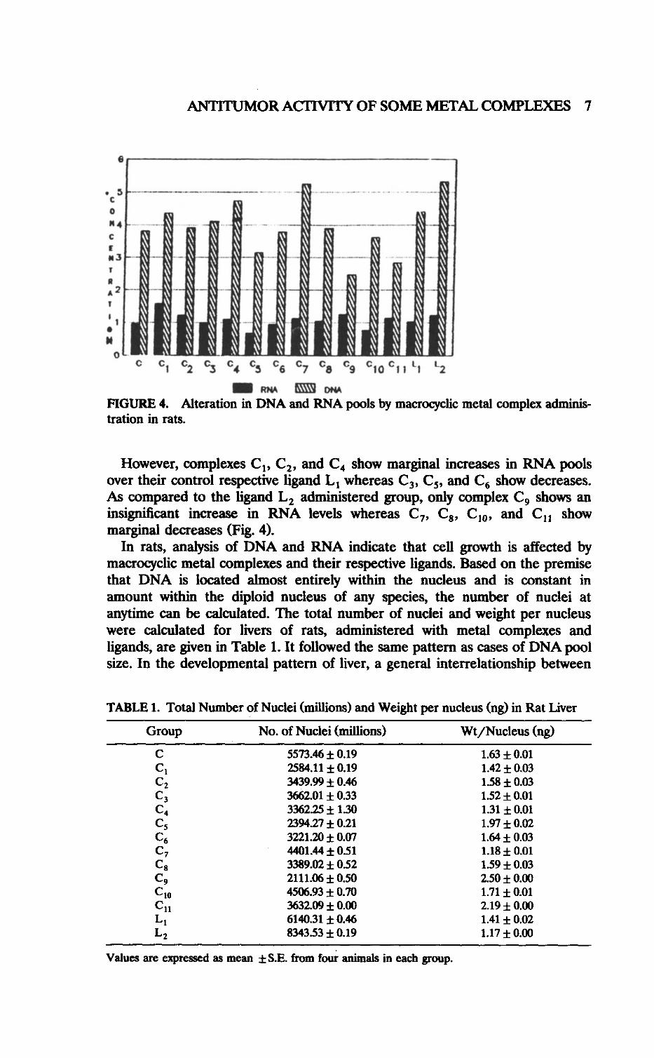
The molecular marker of growth, DNA pools in liver, are shown in Figure 4 .In experimental animals, the DNA pool size is lowered by metal complexesexcept by complexes C 1 , C3, C 4 , C7 , L 1 , and L2 , which showed a markedincrease; whereas C2 and Cg showed an insignificant increase as compared tothe control group. The pattern of decrease in the DNA pool by C6, C10, C5, C11,C9 , as compared to control is shown in Figure 4, when the metal complexes wereadministered for two weeks .However, when compared with control respective ligands, only complex C 4
showed increase in DNA pools with respect to ligand L 1 . All the complexes ofligand L2 show decreases in DNA pools when compared to groups administeredwith L2 ligand (Fig. 4) .
Figure 4 also presents the hepatic RNA pool levels when ligands and metalcomplexes are administered for two weeks . The RNA pool level is elevated bythe metal complexes except by complex C 5 and C 10 , which substantially de-creased the RNA level; while C6 showed at marginal decrease as compared tocontrol. The RNA level is increased quite significantly by C1, C 2 , C4 , C7 , C8 ,C9 , C 11 , L 1 , and L 2 and marginally by C 3 , over control .
ANTITUMOR ACTIVITY OF SOME METAL COMPLEXES 7
03
Ce '09 C IO' t 1 `1 12
(• RNA ® owlFIGURE 4 . Alteration in DNA and RNA pools by macrocycic metal complex adminis-tration in rats .
However, complexes C 1 , C2 , and C 4 show marginal increases in RNA poolsover their control respective ligand L 1 whereas C 3 , C5 , and C6 show decreases .As compared to the ligand L2 administered group, only complex C 9 shows aninsignificant increase in RNA levels whereas C 7, Cs, C 10 , and C11 showmarginal decreases (Fig . 4) .
In rats, analysis of DNA and RNA indicate that cell growth is affected bymacrocyclic metal complexes and their respective ligands. Based on the premisethat DNA is located almost entirely within the nucleus and is constant inamount within the diploid nucleus of any species, the number of nuclei atanytime can be calculated . The total number of nuclei and weight per nucleuswere calculated for livers of rats, administered with metal complexes andligands, are given in Table 1 . It followed the same pattern as cases of DNA poolsize. In the developmental pattern of liver, a general interrelationship between
TABLE 1. Total Number of Nuclei (millions) and Weight per nucleus (ng) in Rat Liver
Values are expressed as mean ±S.E. from four animals in each group .
Group No. of Nuclei (millions) Wt/Nucleus (ng)
C 5573.46 ± 0.19 1 .63 t 0.01C1 2584.11± 0.19 1.42 t 0.03C 2 3439.99 ± 0.46 1.58 t 0.03C 3 3662.01 ± 0.33 1.52 f 0.01C 4 3362.25 ± 1.30 1.31 t 0.01C 5 2394.27 ± 0.21 1.97 t 0.02C6 3221.20 ± 0.07 1.64 t 0.03C 7 4401 .44 ± 0.51 1.18± 0.01C8 3389.02± 0.52 1.59 t 0.03C9 2111.06 ± 0.50 2.50 t 0.00C 10 4506.93 ± 0.70 1 .71± 0.01C 11 3632.09 ± 0.00 2.19 ± 0.00Ll 6140.31 t 0.46 1 .41± 0.02L 2 834353 t 0.19 1 .17 ± 0.00
8 R Chandra et alL
cell number and cell size can be seen. As the cell number increases, the cell sizedecreases. This phenomenon can be explained by the fact that all the anabolicprocesses causing hypertrophy of the cell cannot cope with the rapidly proliferat-ing cells. Despite increases in hepatic DNA pools in rats in some groups, there islittle change in the organ wet weights.
Effect of Administration of Metal Complexes on Hepatic, Renal,and Splenic Total Proteins
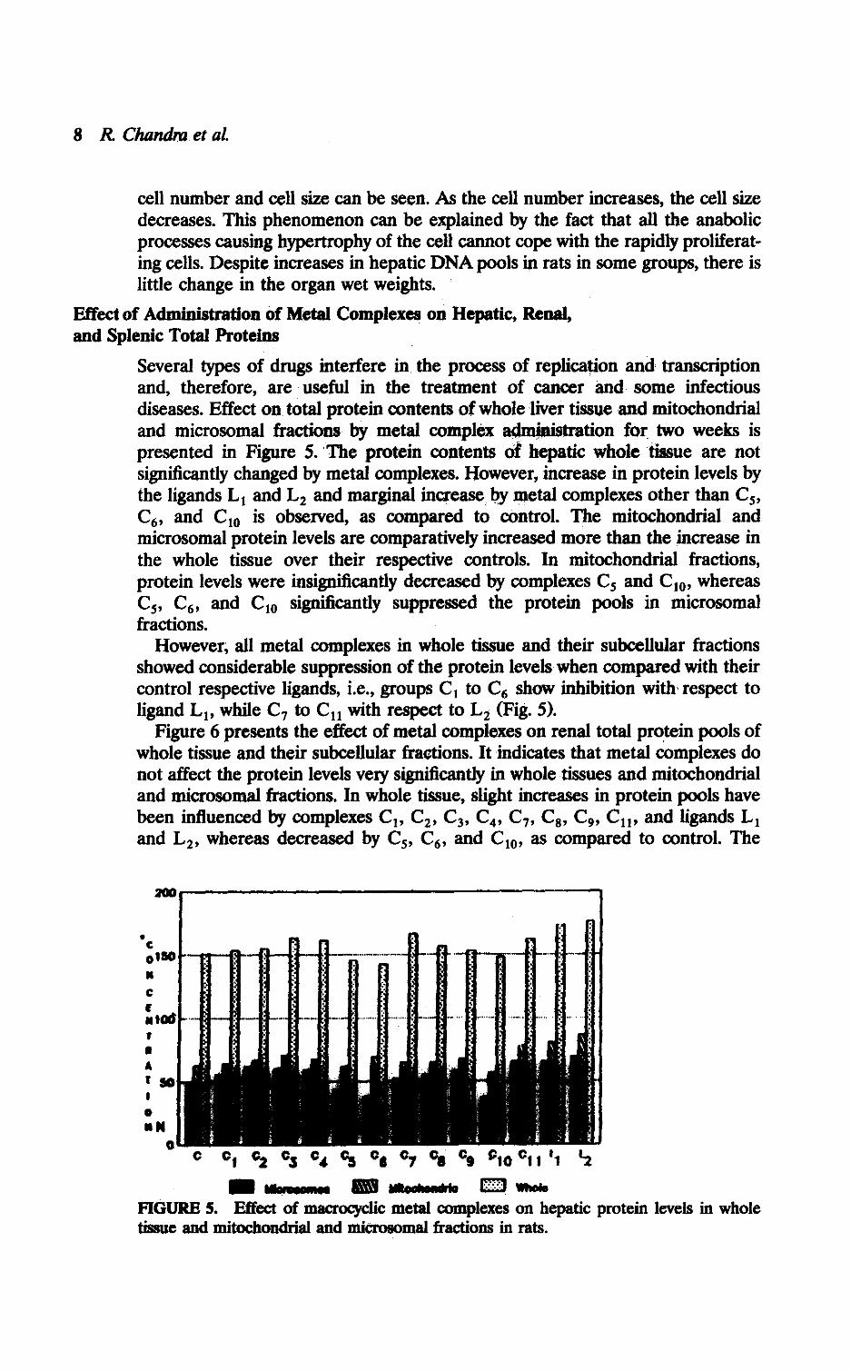
Several types of drugs interfere in . the process of replication and transcriptionand, therefore, are useful in the treatment of cancer and some infectiousdiseases. Effect on total protein contents of whole liver tissue and mitochondrialand microsomal fractions by metal complex administration for two weeks ispresented in Figure 5 . ' The protein contents of hepatic whole tissue are notsignificantly changed by metal complexes . However, increase in protein levels bythe ligands L I and L2 and marginal increase, by metal complexes other than C 5 ,C6 , and C IO is observed, as compared to control . The mitochondrial andmicrosomal protein levels are comparatively increased more than the increase inthe whole tissue over their respective controls . In mitochondrial fractions,protein levels were insignificantly decreased by complexes C5 and C IO , whereasC5, C6 , and C IO significantly suppressed the protein pools in microsomalfractions .
However, all metal complexes in whole tissue and their subcellular fractionsshowed considerable suppression of the protein levels when compared with theircontrol respective ligands, i .e., groups C, to C6 show inhibition with respect toligand L I , while C7 to CII with respect to L2 (Fig. 5).
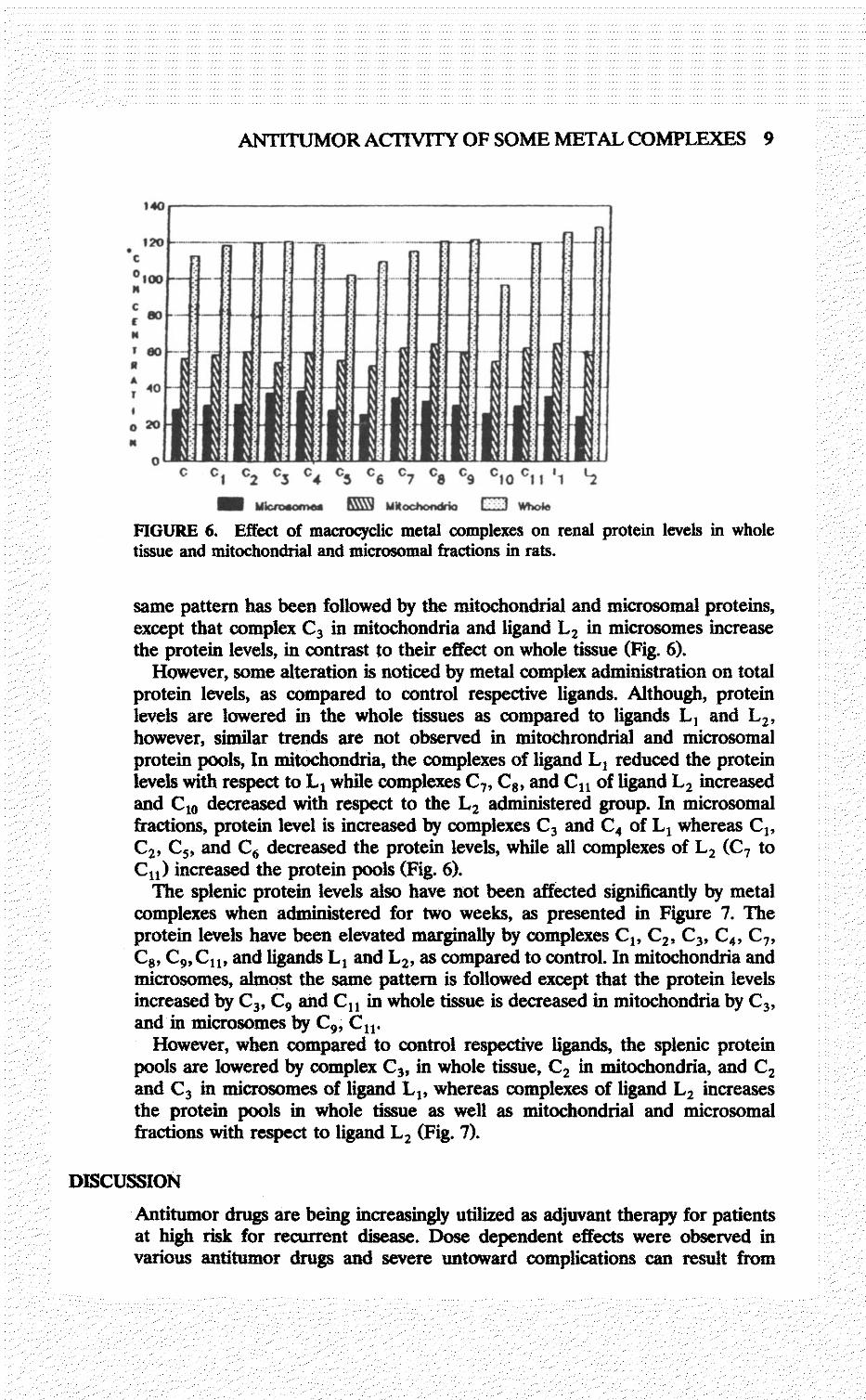
Figure 6 presents the effect of metal complexes on renal total protein pools ofwhole tissue and their subcellular fractions. It indicates that metal complexes donot affect the protein levels very significantly in whole tissues and mitochondrialand microsomal fractions . In whole tissue, slight increases in protein pools havebeen influenced by complexes C I , C2 , C3 , C4 , C7, C8 , C9, C l ,, and ligands, L,and L2 , whereas decreased by C 5 , C6 , and C IO, as compared to control. The
zoo
C
C C 1 02 03 04 es CG C7 ca C010
C1I 'I 2
S IMMWNW ® UNOWWN 1s 01 wn 1.FIGURE 5. Effect of macrocyclic metal complexes on hepatic protein levels in wholetissue and mitochondrial and microsomal fractions in rats .
140
s 120C0 100MCr W
C
ANTITLJMOR AC 1'1VITY OF SOME METAL COMPLEXES 9
0 1 C2 C3 C4
= Mimo8a"wa
Cs 06 C7
® M*.&"1*%
0 c11 I 1
ED Whois
t2
FIGURE 6. Effect of macrocyclic metal complexes on renal protein levels in wholetissue and mitochondrial and microsomal fractions in rats .
same pattern has been followed by the mitochondrial and microsomal proteins,except that complex C3 in mitochondria and ligand L2 in microsomes increasethe protein levels, in contrast to their effect on whole tissue (Fig . 6).
However, some alteration is noticed by metal complex administration on totalprotein levels, as compared to control respective ligands. Although, proteinlevels are lowered in the whole tissues as compared to ligands L, and L 2 ,however, similar trends are not observed in mitochrondrial and microsomalprotein pools, In mitochondria, the complexes of ligand L, reduced the proteinlevels with respect to L, while complexes C7, C8 , and C 11 of ligand L2 increasedand C10 decreased with respect to the L 2 administered group. In microsomalfractions, protein level is increased by complexes C 3 and C4 of L, whereas C,,C2 , C5 , and C6 decreased the protein levels, while all complexes of L2 (C 7 toC11 ) increased the protein pools (Fig. 6).
The splenic protein levels also have not been affected significantly by metalcomplexes when administered for two weeks, as presented in Figure 7 . Theprotein levels have been elevated marginally by complexes C1, C2 , C3, C4 , C7,Cs , C9, C 11 , and ligands L, and L2 , as compared to control . In mitochondria andmicrosomes, almost the same pattern is followed except that the protein levelsincreased by C 3, C9 and C 11 in whole tissue is decreased in mitochondria by C 3,and in microsomes by C9, C11 .
However, when compared to control respective ligands, the splenic proteinpools are lowered by complex C 3 , in whole tissue, C 2 in mitochondria, and C2and C3 in microsomes of ligand L 1 , whereas complexes of ligand L 2 increasesthe protein pools in whole tissue as well as mitochondrial and microsomalfractions with respect to ligand L 2 (Fig. 7).
DISCUSSION
Antitumor drugs are being increasingly utilized as adjuvant therapy for patientsat high risk for recurrent disease . Dose dependent effects were observed invarious antitumor drugs and severe untoward complications can result from
10 R Chandra et aL
Cs
to ° t t 'i
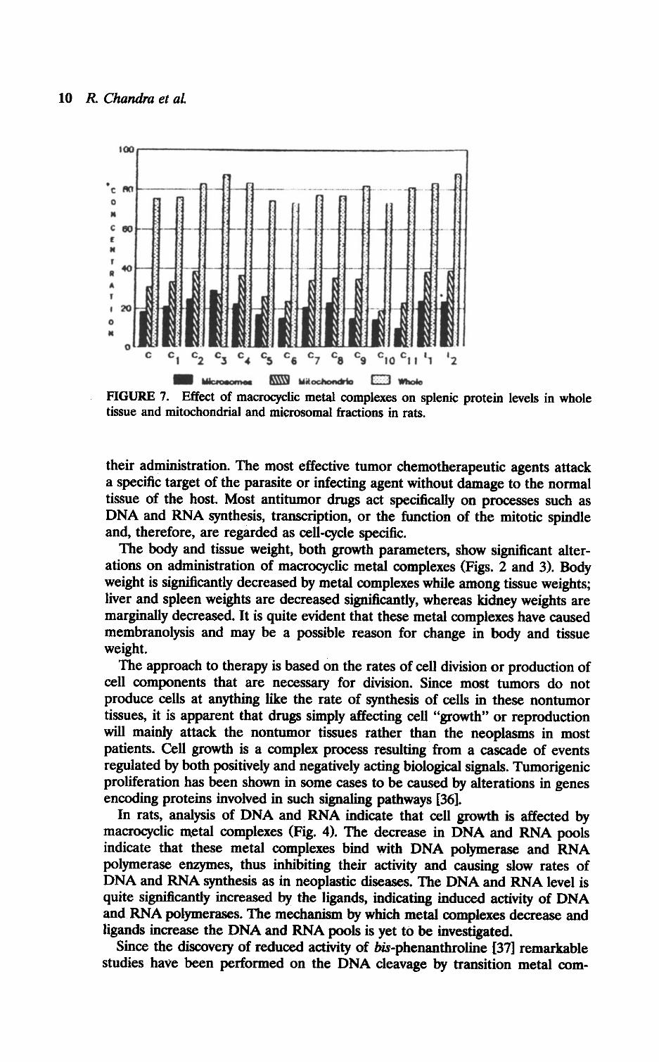
S w .. ® MAOOM"*b a] "winFIGURE 7. Effect of macrocycic metal complexes on splenic protein levels in wholetissue and mitochondrial and microsomal fractions in rats .
their administration. The most effective tumor chemotherapeutic agents attacka specific target of the parasite or infecting agent without damage to the normaltissue of the host. Most antitumor drugs act specifically on processes such asDNA and RNA synthesis, transcription, or the function of the mitotic spindleand, therefore, are regarded as cell-cycle specific.
The body and tissue weight, both growth parameters, show significant alter-ations on administration of macrocyclic metal complexes (Figs . 2 and 3). Bodyweight is significantly decreased by metal complexes while among tissue weights ;liver and spleen weights are decreased significantly, whereas kidney weights aremarginally decreased . It is quite evident that these metal complexes have causedmembranolysis and may be a possible reason for change in body and tissueweight.
The approach to therapy is based on the rates of cell division or production ofcell components that are necessary for division . Since most tumors do notproduce cells at anything like the rate of synthesis of cells in these nontumortissues, it is apparent that drugs simply affecting cell "growth" or reproductionwill mainly attack the nontumor tissues rather than the neoplasms in mostpatients. Cell growth is a complex process resulting from a cascade of eventsregulated by both positively and negatively acting biological signals . Tumorigenicproliferation has been shown in some cases to be caused by alterations in genesencoding proteins involved in such signaling pathways [36].
In rats, analysis of DNA and RNA indicate that cell growth is affected bymacrocyclic nietal complexes (Fig . 4). The decrease in DNA and RNA poolsindicate that these metal complexes bind with DNA polymerase and RNApolymerase enzymes, thus inhibiting their activity and causing slow rates ofDNA and RNA synthesis as in neoplastic diseases . The DNA and RNA level isquite significantly increased by the ligands, indicating induced activity of DNAand RNA polymerases. The mechanism by which metal complexes decrease andligands increase the DNA and RNA pools is yet to be investigated .
Since the discovery of reduced activity of bis-phenanthroline [37] remarkablestudies have been performed on the DNA cleavage by transition metal com-
ANTITUMOR ACTIVITY OF SOME METAL COMPLEXES 11
plexes by photoactivation of light-sensitive complexes, e .g., cobalt or rutheniumcomplexes [38]. Inorganic cobalt complexes containing porphyrinic ligands, i.e.,protoporphyrins, were found to be powerful inhibitors in human patients [39], ofsarcoma 180 Ehrlich ascites tumor in mice and Yoshida sarcoma in rats. Theobserved inhibition of DNA synthesis due to rhodium (II, III) complexes may bedue to the blocking of some thiolic enzyme involved in DNA synthesis, since therhodium (II) carboxylates do not bind to double-stranded DNA [40, 41] . Somemanganese complexes have also been recently reported as being efficient DNAcleavers [42] . Iron complexes are also used in the treatment of human cancer .Studies have been reported on the influence of iron on antitumor properties ofsome anticancer drugs, e.g., 6-mercaptopurine [43]. Although nickel compoundsare generally considered carcinogenic, several nickel complexes were tested forantitumor properties but results were not encouraging [26, 44-47]. Bis-(diethyl-dithiophosphate) nickel (11) was reported to be more active than correspondingplatinum derivatives [48]. The influence of selenium containing compounds onnucleic acid, particularly RNA synthesis in rat liver cells in vivo, has beenstudied [49] and proved to be anticarcinogenic [50, 51] among tetravalentcompounds of selenium and suppressed the synthesis of DNA and RNA inanimal cells [52] .
The biosynthetic pathways for the formation of purines and pyrimidinesdepends on interrelationships with other metabolic processes, i.e., protein syn-thesis. By administering certain compounds or drugs, there results a competitionwith the natural compounds that inhibit purine and pyrimidine synthesis andthus also, the synthesis of nucleic acids [53] . 6-Mercaptopurine can be incorpo-rated into nucleic acids, thereby interfering with protein synthesis . Since acharacteristic of cancer tissue is its uncontrolled cell division, an increasedsynthesis of DNA and RNA is obviously involved . Inhibition of the biosynthesisof purines and pyrimidines would suppress the elevated synthesis of nucleicacids and, therefore, also restrict the growth of cancerous tissue . Unfortunately,the amount of drug needed to block cancer growth often also suppresses normalcell division and, therefore can be toxic .
The effect of the reaction of cis-DDP with DNA has been investigated withregard to DNA synthesis [54] and involves the inhibition of DNA syntheticactivity. Although inhibition of DNA synthesis provides a plausible model toaccount for drug action, it is well established that important control of geneexpression may also occur at the level of DNA transcription through alterationsin DNA structure and conformation [55-57] .
Our studies reveal that protein contents of liver, kidney, and spleen have notbeen affected so significantly by metal complexes (Figs. 5, 6 and 7) . The totalprotein levels are marginally increased by ligands L 1 and L 2 and decreased byC 6, C7, and C 11 in the liver, kidney, and spleen. The hepatic, renal, andmicrosomal protein levels are comparatively increased more than the increaseobserved in whole tissue, as compared to respective controls .
The total protein levels in whole tissue and mitochondrial and microsomalproteins, DNA and RNA profiles indicate that the mitotic indices in rat liverfollowing administration of excess metal complexes at the time of active prolifer-ation and differentiation are affected adversely, leading to decreased cellpopulation. Development of any organ follows exquisitely timed and well coordi-nated events, viz ., proliferation of cells (hyperplasia), hyperplasia with concomi-tant increase in cell (hypertrophy), and then hypertrophy alone [58]. It has been
12 R Chandm et aL
REFERENCES
shown that the primary phase of cell proliferation, when interrupted, may leavea permanent phenotypic impression . In liver, as in any other organ, DNAsynthesis, hence cell division, stops long before growth stops [58]. The under-changed or increased protein levels in metal compound administered groups ofanimals suggest that cell size tries to compensate for the decreased populationof cells, and is able to do it .
The selective uptake of various natural and synthetic antitumor agents bytumor cells offers novel opportunities for cancer detection and therapy . Theagents presently available for cancer chemotherapy modify the growth of normaltissues as well as their neoplastic tissues . The detection of specific inhibitors ofthe synthesis of biomacromolecules is of great importance for a more profoundstudy of the mechanisms of the control and modulation of genetic process innormal cells in the normal state and in the presence of pathological changes. Inaddition to platinum compounds, other coordination compounds of iron, cobalt,and nickel may also exhibit antitumor properties which prove to be promising tosome extent. The current results demonstrate that these metal complexesmarkedly affect the properties of RNA and DNA polymerase, suggesting thatthey may exert their effect in vivo on DNA, RNA, and total protein levels . Inview of the above, it is concluded that our three metal complexes : [Co(FaHh-DAP)](OAc)2 , [Co(FaHh-DAP)](N03)2 , and [Ni(DAP-Th e-DAP.H20)]S04 mayexhibit antitumor properties and seem to emerge as potential antitumor agents,which certainly require detailed screening in animals and later in homo sapiansbefore it is used clinically . This is a preliminary investigation of the toxicity ofthese macrocyclic metal complexes and their side effects on rats bearingtransplanted tumors, to examine their biological properties .
The authors (RC and AS) thankfully acknowledge the University Grants Commission, NewDelhi, India for the Research Scientist Award and Senior Research Fellowship. The otherauthor (RB) thanks the Council of Scientific and Industrial Research, New Delhi, India forthe award of Research Associateship .
1 . B. Rosenberg, L. Van Camp, J. E. Trosko, and V. H. Mansour, Nature (London) 222,385-386 (1969) .
2. J. Loehrer and L. H. Einhorn, Am. Intern. Med. 100, 704-713 (1984).3. H. W. Bruckner, C. J. Cohen, J. D. Goldberg, B. Kabakow, R. C. Wallach, G. Deppe,E. M. Greenspan, S. B. Gusberg, and J. F. Holland, Cancer 47, 2288-2294 (1981) .
4. A. Eastman, Pharmacol. Ther. 34,155-166 (1987).5 . S. J. Lippard, Pure Appl. Chem. 59, 731-742 (1987).6. J. Reedijk, Pure Appl. Chem . 59, 181-192 (1987).7. D. Wilkie, J. Royal Soc. Med. 72, 599-601 (1979).8. B. J. Kennedy, J. W. Yarbo, V. Kickertz, and M. Sanberg Wollbeim, Cancer Res. 28,
91-97 (1968).9. J. Fok and M. J. Warning, Mol. Phannacol. 8, 65-74 (1972) .
10. W. Behr and G. Hartmann, Biochem. Z . 343, 519-527 (1965) .11 . M. G. C. Benjamin and R. F. Keith, Biochem. Biophys. Res. Commun. 160, 517-524
(1989) .12. W. Behr, K. Honikel, and G . Hartman, Eur. J. Biochem. 9, 82-92 (1969).
ANTITUMOR ACTIVITY OF SOME METAL COMPLEXES 13
13. L. V. Johnson, N. D. Walsh, and L B. Chen, Proc. Nat!, Acad. Sci . (U.S .A.) 77,990-994 (1980) .
14. L. V. Johnson, M. D. Walsh, B. J. Bockus, and L B. Chen, J. Cell. Biol. 88, 526-535(1981).
15. W. Tso and C. Lee, Contraception 25, 649-655 (1982).16. Z. Darzynkiewicz, Z. F. Tranganos, L Stanio-Cocio, J . Kapuscinski, and M . Melamid,
Cancer Res. 42, 799-806 (1982) .17. R. D. Williams, Chem. Revs . 72, 203 (1972).18. M. J. Cleare, Coond. Chem. Rev. 12, 349 (1974) .19. P. Kopf-Maier and H. Kopf, Chem Rev. 87, 1137 (1987).20. C. Silvestru and I . Haiduc, Rev. Chim. (Bucharest) 33, 81 (1983).21. T. A. Connors and J. J. Roberts, Platinum Coordination Complexes in Cancer
Chemotherapy, Springer-Verlag, Berlin, 1974.22. A. W. Prestayko, S. T. Crooke, and S. K. Carter, Cisplatin: Current Status and New
Developments, Academic Press, New York, 1980 .23. S. J. Lipard, Platinum, Gold and other Metal Chemotherapeutic Agents : Chemistry and
Biochemistry, ACS Symposium Series No. 209, American Chemical Society, Washing-ton DC, 1983.
24. M. P. Hacker, E. B. Douple, and I. M. KraKoff, Platinum Coordination Complexes inCancer Chemotherapy. Developments in Oncology Series, Martinus Nijhoff, Boston,MA, 1984, Vol. 17 .
25. M. Nicolini, Platinum and other Metal Coordination Compounds in Cancer Chemother-apy, Martinus Nijhoff, Boston, MA, 1988 .
26. M. J. Cleare and H. C. Hydes, in Metal Ions in Biological Systems, H. Siegel, Ed .,Marcel Dekker, New York, 1980, Vol . 11, pp. 1 .
27. M. Gielen, Metal-Based Antitumor Drugs, Freund, London and Tel . Aviv, 1988 .28. I. Haiduc and C. Silvestru, Organometallics in Cancer Chemotherapy, CRC Press, Boca
Raton, FL, 1989.29. 1. Haiduc and C. Silvestru, Coord. Chem. Rev . 99, 253-296 (1990).30. A. Sarkar, Studies of Some Potential Anticancer Agents: Synthesis and Characterization
of Metal Complexes of Macrocyclic Systems Having Resemblance to Biological Systems :An Approach to Develop and Evaluate Model Systems, PhD dissertation, submitted tothe University of Delhi, India (1990) .
31 . G. S. Drummond and A . Kappas, Proc. Natl. Acad. Sci., U.S.A . 76, 5331-5335(1979).
32. H . N. Munro and D. Fleck, in Methods of Biochemical Analysis, D. Glick, Ed.,Interscience Publishers, New York, 1967, Vol. XIV, p. 113 .
33. K. Burton, Biochem. J. 62, 315-323 (1956) .34. W. C. Schneider, in Methods in Enzymology, S . P. Colowick and N . O. Kaplan, Eds.,
Academic Press, Inc. New York, 1957, Vol. III, p. 680.35. O. H. Lowry, N . J. Rosebrough, A . L. Fan, and R. J. Randall, J. Biol. Chem. 193,
265-275 (1951) .36. G. Klein, Science 238, 1539-1545 (1988) .37. D. S. Sigwan, Acc. Chem. Res . 19, 180-186 (1979) .38. P. B. Dervan, Science 232, 464-471 (1986) .39. J. M. Orten and O. W. Neuhas, in Human Biochemistry, J. E. Lotz, Ed., The C. V .
. Mosby Co., St. Louis, MO, 1982, tenth edition, pp . 477-478.40. J. L. Bear, H. B. Gray, L. Rainen, I. M. Chang, R. Howard, G. Serio, and A . P .
Kimball, Cancer Chemother. Rep. Part I, 59, 611 (1975).41 . P. N. Rao, M. L. Smith, S. Pathak, R . A. Howard, and J . L. Bear, J. Nat!. Cancer Inst .
64, 905 (1980).42. B. Ward, A. Skorobogaty, and J. C. Dabrowiak, Biochemistry 25, 6875-6883 (1986).43. W. D. Yang, Q. Le Me, and S. Q. Peng, Inorg. Chimica Acta 106, 65 (1985).
14 R. Chandra et aL
44. P. Lumme, H. Elo, and J. Janne, Inorg. Chim. Acta 92, 241 (1984).45 . H. Elo, Inorg. Chim. Acta 136, 233 (1987) .46. R. M. Synder, C. K Mirabelli, R. K Johnson, C. M. Sung, L. F. Faucette, F. L.
McCabe, J. P. Zimmerman, M. Whiteman, J. C Hempel, and S. T. Crooke, CancerRes. 46, 5054 (1986).
47. M. Mohan, N. S. Gupta, L. Chandra, and N. K Jha, J. Inorg. Bioche. 31, 7 (1987).48 . S. E. Livingstone and A . E. Mihkelson, Inorg. Chem . 9, 2545 (1970).49. F. I. Abdullaev, I . A. Allakhverdeiv, and G. R. Mamedona, Biokhimiya 54,145-148
(1989).50. R. J. Shamberger, Mutat. Res. 154, 29-48 (1985).51 . J. N. Vernie, Biochem. Biophys. Acta 738, 203-217 (1984).52. G. B. Abdullaev, N. Kh. Mekhteiv, F. I. Abdullaev, and K A . Kafiari, Biokhimiya 45,
98-1010980).53. J. M. Orten an O. W. Neuhas, in Human Biochemistry, J. E. Lotz, Ed ., The C. V .
Mosby Co., St Louis, MO, 1982, tenth edition, pp. 393-394 .54. F. Bernges and E. Holler, Biochemistry 27, 6398-6402 (1988).55. P. H. Von Hippel, D. G. Bear, W. D. Morgan, and J . A. McSuriggen, Annu. Rev .
Biochem. 53, 389-446 (1984).56. T. M. Jovin, D. M. Soumpasis, and L P. McIntosh, Annu. Rev. Phys. Chem. 38,
521-560 (1987).57. M. Sarvadogo and A. Sentenac, Annu. Rev. Biochem. 59, 711-754 (1990).58. M. Winick, P . Rosso, and J. A. Brasel, Nutr. Diet. 17, 60 (1972).
Received June 14, 1991, accepted January 4, 1992