
Secondary Metabolites from Plants Plant Secondary Metabolites

Acta agriculturae Slovenica, 117/2, 1–12, Ljubljana 2021
doi:10.14720/aas.2021.117.2.2065 Original research article / izvirni znanstveni članek
Antioxidant defense and secondary metabolites concentration in hyssop (Hyssopus officinalis L.) plants as affected by salt stress
Zhaleh SOHEILIKHAH1*, Nasser KARIMI2, Masoud MODARRESI3, Seyed Yahya SALEHI-LISAR1, Ali MOVAFEGHI1
Received January 25, 2021; accepted March 27, 2021.
Delo je prispelo 25. januarja 2021, sprejeto 27. marca 2021.
Antioxidant defense and secondary metabolites concentra-tion in hyssop (Hyssopus officinalis L.) plants as affected by salt stress
Abstract: Salt stress is one of the major limiting factors for plant production, and the quality of medicinal plants is also affected by soil salinity. Hyssop (Hyssopus officinalis L.) plants were cultivated for four weeks in perlite: sand and irrigated with Hoagland nutrient solution containing 0, 50, 100, 150, and 200 mM NaCl. Plants growth was decreased by salt stress while the leaf relative water content was not affected, and the chloro-phyll content decreased only by the highest salt concentration (200 mM). Sodium was accumulated at small amounts, indicat-ing a high ability of this species to exclude salt. Soluble sugars and proline were accumulated up to 1.6 and 4.5 fold, respec-tively. The antioxidant enzymes activity (peroxidase, catalase, ascorbate peroxidase) were increased by the salt treatments, particularly in the leaves. The levels of secondary metabolites (saponins, phenolics, flavonoids, anthocyanins, and iridoids) were all increased under salt stress, and the total antioxidant capacity of alcoholic extract of the leaves and roots was signifi-cantly higher in the salt-treated compared with control plants. Our results showed that hyssop is a salt-tolerant species, and the quality of this medicinal plant is improved when grown un-der saline conditions.
Key words: salinity; hyssop; Hyssopus officinalis; second-ary metabolites; antioxidant enzymes
1 Department of Plant Sciences, Faculty of Natural Sciences, University of Tabriz, Tabriz, Iran2 Department of Biology, Faculty of Science, Razi University, Kermanshah, Iran3 Department of Pharmacognosy and Pharmaceutical Biotechnology, School of Pharmacy, Kermanshah University of Medical Sciences, Kermanshah, Iran* Corresponding author, email: [email protected];
Antioksidativna obramba in vsebnost sekundarnih metabo-litov v navadnem ožepku (Hyssopus officinalis L.) v razmerah solnega stresa
Izvleček: Solni stres je eden izmed dejavnikov, ki najbolj omejujeje rast rastlin, v razmerah zasoljenih tal je prizadeta tudi kakovost zdravilnih rastlin. Navadni ožepek (Hyssopus officinalis L.) je bil gojen v mešanici perlita in peska in zalivan s Hoaglandovo hranilno raztopino, ki je vsebovala 0, 50, 100, 150, and 200 mM NaCl. Rast rastlin se je s solnim stresom zmanjšala, a relativna vsebnost vode v listih ni bila prizadeta in vsebnost klorofila se je zmanjšala le pri največji koncentraciji (200 mM NaCl). Natrij se je v rastlinah kopičil v majhnih koli-činah, kar nakazuje sposobnost te vrste, da izloča sol. Vsebnost topnih sladkorjev in prolina se je povečala za 1,6 ,oziroma 4,5 krat. Aktivnost antioksidacijskih encimov (peroksidaze, kata-laze, askorbat peroksidaze) se je povečala po obravnavanjih s soljo, še posebej v listih. V razmerah solnega stresa se je pove-čala raven sekundarnih metabolitov (saponinov, fenolov, flavo-noidov, antocianinov in iridoidov), celokupna antioksidacijska sposobnost alkoholnega ekstrakta listov in korenin je bila zna-čilno večja pri rastlinah izpostavljenih soli kot pri kontroli. Re-zultati so pokazali, da je navadni ožepek na sol strpna rastlina in, da se kakovost te zdravilne rastline izboljša, če jo gojimo v razmerah slanosti.
Ključne besede: slanost; navadni ožepek; Hyssopus offici-nalis; sekundarni metaboliti; antioksidacijski encimi

Acta agriculturae Slovenica, 117/2 – 20212
Z. SOHEILIKHAH
This paper is part of PhD thesis of Z.S under co-supervision of A.M & N.K. M.M & SY. SL. were the advi-sors of this thesis.
1 INTRODUCTION
Soil salinity is one of the main abiotic stress factors threatening agricultural production worldwide. Salt in the soil is also considered the main factor limiting the dispersal of plants in their natural habitats (Acosta-Mo-tos et al., 2017; Mushtaq et al., 2020). Salinity causes both osmotic and ionic stresses and affects all the plant major processes such as germination, photosynthesis, growth, water and nutrients balances and yield (Parida & Das, 2005; Parihar et al., 2015).
Salt stress affects the plant’s primary metabolism and alters primary metabolites’ concentrations, includ-ing soluble sugars and amino acids (Gupta and Huang, 2014). Among amino acids, proline plays a pivotal role in the plant’s adaptation to salt stress by protecting cells from damages caused by excess accumulation of ions and salt-induced dehydration (Verbruggen and Hermans, 2008). As the second important group of compatible sol-utes, soluble sugars protect from dehydration and help sustain the structural integrity of plant cells under salt stress (Rosa et al., 2009).
Under environmental stress conditions such as sa-linity, the generation of higher reactive oxygen species (ROS) causes oxidative stress. It results in membrane damage characterized by elevated levels of malondialde-hyde (MDA). Plants employ defensive systems for scav-enging ROS and protecting from damaging oxidative reactions through different antioxidant enzymes such as peroxidases (POD), catalase (CAT), and ascorbate peroxidase (APX) (Foyer et al., 1994; Gupta and Huang, 2014; Akyol et al., 2020). Proline protects plant cells from ionic and osmotic stresses and contributes to the scav-enging ROS such as hydroxyl radicals (Verbruggen and Hermans, 2008).
In addition to primary metabolism, secondary plant metabolism is also influenced by salt stress (Ahmad and Sharma, 2008). The concentration of secondary metabo-lites highly depends on plants’ growth stage, especially environmental conditions, including light intensity and stress factors such as salt (Ahl and Omer, 2011). Salt stress has a positive or negative impact on secondary me-tabolites’ biosynthesis depending on plant species or the severity of stress (Verma and Shukla, 2015). Salt stress led to about 8–35 % increase in total phenolics and about 35 % increase in total flavonoid content in Portulaca ol-eracea L. (Alam et al., 2015). An enhancement of anti-oxidant activity and flavonoid and phenolics contents has
also been observed in Cichorium spinosum L. under salt stress (Petropoulos et al., 2017).
In the members of Lamiaceae, salinity may cause substantial changes in the compositions and yield of secondary metabolites (Taarit et al., 2009). Salt stress led to about 20-40 % increase in the total phenolics and flavonoids content in Thymus species (Zrig et al., 2016). Salt stress significantly induced the biosynthesis of some crucial essential oil and phenolic compounds in Salvia mirzayanii Rech.f. & Esfand. (Valifard et al., 2014). Con-sidering the medicinal application of most plant species from Lamiaceae, it is important to know the effect of salt stress on the quality and quantity of secondary metabo-lites. The content of secondary metabolites is also a de-termining factor for the total antioxidant activity of plant extract defined either by FRAP (Ferric Reducing Ability of Plasma) or through DPPH (2, 2-diphenyl-1-picrylhy-drazyl) assay (Fukumoto and Mazza, 2000; Sethi et al., 2020).
Hyssop (Hyssopus officinalis L.) is a perennial sub-shrub belonging to the Lamiaceae family and distributes in the eastern Mediterranean to central Asia (Fathiazad and Hamedeyazdan, 2011). Hyssop is a popular medici-nal herb with carminative, tonic, antiseptic, and expec-torant properties and is used to remedy congestion, lung complaints, and cardiovascular disorders (Hristova et al., 2015). Extracted essential oils from the shoots of hys-sop plants possess a unique aroma and are widely used in the food, pharmaceutical, and cosmetics industries (Kazazi et al., 2007). Different polyphenolic compounds identified in this species contain different glycones and aglycones such as flavonoids, quercetin, apigenin, dios-min, luteolin, and other phenolic compounds such as chlorogenic, ferulic, protocatechuic, syringic, caffeic, and p-hydroxybenzoic acids (Fathiazad and Hamedeyazdan, 2011).
Hyssop is a xerophyte species and is well adapted to drought conditions (Khazaie e al., 2008). Effect of salt stress on the activity of antioxidant enzymes has been in-vestigated in this species (Jahantigh et al., 2016). There is no report, however, on the secondary metabolites levels under salt stress in hyssop plants. This work aimed to in-vestigate the effect of salinity on growth, ROS scavenging activity, the content of various secondary metabolites, and the antioxidant capacity of the leaf and root extract in this species.
2 MATERIAL AND METHODS
2.1 PLANT MATERIAL AND TREATMENTS
Seeds of hyssop (H. officinalis) were purchased from
Acta agriculturae Slovenica, 117/2 – 2021 3
Antioxidant defense and secondary metabolites concentration in hyssop (Hyssopus officinalis L.) plants as affected by salt stress
Pakan-Bazr Company (Isfahan, Iran). The seeds were surface-sterilized using 1 % sodium hypochlorite then were sown in the 2 L pots (15 seeds in each pot) con-taining sterilized perlite: sand (1:3) mixture and placed at 4 °C for stratification. After 4 days, the pots were trans-ferred to the greenhouse conditions at 25/22 °C day/night temperature regimes, at a 16/8 h day/night cycle, and relative humidity of 60 %. After germination, plants were irrigated with 20 % Hoagland solution once a week and with 100 ml distilled water twice a week throughout the experiment.
Salt stress was imposed at a four-leaf stage with four NaCl concentrations (50, 100, 150, and 200 mM) ap-plied with irrigation water to the pots gradually within one week. Plants were grown for four weeks after start-ing salt stress and then were harvested. At harvest, fresh mass (FM) of shoot and roots were determined, and sub-samples were taken and immediately frozen in liquid ni-trogen and then stored at –80 °C until analysis. Another group of samples was oven-dried and, after determina-tion of dry mass (DM), were used for the analysis of sec-ondary metabolites and elemental concentrations.
2.2 BIOCHEMICAL MEASUREMENTS
Soluble carbohydrate content was determined ac-cording to the phenol-sulfuric acid method (Dubois et al., 1956). Proline was quantified according to the meth-ods of Bates et al. (1973), and the content of soluble pro-teins was determined using the Bradford method (Brad-ford, 1976) with bovine serum albumin as standard.
To determine the leaf content of chlorophyll (Chl) and carotenoids, 0.2 g of fresh leaf samples were homog-enized with 2 ml of 80 % acetone and centrifuged at 4000 g for 10 min. After that, the samples’ absorbance was re-corded by a spectrophotometer (Bausch & Lomb 70) at 663, 645, and 480 nm, and the contents of pigments were calculated according to the following equations where A corresponds to the absorbance (Flores-de-Santiago et al., 2016):
Chl a = 12.21A663 − 2.81 A646 Chl b = 20.13A646 − 5.03A663Carotenoids = [1000A470 − 3.27 (Chl a) – 104 (Chl b)] / 229
The leaf relative water content (RWC, %) was meas-ured according to the following equation: RWC (%) = [(FM − DM) / (TM − DM)] × 100
For determination of turgid mass (TM), leaf disks (5
mm diameter) were submerged for 5 h in distilled water, thereafter, they were blotted dry gently on a paper towel and weighed.
2.3 OXIDATIVE STRESS MARKERS AND ACTIVITIES OF ANTIOXIDANT ENZYMES
Sergiev et al. (1997) method with slight modifica-tion was used for quantification of H2O2 content. 100 mg of samples were ground in liquid nitrogen, extracted with trichloroacetic acid (TCA) in an ice bath, centrifuged at 13,000 g for 15 min. The 500 µl of supernatant was added to potassium phosphate buffer (pH 7.0), and the H2O2 content was determined based on supernatant absorb-ance at 390 nm. Malondialdehyde (MDA) content was evaluated by the method of Heath and Packer (1968). 100 mg of samples were homogenized in 1 ml 0.1 % (v/v) TCA and centrifuged at 12,000 g for 10 min. The extract-ed supernatant (0.1 ml) was mixed with 20 % TCA con-taining 0.5 % (w/v) thiobarbituric acid (TBA). The mix-ture was incubated at 100 °C for 10 min then centrifuged at 10,000 g for 15 min. TBA reactive substances’ content was calculated based on the difference in absorbance at 532 and 600 nm using an extinction coefficient of 155 mM–1 cm–1.
For assay of catalase (CAT), peroxidase (POD), and ascorbate peroxidase (APX), 100 mg of the samples was extracted in 5 ml of 100 mM phosphate buffer. The ho-mogenate was then centrifuged for 10 min and used for enzyme assays. CAT activity was analyzed by Beers and Sizer (1952) method. The 2 ml of reaction mixture con-taining 100 mM potassium phosphate buffer (pH 7.0) was mixed with 400 ml of 6 % H2O2 and 100 µl of enzyme extract. CAT activity was calculated to reduce the H2O2 absorption at a wavelength of 240 nm using an extinction coefficient of 0.036 mM–1 cm–1. POD activity was meas-ured by the method of Lin and Kao (1999). The reaction mixture (2 ml) contained potassium phosphate buffer (50 mM, pH 7), guaiacol solution (9 mM), H2O2 (19 mM) and root or leaf extract (100 µl). POD activity calculated the absorbance changes at 470 nm using the extinction coefficient of 26.6 mM–1 cm–1. For assay of APX, the reac-tion mixture contained 250 mM phosphate buffer (pH 7), 1.2 mM H2O2, 0.5 mM ascorbic acid, and 0.1 mM EDTA. The reaction was started by adding H2O2 to the mixture. Total APX activity was calculated to reduce the absorb-ance at 290 nm for 2 min using the extinction coefficient of ascorbic acid (2.8 mM–1 cm–1) (Dazy et al., 2008).
2.4 SECONDARY METABOLITES QUANTIFICATION
For analyzing secondary metabolites, 100 mg of leaf or root dried samples were dissolved in 80 % ethanol (5 ml) and sonicated for 20 min at room temperature. The resulted mixture was centrifuged at 3,000 g for 15 min.

Acta agriculturae Slovenica, 117/2 – 20214
Z. SOHEILIKHAH
The extraction was repeated three times, and the super-natants were pooled and stored until analysis.
A colorimetric method was used to determine the total amount of saponins (Hiai et al., 1975). In a test tube, 0.5 ml of plant ethanol extract was mixed with 0.5 ml of vanillin and 5 ml of 72 % sulfuric acid. The mixture was shaken and heated for 10 min at 60 °C in a water bath. After cooling in the water at room temperature, the extract’s absorbance was determined spectrophoto-metrically at 545 nm. To determine the concentration of the total phenolics, 2.5 ml of Folin-Ciocalteu-Deniz indicator and 2.5 ml of 2 % sodium carbonate solution were added to 0.5 ml plant ethanol extract. The result-ing mixture was homogenized and incubated in the dark for 30 min. The absorbance of the solution was measured spectrophotometrically at 750 nm (Seevers et al., 1971). The total flavonoids concentration was determined using the aluminum chloride colorimetric method (Zhishen et al., 1999). The extract’s 0.5 ml was mixed with 4.5 ml of distilled water and 0.5 ml of 5 % sodium nitrite solu-tion. After 5 min, 0.5 ml of 10 % aluminum chloride was added, and the mixture was incubated for 6 min. After that, 4 ml of 1 M NaOH was added, and after 15 min, the absorbance of the mixture was read at 510 nm by a spectrophotometer. Quercetin was used for the creation of a standard curve. For analyzing the total anthocyanins concentration, 0.2 ml of alcohol extract was diluted sepa-rately with 4.8 ml potassium chloride (pH 1) and sodium acetate buffer (pH 4.5). The solutions were incubated in the dark for 15 min. The absorbance of both groups of samples was determined at 510 and 700 nm, and the con-centration of anthocyanins (A) was calculated according to the following equation and reported as cyanidin-3-O-glucoside equivalent using an extinction coefficient of 26.9 mM–1 cm–1 (Giusti and Wrolstad, 2001):A = [(A510 nm − A700 nm)] pH 1.0 − [(A510 nm − A700 nm)] pH 4.5
A colorimetric method based on the color reaction of acobin with glycine was used to determine the total iri-doid concentration (Narayanan and Akamanchi, 2003). One ml of ethanol extract was mixed with 2 ml distilled water, 1 ml of 10 % glycine, and 1 ml sulfuric acid (0.1 M). The mixture was shaken and heated in a water bath (95 °C) for 1 h. After cooling at room temperature, the absorbance was measured at 554 nm.
2.5 DETERMINATION OF TOTAL ANTIOXIDANT ACTIVITY
Different plant extract concentrations in methanol (10, 25, 50, 100, 250, and 500 μg ml–1) were prepared and incubated with freshly-prepared 80 μg ml–1 DPPH (2,
2-diphenyl-1-picrylhydrazyl). The mixtures were shaken and placed in the dark for 30 min, then the absorbance of the samples in parallel with a solution without plant extract (as blank) was read at 517 nm. The inhibition (%) of DPPH radical formation was calculated according to the following equation: Inhibition (%) = [(A blank – A sample) /A blank] × 100
Plant extract’s antioxidant activity was reported as the sample concentration providing 50 % inhibi-tion (IC50) calculated by plotting inhibition percentages against the samples’ concentration (Sarkar et al., 2006).
2.6 DETERMINATION OF ELEMENTS CONCENTRATIONS
To determine potassium (K) and sodium (Na) con-centrations, 100 mg milled oven-dried samples were di-gested in concentrated nitric acid overnight, then heated at 80 °C for one hour and dissolved in 1 % HCl. The con-centrations of K and Na were determined by flame pho-tometry (Kalra, 1997).
2.7 STATISTICAL ANALYSIS
The experiment was undertaken as a completely randomized block design with three pots as independ-ent replicates for each treatment. Data were presented as means ± standard deviation (SD). Comparison of means was performed by Tukey test (p < 0.05) using SPSS (ver-sion 23, for Windows; SPSS Inc., Chicago, IL, USA).
3 RESULTS AND DISCUSSION
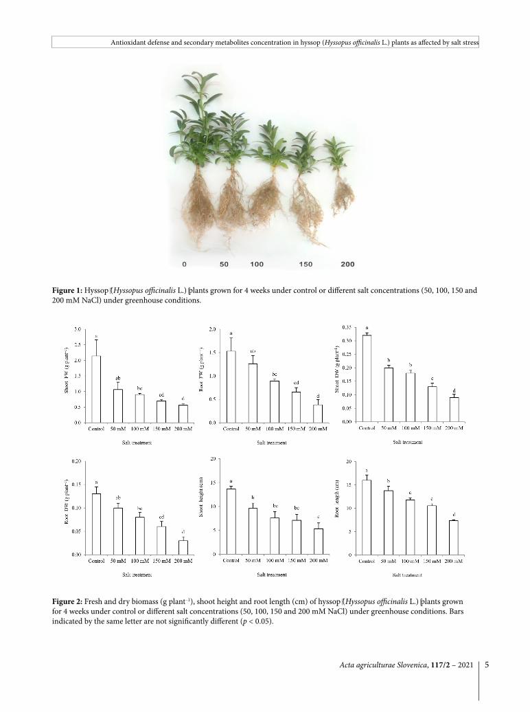
Plants’ growth was expectedly decreased under salt stress conditions (Fig. 1). A significant effect of salt stress on the shoot and root fresh weight was observed at 100 mM salt and higher. Dry biomass of plants was depressed up to 75 % at a salt concentration of 200 mM. Shoot height and length of the taproot significantly decreased by 50 mM salt and higher (Fig. 2).
The leaf content of Chl a and Chl b were not affected by salt concentration up to 150 and 100 mM NaCl, re-spectively. Leaf carotenoid content and RWC, were not significantly influenced by applied salt levels (Table 1).
Activities of all three analyzed antioxidant enzymes were higher in salt-stressed plants both in the leaves and roots (Fig. 3). At 200 mM salt concentration, the leaf ac-tivities of POD, CAT and APX increased up to 2.3, 6.3 and 6.9 fold, respectively, compared to the control treat-
Acta agriculturae Slovenica, 117/2 – 2021 5
Antioxidant defense and secondary metabolites concentration in hyssop (Hyssopus officinalis L.) plants as affected by salt stress
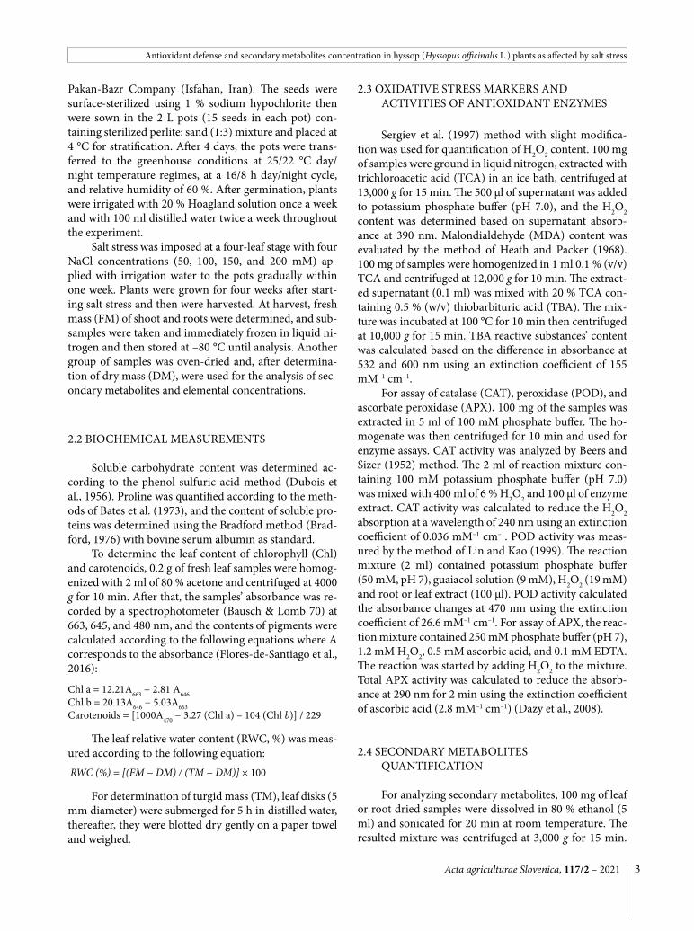
Figure 1: Hyssop (Hyssopus officinalis L.) plants grown for 4 weeks under control or different salt concentrations (50, 100, 150 and 200 mM NaCl) under greenhouse conditions.
Figure 2: Fresh and dry biomass (g plant–1), shoot height and root length (cm) of hyssop (Hyssopus officinalis L.) plants grown for 4 weeks under control or different salt concentrations (50, 100, 150 and 200 mM NaCl) under greenhouse conditions. Bars indicated by the same letter are not significantly different (p < 0.05).

Acta agriculturae Slovenica, 117/2 – 20216
Z. SOHEILIKHAH
Table 1: Content of chlorophyll (Chl) a, b and carotenoids (mg g–1 FM) and relative water content (RWC, %) in the leaves of hyssop (Hyssopus officinalis L.) plants grown for 4 weeks under control or different salt concentrations (50, 100, 150 and 200 mM NaCl) under greenhouse conditions. Data of each column indicated by the same letter are not significantly different (p < 0.05).
Figure 3: Activity of peroxidase (µmol mg–1 protein min–1), catalase (µmol mg–1 protein min–1) and ascorbate peroxidase (µmol mg–1 protein min–1) in the leaves and roots of hyssop (Hyssopus officinalis L.) plants grown for 4 weeks under control or differ-ent salt concentrations (50, 100, 150 and 200 mM NaCl) under greenhouse conditions. Bars indicated by the same letter are not significantly different (p < 0.05).
NaCl concentration Chl a Chl b Carotenoids RWCControl 3.01 ± 0.27 a 1.27 ± 0.18 a 4.95 ± 0.53 a 0.58 ± 0.05 a
50 mM 2.95 ± 0.21 a 1.07 ± 0.06 a 5.40 ± 3.74 a 0.50 ± 0.11 a
100 mM 2.18 ± 0.04 ab 1.09 ± 0.01 a 3.74 ± 0.13 a 0.55 ± 0.12 a
150 mM 2.48 ± 0.19 a 0.74 ± 0.07 b 4.19 ± 0.35 a 0.46 ± 0.04 a
200 mM 1.36 ± 0.79 b 0.76 ± 0.09 b 2.51 ± 0.25 a 0.49 ± 0.03 a
ment. In the roots, the activities of POD, CAT and APX were 1.3, 2.0 and 1.6 fold higher than the control plants (Fig. 3).
The soluble sugar content increased gradually in re-sponse to increasing salt levels in the medium, both in the leaves and roots. The extent of the increase was higher for the leaves (60 % at 200 mM salt) than the roots (20 % at 200 mM salt). The content of soluble proteins decreased by salt stress both in the leaves and roots. However, dif-ferent salt levels did not differ in their effect on the root protein content, while in the leaves, it was continuously decreased by increasing salt levels (Table 2). Proline was accumulated both in the leaves and roots upon exposure
to salt stress. In the leaves, proline content responded to low salt level (50 mM) and accumulated up to 4.5 fold in the presence of 200 mM salt. In comparison, in the roots, salt’s significant effect was not observed at a low level (50 mM) and accumulated to much less extent, i,e, 1.8 fold under 200 mM salt. The concentration of K was steadily decreased under salt treatment while that of Na increased both in the leaves and roots (Table 2).
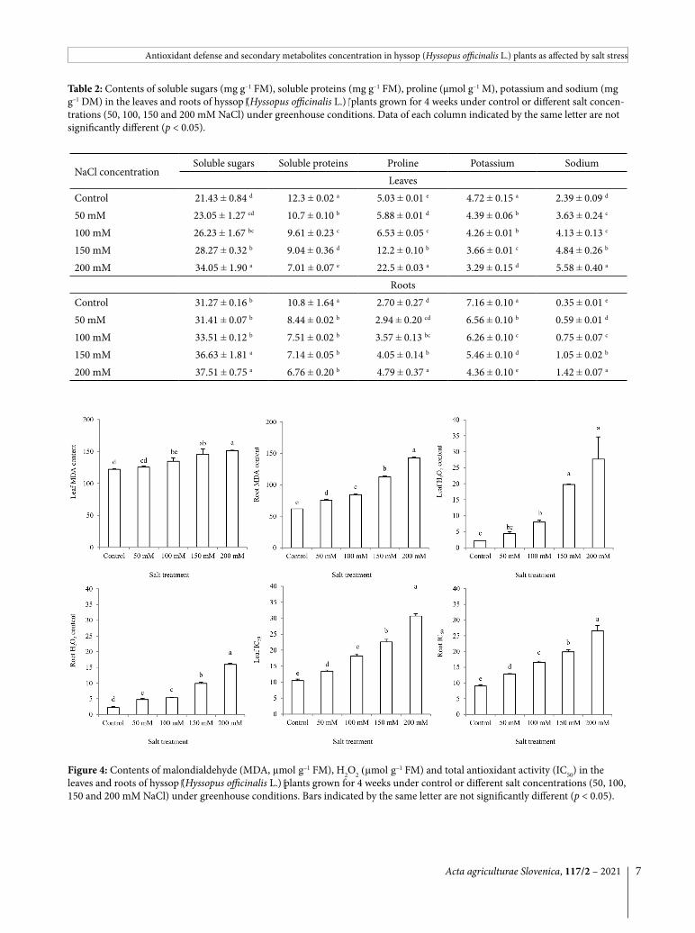
Leaf content of MDA was increased by salt stress both in the leaves and roots. The treatment effect was more prominent in the roots with up to 2.3 fold MDA accumulation at the salt treatment of 200 mM, while the corresponding value for the leaves was only 1.2 fold. The
Acta agriculturae Slovenica, 117/2 – 2021 7
Antioxidant defense and secondary metabolites concentration in hyssop (Hyssopus officinalis L.) plants as affected by salt stress
Table 2: Contents of soluble sugars (mg g–1 FM), soluble proteins (mg g–1 FM), proline (µmol g–1 M), potassium and sodium (mg g–1 DM) in the leaves and roots of hyssop (Hyssopus officinalis L.) plants grown for 4 weeks under control or different salt concen-trations (50, 100, 150 and 200 mM NaCl) under greenhouse conditions. Data of each column indicated by the same letter are not significantly different (p < 0.05).
Figure 4: Contents of malondialdehyde (MDA, µmol g–1 FM), H2O2 (µmol g–1 FM) and total antioxidant activity (IC50) in the leaves and roots of hyssop (Hyssopus officinalis L.) plants grown for 4 weeks under control or different salt concentrations (50, 100, 150 and 200 mM NaCl) under greenhouse conditions. Bars indicated by the same letter are not significantly different (p < 0.05).
NaCl concentrationSoluble sugars Soluble proteins Proline Potassium Sodium
LeavesControl 21.43 ± 0.84 d 12.3 ± 0.02 a 5.03 ± 0.01 e 4.72 ± 0.15 a 2.39 ± 0.09 d
50 mM 23.05 ± 1.27 cd 10.7 ± 0.10 b 5.88 ± 0.01 d 4.39 ± 0.06 b 3.63 ± 0.24 c
100 mM 26.23 ± 1.67 bc 9.61 ± 0.23 c 6.53 ± 0.05 c 4.26 ± 0.01 b 4.13 ± 0.13 c
150 mM 28.27 ± 0.32 b 9.04 ± 0.36 d 12.2 ± 0.10 b 3.66 ± 0.01 c 4.84 ± 0.26 b
200 mM 34.05 ± 1.90 a 7.01 ± 0.07 e 22.5 ± 0.03 a 3.29 ± 0.15 d 5.58 ± 0.40 a
RootsControl 31.27 ± 0.16 b 10.8 ± 1.64 a 2.70 ± 0.27 d 7.16 ± 0.10 a 0.35 ± 0.01 e
50 mM 31.41 ± 0.07 b 8.44 ± 0.02 b 2.94 ± 0.20 cd 6.56 ± 0.10 b 0.59 ± 0.01 d
100 mM 33.51 ± 0.12 b 7.51 ± 0.02 b 3.57 ± 0.13 bc 6.26 ± 0.10 c 0.75 ± 0.07 c
150 mM 36.63 ± 1.81 a 7.14 ± 0.05 b 4.05 ± 0.14 b 5.46 ± 0.10 d 1.05 ± 0.02 b
200 mM 37.51 ± 0.75 a 6.76 ± 0.20 b 4.79 ± 0.37 a 4.36 ± 0.10 e 1.42 ± 0.07 a

Acta agriculturae Slovenica, 117/2 – 20218
Z. SOHEILIKHAH
Table 3: Concentrations of saponins (mg g–1 DM), total phenolics (mg g–1 DM), total flavonoids (µg g–1 DM), anthocyanins (µg g‒1 DM) and iridoids (mg g–1 DM) in the leaves and roots of hyssop (Hyssopus officinalis L.) plants grown for 4 weeks under control or different salt concentrations (50, 100, 150 and 200 mM NaCl) under greenhouse conditions. Data of each column indicated by the same letter are not significantly different (p < 0.05).
NaCl concentrationSaponins Phenolics Flavonoids Anthocyanins Iridoids
LeavesControl 330 ± 2.8 c 19.3 ± 0.10 d 75.4 ± 3.96 d 10.15 ± 0.57 d 50.7 ± 1.59 d
50 mM 371 ± 24.7 c 22.1 ± 0.98 c 87.6 ± 3.22 c 18.70 ± 0.62 c 55.0 ± 2.57 cd
100 mM 475 ± 83.6 c 24.4 ± 0.33 b 93.9 ± 1.83 c 20.55 ± 0.78 c 73.7 ± 3.13 bc
150 mM 644 ± 54.6 b 25.7 ± 0.90 ab 109 ± 4.58 b 26.25 ± 0.83 b 92.2 ± 5.78 b
200 mM 801 ± 68.9 a 27.4 ± 0.48 a 118 ± 2.37 a 50.09 ± 1.98 a 129.1 ± 15.2 a
RootsControl 390 ± 23.7 c 45.31 ± 1.22 d 199 ± 18.31c 0.90 ± 0.10 d 70.5 ± 0.33 a
50 mM 446 ± 10.1 c 48.79 ± 0.82 cd 224 ± 1.64 bc 1.50 ± 0.10 d 75.3 ± 0.98 a
100 mM 610 ± 109 b 52.75 ± 0.38 bc 239 ± 8.38 b 2.52 ± 0.50 c 76.8 ± 0.18 a
150 mM 755 ± 18.3 a 55.03 ± 0.43 b 249 ± 1.77 ab 4.00 ± 0.56 b 78.5 ± 0.64 a
200 mM 859 ± 5.81 a 59.39 ± 3.10 a 267 ± 9.39 a 6.98 ± 0.07 a 75.9 ± 6.79 a
content of H2O2 was consistently increased by increasing salt level. The H2O2 accumulation in response to higher salt levels (150 and 200 mM) was more prominent in the leaves than in the roots. The total antioxidant activ-ity (IC50) was increased up to 2.9 fold in the salt-stressed plants (Fig. 4).
The leaf and root concentrations of all analyzed secondary metabolites were increased by exposure to salt stress in the leaves and roots except iridoids in the roots that remained unchanged (Table 3). Lower salt level (50 mM) was effective in the increasing phenolics, flavonoids, and anthocyanins in the leaves, while in the roots, a significant effect was observed by higher salt level (100 mM). The extent of salt-induced increase in the concentration of analyzed secondary metabolites was in the range of 1.5-2.5 fold except for anthocyanins. This metabolite showed up to 4.9 and 7.8 fold increase upon exposure to 200 mM salt in the leaves and roots, respec-tively (Table 3).
4 DISCUSSION
4.1 EFFECT OF SALT STRESS ON GROWTH, NA CONCENTRATION AND LEAF CHL CONTENTHyssop is a drought-tolerant species (Khazaie e al.,
2008); however, its salt tolerance has not been studied so far. Our data demonstrated that hyssop is also tolerant to salinity stress as the plants in our study survived after 4 weeks of salt treatment of 200 mM. Such high salt toler-
ance has been rarely reported in the members of Lami-aceae. In the studies on the salt tolerance in other Lami-aceae species such as Thymus, Perilla, and Salvia, much higher growth inhibition by salt has been reported, and plants were killed by salt concentrations higher than 100 mM (Paiva et al., 2018; Bistgani et al., 2019; Salachna et al., 2019).
The Na concentration data showed that this spe-cies is a Na-excluder salt-tolerant plant and can avoid root Na uptake. The low Na accumulation in the roots and leaves was accompanied by stable amounts of RWC showing that this species maintains tissue water content despite exposure to low water potentials in the rooting medium. On the other hand, a constitutively lower RWC (0.49-0.58 %) shows that these species cope with low wa-ter potentials through passive water content reduction. A similar mechanism for salt tolerance has been observed in Thellungiella, a halophyte close relative of Arabidopsis (Lugan et al., 2010).
In agreement with the conclusion mentioned above on high salt tolerance in hyssop plants, leaf Chl content remained unaffected by salt treatment up to 150 mM sug-gesting that leaf photosynthetic capacity remained main-ly unaffected under these conditions. The maintenance of photosynthesis and carbon metabolism may help plants retain an ability to synthesize organic osmolytes, includ-ing soluble sugars and proline (Chaves et al., 2009). These two organic osmolytes were accumulated in the leaves up to 1.6 and 4.5 fold, respectively, which may contrib-ute significantly to plants’ osmotic homeostasis under salt stress conditions. In addition to osmotic functions,
Acta agriculturae Slovenica, 117/2 – 2021 9
Antioxidant defense and secondary metabolites concentration in hyssop (Hyssopus officinalis L.) plants as affected by salt stress
these osmolytes contribute to protecting cell structures, ROS scavenging, and nitrogen and carbon sources under stress conditions (Verbruggen and Hermans, 2008; Mat-tioli et al., 2009; Rosa et al., 2009).
4.2 EFFECT OF SALT STRESS ON THE ACTIVITY OF ROS ACCUMULATION, SCAVENGING AND MEMBRANE INTEGRITY
The activities of ROS scavenging enzymes were ex-pectedly increased by salt treatment both in the leaves and roots. The salt-induced activity of all three analyzed enzymes was higher in the leaves compared with the roots that may contribute to high protection of leaves against salt-induced damage. Better protection of leaves than roots was confirmed by the maintenance of a high Chl content under high salinity treatments and much less increase of MDA content under salt stress (24 % at 200 mM salt) compared with the roots (130 % at 200 mM salt).
Nevertheless, the accumulation of H2O2 was higher in the leaf than in the roots indicating that higher en-zyme activities were not sufficient for inhibition of H2O2 accumulation in the leaves. Although H2O2 belongs to ROS, it is known to be much less damaging in compari-son to superoxide and hydroxyl radicals (Cheng et al., 2006). The prevailing effect of H2O2 is a signaling role. It has been observed that H2O2 is an important signal that is raised under salt stress and is responsible for the acti-vation of various defense pathways in salt-stressed plants (Shu-Hsien et al., 2005). We suggest that the higher ca-pability of leaves for H2O2 accumulation, and activation of defense pathways may be partly responsible for higher protection of leaves against salt stress than the roots.
4.3 EFFECT OF SALT STRESS ON THE CONCENTRATION OF SECONDARY METABOLITES
The concentration of all analyzed secondary metab-olites was higher in the salt-stressed hyssop plants in our study. The effect of salt treatment on the levels of pheno-lics, flavonoids, and anthocyanins has been reported in other Lamiaceae species (Kotagiri et al., 2017; Bistgani et al., 2019; Salachna et al., 2019; Becerra-Gudiño et al., 2019). However, in hyssop, the quantity of bioactive com-pounds as affected by salinity has not been investigated so far. Our study is also the first report on the effect of salt stress on the saponins and iridoids. Iridoids are a type of monoterpenoids found in plants, mainly as glyco-
sides (Wang et al., 2020). The iridoids produced by plants act as a defense against herbivores or microorganisms (Fuchs et al., 2004). From a medicinal point of view, these compounds have wound-healing and anti-inflammatory effects with therapeutic potential for Alzheimer’s and Parkinson’s diseases (Dinda et al., 2019; Hussain et al., 2019). Saponins with one or more hydrophilic glycoside moieties combined with a lipophilic triterpene molecule (El Aziz et al., 2019) exhibit medicinal properties such as hemolytic factor, anti-inflammatory, antibacterial, anti-fungal, antiviral, anticancer, and cholesterol-lowering ac-tion in animals and human (Sparg et al., 2004). Besides, saponins formed the backbone of modern medicine or drugs and were considered a starting precursor for the semi-synthesis of steroidal drugs in the pharmaceutical industry (Netala et al., 2015).
It is noteworthy that higher concentrations of the secondary metabolites accompanied by reduction of biomass suggests a ‘concentration-effect’ in our hyssop plants. Nonetheless, it indicates that salt treatment did not inhibit the secondary metabolism in this species.
4.4 EFFECT OF SALT STRESS ON THE ANTIOXIDANT ACTIVITY OF LEAF AND ROOT EXTRACT
The DPPH scavenging activity is defined as the anti-oxidant activity of food and medicinal plants (Fukumoto and Mazza 2000; Sethi et al., 2020), has been reported for hyssop plants (Fathiazad et al., 2011; Pirbalouti et al., 2019; Rezaei Savadkouhi et al., 2020). However, the effect of salt on this parameter has not been studied so far. Here in our work, the DPPH free radical scavenging activity was increased by salt treatment for the leaf and root extracts. Electron donation is an important mecha-nism in which plants bioactive compounds convert free radicals to nonradical forms and thus, end the radical chain reactions (San Miguel-Chávez, 2017; Shahidi and Ambigaipalan, 2015). By analyzing various plant species, it has been observed that the main component of DPPH scavenging activity is phenolics, flavonoids, and antho-cyanins (Fukumoto and Mazza 2000; Kim et al., 2007). Phenolic compounds act as a reducing agent and a hy-drogen donator and show antioxidant effects (Oke et al., 2009).
5 CONCLUSION
Our data demonstrated that, hyssop plants are a salt-tolerant species, and secondary metabolites are in-creased upon growth under salinity. Regarding the fact

Acta agriculturae Slovenica, 117/2 – 202110
Z. SOHEILIKHAH
that, the plants dry matter production was reduced un-der higher salt levels. i.e., 200 mM equivalent with 14.5 dS m–1, cultivation of this species is recommended in the soils with electrical conductivity up to 10 dS m–1. Thus, the cultivation of this species on salinized soils that are unsuitable for most crop species is an alternative for low-income farmers.
6 REFERENCES
Acosta-Motos, J. R., Ortuno, M. F., Bernal-Vicente, A., Diaz-Vi-vancos, P., Sanchez-Blanco, M. J., & Hernandez, J. A. (2017). Plant responses to salt stress: adaptive mechanisms. Agron-omy, 7(1), 18. https://doi.org/10.3390/agronomy7010018
Ahl, S. A., & Omer, E. (2011). Medicinal and aromatic plants production under salt stress. A review. Herba Polonica, 57(1), 72–87.
Ahmad, P., & Sharma, S. )2008(. Salt stress and phyto-biochem-ical responses of plants. Plant and Soil Environment, 54, 89–99. https://doi.org/10.17221/2774-PSE
Akyol, T.Y., Yilmaz, O., Uzilday, B., Uzilday, R. Ö., & Türkan, İ. (2020). Plant response to salinity: an analysis of ROS for-mation, signaling, and antioxidant defense. Turkish Journal of Botany, 44(1), 1–3. https://doi.org/10.3906/bot-1911-15
Alam, M. A., Juraimi, A. S., Rafii, M. Y., Hamid, A. A., Asla-ni, F., & Alam, M. Z. (2015). Effects of salinity and salin-ity-induced augmented bioactive compounds in purslane (Portulaca oleracea L.) for possible economical use. Food Chemistry, 169, 439–47. https://doi.org/10.1016/j.food-chem.2014.08.019
Bates, L. S., Waldren, R. P., & Teare, I. (1973). Rapid determina-tion of free proline for water-stress studies. Plant and Soil, 39(1), 205–207. https://doi.org/10.1007/BF00018060
Becerra-Gudiño, A., Juárez-Rosete, C. R., Bugarín-Montoya, R., & Murillo-Amador, B. (2019). Growth of Rosmarinus officinalis L. and accumulation of secondary metabolites under high salinity. Revista Bio Ciencias, 6, e567.
Beers, R. F & Sizer, I. W. (1952). A spectrophotometric method for measuring the breakdown of hydrogen peroxide by catalase. Journal of Biological chemistry, 195(1), 133–140. https://doi.org/10.1016/S0021-9258(19)50881-X
Bistgani, Z. E., Hashemi, M., DaCosta, M., Craker, L., Maggi, F., & Morshedloo, M. R. (2019). Effect of salinity stress on the physiological characteristics, phenolic compounds and antioxidant activity of Thymus vulgaris L. and Thymus dae-nensis Celak. Industrial Crops and Products, 135, 311–320. https://doi.org/10.1016/j.indcrop.2019.04.055
Bradford, M. M. (1976). A rapid and sensitive method for the quantitation of microgram quantities of protein utilizing the principle of protein-dye binding. Analytical Biochem-istry, 72(1–2), 248–254. https://doi.org/10.1016/0003-2697(76)90527-3
Chaves, M. M., Flexas, J., & Pinheiro, C. (2009). Photosynthe-sis under drought and salt stress: regulation mechanisms from whole plant to cell. Annals of Botany, 103(4), 551–560. https://doi.org/10.1093/aob/mcn125
Cheng, Y., Song, C. (2006). Hydrogen peroxide homeostatis
and signaling in plant cells. Science in China, Series C (Life Sciences-English Edition), 49(1), 1.
Dazy, M., Béraud, E., Cotelle, S., Meux, E., Masfaraud, J.F., & Férard J.F. (2008). Antioxidant enzyme activities as affected by trivalent and hexavalent chromium species in Fontinalis antipyretica Hedw. Chemosphere, 73(3), 281–290. https://doi.org/10.1016/j.chemosphere.2008.06.044
Dinda, B., Dinda, M., Kulsi, G., Chakraborty, A., & Dinda S. (2019). Therapeutic potentials of plant iridoids in Alz-heimer’s and Parkinson’s diseases: A review. European Journal of Medicinal Chemistry, 169,185–199. https://doi.org/10.1016/j.ejmech.2019.03.009
Dubois, M., Gilles, K. A., Hamilton, J. K., Rebers, P., & Smith, F. (1956). Colorimetric method for determination of sugars and related substances. Analytical Chemistry, 28(3): 350–356. https://doi.org/10.1021/ac60111a017
El Aziz, M. M., Ashour, A. S., & Melad, A. S. (2019) A review on saponins from medicinal plants: chemistry, isolation, and determination. Journal of Nanomedicine Research, 8, 6–12.
Fathiazad, F., & Hamedeyazdan, S. (2011) A review on Hyssopus officinalis L.: Composition and biological activities. African Journal of Pharmacy and Pharmacology, 5(17), 1959-1966 https://doi.org/10.5897/AJPP11.527
Fathiazad, F., Mazandarani, M., & Hamedeyazdan, S. (2011) Phytochemical analysis and antioxidant activity of Hysso-pus officinalis L. from Iran. Advanced Pharmaceutical Bul-letin, 1(2), 63.
Flores-de-Santiago, F., Kovacs, J., Wang, J., Flores-Verdugo, F., Zhang, C., & González-Farías, F. (2016). Examining the in-fluence of seasonality, condition, and species composition on mangrove leaf pigment contents and laboratory based spectroscopy data. Remote Sensing, 8(3), 226–246. https://doi.org/10.3390/rs8030226
Foyer, C., Descourvieres, P., & Kunert, K. (1994). Protection against oxygen radicals: an important defence mecha-nism studied in transgenic plants. Plant Cell Environment, 17(5), 507–523. https://doi.org/10.1111/j.1365-3040.1994.tb00146.x
Fuchs, A., & Bowers, M. D. (2004). Patterns of iridoid gly-coside production and induction in Plantago lan-ceolata and the importance of plant age. Journal of Chemical Ecology, 30(9), 1723–1741. https://doi.org/10.1023/B:JOEC.0000042398.13765.83
Fukumoto, L. R., & Mazza, G. (2000). Assessing antioxidant and prooxidant activities of phenolic compounds. Journal of Agricultural and Food Chemistry, 48(8), 3597–3604. htt-ps://doi.org/10.1021/jf000220w
Giusti, M. M., Wrolstad, R. E. (2001). Characterization and measurement of anthocyanins by UV‐visible spectros-copy. Current Protocols in Food Analytical Chemistry, 00(1), F1.2.1-F1.2.13. https://doi.org/10.1002/0471142913.faf0102s00
Gupta, B., & Huang B. (2014). Mechanism of salinity tolerance in plants: physiological, biochemical, and molecular char-acterization. International Journal of Genomics, e701596. https://doi.org/10.1155/2014/701596
Heath, R. L., & Packer, L. (1968). Photoperoxidation in isolated chloroplasts: I. Kinetics and stoichiometry of fatty acid per-

Acta agriculturae Slovenica, 117/2 – 2021 11
Antioxidant defense and secondary metabolites concentration in hyssop (Hyssopus officinalis L.) plants as affected by salt stress
oxidation. Archives of Biochemistry and Biophysics, 125(1), 189–198. https://doi.org/10.1016/0003-9861(68)90654-1
Hiai, S., Oura, H., Odaka, Y., & Nakajima, T. (1975). A col-orimetric estimation of ginseng saponins. Planta Medica, 28(8), 363–369. https://doi.org/10.1055/s-0028-1097871
Hristova, Y., Wanner, J., Jirovetz, L., Stappen, I., Iliev, I., & Gochev, V. (2015). Chemical composition and antifungal activity of essential oil of Hyssopus officinalis L. from Bul-garia against clinical isolates of Candida species. Biotechnol Biotechnol Equip, 29, 592–601. https://doi.org/10.1080/13102818.2015.1020341
Hussain, H., Green, I. R., Saleem, M., Raza, M. L., & Nazir, M. (2019). Therapeutic potential of iridoid derivatives: Patent review. Inventions, 4(2), 29. https://doi.org/10.3390/inven-tions4020029
Jahantigh, O., Najafi, F., Badi, H. N., Khavari-Nejad, R. A., & Sanjarian, F. (2016). Changes in antioxidant enzymes ac-tivities and proline, total phenol and anthocyanine con-tents in Hyssopus officinalis L. plants under salt stress. Acta Biologica Hungarica, 67(2), 195–204. https://doi.org/10.1556/018.67.2016.2.7
Kalra, Y. (1997). Handbook of reference methods for plant anal-ysis. CRC press. https://doi.org/10.1201/9781420049398
Kazazi, H., Rezaei, K., Ghotb-Sharif, S. J., Emam-Djomeh, Z., & Yamini, Y. (2007). Supercriticial fluid extraction of fla-vors and fragrances from Hyssopus officinalis L. cultivated in Iran. Food Chemistry, 105(2), 805–811. https://doi.org/10.1016/j.foodchem.2007.01.059
Khazaie, H. R., Nadjafi, F., & Bannayan, M. (2008). Effect of irri-gation frequency and planting density on herbage biomass and oil production of thyme (Thymus vulgaris) and hyssop (Hyssopus officinalis). Industrial Crops and Products, 27(3), 315–321. https://doi.org/10.1016/j.indcrop.2007.11.007
Kim, M. J., Hyun, J. N., Kim, J. A., Park, J. C., Kim, M. Y., Kim, J. G., Lee, S. J., Chun, S. C., Chung, I. M. (2007). Relation-ship between phenolic compounds, anthocyanins content and antioxidant activity in colored barley germplasm. Jour-nal of Agricultural and Food Chemistry, 55(12), 4802–4809. https://doi.org/10.1021/jf0701943
Kotagiri, D., Beebi, S. K., Chaitanya, K. V. (2017). Secondary metabolites and the antimicrobial potential of five differ-ent Coleus species in response to salinity stress. BioRxiv, https://doi.org/10.1101/220368
Lin, C. C., & Kao, C. H. (1999). NaCl induced changes in ionically bound peroxidase activity in roots of rice seedlings. Plant and Soil, 216(1), 147–153. https://doi.org/10.1023/A:1004714506156
Lugan, R., Niogret, M. F., Leport, L., Guégan, J. P., Larher, F. R., Savouré, A., Kopka, J., Bouchereau, A. (2010). Metabolome and water homeostasis analysis of Thellungiella salsuginea suggests that dehydration tolerance is a key response to os-motic stress in this halophyte. The Plant Journal, 64(2), 215–229. https://doi.org/10.1111/j.1365-313X.2010.04323.x
Mattioli, R., Costantino, P., Trovato, M. (2009). Proline accumu-lation in plants: not only stress. Plant Signaling & Behavior, 4(11), 1016–1018. https://doi.org/10.4161/psb.4.11.9797
Mushtaq, Z., Faizan, S., & Gulzar, B. (2020). Salt stress, its im-pacts on plants and the strategies plants are employing
against it: a review. Journal of Applied Biology and Biotech-nology, 8, 81–91. https://doi.org/10.7324/JABB.2020.80315
Narayanan, P., & Akamanchi, K. (2003). Colorimetric estima-tion of total iridoid content of Picrorhiza kurrooa. Journal of Asian Natural Products Research, 5(2), 105–111. https://doi.org/10.1080/1028602021000054955
Netala, V. R., Ghosh, S. B., Bobbu, P., Anitha, D., & Tartte, V. (2015). Triterpenoid saponins: a review on biosynthesis, applications and mechanism of their action. International Journal of Pharmacy and Pharmaceutical Sciences, 7(1), 24–28.
Oke, F., Aslim, B., Ozturk, S., & Altundag, S. (2009). Essential oil composition, antimicrobial and antioxidant activities of Satureja cuneifolia Ten. Food Chemistry, 112(4), 874–879. https://doi.org/10.1016/j.foodchem.2008.06.061
Paiva, E. P., Torres, S. B., Alves, T. R, Sá, F. V., Leite, M. D., & Dombroski, J. L. (2018). Germination and biochemical components of Salvia hispanica L. seeds at different salinity levels and temperatures. Acta Scientiarum. Agronomy, 40, e39396. https://doi.org/10.4025/actasciagron.v40i1.39396
Parida, A.K., & Das A.B. (2005). Salt tolerance and salin-ity effects on plants: a review. Ecotoxicology and Environ-mental Safety, 60(3), 324–349. https://doi.org/10.1016/j.ecoenv.2004.06.010
Parihar, P., Singh, S., Singh, R., Singh, V.P., & Prasad, S.M. (2015). Effect of salinity stress on plants and its tolerance strategies: a review. Environmental Science and Pollu-tion Research, 22(6), 4056–4075 https://doi.org/10.1007/s11356-014-3739-1
Petropoulos, S. A., Levizou, E., Ntatsi, G., Fernandes, Â., Petro-tos, K., & Akoumianakis, K., et al. (2017). Salinity effect on nutritional value, chemical composition and bioac-tive compounds content of Cichorium spinosum L. Food Chemistry, 214, 129–136. https://doi.org/10.1016/j.food-chem.2016.07.080
Pirbalouti, A. G., Mohamadpoor, H., Bajalan, I., & Malekpoor, F. (2019). Chemical compositions and antioxidant activ-ity of essential oils from inflorescences of two landraces of hyssop [Hyssopus officinalis L. subsp. angustifolius (Bieb.)] cultivated in Southwestern, Iran. Journal of Essential Oil Bearing Plants, 22(4), 1074–1081. https://doi.org/10.1080/0972060X.2019.1641431
Rezaei Savadkouhi, N., Ariaii, P., & Charmchian Langerodi, M. (2020). The effect of encapsulated plant extract of hyssop (Hyssopus officinalis L.) in biopolymer nanoemulsions of Lepidium perfoliatum and Orchis mascula on controlling oxidative stability of soybean oil. Food Science & Nutrition, 8(2), 1264–1271. https://doi.org/10.1002/fsn3.1415
Rosa, M., Prado, C., Podazza, G., Interdonato, R., González, J. A., Hilal, M., & Prado, F. E. (2009). Soluble sugars: Me-tabolism, sensing and abiotic stress: A complex network in the life of plants. Plant Signaling & Behavior, 4(5), 388–393. https://doi.org/10.4161/psb.4.5.8294
Salachna, P., Grzeszczuk, M., Meller, E., & Mizielińska, M. (2019). Effects of gellan oligosaccharide and NaCl stress on growth, photosynthetic pigments, mineral composi-tion, antioxidant capacity and antimicrobial activity in red perilla. Molecules, 24(21), 3925. https://doi.org/10.3390/molecules24213925

Acta agriculturae Slovenica, 117/2 – 202112
Z. SOHEILIKHAH
San Miguel-Chávez, R. (2017). Phenolic antioxidant capacity: A review of the state of the art. Phenolic Compounds-Biologi-cal Activity, 8, 59–74. https://doi.org/10.5772/66897
Sarkar, K., & Sil, P. C. (2006). A 43 kDa protein from the herb Cajanus indicus L. protects thioacetamide induced cyto-toxicity in hepatocytes. Toxicology in vitro, 20(5), 634–640. https://doi.org/10.1016/j.tiv.2005.11.003
Seevers, P., Daly, J., & Catedral, F. (1971). The role of peroxi-dase isozymes in resistance to wheat stem rust disease. Plant Physiology, 48(3), 353–360. https://doi.org/10.1104/pp.48.3.353
Sergiev, I., Alexieva, V., & Karanov, E. (1997). Effect of sper-mine, atrazine and combination between them on some en-dogenous protective systems and stress markers in plants. Comptes Rendus de l ‘Academie Bulgare des Sciences, 51(3), 121–124.
Sethi, S., Joshi, A., Arora, B., Bhowmik, A., Sharma, R. R., & Ku-mar, P. (2020). Significance of FRAP, DPPH, and CUPRAC assays for antioxidant activity determination in apple fruit extracts. European Food Research and Technology, 246(3), 591–598. https://doi.org/10.1007/s00217-020-03432-z
Shahidi, F., & Ambigaipalan, P. (2015). Phenolics and polyphe-nolics in foods, beverages and spices: Antioxidant activity and health effects–A review. Journal of Functional Foods, 18, 820–897. https://doi.org/10.1016/j.jff.2015.06.018
Shu-Hsien, H. U., Chih-Wen, Y. U., & Lin, C. H. (2005). Hydro-gen peroxide functions as a stress signal in plants. Botanical Bulletin of Academia Sinica, 46, 1–10.
Sparg, S. G., Light, M. E., Van Staden, J. (2004). Biological activ-ities and distribution of plant saponins. Journal of ethnop-harmacology, 94(2-3), 219–243. https://doi.org/10.1016/j.jep.2004.05.016
Taarit, M. B., Msaada, K., Hosni, K., Hammami, M., Kchouk, M. E., Marzouk, B. (2009). Plant growth, essential oil yield and composition of sage (Salvia officinalis L.) fruits cultivated under salt stress conditions. Industrial Crops Production, 30, 333–337. https://doi.org/10.1016/j.indcrop.2009.06.001
Valifard, M., Mohsenzadeh, S., Kholdebarin, B., & Rowshan, V. (2014). Effects of salt stress on volatile compounds, total phenolic content and antioxidant activities of Salvia mir-zayanii. South African Journal of Botany, 93, 92–97. https://doi.org/10.1016/j.sajb.2014.04.002
Verbruggen, N., & Hermans, C. (2008). Proline accumulation in plants: a review. Amino acids, 35(4), 753–759. https://doi.org/10.1007/s00726-008-0061-6
Verma, N., & Shukla, S. (2015). Impact of various factors respon-sible for fluctuation in plant secondary metabolites. Journal of Applied Research in Medicinal and Aromatic Plants, 2 (4), 105–113. https://doi.org/10.1016/j.jarmap.2015.09.002
Wang, C., Gong, X., Bo, A., Zhang, L., Zhang, M., Zang, E., Zhang, C., & Li, M. (2020). Iridoids: research advances in their phytochemistry, biological activities, and pharma-cokinetics. Molecules, 25(2), 287. https://doi.org/10.3390/molecules25020287
Zhishen, J., Mengcheng, T., & Jianming, W. (1999). The deter-mination of flavonoid contents in mulberry and their scav-enging effects on superoxide radicals. Food Chemistry, 64, 555–559. https://doi.org/10.1016/S0308-8146(98)00102-2
Zrig, A., Tounekti, T., Hegab, M. M., Ali, S. O., & Khemira, H. (2016). Essential oils, amino acids and polyphenols changes in salt-stressed Thymus vulgaris exposed to open–field and shade enclosure. Industrial Crops and Products, 91, 223–230. https://doi.org/10.1016/j.indcrop.2016.07.012


![Tryptophan-Derived Metabolites Are Required for …...Tryptophan-Derived Metabolites Are Required for Antifungal Defense in the Arabidopsis mlo2 Mutant1[C][W][OA] Chiara Consonni,](https://static.fdocuments.us/doc/165x107/5ed503dead38025d974e448d/tryptophan-derived-metabolites-are-required-for-tryptophan-derived-metabolites.jpg)


