Antimicrobial effects of topical skin cream containing ...vri.cz/docs/vetmed/60-4-202.pdf · 202...
6
202 Original Paper Veterinarni Medicina, 60, 2015 (4): 202–207 doi: 10.17221/8108-VETMED Antimicrobial effects of topical skin cream containing natural oil mixtures against Staphylococcus pseudintermedius and Malassezia pachydermatis J.I. Han, S.J. Park, S.G. Kim, H.M. Park College of Veterinary Medicine, Konkuk University, Seoul, Republic of Korea ABSTRACT: e objective of this study was to evaluate the in vitro efficacy of a topical skin cream containing a mixture of emu oil, jojoba oil, avocado oil, and tea tree oil against the canine skin pathogens Staphylococcus pseudintermedius and Malassezia pachydermatis. ree S. pseudintermedius isolates from dogs and a type strain of M. pachydermatis were used. Based on the standards of the Clinical and Laboratory Standards Institute, the minimal inhibitory concentration (MIC) and the minimal bactericidal/fungicidal concentration (MBC/MFC) were determined. In addition, microbial inactivation time was determined for both pathogens. e MICs against S. pseudintermedius and M. pachydermatis were 0.23% and 0.63%, while the MBC/MFCs were 7.5% and 5%, respectively. In assessments of the microbial inactivation time, after 12 h of incubation with the cream, the growth of both pathogens was com- pletely inhibited. ese results suggest that the skin cream tested here can be used as a substitute for generally used antibiotic/antifungal agents. Keywords: natural oil; Staphylococcus pseudintermedius; Malassezia pachydermatis; antimicrobial effect Supported by High Nature Ltd in 2014 (Grant No. 2013-A012-0038). Superficial skin infections such as superficial pyoderma and otitis externa are commonly en- countered in veterinary practice and account for a significant number of antimicrobial prescriptions (Escher et al. 2011). Staphylococcus pseudinter- medius and Malassezia pachydermatis are the most common pathogens isolated from pyoderma and otitis externa in dogs (Cafarchia et al. 2005; Fitzgerald 2009). In the last decade, resistance to antimicrobial agents has increased, particularly with the emergence and widespread dissemina- tion of methicillin-resistant S. pseudintermedius in dogs as a result of the broad use of certain anti- microbial agents (Neu 1992; Kadlec and Schwarz 2013). The increased resistance to most available antimicrobial agents has created a need to identify alternative treatment strategies. As one possible alternative, the use of essential oils against various pathogens such as bacteria, virus, fungi, parasites, and insects has been widely evaluated (Weseler et al. 2002; Moon et al. 2006; Prabussenivasan et al. 2006; LaPlante et al. 2007; Wu et al. 2010; Freire et al. 2012; Valentine et al. 2012). These oils contain numerous constituents that contribute to the char- acteristic odour and medicinal effects, but they are not a usual choice for the treatment of infection, primarily because of the lack of scientific evidence of their efficacy. The objective of this study was to evaluate the in vitro efficacy of a topical skin cream containing a mixture of natural oils against the canine skin path- ogens S. pseudintermedius and M. pachydermatis. MATERIAL AND METHODS Test materials. A topical skin cream (Dara cream ® , Koreai1, Kimpo, Korea) containing a mixture of emu oil, jojoba oil, avocado oil, and tea tree oil was evaluated. Ampicillin sodium salt and miconazole nitrate were purchased from Sigma-Aldrich Corp. (St. Louis, MO, USA) and used as positive controls for S. pseudintermedius and M. pachydermatis, re- spectively.
Transcript of Antimicrobial effects of topical skin cream containing ...vri.cz/docs/vetmed/60-4-202.pdf · 202...
202
Original Paper Veterinarni Medicina, 60, 2015 (4): 202–207
doi: 10.17221/8108-VETMED
Antimicrobial effects of topical skin cream containing natural oil mixtures against Staphylococcus pseudintermedius and Malassezia pachydermatis
J.I. Han, S.J. Park, S.G. Kim, H.M. Park
College of Veterinary Medicine, Konkuk University, Seoul, Republic of Korea
ABSTRACT: The objective of this study was to evaluate the in vitro efficacy of a topical skin cream containing a mixture of emu oil, jojoba oil, avocado oil, and tea tree oil against the canine skin pathogens Staphylococcus pseudintermedius and Malassezia pachydermatis. Three S. pseudintermedius isolates from dogs and a type strain of M. pachydermatis were used. Based on the standards of the Clinical and Laboratory Standards Institute, the minimal inhibitory concentration (MIC) and the minimal bactericidal/fungicidal concentration (MBC/MFC) were determined. In addition, microbial inactivation time was determined for both pathogens. The MICs against S. pseudintermedius and M. pachydermatis were 0.23% and 0.63%, while the MBC/MFCs were 7.5% and 5%, respectively. In assessments of the microbial inactivation time, after 12 h of incubation with the cream, the growth of both pathogens was com-pletely inhibited. These results suggest that the skin cream tested here can be used as a substitute for generally used antibiotic/antifungal agents.
Keywords: natural oil; Staphylococcus pseudintermedius; Malassezia pachydermatis; antimicrobial effect
Supported by High Nature Ltd in 2014 (Grant No. 2013-A012-0038).
Superficial skin infections such as superficial pyoderma and otitis externa are commonly en-countered in veterinary practice and account for a significant number of antimicrobial prescriptions (Escher et al. 2011). Staphylococcus pseudinter-medius and Malassezia pachydermatis are the most common pathogens isolated from pyoderma and otitis externa in dogs (Cafarchia et al. 2005; Fitzgerald 2009). In the last decade, resistance to antimicrobial agents has increased, particularly with the emergence and widespread dissemina-tion of methicillin-resistant S. pseudintermedius in dogs as a result of the broad use of certain anti-microbial agents (Neu 1992; Kadlec and Schwarz 2013). The increased resistance to most available antimicrobial agents has created a need to identify alternative treatment strategies. As one possible alternative, the use of essential oils against various pathogens such as bacteria, virus, fungi, parasites, and insects has been widely evaluated (Weseler et al. 2002; Moon et al. 2006; Prabussenivasan et al. 2006; LaPlante et al. 2007; Wu et al. 2010; Freire et
al. 2012; Valentine et al. 2012). These oils contain numerous constituents that contribute to the char-acteristic odour and medicinal effects, but they are not a usual choice for the treatment of infection, primarily because of the lack of scientific evidence of their efficacy.
The objective of this study was to evaluate the in vitro efficacy of a topical skin cream containing a mixture of natural oils against the canine skin path-ogens S. pseudintermedius and M. pachydermatis.
MATERIAL AND METHODS
Test materials. A topical skin cream (Dara cream®, Koreai1, Kimpo, Korea) containing a mixture of emu oil, jojoba oil, avocado oil, and tea tree oil was evaluated. Ampicillin sodium salt and miconazole nitrate were purchased from Sigma-Aldrich Corp. (St. Louis, MO, USA) and used as positive controls for S. pseudintermedius and M. pachydermatis, re-spectively.
203
Veterinarni Medicina, 60, 2015 (4): 202–207 Original Paper
doi: 10.17221/8108-VETMED
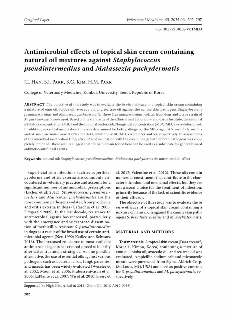
Test organisms. Three S. pseudintermedius iso-lates were obtained from nostrils of healthy dogs at the Veterinary Teaching Hospital of Kunkuk University (Seoul, Korea). Primary identification of the bacteria was made on the basis of colony morphology, complete or incomplete haemolysis, Gram staining, and conventional catalase test. The resultant Staphylococcus isolates were further tested for coagulase synthesis through the tube co-agulase test and DNase test. For identification of S. pseudintermedius, multiplex PCR for the nuc gene was performed for the isolated Staphylococcus spp. as described previously (Figure 1) (Sasaki et al. 2010). Additionally, the 16S ribosomal RNA gene of an isolated S. pseudintermedius was am-plified by a universal primer pair (27F and 1492R) and sequenced directly. Based on the Clinical and Laboratory Standards Institute (CLSI) guideline MM-18A, S. pseudintermedius was finally con-firmed by sequence comparison using the GenBank database.
A type strain of M. pachydermatis (KCTC 17008) was purchased from the Korea Collection for Type Culture (KCTC; Daejeon, Korea). The organisms were cultured on modified Leeming and Notman (LNA) agar plates (BD, Sparks, MD, USA) at 30 °C for seven days and used for the subsequent experiments.
Minimal inhibitory concentration (MIC) determination. For the MIC measurement of S. pseudintermedius and M. pachydermatis, the performance standard for antimicrobial suscep-tibility testing (CLSI, M100-S22) and the micro-
dilution antifungal method (CLSI BMD, M27-A3) according to the CLSI were applied, respectively.
A suspension of S. pseudintermedius was prepared by inoculation of a freshly cultured bacterial colony from an overnight trypticase soy agar plate with 5% sheep blood. The turbidity of the bacterial suspen-sion was adjusted to a 0.5 McFarland Standard. In order to identify the number of bacteria in the sus-pension, colony counting was performed using a standard plate counting technique. Briefly, a series of 10-fold dilutions from 10–1 to 10–6 were made using sterile saline. Three trypticase soy agar plates with 5% sheep blood were prepared and each plate was divided into two sections. One hundred mi-croliters of each dilution was inoculated on each section of the plates. All plates were incubated at 37 °C until colonies formed. The CFU on each sec-tion were recorded. Dilutions with ≤ 50 CFU/sec-tion were selected for calculating the CFU of the bacterial suspension. The final concentration of the bacterial suspension was 1.2 × 10⁵ CFU/ml.
A suspension of M. pachydermatis was prepared from the cultured modified LNA plate by gently scraping the colonies off with sterile saline contain-ing 0.1% Tween® 80. The suspension was filtered through sterile gauze to remove large aggregates of yeast cells. Final concentrations of the yeast cells were determined using a haemocytometer and were adjusted to 1.44 × 10⁴ cells/ml, corresponding to 0.4–5.0 × 10⁴ CFU/ml, as recommended by CLSI.
Ampicillin (128 μg/ml in 0.9% saline), miconazole (128 μg/ml in 0.9% saline), or Dara cream® stock solution (7.5% in 0.9% saline) was prepared by a se-ries of 2-fold dilutions using Mueller-Hinton broth (ampicillin and Dara cream® stock solution for S. pseudintermedius) or RPMI 1640 medium (with 2% l-glutamine) buffered to pH 7.0 (miconazole and Dara cream® stock solution for M. pachyder-matis). Final concentrations of ampicillin, micon-azole, and Dara cream® were 128–0.0625 μg/ml, 128–0.0625 μg/ml, and 7.5–0.0036%, respectively.
For S. pseudintermedius, 100-μl aliquots of ampi-cillin or Dara cream® were dispensed separately into 96-well microplate wells, while for M. pachydermatis, miconazole or Dara cream® were similarly dispensed into 96-well microplate wells. Then, each suspension of pathogenic microbes was dispensed into the corre-sponding microplate wells as 100-μl aliquots and the plates were incubated at 37 °C for 20 h (for bacteria) or at 30 °C until growth (for yeast). The MIC was visually determined as the lowest concentration of
Figure 1. Result of multiplex PCR for the nuc gene of five coagulase-negative (lane 1 to 5) and two coagulase-positive Staphylococci (lane 6 and 7) isolated from the nostrils of healthy dogs. Lanes 6 and 7 showed bands of the expected sizes for S. aureus (359 bp) or S. pseudinter-medius (926 bp) while the reactions were all negative in coagulase-negative Staphylococci
204
Original Paper Veterinarni Medicina, 60, 2015 (4): 202–207
doi: 10.17221/8108-VETMED
the drug or cream to inhibit the growth of bacteria or yeast. The MIC measurements for ampicillin or Dara cream® were performed in triplicate.
Minimal bactericidal/fungicidal concentra-tion (MBC/MFC) determination. After MIC de-termination, MBC/MFC was determined by the culture of the medium in the wells in front of the well with the MIC concentration of Dara cream®. For S. pseudintermedius, cultured media were inoc-ulated on trypticase soy agar with 5% sheep blood and the plates were cultured at 37 °C for 20 h. For M. pachydermatis, cultured media were inoculated on Sabouraud dextrose agar and the plates were cultured at 30 °C for a week. The MBC/MFC was determined to be the lowest concentration of the drugs or cream to kill the bacteria or yeast.
Determination of microbial inactivation time. Dara cream® was further tested for determination of microbial inactivation time by incubation of a mixture containing Dara cream® together with each pathogen. As test materials, a bacterial or yeast sus-pension was adjusted to 1.5 × 10⁴ CFU/ml (for bac-teria) or 4.2 × 10³ cells/ml (for yeast) using the same protocol. One gram of Dara cream® was dissolved in 3 ml of the prepared bacterial or yeast suspension, and the mixtures were incubated for 1, 3, and 12 h at 37 °C (for bacteria) or 30 °C (for yeast). After the incubation, 100 μl of each mixture were inoculated on trypticase soy agar with 5% sheep blood (for bac-teria) or Sabouraud dextrose agar (for yeast). For the bacteria, the plates were incubated for 16 h and colonies were counted. For the yeast, the plates were incubated until the cultured colonies were visible in the positive control and colonies were then counted. As positive controls, 100 μl of bacterial or yeast sus-pension were inoculated and incubated for 1, 3, and 12 h using the same procedures.
Statistical analysis. Microbial inactivation in the Dara cream®-treated or non-treated groups was analysed using the Kruskal-Wallis test followed by the Mann-Whitney U-test (SPSS for Windows, IBM, USA). A P-value of less than 0.05 was re-garded as significant.
RESULTS
MIC and MBC/MFC
The MIC and MBC/MFC values of Dara cream®
to S. pseudintermedius and M. pachydermatis are
summarised in Table 1. The MICs against S. pseud-intermedius and M. pachydermatis were 0.23% and 0.63%, respectively. The MBC/MFC was 7.5% and 5%, respectively, indicating similar antimicrobial effects of Dara cream® against both pathogens.
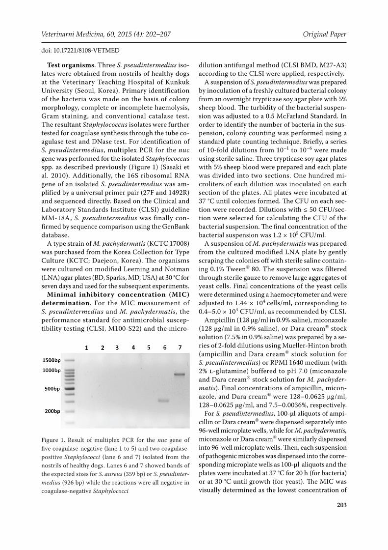
Microbial inactivation time. Changes in the number of S. pseudintermedius following incuba-tion with Dara cream® are shown in Figure 2 and Figure 3. While the number of bacterial CFU in the control (no treatment group) explosively in-creased after 3 h and 12 h incubation, the CFU in the Dara cream®-treated groups (3 h and 12 h incubation groups with Dara cream®) significantly decreased (P = 0.022). After 3 h of incubation with Dara cream®, the CFU count was > 99% lower (8.4 × 101 ± 8.72 CFU/ml) (P = 0.037) and after 12 h of
Table 1. MIC and MBC/MFC of Dara cream against S. pseudintermedius and M. pachydermatis
Test organism MIC (%) MBC/MFC (%)S. pseudintermedius 0.23 7.5M. pachydermatis 0.63 5
MIC = minimal inhibitory concentration, MBC = minimal bactericidal concentration, MFC = minimal fungicidal con-centration
0.1
1
10
100
610
control (1 h)control (3 h)control (12 h)daracream (1 h)daracream (3 h)daracream (12 h)
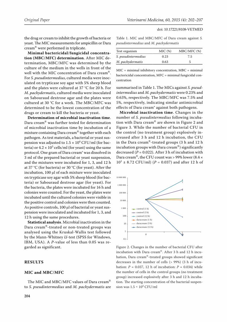
Figure 2. Changes in the number of bacterial CFU after incubation with Dara cream®. After 3 h and 12 h incu-bation, Dara cream®-treated groups showed significant decreases in the number of cells (> 99%) (3 h of incu-bation: P = 0.037, 12 h of incubation: P = 0.034) while the number of cells in the control groups (no treatment group) increased explosively after 3 h and 12 h incuba-tion. The starting concentration of the bacterial suspen-sion was 1.5 × 10⁴ CFU/ml
205
Veterinarni Medicina, 60, 2015 (4): 202–207 Original Paper
doi: 10.17221/8108-VETMED
incubation, bacterial growth was completely in-hibited (P = 0.034).
In testing for M. pachydermatis, the numbers of yeast cells had decreased at 1 h of incubation in the
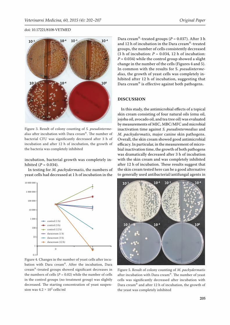
Dara cream®-treated groups (P = 0.037). After 3 h and 12 h of incubation in the Dara cream®-treated groups, the number of cells consistently decreased (3 h of incubation: P = 0.034, 12 h of incubation: P = 0.034) while the control group showed a slight change in the number of the cells (Figures 4 and 5). In common with the results for S. pseudinterme-dius, the growth of yeast cells was completely in-hibited after 12 h of incubation, suggesting that Dara cream® is effective against both pathogens.
DISCUSSION
In this study, the antimicrobial effects of a topical skin cream consisting of four natural oils (emu oil, jojoba oil, avocado oil, and tea tree oil) was evaluated by measurements of MIC, MBC/MFC and microbial inactivation time against S. pseudintermedius and M. pachydermatis, major canine skin pathogens. Overall, the skin cream showed good antimicrobial efficacy. In particular, in the measurement of micro-bial inactivation time, the growth of both pathogens was dramatically decreased after 3 h of incubation with the skin cream and was completely inhibited after 12 h of incubation. These results suggest that the skin cream tested here can be a good alternative to generally used antibacterial/antifungal agents in
Figure 3. Result of colony counting of S. pseudinterme-dius after incubation with Dara cream®. The number of bacterial CFU was significantly decreased after 3 h of incubation and after 12 h of incubation, the growth of the bacteria was completely inhibited
0.1
1
10
100
610
control (1 h)
control (3 h)
control (12 h)
daracream (1 h)
daracream (3 h)
daracream (12 h)
Figure 4. Changes in the number of yeast cells after incu-bation with Dara cream®. After the incubation, Dara cream®-treated groups showed significant decreases in the numbers of cells (P = 0.02) while the number of cells in the control groups (no treatment group) was slightly decreased. The starting concentration of yeast suspen-sion was 4.2 × 10³ cells/ml
Figure 5. Result of colony counting of M. pachydermatis after incubation with Dara cream®. The number of yeast cells was significantly decreased after incubation with Dara cream® and after 12 h of incubation, the growth of the yeast was completely inhibited
206
Original Paper Veterinarni Medicina, 60, 2015 (4): 202–207
doi: 10.17221/8108-VETMED
cases of skin infection, especially for cases in which long-term systemic medication may carry potential risks (i.e., geriatric patients and animals with organ dysfunction or failure).
Many complementary and alternative medicines have been introduced as alternatives to the use of commercial antibacterial/antifungal agents which have potential toxicity. One area of interest is the use of essential oils. Essential oils contain numer-ous constituents that contribute to their charac-teristic odours and effects. The major chemical components that account for the pleasant aromatic odour are terpenoids, monoterpenes, and linalool (Williams 1997). Terpenoids in essential oils are particularly known for their antimicrobial effects. Approximately 60% of essential oil derivatives containing terpenoid compounds are inhibitory to fungi, and 30% are inhibitory to bacteria (Chaurasia and Vyas 1977). Tea tree oil, a constituent of the skin cream used in this study, is derived mainly from the Australian native plant Melaleuca alterni-folia and contains terpenoid hydrocarbons, mainly monoterpenes, sesquiterpenes, and the associated alcohols (Carson et al. 2006). The mechanism of the antimicrobial effect of tea tree oil has not been fully elucidated, but the oil is known to have a broad inhibitory effect on microbes including bacteria, fungi, virus, and protozoa (Carson et al. 2006). Because jojoba oil and avocado oil, emollients for the skin, have not been proven to possess antimi-crobial effects, it is presumed that the antimicrobial effect of the skin cream is primarily attributable to the antimicrobial properties of tea tree oil.
Besides the essential oils, the skin cream test-ed in this study also contains emu oil, extracted from the subcutaneous fat of a ratite species, the emu (Dromaius novaehollandiae) (Abimosleh et al. 2012). Emu oil mainly consists of several types of fatty acids, carotenoids, flavones, polyphenols, tocopherol, and phospholipids (Brown et al. 1995). Several studies have indicated that emu oil reduces the inflammatory response by suppressing the ex-pression of pro-inflammatory cytokines, resulting in reduced oxidative injury and rapid wound heal-ing and recovery (Politis and Dmytrowich 1998; Yoqanathan et al. 2003; Bennett et al. 2008). The anti-inflammatory effect of emu oil in canine skin infection requires further investigation. However, it is presumed that the inclusion of emu oil in the skin cream would be effective for rapid restoration of damaged skin tissues.
REFERENCES
Abimosleh SM, Tran CD, Howarth GS (2012): Emu oil: a novel therapeutic for disorders of the gastrointestinal tract? Journal of Gastroenterology and Hepatology 27, 857–861.
Bennett DC, Code WE, Godin DV, Cheng KM (2008): Com-parison of the antioxidant properties of emu oil with other avian oils. Australian Journal of Experimental Agriculture 48, 1345–1350.
Brown MA, Craig-Schmidt MC, Smith PC (1995): Fatty acid composition of emu. Informs 6, 470.
Cafarchia C, Gallo S, Romito D, Capelli G, Chermette R, Guillot J, Otranto D (2005): Frequency, body distribution, and population size of Malassezia species in healthy dogs and in dogs with localized cutaneous lesions. Journal of Veterinary Diagnostic Investigation 17, 316–322.
Carson CF, Hammer KA, Riley TV (2006): Melaleuca al-ternifolia (Tea Tree) oil: a review of antimicrobial and other medicinal properties. Clinical Microbiology Re-views 19, 50–62.
Chaurasia SC, Vyas KK (1977): In vitro effect of some vol-atile oil against phytophthoraparasitica vr. Piperina. Jour-nal of Research and Education in Indian Medicine. Yoga and Homeopathy 24–26.
Escher M, Vanni M, Intorre L, Caprioli A, Tognetti R, Scavia G (2011): Use of antimicrobials in companion animal practice: a retrospective study in a veterinary teaching hospital in Italy. Journal of Antimicrobial Chemotherapy 66, 920–927.
Fitzgerald JR (2009): The Staphylococcus intermedius group of bacterial pathogens: species re-classification, patho-genesis and the emergence of methicillin resistance. Vet-erinary Dermatology 20, 490–495.
Freire MMJG, Dhingra OD, Jardim CM, Barcelos RC, Va-lente VMM (2012): Composition, antifungal activity and main fungitoxic components of the essential oil of Men-tha piperita L. Journal of Food Safety 32, 29–36.
Kadlec K, Schwarz S (2013): Antimicrobial resistance of Staphylococcus pseudintermedius. Veterinary Dermatol-ogy 23, 276–282, e55.
LaPlante KL, Mermel LA (2007): In vitro activity of dapto-mycin and vancomycin lock solutions on staphylococcal biofilms in a central venous catheter model. Nephrology Dialysis Transplantation 22, 2239–2246.
Moon T, Wilkinson JM, Cavanagh HM (2006): Antiparasitic activity of two Lavandula essential oils against Giardia duodenalis, Trichomonas vaginallis and Hexamita inflate. Parasitology Research 99, 722–728.
Neu HC (1992): The crisis in antibiotic resistance. Science 257, 1064–1071.

207
Veterinarni Medicina, 60, 2015 (4): 202–207 Original Paper
doi: 10.17221/8108-VETMED
Politis MJ, Dmytrowich A (1998): Promotion of second in-tention wound healing by emu oil lotion: comparative results with furasin, polysporin, and cortisone. Plastic and Reconstructive Surgery 102, 2404–2407.
Prabussenivasan S, Jayakumar M, Ignacimuthu S (2006): In vitro antibacterial activity of some plant essential oils. BMC Complementary and Alternative Medicine 6, 39–46.
Sasaki T, Tsubakishita S, Tanaka Y, Sakusabe A, Ohtsuka M, Hirotaki S, Kawakami T, Fukata T, Hiramatsu K (2010): Multiplex PCR method for species identification of co-agulase-positive Staphylococci. Journal of Clinical Mi-crobiology 48, 765–769.
Valentine BK, Dew W, Yu A, Weese JS (2012): In vitro eval-uation of topical biocide and antimicrobial susceptibility of Staphylococcus pseudintermedius from dogs. Veteri-nary Dermatology 23, 493, e95.
Weseler A, Geiss HK, Saller R, Reichling J (2002): Antifun-gal effect of Australian tea tree oil on Malassezia pachy-dermatis isolated from canines suffering from cutaneous skin disease. Schweizer Archiv fur Tierheikunde 144, 215–221.
Williams DG (1997): The chemistry of essential oils: An introduction for aromatherapists, beauticians, retailers and students. 1st ed. Micelle Press, Dorset, UK.
Wu S, Patel KB, Booth LJ, Metcalf JP, Lin HK, Wu W (2010): Protective essential oil attenuates influenza virus infec-tion: an in vitro study in MDCK cells. BMC Complemen-tary and Alternative Medicine 10, 69–81.
Yoqanathan S, Nicolosi R, Wilson T, Handelman G, Scollin P, Tao R, Binford P, Orthoefer F (2003): Antagonism of croton oil inflammation by topical emu oil in CD-1 mice. Lipids 38, 603–607.
Received: 2014–06–23Accepted after corrections: 2015–03–13
Corresponding Author:
Hee-Myung Park, Department of Veterinary Internal Medicine, College of Veterinary Medicine, Konkuk University, Neungdong-ro, Gwangjin-gu, Seoul 143-701, Republic of KoreaE-mail: [email protected]