Antimicrobial Activity of Bio and Chemical Synthesized ...egyptianjournal.xyz/709_13.pdf · The...
14
The Egyptian Journal of Hospital Medicine (January 2018) Vol. 70 (9), Page 1494-1507 1494 Received: 17/11/2017 DOI: 10.12816/0044675 Accepted: 27/11/2017 Antimicrobial Activity of Bio and Chemical Synthesized Cadmium Sulfide Nanoparticles Soheir S. Abd Elsalam 1 , Rania H. Taha 2* , Amany M. Tawfeik 3 , Mohamed O. Abd El-Monem 4 , Hanady A. Mahmoud 5 1 Microbiology Department, Faculty of science, Benha University, 2 Chemistry Department, Faculty of Science for Girls, Al-Azahar University, 3 Microbiology and Immunology Department, Faculty of Medicine for Girls, Al-Azhar University, 4 Botany Department, Faculty of science, Benha University, 5 Botany Department, Faculty of science, for Girls, Al-Azahar University, Egypt Corresponding author: Hanady A. Mahmoud, [email protected] ABSTRACT Background: The green synthesis of cadmium sulfide (CdS) nanoparticles has been regarded as the most promising technique for their prospective applications in biological system. Aim of the work: In this study isolation of different bacterial strains from stool samples of healthy volunteer, selection of the most efficient bacterial strains able to reduce cadmium sulfide metal into nanoparticles. Characterization of biosynthesized metal nanoparticles by standard analytical methods. Mediating the biosynthesized cadmium sulfide nanoparticles in medical applications in comparison to those produced by chemical methods. Materials and methods: Extracellular Escherichia coli E-30 and Klebsiella pneumoniae K-6 isolated from stool samples were the strains used for biosynthesis. Cadmium sulfide nanoparticles were also produced by wet chemical method. The characterizations of cadmium sulfide nanoparticles were done by using UV- Visible Spectroscopy, Transmission electron microscopy (TEM), energy dispersive x-ray (EDX) and Fourier transform infrared spectroscopy (FT-IR). Results: Escherichia coli E-30 has shown to be efficient in synthesizing cadmium sulfide nanoparticles where CdS nanoparticles were with average size ranging from 3.2 to 44.9 nm while average size of CdS nanoparticle was synthesized by Klebsiella pneumoniae K-6 ranging from 8.5 to 44.9 nm. While cadmium sulfide nanoparticles synthesized by wet chemical method, ranging from 8.77 to 16.50 nm. Biosynthesized cadmium sulfide nanoparticles by Escherichia coli E-30 showed highest antimicrobial activity on Aspergillus fumigatus, Geotricum candidum, Bacillus subtilis, Staphylococcus aureus and Escherichia coli than chemical synthesized of CdS nanoparticles. Conclusion: Escherichia coli and Klebsiella pneumoniae isolated from stool samples had the ability to produce cadmium sulfide nanoparticles. This kind of microorganisms can be used for synthesis of nanoparticles and heavy metal absorption for detoxification of environment. Keywords: biosynthesis cadmium sulfide nanoparticles, Escherichia coli and Klebsiella pneumoniae, wet chemical synthesis cadmium sulfide, antimicrobial activity. INTRODUCTION Nanotechnology is the creation, manipulation and use of materials at the nanometer size scale (1 to 100 nm). At this size scale there are significant differences in many material properties that are normally not seen in the same materials at larger scales. Although nanoscale materials can be produced using a variety of traditional physical and chemical processes, it is now possible to biologically synthesize materials via environment- friendly green chemistry based techniques [1, 2] . The microorganisms have the ability to produce nanoparticles either extracellular or intracellular depending on the type of organism used [3, 4] . The biosynthesis mechanism of semiconductor nanoparticles involves the reduction of inorganic metals in the solution which is facilitated by the enzyme sulphate reductase present in most of the bacterial species [4, 5] . In the intracellular production of nanoparticles the transport of ions takes place into the cell which utilizes the intracellular enzymes for the production, whereas in extra cellular production of nanoparticles the metal ions and enzymes are trapped on the cell surface to produce nanoparticles [6, 7] . The decrease in the size of nanoparticle provides more surface to volume ratio which increase the chance of Cd +2 exposure to the bacterial cells. Nanoscale materials have emerged up as novel antimicrobial agents owing to their high surface area to volume ratio and its unique chemical and physical properties [8] . There are reports on antimicrobial activity of nanoparticles such as Ag, Au, MgO, CuO, Cd, Al, TiO2, etc. which are effective against different drug resistant
Transcript of Antimicrobial Activity of Bio and Chemical Synthesized ...egyptianjournal.xyz/709_13.pdf · The...
The Egyptian Journal of Hospital Medicine (January 2018) Vol. 70 (9), Page 1494-1507
1494
Received: 17/11/2017 DOI: 10.12816/0044675
Accepted: 27/11/2017
Antimicrobial Activity of Bio and Chemical Synthesized
Cadmium Sulfide Nanoparticles Soheir S. Abd Elsalam
1, Rania H. Taha
2*, Amany M. Tawfeik
3,
Mohamed O. Abd El-Monem4, Hanady A. Mahmoud
5
1 Microbiology Department, Faculty of science, Benha University,
2Chemistry Department, Faculty of
Science for Girls, Al-Azahar University, 3 Microbiology and Immunology Department, Faculty of
Medicine for Girls, Al-Azhar University, 4 Botany Department, Faculty of science, Benha University,
5
Botany Department, Faculty of science, for Girls, Al-Azahar University, Egypt Corresponding author: Hanady A. Mahmoud, [email protected]
ABSTRACT
Background: The green synthesis of cadmium sulfide (CdS) nanoparticles has been regarded as the most
promising technique for their prospective applications in biological system. Aim of the work: In this study
isolation of different bacterial strains from stool samples of healthy volunteer, selection of the most
efficient bacterial strains able to reduce cadmium sulfide metal into nanoparticles. Characterization of
biosynthesized metal nanoparticles by standard analytical methods. Mediating the biosynthesized cadmium
sulfide nanoparticles in medical applications in comparison to those produced by chemical methods.
Materials and methods: Extracellular Escherichia coli E-30 and Klebsiella pneumoniae K-6 isolated from
stool samples were the strains used for biosynthesis. Cadmium sulfide nanoparticles were also produced by
wet chemical method. The characterizations of cadmium sulfide nanoparticles were done by using UV-
Visible Spectroscopy, Transmission electron microscopy (TEM), energy dispersive x-ray (EDX) and
Fourier transform infrared spectroscopy (FT-IR). Results: Escherichia coli E-30 has shown to be efficient
in synthesizing cadmium sulfide nanoparticles where CdS nanoparticles were with average size ranging
from 3.2 to 44.9 nm while average size of CdS nanoparticle was synthesized by Klebsiella pneumoniae K-6
ranging from 8.5 to 44.9 nm. While cadmium sulfide nanoparticles synthesized by wet chemical method,
ranging from 8.77 to 16.50 nm. Biosynthesized cadmium sulfide nanoparticles by Escherichia coli E-30
showed highest antimicrobial activity on Aspergillus fumigatus, Geotricum candidum, Bacillus subtilis,
Staphylococcus aureus and Escherichia coli than chemical synthesized of CdS nanoparticles.
Conclusion: Escherichia coli and Klebsiella pneumoniae isolated from stool samples had the ability to
produce cadmium sulfide nanoparticles. This kind of microorganisms can be used for synthesis of
nanoparticles and heavy metal absorption for detoxification of environment.
Keywords: biosynthesis cadmium sulfide nanoparticles, Escherichia coli and Klebsiella pneumoniae, wet
chemical synthesis cadmium sulfide, antimicrobial activity.
INTRODUCTION
Nanotechnology is the creation, manipulation
and use of materials at the nanometer size scale (1
to 100 nm). At this size scale there are significant
differences in many material properties that are
normally not seen in the same materials at larger
scales. Although nanoscale materials can be
produced using a variety of traditional physical and
chemical processes, it is now possible to
biologically synthesize materials via environment-
friendly green chemistry based techniques [1, 2]
. The
microorganisms have the ability to produce
nanoparticles either extracellular or intracellular
depending on the type of organism used [3, 4]
. The
biosynthesis mechanism of semiconductor
nanoparticles involves the reduction of inorganic
metals in the solution which is facilitated by the
enzyme sulphate reductase present in most of the
bacterial species [4, 5]
. In the intracellular production
of nanoparticles the transport of ions takes place
into the cell which utilizes the intracellular enzymes
for the production, whereas in extra cellular
production of nanoparticles the metal ions and
enzymes are trapped on the cell surface to produce
nanoparticles [6, 7]
. The decrease in the size of
nanoparticle provides more surface to volume ratio
which increase the chance of Cd+2
exposure to the
bacterial cells. Nanoscale materials have emerged
up as novel antimicrobial agents owing to their high
surface area to volume ratio and its unique
chemical and physical properties [8]
. There are
reports on antimicrobial activity of nanoparticles
such as Ag, Au, MgO, CuO, Cd, Al, TiO2, etc.
which are effective against different drug resistant
Antimicrobial Activity…
1495
bacterial, viral and fungal strains [9]
. CdS has been
studied due to its potential technological
applications in environmental sensors and
biological sensors [10]
. In the past few decades, a
variety of wet chemical methods have been used to
prepare CdS nanoparticles such as chemical
precipitation method [11]
, solvothermal method [12]
,
micro-emulsion [13]
and hydrothermal method [14]
.
AIM OF THE WORK
In this study isolation of different bacterial
strains from stool samples of healthy volunteer,
selection of the most efficient bacterial strains able
to reduce cadmium sulfide metal into
nanoparticles. Characterization of biosynthesized
metal nanoparticles by standard analytical
methods. Mediating the biosynthesized cadmium
sulfide nanoparticles in medical applications in
comparison to those produced by chemical
methods.
MATERIALS AND METHODS
Isolation and identification of bacteria
Sixty two stool samples were taken in sterile
plastic cups and cultured directly on Mac Conky
and C.L.E.D agar plates (about 1 h after collection),
the inoculum on the plates was streaked out for
discrete colonies with a sterile wire, then incubated
at 37◦C for 24 hours. Growing bacteria were
isolated and identified by studying morphological
and biochemical characteristics, including Gram
stain, catalase test, indole production, methyl red
test, oxidase test, hydrogen sulphide production,
citrate test, culture on bile esculin and fermentation
of lactose, glucose, maltose, sucrose, and mannitol [15, 16]
.
Molecular identification of bacterial isolates
Bacterial isolates were further identified via
16S rRNA cataloging DNA was extracted from
bacterial cultures by using protocol of GeneJet
genomic DNA purification Kit (Thermo K0721).
16S rRNA gene was amplified for each isolate by
PCR by using Maxima hot start PCR Master Mix
(Thermo K105). The forward primer: 5'-AGA GTT
TGA TCC TGG CTC AG-3' and the reverse
primer: 3'-GGT TAC CTT GTT ACG ACT T-5'.
The DNA fragment was gel purified using Gene
JET™ PCR Purification Kit (Thermo K0701) [17, 18]
.
Screening of cultures for Cadmium Sulfide
Nanoparticles biosynthesis
Extracellular synthesis of Cadmium Sulfide
Nanoparticles Bacterial strain used for the synthesis of
cadmium sulfide nanoparticles were Escherichia
coli, and Klebsiella sp, were grown on LB broth at
37ºC for 24h. Culture was prepared by transferring
one loop of bacteria into 3 ml nutrient broth and
grown for 24h at 37°C at 150 rpm on rotary shaker.
The culture was further enriched by transferring 1
ml of culture into 50 ml nutrient broth and grown
for 24h at 37°C at 150 rpm. The culture broth was
centrifuged at 8000 rpm for 20 minutes and the
supernatant was collected for further studies. The
synthesis of cadmium sulfide nanoparticles
involves the reaction between cadmium chloride
and sodium sulfide under the influence of bacterial
supernatant. CdCl2+Na2S→CdS+2NaCl. 0.25 M
concentration of cadmium chloride and sodium
sulfide was used for the reaction to synthesize CdS.
Four different ratios of cadmium chloride and
sodium sulfide ranging 1:1, 2:1, 3:1 and 4:1
respectively was taken to check the effect of
cadmium chloride on nanoparticle formation. A
volume of 5, 6.6, 7.5 and 8 ml of cadmium chloride
and 5, 3.3, 2.5 and 2 ml of sodium sulfide
corresponding to ratio 1:1, 2:1, 3:1 and 4:1 were
added in different screw cap tubes and allowed to
react. This reaction produced an orange-yellow
color of cadmium sulfide suspension to which equal
volume of supernatant was added to each of the
tubes and mixed thoroughly. The mixture was kept
in water bath at 60°C for about 10-20 minutes until
there was fluffy orange yellow deposition seen at
the bottom, indicating the formation of
nanoparticles. The suspension was left to cool and
incubated at room temperature overnight.
Following day, the solution was observed for
coalescent orange yellow clusters deposited at the
bottom of the tube [19]
.
Purification of Nanoparticles:
The sodium chloride formed from the
reaction of cadmium chloride and sodium sulfide
was removed without disturbing the CdS
nanoparticle precipitate. The precipitate was
washed with acetone and water to remove if any
contaminants present and dried in hot air oven at
45° - 50°C.
Soheir Abd Elsalam et al.
1496
Wet Chemical Synthesis of Cadmium Sulfide
Nanoparticles:
CdS was prepared by stirring 1 mM of
cadmium chloride with 5 mM sodium citrate along
with addition of 1 mM of sodium sulfide. The
precipitate was washed with double distilled water
twice and dried at 60 °C in air [20]
.
Characterization of Cadmium sulfide
Nanoparticles UV-visible spectra were obtained using a
Shimadzu, U.V-1650-PC spectrophotometer. The
formation of CdS NPs was monitored by UV–vis
spectra of the reaction mixture from 200 to 700 nm.
Furthermore, morphological analysis and particle
size distribution of the nanoparticles were carried
out using Transmission Electron Microscope
(TEM, JEOL-JEM 1010). EDX is used for the
analysis of elemental composition of the CdS NPs.
Finally FT-IR analysis was done using FT/IR
NICOLET 6700.
Antimicrobial Activity of Cadmium Sulfide
Nanoparticles:
The antimicrobial activity of samples was
determined using agar well diffusion method [21]
.
All the compounds were tested in vitro for their
antibacterial activity against Bacillus subtilis,
Streptococcus pneumonia, Staphylococcus aureus
and Staphylococcus epidermidis (Gram Positive
bacteria), Pseudomonas aeruginosa, Escherichia
coli, Proteus mirabilis and Klebsiella pneumoniae
(Gram Negative bacteria) using nutrient agar
medium. Antifungal activity was carried out against
Aspergillus niger, Aspergillus fumigatus,
Geotricum candidum and Candida albicans using
Sabouraud`s dextrose agar medium. Ampicillin,
Gentamicine and Amphotricine B were used as
standard drugs for Gram positive, Gram negative
and antifungal activity respectively. The
compounds were tested at a concentration of 5
mg/ml against both bacterial and fungal strains. The
sterilized media was poured onto the sterilized Petri
dishes (20 ml, each petri dish) and allowed to
solidify. Wells of 6 mm diameter were made in the
solidified media with the help of sterile borer. A
sterile swab was used to evenly distribute microbial
suspension over the surface of solidified media and
solutions of the tested samples were added to each
well with the help of micropipette. The plates were
incubated at 37°C for 24h in case of antibacterial
activity and 48h at 25°C for antifungal activity.
This experiment was carried out in triplicate and
zones of inhibition were measured in mm. scale.
The study was approved by the Ethics
Board of Banha University.
Statistical Analysis The data were coded, entered and processed
on computer using SPSS (version 18) [22]
. The
results were represented in tabular and
diagrammatic forms then interpreted. Mean,
standard deviation, range, frequency, and
percentage were use as descriptive statistics. Chi-
Square test Χ² was used to test the association
variables for categorical data. Student's t-test was
used to assess the statistical significance of the
difference between two population means in a study
involving independent samples. ANOVA (F test)
for normally quantitative variables, to compare
between more than two groups, and Post Hoc test
(LSD) for pairwise comparisons.
RESULTS
Screening of Escherichia coli and Klebsiella sp
for biosynthesis of cadmium sulfide
nanoparticles.
The obtained bacterial isolates were
identified by Gram stain and Conventional
biochemical tests into E. coil (31 isolates) and
Klebsiella sp (19 isolates). 50 bacterial isolates
were obtained from stool samples of healthy
volenteer. These bacterial isolates 31 Escherichia
coli and 19 Klebsiella sp were screened for
cadmium sulfide nanoparticles synthesis. 5
bacterial isolates (3 Escherichia coli and 2
Klebsiella sp) were found to synthesize cadmium
sulfide nanoparticles Fig. (1).
Antimicrobial Activity…
1497
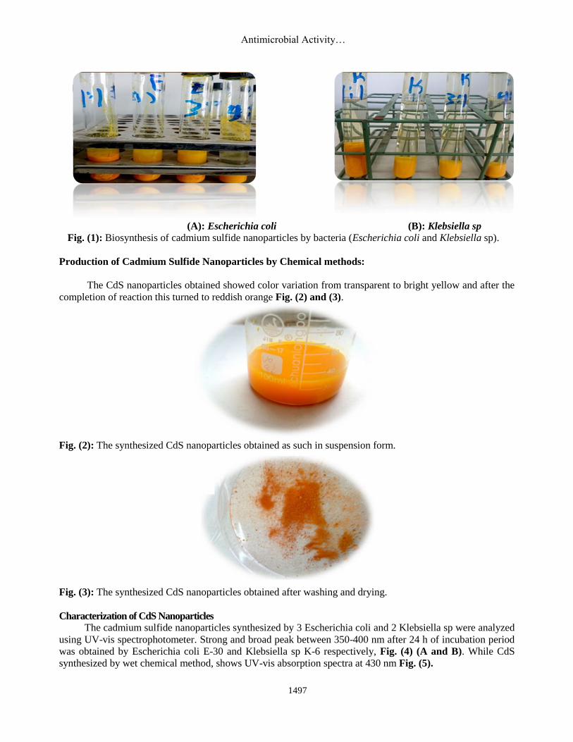
(A): Escherichia coli (B): Klebsiella sp Fig. (1): Biosynthesis of cadmium sulfide nanoparticles by bacteria (Escherichia coli and Klebsiella sp).
Production of Cadmium Sulfide Nanoparticles by Chemical methods:
The CdS nanoparticles obtained showed color variation from transparent to bright yellow and after the
completion of reaction this turned to reddish orange Fig. (2) and (3).
Fig. (2): The synthesized CdS nanoparticles obtained as such in suspension form.
Fig. (3): The synthesized CdS nanoparticles obtained after washing and drying.
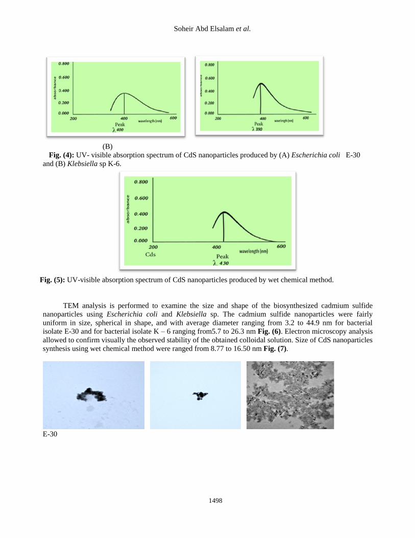
Characterization of CdS Nanoparticles
The cadmium sulfide nanoparticles synthesized by 3 Escherichia coli and 2 Klebsiella sp were analyzed
using UV-vis spectrophotometer. Strong and broad peak between 350-400 nm after 24 h of incubation period
was obtained by Escherichia coli E-30 and Klebsiella sp K-6 respectively, Fig. (4) (A and B). While CdS
synthesized by wet chemical method, shows UV-vis absorption spectra at 430 nm Fig. (5).
Soheir Abd Elsalam et al.
1498
(A) (B)
Fig. (4): UV- visible absorption spectrum of CdS nanoparticles produced by (A) Escherichia coli E-30
and (B) Klebsiella sp K-6.
Fig. (5): UV-visible absorption spectrum of CdS nanoparticles produced by wet chemical method.
TEM analysis is performed to examine the size and shape of the biosynthesized cadmium sulfide
nanoparticles using Escherichia coli and Klebsiella sp. The cadmium sulfide nanoparticles were fairly
uniform in size, spherical in shape, and with average diameter ranging from 3.2 to 44.9 nm for bacterial
isolate E-30 and for bacterial isolate K – 6 ranging from5.7 to 26.3 nm Fig. (6). Electron microscopy analysis
allowed to confirm visually the observed stability of the obtained colloidal solution. Size of CdS nanoparticles
synthesis using wet chemical method were ranged from 8.77 to 16.50 nm Fig. (7).
E-30
Antimicrobial Activity…
1499
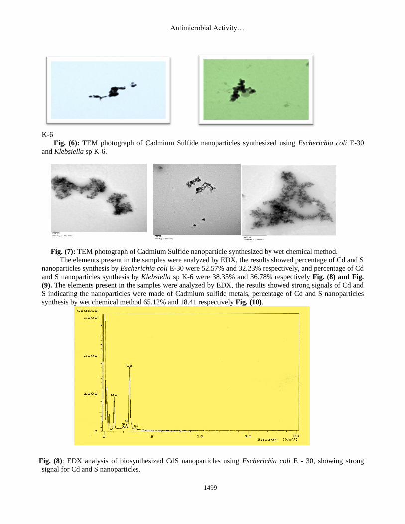
K-6
Fig. (6): TEM photograph of Cadmium Sulfide nanoparticles synthesized using Escherichia coli E-30
and Klebsiella sp K-6.
Fig. (7): TEM photograph of Cadmium Sulfide nanoparticle synthesized by wet chemical method.
The elements present in the samples were analyzed by EDX, the results showed percentage of Cd and S
nanoparticles synthesis by Escherichia coli E-30 were 52.57% and 32.23% respectively, and percentage of Cd
and S nanoparticles synthesis by Klebsiella sp K-6 were 38.35% and 36.78% respectively Fig. (8) and Fig.
(9). The elements present in the samples were analyzed by EDX, the results showed strong signals of Cd and
S indicating the nanoparticles were made of Cadmium sulfide metals, percentage of Cd and S nanoparticles
synthesis by wet chemical method 65.12% and 18.41 respectively Fig. (10).
Fig. (8): EDX analysis of biosynthesized CdS nanoparticles using Escherichia coli E - 30, showing strong
signal for Cd and S nanoparticles.
Soheir Abd Elsalam et al.
1500
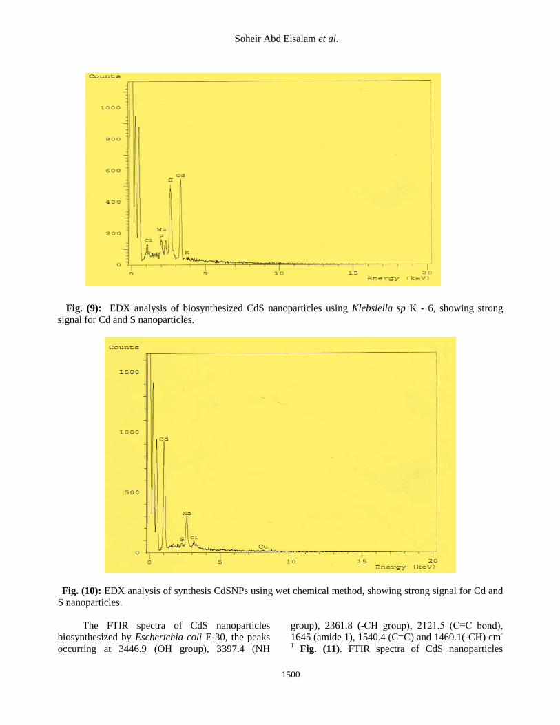
Fig. (9): EDX analysis of biosynthesized CdS nanoparticles using Klebsiella sp K - 6, showing strong
signal for Cd and S nanoparticles.
Fig. (10): EDX analysis of synthesis CdSNPs using wet chemical method, showing strong signal for Cd and
S nanoparticles.
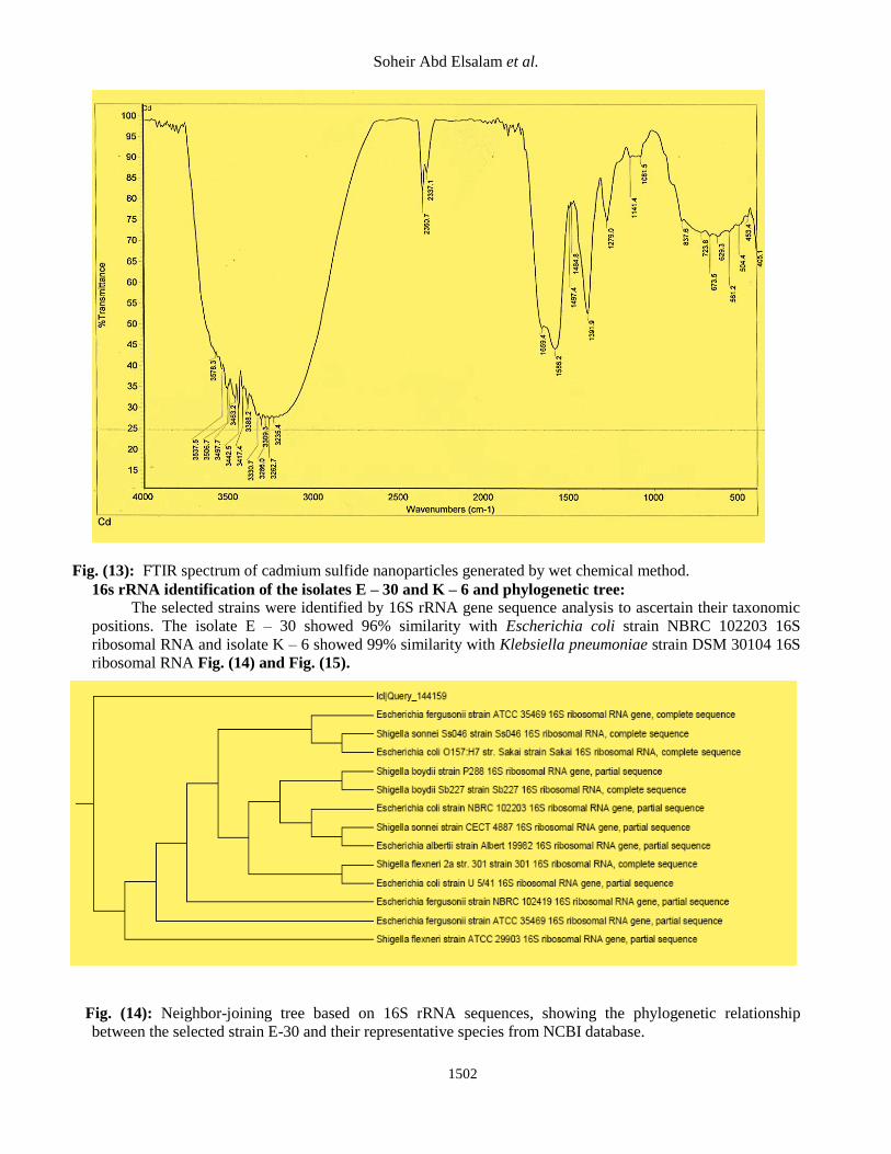
The FTIR spectra of CdS nanoparticles
biosynthesized by Escherichia coli E-30, the peaks
occurring at 3446.9 (OH group), 3397.4 (NH
group), 2361.8 (-CH group), 2121.5 (C≡C bond),
1645 (amide 1), 1540.4 (C=C) and 1460.1(-CH) cm-
1 Fig. (11). FTIR spectra of CdS nanoparticles

Antimicrobial Activity…
1501
biosynthesized by Klebsiella sp K-6, the peaks
occurring at 3502.9 and 3298.9 (OH group), 2121.7
(C≡C bond), 1659.3 (amide 1), 1552.0 (amide 11),
1455.1(-CH group) and 1230.5 (C-O bond) cm-1
Fig. (12). Fig. (13) show the peaks obtained by all
the synthesized CdS nanoparticles. The peak in the
range of 3578.3-3235.4cm-1
assigned to stretching
vibration of hydroxyl group with strong hydrogen
bond. Two strong peaks at 2945.1 and 2910.0 cm−1
are the characteristic bands of the asymmetric and
symmetric aliphatic C–H stretching vibration
respectively. The characteristic infrared peak of
C═C appeared at 1650.0 cm-1
.The two peaks
observed at 1462.1 and 1391.9 cm-1
assigned as
CH2 bending vibration and the deformation
vibration of C–CH3 respectively. The infrared
absorption peak at 1141.4 cm-1
was assigned as C–C
and C–O–C stretching vibrations.
Fig. (11): FTIR spectrum of CdS nanoparticles generated by Escherichia coli E-30.
Fig. (12): FTIR spectrum of CdS nanoparticles generated by Klebsiella sp K-30.
Soheir Abd Elsalam et al.
1502
Fig. (13): FTIR spectrum of cadmium sulfide nanoparticles generated by wet chemical method.
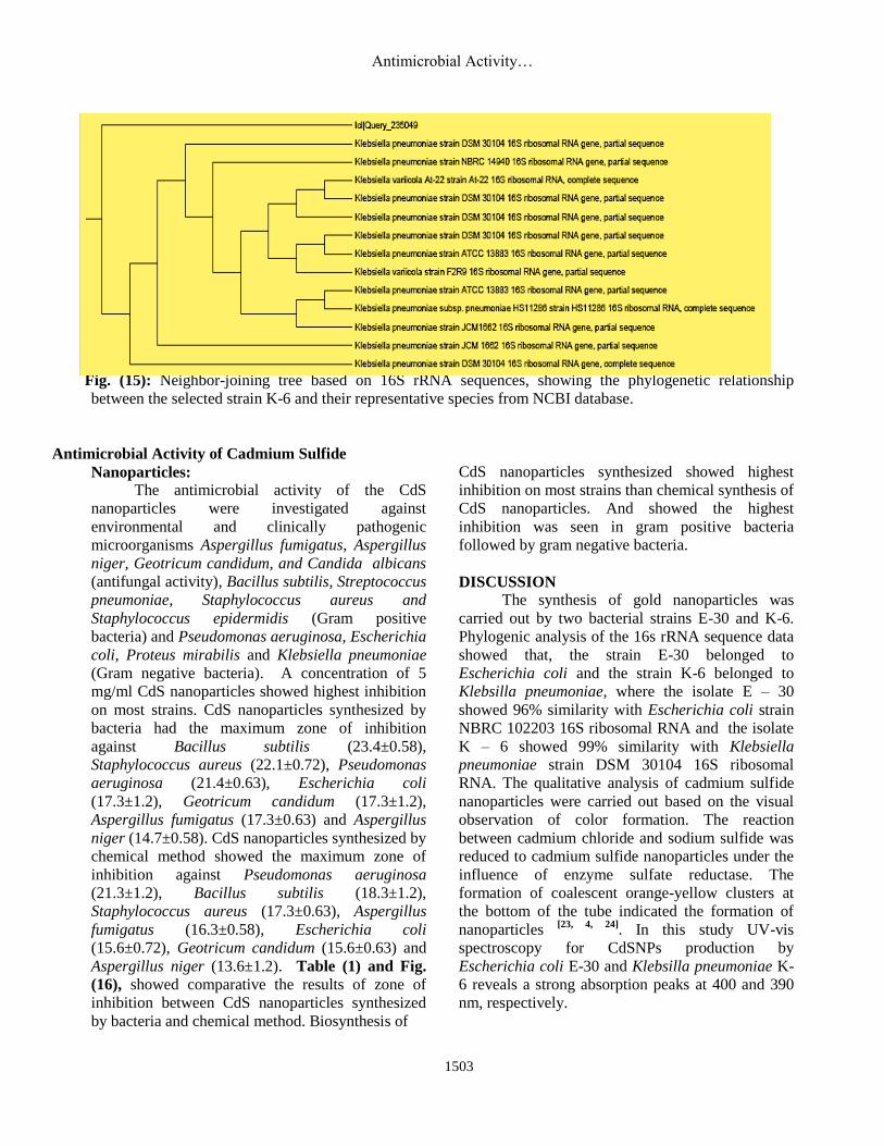
16s rRNA identification of the isolates E – 30 and K – 6 and phylogenetic tree:
The selected strains were identified by 16S rRNA gene sequence analysis to ascertain their taxonomic
positions. The isolate E – 30 showed 96% similarity with Escherichia coli strain NBRC 102203 16S
ribosomal RNA and isolate K – 6 showed 99% similarity with Klebsiella pneumoniae strain DSM 30104 16S
ribosomal RNA Fig. (14) and Fig. (15).
Fig. (14): Neighbor-joining tree based on 16S rRNA sequences, showing the phylogenetic relationship
between the selected strain E-30 and their representative species from NCBI database.
Antimicrobial Activity…
1503
Fig. (15): Neighbor-joining tree based on 16S rRNA sequences, showing the phylogenetic relationship
between the selected strain K-6 and their representative species from NCBI database.
Antimicrobial Activity of Cadmium Sulfide
Nanoparticles:
The antimicrobial activity of the CdS
nanoparticles were investigated against
environmental and clinically pathogenic
microorganisms Aspergillus fumigatus, Aspergillus
niger, Geotricum candidum, and Candida albicans
(antifungal activity), Bacillus subtilis, Streptococcus
pneumoniae, Staphylococcus aureus and
Staphylococcus epidermidis (Gram positive
bacteria) and Pseudomonas aeruginosa, Escherichia
coli, Proteus mirabilis and Klebsiella pneumoniae
(Gram negative bacteria). A concentration of 5
mg/ml CdS nanoparticles showed highest inhibition
on most strains. CdS nanoparticles synthesized by
bacteria had the maximum zone of inhibition
against Bacillus subtilis (23.4±0.58),
Staphylococcus aureus (22.1±0.72), Pseudomonas
aeruginosa (21.4±0.63), Escherichia coli
(17.3±1.2), Geotricum candidum (17.3±1.2),
Aspergillus fumigatus (17.3±0.63) and Aspergillus
niger (14.7±0.58). CdS nanoparticles synthesized by
chemical method showed the maximum zone of
inhibition against Pseudomonas aeruginosa
(21.3±1.2), Bacillus subtilis (18.3±1.2),
Staphylococcus aureus (17.3±0.63), Aspergillus
fumigatus (16.3±0.58), Escherichia coli
(15.6±0.72), Geotricum candidum (15.6±0.63) and
Aspergillus niger (13.6±1.2). Table (1) and Fig.
(16), showed comparative the results of zone of
inhibition between CdS nanoparticles synthesized
by bacteria and chemical method. Biosynthesis of
CdS nanoparticles synthesized showed highest
inhibition on most strains than chemical synthesis of
CdS nanoparticles. And showed the highest
inhibition was seen in gram positive bacteria
followed by gram negative bacteria.
DISCUSSION
The synthesis of gold nanoparticles was
carried out by two bacterial strains E-30 and K-6.
Phylogenic analysis of the 16s rRNA sequence data
showed that, the strain E-30 belonged to
Escherichia coli and the strain K-6 belonged to
Klebsilla pneumoniae, where the isolate E – 30
showed 96% similarity with Escherichia coli strain
NBRC 102203 16S ribosomal RNA and the isolate
K – 6 showed 99% similarity with Klebsiella
pneumoniae strain DSM 30104 16S ribosomal
RNA. The qualitative analysis of cadmium sulfide
nanoparticles were carried out based on the visual
observation of color formation. The reaction
between cadmium chloride and sodium sulfide was
reduced to cadmium sulfide nanoparticles under the
influence of enzyme sulfate reductase. The
formation of coalescent orange-yellow clusters at
the bottom of the tube indicated the formation of
nanoparticles [23, 4, 24]
. In this study UV-vis
spectroscopy for CdSNPs production by
Escherichia coli E-30 and Klebsilla pneumoniae K-
6 reveals a strong absorption peaks at 400 and 390
nm, respectively.
Soheir Abd Elsalam et al.
1504
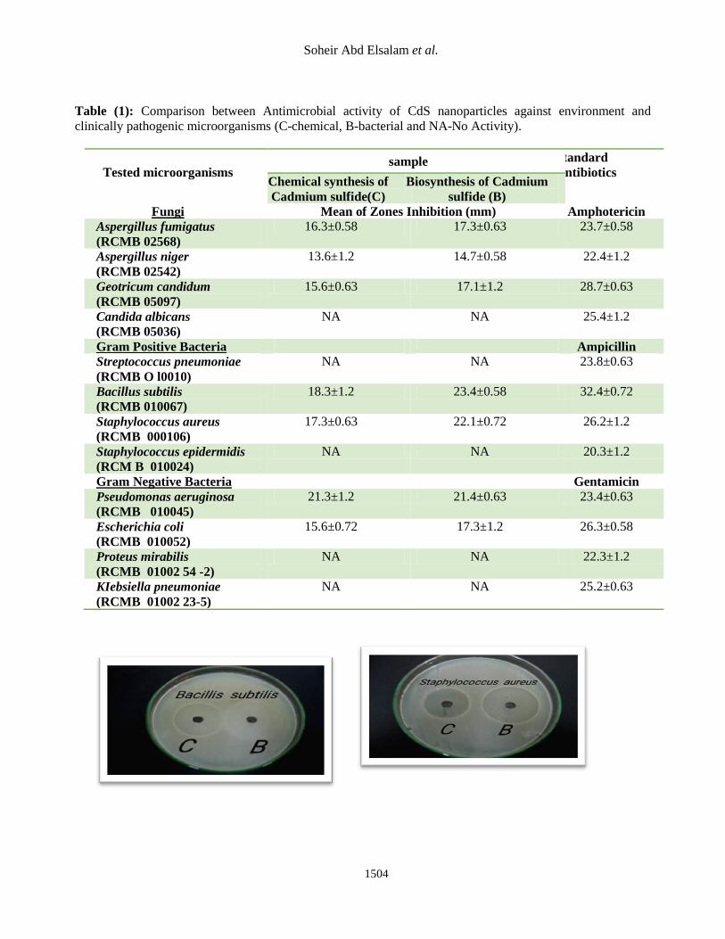
Table (1): Comparison between Antimicrobial activity of CdS nanoparticles against environment and
clinically pathogenic microorganisms (C-chemical, B-bacterial and NA-No Activity).
Tested microorganisms sample Standard
Antibiotics Chemical synthesis of
Cadmium sulfide(C)
Biosynthesis of Cadmium
sulfide (B)
Fungi Mean of Zones Inhibition (mm) Amphotericin
Aspergillus fumigatus
(RCMB 02568)
16.3±0.58 17.3±0.63 23.7±0.58
Aspergillus niger
(RCMB 02542)
13.6±1.2 14.7±0.58 22.4±1.2
Geotricum candidum
(RCMB 05097)
15.6±0.63 17.1±1.2 28.7±0.63
Candida albicans
(RCMB 05036)
NA NA 25.4±1.2
Gram Positive Bacteria Ampicillin
Streptococcus pneumoniae
(RCMB O l0010)
NA NA 23.8±0.63
Bacillus subtilis
(RCMB 010067)
18.3±1.2 23.4±0.58 32.4±0.72
Staphylococcus aureus
(RCMB 000106)
17.3±0.63 22.1±0.72 26.2±1.2
Staphylococcus epidermidis
(RCM B 010024)
NA NA 20.3±1.2
Gram Negative Bacteria Gentamicin
Pseudomonas aeruginosa
(RCMB 010045)
21.3±1.2 21.4±0.63 23.4±0.63
Escherichia coli
(RCMB 010052)
15.6±0.72 17.3±1.2 26.3±0.58
Proteus mirabilis
(RCMB 01002 54 -2)
NA NA 22.3±1.2
KIebsiella pneumoniae
(RCMB 01002 23-5)
NA NA 25.2±0.63
Antimicrobial Activity…
1505
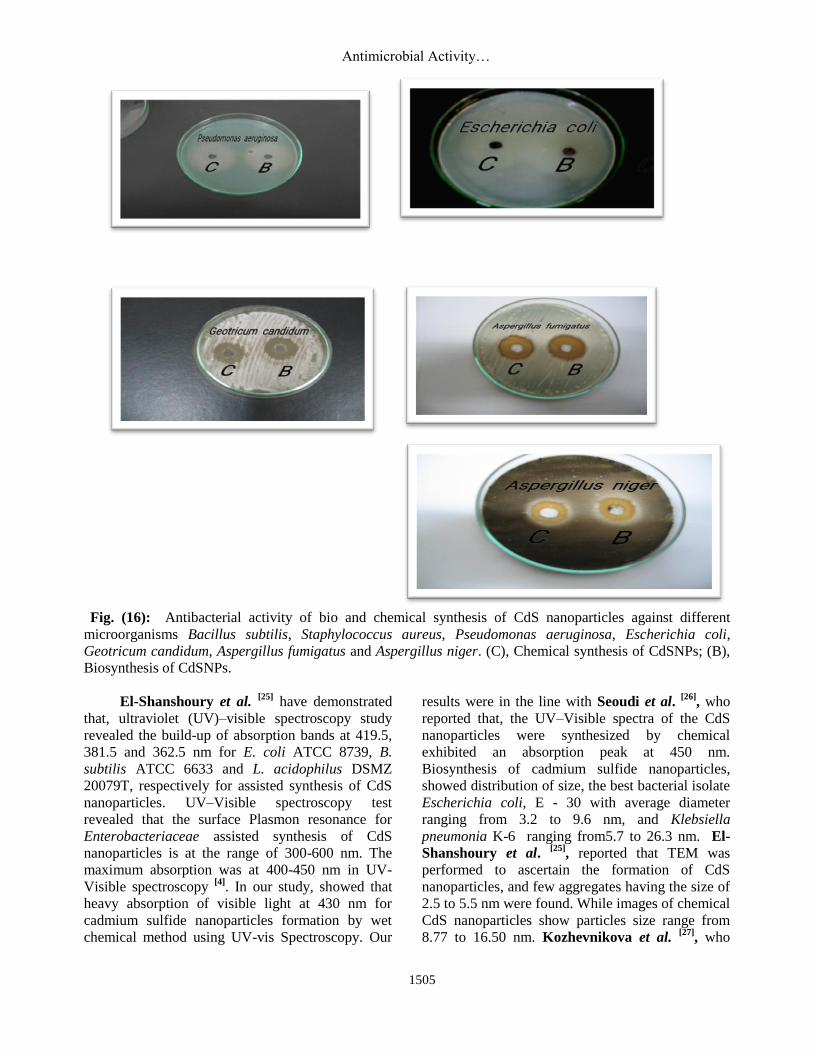
Fig. (16): Antibacterial activity of bio and chemical synthesis of CdS nanoparticles against different
microorganisms Bacillus subtilis, Staphylococcus aureus, Pseudomonas aeruginosa, Escherichia coli,
Geotricum candidum, Aspergillus fumigatus and Aspergillus niger. (C), Chemical synthesis of CdSNPs; (B),
Biosynthesis of CdSNPs.
El-Shanshoury et al. [25]
have demonstrated
that, ultraviolet (UV)–visible spectroscopy study
revealed the build-up of absorption bands at 419.5,
381.5 and 362.5 nm for E. coli ATCC 8739, B.
subtilis ATCC 6633 and L. acidophilus DSMZ
20079T, respectively for assisted synthesis of CdS
nanoparticles. UV–Visible spectroscopy test
revealed that the surface Plasmon resonance for
Enterobacteriaceae assisted synthesis of CdS
nanoparticles is at the range of 300-600 nm. The
maximum absorption was at 400-450 nm in UV-
Visible spectroscopy [4]
. In our study, showed that
heavy absorption of visible light at 430 nm for
cadmium sulfide nanoparticles formation by wet
chemical method using UV-vis Spectroscopy. Our
results were in the line with Seoudi et al. [26]
, who
reported that, the UV–Visible spectra of the CdS
nanoparticles were synthesized by chemical
exhibited an absorption peak at 450 nm.
Biosynthesis of cadmium sulfide nanoparticles,
showed distribution of size, the best bacterial isolate
Escherichia coli, E - 30 with average diameter
ranging from 3.2 to 9.6 nm, and Klebsiella
pneumonia K-6 ranging from5.7 to 26.3 nm. El-
Shanshoury et al. [25]
, reported that TEM was
performed to ascertain the formation of CdS
nanoparticles, and few aggregates having the size of
2.5 to 5.5 nm were found. While images of chemical
CdS nanoparticles show particles size range from
8.77 to 16.50 nm. Kozhevnikova et al. [27]
, who
Soheir Abd Elsalam et al.
1506
observed that electron micrograph of particles of the
disperse phase of chemical CdS nanoparticles are
seen of the size 10-100 nm. EDX spectrum was
showed strong signals of Cd and S indicating the
nanoparticles were made of cadmium sulfide
metals. The biosynthesis of CdS nanoparticles using
the bacteria of Enterobacteriaceae, EDX spectrum
was recorded strong signals showed the presence of
Cd and S [4]
. Our results showed strong signals of
Cd and S indicating the nanoparticles were made of
chemical Cadmium sulfide metals. Qutub [28]
, who
observed that EDX spectra of the chemical
synthesis of CdS NP reveals the presence Cd
(51.5%) and S (48.5%) strong peaks and presence of
other peaks. The FTIR spectra of CdS nanoparticles
biosynthesized by Escherichia coli E-30, the peaks
occurring at 3446.9, 3397.4, 2361.8, 2121.5, 1645,
1540.4 and 1460.1 cm-1. While Klebsiella
pneumonia K-6, the peaks occurring at 3502.9,
3298.9, 2121, 1659.3, 1552.0, 1455.1 and 1230.5
cm-1. This is in agreement with Tripathi et al. [29]
,
who found that (FTIR) provides the evidence for the
presence of proteins as possible biomolecules
responsible for the stabilization of the synthesized
CdS nanoparticles. In the present work, the FTIR
spectra show the peaks obtained by all the chemical
synthesized CdS nanoparticles. The peak in the
range of 3578.3-3235.4(OH group), 2945.1 and
2910.0 (C-H), 1650 (C=C), 1462.1 (CH2), 1391.9
(C-CH3) and 1141.4 (C-C and C-O-C) cm−1. The
infrared spectral data confirmed that the -OH group
used as a coordinated site to aggregate the cadmium
ions and different uniform sizes of CdS
nanoparticles were formed at this site by releasing
of S-2
ions [26]
. The efficiency of antimicrobial
activity also depends on the size of the
nanoparticles. Smaller nanoparticles have more
surface atoms which gives them a larger surface
area for interaction with the bacterial cell. It has also
been shown that smaller nanoparticles have larger
fractions of atoms on low coordination and high
energy sites like corners, edges and steps which
makes them more active than larger particles [30].
Biosynthesis of CdS nanoparticles synthesized
showed highest inhibition on most strains than
chemical synthesis of CdS nanoparticles.
Shivashankarappa et al. [19]
, who observed that,
biosynthesis of CdS nanoparticles showed the
highest antimicrobial activity was seen in the order
of Pseudomonas aeruginosa (26.5±0.70) followed
by Bacillus licheniformis (23.5±0.70), Bacillus
cereus (22±0.01), E coli (19.1±0.14) and
Staphylococcus aureus (18.25±0.35).
CONCLUSION
Escherichia coli and Klebsiella pneumoniae
isolated from stool samples had the ability to
produce cadmium sulfide nanoparticles. They had
been confirmed with UV, XRD, TEM and FTIR.
And the nanoparticles has potential antibacterial
activity against clinical and environmental isolates.
This is an inexpensive procdure and ecofriendly
process to produce this nanoparticles. Hence, this
kind of microorganisms can be used for synthesis
of nanoparticles and heavy metal absorption for
detoxification of environment.
REFERENCES 1.Shah M, Fawcett D, Sharma S, Tripathy SK, Jai
Poinern GE (2015): Green Synthesis of Metallic
Nanoparticles via Biological Entities. Materials,
8:7278–7308.
2. Kanude KR and Jain P (2017): CdS Nanoparticles:
Green Synthesis Method and Capping Agent’s Effect
on its Crystallite Size. Journal of Chemical and
Pharmaceutical Research, 9(6):86-89.
3. Mandal D, Bolander M E, Mukhopadhyay D,
Sarkar G, Mukherjee P (2006): The use of
microorganisms for the formation of metal
nanoparticles and their application. Appl Microbiol
Biotechnol., 69(5):485-492.
4. Mousavi RA, Sepahy AA, Fazel MR (2012): Biosynthesis, Purification and Characterization of
Cadmium Sulfide Nanoparticles Using
Enterobacteriaceae and their Application. Proceedings
of the international conference nanomaterials:
applications and properties. doi: 10.1016
5. Thakkar KN, Mhatre SS, Parikh RY (2009): Biological synthesis of metallic nanoparticles,
Nanomedicine: Nanotechnology, Biology and
Medicine, 6(2): 257-262.
6. Hossain S T, Mukherjee S K (2013): Toxicity of
cadmium sulfide (CdS) nanoparticles against
Escherichia coli and HeLa cells, Journal of Hazardous
Materials, 260: 1073-1082.
7. Li KG, Chen JT, Bai SS, Wen X, Song SY, Yu Q, Li
J, Wang YQ (2009): Intracellular oxidative stress
and cadmium ions release induce cytotoxicity of
unmodified cadmium sulfide quantum dots,
Toxicology In Vitro., 23(6): 1007-1013.
8. Durga B, Raziya S, Rajamahanti SG, Govindh B,
Raju KV, Annapurna N (2017): Synthesis and
Characterization of CdS Nanoparticles Using
Artabotrys hexapetalus Leaf Extract as Capping
Agent. Der Pharma Chemica, 9(14):157-162.
Antimicrobial Activity…
1507
9. Rai RV, Bai JA (2011): Nanoparticles and their
potential application as antimicrobials,
Communicating current research and technological
advances. Formatex research centre,3(1): 197-209.
10. Yang J, Peng JJ, Zou R, Peng F, Wang H, Yu HL,
JY (2008): Mesoporous zinc blende ZnS
nanoparticles: synthesis, characterization and superior
photocatalytic properties, J. Nanotechnol., 19: 1-7.
11. Lee HL, Issam AM, Belmahi M, Assouar MB,
Rinnert H, Alnot M (2009): Synthesis and
characterization of bare CdS nanocrystals using
chemical precipitation method for photoluminescence
application. J. Nanomaterials., 9:44-48.
12. Zhong S, Zhang L, Huang Z, Wang S (2010): Mixed -solvothermal synthesis of CdS
micro/nanostructures and their optical properties. J.
Apsusc., 257: 2599-2603.
13. Ghows N, Entezari MH (2010): A novel method for
the synthesis of CdS nanoparticles without surfactant.
J. Ultsonch., 18: 269-275.
14. Lu Y, Li L, Din Y, Zhang F, Wang Y, Yu W
(2011): Hydrothermal synthesis of functionalized CdS
nanoparticles and their application as fluorescence
probe in the determination of uracil and thymine. J.
Lumin., 132: 244-249.
15. Balows A, Hausler Jr, W.J., Herrmann, K.L.,
Isenberg, H.D., Shadomy, H.J (1991): Manual of
Clinical Microbiology (Fifth edition). American
Society for Microbiology, Washington.
16. Elmer WK, Stephen DA, William MJ, Paul CS,
Washington CW (1997): Color atlas and textbook of
diagnostic microbiology, 5th edition. Lippincott-
Raven Publishers, Philadelphia, NewYork.
17. Marko MA, Chipperfield R, Birnboim HC (1982): A procedure for the large-scale isolation of highly
purified plasmid DNA using alkaline extraction and
binding to glass powder. Anal. Biochem., 121: 382–7.
18. Boom R, Sol CJ, Salimans M, Jansen CL,
Wertheim-van Dillen PM, van der Noordaa J
(1990): Rapid and simple method for purification of
nucleic acids. J. Clin. Microbiol., 28: 495–503.
19. Shivashankarappa A, Sanjay KR (2015): Study on
Biological Synthesis of Cadmium Sulfide
Nanoparticles by Bacillus licheniformis and Its
Antimicrobial Properties against Food Borne
Pathogens. Nanoscience and Nanotechnology
Research, 3: No. 1, 6-15.
20. Ramesh S, Narayanan V (2013): Wet Chemical
Synthesis of Cadmium Sulphide
Nanoparticles and its Characterization. Chem Sci Trans.,
2(S1): S192-S194.
21. Scott AC (1989): Laboratory control of antimicrobial
therapy. In: Collee JG et al. eds. Practical Medical
Microbiology, 13th edn. Edinburgh: Churchill
Livingstone, 161-181.
22. Levesque R (2007): SPSS Programming and Data
Management: A Guide for SPSS and SAS Users,
SPSS Inc.
23. Bai H, Zhang Z, Guo Y, Jia W (2009): Biological
synthesis of size-controlled cadmium sulfide
nanoparticles using immobilized Rhodobacter
sphaeroides Nanoscale Research Letters, 4: 717-723.
24. Sweeney R Y, Mao C, Gao X, Burt J L, Belcher A
M, Georgiou G, Iverson B L (2004): Bacterial
biosynthesis of cadmium sulfide nanoparticles,
Chemistry and Biology, 11(11): 1553-1559.
25. El-Shanshoury AR, Elsilk SE, Ebeid ME (2012): Rapid biosynthesis of cadmium sulfide (CdS)
nanoparticles using culture supernatants of
Escherichia coli ATCC 8739, Bacillus subtilis ATCC
6633 and Lactobacillus acidophilus DSMZ 20079T.
African Journal of Biotechnology , 11(31): 7957-
7965.
26. Seoudi R, El- Bailly AB, Eisa W, Shabaka AA,
Soliman SI, Abd El Hamid RK, Ramadan RA
(2012): Synthesis, Optical and Dielectric Properties
of (PVA/ CdS) Nanocomposites. Journal of Applied
Sciences Research, 8(2): 658-667.
27. Kozhevnikova NS, Vorokh AS, Rempel AA (2010): Preparation of Stable Colloidal Solution of Cadmium
Sulfide CdS Using Ethylenediaminetetraacetic Acid.
Russian Journal of General Chemistry, 80(3): 391–
394.
28. Qutub N(2013):Titanium dioxide nanoparticles and
its nanocomposites. Ph.D. Thesis A.M.U., India.
shodhganga.inflibnet.ac.in/bitstream/10603/21086/.../
12_chapter3.pdf
29. Tripathi RM, Bhadwal AS, Singh P, Shrivastav A,
Singh MP, Shrivastav BR (2014): Mechanistic
aspects of biogenic synthesis of CdS nanoparticles
using Bacillus licheniformis. Adv. Nat. Sci.: Nanosci.
Nanotechnol., 5: 025006 .
30. Zhang Q, Iwakuma N, Sharma P, Moudgil B, Wu
C, McNeill J, Jiang H, Grobmyer S (2009): Gold
nanoparticles as a contrast agent for in vivo tumor
imaging with photoacoustic tomography.
Nanotechnology: 20:88-90.