
Antigen Processing PPT
46
Transcript of Antigen Processing PPT
• That T and B cells recognise antigen differently
• The experimental evidence that antigen catabolism takes place
• Antigen processing generates antigenic peptides
• That antigen processing can take place in lysosomes
• That there is a non-lysosomal mechanism of antigen processing
• The mechanism of antigen processing depends upon the compartment in which the pathogen replicates
• Antigen processing includes uptake, degradation, complex formation and presentation
• The role of invariant chain HLA-DM and CLIP in antigen processing
• The role of the proteasome and transporters in antigen processing
• How pathogens evade immunity by disrupting antigen processing
What you should know by the end of this lecture
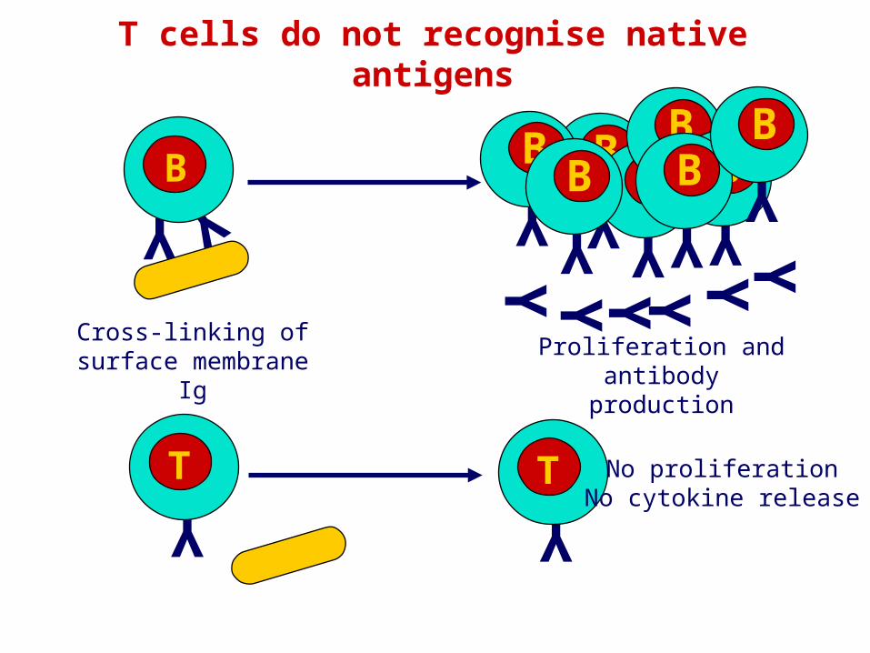
T cells do not recognise native antigens
YYBYY Y Y YY
Y
BYT
YT
Proliferation and antibody production
No proliferationNo cytokine release
Cross-linking of surface membrane Ig
Y
B
Y
B Y
B
Y
B
Y
B Y
B
Y
B
Cell surfacepeptides
of Ag
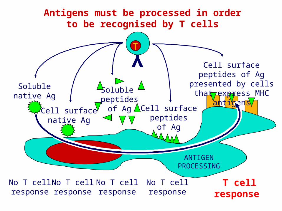
Antigens must be processed in orderto be recognised by T cells
YT
T cellresponse
No T cellresponse
No T cellresponse
No T cellresponse
No T cellresponse
Solublenative Ag
Cell surfacenative Ag
Soluble peptides
of Ag
Cell surface peptides of Ag presented by cells that
express MHC antigens
ANTIGENPROCESSING
M
M
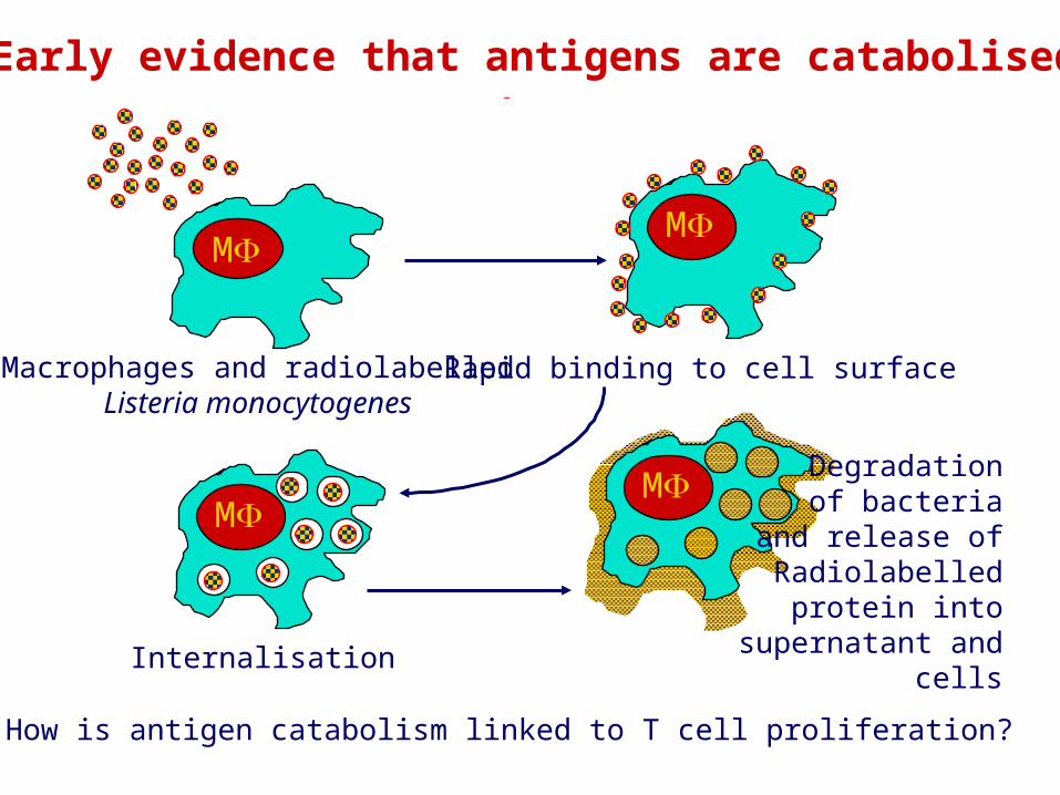
Early evidence that antigens are catabolised
M
Macrophages and radiolabelledListeria monocytogenes
Internalisation
Rapid binding to cell surface
M Degradationof bacteria
and release ofRadiolabelled
protein into supernatant and cells
How is antigen catabolism linked to T cell proliferation?
MM MM
0mins 60mins
T
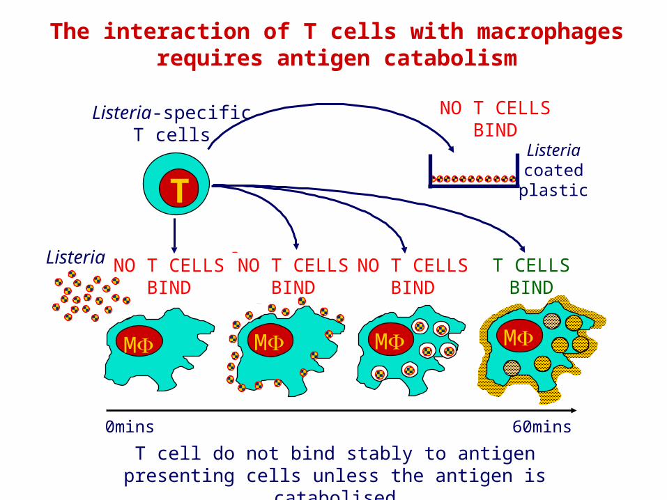
Listeria-specificT cells
The interaction of T cells with macrophagesrequires antigen catabolism
Listeria
NO T CELLSBIND
NO T CELLSBIND
T CELLSBIND
Listeriacoatedplastic
NO T CELLSBIND
NO T CELLSBIND
T cell do not bind stably to antigen presenting cells unless the antigen is catabolised
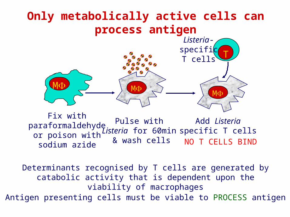
Only metabolically active cells can process antigen
Determinants recognised by T cells are generated by catabolic activity that is dependent upon the viability of macrophages
Fix withparaformaldehyde
or poison withsodium azide
M
Pulse withListeria for 60min
& wash cells
M
Add Listeriaspecific T cells
M
TListeria-specificT cells
NO T CELLS BIND
Antigen presenting cells must be viable to PROCESS antigen
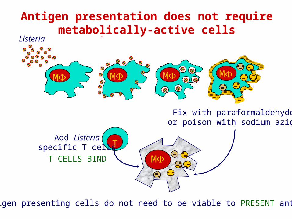
Fix with paraformaldehydeor poison with sodium azide
MM MM
Listeria
TAdd Listeria
specific T cellsT CELLS BIND M
Antigen presenting cells do not need to be viable to PRESENT antigen
Antigen presentation does not requiremetabolically-active cells
MMM
MMM
Listeria
Listeria
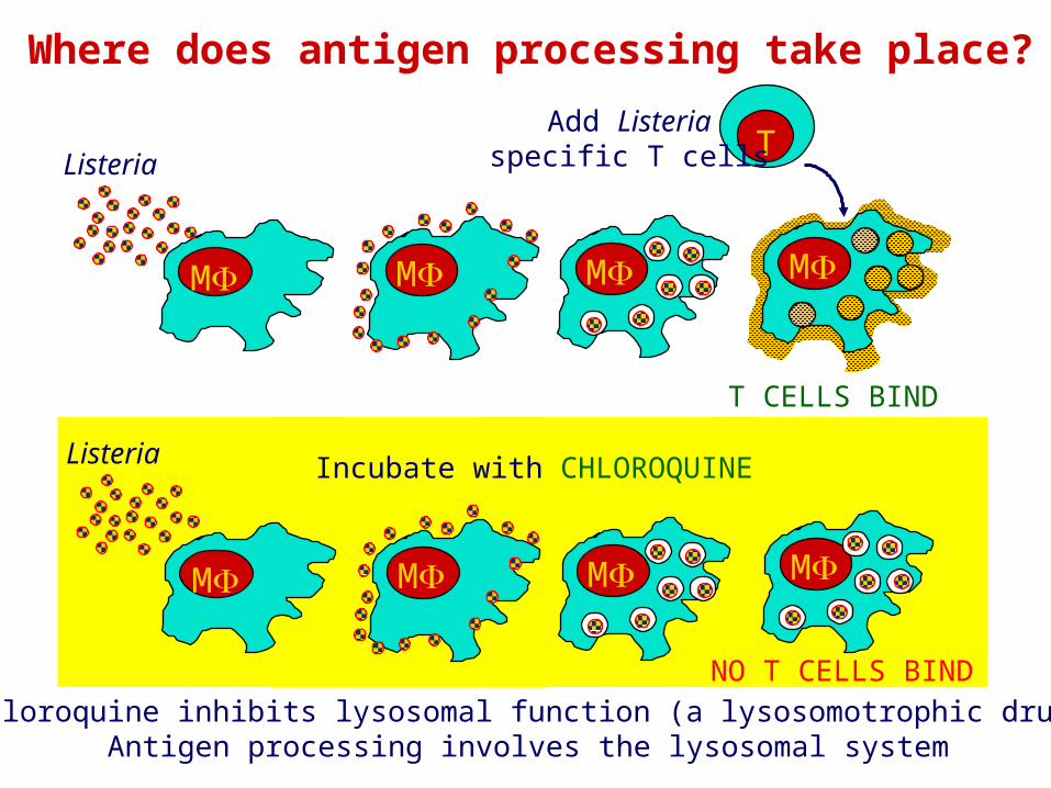
Where does antigen processing take place?
M
M
Incubate with CHLOROQUINE
TAdd Listeria
specific T cells
T CELLS BIND
NO T CELLS BINDChloroquine inhibits lysosomal function (a lysosomotrophic drug)
Antigen processing involves the lysosomal system
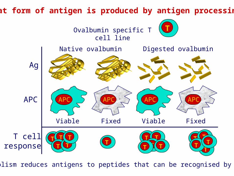
What form of antigen is produced by antigen processing?
TOvalbumin specific T cell line
Catabolism reduces antigens to peptides that can be recognised by T cells
APC
Viable
APC
Viable
T TT T
TT
T TT TT
T T
TT T
Digested ovalbumin
Fixed
APC
Fixed
APC
Native ovalbumin
Ag
APC
T cellresponse
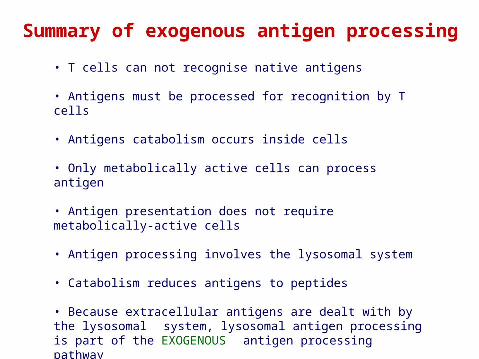
Summary of exogenous antigen processing
• T cells can not recognise native antigens
• Antigens must be processed for recognition by T cells
• Antigens catabolism occurs inside cells
• Only metabolically active cells can process antigen
• Antigen presentation does not require metabolically-active cells
• Antigen processing involves the lysosomal system
• Catabolism reduces antigens to peptides
• Because extracellular antigens are dealt with by the lysosomal system, lysosomal antigen processing is part of the EXOGENOUS antigen processing pathway
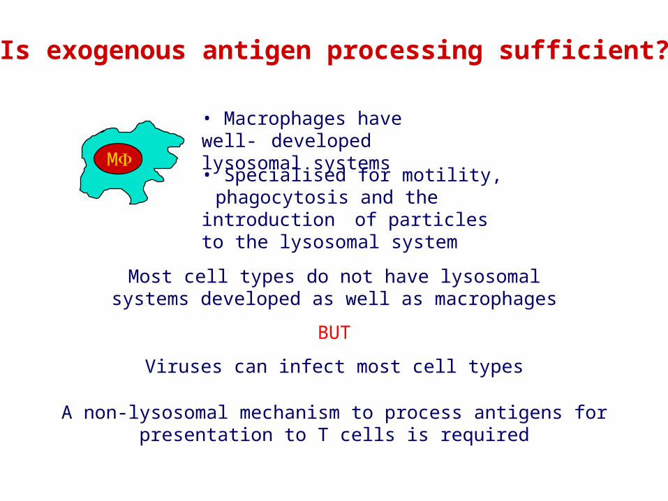
Is exogenous antigen processing sufficient?
Most cell types do not have lysosomal systems developed as well as macrophages
BUT
Viruses can infect most cell types
M
• Macrophages have well-developed lysosomal systems
• Specialised for motility, phagocytosis and the introduction of particles to the lysosomal system
A non-lysosomal mechanism to process antigens for presentation to T cells is required
+ Chloroquine
Infectious influenza
CTLCTL
CTLCTL CTLCTLCTL
CTL
CTLCTLCTL
CTLCTL CTLCTL CTL
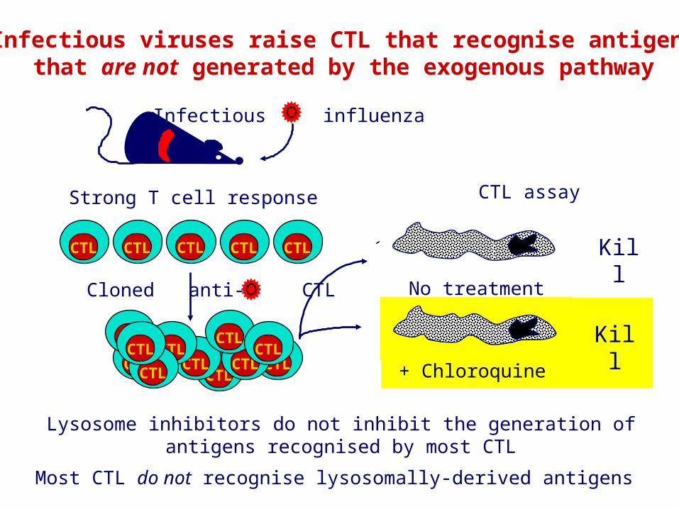
Infectious viruses raise CTL that recognise antigensthat are not generated by the exogenous pathway
Most CTL do not recognise lysosomally-derived antigens
Cloned anti- CTL No treatment
CTL assay
Lysosome inhibitors do not inhibit the generation ofantigens recognised by most CTL
Kill
Kill
Strong T cell response
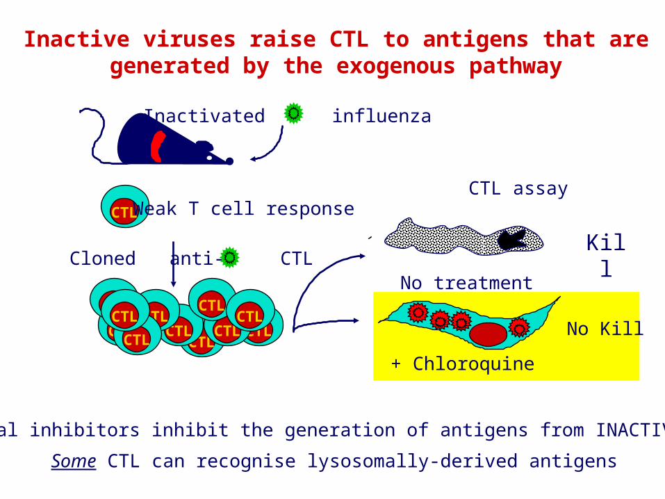
Lysosomal inhibitors inhibit the generation of antigens from INACTIVE virus
Some CTL can recognise lysosomally-derived antigens
Inactivated influenza
Cloned anti- CTL
Inactive viruses raise CTL to antigens that aregenerated by the exogenous pathway
CTLCTL
CTLCTL CTLCTLCTL
CTL
CTLCTLCTL
CTL Weak T cell response
+ Chloroquine
No treatment
CTL assay
Kill
No Kill
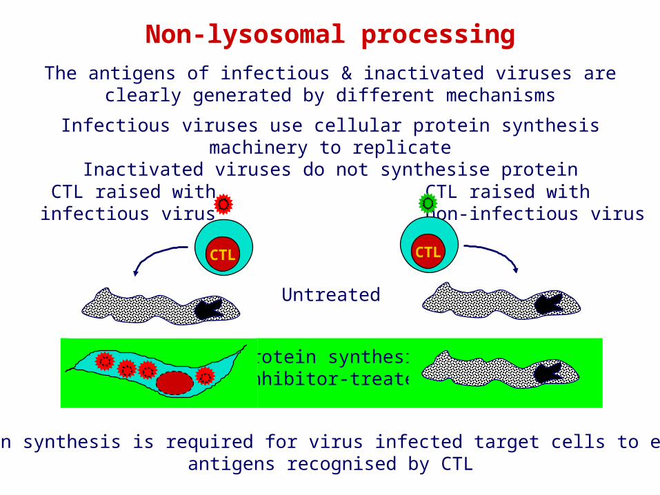
Non-lysosomal processingThe antigens of infectious & inactivated viruses are clearly generated by
different mechanisms
Protein synthesisinhibitor-treated
Protein synthesis is required for virus infected target cells to expressantigens recognised by CTL
CTL raised withinfectious virus
CTL
CTL raised withnon-infectious virus
CTL
Untreated
Infectious viruses use cellular protein synthesis machinery to replicateInactivated viruses do not synthesise protein
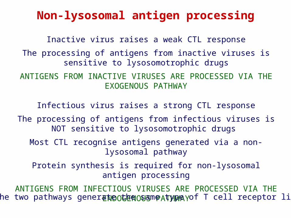
Inactive virus raises a weak CTL response
The processing of antigens from inactive viruses is sensitive to lysosomotrophic drugs
ANTIGENS FROM INACTIVE VIRUSES ARE PROCESSED VIA THE EXOGENOUS PATHWAY
Infectious virus raises a strong CTL response
The processing of antigens from infectious viruses is NOT sensitive to lysosomotrophic drugs
Most CTL recognise antigens generated via a non-lysosomal pathway
Protein synthesis is required for non-lysosomal antigen processing
ANTIGENS FROM INFECTIOUS VIRUSES ARE PROCESSED VIA THE ENDOGENOUS PATHWAY
Non-lysosomal antigen processing
Do the two pathways generate the same type of T cell receptor ligand?
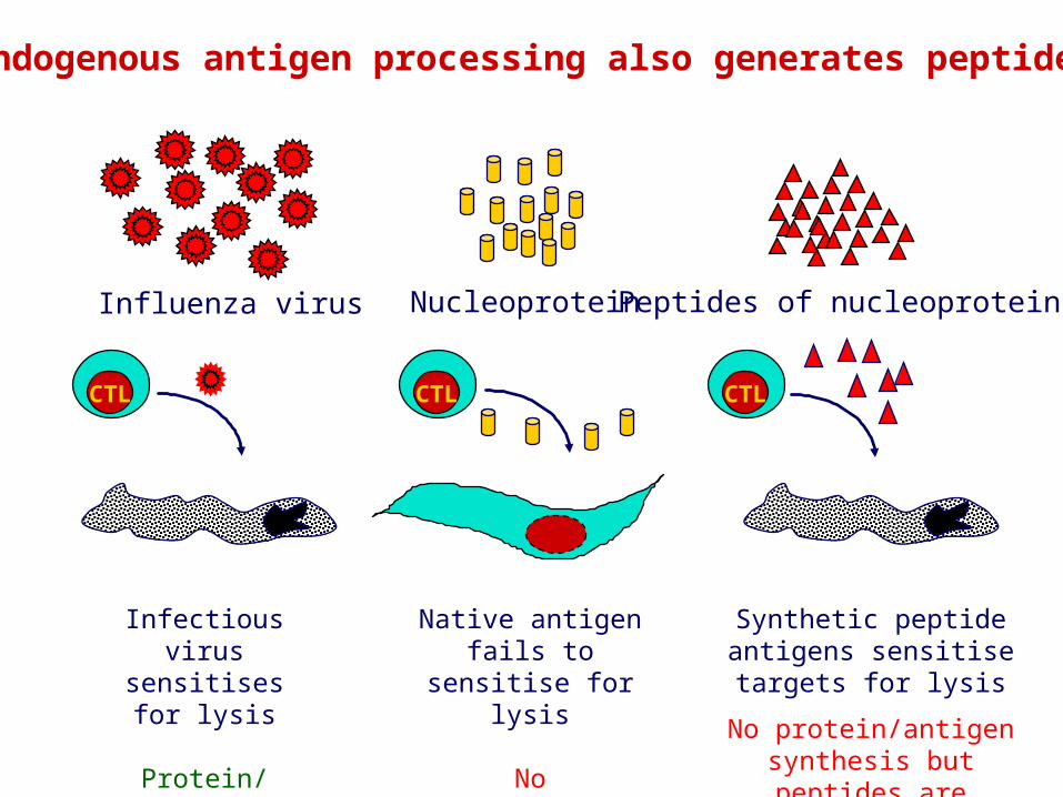
Endogenous antigen processing also generates peptides
Peptides of nucleoprotein
Native antigen fails to sensitise for lysis
No protein/antigensynthesis
Infectious virussensitises for lysis
Protein/antigensynthesis
CTL
Influenza virus Nucleoprotein
CTL
Synthetic peptide antigens sensitise targets for lysis
No protein/antigensynthesis but peptides are
pre-formed
CTL

YThe site of pathogen replication or mechanism of antigen uptake determines the antigen processing pathway used
Y
Cytosolic compartmentEndogenous processing
(Viral antigens)
Vesicular CompartmentContiguous with extracellular fluid
Exogenous processing(Streptococcal, Mycobacterial antigens)
Distinct mechanisms of antigen generation are used to raiseT cells suited to the elimination of endogenous or exogenous pathogens
INTRACELLULAR REPLICATION
EXTRACELLULAR ORENDOSOMAL REPLICATION
Y
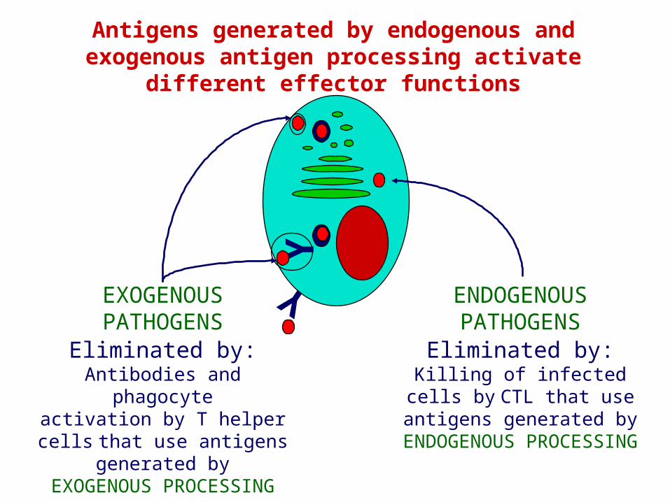
Eliminated by:Killing of infected cells by CTL that use antigens generated by
ENDOGENOUS PROCESSING
YEliminated by:
Antibodies and phagocyteactivation by T helper cells that
use antigens generated byEXOGENOUS PROCESSING
Antigens generated by endogenous and exogenous antigen processing activate different effector functions
ENDOGENOUS PATHOGENS
EXOGENOUSPATHOGENS
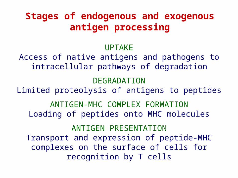
Stages of endogenous and exogenousantigen processing
UPTAKEAccess of native antigens and pathogens to intracellular
pathways of degradation
DEGRADATIONLimited proteolysis of antigens to peptides
ANTIGEN-MHC COMPLEX FORMATIONLoading of peptides onto MHC molecules
ANTIGEN PRESENTATIONTransport and expression of peptide-MHC complexes on the
surface of cells for recognition by T cells
Y Y
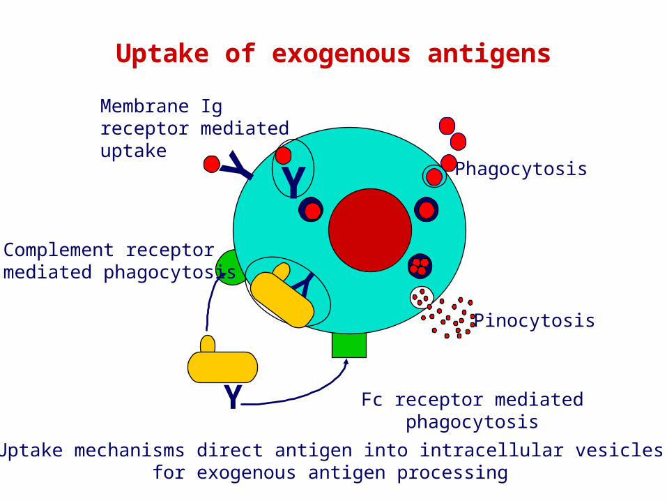
Pinocytosis
Phagocytosis
Membrane Igreceptor mediateduptake
Y
Uptake of exogenous antigens
Complement receptormediated phagocytosis Y
Fc receptor mediated phagocytosis
Uptake mechanisms direct antigen into intracellular vesiclesfor exogenous antigen processing
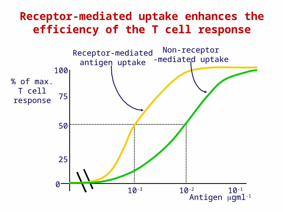
100
50
75
25
0
% of max.T cell
response
10-110-210-3
Antigen gml-1
Receptor-mediatedantigen uptake
Non-receptor-mediated uptake
Receptor-mediated uptake enhances theefficiency of the T cell response
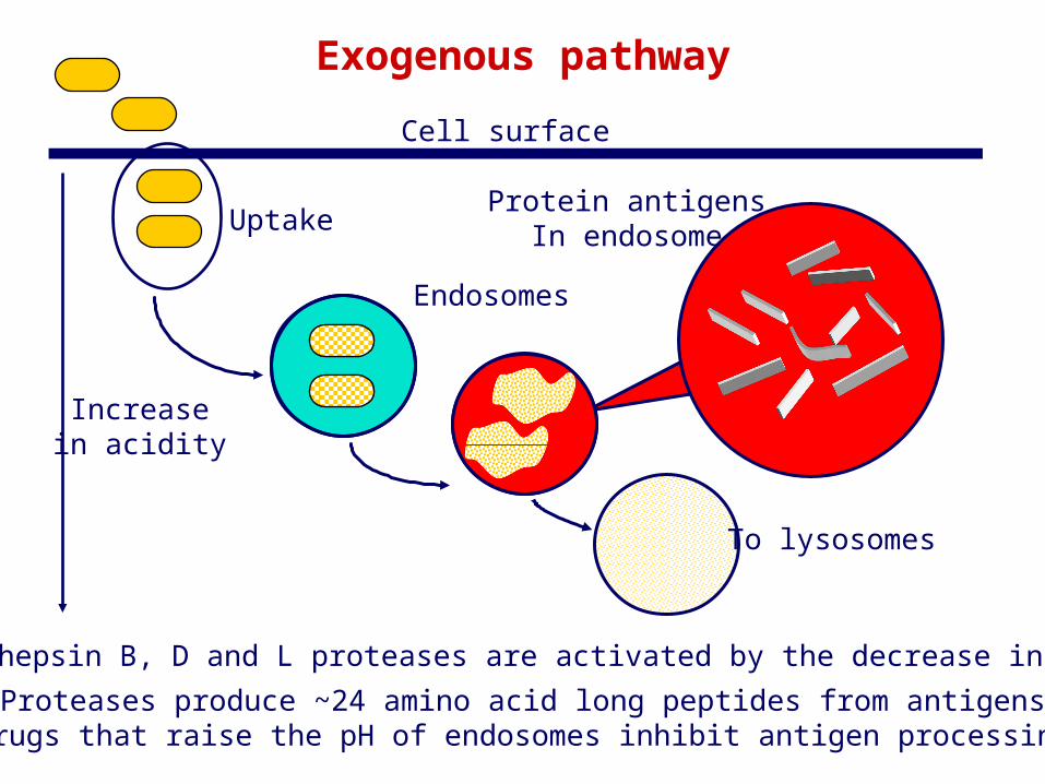
Proteases produce ~24 amino acid long peptides from antigensDrugs that raise the pH of endosomes inhibit antigen processing
Endosomes
Exogenous pathway
Increasein acidity
Cell surface
To lysosomes
UptakeProtein antigens
In endosome
Cathepsin B, D and L proteases are activated by the decrease in pH
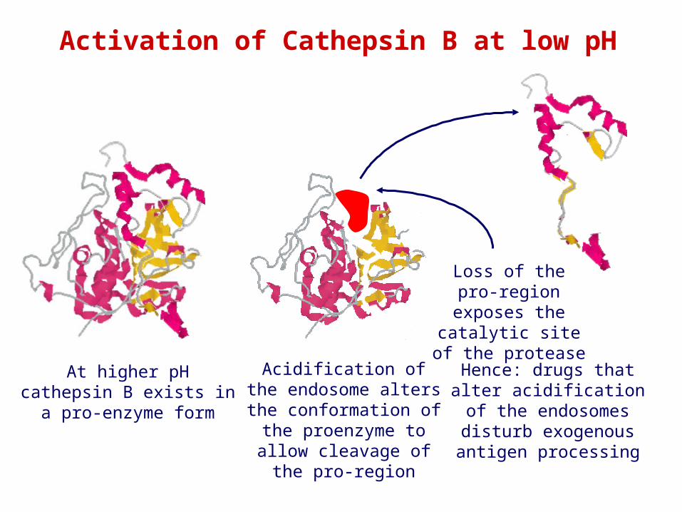
Activation of Cathepsin B at low pH
At higher pH cathepsin B exists in a pro-enzyme form
Acidification of the endosome alters the conformation of the proenzyme to allow
cleavage of the pro-region
Loss of the pro-region exposes the catalytic site of the
protease
Hence: drugs that alter acidification of the endosomes disturb exogenous antigen
processing

Proteases produce ~24 amino acid long peptides from antigensDrugs that raise the pH of endosomes inhibit antigen processing
Endosomes
Exogenous pathway
Increasein acidity
Cell surface
To lysosomes
UptakeProtein antigens
In endosome
Cathepsin B, D and L proteases are activated by the decrease in pH
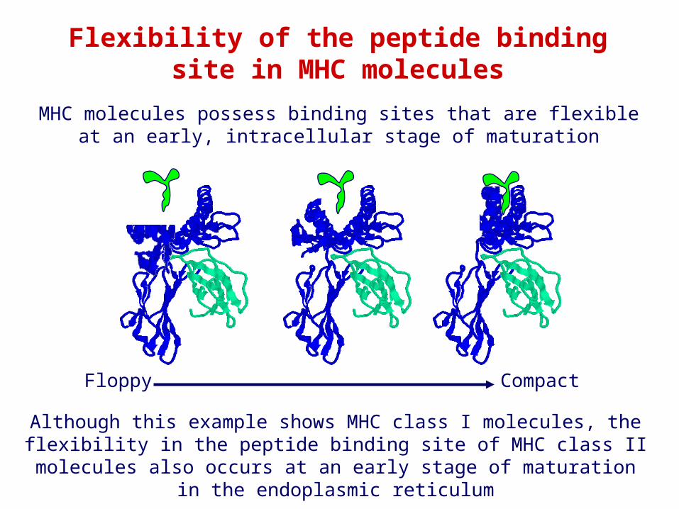
MHC molecules possess binding sites that are flexible at an early, intracellular stage of maturation
Floppy Compact
Flexibility of the peptide bindingsite in MHC molecules
Although this example shows MHC class I molecules, the flexibility in the peptide binding site of MHC class II molecules also occurs at an early
stage of maturation in the endoplasmic reticulum
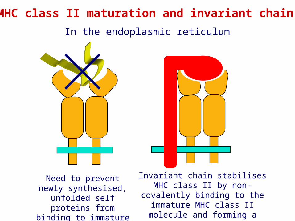
Need to prevent newly synthesised, unfolded
self proteins from binding to immature MHC
Invariant chain stabilises MHC class II by non- covalently binding to the
immature MHC class II molecule and forming a nonomeric complex
In the endoplasmic reticulum
MHC class II maturation and invariant chain
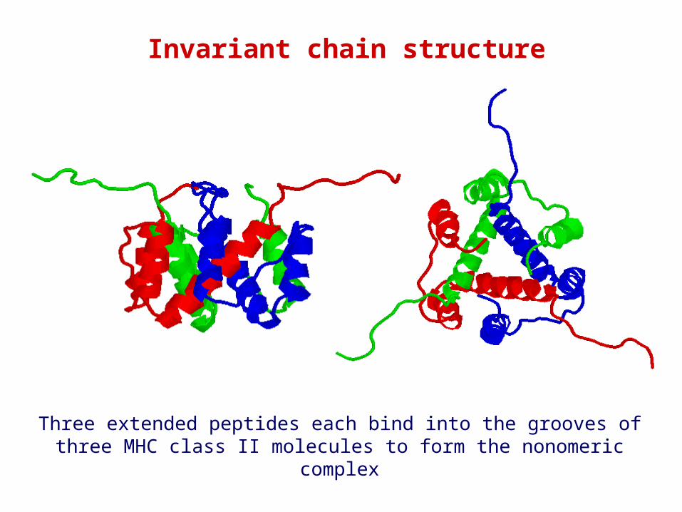
Invariant chain structure
Three extended peptides each bind into the grooves of three MHC class II molecules to form the nonomeric complex
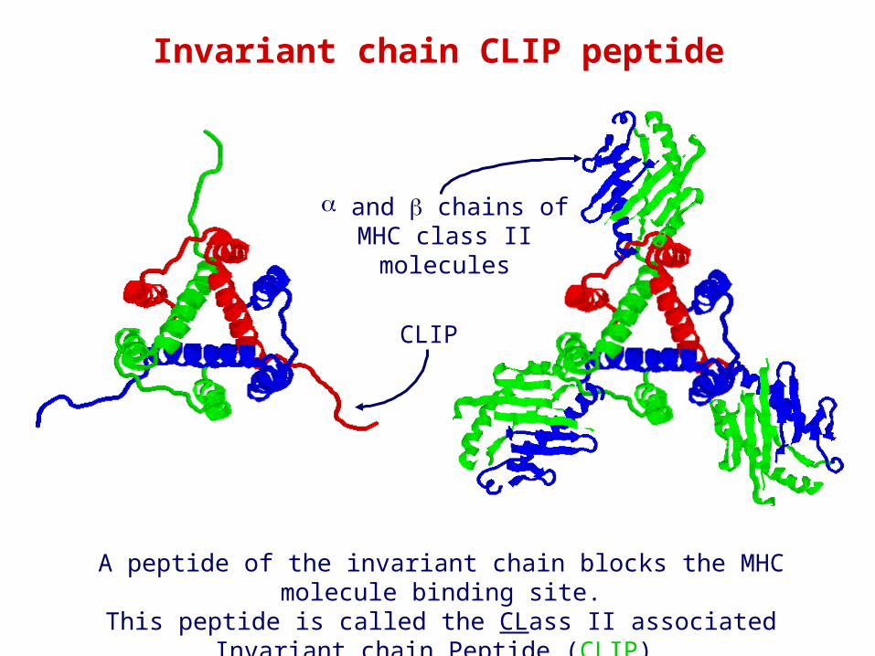
A peptide of the invariant chain blocks the MHC molecule binding site.This peptide is called the CLass II associated Invariant chain Peptide
(CLIP)
Invariant chain CLIP peptide
and chains of MHC class II molecules
CLIP
Endosomes
Cell surface
Uptake
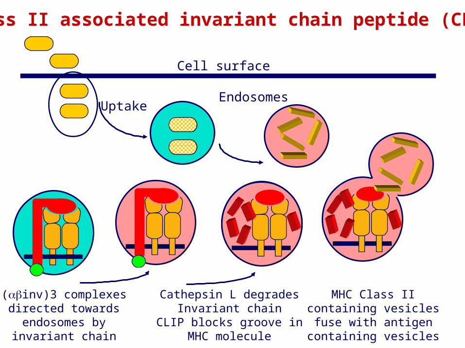
Class II associated invariant chain peptide (CLIP)
(inv)3 complexesdirected towardsendosomes byinvariant chain
Cathepsin L degrades Invariant chain
CLIP blocks groove in MHC molecule
MHC Class IIcontaining vesiclesfuse with antigen
containing vesicles
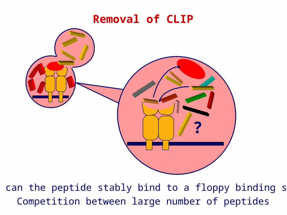
Removal of CLIP
?
How can the peptide stably bind to a floppy binding site?Competition between large number of peptides
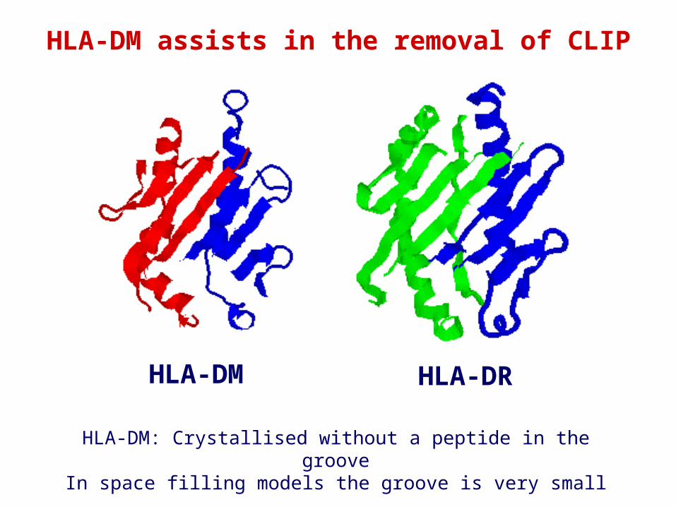
HLA-DM HLA-DR
HLA-DM assists in the removal of CLIP
HLA-DM: Crystallised without a peptide in the grooveIn space filling models the groove is very small
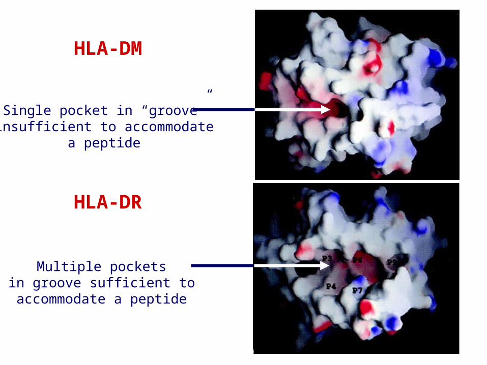
HLA-DM
HLA-DR
Single pocket in “groove”insufficient to accommodate
a peptide
Multiple pocketsin groove sufficient to
accommodate a peptide
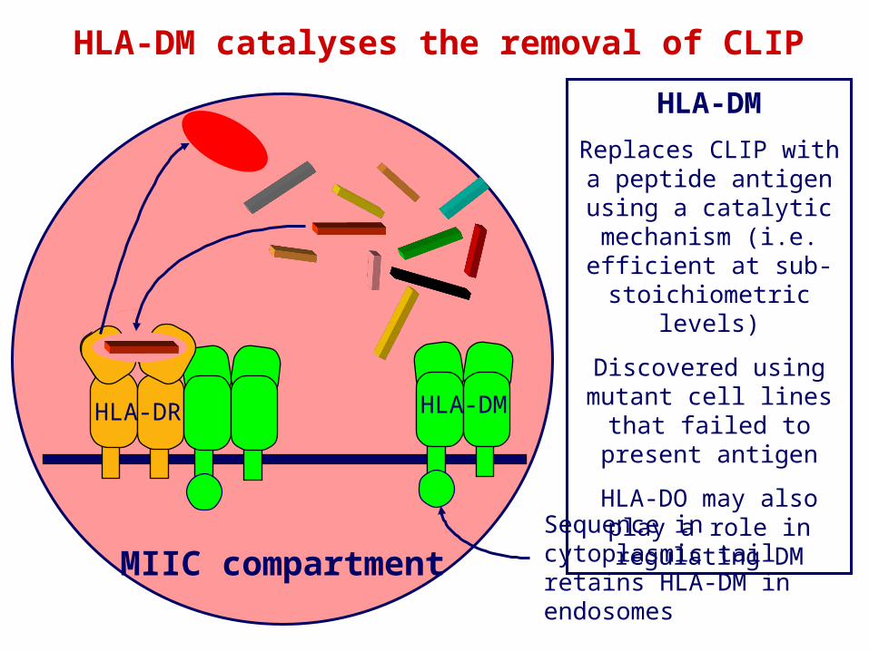
HLA-DM catalyses the removal of CLIP
MIIC compartment
HLA-DMReplaces CLIP with a
peptide antigen using a catalytic mechanism (i.e.
efficient at sub-stoichiometric levels)
Discovered using mutant cell lines that failed to
present antigen
HLA-DO may also play a role in regulating DM
Sequence in cytoplasmic tail retains HLA-DM in endosomes
HLA-DMHLA-DR
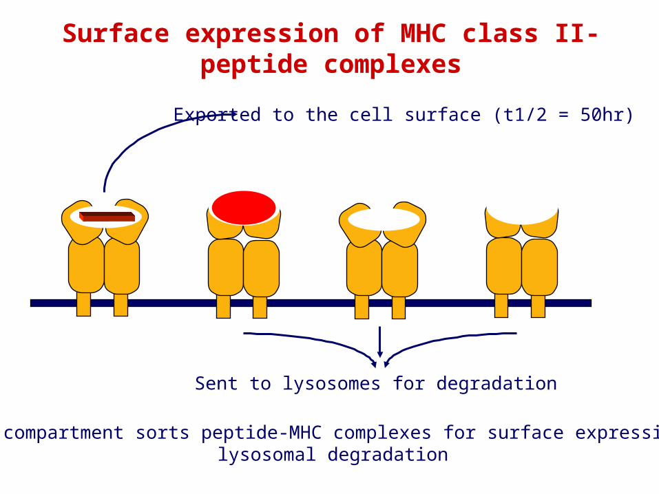
MIIC compartment sorts peptide-MHC complexes for surface expression orlysosomal degradation
Surface expression of MHC class II-peptide complexes
Exported to the cell surface (t1/2 = 50hr)
Sent to lysosomes for degradation
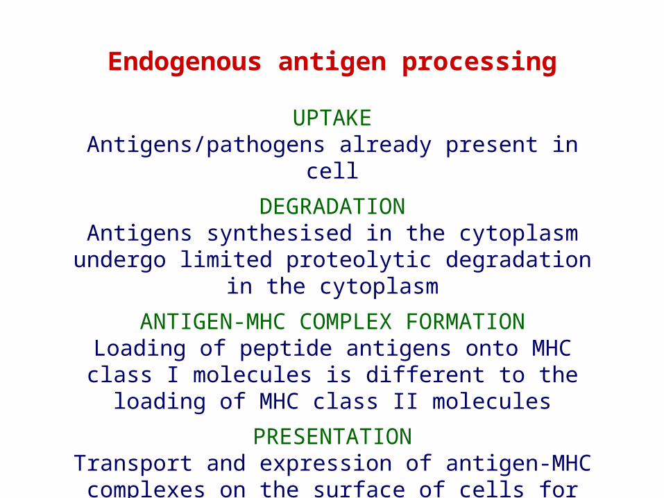
UPTAKEAntigens/pathogens already present in cell
DEGRADATIONAntigens synthesised in the cytoplasm undergo limited
proteolytic degradation in the cytoplasmANTIGEN-MHC COMPLEX FORMATION
Loading of peptide antigens onto MHC class I molecules is different to the loading of MHC class II molecules
PRESENTATIONTransport and expression of antigen-MHC complexes on
the surface of cells for recognition by T cells
Endogenous antigen processing
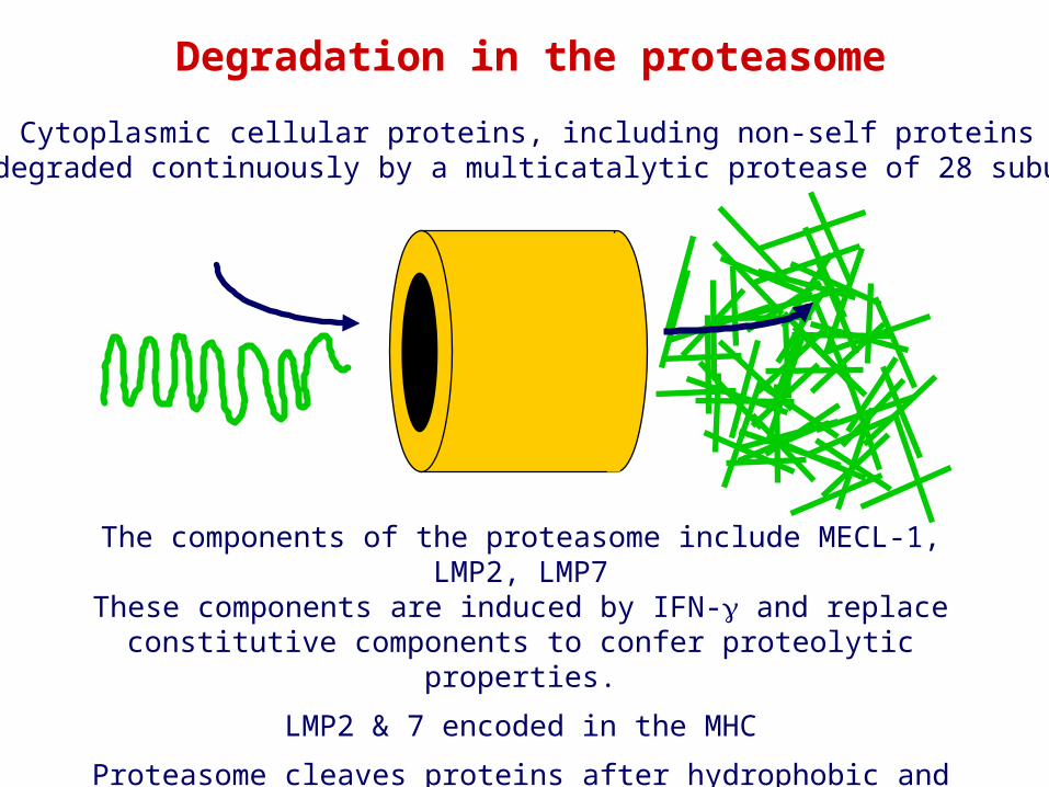
Degradation in the proteasome
The components of the proteasome include MECL-1, LMP2, LMP7These components are induced by IFN- and replace constitutive
components to confer proteolytic properties.
LMP2 & 7 encoded in the MHC
Proteasome cleaves proteins after hydrophobic and basic amino acids and releases peptides into the cytoplasm
Cytoplasmic cellular proteins, including non-self proteinsare degraded continuously by a multicatalytic protease of 28 subunits
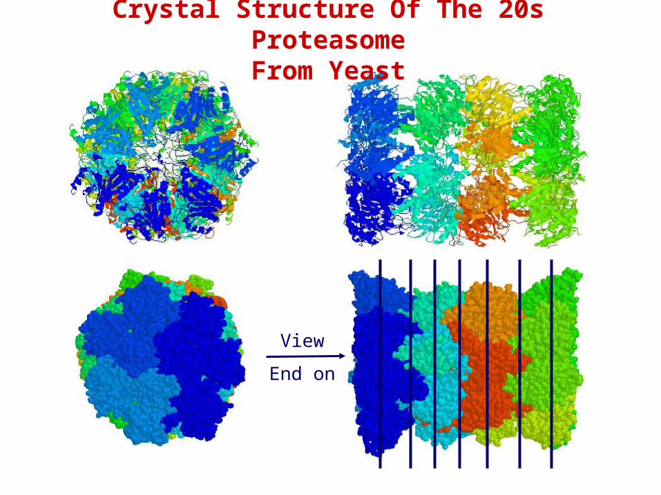
Crystal Structure Of The 20s ProteasomeFrom Yeast
View
End on
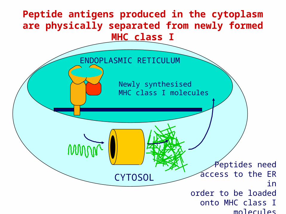
ENDOPLASMIC RETICULUM
CYTOSOL
Peptide antigens produced in the cytoplasm are physically separated from newly formed MHC class I
Newly synthesisedMHC class I molecules
Peptides needaccess to the ER in
order to be loaded onto MHC class I molecules
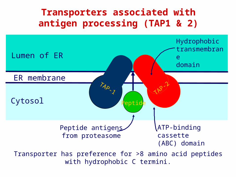
ER membrane
Lumen of ER
Cytosol
Transporters associated withantigen processing (TAP1 & 2)
Transporter has preference for >8 amino acid peptideswith hydrophobic C termini.
TAP-1 TAP-2
Peptide
TAP-1 TAP-2
PeptideTAP-1 TAP-2
Peptide
TAP-1 TAP-2
PeptideTAP-1 TAP-2
Peptide
TAP-1 TAP-2
PeptideTAP-1 TAP-2
Peptide
TAP-1 TAP-2
PeptideTAP-1 TAP-2
Peptide
TAP-1 TAP-2
Peptide
ER membrane
Lumen of ER
Cytosol
TAP-1 TAP-2
Peptide
ATP-binding cassette(ABC) domain
Hydrophobictransmembranedomain
Peptide antigensfrom proteasome
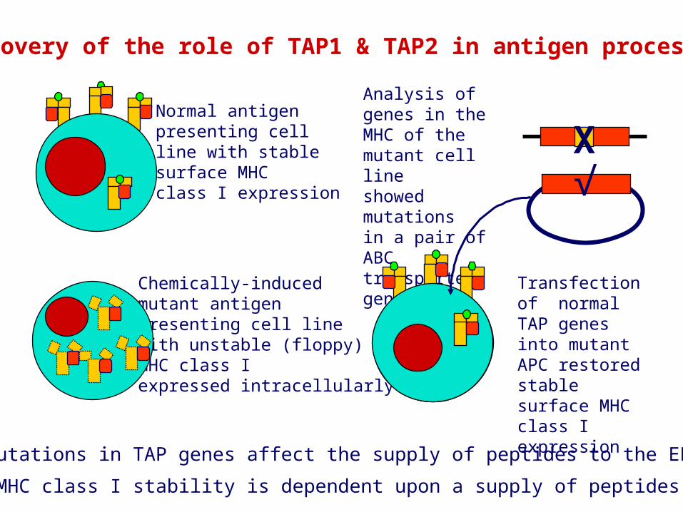
Discovery of the role of TAP1 & TAP2 in antigen processing
Transfection of normal TAP genes into mutant APC restored stablesurface MHC class Iexpression
Mutations in TAP genes affect the supply of peptides to the ER
MHC class I stability is dependent upon a supply of peptides
Analysis of genes in the MHC of the mutant cell lineshowed mutationsin a pair of ABC transporter genes
Normal antigen presenting cellline with stablesurface MHCclass I expression
Chemically-inducedmutant antigenpresenting cell linewith unstable (floppy)MHC class Iexpressed intracellularly
√X
Endoplasmic reticulum
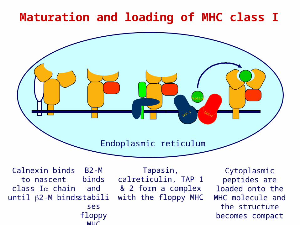
Calnexin bindsto nascent
class I chainuntil 2-M binds
TAP-1 TAP-2
Peptide
TAP-1 TAP-2
PeptideTAP-1 TAP-2
Peptide
TAP-1 TAP-2
PeptideTAP-1 TAP-2
Peptide
TAP-1 TAP-2
PeptideTAP-1 TAP-2
Peptide
TAP-1 TAP-2
PeptideTAP-1 TAP-2
Peptide
TAP-1 TAP-2
PeptideTAP-1 TAP-2
Peptide
B2-M binds and stabilises
floppy MHC
Tapasin, calreticulin, TAP 1 & 2 form a complex with
the floppy MHC
Cytoplasmic peptides are loaded onto the
MHC molecule and the structure becomes
compact
Maturation and loading of MHC class I
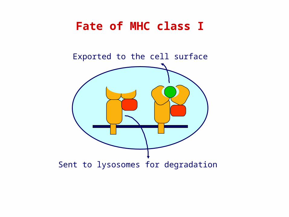
Fate of MHC class I
Sent to lysosomes for degradation
Exported to the cell surface
Endoplasmic reticulum
TAP-1 TAP-2
Peptide
TAP-1 TAP-2
PeptideTAP-1 TAP-2
Peptide
TAP-1 TAP-2
Peptide
TAP-1 TAP-2
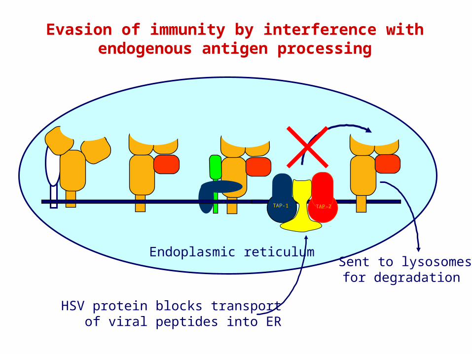
HSV protein blocks transportof viral peptides into ER
Sent to lysosomesfor degradation
Evasion of immunity by interference with endogenous antigen processing
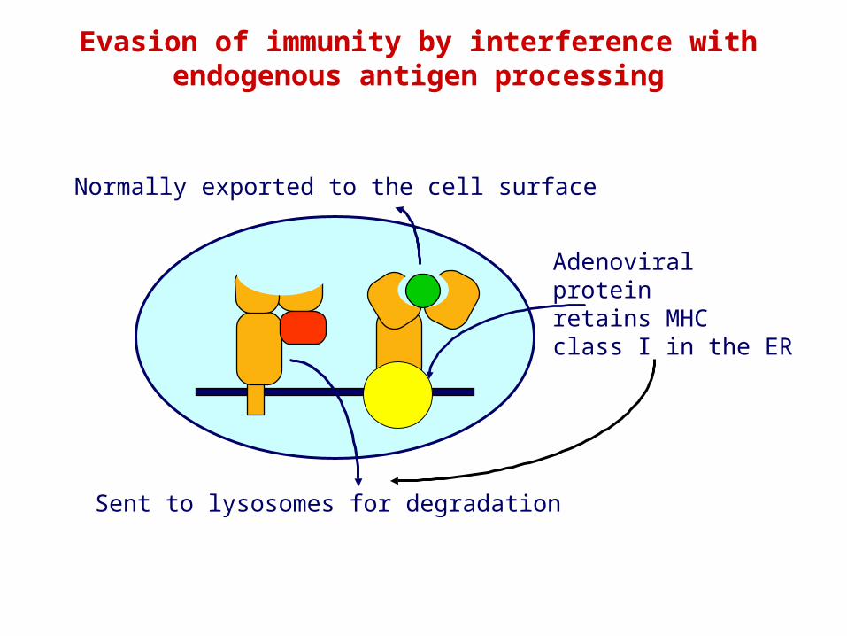
Sent to lysosomes for degradation
Normally exported to the cell surface
Adenoviralproteinretains MHCclass I in the ER
Evasion of immunity by interference withendogenous antigen processing
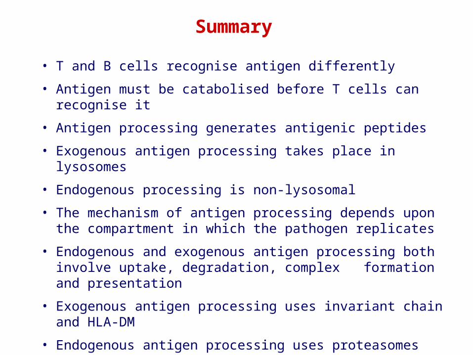
• T and B cells recognise antigen differently
• Antigen must be catabolised before T cells can recognise it
• Antigen processing generates antigenic peptides
• Exogenous antigen processing takes place in lysosomes
• Endogenous processing is non-lysosomal
• The mechanism of antigen processing depends upon the compartment in which the pathogen replicates
• Endogenous and exogenous antigen processing both involve uptake, degradation, complex formation and presentation
• Exogenous antigen processing uses invariant chain and HLA-DM
• Endogenous antigen processing uses proteasomes and peptide transporters in antigen processing
• Pathogens can evade immunity by disrupting antigen processing
Summary