Antifungal Susceptibility Testing - BioMérieux · bioMérieux SA English - 1 INTENDED USE Etest is...
6
bioMérieux SA English - 1 INTENDED USE Etest is an agar-based gradient technique for quantitative antifungal susceptibility testing (AFST). The system comprises a predefined concentration gradient of a specific antifungal agent which is used to determine the Minimum Inhibitory Concentration (MIC), in µg/mL, that inhibits the growth of the test organism under defined testing conidtions. SUMMARY AND EXPLANATION Current antifungal susceptibility testing methods are based either on quantitative dilution techniques or qualitative diffusion procedures. Dilution methods are based on two-fold serial dilutions of antifungal agents in broth. These methods generate the MIC value i.e. Minimum Inhibitory Concentration of a given antifungal agent in µg/mL that will inhibit the growth of a particular fungus under defined experimental conditions. PRINCIPLES OF USE The Etest gradient technology is based on a combination of the concepts of dilution and diffusion principles for susceptibility testing. As with other dilution methods, Etest directly quantifies antifungal susceptibility in terms of discrete MIC values. However, in using a predefined, stable and continuous concentration gradient, Etest MIC values can be more precise and reproducible than results obtained from conventional procedures based on discontinuous two-fold serial dilutions. The concentration gradient per se is a format that is considered useful for detection of various resistance mechanisms. Studies have shown, for example, that Etest can efficiently detect heteroresistance and intrinsic resistance to azoles in Candida krusei and C. glabrata. Although processed like the disc diffusion test, the preformed and stable antifungal concentration gradient in Etest clearly differentiates the two methods. Unlike disc diffusion, Etest MIC results are not similarly affected by the physico-chemical properties of the drug e.g. molecular weight, solubility and diffusion coefficient nor the varying growth characteristics of different organisms. Etest is a thin, inert and non-porous plastic strip. One side of the strip (A) carries the MIC reading scale in µg/mL and a two or three-letter code on the handle to designate the identity of the antifungal agent. A predefined, exponential gradient of antifungal agent, dried and stabilised, is immobilised on the other side of the strip (B) with the concentration maximum at a, and the minimum at b (Figure 1). The gradient covers a continuous concentration range across 15 two-fold dilutions of a conventional MIC method. When an Etest gradient strip is applied to an inoculated agar surface, there is immediate and effective transfer of the preformed antifungal gradient on the plastic carrier surface into the agar matrix. A stable, continuous and exponential gradient of antifungal agent concentrations is formed directly underneath the strip. After incubation, whereby bacterial growth becomes visible, a symmetrical inhibition ellipse centred along the strip is seen. The MIC value is read from the scale in terms of µg/mL where the pointed end of the ellipse intersects the strip. To obtain reproducible MICs from a gradient based system, the stability of the gradient must be maintained throughout the critical period when the position of the growth/inhibition edge for a particular fungus/antifungal agent combination is determined. Due to the stability and precision of the Etest predefined gradient, MIC values have been shown to be reproducible and equivalent to those of the CLSI ® reference dilution procedures. REAGENTS Etest is supplied in a package of 30 or 100 test strips (depending on package format) of one antifungal agent. STORAGE Etest should always be stored according to the temperature specified on the packaging, until the given expiry date. Products can always be stored lower than the maximum temperature specified. Etest gradient strips left over from an opened package must be kept dry. The opened package should be either re-sealed with a sealing clamp or placed in an airtight storage container with active desiccant, and stored within the temperature range stated on the label. Left-over strips in storage containers can be used until the expiry date if correctly stored and handled. Ensure that the batch number and expiry date are marked on the storage container. Protect Etest strips from moisture, heat and direct exposure to strong light at all times. Prevent moisture from penetrating into or forming within the package or storage container. Etest strips must be kept dry with active desiccant. HANDLING • Before using the Etest gradient strips from an unopened package, visually inspect to ensure the package is intact. Do not use the Etest gradient strips if the package has been damaged. • When removed from the refrigerator/freezer, allow the original package or storage container to reach room temperature before opening (+4 °C/ approx. 15 minutes, -20 °C/ approx. 30 minutes). Ensure that moisture condensing on the outer surface has evaporated completely before opening the package. Packages stored at room temperature can be used immediately. • Opening instructions: • Single Pack (refer to diagram below) 1. Hold the packaging between the thumb and the index finger, placing the thumb tip on the indented area on the back. 2. Press forward with the thumb and back with the index finger to break open the aluminium film, ensuring that the desiccant remains in the top part of the packaging. 3. Bend the top part back to open the packaging completely. 4. Remove the Etest strip from the packaging using forceps or other manual applicator. • Blister - Open one blister compartment by cutting the packaging along the dotted line using scissors. • Foam - Open the packaging by cutting off one end of the aluminium pouch using scissors. • When handling Etest strips manually, grip only the handle of the strip i.e. the area containing the two or three-letter code. Do not touch the surface of the strip with the antifungal gradient, i.e. the side opposite the MIC scale (Figure 1, B). Strips can be placed in an applicator tray until ready to use (Figure 3). A manual applicator (e.g., Mini Grip-It, forceps or similar device) or the vacuum pen Nema C88™ (bioMérieux) can be used to efficiently pick up Etest strips (Figures 3 & 4). The foam cartridges carrying the Etest strips should be directly loaded onto the automatic applicator instrument Simplex C76™ (bioMérieux). Figure 1: Etest gradient configuration Exponential antifungal gradient a b MIC Reading scale (µg/mL) A B Antifungal code ® 9305056D - en - 2013/02 Antifungal Susceptibility Testing 1. 2. 3. 4.
Transcript of Antifungal Susceptibility Testing - BioMérieux · bioMérieux SA English - 1 INTENDED USE Etest is...

bioMérieux SA English - 1
INTENDED USEEtest is an agar-based gradient technique for quantitative antifungal susceptibility testing (AFST). The system comprises a predefined concentration gradient of a specific antifungal agent which is used to determine the Minimum Inhibitory Concentration (MIC), in µg/mL, that inhibits the growth of the test organism under defined testing conidtions.
SUMMARY AND EXPLANATIONCurrent antifungal susceptibility testing methods are based either on quantitative dilution techniques or qualitative diffusion procedures. Dilution methods are based on two-fold serial dilutions of antifungal agents in broth. These methods generate the MIC value i.e. Minimum Inhibitory Concentration of a given antifungal agent in µg/mL that will inhibit the growth of a particular fungus under defined experimental conditions.
PRINCIPLES OF USEThe Etest gradient technology is based on a combination of the concepts of dilution and diffusion principles for susceptibility testing. As with other dilution methods, Etest directly quantifies antifungal susceptibility in terms of discrete MIC values. However, in using a predefined, stable and continuous concentration gradient, Etest MIC values can be more precise and reproducible than results obtained from conventional procedures based on discontinuous two-fold serial dilutions.
The concentration gradient per se is a format that is considered useful for detection of various resistance mechanisms. Studies have shown, for example, that Etest can efficiently detect heteroresistance and intrinsic resistance to azoles in Candida krusei and C. glabrata.
Although processed like the disc diffusion test, the preformed and stable antifungal concentration gradient in Etest clearly differentiates the two methods. Unlike disc diffusion, Etest MIC results are not similarly affected by the physico-chemical properties of the drug e.g. molecular weight, solubility and diffusion coefficient nor the varying growth characteristics of different organisms.
Etest is a thin, inert and non-porous plastic strip. One side of the strip (A) carries the MIC reading scale in µg/mL and a two or three-letter code on the handle to designate the identity of the antifungal agent. A predefined, exponential gradient of antifungal agent, dried and stabilised, is immobilised on the other side of the strip (B) with the concentration maximum at a, and the minimum at b (Figure 1). The gradient covers a continuous concentration range across 15 two-fold dilutions of a conventional MIC method.
When an Etest gradient strip is applied to an inoculated agar surface, there is immediate and effective transfer of the preformed antifungal gradient on the plastic carrier surface into the agar matrix. A stable, continuous and exponential gradient of antifungal agent concentrations is formed directly underneath the strip. After incubation, whereby bacterial growth becomes visible, a symmetrical inhibition ellipse centred along the strip is seen. The MIC value is read from the scale in terms of µg/mL where the pointed end of the ellipse intersects the strip.
To obtain reproducible MICs from a gradient based system, the stability of the gradient must be maintained throughout the critical period when the position of the growth/inhibition edge for a particular fungus/antifungal agent combination is determined. Due to the stability and precision of the Etest predefined gradient, MIC values have been shown to be reproducible and equivalent to those of the CLSI® reference dilution procedures.
REAGENTSEtest is supplied in a package of 30 or 100 test strips (depending on package format) of one antifungal agent.
STORAGEEtest should always be stored according to the temperature specified on the packaging, until the given expiry date. Products can always be stored lower than the maximum temperature specified.
Etest gradient strips left over from an opened package must be kept dry. The opened package should be either re-sealed with a sealing clamp or placed in an airtight storage container with active desiccant, and stored within the temperature range stated on the label. Left-over strips in storage containers can be used until the expiry date if correctly stored and handled. Ensure that the batch number and expiry date are marked on the storage container. Protect Etest strips from moisture, heat and direct exposure to strong light at all times.
Prevent moisture from penetrating into or forming within the package or storage container. Etest strips must be kept dry with active desiccant.
HANDLING• Before using the Etest gradient strips from an unopened package, visually
inspect to ensure the package is intact. Do not use the Etest gradient strips if the package has been damaged.
• When removed from the refrigerator/freezer, allow the original package or storage container to reach room temperature before opening (+4 °C/ approx. 15 minutes, -20 °C/ approx. 30 minutes). Ensure that moisture condensing on the outer surface has evaporated completely before opening the package. Packages stored at room temperature can be used immediately.
• Opening instructions:• Single Pack (refer to diagram below)
1. Hold the packaging between the thumb and the index finger, placing the thumb tip on the indented area on the back.
2. Press forward with the thumb and back with the index finger to break open the aluminium film, ensuring that the desiccant remains in the top part of the packaging.
3. Bend the top part back to open the packaging completely.4. Remove the Etest strip from the packaging using forceps or other
manual applicator.
• Blister- Open one blister compartment by cutting the packaging along the
dotted line using scissors.• Foam
- Open the packaging by cutting off one end of the aluminium pouch using scissors.
• When handling Etest strips manually, grip only the handle of the strip i.e. the area containing the two or three-letter code. Do not touch the surface of the strip with the antifungal gradient, i.e. the side opposite the MIC scale (Figure 1, B). Strips can be placed in an applicator tray until ready to use (Figure 3). A manual applicator (e.g., Mini Grip-It, forceps or similar device) or the vacuum pen Nema C88™ (bioMérieux) can be used to efficiently pick up Etest strips (Figures 3 & 4). The foam cartridges carrying the Etest strips should be directly loaded onto the automatic applicator instrument Simplex C76™ (bioMérieux).
Figure 1: Etest gradient configuration
Exponential antifungal gradient
a
b
MIC Reading scale
(µg/mL)
A BAntifungal
code
®
9305056D - en - 2013/02
Antifungal Susceptibility Testing
1. 2. 3. 4.
PRECAUTIONS AND WARNINGS• Etest is intended for in vitro diagnostic use only.• Although based on a simple procedure, Etest should only be used by
trained personnel.• Aseptic procedures and precautions against microbiological hazards should
be used when handling fungal specimens.
PROCEDURESMaterials provided• 30 or 100 Etest strips of one antifungal agent• 1 package insert + TABLE 1 provided in the kit or downloadable from
www.biomerieux.com/techlib
Materials required but not provided• RPMI agar plates (depth 4.0 ± 0.5 mm) • Sabouraud Dextrose Agar (bioMérieux Ref. 43 555) or Potato Dextrose
Agar• Sterile saline (0.85% NaCl)• Swabs (sterile, non-toxic and not too tightly spun), sterile loops, test
tubes, vortex-type mixer and scissors• Manual applicator [e.g., Mini Grip-It (bioMérieux Ref. 411200), forceps
or similar device] or bioTools [Retro C80™ (bioMérieux Ref. 559803), Nema C88™ (bioMérieux Ref. 559804), Simplex C76™ (bioMérieux Ref. 559802)]
• 0.5 and 1 McFarland turbidity standards (bioMérieux Ref. 70 900) or DENSIMAT (bioMérieux Ref. 99 234)
• Incubator (35 ± 2 °C)• Quality control organisms (e.g., LyfoCults® Plus)• Storage containers with active desiccant capsules (bioMérieux Ref.
501603, 559900, 559901 or 559902), or pouches and/or sealing clamps (bioMérieux Ref. 559809)
MediumRPMI 1640 broth (with L-glutamine and phenol red without bicarbonate) + MOPS (0.165 mol/L) + 2% glucose + 1.5% Bacto agar (pH 7.0 ± 0.1) should be used. When using RPMI 1640 media from different commercial sources, quality control (QC) should always be performed using the specified reference strains to ensure that MIC results obtained are within QC specifications (see QUALITY CONTROL). Do not use a particular media brand or batch if results are not within the QC limits.
Inoculum preparationHomogenise several well-isolated colonies from a 24- to 48-hour pure culture on Sabouraud dextrose agar in saline to obtain a turbidity equivalent to 0.5 McFarland standard. For mucoid organisms, use a 1 McFarland standard to compensate for turbidity associated with the strain’s capsular material. Further technical information is provided in the Etest Technical Guide No. 10 (ETG10) available at www.biomerieux.com/techlib.
InoculationSoak a sterile, non-toxic and not too tightly spun swab in the inoculum suspension and remove excess fluid by pressing it against the inside wall of the test tube. Carefully streak the entire agar surface three times, rotating the plate 60 degrees each time to evenly distribute the inoculum. Soak the swab again and repeat the streaking procedure a second time. Alternatively, use Retro C80 (rota-plater, bioMérieux) to efficiently streak the inoculum over the agar surface and then soak the swab again before streaking a second time. Allow excess moisture to be absorbed for approximately 15 to 20 minutes so that the surface is completely dry before applying the Etest gradient strips.
Notes: 1. For echinocandins, it may be necessary to pre-dry the RPMI plates in an
incubator (approx. 15 minutes). 2. When the inoculum and inoculation are optimal, an even confluent growth
will be obtained.3. McFarland turbidity standards do not guarantee correct number of viable
cells in the suspension. Perform colony counts regularly to verify that the inoculum procedure gives the correct number of viable cells in CFU/mL. Please refer to the QUALITY CONTROL section.
4. Additional technical information is provided in the Etest Application Guide (EAG) and information on media for Antifungal Susceptibility Testing in the Etest Customer Information Sheet (CIS 005), available at www.biomerieux.com/techlib.
ApplicationCheck that the inoculated agar surface is completely dry before applying Etest gradient strips.Open the package and handle the Etest strips as described under HANDLING.A template can be used to optimally position Etest strips in an equidistant pattern on an agar plate. Four to six (maximum) Etest strips can be placed on a 150 mm agar plate (Figure 2a). For single MICs, one or two strips can be used on a 90 mm agar plate (Figure 2b). Strip placement is automatically optimised when using Simplex C76 (Figure 6). For organisms expected to be highly susceptible, use fewer strips per 150 mm plate and only one on a 90 mm plate.
Etest strips can be applied to the inoculated agar surface with forceps, a manual applicator, Nema C88 (Figure 5) or Simplex C76 (Figure 6). Position the Etest gradient strip with the MIC scale facing upwards (towards the opening of the plate) and the concentration maximum nearest the rim of the plate (Figure 2a).
bioMérieux SA English - 2
Etest® Antifungal Susceptibility Testing 9305056D - en - 2013/02
Ensure that the whole strip is in complete contact with the agar surface. Do not place the strip upside down as no inhibition ellipse will form since the antifungal agent will not diffuse across the non-porous plastic strip. If air pockets are seen under the strip, remove them by pressing gently on the strip (without moving it) with the applicator tip or forceps, working from the lowest concentration upwards. Small bubbles will not affect results. Once applied, the strip cannot be moved because of instantaneous release of antifungal agent into the agar.
IncubationIncubate the inverted agar plates in an ambient and moist atmosphere in an incubator at 35 ± 2 °C until growth is clearly seen. To maintain moisture, agar plates can be placed in a loosely folded plastic bag before placing in the incubator. Most Candida species will grow well within 24 hours although the confirmatory reading should be done at 48 hours. This will accommodate slower growing Candida species and ensure that delayed expression of heteroresistance is detected. Please refer to the Etest Application Guide (EAG) available at www.biomerieux.com/techlib for information on incubation conditions.
Figure 2b. Template for 2 strips per 90 mm plate.
Figure 2a. Template for 6 strips per 150 mm plate.
Figure 5. Applying the Etest strip to the agar
surface using Nema C88
Figure 6. Simplex C76 automatic applicator
Figure 3. Picking up an Etest strip from the tray
using a Mini Grip-It
Figure 4. Etest strips can be used directly from the
cartridge with Nema C88

INTERPRETATION OF RESULTSReading the MICAfter the required incubation period, and only when an even lawn of growth is distinctly visible, read the MIC value where the pointed end of the inhibition ellipse intersects the side of the strip. Do not read the plate if the culture appears mixed or if the lawn of growth is too light or too heavy; repeat the test.
Etest MIC endpoints are usually clear-cut although different growth/inhibition patterns and trailing endpoints may be seen. Please consult the guidelines below and illustrations in the ETEST ANTIFUNGAL READING GUIDE (Figures 7 to 24).
IMPORTANT READING OBSERVATIONS• The yeast species, antifungal agent, medium, incubation period, inoculum
and resistance mechanism can affect the appearance of the MIC endpoint, especially for azoles.
• For flucytosine, read trailing endpoints at approximately 90% inhibition of growth, ignoring faint hazes and minute colonies.
• Azoles may give diffuse endpoints, more so for some Candida species than others. Read the MIC at the first point of significant inhibition or marked decrease in growth density. Use the principle of the so-called 80% inhibition to visually select the MIC endpoint.
• Use C. albicans ATCC® 90028TM to practise reading 80% inhibition when azole trailing occurs.
• Amphotericin B endpoints should be read at 100% inhibition, i.e. at complete inhibition of growth including all microcolonies, hazes and isolated colonies.
• For echinocandins (e.g. caspofungin), in case of trailing endpoints, read at 80% inhibition, i.e. the first point of significant inhibition as judged by the naked eye.
• The inoculum density may affect the clarity of the endpoint, especially for azoles. A lighter but correct inoculum generally gives clearer results.
• The inoculation technique may affect the appearance of the endpoint. Streak evenly in three directions or use the Retro C80™ to optimise inoculation. Perform the streaking twice (dipping in the inoculum suspension in-between streaking) to ensure complete and even coverage of the agar surface.
• Excessively wet plates prior to inoculation, insufficient drying before applying strips and/or unevenly streaked surfaces may give non-confluent growth, jagged ellipse edges, deformed ellipses or uneven MIC intersections. In the case of uneven intersections, read the higher value. If >1 dilution, repeat the test.
• The RPMI media brand and batch may affect results: - RPMI 1640 as recommended by CLSI® M27-A standard supports
growth of most Candida species with the exception of some isolates of C. glabrata, C. lusitaniae, C. krusei and C. parapsilosis. Weaker growth can potentially result in false susceptibility, thus confirmation after 36-48 hour incubation is recommended, especially when overnight growth is weak.
- Use a well defined and high quality RPMI 1640 medium with glucose to support good growth and minimise excessive trailing endpoints for azoles. The brand of media chosen should also have good batch-to-batch reproducibility so that accurate and reliable MIC values can be obtained.
• The incubation period can affect the clarity of endpoints, especially for azoles. When testing Candida species with azoles, plates showing good growth should preferably be read after 18-24 hours and confirmed at 36-48 hours.
• For Itraconazole, resistant results after 18-24 hours can be reported but other category results should be confirmed at 36-48 hours.
• When growth occurs along the entire strip i.e. no inhibition ellipse is seen, report the MIC as ≥ the highest value on the MIC scale. When the inhibition ellipse is below the strip (does not intersect the strip), report the MIC as < the lowest value on the MIC scale.
InterpretationMIC breakpoints for defining interpretive categories as published by the CLSI and/or national reference groups may be used for interpreting Etest MIC values.
Being a fully quantitative MIC method, Etest enables the laboratory to report the exact MIC value together with the interpretive category. Etest generates MIC values from a continuous scale and can give results in-between conventional two-fold dilutions i.e. half dilutions. An Etest MIC value which falls between standard two-fold dilutions must be rounded up to the next upper two-fold value before categorisation.
Example: an Etest Fluconazole MIC value of 48 µg/mL is first rounded up to 64 µg/mL and the category reported as resistant (R ≥ 64), according to interpretive criteria in TABLE 1.
QUALITY CONTROLTo check the performance of Etest reagents, quality of media, inoculum and procedure used, test appropriate quality control strains as outlined under PROCEDURE. The reagents and test procedure are considered satisfactory if MIC values obtained fall within the quality control specifications provided in TABLE 1.
Do not report patient results when quality control results are outside the stated QC ranges. Frequency of quality control testing should be established by the individual laboratory. Guidelines provided in CLSI® M27-A standard are recommended.
Etest quality control ranges may not be identical to CLSI specifications in all cases. Etest QC ranges at 48 hours are based on extensive data generated from QC testing of a large number of reagent lots over several years and include data from multi-site studies. Consult TABLE 1 for QC specifications.
MIC results for a quality control (QC) strain that fall a half dilution below the lower QC limit should be rounded up to the next upper two-fold value before establishing QC compliance. Similarly, MIC results that are a half dilution above the upper limit show non-QC compliance.
Perform regular colony counts to verify the density of the inoculum suspension in terms of CFU/mL of viable cells. For example, dilute the inoculum suspension 1:1000 and subculture 10 µL onto a Sabouraud Dextrose Agar plate and incubate for 36-48 hours. An acceptable inoculum for Etest should give approximately 10 to 50 colonies per plate, i.e. 1 - 5 x 10⁶ CFU/mL.
McFarland turbidity standards do not guarantee the correct number of viable cells in CFU/mL.
bioMérieux SA English - 3
Etest® Antifungal Susceptibility Testing 9305056D - en - 2013/02

PERFORMANCE CHARACTERISTICSEtest performance characteristics have been established using comparative evaluations at three or more clinical sites and in-house testing. These investigations compared Etest to the reference CLSI® broth microdilution procedure as described in the CLSI M27-A2 standard. Agreement within two dilutions was used to assess the essential agreement (EA) of MIC values obtained with Etest and the reference method. This is accepted praxis in antifungal testing evaluations in order to accommodate the general difficulty in reading antifungal MIC endpoints. Category agreement (CA) was based on comparisons of the interpretive category result obtained with each test method.
Performance data are summarised in TABLE 1. Etest 24- and 48-hour results were comparable and showed satisfactory correlation to the reference method read at 48 hours. Whenever categorical agreement was lower than essential agreement, it was usually associated with clustering of MIC results for certain species around the MIC interpretive breakpoints. For some strains, Itraconazole resistance as defined by the reference method at 48 hours was better detected at 48 hours by Etest.
LIMITATIONS1. The ability of Etest to detect resistance to Voriconazole in Candida
krusei, Candida parapsilosis and Candida tropicalis is unknown because resistant organisms were not available at the time of comparative testing.
2. Antifungal susceptibility testing with all methods, including Etest, requires experience and should be performed only by personnel trained in mycology and antifungal susceptibility testing.
3. Antifungal susceptibility testing with all methods, including Etest, can only be performed with isolated colonies.
4. As azoles can have various types of trailing endpoints, judgement of the MIC may be difficult, particularly for inexperienced personnel. Users should practise reading the different types of endpoints and compare their performance with trained personnel to improve proficiency and achieve consistency in selecting the correct Etest MIC endpoints.
5. Certain differences between Etest as an agar-based gradient method, and broth dilution procedures based on other technical principles may occur due to characteristics inherent in these test formats. An example could be more efficient detection of intrinsic azole resistance by Etest in C. krusei and C. glabrata.
6. As with all methods for antifungal susceptibility testing, Etest results are in vitro values only and may provide an indication of the organism’s potential in vivo susceptibility. The use of the results to guide therapy selection must be the sole decision and responsibility of the attending physician, who should base judgement on the particular medical history and knowledge of the patient, pharmacokinetics/pharmacodynamics of the antifungal agent and clinical experience in treating infections caused by the particular species of fungal pathogen with the antifungal agent being considered.
WASTE DISPOSALUnused reagents may be considered as non hazardous waste and disposed of accordingly.
Dispose of used reagents as well as any other contaminated disposable materials following procedures for infectious or potentially infectious products.
It is the responsibility of each laboratory to handle waste and effluents produced according to their nature and degree of hazardousness and to treat and dispose of them (or have them treated and disposed of) in accordance with any applicable regulations.
bioMérieux SA English - 4
Etest® Antifungal Susceptibility Testing 9305056D - en - 2013/02
bioMérieux SA English - 5
Etest® Antifungal Susceptibility Testing 9305056D - en - 2013/02
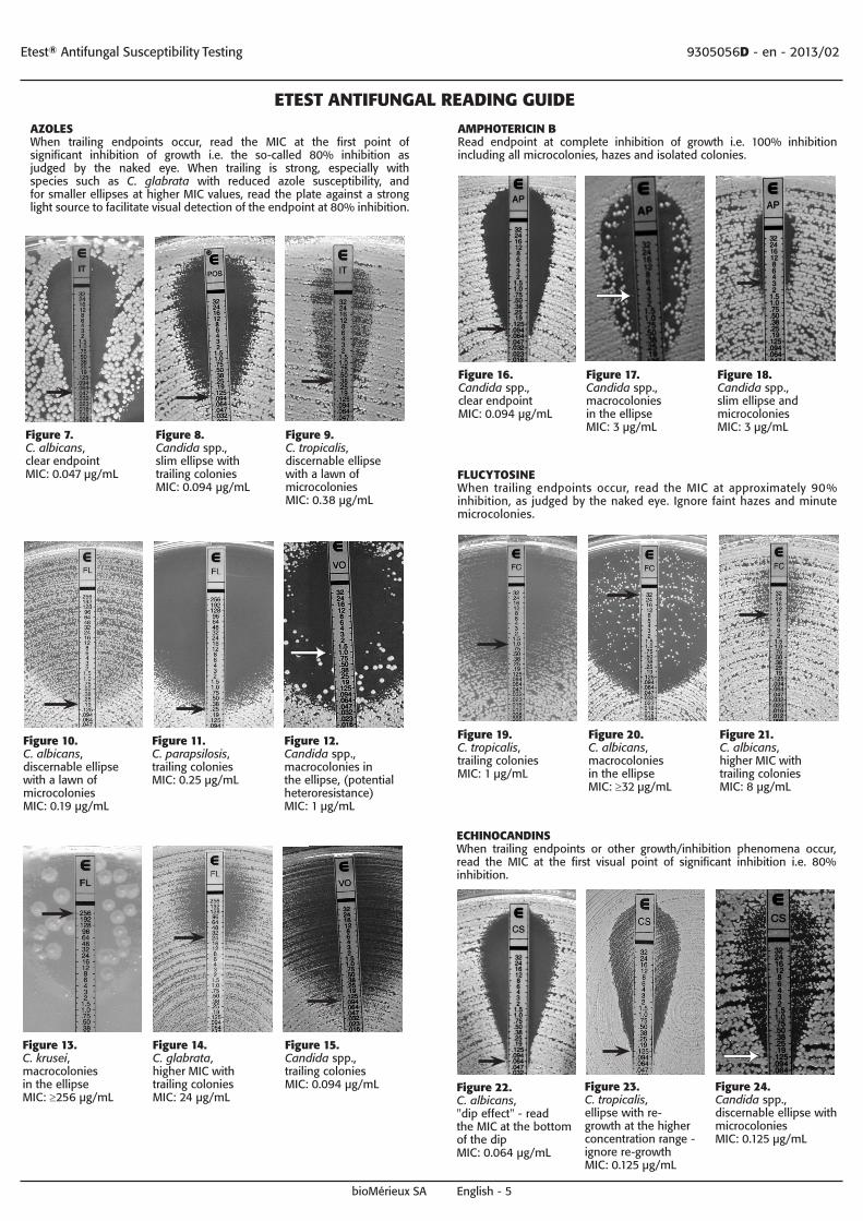
ETEST ANTIFUNGAL READING GUIDE
AZOLESWhen trailing endpoints occur, read the MIC at the first point of significant inhibition of growth i.e. the so-called 80% inhibition as judged by the naked eye. When trailing is strong, especially with species such as C. glabrata with reduced azole susceptibility, and for smaller ellipses at higher MIC values, read the plate against a strong light source to facilitate visual detection of the endpoint at 80% inhibition.
Figure 7. C. albicans, clear endpoint MIC: 0.047 µg/mL
Figure 8. Candida spp., slim ellipse with trailing coloniesMIC: 0.094 µg/mL
Figure 9. C. tropicalis, discernable ellipse with a lawn of microcolonies MIC: 0.38 µg/mL
Figure 10. C. albicans, discernable ellipse with a lawn of microcolonies MIC: 0.19 µg/mL
Figure 11.C. parapsilosis, trailing colonies MIC: 0.25 µg/mL
Figure 12. Candida spp., macrocolonies in the ellipse, (potential heteroresistance) MIC: 1 µg/mL
Figure 13.C. krusei, macrocolonies in the ellipse MIC: ≥256 µg/mL
Figure 14.C. glabrata, higher MIC with trailing colonies MIC: 24 µg/mL
Figure 15. Candida spp., trailing colonies MIC: 0.094 µg/mL
Figure 16. Candida spp., clear endpoint MIC: 0.094 µg/mL
Figure 17. Candida spp., macrocolonies in the ellipse MIC: 3 µg/mL
Figure 18. Candida spp., slim ellipse and microcoloniesMIC: 3 µg/mL
AMPHOTERICIN BRead endpoint at complete inhibition of growth i.e. 100% inhibition including all microcolonies, hazes and isolated colonies.
FLUCYTOSINEWhen trailing endpoints occur, read the MIC at approximately 90% inhibition, as judged by the naked eye. Ignore faint hazes and minute microcolonies.
Figure 19. C. tropicalis, trailing colonies MIC: 1 µg/mL
Figure 20.C. albicans, macrocolonies in the ellipse MIC: ≥32 µg/mL
Figure 21. C. albicans, higher MIC with trailing colonies MIC: 8 µg/mL
ECHINOCANDINSWhen trailing endpoints or other growth/inhibition phenomena occur, read the MIC at the first visual point of significant inhibition i.e. 80% inhibition.
Figure 22. C. albicans, "dip effect" - read the MIC at the bottom of the dip MIC: 0.064 µg/mL
Figure 23. C. tropicalis, ellipse with re-growth at the higher concentration range - ignore re-growth MIC: 0.125 µg/mL
Figure 24. Candida spp., discernable ellipse with microcoloniesMIC: 0.125 µg/mL

REFERENCES
1. Reference Method for Broth Dilution Antifungal Susceptibility Testing of Yeasts; Approved Standard. CLSI M27-A (latest version).
2. Reference Method for Broth Dilution Antifungal Susceptibility Testing of Filamentous Fungi; Approved Standard. CLSI M38-A (latest edition).
3. Pfaller M.A., Messer S.A., Bolmström A., Odds F.C., Rex J.H. (1996). Multi-site reproducibility of the Etest method for Antifungal susceptibility testing of yeast isolates. Journal of Clinical Microbiology (JCM). 34(7): 1691-1693.
4. Espinel-Ingroff A., Pfaller M.A., Erwin M.E., Jones R.N. (1996). Interlaboratory evaluation of Etest method for testing antifungal susceptibilities of pathogenic yeasts to five antifungal agents by using casitone agar and solidified RPMI 1640 medium with 2% glucose. JCM. 34(4): 848-852.
5. Pfaller M.A., Messer S.A., Karlsson Å., Bolmström A. (1998). Evaluation of the Etest method for determining fluconazole susceptibilities of 402 clinical yeast isolates by using three different agar media. JCM. 36(9): 2586-2589.
6. Pfaller M.A., Messer S.A., Mills K., Bolmström A., Jones R.N. (2000). Evaluation of the Etest method for determining voriconazole susceptibilities of 312 clinical isolates of Candida species by using three different agar media. JCM. 38(10): 3715-3717.
7. Pfaller M.A., Messer S.A., Mills K., Bolmström A., Jones R.N. (2001). Evaluation of the Etest method for determining caspofungin susceptibilities of 726 clinical isolates of Candida species. JCM. 39(12): 4387-4389.
8. Pfaller M.A., Messer S.A., Mills K., Bolmström A., Jones RN. (2001). Evaluation of the Etest method for determining posaconazole MICs for 314 clinical isolates of Candida species. JCM. 39(11): 3952-3954.
9. Barry A.L., Pfaller M.A., Rennie R.P., Fuchs P.C., Brown S.D. (2002). Precision and accuracy of fluconazole susceptibility testing by broth microdilution, Etest and disk diffusion methods. Antimicrobial Agents and Chemotherapy (AAC). 46(6): 1781-1784.
10. Maxwell M.J., Messer S.A., Hollis R.J., Boyken L., Tendolkar S., Diekema D.J., Pfaller M.A, and the International Fungal Surveillance Participant Group. (2003). Evaluation of Etest method for determining fluconazole and voriconazole MICs for 279 clinical isolates of Candida species infrequently isolated from blood. JCM. 41(3): 1087-1090.
11. Pfaller M.A., Diekema D.J., Messer S.A., Boyken L., Hollis R.J. (2003). Activities of fluconazole and voriconazole against 1,586 recent clinical isolates of Candida species determined by broth microdilution, disk diffusion and Etest methods: Report from Artemis global antifungal susceptibility program 2001. JCM. 41(4): 1440-1446.
12. Pfaller M.A., Diekema D.J., Boyken L., Messer S.A., Tendolkar S., Hollis R.J. (2003). Evaluation of Etest and disk diffusion methods for determining susceptibilities of 235 bloodstream isolates of C. glabrata to fluconazole and voriconazole. JCM. 41(5): 1875-1880.
13. Pfaller M.A., Messer S.A., Mills K., Bolmstrom A., Jones R.N. (2001). Evaluation of Etest method for determining caspofungin (MK-0991) susceptibilities of 726 clinical isolates of Candida species. JCM. 39(12): 4387-4389.
14. Laverdiere M., Restieri C., Habel F. (2002). Evaluation of the in vitro activity of caspofungin against bloodstream isolates of Candida species from cancer patients: comparison of Etest and NCCLS reference methods. International Journal of Antimicrobial Agents. 20(6): 468-471.
BIOMERIEUx, the blue logo, Etest, the Etest gradient strip, LyfoCults, Nema C88, Retro C80 and Simplex C76 are used pending and/or registered trademarks belonging to bioMérieux, or one of its subsidiaries, or one of its companies.CLSI is a registered trademark belonging to Clinical Laboratory and Standards Institute, Inc.The ATCC trademark and trade name and any and all ATCC catalog numbers are trademarks of the American Type Culture Collection.Any other name or trademark is the property of its respective owner.
Etest® Antifungal Susceptibility Testing 9305056D - en - 2013/02
bioMérieux SAChemin de l’Orme 69280 Marcy-l’Etoile - France
RCS LYON 673 620 399 Tel. 33 (0)4 78 87 20 00 Fax 33 (0)4 78 87 20 90 www.biomerieux.com
WARRANTY AND DISCLAIMER
ExPRESS LIMITED WARRANTY AND DISCLAIMER
bioMérieux expressly warrants that Etest will determine the MIC of the antimicrobial agent on each test strip, if the procedures, precautions and important observations indicated in the package insert are strictly complied with.
bioMérieux makes no other warranties, expressed or implied, including the implied warranty of merchantability or fitness for particular purpose.
Any change or modification of the product instructions may affect results. bioMérieux shall not be liable for any damages resulting from product tampering, variance in transportation, stated storage, handling, testing procedures, precautions and other instructions of the most recently revised version of the package insert.
Photos: bioMérieux SA
INDEX OF SYMBOLS
Symbol Meaning
Catalogue number
In Vitro Diagnostic Medical Device
Manufacturer
Temperature limitation
Upper limit of temperature
Use by
Batch code
Consult Instructions for Use
Contains sufficient for <n> tests