Antibiotics induce redox-related physiological alterations ... · resulting in cell death. Rather...
10
Antibiotics induce redox-related physiological alterations as part of their lethality Daniel J. Dwyer a,b,c,1 , Peter A. Belenky a,b,c,1 , Jason H. Yang a,b,c,1 , I. Cody MacDonald a,b,c , Jeffrey D. Martell d , Noriko Takahashi e , Clement T. Y. Chan a,b,c , Michael A. Lobritz a,b,c,f,g , Dana Braff a,b,c , Eric G. Schwarz a,b,c , Jonathan D. Ye a,b,c , Mekhala Pati h , Maarten Vercruysse e , Paul S. Ralifo h , Kyle R. Allison i , Ahmad S. Khalil b,c,f , Alice Y. Ting d , Graham C. Walker e , and James J. Collins a,b,c,f,j,2 a Howard Hughes Medical Institute, Departments of b Biomedical Engineering and h Chemistry, and c Center of Synthetic Biology, Boston University, Boston, MA 02215; Departments of d Chemistry and e Biology, Massachusetts Institute of Technology, Cambridge, MA 02139; f Wyss Institute for Biologically Inspired Engineering, Harvard University, Boston, MA 02115; g Division of Infectious Diseases, Massachusetts General Hospital, Boston, MA 02114; i Department of Systems Biology, Columbia University, New York, NY 10032; and j Department of Medicine, Boston University School of Medicine, Boston, MA 02118 Edited* by Charles R. Cantor, Sequenom, Inc., San Diego, CA, and approved April 9, 2014 (received for review January 30, 2014) Deeper understanding of antibiotic-induced physiological responses is critical to identifying means for enhancing our current antibiotic arsenal. Bactericidal antibiotics with diverse targets have been hypothesized to kill bacteria, in part by inducing production of damaging reactive species. This notion has been supported by many groups but has been challenged recently. Here we robustly test the hypothesis using biochemical, enzymatic, and biophysical assays along with genetic and phenotypic experiments. We first used a novel intracellular H 2 O 2 sensor, together with a chemically diverse panel of fluorescent dyes sensitive to an array of reactive species to demonstrate that antibiotics broadly induce redox stress. Subsequent gene-expression analyses reveal that complex antibiotic-induced oxidative stress responses are distinct from ca- nonical responses generated by supraphysiological levels of H 2 O 2 . We next developed a method to quantify cellular respiration dy- namically and found that bactericidal antibiotics elevate oxygen consumption, indicating significant alterations to bacterial redox physiology. We further show that overexpression of catalase or DNA mismatch repair enzyme, MutS, and antioxidant pretreat- ment limit antibiotic lethality, indicating that reactive oxygen spe- cies causatively contribute to antibiotic killing. Critically, the killing efficacy of antibiotics was diminished under strict anaerobic condi- tions but could be enhanced by exposure to molecular oxygen or by the addition of alternative electron acceptors, indicating that envi- ronmental factors play a role in killing cells physiologically primed for death. This work provides direct evidence that, downstream of their target-specific interactions, bactericidal antibiotics induce complex redox alterations that contribute to cellular damage and death, thus supporting an evolving, expanded model of antibiotic lethality. reactive oxygen species | DNA repair | mutagenesis T he increasing incidence of antibiotic-resistant infections cou- pled with a declining antibiotic pipeline has created a global public health threat (1–6). Therefore there is a pressing need to expand our conceptual understanding of how antibiotics act and to use insights gained from such efforts to enhance our antibiotic arsenal. It has been proposed that different classes of bactericidal antibiotics, regardless of their drug–target interactions, generate varying levels of deleterious reactive oxygen species (ROS) that contribute to cell killing (7, 8). This unanticipated notion, built upon important prior work (9–11), has been extended and sup- ported by multiple laboratories investigating wide-ranging drug classes (e.g., β-lactams, aminoglycosides, and fluoroquinolones) and bacterial species (e.g., Escherichia coli, Pseudomonas aeruginosa, Salmonella enterica, Mycobacterium tuberculosis, Bacillus subtilis, Staphylococcus aureus, Acinetobacter baumannii , Burkholderia cepecia, Streptococcus pneumonia, Enterococcus faecalis) using independent lines of evidence (12–39). Importantly, these ongoing efforts have served to refine aspects of the initial model and show that antibiotic- induced ROS generation is a more complex process than originally suggested, likely involving additional means of production. The redox stress component of antibiotic lethality is hypothe- sized to derive from alterations to multiple core aspects of cel- lular physiology and stress response activation. Specifically, this component includes alterations to central metabolism, cellular respiration, and iron metabolism initiated by drug-mediated dis- ruptions of target-specific processes and resulting cellular damage (Fig. 1A). Important support for this hypothesis can be found in pathogenic clinical isolates whose drug tolerance involves muta- tions in oxidative stress response and defense genes and not ex- clusively in drug target mutagenesis (40–48). Recent critiques of this evolving model have misinterpreted an essential aspect of the hypothesis. Specifically, these recent studies (49–51) are predicated on the notion that ROS are the sole arbiters of antibiotic lethality, thereby implying that the model suggests that antibiotics do not kill by disrupting their well-established, target-specific processes. However, the evolving model is completely consistent with the literature indicating that bactericidal antibiotics are capable of inducing lethal cellular damage via interference with target-specific processes, ultimately Significance Substantial knowledge exists about how antibiotics interfere with core bacterial processes by binding to specific targets. Re- cently it has become appreciated that blocking these functions alters cellular redox state, and these perturbations may contribute to the lethality of antibiotics. In this work we explore whether antibiotic treatment of bacteria affects cellular oxidative stress and the role of such stress in antibiotic-mediated killing. We find that antibiotics dynamically alter cellular respiration and induce lethal levels of intracellular hydrogen peroxide. Antioxidants, in- cluding oxidative stress defense proteins, significantly reduce the killing by antibiotics, which is highly sensitive to the presence of molecular oxygen. These findings underscore the complex nature of antibiotic action and suggest practical approaches to enhancing our current antibiotic arsenal. Author contributions: D.J.D., P.A.B., J.H.Y., I.C.M., J.D.M., N.T., C.T.Y.C., A.Y.T., G.C.W., and J.J.C. designed research; D.J.D., P.A.B., J.H.Y., I.C.M., J.D.M., N.T., C.T.Y.C., D.B., E.G.S., J.D.Y., M.P., M.V., and P.S.R. performed research; D.J.D., P.A.B., J.D.M., A.Y.T., and J.J.C. contrib- uted new reagents/analytic tools; D.J.D., P.A.B., J.H.Y., I.C.M., J.D.M., C.T.Y.C., M.A.L., D.B., E.G.S., M.P., K.R.A., A.S.K., A.Y.T., G.C.W., and J.J.C. analyzed data; and D.J.D., P.A.B., J.H.Y., J.D.M., M.A.L., G.C.W., and J.J.C. wrote the paper. Conflict of interest statement: J.J.C. is a scientific cofounder and Scientific Advisory Board chair of EnBiotix, Inc., a start-up focused on antibiotic development. *This Direct Submission article had a prearranged editor. Freely available online through the PNAS open access option. Data deposition: The microarray data reported in this paper have been deposited in the Gene Expression Omnibus (GEO) database (accession no. GSE56133). 1 D.J.D., P.A.B., and J.H.Y. contributed equally to this work. 2 To whom correspondence should be addressed. E-mail: [email protected]. This article contains supporting information online at www.pnas.org/lookup/suppl/doi:10. 1073/pnas.1401876111/-/DCSupplemental. E2100–E2109 | PNAS | Published online May 6, 2014 www.pnas.org/cgi/doi/10.1073/pnas.1401876111 Downloaded by guest on December 25, 2019
Transcript of Antibiotics induce redox-related physiological alterations ... · resulting in cell death. Rather...

Antibiotics induce redox-related physiologicalalterations as part of their lethalityDaniel J. Dwyera,b,c,1, Peter A. Belenkya,b,c,1, Jason H. Yanga,b,c,1, I. Cody MacDonalda,b,c, Jeffrey D. Martelld,Noriko Takahashie, Clement T. Y. Chana,b,c, Michael A. Lobritza,b,c,f,g, Dana Braffa,b,c, Eric G. Schwarza,b,c,Jonathan D. Yea,b,c, Mekhala Patih, Maarten Vercruyssee, Paul S. Ralifoh, Kyle R. Allisoni, Ahmad S. Khalilb,c,f,Alice Y. Tingd, Graham C. Walkere, and James J. Collinsa,b,c,f,j,2
aHoward Hughes Medical Institute, Departments of bBiomedical Engineering and hChemistry, and cCenter of Synthetic Biology, Boston University, Boston,MA 02215; Departments of dChemistry and eBiology, Massachusetts Institute of Technology, Cambridge, MA 02139; fWyss Institute for Biologically InspiredEngineering, Harvard University, Boston, MA 02115; gDivision of Infectious Diseases, Massachusetts General Hospital, Boston, MA 02114; iDepartment ofSystems Biology, Columbia University, New York, NY 10032; and jDepartment of Medicine, Boston University School of Medicine, Boston, MA 02118
Edited* by Charles R. Cantor, Sequenom, Inc., San Diego, CA, and approved April 9, 2014 (received for review January 30, 2014)
Deeper understanding of antibiotic-induced physiological responsesis critical to identifying means for enhancing our current antibioticarsenal. Bactericidal antibiotics with diverse targets have beenhypothesized to kill bacteria, in part by inducing production ofdamaging reactive species. This notion has been supported bymany groups but has been challenged recently. Here we robustlytest the hypothesis using biochemical, enzymatic, and biophysicalassays along with genetic and phenotypic experiments. We firstused a novel intracellular H2O2 sensor, together with a chemicallydiverse panel of fluorescent dyes sensitive to an array of reactivespecies to demonstrate that antibiotics broadly induce redoxstress. Subsequent gene-expression analyses reveal that complexantibiotic-induced oxidative stress responses are distinct from ca-nonical responses generated by supraphysiological levels of H2O2.We next developed a method to quantify cellular respiration dy-namically and found that bactericidal antibiotics elevate oxygenconsumption, indicating significant alterations to bacterial redoxphysiology. We further show that overexpression of catalase orDNA mismatch repair enzyme, MutS, and antioxidant pretreat-ment limit antibiotic lethality, indicating that reactive oxygen spe-cies causatively contribute to antibiotic killing. Critically, the killingefficacy of antibiotics was diminished under strict anaerobic condi-tions but could be enhanced by exposure to molecular oxygen or bythe addition of alternative electron acceptors, indicating that envi-ronmental factors play a role in killing cells physiologically primed fordeath. This work provides direct evidence that, downstream of theirtarget-specific interactions, bactericidal antibiotics induce complexredox alterations that contribute to cellular damage and death, thussupporting an evolving, expanded model of antibiotic lethality.
reactive oxygen species | DNA repair | mutagenesis
The increasing incidence of antibiotic-resistant infections cou-pled with a declining antibiotic pipeline has created a global
public health threat (1–6). Therefore there is a pressing need toexpand our conceptual understanding of how antibiotics act andto use insights gained from such efforts to enhance our antibioticarsenal. It has been proposed that different classes of bactericidalantibiotics, regardless of their drug–target interactions, generatevarying levels of deleterious reactive oxygen species (ROS) thatcontribute to cell killing (7, 8). This unanticipated notion, builtupon important prior work (9–11), has been extended and sup-ported by multiple laboratories investigating wide-ranging drugclasses (e.g., β-lactams, aminoglycosides, and fluoroquinolones)and bacterial species (e.g., Escherichia coli, Pseudomonas aeruginosa,Salmonella enterica, Mycobacterium tuberculosis, Bacillus subtilis,Staphylococcus aureus, Acinetobacter baumannii, Burkholderia cepecia,Streptococcus pneumonia, Enterococcus faecalis) using independentlines of evidence (12–39). Importantly, these ongoing efforts haveserved to refine aspects of the initial model and show that antibiotic-induced ROS generation is a more complex process than originallysuggested, likely involving additional means of production.
The redox stress component of antibiotic lethality is hypothe-sized to derive from alterations to multiple core aspects of cel-lular physiology and stress response activation. Specifically, thiscomponent includes alterations to central metabolism, cellularrespiration, and iron metabolism initiated by drug-mediated dis-ruptions of target-specific processes and resulting cellular damage(Fig. 1A). Important support for this hypothesis can be found inpathogenic clinical isolates whose drug tolerance involves muta-tions in oxidative stress response and defense genes and not ex-clusively in drug target mutagenesis (40–48).Recent critiques of this evolving model have misinterpreted
an essential aspect of the hypothesis. Specifically, these recentstudies (49–51) are predicated on the notion that ROS are thesole arbiters of antibiotic lethality, thereby implying that themodel suggests that antibiotics do not kill by disrupting theirwell-established, target-specific processes. However, the evolvingmodel is completely consistent with the literature indicating thatbactericidal antibiotics are capable of inducing lethal cellulardamage via interference with target-specific processes, ultimately
Significance
Substantial knowledge exists about how antibiotics interferewith core bacterial processes by binding to specific targets. Re-cently it has become appreciated that blocking these functionsalters cellular redox state, and these perturbations may contributeto the lethality of antibiotics. In this work we explore whetherantibiotic treatment of bacteria affects cellular oxidative stressand the role of such stress in antibiotic-mediated killing. We findthat antibiotics dynamically alter cellular respiration and inducelethal levels of intracellular hydrogen peroxide. Antioxidants, in-cluding oxidative stress defense proteins, significantly reduce thekilling by antibiotics, which is highly sensitive to the presence ofmolecular oxygen. These findings underscore the complex natureof antibiotic action and suggest practical approaches to enhancingour current antibiotic arsenal.
Author contributions: D.J.D., P.A.B., J.H.Y., I.C.M., J.D.M., N.T., C.T.Y.C., A.Y.T., G.C.W., andJ.J.C. designed research; D.J.D., P.A.B., J.H.Y., I.C.M., J.D.M., N.T., C.T.Y.C., D.B., E.G.S., J.D.Y.,M.P., M.V., and P.S.R. performed research; D.J.D., P.A.B., J.D.M., A.Y.T., and J.J.C. contrib-uted new reagents/analytic tools; D.J.D., P.A.B., J.H.Y., I.C.M., J.D.M., C.T.Y.C., M.A.L., D.B.,E.G.S., M.P., K.R.A., A.S.K., A.Y.T., G.C.W., and J.J.C. analyzed data; and D.J.D., P.A.B., J.H.Y.,J.D.M., M.A.L., G.C.W., and J.J.C. wrote the paper.
Conflict of interest statement: J.J.C. is a scientific cofounder and Scientific Advisory Boardchair of EnBiotix, Inc., a start-up focused on antibiotic development.
*This Direct Submission article had a prearranged editor.
Freely available online through the PNAS open access option.
Data deposition: The microarray data reported in this paper have been deposited in theGene Expression Omnibus (GEO) database (accession no. GSE56133).1D.J.D., P.A.B., and J.H.Y. contributed equally to this work.2To whom correspondence should be addressed. E-mail: [email protected].
This article contains supporting information online at www.pnas.org/lookup/suppl/doi:10.1073/pnas.1401876111/-/DCSupplemental.
E2100–E2109 | PNAS | Published online May 6, 2014 www.pnas.org/cgi/doi/10.1073/pnas.1401876111
Dow
nloa
ded
by g
uest
on
Dec
embe
r 25
, 201
9
resulting in cell death. Rather than refute this traditional view ofantibiotic action, the hypothesis extends it by suggesting that anadditional component of toxicity results from ROS, which aregenerated as a downstream physiological consequence of antibioticsinteracting with their traditional targets. In this respect, reactivespecies are thought to contribute causatively to drug lethality.However, an important gap exists in our general understanding
of how bacteria respond physiologically to antibiotic–target inter-actions on a system-wide level, how these responses contribute toantibiotic killing, and how the extracellular environment protectsor exacerbates the intracellular contributions to cell death. Here,we use a multidisciplinary set of biochemical, enzymatic, bio-physical, and genetic assays to address these issues and expand ourunderstanding of antibiotic-induced physiological responses andfactors contributing to antibiotic lethality. Data from the presentwork indicate that antibiotic lethality is accompanied by ROSgeneration and that such reactive species causatively contribute toantibiotic lethality.
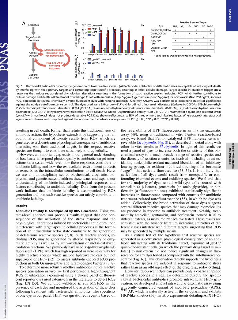
ResultsAntibiotic Lethality Is Accompanied by ROS Generation. Using sys-tems-level analyses, our previous results suggest that one con-sequence of the activation of the stress response and thephysiological alterations induced by bactericidal antibiotics uponinterference with target-specific cellular processes is the forma-tion of an intracellular redox state conducive to the generationof deleterious reactive species (7, 8). Such reactive species, in-cluding ROS, may be generated by altered respiratory or enzy-matic activity as well as by auto-oxidation or metal-catalyzedoxidation reactions. We previously have used 3′-(p-hydroxyphenyl)fluorescein (HPF), which has high reported in vitro selectivity forhighly reactive species which include hydroxyl radicals but notsuperoxide or H2O2 (52), to assess antibiotic-induced ROS pro-duction in both Gram-negative and Gram-positive bacteria (7, 8).To determine more robustly whether antibiotics induce reactive
species generation in vivo, we first performed a high-throughputROS quantification experiment using a diverse panel of fluores-cent reporter dyes used extensively in the literature to detect ROS(Fig. 1B) (53). We cultured wild-type E. coli MG1655 in thepresence of each dye and monitored the activation of these dyesfollowing treatment with bactericidal antibiotics. The specificityof one dye in our panel, HPF, was questioned recently based on
the reversibility of HPF fluorescence in an in vitro enzymaticassay (49); using a traditional in vitro Fenton reaction-basedassay, we found that Fenton-catalyzed HPF fluorescence is ir-reversible (SI Appendix, Fig. S1), as described in detail along withother in vitro results in SI Appendix. In light of this result, weused a panel of dyes to increase both the sensitivity of this bio-chemical assay to a much broader range of reactive species andthe diversity of reaction chemistries involved—including direct ox-idation, nucleophilic oxidant-mediated liberation of an inhibitoryleaving group, or oxidant-mediated opening of a boronate“cage”—that activate fluorescence (53, 54). It is unlikely thatactivation of all dyes would result from nonspecific or con-founding chemical events and oxidizing species. We found thatfor the majority of dyes tested, wild-type cells treated withampicillin (a β-lactam), gentamicin (an aminoglycoside), or nor-floxacin (a fluoroquinolone) exhibited statistically significantincreases in fluorescence compared with controls for antibiotictreatment-related autofluorescence (55), in which no dye wasadded. Collectively, the broad activation of these dyes suggestsseveral different reactive species that may damage biomoleculesare produced in response to antibiotic treatment. Notably, treat-ment by ampicillin, gentamicin, and norfloxacin induced ROS todifferent extents, as measured by each dye tested. These results areconsistent with the broader literature indicating antibiotics of dif-ferent classes interfere with different targets, suggesting that ROSmay be generated by multiple means.As a critical test of the hypothesis that reactive species are
generated as a downstream physiological consequence of an anti-biotic interacting with its traditional target, exposure of gyrA17quinolone-resistant cells (in which the primary drug target is mu-tated) to norfloxacin did not induce significant changes in fluo-rescence for any dyes tested as compared with the autofluorescencecontrol (Fig. 1C). This observation directly supports the hypothesisthat reactive species are induced in response to antibiotic stressrather than as an off-target effect of the drug (e.g., redox cycling).However, fluorescent dyes can provide only a coarse snapshot
of reactive species in a cell. To determine directly and specifi-cally if bactericidal antibiotics promote intracellular H2O2 gen-eration, we developed a novel intracellular enzymatic assay usinga recently engineered variant of ascorbate peroxidase (APX),W41FAP, which is naturally active in the cytoplasm and exhibitsHRP-like kinetics (56). In vitro experiments detailing APX H2O2
Fig. 1. Bactericidal antibiotics promote the generation of toxic reactive species. (A) Bactericidal antibiotics of different classes are capable of inducing cell deathby interfering with their primary targets and corrupting target-specific processes, resulting in lethal cellular damage. Target-specific interactions trigger stressresponses that induce redox-related physiological alterations resulting in the formation of toxic reactive species, including ROS, which further contribute tocellular damage and death. (B) Treatment of wild-type E. coliwith ampicillin (Amp, 5 μg/mL), gentamicin (Gent, 5 μg/mL), or norfloxacin (Nor, 250 ng/mL) inducesROS, detectable by several chemically diverse fluorescent dyes with ranging specificity. One-way ANOVA was performed to determine statistical significanceagainst the no-dye autofluorescence control. The dyes used were 5/6-carboxy-2’,7’-dichlorodihydrofluorescein diacetate (Carboxy-H2DCFDA); 5/6-chloromethyl-2’,7’-dichlorodihydrofluorescein diacetate (CM-H2DCFDA); 4-amino-5-methylamino-2´,7´-diflurorescein diacetate (DAF-FM); 2’,7’-dichlorodihydrofluoresceindiacetate (H2DCFDA); 3′-(p-hydroxyphenyl) fluorescein (HPF); OxyBURST Green (Oxyburst); and Peroxy-Fluor 2 (PF2). (C) Treatment of a quinolone-resistant strain(gyrA17) with norfloxacin does not produce detectable ROS. Data shown reflect mean ± SEM of three or more technical replicates. Where appropriate, statisticalsignificance is shown and computed against the no-treatment control or no-dye control (*P ≤ 0.05; **P ≤ 0.01; ***P ≤ 0.001).
Dwyer et al. PNAS | Published online May 6, 2014 | E2101
BIOPH
YSICSAND
COMPU
TATIONALBIOLO
GY
PNASPL
US
Dow
nloa
ded
by g
uest
on
Dec
embe
r 25
, 201
9

specificity are described in SI Appendix. The H2O2 measurementis made using Amplex Red, a fluorogenic dye that diffuses acrosscell membranes into the cytoplasm. Within the cell, APX cata-lyzes the rapid H2O2-dependent conversion of Amplex Red intoa readily detectable fluorescent product. This method improveson the common, indirect method for measuring H2O2 in bi-ological systems in which Amplex Red is oxidized extracellularlyby exogenous HRP and H2O2 in the culture medium (49, 57).The extracellular assay assumes that the external concentrationof H2O2 in the culture supernatant reflects the intracellularH2O2 concentration because of rapid diffusion. This assumption,however, is problematic because of biological constraints on H2O2diffusion (58), scavenging compartmentation (59–61), and rapidFenton chemistry destruction of intracellular H2O2 (62, 63).Importantly, intracellular APX uses the same dye as extracellularHRP to report on H2O2, and APX expression has no discernableeffect on the rate of cell growth.We found that untreated cells expressing APX exhibited stable
baseline levels of Amplex Red fluorescence (Fig. 2 A and B). In
comparison, when APX-expressing cells were treated with am-picillin, gentamicin, or norfloxacin, we observed significant two-to threefold changes in the level of Amplex Red fluorescence inour assay in samples taken at 1 and 2 h posttreatment. Notably,the level of Amplex Red fluorescence induced by bactericidalantibiotics at 1 h posttreatment was comparable to our 10 μMH2O2 spike-in control and was significantly smaller than thesupraphysiological levels of H2O2 typically used to study oxidativestress responses (49). In contrast, treatment of APX-expressingcells with the bacteriostatic drug chloramphenicol yielded noeffect on Amplex Red fluorescence.These findings contrast with the recent failure to detect in-
creased extracellular H2O2 by HRP when an E. coli strain lackingthe three best-characterized H2O2-scavenging enzymes (ahpCFkatE, and katG, referred to as “Hpx−”) was treated with bac-tericidal antibiotics. These earlier experiments assumed thatdrug-induced increases in extracellular H2O2 would be readilydetectable over the naturally elevated steady-state concentrationfound in this strain (49, 60) and that the absence of H2O2 de-tection in the supernatant implied that H2O2 was not generatedfollowing antibiotic treatment (49, 60). The failure of this ex-tracellular assay to detect antibiotic-induced H2O2 productionmay be caused by two confounding factors. First, recent work hasshown that cytochrome bd oxidase displays high catalase activity(59), which may compensate for the loss of known H2O2-scavenging activity by the catalase and peroxidase deletionsin the Hpx− strain. Moreover, intracellular H2O2 could bedestroyed by Fenton chemistry before diffusing into the medium.Second, H2O2’s dipole moment, similar to that of water (58),may prevent extracellular H2O2 from equilibrating with intra-cellular H2O2 by limiting H2O2 diffusion across the membrane.Indeed, such limitations in H2O2 permeability rationalized theinitial observation that H2O2 scavenging is compartmented (60).In addition to these biochemical and enzymatic approaches, we
determined whether antibiotic-induced ROS could trigger redoxstress responses in vivo by using a genetic reporter assay. Wereasoned that if bactericidal antibiotic interference with target-specific processes results in ROS generation, then one wouldexpect to observe activation of oxidative stress regulons. To testthis hypothesis, we constructed GFP-based promoter reportersystems that report on oxidant stress and coregulated metabolicresponse activation (Fig. 2C and SI Appendix, Fig. S4); the fullcomplement of reporter constructs is detailed in SI Appendix. Inour assay, we focused on the activity of the promoters pOxyS andpSoxS, which are activated by the main regulators of the responseto H2O2 and superoxide, OxyR and SoxR, respectively (64). Weassessed whether ampicillin or norfloxacin induces expressionfrom a diverse set of promoters; gentamicin was not testedbecause of the confounding effects of aminoglycosides onGFP reporter assays. We found that treatment with ampicillinor norfloxacin elicited significantly increased pOxyS and pSoxSactivity, indicating bactericidal antibiotic-induced activation ofOxyR and SoxR (Fig. 2C and SI Appendix, Fig. S4).Because this assay was quantitative, we assessed the sensitivity
of OxyR to antibiotic treatment by comparing antibiotic-inducedpOxyS-gfp expression with that of a dose-range of H2O2 (Fig.2D). We quantified GFP expression induction over a range ofH2O2 concentrations spanning six orders of magnitude (1 mM–1 nM),which encompasses the minimum levels of H2O2 reported tofully oxidize OxyR both in vivo (5 μM) and in vitro (50 nM, in thepresence of antioxidants) (61, 65). Significant changes in pOxyS-gfp expression were detected at a threshold near 1 μM H2O2as compared with untreated cells; this finding is consistent withreports of OxyR activation by submicromolar H2O2 (66, 67).GFP expression was increased by two orders of magnitude overthat in untreated cells in response to exogenous H2O2 appliedat concentrations approaching lethality (100 μM and 1 mM)(67), whereas ampicillin and norfloxacin induced GFP expres-sion most comparable to 10 μM exogenous H2O2, similar tomeasurements by our enzymatic Amplex Red assay (Fig. 2A).
Fig. 2. Antibiotics trigger physiologically relevant generation of H2O2. (A)Treatment of wild-type E. coli with ampicillin (Amp, 5 μg/mL), gentamicin(Gent, 5 μg/mL) or norfloxacin (Nor, 250 ng/mL) induces H2O2 production,detected by the intracellular enzymatic sensor APX, using Amplex Redfluorescence as an output. Antibiotic induction was compared with an ex-ogenous H2O2 dose-range control. (B) Fold change of antibiotic-induced,H2O2-mediated Amplex Red fluorescence at 1 h and 2 h posttreatment,compared with the no-treatment control with basal H2O2 production. (C)Antibiotic-induced ROS trigger endogenous oxidative stress responses.Treatment with ampicillin (5 μg/mL) or norfloxacin (250 ng/mL) induces GFPexpression from promoters regulated by H2O2-sensitive OxyR (pOxyS-gfp)and superoxide-sensitive SoxR (pSoxS-gfp) in wild-type E. coli. Data fromadditional GFP promoter reporters are included in SI Appendix. (D) Anti-biotics induce physiologically relevant levels of oxidative stress. Antibioticinduction of GFP expression from pOxyS was compared with an exogenousH2O2 dose-range control. (E) Antibiotic-induced redox stress is comparableto physiologically relevant oxidative stress perturbations at the transcrip-tional level. Microarrays were performed on cells treated with H2O2 (10 μM),ampicillin (5 μg/mL), gentamicin (5 μg/mL), or norfloxacin (250 ng/mL) toquantify altered expression. Data shown reflect mean ± SEM o three or moretechnical replicates. Where appropriate, statistical significance is shown(*P ≤ 0.05; **P ≤ 0.01; ***P ≤ 0.001). In each instance, an untreated controlwas used for normalization and determination of statistical significance.
E2102 | www.pnas.org/cgi/doi/10.1073/pnas.1401876111 Dwyer et al.
Dow
nloa
ded
by g
uest
on
Dec
embe
r 25
, 201
9

Because submicromolar H2O2 is sufficient to induce cytotoxicity,including significant DNA damage (68), these results suggestthat the dynamic range for OxyR activation exceeds the trueoxidative stress capacity of wild-type cells by orders of magni-tude. Consequently, supraphysiological H2O2 perturbations (49)poorly simulate the cytotoxic oxidative stresses experienced bycells in culture and suggest that such perturbations are inap-propriate controls for studying OxyR-regulated responses toantibiotic-induced oxidative stress.To validate our estimates that bactericidal antibiotics induce
oxidative stresses similar to exogenous treatment with 10 μMH2O2,we compared microarray gene-expression profiles from untreatedwild-type cells with those from cells treated with ampicillin, gen-tamicin, norfloxacin, or 10 μM H2O2. Similar to our results usingpOxyS-gfp (Fig. 2C), we found that all treatments increased oxySexpression in comparison with our untreated control (Fig. 2E).Interestingly, soxS expression was increased by bactericidal anti-biotics but was decreased significantly by 10 μM H2O2, suggestingsoxS activation by antibiotic-induced superoxide but not by H2O2.Nonetheless, the expression of many genes in both the OxyR andSoxRS regulons was induced similarly by bactericidal antibioticsand 10 μM H2O2 (SI Appendix, Tables S1 and S2).Given that bactericidal antibiotics induce substantial activa-
tion of the oxidative stress regulon, by extension it may beexpected that H2O2-scavenger genes would be activated similarlyby 10 μM H2O2. Significant induction of ahpC and katG bysupraphysiologic H2O2 doses was reported previously (49);however, we found that neither 10 μM H2O2 nor bactericidalantibiotics induced significant up-regulation of ahpC or katGexpression (Fig. 2E and SI Appendix, Table S2). This observationhighlights the complexity in the oxidative stress response andsuggests that intrinsically induced stress responses, such as thosearising from antibiotic treatments, may be similar but not nec-essarily equivalent to those canonically induced by exogenousoxidants. In particular, these data demonstrate that bactericidalantibiotics trigger oxidative stress responses similar to 10 μM H2O2and emphasize the importance of using physiologically relevantcontrols for investigating cellular responses to antibiotic stress.Collectively, our biochemical, enzymatic, genetic, and micro-
array experiments indicate that bactericidal antibiotics inducethe formation of reactive species and trigger stress responsessimilar to those elicited by 10 μM H2O2. Possible sources forantibiotic-induced ROS are auto-oxidation reactions involvingcofactor-bearing respiratory enzymes or electron leakage, bothof which would be enhanced by elevated respiratory activity andincreased respiratory enzyme titer, as well as by stress-induceduncoupling (69–72). We hypothesized that the antibiotic-inducedincreases in ROS would be accompanied by elevated respiratoryactivity. To test this hypothesis, we developed a flexible assaythat dynamically measures bacterial oxygen consumption rate(OCR) using the Seahorse Extracellular Flux Analyzer platform.This assay permits continuous quantification of real-time oxygenconsumption of multiple samples in parallel with high sensitivityand with detection limited only by the diffusion rate of molecularoxygen in the culture medium. Overall, we found that ampicillinand norfloxacin significantly elevated OCR, whereas bacterio-static chloramphenicol led to a rapid reduction in oxygen con-sumption when cells were grown in defined minimal mediumwith a single carbon source (10 mM glucose) (Fig. 3 and SIAppendix, Fig. S3). These results support recent observationsthat antibiotic stress can trigger sharp increases in the ATP/ADPratio (73). Relative to the untreated control, gentamicin inducedan immediate but transient increase in OCR, whereas ampicillinand norfloxacin induced delayed but sustained OCR increases.The varied respiratory activities stimulated by these differentantibiotics parallel the varied increases in reactive species dosagewe detected biochemically with the fluorescent dyes (Fig. 1B),suggesting that differential respiratory behaviors may give rise todifferent levels of ROS. Together, these results are consistentwith the hypothesis that bactericidal antibiotics induce redox-
related alterations to cell physiology, and that these alterationsare sensitive to respective primary target effects.
ROS Causatively Contribute To Antibiotic Lethality. If bactericidalantibiotics induce ROS-mediated cellular damage as part of theirlethality, then one would anticipate oxidative damage to nucleicacids and their building blocks during drug treatment. Althoughsuperoxide and H2O2 do not cause oxidative damage to nucle-otides, Fenton chemistry does, through the formation of eitherhighly reactive hydroxyl radicals (62, 63, 74, 75) or iron-oxointermediates (76). Accordingly, these and previous data point tohydroxyl radicals as agents of antibiotic mutagenesis, and thisnotion is supported by observations that anaerobic growth orthiourea addition reduces mutation rates to near normal levels(77). DNA polymerase IV critically mediates such mutagenesisby incorporating oxidized dNTPs as replicative substrates (78)under both sublethal (79) and lethal (25) antibiotic doses. Becauseoverexpression of the mismatch repair enzyme MutS reducesantibiotic mutagenesis (79), we tested whether MutS overex-pression would similarly protect against antibiotic lethality. Wefound that increases in MutS expression that do not discerniblyaffect growth rate strikingly reduce the killing by bactericidalantibiotics (Fig. 4). As controls, we tested mutations affecting theability of MutS to recognize mismatches (F36A) (80) or itsATPase (K620A) (81) and observed reduced ability for MutS tosuppress killing by all three classes of bactericidal antibiotics.These results suggest that the well-characterized roles of MutS inlong-patch postreplicative mismatch repair account for much of thesuppression, although our results do not exclude the possibility thatMutS plays additional roles (82, 83). These observations, coupledto the protection elicited by MutS and MutT overexpression (25)and enhanced killing elicited by RecA deletion (8, 20), indicatea common DNA-damage component to killing by bactericidalantibiotics, including those whose targets are at the cell membrane(β-lactams) or the ribosome (aminoglycosides).We hypothesize that such damage is best explained by nucle-
otide oxidation following ROS formation and that the sub-sequent incorporation of these nucleotides into DNA leads todouble-strand breaks. We note that iron (II) chelation by nu-cleoside triphosphates, which is biologically significant (84),would favor the localized production of hydroxyl radicals byFenton chemistry (85). In purine nucleotides, the C8 position isparticularly close to the complexed Fe2+ (86) and hence wouldbe favorably disposed to react with a newly generated hydroxylradical (71) or with an iron-oxo radical (76). Moreover, chelationof Fe2+ by a nucleoside triphosphate increases the rate of theFenton reaction comparably to chelation by EDTA or nitrilo-triacetate (87). Minor amounts of nucleotide oxidation can besignificant because they impart a gain of function to the target(88), and even trace amounts of 8-oxo-dG in dNTP pools havebeen shown to have significant biological effects (89). In additionto the elevated respiratory activity, we observed antibiotic-inducedalterations to iron homeostasis that may further enhance nucleotideoxidation, as described in SI Appendix.
Fig. 3. Antibiotic stress induces redox-related alterations to cell physiology.Treatment of wild-type E. coli with bactericidal ampicillin (Amp, 5 μg/mL),gentamicin (Gent, 5 μg/mL), or norfloxacin (Nor, 250 ng/mL), but not bacte-riostatic chloramphenicol (Chlor, 10 μg/mL), elevates respiratory activity asindicated by elevated OCR, measured by the Seahorse Extracellular Flux An-alyzer. Data shown reflect mean ± SEM of three or more technical replicates.
Dwyer et al. PNAS | Published online May 6, 2014 | E2103
BIOPH
YSICSAND
COMPU
TATIONALBIOLO
GY
PNASPL
US
Dow
nloa
ded
by g
uest
on
Dec
embe
r 25
, 201
9

To determine if reactive species causatively contribute to an-tibiotic killing, we investigated drug lethality under conditions inwhich ROS accumulation is limited. With direct evidence thatbactericidal drugs increase H2O2 levels, we first tested the hy-pothesis that increased dosage of H2O2-scavenging enzymeswould reduce killing by bactericidal antibiotics. We found thatincreased dosage of bifunctional hydroperoxidase I, KatG [theprimary scavenger of H2O2 at high levels (66)] markedly de-creased antibiotic killing at expression levels that did not dis-cernibly affect growth rate (Fig. 5A). To distinguish between thereduction of toxicity being caused by increases in KatG catalyticactivity or by an indirect effect of protein overproduction, wetested a KatG mutant with significantly reduced catalytic activity,H106Y (90); this mutant also possesses some peroxidase activityand, importantly, retains the ability to bind heme, thus confer-ring a similar pleiotropic effect on iron metabolism. The H106Ymutant KatG exhibited a reduced ability to suppress killing byampicillin, gentamicin, and norfloxacin, implying that the com-bined catalase and peroxidase activity of KatG are responsiblefor much of this effect. Similar results were obtained whenwe tested native and mutant forms [AhpF C348S (29)] of thealkylhydroperoxide reductase, AhpCF, which is the primaryscavenger of H2O2 at low concentrations (SI Appendix, Fig. S8A)(66). These results complement independent observations byothers that deficiencies in KatG or AhpC increase killing byampicillin, kanamycin, and norfloxacin (15).Although treatment with ampicillin, gentamicin, and norfloxacin
did not strongly induce these native scavenging enzymes, we hy-pothesized that pretreating cells with H2O2 at higher concentrationswould induce these endogenous oxidative stress defense mecha-nisms and also would exert protection against antibiotic treatment.Pretreating cells with 1 mM H2O2, which strongly induced OxyRactivity (Fig. 2D) and exceeded the threshold required for stronginduction of both ahpC and katG (49), we observed a transient 1-log protection from lethality by ampicillin, gentamicin, and nor-floxacin (SI Appendix, Fig. S5D). This protection could beincreased by pretreating cells with 5 mM H2O2 (Fig. 5B), in-dicating sensitivity to the magnitude of oxidative stress defen-ses induced. Moreover, these results strongly suggest that thesignificant protection against bactericidal antibiotics observed incatalase- and peroxidase-deficient Hpx− cells under fully aerobicconditions (SI Appendix, Fig. S5A) is caused not by the lack ofROS formation (49) but instead by the compensatory inductionof native oxidative stress defenses (SI Appendix, Fig. S5B) tomanage the elevated oxidative stress experienced by these cells inaerobic growth and culture (60, 68). Indeed, such protection isabsent when Hpx− cells are cultured and treated under fullyanaerobic conditions (SI Appendix, Fig. S5E), as described in SIAppendix. Together, the protection observed by KatG or AhpCFoverexpression and by induction of native oxidative stress defenseswith H2O2 pretreatment directly indicates that antibiotic-inducedoxidative stress contributes to lethality.
As an independent test of the hypothesis that ROS contributeto killing by antibiotics, we asked whether antioxidants also couldreduce drug lethality. We found that pretreatment with gluta-thione, a natural antioxidant known to protect E. coli from avariety of biological stresses (11, 91), provided at least 1-log ofprotection from cell death induced by ampicillin, gentamicin,and norfloxacin at 4 h posttreatment (Fig. 5C). Similar resultswere obtained when cultures were pretreated with ascorbic acid,another antioxidant that also reacts with a range of radicalspecies (Fig. 5D). Importantly, pretreatment with glutathione orascorbic acid did not introduce growth defects (SI Appendix,Fig. S8 B and C). Additional supportive in vitro results relatedto the scavenging capacity of antioxidants, including the recentlychallenged use of thiourea (49), are presented in SI Appendix.Interestingly, the protection given by KatG or AhpCF over-
expression or by glutathione or ascorbic acid pretreatment dif-fered among the different classes of antibiotics administered.In most cases, the greatest protection was seen with ampicillin
Fig. 4. Antibiotic-induced ROS damage DNA nucleotides. Overexpression ofthe DNA mismatch repair protein MutS inhibits killing by ampicillin (Amp,5 μg/mL), kanamycin (Kan, 5 μg/mL), or norfloxacin (Nor, 250 ng/mL). Over-expression of recognition (F36A) or ATPase (K620A) mutants reduces MutS’sability to suppress killing, indicating oxidative nucleotide damage. Datashown reflect mean ± SEM of three or more technical replicates for all datapoints. Where SEM is small, error bars are present but are inside symbols.
Fig. 5. ROS contribute to the lethality elicited by bactericidal antibiotics. (A)Overexpression of the bifunctional peroxidase/catalase KatG inhibits killingby ampicillin (Amp, 10 μg/mL), gentamicin (Gent, 5 μg/mL), or norfloxacin(Nor, 125 ng/mL). Overexpression of heme cofactor-bearing mutant (H106Y),with markedly decreased catalase activity, reduces KatG’s ability to suppresskilling. (B) Pretreatment with H2O2 (5 mM) for 15 min induces transientprotection against killing by ampicillin (5 μg/mL), gentamicin (5 μg/mL), ornorfloxacin (250 ng/mL). (C) Preincubation with glutathione (GSH, 50 mM),a natural antioxidant, inhibits killing by ampicillin (10 μg/mL), gentamicin(5 μg/mL), or norfloxacin (250 ng/mL). (D) Preincubation with ascorbic acid(AsA), another natural antioxidant, inhibits antibiotic killing by ampicillin(10 μg/mL, 1 mM AsA), gentamicin (5 μg/mL, 50 mM AsA), or Nor (250 ng/mL,50 mM AsA). Transient protection of killing by ampicillin with 50 mM AsApretreatment is depicted in SI Appendix, Fig. S8D. Data shown reflect mean ±SEM of three or more technical replicates for all data points. Where SEM issmall, error bars are present but are inside symbols.
E2104 | www.pnas.org/cgi/doi/10.1073/pnas.1401876111 Dwyer et al.
Dow
nloa
ded
by g
uest
on
Dec
embe
r 25
, 201
9

treatment and the least with norfloxacin treatment, although ourpanel of ROS-sensitive fluorescent dyes detected similar amountsof ROS with ampicillin and norfloxacin treatment (Fig. 1B).Although ROS are known to damage many aspects of cell phys-iology, including proteins, lipids, and metabolites, these resultshighlight the lethal effects of nucleotide oxidation. DNA in-corporation of oxidized nucleotides can lead to double-strandbreaks (25, 32), which may be redundant with the primary dam-age generated by gyrase inhibition under quinolone treatment.Such redundancy may explain why ampicillin treatment confersthe greatest protection, because damage by nucleotide oxidationwould be least redundant with insults to the cell wall by β-lactams.Moreover, the significant increase in protection over that con-ferred by gentamicin, despite the low level of ROS detected,supports observations that very little ROS production is requiredto potentiate antibiotic killing (92). Because the level of ROSinduced by antibiotics is small relative to the cell’s normal ca-pacity to handle oxidative stress (Fig. 2), these results also suggestthat the lethality induced by such ROS is synergistic with thedamage directly caused by interference with the primary target,because the contribution of ROS to killing is sensitive to thebackground state of cells already stressed by antibiotic treatment.It was suggested recently (93) that, in studies attempting to
correlate changes in HPF fluorescence with the extent of killing bynorfloxacin (50), the fluorescent dye HPF may have acted as anantioxidant. This effect is plausible, given that fluorescent dyesused to detect ROS in vivo interact with reactive species as part oftheir chemistry, thereby quenching such species. Similar to ourobservations using glutathione or ascorbic acid, we found that HPFattenuated norfloxacin-induced cell death (SI Appendix, Fig. S8E),as is consistent with the notion that an antioxidant removes, pre-vents, or delays oxidative damage to biomolecular targets (74).These results confound recent attempts to correlate HPF fluores-cence with antibiotic killing (50), because the dyes themselves maypossess antioxidant activity and affect ROS-dependent lethality.As a third independent test, we examined drug lethality under
strict anaerobic conditions, predicting that the absence of envi-ronmental oxygen would limit the formation of reactive species andtherefore would diminish antibiotic killing. To test this possibility,we compared antibiotic killing in conditions of strictly aerobic andstrictly anaerobic growth. To ensure strict anaerobic growth con-ditions for the culture, treatment, sample dilution, and incubationof cells for quantifying cfus, we performed experiments in an an-aerobic chamber (under nitrogen, with catalyst- and air-lockedpass-through) and incubated survival assay samples in an anaerobicBD GasPak EZ container within the chamber. To ensure themost stringent anaerobic conditions achievable, we did notremove the culture vessels from the chamber during anaerobic drugincubations (50) or plate the bacteria aerobically before countingcolonies (49). Previous observations usingmutTmutants have notedequivalent mutation frequencies when grown either aerobically oranaerobically in undefined rich (but not in minimal) medium (94),indicating that undefined rich medium may promote confoundingredox reactions under anaerobic conditions. Consequently, wechose Neidhardt fully defined complete medium, which containsonly sulfate as a potential terminal electron acceptor (95), for an-aerobic culturing. Using this defined medium, we maintained ex-perimental consistency by treating aerobically and anaerobicallygrown cells at the same optical density at entrance to exponentialphase, circumventing pitfalls associated with differentially treatinganaerobic cultures at a lower density due to culture medium (49).Compared with aerobically treated cells at concentrations based
on the aerobic minimum inhibitory concentrations (MICs), wefound that strict anaerobic conditions attenuated killing by bac-tericidal antibiotics at many drug concentrations (Fig. 6A). In eachcase, we observed 1- to 4-log increased survival compared with thekilling achieved under aerobic conditions. In examining high an-tibiotic concentrations, we found drug lethality to be significantlyreduced but not completely eliminated under strict anaerobicconditions (Fig. 6B), supporting the hypothesis that ROS are
contributors to but not the sole arbiters of antibiotic-mediatedkilling. This effect also was observed with other clinically relevantβ-lactams (meropenem and ceftriaxone) and fluoroquinolones(moxifloxacin) (Fig. 6B and SI Appendix, Fig. S9). These resultsclearly indicate that the availability of molecular oxygen plays
Fig. 6. Antibiotic killing efficacy is sensitive to the availability of molecularoxygen and to alternative electron acceptors. (A) Strict, fully anaerobictreatment and plating reduces killing by 1–4 log over fully aerobic treatmentand plating. Survival upon treatment (at indicated concentrations) withampicillin (Amp), gentamicin (Gent), or norfloxacin (Nor) was assessed innitrate-free Neidhardt complete defined medium. (B) Strict anaerobic con-ditions significantly inhibit antibiotic killing for high-concentration treat-ments with ampicillin (15 μg/mL), gentamicin (1 μg/mL), norfloxacin (400ng/mL), and clinically relevant β-lactams [meropenem (Mero, 900 ng/mL)],ceftriaxone (Ceft, 3.5 μg/mL), or fluoroquinolones [moxifloxacin (Moxi, 275ng/mL)]. (C) Exposure of anaerobically treated cells to environmental oxygenenhances antibiotic lethality. Cells were treated with ampicillin (25 μg/mL),gentamicin (1.25 μg/mL), or norfloxacin (650 ng/mL) under strict anaerobicconditions and then were diluted, plated, and incubated under either strictanaerobic or aerobic conditions. (D) Alternative electron acceptors enhancekilling under strict anaerobic conditions by ampicillin (15 μg/mL, 10 mMKNO3), gentamicin (5 μg/mL, 5 mM KNO3), or norfloxacin (250 ng/mL, 5 mMKNO3). Cells were supplemented with up to 10 mM nitrate, similar to con-centrations found in LB (98). Data shown reflect mean ± SEM of three ormore technical replicates for all data points. Where SEM is small, error barsare present but are inside symbols. Where appropriate, statistical signifi-cance is shown (*P ≤ 0.05; **P ≤ 0.01; ***P ≤ 0.001).
Dwyer et al. PNAS | Published online May 6, 2014 | E2105
BIOPH
YSICSAND
COMPU
TATIONALBIOLO
GY
PNASPL
US
Dow
nloa
ded
by g
uest
on
Dec
embe
r 25
, 201
9

a significant role in the lethality of bactericidal antibiotics. Theseresults, along with recent observations that kanamycin can damageDNA bases (26) and induce SOS response in strains with mutated8-oxo-dG–processing genes (mutM, mutY, and mutT) (96), high-light a DNA-damaging component to aminoglycoside lethality, asis consistent with previous MutT results (25). More broadly, thesefindings suggest that suppressive alterations to proton motor forcecannot wholly explain anaerobic protection against aminoglycosidelethality (49–51).Collectively, these genetic, biochemical, and environmental
perturbations indicate that reactive species contribute to antibi-otic lethality. Given the redox-related metabolic alterations as-sociated with ROS formation observed under aerobic antibioticstress, we questioned whether antibiotic treatment under strictanaerobic conditions would prime cells for enhanced killing uponsubsequent environmental oxygen exposure, thereby exacerbatingcellular damage and augmenting drug lethality. When cells werecultured and treated under strict anaerobic conditions but thenwere diluted, plated, and incubated under aerobic conditions aspreviously described (49), we observed a greater than 1-log en-hancement in killing by ampicillin, gentamicin, or norfloxacin(Fig. 6C). These results extend observations by others that anaer-obic culture and treatment inhibits killing by quinolones (97).If the protection incurred by the strict anaerobic conditions
were caused by the inhibition of reactive species formation, wehypothesized that supplementation with an alternative electronacceptor that enhances respiratory electron flow, such as nitrate,also might enhance antibiotic killing. Relative to sulfate assimi-lation, denitrification involving nitrate as a redox acceptor isfavored during anaerobic respiration. When Neidhardt mediumwas supplemented with up to 10 mM nitrate at the time of an-tibiotic treatment, cells treated under strict anaerobic conditionswith high concentrations of ampicillin, gentamicin, or norfloxacinyielded ∼1-log greater killing (Fig. 6D). Because LB contains ∼10mM nitrate (98), the use of LB under anaerobic conditions mayconfound interpretations regarding the impact of environmentaloxygen on antibiotic lethality (49, 50). These measurements supportour overall hypothesis that antibiotic-associated changes in redoxphysiology contribute to antibiotic toxicity and indicate a criticalrole for cellular respiration in the resulting antibiotic killing. Thesedata also may support a potential role for reactive nitrogen speciesin cellular killing (99, 100). Considered together, these data broadlyimply that, even under growth conditions that constrain maximumlethality, antibiotic-treated cells are well-poised for death caused bytarget-specific interference and are primed for extrinsic factors totrigger intrinsic contributions to lethality.
DiscussionIn this work, we show that bactericidal antibiotics induce physi-ological alterations to the cellular redox state, promoting theformation of reactive species including ROS. Although numer-ous studies similarly have found that oxidative stress is associatedwith antibiotic treatment (12–17, 19–23, 25–36), some have sug-gested that ROS production is an epiphenomenon of the deathprocess and not a causal contributor to drug lethality. Usinga broad set of independent methodologies, we demonstrate thatROS generated by antibiotic treatment contribute directly toantibiotic lethality. Notably, we also find that redox-related al-terations triggered by bactericidal antibiotic stress are sensitiveto the class of antibiotics used, indicating a role for the target-specific effects of antibiotics on the extent of these alterations.Further, our results highlight the influence of environmentalconditions tested on the intracellular contributions to cell death.For example, depending on the specific drug being assayed, weobserve between 1–4 logs of killing, which can be perturbed byextrinsic factors such as the level of aeration and availability ofterminal electron acceptors. Interesting recent work has illustratedthat phenotypes such as cell death, mutagenesis, oxidative stress,and related activation of OxyR can all be reduced, depending onthe method of culturing (101). These findings are consistent with
a recent commentary on the current ROS debate (93), whichsuggested that differences in experimental conditions may helpexplain the incongruent conclusions regarding the ROS hypothesisreached by others based on the absence or minimal extent of suchphenotypes (49–51). Our related recent work involving metabolicmodeling of ROS production predicted and experimentally con-firmed that even small increases in ROS levels can enhance anti-biotic lethality (92). It would be interesting to pursue studies thatexamine antibiotic lethality in strains in which basal ROS levels areincreased genetically or chemically but experimental conditionsconstrain drug-induced ROS production.In association with the understanding that bactericidal anti-
biotics alter cellular metabolism as part of their killing, our dataprovide a foundation for rationally designing strategies to im-prove therapeutic options against bacterial infections. Our find-ings broadly indicate that antibiotic-treated cells are well-poisedfor death by their target-specific effects and are primed for ad-ditional lethality by extrinsic factors. For instance, our observa-tion that nitrate supplementation enhances antibiotic killing inanaerobic cultures not only supports the notion that cellularrespiration is involved in antibiotic lethality but also implies thatagents boosting respiratory activity or electron transport mayimprove antibiotic killing. This notion suggests extrinsic manip-ulation of bacterial cell metabolism may be exploited to enhancethe killing efficacy of existing antibiotics. Indeed, recent workdemonstrates that metabolic perturbations (102) or ROS eleva-tion (92) can improve bactericidal lethality.Observations of antibiotic-induced ROS generation in a di-
verse range of pathogens (12–17, 19–23, 25–36) suggest that thisphenotype is highly conserved. It is interesting to hypothesizethat ROS derived from antibiotic-induced physiological alter-ations may participate in a generalized stress response thataugments death at lethal levels but promotes mutagenesis undersublethal doses. This differential activity may provide an expla-nation for an otherwise paradoxical aspect of the reactive speciescontribution to antibiotic lethality—that bacteria possess a com-plex conserved physiological response to lethal doses of antibi-otic that contributes to their own demise and is influenced by thegrowth environment. In nature, bacteria are most likely to beexposed to sublethal concentrations of antibiotics (103, 104),which have been shown to induce beneficial antibiotic muta-genesis capably (79). Consequently, antibiotic-induced and me-tabolism-fueled ROS may provide a mechanism for acquiringbeneficial mutations when stresses are small (77) but induce le-thality when stresses are large (25). Dual-function stress respon-ses may facilitate these processes by initiating protective defensemechanisms under low stresses and inducing programmed celldeath pathways under large stresses (105). Of note, the differ-ences between sublethal and lethal antibiotic doses are quitesmall relative to prescribed concentrations, thereby conferringparticular relevance to studies analyzing phenotypes at respectivedrug-dose thresholds. By extension, studies assessing the levels ofROS that are sufficient to enhance antibiotic lethality will be ofsimilar importance in light of results suggesting that this thresholdmay be quite small in comparison with cytotoxic concentrations ofdirectly applied exogenous oxidants (92).The significant threat of antibiotic resistance requires that we
expand our understanding of how antibiotics affect bacterialmetabolism and achieve lethality. Our work exemplifies how sys-tems-level analyses can help dissect the complexity involved in re-sponses to drug–target interactions and clarify their downstreamregulatory and biochemical contributions to cellular damage anddeath. Information from such studies can be leveraged for thedevelopment of novel treatment strategies for resistant and re-calcitrant infections while providing a foundation for rationallydesigned strategies to improve current therapeutic options.
Materials and MethodsBacterial Strains. In our study, we compared the physiological changes as-sociated with treatment of wild-type MG1655 E. coli (ATCC no. 700926) with
E2106 | www.pnas.org/cgi/doi/10.1073/pnas.1401876111 Dwyer et al.
Dow
nloa
ded
by g
uest
on
Dec
embe
r 25
, 201
9

ampicillin, gentamicin, or norfloxacin with observations of untreated cultures.Where indicated, we also studied the effects of kanamycin, meropenem, cef-triaxone, or moxifloxacin treatment. All antibiotics were obtained from Sigmaand Acros Organics. For experiments requiring tightly controlled, plasmid-based protein expression, we used the previously described MG1655prostrain (7). Hpx− MG1655 (ahpCF::kanR, katG, katE) was constructed for thisstudy. MIC90s were determined as described below. For killing and pro-tection experiments, concentrations used were 1–5× MIC90 unless otherwiseindicated. Additional details are given in SI Appendix.
Determination of MIC. Overnight cultures were diluted 1:10,000 in an ap-propriate medium (LB, M9, or Mops EZ Rich Minimal Medium) at MIC90 foreach drug and medium combination using the microbroth plate dilutionmethod. Aerobic MICs were measured after 24 h of incubation in a light-protected, humidity-controlled shaker outfitted for microplates. AnaerobicMICs were measured after 24 h of incubation in a Coy Type B AnaerobicChamber (Coy Lab Products) on a platform shaker.
Plasmid Construction. All plasmids were transformed into MG1655 orMG1655pro cells by standard molecular biology protocols. Most plasmids usedin this study were constructed using the pZE21-mcs1 vector (106), with kanRgene for selection, and the TetR-regulated pL(tetO) promoter for gene ofinterest expression control. The APX reporter gene (56) and alleles of katG andahpCF for dosage studies were subcloned into pZE21-mcs1. The katG and ahpCFgenes were PCR amplified from MG1655. Mutant alleles of katG (H106Y) andahpF (C345S) were constructed by standard site-directed mutagenesis [Q5 site-directed mutagenesis kit (New England Biolabs)] of the pZE21-katG and -ahpCFplasmids, respectively, and were verified by sequencing. The pCA24N::MutSplasmid for MutS dosage studies was obtained from the ASKA overexpressionplasmid library (107). Mutant alleles of mutS (F36A, K620A) were constructedby standard site-directed mutagenesis of the pCA24N::MutS plasmid and wereverified by sequencing. Additional details are given in SI Appendix.
Fluorescent Dye-Based ROS Detection. Fluorescent dyes were used to quan-titate ROS production in MG1655 or quinolone-resistant gyrA17 (E. coliGenetic Stock Center no. 4366). The following dyes were used: carboxy-H2DCFDA (mixed isomers), chloromethal-H2DCFDA (mixed isomers), DAF-FMdiacetate, H2DCFDA, HPF, OxyBURST Green, and Peroxy-Fluor 2 (PF2), or anequivalent volume of dH2O (our no-dye control). PF2 was a generous gift ofthe Chang laboratory at the University of California, Berkeley; all other dyeswere from Life Technologies. Cells were grown in 200 μL of Luria-Bertanimedium (Fisher) in 96-well, 2-mL deep-well culture plates at 37 °C and 900rpm in a light-protected, humidity-controlled incubator shaker outfitted formicroplate experiments (Multitron II; ATR). Cultures were grown to an OD600
of ∼0.2 before antibiotic treatment. OD600 measurements were made usinga SpectraMax M5 Microplate Reader spectrophotometer (Molecular Devi-ces). For analysis, samples were diluted ∼100-fold in 1× PBS (pH 7.2) intoa 96-well microplate for fluorescence determination using a Fortessa flowcytometer (Becton Dickinson) outfitted with a microplate autosampler.Mean GFP fluorescence (FL1-A) was quantified using the following photo-multiplier tube (PMT) voltages: forward scatter (FSC) 500, side scatter (SSC)250, FL1-A 500. Acquisition was performed at the lowest flow rate (∼2 μL/s),with thresholding on FSC at a value of 500. This method and statisticalanalysis are described further in SI Appendix.
Intracellular H2O2 Measurement. MG1655pro cells with the pZE21-APX plasmidwere used to quantitate intracellular hydrogen peroxide. Cells were grown in10 mL LB (with selection) in 250-mL baffled flasks to an OD600 of ∼0.2 at 37 °Cand 300 rpm in a light-protected, humidity-controlled incubator shaker(Multitron II; ATR) before the addition of anhydrotetracycline (30 ng/mL) toinduce APX expression. Cultures were grown to an OD600 of ∼1.2 before di-lution to an OD600 of 0.3 in LB containing indicated perturbations. H2O2 doseresponsiveness was assessed using freshly prepared H2O2 dilutions made indH2O from a stabilized 10-M stock solution (Sigma). At designated time points,samples were concentrated 10-fold in 1× PBS containing 50 μM AmplexUltraRed (Life Technologies) and were transferred to black, opaque-bottomed96-well microplates. Amplex UltraRed fluorescence then was measured usingthe SpectraMAX M5 Microplate Reader using the 568/591 filter combination.This method and statistical analysis are described further in SI Appendix.
GFP Promoter–Reporter Fusion. MG1655 cells expressing the pZE2-pOxyS-gfp,pSoxS-gfp, pL(FurO)-gfp, pHemH-gfp, pTrxC-gfp, or pL(MetO)-gfp reporterplasmids were used to quantitate promoter activity after the addition ofantibiotics or H2O2. Cells were grown in 25 mL LB (with selection) in 250-mLbaffled flasks at 37 °C and 300 rpm in a light-protected, humidity-controlled
incubator shaker to an OD600 of ∼0.3 before transfer to 24-well microplatescontaining indicated treatments. Microplates were grown at 37 °C and 900rpm in a light-protected, humidity-controlled incubator shaker. H2O2 dose–response curves were prepared as above. For analysis, samples were diluted∼100-fold in 1× PBS (pH 7.2) into a 96-well microplate for fluorescence de-termination using a FACS Aria II flow cytometer. Mean GFP fluorescence(FL1-A) was quantified using the following PMT voltages: FSC 200, SSC 200,FL1-A 325. Acquisition was performed at a low flow rate (∼30 events/s), withthresholding on FSC at a value of 1,000. This method and statistical analysisare described further in SI Appendix.
Genomewide Microarrays. Microarray analysis was performed on MG1655cells treated with ampicillin, gentamicin, norfloxacin, kanamycin, or 10 μMH2O2. H2O2 was prepared as above. Cells were grown in 10 mL LB in 250-mLbaffled flasks at 37 °C and 300 rpm in a light-protected, humidity-controlledincubator shaker to an OD600 of ∼0.3 before transfer to 14-mL polypropylenetubes and application of indicated treatments. Samples for total RNA collec-tion were taken immediately before treatment (time 0) and at 1 h post-treatment. Total RNA was obtained using RNAprotect (Qiagen), the RNeasyProtect Bacteria Mini Kit (Qiagen), and Turbo DNA-free (Life Technologies)DNase treatment according to respective manufacturers’ instructions. cDNApreparation and hybridization to Affymetrix GeneChip E. coli Genome 2.0microarrays were performed as previously described (7). CEL files for theresulting expression profiles were background adjusted and normalized usingRobust Multiarray Averaging (108). Statistical significance was computed usingWelch’s t test. Triplicate (technical replicate) measurements from treatedMG1655 samples were compared with the triplicate (technical replicate) un-treated samples. For each set of comparisons, P values were corrected for false-discovery rate (FDR) (109). Genes with FDR-corrected P values ≤0.05 weredeemed statistically significant. Regulation for genes in the OxyR and SoxRSregulons was identified as annotated in RegulonDB v8.2 (110). Microarray datacollected in this study are available for download on the Gene ExpressionOmnibus (GEO), accession no. GSE56133. Detailed descriptions of the methodand analysis are given in SI Appendix.
Bacterial Respiration. Bacterial respiration, expressed as OCR, was quantifiedusing an XFe96 Extracellular Flux Analyzer (Seahorse Bioscience). Cells weregrown in 25 mL M9 minimal medium (with 10 mM glucose carbon source;Fisher) in 250-mL baffled flasks at 37 °C and 300 rpm in a light-protected,humidity-controlled incubator shaker to an OD600 of ∼0.3 before dilution toan OD600 of 0.02 in M9 glucose for measurements. For OCR measurements,cells were seeded onto poly-D-lysine–coated XF cell culture microplates, andcellular respiration was quantified. Basal OCR was measured for ∼15 minbefore automated antibiotic injection to assure uniform cellular seeding andthen was quantified every 6 min for the duration of the experiment post-treatment. Additional details are given in SI Appendix.
MutS Overexpression. The effect of increased expression of MutS, MutS(F36A), or MutS (K620A) on antibiotic killing was assayed in MG1655 cells.Cells were grown in 10 mL LB (with selection and 1 mM isopropyl β-D-1-thiogalactopyranoside inducer) in 250-mL baffled flasks at 37 °C and 300rpm in a light-protected, humidity-controlled incubator shaker to an OD600
of ∼0.1 before indicated treatments. At designated time points, samples forsurvival measurements (change in colony cfu/mL) were serially diluted in 1×PBS. Samples were plated onto LB agar plates and incubated at 37 °Covernight before cfu determination. Log percent survival (% cfu/mL) wasdetermined by calculating the change in cfu/mL at each time point com-pared with that at pretreatment (time 0). Mean survival and SEM werecalculated across all experiments for each treatment from at least three in-dependent technical replicates. Additional details are given in SI Appendix.
KatG Overexpression. The effect of increased expression of KatG or KatG(H106Y) on antibiotic killing was assayed in MG1655pro cells. Cells weregrown in 10 mL LB (with selection and 5 ng/mL aTc for KatG expression) in250-mL baffled flasks at 37 °C and 300 rpm in a light-protected, humidity-controlled incubator shaker to an OD600 of ∼0.3 before transfer to 24-wellmicroplates containing indicated treatments. Microplates were grown at 37 °Cand 900 rpm in the shaker described above. Log percent survival (% cfu/mL) wasdetermined as described above for at least three independent technical repli-cates. Additional details are given in SI Appendix.
Antioxidant Pretreatment. The effect of pretreatment with the antioxidantsL-glutathione (Sigma) or (+)-Sodium L-ascorbate (Sigma) on antibiotic killingwas assayed in MG1655 cells. Cells were grown in 25 mL LB in 250-mL baffledflasks at 37 °C and 300 rpm in a light-protected, humidity-controlled
Dwyer et al. PNAS | Published online May 6, 2014 | E2107
BIOPH
YSICSAND
COMPU
TATIONALBIOLO
GY
PNASPL
US
Dow
nloa
ded
by g
uest
on
Dec
embe
r 25
, 201
9
incubator shaker to an OD600 of ∼0.3 before 10-min pretreatment withantioxidants. Cultures then were transferred to 24-well microplates containingthe indicated treatments and were grown as described above. Log percentsurvival (% cfu/mL) was determined as described above for at least threeindependent technical replicates. Additional details and the HPF experimentmethod are given in SI Appendix.
H2O2 Pretreatment. The effect of pretreatment with H2O2 (Acros) on antibiotickilling was assayed in MG1655 cells. Cells were grown in 25 mL LB in 250-mLbaffled flasks at 37 °C and 300 rpm in a light-protected, humidity-controlledincubator shaker to an OD600 of ∼0.3 before 15-min pretreatment with H2O2 in15-mL conical centrifuge tubes. Cultures then were pelleted and resuspendedin fresh LB before they were transferred to 24-well microplates containingindicated treatments and grown as described above. Log percent survival(% cfu/mL) was determined as described above for at least three independenttechnical replicates. Additional details are in SI Appendix.
Strict Anaerobic Growth and Survival, With and Without Nitrate. For all an-aerobic growth-related experiments, a Coy Type B Vinyl Anaerobic Chamber(Coy Lab Products) was used for the growth, treatment, dilution, and in-cubation of MG1655 cells at 37 °C as well as for the anaerobic equilibrationof media, PBS, and survival assay plates. Twin palladium catalysts, a CoyOxygen/Hydrogen Analyzer (Coy Lab Products) and 5% hydrogen in nitrogengas mix (AirGas) were used to maintain the steady-state anaerobic environ-ment at <1 ppm.
Cells were grown in 1 mL Neidhardt Complete Minimal Medium for Enter-obacteria (95) with glucose (Mops EZ Rich Minimal Medium; Teknova) in 14-mLculture tubes on a platform shaker at ∼300 rpm to an OD600 at ∼0.2 in the an-aerobic chamber before treatment. OD600 samples were taken in the chamber,and the 96-well sample microplate was transferred out of the chamber forSpectraMAX M5 microplate reader measurements. Analogous conditions wereused for aerobic comparison. At designated time points, samples were seriallydiluted in 1× PBS and plated onto LB agar plates. Plates were incubated withina GasPak EZ Standard Incubation Container (Becton Dickinson) along witha GasPak EZ Anaerobe Sachet with Indicator (Becton Dickinson), sealed, andincubated within the anaerobic chamber at 37 °C for 24 h. To test the effects ofmolecular oxygen exposure on survival, samples were removed from thechamber and were diluted, plated, and incubated on LB agar plates under am-bient conditions. To test the effects of nitrate addition on survival, up to 10mMKNO3 was added with antibiotic treatment, and samples were prepared andincubated under strict anaerobic conditions. For all assays, log percent survival(% cfu/mL) was determined as described above for at least three independenttechnical replicates. All methods are described in SI Appendix.
ACKNOWLEDGMENTS. This work was supported by the National Institutesof Health (NIH) Director’s Pioneer Award Program (DP1 OD003644 to J.J.C.;DP1 OD003961 to A.Y.T.), Howard Hughes Medical Institute (J.J.C.), NIH (R01CA021615 to G.C.W.), Wyss Institute (J.J.C.), and Institut Merieux (J.J.C.; A.S.K.).J.D.M. is a National Defense Science and Engineering Graduate Fellow. G.C.W.is an American Cancer Society Professor.
1. Centers for Disease Control and Prevention (2013) Antibiotic Resistance Threats inthe United States (CDC, Atlanta). Available at http://www.cdc.gov/drugresistance/threat-report-2013. Accessed on April 18, 2014.
2. Wright GD (2007) The antibiotic resistome: The nexus of chemical and genetic di-versity. Nat Rev Microbiol 5(3):175–186.
3. Spellberg B, et al.; Infectious Diseases Society of America (2008) The epidemic ofantibiotic-resistant infections: A call to action for the medical community from theInfectious Diseases Society of America. Clin Infect Dis 46(2):155–164.
4. Fischbach MA, Walsh CT (2009) Antibiotics for emerging pathogens. Science325(5944):1089–1093.
5. Davies J, Davies D (2010) Origins and evolution of antibiotic resistance. MicrobiolMol Biol Rev 74(3):417–433.
6. Piddock LJ (2012) The crisis of no new antibiotics—what is the way forward? LancetInfect Dis 12(3):249–253.
7. Dwyer DJ, Kohanski MA, Hayete B, Collins JJ (2007) Gyrase inhibitors induce an ox-idative damage cellular death pathway in Escherichia coli. Mol Syst Biol 3(91):1–15.
8. Kohanski MA, Dwyer DJ, Hayete B, Lawrence CA, Collins JJ (2007) A commonmechanism of cellular death induced by bactericidal antibiotics. Cell 130(5):797–810.
9. Becerra MC, Albesa I (2002) Oxidative stress induced by ciprofloxacin in Staphylo-coccus aureus. Biochem Biophys Res Commun 297(4):1003–1007.
10. Albesa I, Becerra MC, Battán PC, Páez PL (2004) Oxidative stress involved in theantibacterial action of different antibiotics. Biochem Biophys Res Commun 317(2):605–609.
11. Goswami M, Mangoli SH, Jawali N (2006) Involvement of reactive oxygen species inthe action of ciprofloxacin against Escherichia coli. Antimicrob Agents Chemother50(3):949–954.
12. Kohanski MA, Dwyer DJ, Wierzbowski J, Cottarel G, Collins JJ (2008) Mistranslationof membrane proteins and two-component system activation trigger antibiotic-mediated cell death. Cell 135(4):679–690.
13. Breidenstein EB, Khaira BK, Wiegand I, Overhage J, Hancock RE (2008) Complexciprofloxacin resistome revealed by screening a Pseudomonas aeruginosa mu-tant library for altered susceptibility. Antimicrob Agents Chemother 52(12):4486–4491.
14. Davies BW, et al. (2009) Hydroxyurea induces hydroxyl radical-mediated cell death inEscherichia coli. Mol Cell 36(5):845–860.
15. Wang X, Zhao X (2009) Contribution of oxidative damage to antimicrobial lethality.Antimicrob Agents Chemother 53(4):1395–1402.
16. Gusarov I, Shatalin K, Starodubtseva M, Nudler E (2009) Endogenous nitric oxideprotects bacteria against a wide spectrum of antibiotics. Science 325(5946):1380–1384.
17. Girgis HS, Hottes AK, Tavazoie S (2009) Genetic architecture of intrinsic antibioticsusceptibility. PLoS ONE 4(5):e5629.
18. Bizzini A, Zhao C, Auffray Y, Hartke A (2009) The Enterococcus faecalis superoxidedismutase is essential for its tolerance to vancomycin and penicillin. J AntimicrobChemother 64(6):1196–1202.
19. Yeom J, Imlay JA, Park W (2010) Iron homeostasis affects antibiotic-mediated celldeath in Pseudomonas species. J Biol Chem 285(29):22689–22695.
20. Liu A, et al. (2010) Antibiotic sensitivity profiles determined with an Escherichia coligene knockout collection: Generating an antibiotic bar code. Antimicrob AgentsChemother 54(4):1393–1403.
21. Wang X, Zhao X, Malik M, Drlica K (2010) Contribution of reactive oxygen species topathways of quinolone-mediated bacterial cell death. J Antimicrob Chemother65(3):520–524.
22. Shatalin K, Shatalina E, Mironov A, Nudler E (2011) H2S: A universal defense againstantibiotics in bacteria. Science 334(6058):986–990.
23. Nguyen D, et al. (2011) Active starvation responses mediate antibiotic tolerance in
biofilms and nutrient-limited bacteria. Science 334(6058):982–986.24. Calhoun LN, Kwon YM (2011) The ferritin-like protein Dps protects Salmonella en-
terica serotype Enteritidis from the Fenton-mediated killing mechanism of bacteri-
cidal antibiotics. Int J Antimicrob Agents 37(3):261–265.25. Foti JJ, Devadoss B, Winkler JA, Collins JJ, Walker GC (2012) Oxidation of the guanine
nucleotide pool underlies cell death by bactericidal antibiotics. Science 336(6079):315–319.26. Kang TM, et al. (2012) The aminoglycoside antibiotic kanamycin damages DNA bases
in Escherichia coli: Caffeine potentiates the DNA-damaging effects of kanamycin
while suppressing cell killing by ciprofloxacin in Escherichia coli and Bacillus an-
thracis. Antimicrob Agents Chemother 56(6):3216–3223.27. Grant SS, Kaufmann BB, Chand NS, Haseley N, Hung DT (2012) Eradication of bac-
terial persisters with antibiotic-generated hydroxyl radicals. Proc Natl Acad Sci USA
109(30):12147–12152.28. Liu Y, et al. (2012) Inhibitors of reactive oxygen species accumulation delay and/or
reduce the lethality of several antistaphylococcal agents. Antimicrob Agents Che-
mother 56(11):6048–6050.29. Ling J, et al. (2012) Protein aggregation caused by aminoglycoside action is pre-
vented by a hydrogen peroxide scavenger. Mol Cell 48(5):713–722.30. Sampson TR, et al. (2012) Rapid killing of Acinetobacter baumannii by polymyxins
is mediated by a hydroxyl radical death pathway. Antimicrob Agents Chemother
56(11):5642–5649.31. Luo Y, Helmann JD (2012) Analysis of the role of Bacillus subtilis σ(M) in β-lactam
resistance reveals an essential role for c-di-AMP in peptidoglycan homeostasis. Mol
Microbiol 83(3):623–639.32. Dwyer DJ, Camacho DM, Kohanski MA, Callura JM, Collins JJ (2012) Antibiotic-
induced bacterial cell death exhibits physiological and biochemical hallmarks of
apoptosis. Mol Cell 46(5):561–572.33. Van Acker H, et al. (2013) Biofilm-grown Burkholderia cepacia complex cells survive
antibiotic treatment by avoiding production of reactive oxygen species. PLoS ONE
8(3):e58943.34. Vilchèze C, Hartman T, Weinrick B, Jacobs WR, Jr. (2013) Mycobacterium tuberculosis
is extraordinarily sensitive to killing by a vitamin C-induced Fenton reaction. Nat
Commun 4(1881):1–10.35. Frawley ER, et al. (2013) Iron and citrate export by a major facilitator superfamily
pump regulates metabolism and stress resistance in Salmonella Typhimurium. Proc
Natl Acad Sci USA 110(29):12054–12059.36. Morones-Ramirez JR, Winkler JA, Spina CS, Collins JJ (2013) Silver enhances antibiotic
activity against gram-negative bacteria. Sci Transl Med 5(190):90ra81.37. Khakimova M, Ahlgren HG, Harrison JJ, English AM, Nguyen D (2013) The stringent
response controls catalases in Pseudomonas aeruginosa and is required for hydrogen
peroxide and antibiotic tolerance. J Bacteriol 195(9):2011–2020.38. Jensen PØ, et al. (2014) Formation of hydroxyl radicals contributes to the bactericidal
activity of ciprofloxacin against Pseudomonas aeruginosa biofilms. Pathog Dis 70(3):
440–443.39. Lupien A, et al. (2013) Genomic characterization of ciprofloxacin resistance in
a laboratory-derived mutant and a clinical isolate of Streptococcus pneumoniae.
Antimicrob Agents Chemother 57(10):4911–4919.40. Kanafani H, Martin SE (1985) Catalase and superoxide dismutase activities in virulent
and nonvirulent Staphylococcus aureus isolates. J Clin Microbiol 21(4):607–610.41. Sherman DR, et al. (1996) Compensatory ahpC gene expression in isoniazid-resistant
Mycobacterium tuberculosis. Science 272(5268):1641–1643.
E2108 | www.pnas.org/cgi/doi/10.1073/pnas.1401876111 Dwyer et al.
Dow
nloa
ded
by g
uest
on
Dec
embe
r 25
, 201
9

42. Kelley CL, Rouse DA, Morris SL (1997) Analysis of ahpC gene mutations in isoniazid-resistant clinical isolates of Mycobacterium tuberculosis. Antimicrob Agents Che-mother 41(9):2057–2058.
43. McMurry LM, Oethinger M, Levy SB (1998) Overexpression of marA, soxS, or acrABproduces resistance to triclosan in laboratory and clinical strains of Escherichia coli.FEMS Microbiol Lett 166(2):305–309.
44. Oethinger M, Podglajen I, Kern WV, Levy SB (1998) Overexpression of the marA orsoxS regulatory gene in clinical topoisomerase mutants of Escherichia coli. Anti-microb Agents Chemother 42(8):2089–2094.
45. Koutsolioutsou A, Martins EA,White DG, Levy SB, Demple B (2001) A soxRS-constitutivemutation contributing to antibiotic resistance in a clinical isolate of Salmonella enterica(Serovar typhimurium). Antimicrob Agents Chemother 45(1):38–43.
46. Koutsolioutsou A, Peña-Llopis S, Demple B (2005) Constitutive soxR mutations con-tribute to multiple-antibiotic resistance in clinical Escherichia coli isolates. Anti-microb Agents Chemother 49(7):2746–2752.
47. Chittezham Thomas V, et al. (2013) A dysfunctional tricarboxylic acid cycle enhancesfitness of Staphylococcus epidermidis during β-lactam stress. MBio 4(4).
48. Páez PL, Becerra MC, Albesa I (2010) Antioxidative mechanisms protect resistantstrains of Staphylococcus aureus against ciprofloxacin oxidative damage. FundamClin Pharmacol 24(6):771–776.
49. Liu Y, Imlay JA (2013) Cell death from antibiotics without the involvement of re-active oxygen species. Science 339(6124):1210–1213.
50. Keren I, Wu Y, Inocencio J, Mulcahy LR, Lewis K (2013) Killing by bactericidal anti-biotics does not depend on reactive oxygen species. Science 339(6124):1213–1216.
51. Ezraty B, et al. (2013) Fe-S cluster biosynthesis controls uptake of aminoglycosides ina ROS-less death pathway. Science 340(6140):1583–1587.
52. Setsukinai K, Urano Y, Kakinuma K, Majima HJ, Nagano T (2003) Development ofnovel fluorescence probes that can reliably detect reactive oxygen species and dis-tinguish specific species. J Biol Chem 278(5):3170–3175.
53. Wardman P (2007) Fluorescent and luminescent probes for measurement of oxida-tive and nitrosative species in cells and tissues: Progress, pitfalls, and prospects. FreeRadic Biol Med 43(7):995–1022.
54. Lin VS, Dickinson BC, Chang CJ (2013) Boronate-based fluorescent probes: Imaginghydrogen peroxide in living systems. Methods Enzymol 526:19–43.
55. Renggli S, Keck W, Jenal U, Ritz D (2013) Role of autofluorescence in flow cytometricanalysis of Escherichia coli treated with bactericidal antibiotics. J Bacteriol 195(18):4067–4073.
56. Martell JD, et al. (2012) Engineered ascorbate peroxidase as a genetically encodedreporter for electron microscopy. Nat Biotechnol 30(11):1143–1148.
57. Zhou M, Diwu Z, Panchuk-Voloshina N, Haugland RP (1997) A stable nonfluorescentderivative of resorufin for the fluorometric determination of trace hydrogen per-oxide: Applications in detecting the activity of phagocyte NADPH oxidase and otheroxidases. Anal Biochem 253(2):162–168.
58. Bienert GP, et al. (2007) Specific aquaporins facilitate the diffusion of hydrogenperoxide across membranes. J Biol Chem 282(2):1183–1192.
59. Borisov VB, et al. (2013) Cytochrome bd oxidase from Escherichia coli displays highcatalase activity: An additional defense against oxidative stress. FEBS Lett 587(14):2214–2218.
60. Seaver LC, Imlay JA (2001) Hydrogen peroxide fluxes and compartmentalizationinside growing Escherichia coli. J Bacteriol 183(24):7182–7189.
61. Mishra S, Imlay J (2012) Why do bacteria use so many enzymes to scavenge hydrogenperoxide? Arch Biochem Biophys 525(2):145–160.
62. Imlay JA, Chin SM, Linn S (1988) Toxic DNA damage by hydrogen peroxide throughthe Fenton reaction in vivo and in vitro. Science 240(4852):640–642.
63. Halliwell B, Gutteridge JM (1984) Oxygen toxicity, oxygen radicals, transition metalsand disease. Biochem J 219(1):1–14.
64. Chiang SM, Schellhorn HE (2012) Regulators of oxidative stress response genes inEscherichia coli and their functional conservation in bacteria. Arch Biochem Biophys525(2):161–169.
65. Aslund F, Zheng M, Beckwith J, Storz G (1999) Regulation of the OxyR transcriptionfactor by hydrogen peroxide and the cellular thiol-disulfide status. Proc Natl Acad SciUSA 96(11):6161–6165.
66. González-Flecha B, Demple B (1997) Homeostatic regulation of intracellular hydro-gen peroxide concentration in aerobically growing Escherichia coli. J Bacteriol179(2):382–388.
67. Stone JR, Yang S (2006) Hydrogen peroxide: A signaling messenger. Antioxid RedoxSignal 8(3-4):243–270.
68. Park S, You X, Imlay JA (2005) Substantial DNA damage from submicromolar in-tracellular hydrogen peroxide detected in Hpx- mutants of Escherichia coli. Proc NatlAcad Sci USA 102(26):9317–9322.
69. González-Flecha B, Demple B (1995) Metabolic sources of hydrogen peroxide inaerobically growing Escherichia coli. J Biol Chem 270(23):13681–13687.
70. Cabiscol E, Tamarit J, Ros J (2000) Oxidative stress in bacteria and protein damage byreactive oxygen species. Int Microbiol 3(1):3–8.
71. Imlay JA (2013) The molecular mechanisms and physiological consequences of oxi-dative stress: Lessons from a model bacterium. Nat Rev Microbiol 11(7):443–454.
72. Messner KR, Imlay JA (1999) The identification of primary sites of superoxide andhydrogen peroxide formation in the aerobic respiratory chain and sulfite reductasecomplex of Escherichia coli. J Biol Chem 274(15):10119–10128.
73. Akhova AV, Tkachenko AG (2014) ATP/ADP alteration as a sign of the oxidativestress development in Escherichia coli cells under antibiotic treatment. FEMS Mi-crobiol Lett 353(1):69–76.
74. Halliwell B, Gutteridge JMC (2007) Free Radicals in Biology and Medicine (OxfordUniv Press, New York, NY), 4th Ed.
75. Imlay JA (2003) Pathways of oxidative damage. Annu Rev Microbiol 57:395–418.76. Yamamoto N, Koga N, Nagaoka M (2012) Ferryl-oxo species produced from Fenton’s
reagent via a two-step pathway: Minimum free-energy path analysis. J Phys Chem B116(48):14178–14182.
77. Kohanski MA, DePristo MA, Collins JJ (2010) Sublethal antibiotic treatment leads tomultidrug resistance via radical-induced mutagenesis. Mol Cell 37(3):311–320.
78. Yamada M, et al. (2006) Involvement of Y-family DNA polymerases in mutagenesiscaused by oxidized nucleotides in Escherichia coli. J Bacteriol 188(13):4992–4995.
79. Gutierrez A, et al. (2013) β-Lactam antibiotics promote bacterial mutagenesis via anRpoS-mediated reduction in replication fidelity. Nat Commun 4(1610):1–9.
80. Yamamoto A, Schofield MJ, Biswas I, Hsieh P (2000) Requirement for Phe36 for DNAbinding and mismatch repair by Escherichia coli MutS protein. Nucleic Acids Res28(18):3564–3569.
81. Haber LT, Walker GC (1991) Altering the conserved nucleotide binding motif in theSalmonella typhimurium MutS mismatch repair protein affects both its ATPase andmismatch binding activities. EMBO J 10(9):2707–2715.
82. Klocko AD, et al. (2011) Mismatch repair causes the dynamic release of an essentialDNA polymerase from the replication fork. Mol Microbiol 82(3):648–663.
83. Wang G, Alamuri P, Humayun MZ, Taylor DE, Maier RJ (2005) The Helicobacter pyloriMutS protein confers protection from oxidative DNA damage. Mol Microbiol 58(1):166–176.
84. Weaver J, Pollack S (1989) Low-Mr iron isolated from guinea pig reticulocytes asAMP-Fe and ATP-Fe complexes. Biochem J 261(3):787–792.
85. Floyd RA (1983) Direct demonstration that ferrous ion complexes of di- and tri-phosphate nucleotides catalyze hydroxyl free radical formation from hydrogenperoxide. Arch Biochem Biophys 225(1):263–270.
86. Richter Y, Fischer B (2003) Characterization and elucidation of coordination re-quirements of adenine nucleotides complexes with Fe(II) ions. Nucleosides Nucleo-tides Nucleic Acids 22(9):1757–1780.
87. Rush JD, Maskos Z, Koppenol WH (1990) Reactions of iron (II) nucleotide complexeswith hydrogen peroxide. FEBS Lett 261(1):121–123.
88. Winterbourn CC (2008) Reconciling the chemistry and biology of reactive oxygenspecies. Nat Chem Biol 4(5):278–286.
89. Pursell ZF, McDonald JT, Mathews CK, Kunkel TA (2008) Trace amounts of 8-oxo-dGTP in mitochondrial dNTP pools reduce DNA polymerase gamma replication fi-delity. Nucleic Acids Res 36(7):2174–2181.
90. Hillar A, et al. (2000) Modulation of the activities of catalase-peroxidase HPI of Es-cherichia coli by site-directed mutagenesis. Biochemistry 39(19):5868–5875.
91. Smirnova G, Muzyka N, Oktyabrsky O (2012) Transmembrane glutathione cycling ingrowing Escherichia coli cells. Microbiol Res 167(3):166–172.
92. Brynildsen MP, Winkler JA, Spina CS, MacDonald IC, Collins JJ (2013) Potentiatingantibacterial activity by predictably enhancing endogenous microbial ROS pro-duction. Nat Biotechnol 31(2):160–165.
93. Fang FC (2013) Antibiotic and ROS linkage questioned. Nat Biotechnol 31(5):415–416.94. Fowler RG, Erickson JA, Isbell RJ (1994) Activity of the Escherichia coli mutT mutator
allele in an anaerobic environment. J Bacteriol 176(24):7727–7729.95. Neidhardt FC, Bloch PL, Smith DF (1974) Culture medium for enterobacteria.
J Bacteriol 119(3):736–747.96. Baharoglu Z, Krin E, Mazel D (2013) RpoS plays a central role in the SOS induction by-
sub-lethal aminoglycoside concentrations in Vibrio cholerae. PLoS Genet 9(4):e1003421.97. Malik M, Hussain S, Drlica K (2007) Effect of anaerobic growth on quinolone lethality
with Escherichia coli. Antimicrob Agents Chemother 51(1):28–34.98. Xu J, Xu X, Verstraete W (2000) Adaptation of E. coli cell method for micro-scale
nitrate measurement with the Griess reaction in culture media. J Microbiol Methods41(1):23–33.
99. Burney S, Caulfield JL, Niles JC, Wishnok JS, Tannenbaum SR (1999) The chemistry ofDNA damage from nitric oxide and peroxynitrite. Mutat Res 424(1-2):37–49.
100. Fang FC (2004) Antimicrobial reactive oxygen and nitrogen species: Concepts andcontroversies. Nat Rev Microbiol 2(10):820–832.
101. Kram KE, Finkel SE (2013) Culture volume and vessel affect long-term survival, muta-tion frequency, and oxidative stress in E. coli. Appl Environ Microbiol 80(5):1732–1738.
102. Allison KR, Brynildsen MP, Collins JJ (2011) Metabolite-enabled eradication of bac-terial persisters by aminoglycosides. Nature 473(7346):216–220.
103. Davies J, Spiegelman GB, Yim G (2006) The world of subinhibitory antibiotic con-centrations. Curr Opin Microbiol 9(5):445–453.
104. Kümmerer K (2003) Significance of antibiotics in the environment. J AntimicrobChemother 52(1):5–7.
105. Dorsey-Oresto A, et al. (2013) YihE kinase is a central regulator of programmed celldeath in bacteria. Cell Rep 3(2):528–537.
106. Lutz R, Bujard H (1997) Independent and tight regulation of transcriptional units inEscherichia coli via the LacR/O, the TetR/O and AraC/I1-I2 regulatory elements. Nu-cleic Acids Res 25(6):1203–1210.
107. Kitagawa M, et al. (2005) Complete set of ORF clones of Escherichia coli ASKA library(a complete set of E. coli K-12 ORF archive): Unique resources for biological research.DNA Res 12(5):291–299.
108. Bolstad BM, Irizarry RA, Astrand M, Speed TP (2003) A comparison of normalizationmethods for high density oligonucleotide array data based on variance and bias.Bioinformatics 19(2):185–193.
109. Storey JD, Tibshirani R (2003) Statistical significance for genomewide studies. ProcNatl Acad Sci USA 100(16):9440–9445.
110. Salgado H, et al. (2013) RegulonDB v8.0: Omics data sets, evolutionary conservation,regulatory phrases, cross-validated gold standards and more. Nucleic Acids Res41:(Database issue):D203–D213.
Dwyer et al. PNAS | Published online May 6, 2014 | E2109
BIOPH
YSICSAND
COMPU
TATIONALBIOLO
GY
PNASPL
US
Dow
nloa
ded
by g
uest
on
Dec
embe
r 25
, 201
9