Antibacterial-Nanocomposite Bone Filler Based on Silver ... · Nejadnik et al. 2014; Page et al....
40
This article has been accepted for publication and undergone full peer review but has not been through the copyediting, typesetting, pagination and proofreading process which may lead to differences between this version and the Version of Record. Please cite this article as doi: 10.1002/term.2365 This article is protected by copyright. All rights reserved. Antibacterial-Nanocomposite Bone Filler Based on Silver Nanoparticles and Polysaccharides Corresponding author: Porrelli Davide Email: [email protected] Phone: +39 040 558 8708 Author Names: Porrelli Davide, a,b Travan Andrea, b Turco Gianluca, a Crosera Matteo, c Borgogna Massimiliano, b Donati Ivan, b Paoletti Sergio, b Adami Gianpiero c and Marsich Eleonora. a a Department of Medicine, Surgery and Health Sciences, University of Trieste, Piazza dell'Ospitale 1, I-34129 Trieste, Italy. b Department of Life Sciences, University of Trieste, Via Licio Giorgieri 5, I-34127 Trieste, Italy. c Department Chemical and Pharmaceutical Science, University of Trieste, Via Licio Giorgieri 1, I-34127 Trieste, Italy
Transcript of Antibacterial-Nanocomposite Bone Filler Based on Silver ... · Nejadnik et al. 2014; Page et al....

This article has been accepted for publication and undergone full peer review but has not been through the copyediting, typesetting, pagination and proofreading process which may lead to differences between this version and the Version of Record. Please cite this article as doi: 10.1002/term.2365
This article is protected by copyright. All rights reserved.
Antibacterial-Nanocomposite Bone Filler Based on Silver
Nanoparticles and Polysaccharides
Corresponding author: Porrelli Davide
Email: [email protected]
Phone: +39 040 558 8708
Author Names: Porrelli Davide,a,b
Travan Andrea,b Turco Gianluca,
a Crosera Matteo,
c
Borgogna Massimiliano,b Donati Ivan,
b Paoletti Sergio,
b Adami Gianpiero
c and Marsich
Eleonora.a
aDepartment of Medicine, Surgery and Health Sciences, University of Trieste, Piazza dell'Ospitale 1, I-34129
Trieste, Italy.
bDepartment of Life Sciences, University of Trieste, Via Licio Giorgieri 5, I-34127 Trieste, Italy.
cDepartment Chemical and Pharmaceutical Science, University of Trieste, Via Licio Giorgieri 1, I-34127
Trieste, Italy

This article is protected by copyright. All rights reserved.
Abstract
Injectable bone fillers represent an attractive strategy for the treatment of bone defects. These
injectable materials should be biocompatible, capable of supporting cell growth and possibly
able to exert antibacterial effects.
In this work, nanocomposite microbeads based on alginate, chitlac, hydroxyapatite and silver
nanoparticles were prepared and characterized. The dried microbeads displayed a rapid
swelling in contact with simulated body fluid and maintained their integrity for more than 30
days. The evaluation of silver leakage from the microbeads showed that the antibacterial
metal is slowly released in saline solution, with less than 6% of silver released after 1 week.
Antibacterial tests proved that the microbeads displayed bactericidal effects toward S. aureus,
P. aeruginosa and S. epidermidis and were also able to damage pre-formed bacterial biofilms.
On the other hand, the microbeads did not exert any cytotoxic effect towards osteoblast-like
cells.
After characterization of the bioactive microbeads, a possible means to embed them in a fluid
medium was explored in order to obtain an injectable paste. Upon suspension of the particles
in alginate solution or alginate/hyaluronic acid mixtures, a homogenous and time-stable paste
was obtained. Mechanical tests enabled to quantify the extrusion forces from surgical
syringes, pointing out the proper injectability of the material. This novel antibacterial bone-
filler appears as a promising material for the treatment of bone defects, in particular when
possible infections could compromise the bone-healing process.
Keywords: bone healing; injectable fillers; polysaccharides; antibacterial properties; silver
nanoparticles

This article is protected by copyright. All rights reserved.
1 Introduction
Injectable bone fillers represent an attractive strategy for the treatment of bone defects caused
by traumatic injuries, cysts and pathologies characterized by an altered balance between bone
tissue deposition and resorption (Lewis 2011; Oliveira et al. 2008; Page et al. 2013). These
materials can be employed when these defects are small and not treatable with bone auto or
allo-grafts, such as in case of scarcity of the donors or in immunocompromised patients
(Mauffrey et al. 2015).
Novel injectable materials used as bone fillers should be biocompatible, support cell growth
and possibly exert antibacterial effects in order to prevent infections (Kneser et al. 2006;
Page et al. 2013). Moreover, upon injection, the material should tightly fit the defect cavity
and remain in situ for the time required to form the new bone tissue (Ghanaati et al. 2011;
Kneser et al. 2006).
Bone filler materials can be prepared using various components, like synthetic or natural
polymers and bioceramic compounds (Alves Cardoso et al. 2014; Bongio et al. 2015;
Nejadnik et al. 2014; Page et al. 2013; Sohrabi et al. 2014; Tadier et al. 2014). Among the
bioceramic, hydroxyapatite (HAp) or β-tricalcium phosphate (β-TCP) have been widely
employed owing to their osteoconductive properties (Bongio et al. 2015; Kneser et al. 2006).
Although these materials are effective in supporting bone tissue growth, the risk of
periprosthetic infections remains a major threaten, especially in body districts with particular
exposure to bacteria (e.g. mouth environment) (Norowski and Bumgardner 2009). These
infections are a sever complication that can be as high as 5 % of the total number of implants
and, in the case of bone implants, can lead to tissue damage, implant failure or mortality
(Campoccia et al. 2006; Gristina 1987). Despite the local delivery of antibiotics loaded on
implantable biomaterials can be achieved (Wei et al. 2012), the development of antibiotic

This article is protected by copyright. All rights reserved.
resistances remains a severe problem (Campoccia et al. 2010; Campoccia et al. 2006). To
overcome this issue, the employment of alternative wide spectrum antibacterial agents is
sought (Lara et al. 2011; Morones-Ramirez et al. 2013); among them, silver ions and
nanoparticles (nAg) have been successfully employed for the manufacturing of antibacterial
bone implants (Goudouri et al. 2014; Marsich et al. 2013; Reithofer et al. 2014; Stojkovska et
al. 2014; Taglietti et al. 2014; Travan et al. 2009). Recently, a lactose-modified chitosan
(Chitlac) has been employed for the synthesis of nAg and the resulting system (Chitlac-nAg)
was used to prepare antibacterial coatings (Marsich et al. 2013; Nganga et al. 2013; Travan et
al. 2012). Moreover, the miscibility of Chitlac with alginate was previously exploited to
prepare bioactive hydrogels in the form of scaffolds and microbeads, exploiting the gel-
forming properties of alginate (Marsich et al. 2008; Marsich et al. 2013; Travan et al. 2009).
Given these premises, the scope of this work was to prepare and characterize microbeads
based on alginate/Chitlac-nAg and HAp for bone tissue regeneration. Moreover, this study
aimed at obtaining a preliminary evaluation of the possibility to obtain an injectable paste by
embedding the microbeads in fluid medium, thus combining the osteoconductive and the
antibacterial properties of the components in an injectable bone-filler device.

This article is protected by copyright. All rights reserved.
2 Materials and methods
2.1 Materials
Sodium alginate samples isolated from Laminaria hyperborea were provided by FMC
BioPolymer AS (Norway). The (viscosity average) relative molecular mass (“molecular
weight”, MW) was found to be approximately 120 000 as determined by capillary
viscosimetry according to Vold et al (Vold et al. 2006). The composition of the alginate
sample was determined by means of 1H-NMR and resulted to be FG = 0.68, FM = 1 - FG =
0.32, FGG = 0.57, FGM+MG = 0.22, FMM = 0.21, NG>1 = 14, where FG and FM denote the mole
fraction of alginate monomers as α-L-guluronic acid (G) and β-D-mannuronic acid (M),
respectively, FGG indicates the fraction of G dimers, FMM indicates the fraction of M dimers
and FGM+MG indicates the fraction of any mixed sequence of G and M (irrespective of
sequence). NG>1 is the number-average number ( ) of G monomer in homopolymeric
sequences having ≥ 2. Hyaluronic Acid (HA150, MW 1 500 000) was provided by FMC
BioPolymer AS (Norway). Highly deacetylated chitosan (residual acetylation degree
approximately 16 % as determined by means of 1H-NMR), was purchased from Sigma-
Aldrich (Chemical Co. USA). The relative MW of chitosan, determined by intrinsic viscosity
measurements, was found to be around 690 000 (Donati et al. 2005). Chitlac (lactose-
modified chitosan, CAS registry number 85941-43-1) was prepared according to the
procedure reported elsewhere starting from highly deacetylated chitosan (Donati et al. 2005;
Yalpani and Hall 1984). The composition of Chitlac was determined by means of 1H-NMR
and resulted to be: glucosamine residue 27 %, N-acetylglucosamine 18 % and 2-(lactit-1-yl)-
glucosamine 55 %. The calculated relative MW of Chitlac is around 1.5 106. Silver nitrate
(AgNO3), ascorbic acid (C6H8O6), LDH (lactate dehydrogenase)-based TOX-7 kit, phosphate
buffered saline (PBS), Luria–Bertani (LB) broth, LB Agar and Brain Heart Infusion (BHI)
were purchased from Sigma-Aldrich (Chemical Co. USA). Trypsin/EDTA solutions, Fetal

This article is protected by copyright. All rights reserved.
Bovine Serum (FBS), penicillin streptomycin 100X, l-glutamine 100X, Dulbecco’s modified
Eagle’s medium (DMEM) were purchased from EuroClone (Milan, Italy). FilmTracer™
FM® 1-43 Green Biofilm Cell Stain and FilmTracer Live/Dead biofilm viability kit were
purchased from Invitrogen (U.S.A.). All other chemicals were of analytical grade.
2.2 Synthesis of Chitlac–silver nanoparticles (Chitlac-nAg)
Silver nanoparticles (nAg) were obtained by reducing silver ions with ascorbic acid in Chitlac
solution. Freeze-dried Chitlac was dissolved in deionized water to obtain a 4 g/L solution.
Silver nitrate (AgNO3) was added to Chitlac at final concentration of 1 mM; then, ascorbic
acid was added at final concentration of 0.5 mM. The solution was kept for 4 hours at room
temperature in darkness and then stored at 4 °C.
2.3 Preparation of Microbeads
Microbeads were prepared following a well-defined protocol previously reported by some of
the authors (Marsich et al. 2008; Travan et al. 2009). Two types of microbeads have been
prepared and tested: microbeads with silver nanoparticles (nAg-MB) and without silver
nanoparticles (MB); both microbeads contained alginate, Chitlac and HAp. The MB have
been prepared from an aqueous mixture composed by alginate (final concentration, 20 g/L),
HAp (final concentration, 3% w/V) and Chitlac (final concentration, 2% g/L). In order to
achieve a good miscibility of alginate and Chitlac, NaCl (final concentration 0.15 M) and
HEPES (final concentration 0.01 M, pH 7.4) have been added to the mixture. The nAg-MB
were prepared with the same procedure of the MB, employing Chitlac-nAg instead of
Chitlac, as detailed in paragraph 2.2, at the final concentration of 2g/L.
MB and nAg-MB were obtained by dripping the mixed polymeric solutions into a gelling
solution (aqueous 0.05 M CaCl2). The droplet size was controlled by use of a high voltage
electrostatic bead generator (7.5 kV, 162 mL/h, steel needle with 0.7 mm outer diameter, 1

This article is protected by copyright. All rights reserved.
cm distance from the needle to the gelling solution) according to a procedure previously
described (Travan et al. 2009). The gel microbeads obtained were stirred for 30 min in the
gelling solution and washed three times in deionized water.
Then, in order to obtain a material that could be easily handled and sterilized, microbeads
were dried under air flux. Microbeads were sterilized for 1 h, under UV irradiation, before the
use in the biological tests.
2.4 Evaluation of total silver content and of silver release
The total amount of silver in the nAg-MB and released from nAg-MB, soaked in saline or in
deionized water, was determined by Inductively Coupled Plasma - Optical Emission
Spectroscopy (ICP-OES) using an Optima 8000 ICP-OES Spectrometer (PerkinElmer, USA).
The analysis were conducted using a calibration curve obtained by dilution (range: 0–10
mg/L) of a silver standard solution (10015 µg/mL) for ICP-OES analyses (Sigma-Aldrich,
USA). The limit of detection (LOD) at the operative wavelength of 328.068 was 0.016 mg/L.
The precision of the measurements as relative standard deviation for the analysis was always
less than 5%.
The total amount of silver in the nAg-MB (ng Ag/mg beads) was measured upon treatment
with concentrated H2SO4 and solubilization with concentrated HNO3. About 9 mg of
microbeads were degraded in 60 µL of H2SO4, than the volume was adjusted to 1.2 mL with
HNO3 to ensure the solubilization of silver precipitates. At the end, the volume was adjusted
to 5 mL with deionized water. The average amount of silver was calculated as the mean of
silver quantity measured in three samples.
For the quantification of silver released from the nAg-MB, about 50 mg of sample were
incubated, in agitation, with a volume ratio solution/microbeads of 10. Every 24 h,
supernatants from the microbeads suspensions were collected and analyzed and fresh solution

This article is protected by copyright. All rights reserved.
was added to the microbeads. After the last solution change, the microbeads were washed
with filtered deionized water to recover all the precipitated silver salts; the solution was then
filtered (0.22 µm) and collected.
2.5 Scanning Electron Microscopy
The air-dried microbeads were mounted on aluminum stubs covered with two-sides
conductive carbon adhesive tape. Next, the samples were sputtered with gold (Sputter Coater
K550X, Emitech, Quorum Technologies Ltd, UK) and immediately analyzed by means of a
scanning electron microscope (Quanta250 SEM, FEI, Oregon, USA) operated in secondary
electron detection mode. The working distance was adjusted in order to obtain the suitable
magnification; the accelerating voltage was set to 30kV.
2.6 Microbeads swelling and stability
The swelling and the stability of microbeads were measured as the average diameter variation
upon immersion in Simulated Body Fluid (SBF) prepared as reported by Kokubo et al.
(Kokubo et al. 1990). Each test was performed in triplicate on a known number of beads (70-
100 range). The diameter variation of the microbeads was measured by collecting the images
with a Pentax K100D camera mounted on an optical microscope (Olympus CK 2, Tokyo,
Japan); the diameter of beads population was measured by means of an image analysis
software (ImageJ, U.S.A.). The microbeads were analyzed every two days from the
beginning of the swelling experiment. From day 15, diameters were recorded every week.
The soaking solution was changed every two days.
This article is protected by copyright. All rights reserved.
2.7 Preparation of the injectable bone-filler (paste)
The microbeads (30%w/w) were dispersed in the polysaccharide solution and transferred into
syringes (1 mL, nozzle diameter 2 mm, Chemil, Italia). The solution was composed either by
alginate (4%w/V) or by alginate (3%w/V) plus hyaluronic acid (1%w/V).
2.8 Injectability tests
The injectability tests were performed by applying an axial compression load to the syringe
plunger by means of a Universal Testing Machine (Mecmesin MultiTest 2.5-I) coupled with a
100 N load cell. A compression rate of 15 mm/min was applied along 50 mm of plunger
displacement, recording the load applied. For each formulation, five replicates have been
used; the average load in the plateau region was measured and standard deviations calculated.
2.9 Antibacterial tests
The antibacterial activity of nAg-MB was evaluated using strains of Staphylococcus
epidermidis (ATCC® 12228
TM), Staphylococcus aureus (ATCC
® 25923
TM) and Pseudomonas
aeruginosa (ATCC® 27853
TM), and using MB as a control.
2.10 Growth inhibition assay
Bacterial suspensions were prepared by adding 20 µL of bacteria, preserved in glycerol, to 5
mL of LB broth. The obtained suspensions were incubated overnight at 37 °C. After 24 h,
500 µL of bacterial suspension was diluted in 10 mL of broth and grown up for 90 min at 37
°C in order to restore an exponential growth phase. Bacterial concentration was measured by
means of optical density (OD) at 600 nm. The bacterial suspension was then diluted in 10 %
(v/v) LB broth in PBS to obtain a final concentration of 106 bacteria/mL. 1 mL of bacterial
suspension was added to each microbeads sample (50 mg). S. aureus and P. aeruginosa were
incubated at 37 °C for 4 h, S. epidermidis for 24 h. Tests were carried out in shaking

This article is protected by copyright. All rights reserved.
condition (140 r.p.m.). At the end of incubation, bacterial suspension was collected and
serially diluted in PBS (from 10-1
to 10-5
) and 25 µL of each suspension were plated on LB
agar. After overnight incubation at 37 °C, the colony forming units (CFU) were counted.
Outcomes were compared with a suspension of bacteria grown in liquid medium as control.
Data are reported as the mean of three independent determinations.
2.11 Biofilm formation
Bacterial suspensions of S. aureus and P. aeruginosa were prepared by adding 20 µL of
bacteria, preserved in glycerol, to 5 mL of BHI broth enriched with 3% w/v sucrose. The
obtained suspensions were incubated overnight at 37 °C. After 24 h, bacteria were diluted
1:100 in the same broth and plated (200 µL/well) into 96-well plates. For confocal laser
scanning microscopy analyses, bacteria were plated on sterile 13 mm tissue culture coverslips
(Sarstedt, USA) placed on the bottom of culture plate wells. Plates were incubated at 37 °C
for 24 h allowing biofilm formation. After 24 h, broth was removed and formed biofilm was
carefully rinsed with 100 µL of sterile PBS in order to remove non-adherent cells. 200 µL of
10% LB in PBS were then added to each well and microbeads were deposited on the bacterial
layer. Biofilms treated with microbeads were then incubated at 37 °C for
4 h; then the viability of the biomass was assessed, as described in the following paragraph.
2.12 Viable biomass assessment
The viable biomass assessment was performed staining the biofilm with the FilmTracer™
FM® 1-43 Green Biofilm Cell Stain. The staining solution was prepared by diluting 10 μL of
stock solution into 990 μL of DMSO, followed by diluting 100 μL into 0.9 mL of filter-
sterilized water. After the biofilm incubation period, microbeads and medium were gently
removed from the plates and each well was carefully rinsed with filter-sterilized deionized
water, in order to remove non-adherent cells. 20 µL of staining solution were placed into each

This article is protected by copyright. All rights reserved.
well and the plates were incubated for 30 minutes under lightproof conditions at room
temperature. After the incubation period, each well was washed with filter-sterilized
deionized water; then 80 µL of deionized water were added and the fluorescence was read
with a spectrofluorimeter (λexc 485 nm, λem 520 nm, FLUOstar Omega, BMG LABTECH,
Germany). Outcomes were expressed as fluorescence units.
2.13 Confocal laser scanning microscopy
Confocal laser scanning microscopy (CLSM) studies were addressed at detecting
viability/death of bacteria grown in the biofilm community. FilmTracer Live/Dead biofilm
viability kit was used. Dead cells were stained by propidium iodide, (red fluorescence: λexc
514 nm; λem 590 nm) whereas live cells by SYTO® 9 (green fluorescence: λexc 488 nm; λem
515 nm). Staining was performed on biofilms grown on coverslips as described above,
according to the manufacture’s protocol. Images were acquired on a Nikon Eclipse C1si
confocal laser-scanning microscope with a Nikon Plan Fluor 20 as objective. Resulting
stacks of images were analyzed using ImageJ software.
2.14 Osteoblast cell culture
Osteosarcoma MG-63 (ATCC® CRL-1427) human cell line was cultured in Dulbecco’s
Modified Eagle’s Medium high glucose (Euro-Clone, Italy), 10 % heat-inactivated fetal
bovine serum (Sigma Aldrich, Chemical Co. USA), 100 U/mL penicillin, 100 µg/mL-1
streptomycin and 2 mM
L-glutamine in a humidified atmosphere of 5 % CO2 at 37 °C. Cells were passaged at 80-90%
of confluence and medium was changed every 2 days. For the experimental procedures, cells
at the fourth/fifth passage were used.

This article is protected by copyright. All rights reserved.
2.15 Cytotoxicity tests
In vitro cytotoxicity of nAg-MB was evaluated by using lactate dehydrogenase cytotoxicity
assay (SIGMA TOX-7LDH assay), and using MB as a control. UV-sterilized microbeads
were placed in Dulbecco’s modified Eagle’s medium, inactivated fetal bovine serum 10 %,
penicillin 100 U/mL, streptomycin 100 µg/mL and L-glutamine 2 mM for 24 h. After 24 h of
incubation, the cytotoxicity test was performed by direct contact of the cells with the swollen
microbeads (20 mg per well). Cells were seeded into 24-well plates (30 000 cells per well)
and incubated 24 hours before the cytotoxicity test. After the first day, the medium was
changed and the cells were incubated for 24 and 72 hours with microbeads. After 24 and 72
hours, the medium was collected and the test was performed following the manufacturer’s
protocol. The experiments were performed in triplicate. The absorbance was measured at 490
nm and 690 nm, with a Tecan Nano Quant Infinite M200 Pro plate reader. The cytotoxicity
was calculated using the following equation:
normalizing the values for the total LDH of the control cell lysate. Polystyrene (PS) was used
as a negative control; zinc embedded polyurethane (PU/Zn) membrane was used as positive
control.
Moreover, at 72 hours, before collection of the medium, images of cells were taken with a
Pentax K100D camera mounted on an optical microscope (Olympus CK 2, Tokyo, Japan).
This article is protected by copyright. All rights reserved.
2.16 Statistical analyses
Statistical analyses were performed by means of SPSS Statistics 21 (IBM SPSS Statistics;
SPSS Inc., Chicago, IL, USA). Data of the bacterial growth inhibition satisfied both the
normality (Kolmogorov–Smirnov test) and equality variance (Levene test) assumptions
allowing therefore to use a t test. Both the data for the intensity of fluorescence of the biofilm
and the LDH release were not normally distributed according to Levene’s test. Kruskal-
Wallis and by Mann–Whitney non-parametric tests were therefore used. Statistical
significance was pre-set at α=0.05.
3 Results and discussion
3.1 Microbeads preparation
This work aimed at the preparation and the characterization of nanocomposite microbeads to
be used as bioactive bone fillers. Microbeads based on the mixture of the selected
polysaccharides were prepared by exploiting the gel-forming properties of alginate: the
hydrogel beads were prepared dropping the aqueous mixtures of the biopolymers into an
aqueous solution of CaCl2. In order to control the size of the beads, an electrostatic generator
was used (Figure 1A). This technique is widely used in literature for the preparation of
biocompatible polysaccharide microbeads (Morch et al. 2006; Travan et al. 2009) and can be
used for the encapsulation of cells within alginate gel matrices (Marsich et al. 2008).
To further implement the bioactivity of the beads, the lactose-modified chitosan (Chitlac) was
added to the polymer mixture. Previous studies by some of the authors have demonstrated
that Chitlac possesses bioactive properties: in Marsich et al. (Marsich et al. 2008) and in
Donati et al. (Donati et al. 2005) it was reported that Chitlac is able to stimulate the growth
This article is protected by copyright. All rights reserved.
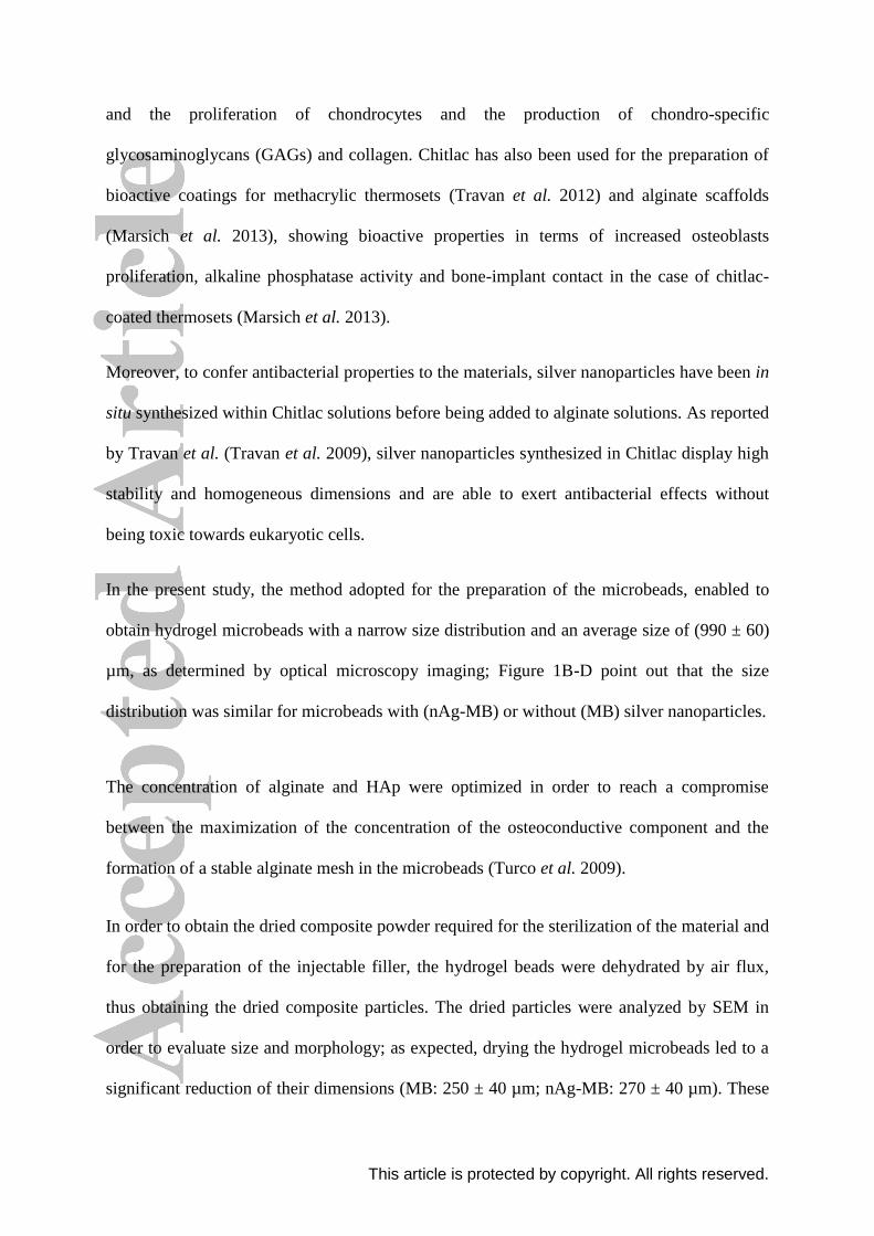
and the proliferation of chondrocytes and the production of chondro-specific
glycosaminoglycans (GAGs) and collagen. Chitlac has also been used for the preparation of
bioactive coatings for methacrylic thermosets (Travan et al. 2012) and alginate scaffolds
(Marsich et al. 2013), showing bioactive properties in terms of increased osteoblasts
proliferation, alkaline phosphatase activity and bone-implant contact in the case of chitlac-
coated thermosets (Marsich et al. 2013).
Moreover, to confer antibacterial properties to the materials, silver nanoparticles have been in
situ synthesized within Chitlac solutions before being added to alginate solutions. As reported
by Travan et al. (Travan et al. 2009), silver nanoparticles synthesized in Chitlac display high
stability and homogeneous dimensions and are able to exert antibacterial effects without
being toxic towards eukaryotic cells.
In the present study, the method adopted for the preparation of the microbeads, enabled to
obtain hydrogel microbeads with a narrow size distribution and an average size of (990 ± 60)
µm, as determined by optical microscopy imaging; Figure 1B-D point out that the size
distribution was similar for microbeads with (nAg-MB) or without (MB) silver nanoparticles.
The concentration of alginate and HAp were optimized in order to reach a compromise
between the maximization of the concentration of the osteoconductive component and the
formation of a stable alginate mesh in the microbeads (Turco et al. 2009).
In order to obtain the dried composite powder required for the sterilization of the material and
for the preparation of the injectable filler, the hydrogel beads were dehydrated by air flux,
thus obtaining the dried composite particles. The dried particles were analyzed by SEM in
order to evaluate size and morphology; as expected, drying the hydrogel microbeads led to a
significant reduction of their dimensions (MB: 250 ± 40 µm; nAg-MB: 270 ± 40 µm). These

This article is protected by copyright. All rights reserved.
values are in the range commonly employed for the preparation of composite bone fillers
based on HAp or β-TCP (Suzuki et al. 2014; Tadier et al. 2014).
The SEM analysis enabled also to highlight how the bead surfaces are roughened by the
presence of HAp crystals that protrude from both MB and nAg-MB particles (Figure 2B and
2D).
3.2 Swelling and stability
The swelling behavior and stability were investigated by incubating the dried microbeads in
Simulated Body Fluid (SBF) at 37 °C and replacing it at fixed days. SBF is widely used for
the evaluation of the stability and of the apatite-forming properties of biomaterials for bone
tissue regeneration (Díez-Pascual and Díez-Vicente 2015; Gervaso et al. 2016; Tan et al.
2010; Turco et al. 2009). In this work, the stability evaluation was performed without the use
of degradation enzymes since it is known that there are not specific enzymes in mammalians
for the degradation of alginate (Guarino et al. 2015; Lee and Mooney 2012). Moreover, it has
been demonstrated that the enzyme lysozyme has only a minor effect on chitosan, and that
this effect becomes even smaller when chitosan is functionalized with lactose (Diolosá et al.
2014).
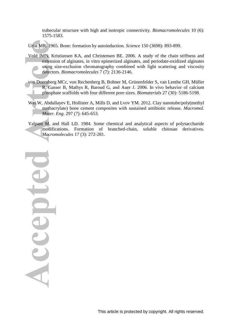
The size variation of the microbeads soaked in SBF over time is reported in Figure 3A: the
data show how the microbeads underwent a considerable swelling leading to an increase of
their dimension, owing to the presence of the hydrophilic polysaccharides. The microbeads
rapidly (approximately 2 days) reached a swelling equilibrium, with an increase of diameter
and of volume which was respectively 3.5-fold and 43-fold compared to the initial values.
The swelling rate of MB and

This article is protected by copyright. All rights reserved.
nAg-MB was similar (Figure 3A) and did not affect the morphology of the microbeads
(Figure 3 B and C).
The experiment went on for 31 days, pointing out the excellent morphological stability of the
microbeads, which did not show any significant degradation in physiological-like conditions.
These data confirm the stability of biomaterials based on alginate hydrogels containing HAp,
as already observed in the case of tridimensional scaffolds for bone tissue regeneration
(Porrelli et al. 2015; Turco et al. 2009).
Considering the final application (the preparation of an injectable system), the swelling and
degradation behavior found represent a positive feature of the material. This feature enables
the injectable filler to adapt to the bone defect and remain firmly in situ for several weeks,
thus assisting the natural bone regrowth process. In fact, the prolonged stability of an
injectable material is a key factor in the regeneration of the bone tissue (Ghanaati et al. 2011;
Grynpas et al. 2002; von Doernberg et al. 2006), as new bone tissue formation requires
several weeks (Urist 1965).
3.3 Silver release from nAg-MB
The amount of silver contained in the nAg-MB has been quantified by means of ICP-OES;
the analysis revealed that 1 mg of microbeads contains (0.978 ± 0.146) μg of silver (data
averaged on three samples).
The silver release from the nAg-MB has been measured soaking the microbeads in deionized
water and in saline solution (NaCl 0.15 M); in order to put the particles in contact with
abundant liquid, the ratio between the volume of water/solution and the volume of
microbeads was 10. To mimic real conditions, the solutions were changed every 24 h and the
microbeads were subjected to mechanical agitation. The silver released from the microbeads

This article is protected by copyright. All rights reserved.
over time was reported both as the percentage of silver released each day (Figure 4A) or as
the cumulative release (Figure 4B).
The data point out that for both water and saline solution the silver released upon daily shifts
is typically lower than 1%, while after 7 days the cumulative silver release was (5.69 ±
0.95)% in saline solution and (0.36 ± 0.12)% in water. The higher release in saline is
explainable considering that the presence of ions can accelerate the swelling of the polymer
mesh, which increases the exposure of silver nanoparticles to the environment. Moreover, it
is known that the dissolution of silver nanoparticles and the release of silver ions are
influenced by ionic strength and the chloride concentration of the solution (Chambers et al.
2014). However, in both cases, the silver release was very low since only 0.56 ng per mg of
bead has been released after 7 days, thus proving the structural stability of the polymer mesh.
Comparing the results of silver release and swelling behavior of the microbeads, one could
expect a burst release of silver in a timeframe compatible with the swelling of the microbeads
(see Figure 3); however, the results did not point out such behavior. The absence of a burst
release can be explained considering that in this system the silver nanoparticles are
chemically stabilized by Chitlac, which in turn is closely embedded in the alginate matrix of
microbeads.
The amount of silver released from nAg-MB is higher if compared to that of alginate/Chitlac-
nAg hydrogels devoid of HAp reported by Travan et al. (Travan et al. 2009); this result can
be explained by the fact that the introduction of HAp can affect the formation of alginate egg-
box, thus reducing the stability of the material. However, the silver released from nAg-MB is
lower than that reported for tridimensional alginate/HAp scaffolds coated with Chitlac-nAg
(Marsich et al. 2013) thus pointing out that silver release can be tuned by employing different
methods of incorporation in the biomaterial.
This article is protected by copyright. All rights reserved.
Overall, the profile of the silver released from the nAg-MB appears particularly appealing for
bone tissue engineering applications, since it ensures a long-term stability of the antibacterial
agent, thus avoiding a burst release of metal ions that could potentially be toxic for the cells
of the surrounding tissues. Moreover, the slow release of silver contributes to prolong the
antibacterial effect of the nanoparticles, which is also related to the direct contact with
bacterial cells (Travan et al. 2009).
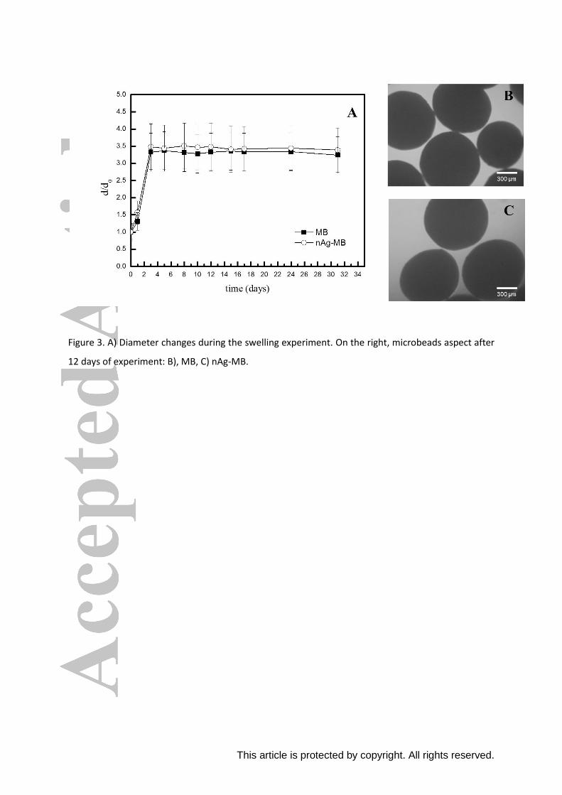
3.4 Antibacterial properties
The antibacterial properties of nAg-MB were assessed in terms of inhibition of bacterial
growth and eradication of biofilms produced by three bacterial strains: S. aureus, P.
aeruginosa and
S. epidermidis. These strains have been selected because of their role in bone-related
infections and their antibiotic-resistance mechanisms (Gottenbos et al. 2000; Kilgus et al.
2002; Moran et al. 2010; Parvizi et al. 2009; Toms et al. 2006). The assays have been
performed incubating the microbeads (nAg-MB or MB) in direct contact with bacteria for
chosen times.
For S. aureus and P. aeruginosa, the growth inhibition assay was performed by incubating
the bacterial suspension with the dried microbeads for 4 hours, after which the colony
forming units (CFU) were measured; in both cases, the nAg-MB induced a significant
decrease of the CFU, whose number was reduced by several orders of magnitude (Figure 5A-
B).
In the case of S. epidermidis, no antibacterial effect was detected after 4 hours of incubation
(data not shown). For this reason, the incubation was prolonged to 24 hours, which revealed
to be a sufficient time for the nAg-MB to exert a bactericidal effect; in fact, a decrease of

This article is protected by copyright. All rights reserved.
more than 4 orders of magnitude was found in the case of the silver-containing particles
(Figure 5C). The observed higher resistance of S. epidermidis was in line with the results
reported in a previous work by Marsich et al. (Marsich et al. 2013).
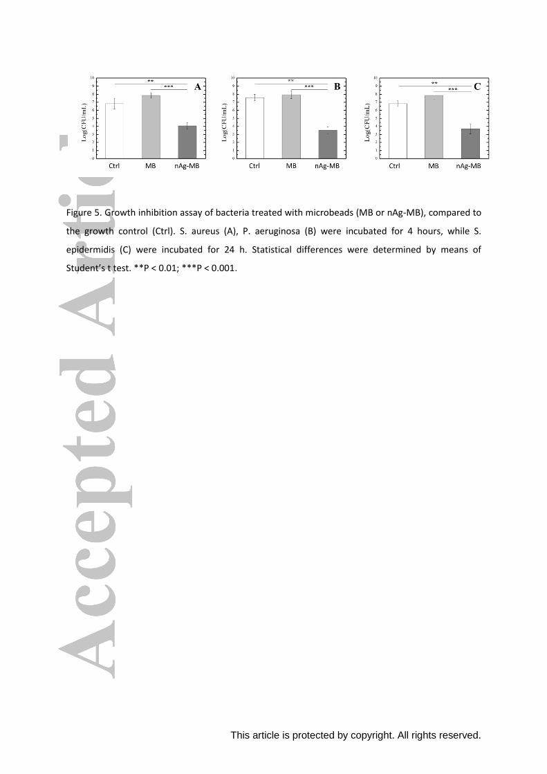
Once the effectiveness of the silver-containing particles in inhibiting bacterial growth had
been verified, a further test has been carried out to evaluate their effect towards pre-formed
biofilms. This assay was performed on S. aureus and P. aeruginosa strains, since S.
epidermidis does not produce a self-protecting biofilm. Bacterial biofilms were put in contact
with the microbeads for four hours and the bacteria viability was quantified using the Green
Biofilm Cell Stain assay, which exploits the fluorescence intensity of the biomass as an
indicator of viable bacteria within the biofilm. The results are reported in Figure 6. In the case
of S. aureus, the nAg-MB displayed a strong anti-biofilm activity, since a 69% decrease of
the fluorescence intensity was measured with respect to the control. In the case of P.
aeruginosa the nAg-MB determined a 26% reduction of the biofilm fluorescence intensity;
this milder effect could be ascribed to the high content of alginate in the P. aeruginosa
biofilm, which represents a physical barrier towards antibacterial agents (Leid et al. 2005).
The viability of bacteria within biofilms was also evaluated by the Live/Dead assay, which,
by means of a fluorescence microscope, enables to distinguish between viable cells (green)
and dead cells (orange-red); figure 7 collects the images of the biofilms after 4 hours of
treatment with the particles, compared to untreated (control) biofilms.

This article is protected by copyright. All rights reserved.
In the case of S. aureus, the images clearly show the abundance of viable bacteria (green) for
untreated (Figure 7A) and MB-treated biofilms (Figure 7B); at variance, the treatment with
nAg-MB causes a clear inactivation of bacterial cells, appearing as red particles (Figure 7C).
In the case of P. aeruginosa, the antibacterial effect of the nAg-MB can be inferred by the
abundance of orange/red biomass (Figure 7G), at variance with control (Figure 7E) and MB-
treated bacteria (Figure 7F). Thus, the Live/Dead results are in line with the results of the
Green Biofilm Cell Stain assay, both pointing out the antibacterial effect of nAg-MB on pre-
formed biofilms.
Overall, the assays performed confirm the antibacterial properties of nAg-MB, in
concordance with the data previously reported for alginate-based hydrogels (Travan et al.
2009) or scaffolds (Marsich et al. 2013) enriched with Chitlac-nAg.
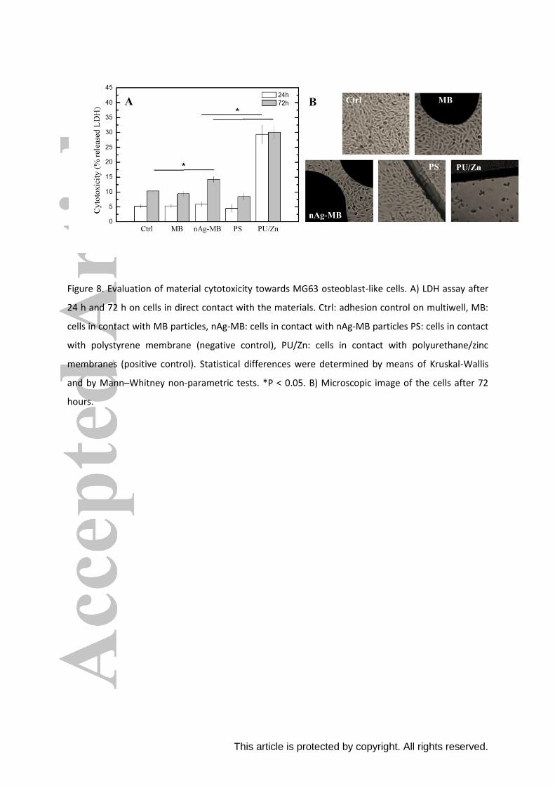
3.5 Cytotoxicity and viability
After studying the antibacterial activity of the microbeads, their effect towards eukaryotic
cells was evaluated by the lactate dehydrogenase (LDH) assay, which enables to quantify the
release of the LDH enzyme due to cellular damage; the assay has been carried out by putting
an osteoblasts cell line (MG63) in direct contact the cells with microbeads for 24 and 72
hours (accordingly to the ISO 10993−5 standard(1999)). Figure 8 shows the results of this
cytotoxicity study as well as a qualitative evaluation of the morphology of the cells in contact
with the materials.
This article is protected by copyright. All rights reserved.
The results of Figure 8A pointed out that both MB and nAg-MB particles were associated
with low values (<15%) of LDH release, which remained significantly lower than the positive
(cytotoxic) control (polyurethane/zinc). These quantitative data were confirmed by the
qualitative investigation of cell morphology (Figure 8B), which highlighted the healthy
conditions of the cells proliferated on the multiwell floor in direct contact with both types of
microbeads.
The data here reported confirm the biocompatibility of silver nanoparticles stabilized within
the Chitlac matrix and the suitability of Chitlac-nAg for the preparation of bioactive
polysaccharide-based biomaterials, in line with previous approaches by some of the authors
(Marsich et al. 2013; Travan et al. 2009; Travan et al. 2012). The slight increase of
cytotoxicity observed at 72 hours can be due to two different factors: i) the accumulation of
silver in the culture medium, which was not changed during the test according to the
standardized protocol; ii) the limited mimicking of the in vivo conditions in a cytotoxicity
assay on 2D-cultured cells. These hypotheses are supported by Stojkovska et al. (Stojkovska
et al. 2014), who reported a higher cytotoxicity of silver nanoparticles on 2D cultures of
chondrocytes with respect to the 3D environment of perfused bioreactors. Moreover,
according to the standards for the determination of the cytotoxicity of materials (accordingly
to the ISO 10993−5 standard(1999)), the values of LDH released by the cells in contact with
nAg-MB are significantly lower than the threshold considered as cytotoxic (30% of LDH
released over the total content).

This article is protected by copyright. All rights reserved.
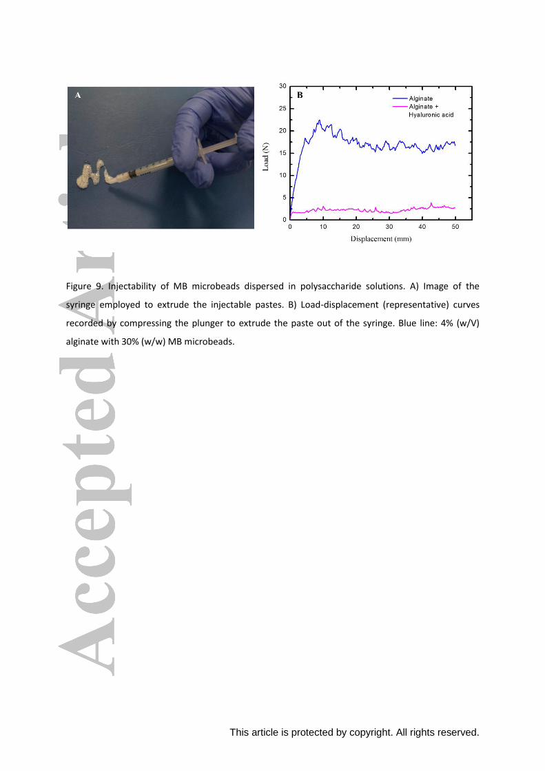
3.6 Injectability studies
As soon as the composite microbeads were characterized and their biological properties
assessed, the material was employed for the preparation of an injectable system based on
dried MB suspended in a suspending liquid medium. The latter was selected through a
preliminary screening of polysaccharide solutions by evaluating the homogeneity and
stability over time of the suspension resulting from the dispersion of the microbeads in the
viscous polysaccharide solution. This screening study enabled to select alginate solution (4%
w/V) with 30% w/w of microbeads as the best performing formulation, since this
composition could be stored within syringes for ten days maintaining the particles
homogeneously distributed within the alginate medium (Figure 9A). The choice of alginate as
a dispersant agent is also supported by literature studies: for example, Alves Cardoso et al.
(Alves et al. 2014) and Oliveira et al. (Oliveira et al. 2008) reported the use of alginate for
the preparation of an injectable, osteoconductive material based on HAp or calcium
phosphate.
In order to assess the injectability of this formulation, the force required to extrude it through
a syringe with a 2 mm nozzle diameter was tested by means of a universal testing machine.
This diameter nozzle is in the typical range for cannulas used for bone cement injections
(Tadier et al. 2014). The results of the mechanical tests are reported in Figure 9B (blue line).
The data showed that it was possible to push the syringe plunger for the whole length of the
syringe
(50 mm) without stacking the particles or blocking the nozzle with an average compression
load of 17 N (± 5 N), thus achieving a 100% extrusion of the paste. These values are in line
with injectable materials developed by other authors (Perut et al. 2011; Sohrabi et al. 2013;

This article is protected by copyright. All rights reserved.
Sohrabi et al. 2014; Tadier et al. 2014) and highlight the capability of this bone filler to be
injected in a surgical procedure.
Another important feature of the formulation here proposed is that during the storage and the
extrusion of the formulation, no phase-separation phenomena of the microbeads from the
liquid phase was observed. Phase separation is a critical issue for the injectable fillers as it
may negatively affect the homogeneity and the injectability of the materials (Tadier et al.
2014).
In order to evaluate the morphology of the particles in the dispersion medium, a SEM
investigation has been performed after withdrawing the microbeads from the alginate solution
(Figure 10): the images showed that the particles displayed a smoother surface than the native
microbeads. This could be ascribed to the adsorption of some alginate from the solution onto
the microbeads surface.
In addition to the results obtained using alginate as dispersing agent, the possibility to add
another polysaccharide to the solution has been explored with the aim to implement the
bioactive properties of the injectable paste. To this end, hyaluronan has been selected as the
additional solution component, given its healing capability and lubricating properties (Dicker
et al. 2014). A high MW (1.5 106) HA sample was chosen (HA 150). A mixed
alginate/HA150 solution was prepared in which the concentration of the two polysaccharides
was 3% w/V and 1% w/V, respectively, thus providing the same polymer mass concentration
as in the HA-free case (4% w/V). Microbeads were added at the same content as in the pure
alginate condition (30% w/w), also in this case obtaining a homogeneous dispersion that was
tested in terms of injectability. The mechanical tests revealed that the presence of HA led to a
considerable decrease of the force required for the extrusion of the injectable filler, with an
average compression load of 2 N (± 1 N) required for the plunger to push the paste out of the

This article is protected by copyright. All rights reserved.
syringe (Figure 9B, magenta line). Moreover, as observed in the formulation with alginate
4% w/V, phase-separation phenomena were not observed during the extrusion.
This finding suggests that the addition of HA to the alginate-microbeads paste is able to
lubricate the particles during the extrusion through the syringe, while implementing the
bioactive properties of the injectable biomaterial.
4 Conclusions
Microbeads based on hydroxyapatite, alginate, and Chitlac-nAg were developed and
characterized for the preparation of injectable bone fillers. The dried microbeads displayed a
rapid swelling in contact with simulated body fluids and maintained their integrity for more
than 30 days. The evaluation of silver leakage from the microbeads showed that the
antibacterial metal is slowly released in saline solution, with less than 6% of silver released
after 1 week. Antibacterial tests in vitro proved that the microbeads displayed bactericidal
effects toward S. aureus, P. aeruginosa and S. epidermidis and that they were also able to
damage pre-formed bacterial biofilms. The microbeads did not exert any cytotoxic effect
towards osteoblast-like cells. Upon suspension of the particles in alginate (or
alginate/hyaluronic acid) solution, a homogenous and time-stable paste was obtained;
mechanical tests enabled to quantify the extrusion forces from surgical syringes, pointing out
the good injectability of the material.
Overall, this novel antibacterial bone-filler appears as a promising material for the treatment
of bone defects, in particular when possible infections could compromise the bone-healing
process. Moreover, the components used for the preparation of the material (Chitlac and
hydroxyapatite) could also provide the filler with osteoconductive properties. A detailed

This article is protected by copyright. All rights reserved.
characterization of the injectable paste, in terms of material properties and in vivo behavior
will be the subject of a forthcoming study.
Acknowledgment.
The scientific support from the inter-university Center for Biomaterials and Regenerative
Medicine, BIOMA-TS, is gratefully acknowledged. This work has been carried out within the
Cluster IRMI, Italian Regenerative Medicine Infrastructure. Mister Mattia Norrito is thanked
for her skillful assistance in the experimental part.
Table of Contents Graphic

This article is protected by copyright. All rights reserved.
References
1999. Biological Evaluation of Medical Devices - Part 5: Tests for In Vitro Cytotoxicity, ISO
10993-5. International Organization for Standardization: Geneva, Switzerland.
Alves Cardoso D, van den Beucken JJJP, Both LLH, Bender J, Jansen JA, and
Leeuwenburgh SCG. 2014. Gelation and biocompatibility of injectable alginate-
calcium phosphate gels for bone regeneration. J. Biomed. Mater. Res., Part A 102 (3):
808-817.
Bongio M, van den Beucken JJJP, Leeuwenburgh SCG, and Jansen JA. 2015. Preclinical
evaluation of injectable bone substitute materials. J. TIssue Eng. Regen. Med. 9 (3):
191-209.
Campoccia D, Montanaro L, and Arciola CR. 2006. The significance of infection related to
orthopedic devices and issues of antibiotic resistance. Biomaterials 27 (11): 2331-
2339.
Campoccia D, Montanaro L, Speziale P, and Arciola CR. 2010. Antibiotic-loaded
biomaterials and the risks for the spread of antibiotic resistance following their
prophylactic and therapeutic clinical use. Biomaterials 31 (25): 6363-6377.
Chambers BA, Afrooz ARMN, Bae S, Aich N, Katz L, Saleh NB, and Kirisits MJ. 2014.
Effects of chloride and ionic strength on physical morphology, dissolution, and
bacterial toxicity of silver nanoparticles. Environ. Sci. Technol. 48 (1): 761-769.
Dicker KT, Gurski LA, Pradhan-Bhatt S, Witt RL, Farach-Carson MC, and Jia X. 2014.
Hyaluronan: a simple polysaccharide with diverse biological functions. Acta
Biomater. 10 (4): 1558-1570.
Díez-Pascual AM, and Díez-Vicente AL. 2015. Wound healing bionanocomposites based on
castor oil polymeric films reinforced with chitosan-modified ZnO nanoparticles.
Biomacromolecules 16 (9): 2631-2644.
Diolosá M, Donati I, Turco G, Cadenaro M, Di Lenarda R, Breschi L, and Paoletti S. 2014.
Use of methacrylate-modified chitosan to increase the durability of dentine bonding
systems. Biomacromolecules 15 (12): 4606-4613.
Donati I, Stredanska S, Silvestrini G, Vetere A, Marcon P, Marsich E, Mozetic P, Gamini A,
Paoletti S, and Vittur F. 2005. The aggregation of pig articular chondrocyte and
synthesis of extracellular matrix by a lactose-modified chitosan. Biomaterials 26:
987-998.
Gervaso F, Padmanabhan SK, Scalera F, Sannino A, and Licciulli A. 2016. Mechanical
stability of highly porous hydroxyapatite scaffolds during different stages of in vitro
studies. Mater. Lett. 185: 239-242.
Ghanaati S, Barbeck M, Hilbig U, Hoffmann C, Unger RE, Sader RA, Peters F, and
Kirkpatrick CJ. 2011. An injectable bone substitute composed of beta-tricalcium
This article is protected by copyright. All rights reserved.
phosphate granules, methylcellulose and hyaluronic acid inhibits connective tissue
influx into its implantation bed in vivo. Acta Biomater. 7 (11): 4018-4028.
Gottenbos B, van der Mei HC, and Busscher HJ. 2000. Initial adhesion and surface growth of
Staphylococcus epidermidis and Pseudomonas aeruginosa on biomedical polymers. J.
Biomed. Mater. Res. 50 (2): 208-214.
Goudouri OM, Kontonasaki E, Lohbauer U, and Boccaccini AR. 2014. Antibacterial
properties of metal and metalloid ions in chronic periodontitis and peri-implantitis
therapy. Acta Biomater. 10 (8): 3795-3810.
Gristina AG. 1987. Biomaterial-centered infection: microbial adhesion versus tissue
integration. Science 237 (4822): 1588-1595.
Grynpas MD, Pilliar RM, Kandel RA, Renlund R, Filiaggi M, and Dumitriu M. 2002. Porous
calcium polyphosphate scaffolds for bone substitute applications in vivo studies.
Biomaterials 23 (9): 2063-2070.
Guarino V, Caputo T, Altobelli R, and Ambrosio L. 2015. Degradation properties and
metabolic acrivity of alginate and chitosan polyelectrolytes for drug delivery and
tissue engineering applications. AIMS Mater. Sci. 2 (4): 497-502.
Kilgus DJ, Howe DJ and Strang A. 2002. Results of periprosthetic hip and knee infections
caused by resistant bacteria. Clin. Orthop. Relat. Res. 404: 116-124.
Kneser U, Schaefer DJ, Polykandriotis E, and Horch RE. 2006. Tissue engineering of bone:
the reconstructive surgeon's point of view. J. Cell. Mol. Med. 10 (1): 7-19.
Kokubo T, Kushitani H, Sakka S, Kitsugi T, and Yamamuro T. 1990. Solutions able to
reproduce in vivo surface-structure changes in bioactive glass-ceramic A-W3. J.
Biomed. Mater. Res., Part A 24: 721-734.
Lara H, Garza-Trevino E, Ixtepan-Turrent L, and Singh D. 2011. Silver nanoparticles are
broad-spectrum bactericidal and virucidal compounds. J. Nanobiotechnol. 9 (1): 30.
Lee KY, and Mooney DJ. 2012. Alginate: properties and biomedical applications. Progr.
Polym. Sci. 37 (1): 106-126.
Leid JG, Willson CJ, Shirtliff ME, Hassett DJ, Parsek MR, and Jeffers AK. 2005. The
exopolysaccharide alginate protects Pseudomonas aeruginosa biofilm bacreria from
IFN-gamma-mediated macrophage killing. J. Immunol. 175 (11): 7512-7518.
Lewis G. 2011. Viscoelastic properties of injectable bone cements for orthopaedic
applications: State-of-the-art review. J. Biomed. Mater. Res., Part B 98B (1): 171-
191.
Marsich E, Borgogna M, Donati I, Mozetic P, Strand BL, Salvador SG, Vittur F, and Paoletti
S. 2008. Alginate/lactose-modified chitosan hydrogels: a bioactive biomaterial for
chondrocyte encapsulation. J. Biomed. Mater. Res., Part A 84A (2): 364-376.
Marsich E, Bellomo F, Turco G, Travan A, Donati I, and Paoletti S. 2013. Nano-composite
scaffolds for bone tissue engineering containing silver nanoparticles: preparation,

This article is protected by copyright. All rights reserved.
characterization and biological properties. J. Mater. Sci., Mater. Med. 24 (7): 1799-
1807.
Marsich E, Travan A, Donati I, Turco G, Kulkova J, Moritz N, Aro HT, Crosera M, and
Paoletti S. 2013. Biological responses of silver-coated thermosets: an in vitro and in
vivo study. Acta Biomater. 9 (2): 5088-5099.
Marsich E, Travan A, Feresini M, Lapasin R, Paoletti S, and Donati I. 2013. Polysaccharide-
based polyanion-polycation-polyanion ternary systems in the concentrated regime and
hydrogel form. Macromol. Chem. Phys. 214 (12): 1309-1320.
Mauffrey C, Barlow BT, and Smith W. 2015. Management of Segmental Bone Defects.
JAOSS 23 (3): 143-153.
Moran E, Byren I, and Atkins BL. 2010. The diagnosis and management of prosthetic joint
infections. J. Antimicrob. Chemother. 65 (suppl 3): iii45-iii54.
Morch YA, Donati I, Strand BL, and Skjak-Braek G. 2006. Effect of Ca2+
, Ba2+
, and Sr2+
on
alginate microbeads. Biomacromolecules 7 (5): 1471-1480.
Morones-Ramirez JR, Winkler JA, Spina CS, and Collins JJ. 2013. Silver enhances antibiotic
activity against gram-negative bacteria. Sci. Transl. Med. 5 (190).
Nejadnik MR, Yang X, Bongio M, Alghamdi HS, van den Beucken JJJP, Huysmans MC,
Jansen JA, Hilborn J, Ossipov D, and Leeuwenburgh SCG. 2014. Self-healing hybrid
nanocomposites consisting of bisphosphonated hyaluronan and calcium phosphate
nanoparticles. Biomaterials 35 (25): 6918-6929.
Nganga S, Travan A, Marsich E, Donati I, Söderling E, Moritz N, Paoletti S, and Vallittu P.
2013. In vitro antimicrobial properties of silver-polysaccharide coatings on porous
fiber-reinforced composites for bone implants. J. Mater. Sci., Mater. Med. 24 (12):
2775-2785.
Norowski PA, and Bumgardner JD. 2009. Biomaterial and antibiotic strategies for peri-
implantitis: a review. J. Biomed. Mater. Res., Part B 88B (2): 530-543.
Oliveira SM, Barrias CC, Almeida IF, Costa PC, Ferreira MR, Bahia MF, and Barbosa MA.
2008. Injectability of a bone filler system based on hydroxyapatite microspheres and a
vehicle with in situ gel-forming ability. J. Biomed. Mater. Res., Part B 87 (1): 49-58.
Page JM, Harmata AJ, and Guelcher SA. 2013. Design and development of reactive
injectable and settable polymeric biomaterials. J. Biomed. Mater. Res., Part A 101
(12): 3630-3645.
Parvizi J, Azzam K, Ghanem E, Austin MS, and Rothman RH. 2009. Periprosthetic Infection
Due to Resistant Staphylococci: Serious Problems on the Horizon. Clin. Orthop. Rel.
Res. 467 (7): 1732-1739.
Perut F, Montufar EB, Ciapetti G, Santin M, Salvage J, Traykova T, Planell JA, Ginebra MP,
and Baldini N. 2011. Novel soybean/gelatine-based bioactive and injectable
hydroxyapatite foam: Material properties and cell response. Acta Biomater. 7 (4):
1780-1787.

This article is protected by copyright. All rights reserved.
Porrelli D, Travan A, Turco G, Marsich E, Borgogna M, Paoletti S, and Donati I. 2015.
Alginate-hydroxyapatite bone scaffolds with isotropic or anisotropic pore structure:
material properties and biological behavior. Macromol. Mater. Eng 300 (10): 989-
1000.
Reithofer MR, Lakshmanan A, Ping ATK, Chin JM, and Hauser CAE. 2014. In situ synthesis
of size-controlled, stable silver nanoparticles within ultrashort peptide hydrogels and
their anti-bacterial properties. Biomaterials 35 (26): 7535-7542.
Sohrabi M, Hesaraki S, and Kazemzadeh A. 2014. Injectable bioactive glass/polysaccharide
polymers nanocomposites for bone sostitution. Key Eng. Mater. 614: 41-46.
Sohrabi M, Hesaraki S, Kazemzadeh A, and Alizadeh M. 2013. Development of injectable
biocomposites from hyaluronic acid and bioactive glass nano-particles obtained from
different sol-gel routes. Mater. Sci. Eng., C 33 (7): 3730-3744.
Stojkovska J, Kostic D, Jovanovic Z, Vukasinovic Sekulic M, Miskovic Stankovic V, and
Obradovic B. 2014. A comprehensive approach to in vitro functional evaluation of
Ag/alginate nanocomposite hydrogels. Carbohydr. Polym. 111: 305-314.
Suzuki K, Anada T, Miyazaki T, Miyatake N, Honda Y, Kishimoto KN, Hosaka M,
Imaizumi H, Itoi E, and Suzuki O. 2014. Effect of addition of hyaluronic acids on the
osteoconductivity and biodegradability of synthetic octacalcium phosphate. Acta
Biomater. 10 (1): 531-543.
Tadier S, Galea L, Charbonnier B, Baroud G, and Bohner M. 2014. Phase and size
separations occurring during the injection of model pastes composed of b-tricalcium
phosphate powder, glass beads and aqueous solutions. Acta Biomater. 10 (5): 2259-
2268.
Taglietti A, Arciola CR, D'Agostino A, Dacarro G, Montanaro L, Campoccia D, Cucca L,
Vercellino M, Poggi A, Pallavicini P, and Visai L. 2014. Antibiofilm activity of a
monolayer of silver nanoparticles anchored to an amino-silanized glass surface.
Biomaterials 35 (6): 1779-1788.
Tan R, Feng Q, She Z, Wang M, Jin H, Li J, and Yu X. 2010. In vitro and in vivo degradation
of an injectable bone repair composite. Polym. Degrad. Stab. 95 (9): 1736-1742.
Toms AD, Davidson D, Masri BA, and Duncan CP. 2006. The management of peri-prosthetic
infection in total joint arthroplasty. J. Bone Joint Surg., Br. Vol. 88-B (2): 149-155.
Travan A, Marsich E, Donati I, Foulc MP, Moritz N, Aro HT, and Paoletti S. 2012.
Polysaccharide-coated thermosets for orthopedic applications: from material
characterization to in vivo tests. Biomacromolecules 13 (5): 1564-1572.
Travan A, Pelillo C, Donati I, Marsich E, Benincasa M, Scarpa T, Semeraro S, Turco G,
Gennaro R, and Paoletti S. 2009. Non-cytotoxic silver nanoparticle-polysaccharide
nanocomposites with antimicrobial activity. Biomacromolecules 10 (6): 1429-1435.
Turco G, Marsich E, Bellomo F, Semeraro S, Donati I, Brun F, Grandolfo M, Accardo A, and
Paoletti S. 2009. Alginate/hydroxyapatite biocomposite for bone ingrowth: a
This article is protected by copyright. All rights reserved.
trabecular structure with high and isotropic connectivity. Biomacromolecules 10 (6):
1575-1583.
Urist MR. 1965. Bone: formation by autoinduction. Science 150 (3698): 893-899.
Vold IMN, Kristiansen KA, and Christensen BE. 2006. A study of the chain stiffness and
extension of alginates, in vitro epimerized alginates, and periodate-oxidized alginates
using size-exclusion chromatography combined with light scattering and viscosity
detectors. Biomacromolecules 7 (7): 2136-2146.
von Doernberg MCc, von Rechenberg B, Bohner M, Grünenfelder S, van Lenthe GH, Müller
R, Gasser B, Mathys R, Baroud G, and Auer J. 2006. In vivo behavior of calcium
phosphate scaffolds with four different pore sizes. Biomaterials 27 (30): 5186-5198.
Wei W, Abdullayev E, Hollister A, Mills D, and Lvov YM. 2012. Clay nanotube/poly(methyl
methacrylate) bone cement composites with sustained antibiotic release. Macromol.
Mater. Eng. 297 (7): 645-653.
Yalpani M, and Hall LD. 1984. Some chemical and analytical aspects of polysaccharide
modifications. Formation of branched-chain, soluble chitosan derivatives.
Macromolecules 17 (3): 272-281.

This article is protected by copyright. All rights reserved.
Figure 1. A) Schematic representation of the preparation of the microbeads: the
alginate/HAp/Chitlac (with or without nAg) solution is dropped in a CaCl2 solution under the
application of a voltage. B) Dimension distribution of MB and nAg-MB. On the right, microbeads
aspect: C), MB, D) nAg-MB.

This article is protected by copyright. All rights reserved.
Figure 2. SEM micrographs of MB (A, B) and nAg-MB (C, D). E) Distribution of microbeads dimension:
MB and nAg-MB.
This article is protected by copyright. All rights reserved.
Figure 3. A) Diameter changes during the swelling experiment. On the right, microbeads aspect after
12 days of experiment: B), MB, C) nAg-MB.

This article is protected by copyright. All rights reserved.
Figure 4. Silver released by the microbeads (nAg-MB) soaked in deionized water or in saline solution
expressed as percentage of the total silver contained: (A) release at given solution shift; (B)
cumulative release. Data were averaged on three independent experiments.
This article is protected by copyright. All rights reserved.
Figure 5. Growth inhibition assay of bacteria treated with microbeads (MB or nAg-MB), compared to
the growth control (Ctrl). S. aureus (A), P. aeruginosa (B) were incubated for 4 hours, while S.
epidermidis (C) were incubated for 24 h. Statistical differences were determined by means of
Student’s t test. **P < 0.01; ***P < 0.001.
This article is protected by copyright. All rights reserved.
Figure 6. Effect of microbeads on biofilms of S. aureus (A) and P. aeruginosa (B) after 4 hours of
contact with the materials (Green Biofilm Cell Stain assay). Ctrl: untreated biofilm; MB: biofilm
treated with MB particles; nAg-MB: biofilm treated with nAg-MB particles. Statistical significance
was evaluated by Kruskal-Wallis and by Mann–Whitney non-parametric tests. *P < 0.05; **P < 0.01;
***P < 0.001.

This article is protected by copyright. All rights reserved.
Figure 7. Effect of microbeads on biofilms of S. aureus (A-C) and P. aeruginosa (E-G) after 4 hours of
contact with the materials (Live/Dead assay). Control: untreated biofilm; MB: biofilm treated with
MB particles; nAg-MB: biofilm treated with nAg-MB particles. For all images, green fluorescence
(SYTO® 9) indicates live cells whereas red fluorescence (propidium iodide) refers to dead ones.
This article is protected by copyright. All rights reserved.
Figure 8. Evaluation of material cytotoxicity towards MG63 osteoblast-like cells. A) LDH assay after
24 h and 72 h on cells in direct contact with the materials. Ctrl: adhesion control on multiwell, MB:
cells in contact with MB particles, nAg-MB: cells in contact with nAg-MB particles PS: cells in contact
with polystyrene membrane (negative control), PU/Zn: cells in contact with polyurethane/zinc
membranes (positive control). Statistical differences were determined by means of Kruskal-Wallis
and by Mann–Whitney non-parametric tests. *P < 0.05. B) Microscopic image of the cells after 72
hours.
This article is protected by copyright. All rights reserved.
Figure 9. Injectability of MB microbeads dispersed in polysaccharide solutions. A) Image of the
syringe employed to extrude the injectable pastes. B) Load-displacement (representative) curves
recorded by compressing the plunger to extrude the paste out of the syringe. Blue line: 4% (w/V)
alginate with 30% (w/w) MB microbeads.

This article is protected by copyright. All rights reserved.
Figure 10. SEM micrographs of alginate-coated MB microbeads (A, B) and of native MB microbeads
(C, D).