ANTIBACTERIAL ACTIVITY - Shodhgangashodhganga.inflibnet.ac.in/bitstream/10603/2520/11/11_chapter...
43
Chapter - IV: Antimicrobial Activity ANTIBACTERIAL ACTIVITY INTRODUCTION The science dealing with the study of the prevention and treatment of diseases caused by micro-organisms is known as medical microbiology. Its sub- disciplines are virology (study of viruses), bacteriology (study of bacteria), mycology (study of fungi), phycology (study of algae) and protozoology (study of protozoa). For the treatment of diseases inhibitory chemicals employed to kill micro-organisms or prevent their growth, are called antimicrobial agents. These are classified according to their application and spectrum of activity, as germicides that kill micro-organisms, whereas micro-biostatic agents inhibit the growth of pathogens and enable the leucocytes and other defense mechanism of the host to cope up with static invaders. The germicides may exhibit selective toxicity depending on their spectrum of activity. They may act as viricides (killing viruses), bacteriocides (killing bacteria), algicides (killing algae) or fungicides (killing fungi). The beginning of modern chemotherapy has largely been due to the efforts of Dr. Paul Ehrlich (1910), who used salvarsan, as arsenic derivative effective against syphilis. Paul Ehrlich used the term chemotherapy for curing the infectious disease without injury to the host’s tissue, known as chemotherapeutic agents such as antibacterial, antiprotosoal, antiviral, antineoplastic, antitubercular and antifungal agents. Later on, Domagk (1953) prepared an important chemotherapeutic agent sulfanilamide. CLASSIFICATION OF ANTIBACTERIAL AGENTS The antibacterial agents are classified in three categories: 197
Transcript of ANTIBACTERIAL ACTIVITY - Shodhgangashodhganga.inflibnet.ac.in/bitstream/10603/2520/11/11_chapter...
Chapter - IV: Antimicrobial Activity
ANTIBACTERIAL ACTIVITY
INTRODUCTION
The science dealing with the study of the prevention and treatment of
diseases caused by micro-organisms is known as medical microbiology. Its sub-
disciplines are virology (study of viruses), bacteriology (study of bacteria),
mycology (study of fungi), phycology (study of algae) and protozoology (study of
protozoa). For the treatment of diseases inhibitory chemicals employed to kill
micro-organisms or prevent their growth, are called antimicrobial agents. These
are classified according to their application and spectrum of activity, as germicides
that kill micro-organisms, whereas micro-biostatic agents inhibit the growth of
pathogens and enable the leucocytes and other defense mechanism of the host to
cope up with static invaders. The germicides may exhibit selective toxicity
depending on their spectrum of activity. They may act as viricides (killing viruses),
bacteriocides (killing bacteria), algicides (killing algae) or fungicides (killing fungi).
The beginning of modern chemotherapy has largely been due to the efforts
of Dr. Paul Ehrlich (1910), who used salvarsan, as arsenic derivative effective
against syphilis. Paul Ehrlich used the term chemotherapy for curing the infectious
disease without injury to the host’s tissue, known as chemotherapeutic agents
such as antibacterial, antiprotosoal, antiviral, antineoplastic, antitubercular and
antifungal agents. Later on, Domagk (1953) prepared an important
chemotherapeutic agent sulfanilamide.
CLASSIFICATION OF ANTIBACTERIAL AGENTS
The antibacterial agents are classified in three categories:
197
Chapter - IV: Antimicrobial Activity
(I) Antibiotics and chemically synthesized
chemotherapeutic agents.
(II) Non-antibiotic chemotherapeutic agents
(Disinfectants, antiseptics and preservatives)
(III) Immunological products.
(I) Antibiotics
They are produced by micro-organisms or they might be fully or partly
prepared by chemical synthesis. They inhibit the growth of micro-organisms in
minimal concentrations. Antibiotics may be of microbial origin or purely synthetic
or semisynthetic.1 They can be classified by manner of biosynthesis or chemical
structure. Structurally, they are classified into different classes as shown in the
following table.
CLASSIFICATION OF ANTIBIOTICS ACCORDING TO THEIR CHEMICAL
STRUCTURE (Berdy, 1974)2
No. Name of the group Example
1. Carbohydrate-containing antibiotics
Pure sugars
Aminoglycosides
Orthosymycins
N-Glycosides
C-Glycosides
Glycolipids
Nojirimycin
Streptomycin
Everninomicin
Streptothricin
Vancomycin
Moenomycin
2. Macrocyclic lactones
Macrolide antibiotics
Polyene antibiotics
Erythromycin
Candicidin
198
Chapter - IV: Antimicrobial Activity
Ausamycins
Macrotetrolides
Rifamycin
Tetranactin
3. Quinones and related antibiotics
Tetracyclines
Anthracyclines
Naphthoquinones
Benzoquinones
Tetracycline
Adriamycin
Actinorhodin
Mitomycin
4. Amino acid and peptide antibiotics
Amino acid derivatives
β-Lactum antibiotics
Peptide antibiotics
Chromopeptides
Depsipeptides
Chelate forming peptides
Cycloserine
Penicillin
Bacteriacin
Actinomycins
Valinomycin
Bleomycins
5. Heterocyclic antibiotics containing oxygen
Polyether antibiotics Monensin
6. Heterocyclic antibiotics containing nitrogen
Nucleoside antibiotics Polyoxins
7. Aromatic antibiotics
Cycloalkane derivatives
Steriod antibiotics
Cycloheximide
Fusidic acid
8. Aromatic antibiotics
Benzene derivatives
Condensed aromatic antibiotics
Aromatic ether
Chloramphenicol
Griseofulvin
Novobiocin
9. Aliphatic antibiotics
Compounds containing phosphorous Fosfomycins
199
Chapter - IV: Antimicrobial Activity
Synthetic antimicrobial agents include sulfonamides, diamino pyrimidine
derivatives, antitubercular compounds, nitrofuran compounds, 4-quinoline
antibacterials, imidazole derivatives, flucytosine etc.
(II) Non-antibiotics
The second category of antibacterial agents includes non-antibiotic
chemotherapeutic agents which are as follows:
1) Acids and their derivatives
Some organic acids such as sorbic, benzoic, lactic and propionic acids are
used for preserving food and pharmaceuticals. Salicyclic acid has strong antiseptic
and germicidal properties as it is a carboxylated phenol. The presence of –COOH
group appears to enhance the antiseptic property and to decrease the destructive
effect. Benzoic acid is used externally as an antiseptic and is employed in lotion
and ointment. Benzoic acid and salicylic acid are used to control fungi that cause
disease such as athlete’s foot. Benzoic acid and sodium benzoate are used as
antifungal preservatives. Mandolic acid possesses good bacteriostatic and
bactericidal properties.
2) Alcohols and related compounds
They are bactericidal and fungicidal, but are not effective against
endospores and some viruses. Various alcohols and their derivatives have been
used as antiseptics e.g. ethanol and propanol. The antibacterial value of straight
chain alcohols increases with an increase in the molecular weight and beyond C8-
the activity begins to fall off. The isomeric alcohol shows a drop in activity from
primary, secondary to tertiary. Ethanol has extremely numerous uses in
pharmacy.
200
Chapter - IV: Antimicrobial Activity
3) Chlorination and compound containing chlorine
Chlorination is extensively used to disinfect drinking water, swimming pools
and for the treatment of effluent from industries. Robert Koch in 1981 first
referred to the bactericidal properties of hypochlorites. N-chloro compounds are
represented by amides, imides and amidines wherein one or more hydrogen
atoms are replaced by chlorine.
4) Iodine containing compounds
Iodine containing compounds are widely used as antiseptic, fungicide and
amoebicide. Iodophores are used as disinfectants and antiseptics. The soaps used
for surgical scrubs often contain iodophores.
5) Heavy metals
Heavy metals such as silver, copper, mercury and zinc have antimicrobial
properties and are used in disinfectant and antiseptic formulations.
Mercurochrome and merthiolate are applied to skin after minor wounds. Zinc is
used in antifungal antiseptics. Copper sulfate is used as algicides.
6) Oxidising agents
Their value as antiseptics depends on the liberation of oxygen and all are
organic compounds.
7) Dyes
Organic dyes have been extensively used as antibacterial agents. Their
medical significance was first recognized by Churchman3 in 1912. He reported
inhibitory effect of Crystal violet on Gram-positive organism. The acridines exert
bactericidal and bacteriostatic action against both Gram-positive and Gram-
negative organisms.
8) 8-Hydroxyquinolines
201
Chapter - IV: Antimicrobial Activity
8-Hydroxyquinoline or oxine is unique among the isomeric hydroxyl-
quinolines, for it alone exhibits antimicrobial activity. This attributes to its ability
to chelate metals4, which the other isomers do not exhibit.
9) Surface active agents
Soaps and detergents are used to remove microbes mechanically from the
skin surface. Anionic detergents remove microbes mechanically; cationic
detergents have antimicrobial activities and can be used as disinfectants and
antiseptics.
(III) Immunological products
Certain immunological products such as vaccines and monoclonal
antibodies are used to control the diseases as a prophylactic measure.
MODE OF ACTION
Antimicrobial drugs interfere chemically with the synthesis of function of
vital components of micro organisms. The cellular structure and functions of
eukaryotic cells of the human body. These differences provide us with selective
toxicity of chemotherapeutic agents against bacteria.
Antimicrobial drugs may either kill microorganisms outright or simply
prevent their growth. There are various ways in which these agents exhibit their
antimicrobial activity.5 They may inhibit
(1) Cell-wall synthesis
(2) Protein synthesis
(3) Nucleic acid synthesis
(4) Enzymatic activity
(5) Folate metabolism or
(6) Damage cytoplasmic membrane
202
Chapter - IV: Antimicrobial Activity
Bacteriostatic dyes
Stearn and Stearn6 attributed the bacteriostatic activity to
triphenylmethane dyes. Fischer and Munzo7 have found the relationship between
their structure and effectiveness of such dyes.
A number of drugs are metal-binding agents. The chelates are the active
form of drugs. The site of action within the cell or on the cell surface has not
been established. The site of action of oxine and its analogs has been suggested
inside the bacterial cell8 or on cell surface.9
Detoxification of antibacterials
P-Aminobenzoic acid is a growth factor for certain micro-organisms and
competitively inhibits the bacteriostatic action of sulfonamides. The metabolites
identified in man are p-amino-benzoylglucoronide; p-aminohippuric acid,
p-acetylaminobenzoic acid. 8-Hydroxyquinoline (oxine) and 4-hydroxyquinoline
are excerted as sulfate esters or glucorinides.
Bacteria
The bacteria are microscopic organisms with relatively simple and primitive
forms of prokaryotic type. Danish Physician Christian Grams, discovered the
differential staining technique known as Gram staining, which differentiates the
bacteria into two groups “Gram positive” and “Gram negative”, Gram positive
bacteria retain the crystal violet and resist decolorization with acetone or alcohol
and hence appear deep violet in colour; while Gram negative bacteria, which
loose the crystal violet, are counter-stained by saffranin and hence appear red in
colour.
203
Chapter - IV: Antimicrobial Activity
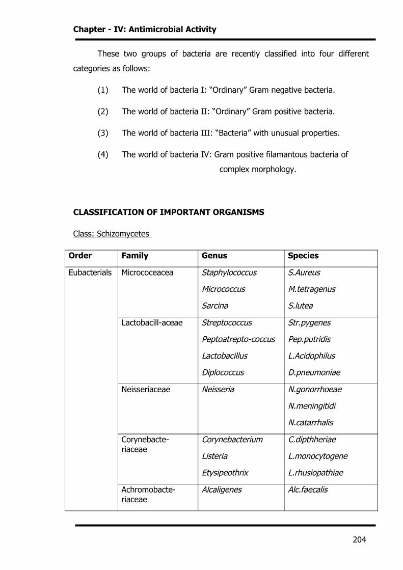
These two groups of bacteria are recently classified into four different
categories as follows:
(1) The world of bacteria I: “Ordinary” Gram negative bacteria.
(2) The world of bacteria II: “Ordinary” Gram positive bacteria.
(3) The world of bacteria III: “Bacteria” with unusual properties.
(4) The world of bacteria IV: Gram positive filamantous bacteria of
complex morphology.
CLASSIFICATION OF IMPORTANT ORGANISMS
Class: Schizomycetes
Order Family Genus Species
Eubacterials Micrococeacea Staphylococcus
Micrococcus
Sarcina
S.Aureus
M.tetragenus
S.lutea
Lactobacill-aceae Streptococcus
Peptoatrepto-coccus
Lactobacillus
Diplococcus
Str.pygenes
Pep.putridis
L.Acidophilus
D.pneumoniae
Neisseriaceae Neisseria N.gonorrhoeae
N.meningitidi
N.catarrhalis
Corynebacte-riaceae
Corynebacterium
Listeria
Etysipeothrix
C.dipthheriae
L.monocytogene
L.rhusiopathiae
Achromobacte-riaceae
Alcaligenes Alc.faecalis
204
Chapter - IV: Antimicrobial Activity
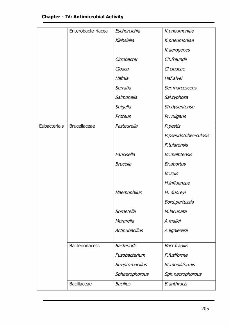
Enterobacte-riacea Eschercichia
Klebsiella
Citrobacter
Cloaca
Hafnia
Serratia
Salmonella
Shigella
Proteus
K.pneumoniae
K.pneumoniae
K.aerogenes
Cit.freundii
Cl.cloacae
Haf.alvei
Ser.marcescens
Sal.typhosa
Sh.dysenterise
Pr.vulgaris
Eubacterials Brucellaceae Pasteurella
Fancisella
Brucella
Haemophilus
Bordetella
Morarella
Actinubacillus
P.pestis
P.pseudotuber-culosis
F.tularensis
Br.meltitensis
Br.abortus
Br.suis
H.influenzae
H. duoreyi
Bord.pertussia
M.lacunata
A.mallei
A.lignieresii
Bacteriodacess Bacteriods
Fusobacterium
Strepto-bacillus
Sphaerophorous
Bact.fragilis
F.fusiforme
St.moniliformis
Sph.nacrophorous
Bacillaceae Bacillus B.anthracis
205
Chapter - IV: Antimicrobial Activity
Clostridium
B.subtilis
Cl.tetani
Cl.welchii
Order Family Genus Species
Pseudo-
monadales
Pseudo-
monadaceae
Pseudomonas Ps.aeruginosa
Spirillaceae Vibrio
Spirillum
V.cholerae
Sp.minus
Mycoplasm-
atales
Mycoplasma-taceae Mycoplasma M.pneumoniae
M.mycoids
Actinomyce-
tates
Mycobacteri-aceae Mycobacterium M.tuberculosis
M.laprae
Actinomycet-aceae Actinomyces
Nocardia
A.israeli
A.bovis
N.modurae
Streptomycet
aceae
Streptomyces Strepto.griseus
Spirochaetel
-es
Spirochaet-aceae Spirochaeta
Saprospira
Nonpathogenic
Treponenat-
aceae
Borrelia Bor.duttoni
Bor.recurrentis
Bor.vincenti
206
Chapter - IV: Antimicrobial Activity
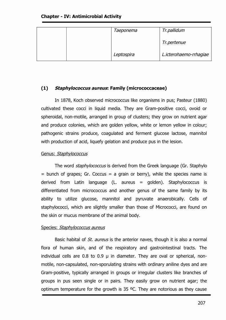
Taeponema
Leptospira
Tr.pallidum
Tr.pertenue
L.icterohaemo-rrhagiae
(1) Staphylococcus aureus: Family (micrococcaceae)
In 1878, Koch observed micrococcus like organisms in pus; Pasteur (1880)
cultivated these cocci in liquid media. They are Gram-positive cocci, ovoid or
spheroidal, non-motile, arranged in group of clusters; they grow on nutrient agar
and produce colonies, which are golden yellow, white or lemon yellow in colour;
pathogenic strains produce, coagulated and ferment glucose lactose, mannitol
with production of acid, liquefy gelation and produce pus in the lesion.
Genus: Staphylococcus
The word staphylococcus is derived from the Greek language (Gr. Staphylo
= bunch of grapes; Gr. Coccus = a grain or berry), while the species name is
derived from Latin language (L. aureus = golden). Staphylococcus is
differentiated from micrococcus and another genus of the same family by its
ability to utilize glucose, mannitol and pyruvate anaerobically. Cells of
staphylococci, which are slightly smaller than those of Micrococci, are found on
the skin or mucus membrane of the animal body.
Species: Staphylococcus aureus
Basic habital of St. aureus is the anterior naves, though it is also a normal
flora of human skin, and of the respiratory and gastrointestinal tracts. The
individual cells are 0.8 to 0.9 μ in diameter. They are oval or spherical, non-
motile, non-capsulated, non-sporulating strains with ordinary aniline dyes and are
Gram-positive, typically arranged in groups or irregular clusters like branches of
groups in pus seen single or in pairs. They easily grow on nutrient agar; the
optimum temperature for the growth is 35 ºC. They are notorious as they cause
207
Chapter - IV: Antimicrobial Activity
suppurative (pyogenic or pus forming) conditions, mostitis of women and cows,
boils and food poisioning. St. aureus grows rapidly and produce circular (1-2 mm)
endive edge, convex, soft, glistening colonies having a golden yellow pigment. St.
aureus can tolerate moderately high concentration of NaCl, hence they can be
selectively isolated on the nutrient medium containing 7.5 % sodium chloride. It is
also able to ferment mannitol to organic acid. St. aureus also produce the
coagulase which is able to clot citrated plasma. It also produces the enzymes
catalase, hyaluronidase as well as other virulent factors like hemolysins,
leucocidins, enterotoxins and exofoliatin.
(2) STREPTOCOCCUS PYOGENES
Genus: Streptococcus
The term Streptococcus was first introduced by Bilroth [1874] and the term
Streptococcus Pyogenes was used by Rosenbach [1884]. These are spherical or
ovoid cells; divide in one axis and form chains; nonmotile and nonsporing. The
growth is absence of native proteins in the medium; they produce characteristic
haemolytic changes in media containing blood; produce acid only by fermentation
of carbohydrates; often fail t liquefy gelatin; some strains produce exotoxin and
extracellular products; a few of them are Anaerobic.
Species: Streptococcus Pyogenes
Streptococcus Pyogenes is pathogenic to human and found in sore throat,
follicular tonsillitis, septicemia, acute or malignant ulcerative endocarditis etc.
These are spherical Cocci 0.5 to 0.75 micro in diameter, arranged in moderately
long chains of round Cocci and easily differentiated from Enterococci that from
short chains of 2 to 4 spheres. Streptococcus Pyogenes is recently isolated from
throat or other lesions; they show either mucoid or matt colonies. On keeping in
the laboratory, they undergo varation to a glossy type. Streptococci are
susceptible to destructive agents, and to penicillin and sulphomamides.
208
Chapter - IV: Antimicrobial Activity
(3) Escherichia coli : Enterobacteriaceae
They are Gram-negative rods, motile with peritrichate flagella or non-
motile. They do not form spores. All are sometimes (i.e. from rarely to,
invariably ) found in intestinal treatment of man or lower animals.
Genus: Escherichia
This genus comprises Escherichia coli and several variants.
Species: Escherichia coli
Escherichia in 1885 discovered Escherichia coli which is a commensal of the
human intestine and is found in the sewage, water or soil contaminated by faecal
matters. These are Gram-negative rods, 2 to 4 μ, commonly seen in
coccobacillary form, which do not form any spore and have 4 to 8 paritrichate
flagella, are sluggishly motile, are facultative anaerobes and grow in laboratory
media. E. coli are generally non-pathogenic and are incriminated as pathogens,
because in certain instance some strains have been found to produce septicemia,
inflammation of liver and gall bladder, appendix and other infections and this
species is a recognized pathogen in the veterinary field.
(4) Pseudomonas aeruginosa
Genus: Pseudomonas
Pseudomonas is a Greek word (Gr. Pseudo = false, Gr. Monas = a unit)
while the word aeruginosa is of Latin origin (L. aeruginosa = full of copper rust
i.e. green).
Species: Pseudomonas aeruginosa
P.aeruginosa is Gram-negative short rod with variable length (1.5-3.0 x 0.5
μm). They are motile by means of one or two polar flagella. Organisms are non-
sporulating and non-capsulated, however, few strains possess slime layer up of
polysaccharide. Primary habitat of P.aeruginosa is human and animal gastro-
209
Chapter - IV: Antimicrobial Activity
intestinal tract, water, sewage, soil and vegetation. It is physiologically versatile
and flourishes as a saprophyte in warm moist situations in the human
environment, including sinks, drains, respirators, humidifiers, etc. P.aeruginosa
produces several virulence factors, including exotoxin A., proteases, a leukocidin,
and phospholipase C. pseudomonas is an opportunistic pathogen which is able to
cause infections when the natural resistance of the body is low. They are mostly
related with hospital infections and post burn infections. They also cause
infections of middle ear, eyes and urinary tracts. It is also associated with
diarrhoea, pneumonia and osteomyelitis. Due to drug resistant nature of P.
aeruginosa it causes infection in patients receiving long term antibiotic therapy for
wounds, burns and cystic fibrosis and other illness. Approximately 25% of burn
victims develop infection which frequently leads to fatal septicemia.
EVALUTION TECHNIQUES
The following conditions must be met for the screening of antimicrobial
activity:
There should be intimate contact between the test organisms and
substance to be evaluated.
Required conditions should be provided for the growth of
microorganisms.
Conditions should be same through the study.
Aseptic / sterile environment should be maintained.
Various methods have been used from time to time by several workers to
evaluate the antimicrobial activity. The evaluation can be done by the following
methods:
• Turbidometric method.
• Agar streak dilution method.
• Serial dilution method.
210
Chapter - IV: Antimicrobial Activity
• Agar diffusion method.
Following Techniques are used as agar diffusion method:
• Agar Cup method.
• Agar Ditch method.
• Paper Disc method.
We have used the Agar cup Method to evaluate the antibacterial activity. It is
one of the non automated in vitro bacterial susceptibility tests. This classic
method yields a zone of inhibition in mm result for the amount of antimicrobial
agents that is needed to inhibit growth of specific microorganisms. It is carried
out in Petri plates.
EXPERIMENTAL
MATERIALS AND METHODS
211
Chapter - IV: Antimicrobial Activity
The bacteriostatic property of the compounds was tested by disc diffusion
method as described by Bauer kirby’s method.
[A] Preparation of Mueller-Hinton agar
(1) Beef infusion : 300 g
(2) Acid hydrolysate of casein : 17.5 g
(3) Starch : 1.5 g
(4) Agar : 17 g
(5) Distilled water : 1 Lit.
The above constituents were weighed and dissolved in water. The mixture
was warmed on water bath till agar dissolved. This was then sterilized in an
autoclave at 15 lbs pressure and 121 oC for fifteen minutes. The sterilized medium
(20 ml) was poured in sterilized Petri dishes under aseptic condition, allowing
them to solidify on a plane table.
[B] Preparation of Antibacterial Solution
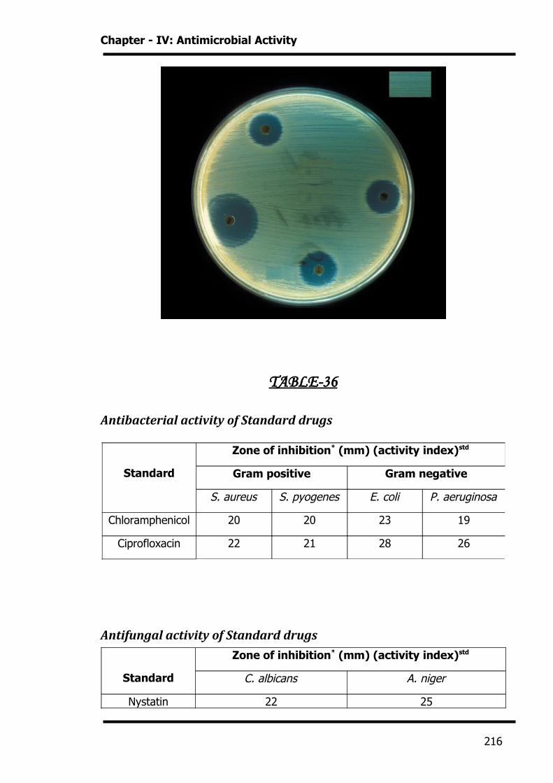
All the compounds were dissolved in dimethyl formamide (DMF). Proper
drug controls were used. Fig. 1 and Fig. 2 represent the zone of inhibition of the
control and the compound.
Compound was taken at concentration of 100 µg/ml for testing
antibacterial activity. The compound diffused into the medium produced a
concentration gradient. After the incubation period, the zones of inhibition were
measured in mm. The tabulated results represent the actual readings control.
[C] Test cultures
212
Chapter - IV: Antimicrobial Activity
Following common standard strains were used for screening of antibacterial
and antifungal activities:
• Escherichia coli [Gram negative] MTCC – 443
• Pseudomonas aeruginosa [Gram negative] MTCC – 424
• Staphylococcus aureus [Gram positive] MTCC – 96
• Streptococcus Pyogenes [Gram positive] MTCC – 442
• Candida albicans [Fungus] MTCC – 227
• Aspergillus Niger [Fungus] MTCC – 282
[D] Inoculum’s preparation
The inoculum was standardized at 1* 106 CFU/ml comparing with turbidity
standard (0.5 MacFarland tube)
[E] Swabs preparation
A supply of cotton wool swabs on wooden applicator sticks was prepared.
They were sterilized in tins, culture tubes, or on paper, either in the autoclave or
by dry heat.
[F] Experimental procedure
213
Chapter - IV: Antimicrobial Activity
1) The plates were inoculated by dipping a sterile swab into inoculums.
Excess inoculum was removed by pressing and rotating the swab firmly
against the side of the tube, above the level of the liquid.
2) The swab was streaked all over the surface of the medium three times,
rotating the plate through an angle of 60 oC after each application. Finally
the swab was passed round the edge of the agar surface. The inoculation
was dried for a few minutes, at room temperature, with the lid closed.
3) Ditch the bore in plate. Add compounds solution in bore.
4) The plates were placed in an incubator at 37 oC within 30 minutes of
preparation for bacteria and 22 oC for fungal.
5) After 48 hrs incubation for bacteria and 7-days for fungal, the diameter of
zone (including the diameter disc) was measured and recorded in mm. The
measurements were taken with a ruler, from the bottom of the plate,
without opening the lid.
Results are presented in Table-36 to 42.
214
Chapter - IV: Antimicrobial Activity
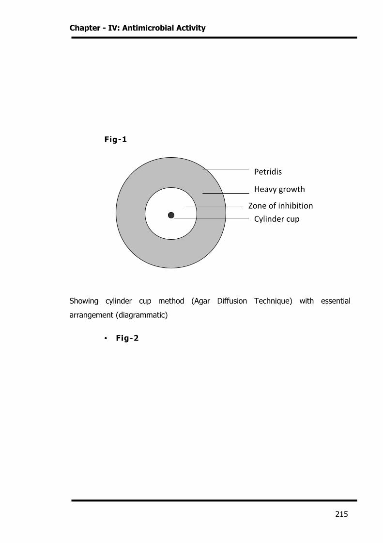
Fig-1
Showing cylinder cup method (Agar Diffusion Technique) with essential
arrangement (diagrammatic)
• Fig-2
215
Petridis
Heavy growth
Zone of inhibition
Cylinder cup
Chapter - IV: Antimicrobial Activity
TABLE-36
Antibacterial activity of Standard drugs
Antifungal activity of Standard drugs
Standard
Zone of inhibition* (mm) (activity index)std
C. albicans A. niger
Nystatin 22 25
Standard
Zone of inhibition* (mm) (activity index)std
Gram positive Gram negative
S. aureus S. pyogenes E. coli P. aeruginosa
Chloramphenicol 20 20 23 19
Ciprofloxacin 22 21 28 26
216
Chapter - IV: Antimicrobial Activity
Greseofulvin 27 29
*= average zone of inhibition in mm,
Activity index = Inhibition zone of the sample / Inhibition zone of the standard
TABLE-37
Antibacterial activity: Section-II
N N
NH2
R R'
R= 2,4-(Cl)2-5-F, 4-Cl, 4-OCH3, 4-CH3
R'= 4'-F, 4'-Cl, 3'-NO2, 3'-Br
Compound
Zone of Inhibition* (mm) (Activity Index) std.
Gram positive Gram negative
S. aureus S. pyogenus E. coli P. aeruginosa
A-17 10 09 10 08
A -18 18 15 19 16
A -19 16 15 18 14
217
Chapter - IV: Antimicrobial Activity
A -20 11 10 13 10
A-21 17 16 20 15
A-22 10 11 12 11
A-23 15 16 15 14
A-24 17 17 20 18
A-25 14 12 14 13
A-26 10 10 12 11
A-27 07 09 11 08
A-28 11 11 13 10
A-29 08 10 11 09
A-30 15 14 16 14
A-31 15 16 17 15
A-32 10 11 08 07
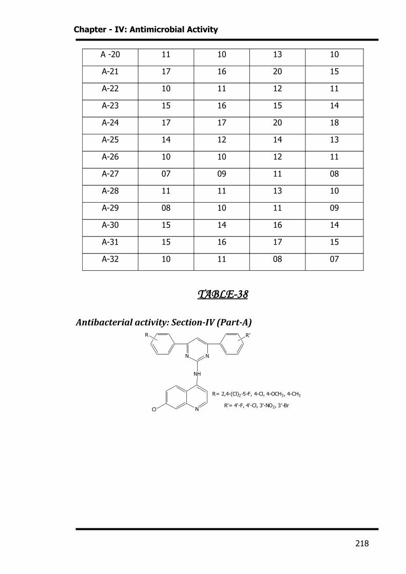
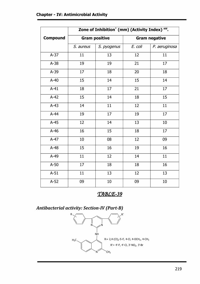
TABLE-38
Antibacterial activity: Section-IV (Part-A)
N N
NH
R R'
NCl
R= 2,4-(Cl)2-5-F, 4-Cl, 4-OCH3, 4-CH3
R'= 4'-F, 4'-Cl, 3'-NO2, 3'-Br
218
Chapter - IV: Antimicrobial Activity
TABLE-39
Antibacterial activity: Section-IV (Part-B)
N N
NH
R R'
N
R= 2,4-(Cl)2-5-F, 4-Cl, 4-OCH3, 4-CH3
R'= 4'-F, 4'-Cl, 3'-NO2, 3'-Br
CH3
H3C
Compound
Zone of Inhibition* (mm) (Activity Index) std.
Gram positive Gram negative
S. aureus S. pyogenus E. coli P. aeruginosa
A-37 11 13 12 11
A-38 19 19 21 17
A-39 17 18 20 18
A-40 15 14 15 14
A-41 18 17 21 17
A-42 15 14 18 15
A-43 14 11 12 11
A-44 19 17 19 17
A-45 12 14 13 10
A-46 16 15 18 17
A-47 10 08 12 09
A-48 15 16 19 16
A-49 11 12 14 11
A-50 17 18 18 16
A-51 11 13 12 13
A-52 09 10 09 10
219
Chapter - IV: Antimicrobial Activity
TABLE-40
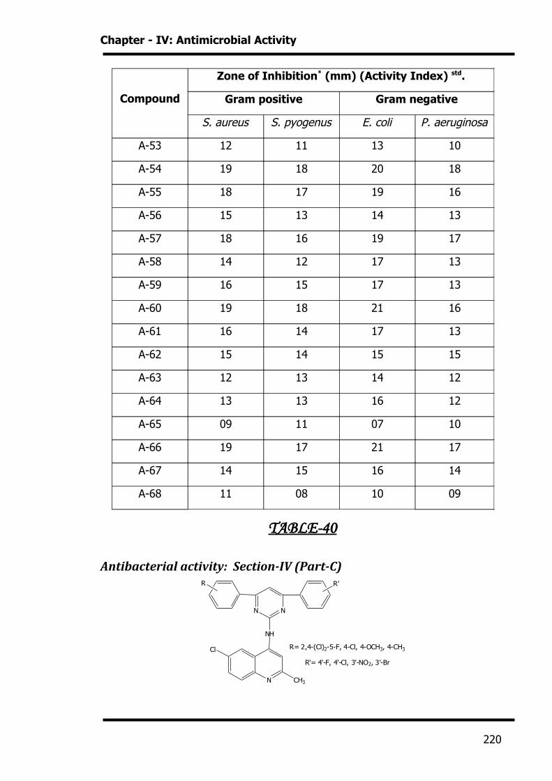
Antibacterial activity: Section-IV (Part-C)
N N
NH
R R'
N
R= 2,4-(Cl)2-5-F, 4-Cl, 4-OCH3, 4-CH3
R'= 4'-F, 4'-Cl, 3'-NO2, 3'-Br
CH3
Cl
Compound
Zone of Inhibition* (mm) (Activity Index) std.
Gram positive Gram negative
S. aureus S. pyogenus E. coli P. aeruginosa
A-53 12 11 13 10
A-54 19 18 20 18
A-55 18 17 19 16
A-56 15 13 14 13
A-57 18 16 19 17
A-58 14 12 17 13
A-59 16 15 17 13
A-60 19 18 21 16
A-61 16 14 17 13
A-62 15 14 15 15
A-63 12 13 14 12
A-64 13 13 16 12
A-65 09 11 07 10
A-66 19 17 21 17
A-67 14 15 16 14
A-68 11 08 10 09
220

Chapter - IV: Antimicrobial Activity
TABLE-41
Antibacterial activity: Section-IV (Part-D)
N N
NH
R R'
N
R= 2,4-(Cl)2-5-F, 4-Cl, 4-OCH3, 4-CH3
R'= 4'-F, 4'-Cl, 3'-NO2, 3'-Br
CH3
H3CO
Compound
Zone of Inhibition* (mm) (Activity Index) std.
Gram positive Gram negative
S. aureus S. pyogenus E. coli P. aeruginosa
A-69 11 10 13 10
A-70 17 18 20 17
A-71 18 17 19 16
A-72 15 13 14 15
A-73 18 17 19 17
A-74 14 13 17 14
A-75 12 11 14 13
A-76 18 18 20 17
A-77 11 13 10 11
A-78 15 14 15 14
A-79 09 10 13 08
A-80 13 14 16 14
A-81 10 09 13 11
A-82 14 13 13 11
A-83 12 11 16 13
A-84 11 09 07 10
221
Chapter - IV: Antimicrobial Activity
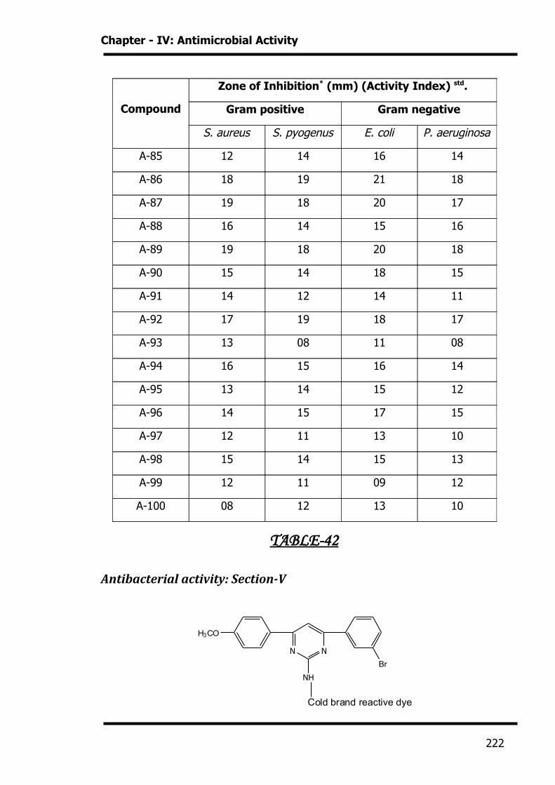
TABLE-42
Antibacterial activity: Section-V
N N
NH
Br
H3CO
Cold brand reactive dye
Compound
Zone of Inhibition* (mm) (Activity Index) std.
Gram positive Gram negative
S. aureus S. pyogenus E. coli P. aeruginosa
A-85 12 14 16 14
A-86 18 19 21 18
A-87 19 18 20 17
A-88 16 14 15 16
A-89 19 18 20 18
A-90 15 14 18 15
A-91 14 12 14 11
A-92 17 19 18 17
A-93 13 08 11 08
A-94 16 15 16 14
A-95 13 14 15 12
A-96 14 15 17 15
A-97 12 11 13 10
A-98 15 14 15 13
A-99 12 11 09 12
A-100 08 12 13 10
222
Chapter - IV: Antimicrobial Activity
ANTIFUNGAL ACTIVITY
INTRODUCTON
There are perhaps over 10,000 species of fungi, but less than 100 cause
diseases in human.10 Fungi may cause benign, but unsightly infections of the skin,
nail or hair, relatively trivial infection of mucous membranes (thrush) or systemic
infection causing progressive often fatal disease.
Compound
Zone of Inhibition* (mm) (Activity Index) std.
Gram positive Gram negative
S. aureus S. pyogenus E. coli P. aeruginosa
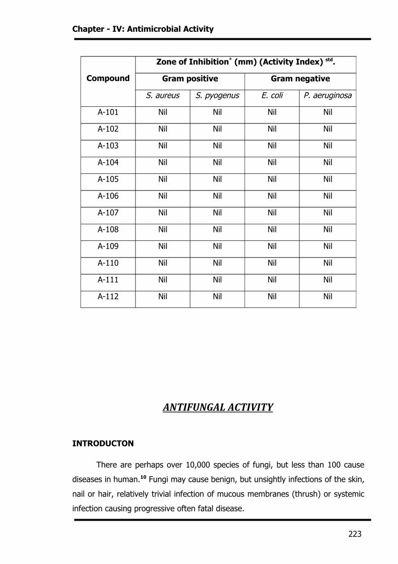
A-101 Nil Nil Nil Nil
A-102 Nil Nil Nil Nil
A-103 Nil Nil Nil Nil
A-104 Nil Nil Nil Nil
A-105 Nil Nil Nil Nil
A-106 Nil Nil Nil Nil
A-107 Nil Nil Nil Nil
A-108 Nil Nil Nil Nil
A-109 Nil Nil Nil Nil
A-110 Nil Nil Nil Nil
A-111 Nil Nil Nil Nil
A-112 Nil Nil Nil Nil
223
Chapter - IV: Antimicrobial Activity
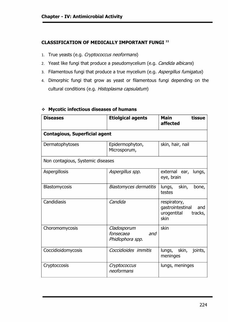
CLASSIFICATION OF MEDICALLY IMPORTANT FUNGI 11
1. True yeasts (e.g. Cryptococcus neoformans)
2. Yeast like fungi that produce a pseudomycelium (e.g. Candida albicans)
3. Filamentous fungi that produce a true mycelium (e.g. Aspergillus fumigatus)
4. Dimorphic fungi that grow as yeast or filamentous fungi depending on the
cultural conditions (e.g. Histoplasma capsulatum)
Mycotic infectious diseases of humans
Diseases Etiolgical agents Main tissue affected
Contagious, Superficial agent
Dermatophytoses Epidermophyton, Microsporum,
skin, hair, nail
Non contagious, Systemic diseases
Aspergillosis Aspergillus spp. external ear, lungs, eye, brain
Blastomycosis Biastomyces dermatitis lungs, skin, bone, testes
Candidiasis Candida respiratory, gastrointestinal and urogentital tracks, skin
Choromomycosis Cladosporum fonsecaea and Phidlophora spp.
skin
Coccidioidomycosis Coccidioides immitis lungs, skin, joints, meninges
Cryptoccosis Cryptococcus neoformans
lungs, meninges
224
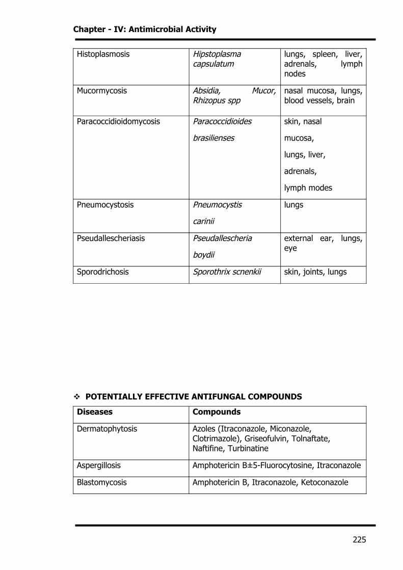
Chapter - IV: Antimicrobial Activity
Histoplasmosis Hipstoplasma capsulatum
lungs, spleen, liver, adrenals, lymph nodes
Mucormycosis Absidia, Mucor, Rhizopus spp
nasal mucosa, lungs, blood vessels, brain
Paracoccidioidomycosis Paracoccidioides
brasilienses
skin, nasal
mucosa,
lungs, liver,
adrenals,
lymph modes
Pneumocystosis Pneumocystis
carinii
lungs
Pseudallescheriasis Pseudallescheria
boydii
external ear, lungs, eye
Sporodrichosis Sporothrix scnenkii skin, joints, lungs
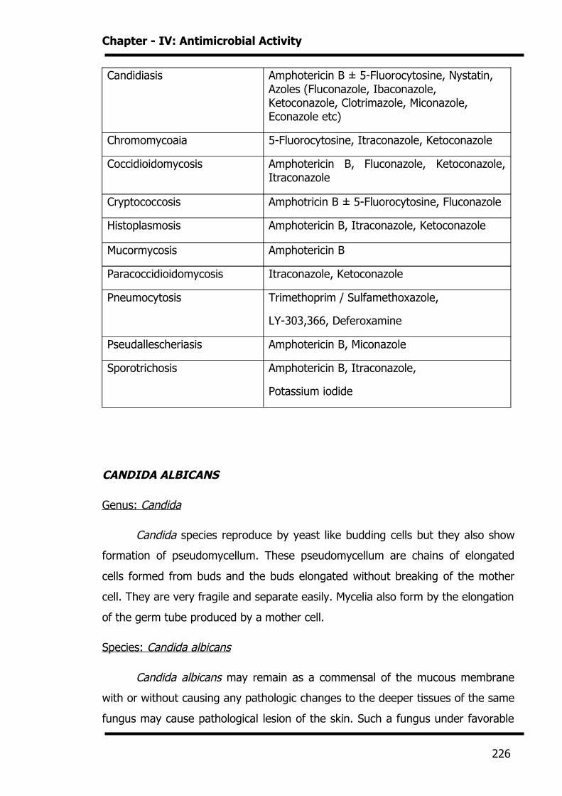
POTENTIALLY EFFECTIVE ANTIFUNGAL COMPOUNDS
Diseases Compounds
Dermatophytosis Azoles (Itraconazole, Miconazole, Clotrimazole), Griseofulvin, Tolnaftate, Naftifine, Turbinatine
Aspergillosis Amphotericin B±5-Fluorocytosine, Itraconazole
Blastomycosis Amphotericin B, Itraconazole, Ketoconazole
225
Chapter - IV: Antimicrobial Activity
Candidiasis Amphotericin B ± 5-Fluorocytosine, Nystatin, Azoles (Fluconazole, Ibaconazole, Ketoconazole, Clotrimazole, Miconazole, Econazole etc)
Chromomycoaia 5-Fluorocytosine, Itraconazole, Ketoconazole
Coccidioidomycosis Amphotericin B, Fluconazole, Ketoconazole, Itraconazole
Cryptococcosis Amphotricin B ± 5-Fluorocytosine, Fluconazole
Histoplasmosis Amphotericin B, Itraconazole, Ketoconazole
Mucormycosis Amphotericin B
Paracoccidioidomycosis Itraconazole, Ketoconazole
Pneumocytosis Trimethoprim / Sulfamethoxazole,
LY-303,366, Deferoxamine
Pseudallescheriasis Amphotericin B, Miconazole
Sporotrichosis Amphotericin B, Itraconazole,
Potassium iodide
CANDIDA ALBICANS
Genus: Candida
Candida species reproduce by yeast like budding cells but they also show
formation of pseudomycellum. These pseudomycellum are chains of elongated
cells formed from buds and the buds elongated without breaking of the mother
cell. They are very fragile and separate easily. Mycelia also form by the elongation
of the germ tube produced by a mother cell.
Species: Candida albicans
Candida albicans may remain as a commensal of the mucous membrane
with or without causing any pathologic changes to the deeper tissues of the same
fungus may cause pathological lesion of the skin. Such a fungus under favorable
226
Chapter - IV: Antimicrobial Activity
conditions can cause superficial, intermediate of deep mycoses depending on the
condition of the host.
ASPERGILLUS NIGER
Genus: Aspergillus
The Aspergilli are widespread in nature, being found on fruits, vegetables
and other substrates, which may provide nutriment. Some species are involved in
food spoilage. They are important economically because they are used in a
number of industrial fermentations, including the production of citric acid gluconic
acid. Aspergilli grow in high concentrations of sugar and salt, indicating that they
can extract water required for their growth from relatively dry substances.
Derivatives of N-methyl piperazine are therapeutically useful having
antibacterial and antiprotozoal activity. These are active against Gram positive
and Gram negative organisms. In addition, they are active against Candida
albicans and other mycetes. They are also cytostatic.12
Keto oximes esters, i.e. 1,3-Dichloro-2-propan-1-o-(benzoyl)-oxime, are
useful in inhibiting the growth of bacteria and fungi. They also find use as
herbicides and acaricides.13 Substituted 5-nitro-2-furylaminoximes and their
herbicides are active antibacterial and antifungal agents and can be used in
disinfectant compositions to control a variety of micro-organisms.14 O-(N-
methylcarbamolyl)-carbethoxy-chloroform-aldoxime shows biocidal activity against
Aerobacter derogenes in paper pulp and fungicidal activity against Septoria in
weight grains.15
Substituted glyoxal dithiosemicarbazones possess anaplasmicidal activity,
being effective in the control of Anaplasmamarginale.16 Alloxan-5-
thiosemicarbazone possesses bacteriostatic, bactericidal, and fungicidal and
defoliant activities. It is especially useful for the control of SPP of Erwinia,
Staphylococcus and Salmonella.17 Aryl-5-fluoro-2-methoxyphenyl ketoximes and
their thiosemicarbazones were found to be active against Aspergillus Niger and
227
Chapter - IV: Antimicrobial Activity
Aspergillus flavus.18 Fluorinated diaryl ketoximes and their thiosemicarbazones
possess antifungal activity.19
1-Propyl-1,2,4-triazolyl derivatives and their salts have a very broad
spectrum of fungicidal action and can be used in particular against parasitic fungi
which attack above ground parts of plants, such as Erysiphe, Podosphrea,
Piricularia and Pellicularia SPP and also against fungi attaking plants through the
soil and against seed-borne fungi.20
Chemical complexes of urea or its derivatives with a completely
halogenated acetone may be used as active components in fungicidal and
herbicidal compositions. They are particularly useful as systemic toxicants for
protecting food crops against harmful fungi, blights and similar pesticidal micro-
organisms.21 N2 -ethylhexyl-N1 -aryl ureas have a broad antibacterial activity
spectrum, including both Gram positive and Gram negative bacteria. They may be
used on a very broad basis, particularly for protecting organic substrates from
infection by destructive and pathogenic bacteria. They are suitable for use as
preservatives and disinfectants for textile and in cosmetics.22 The 2-Oxazolyl
thiourea compounds are useful antifungal agents, being effective in the control of
phytopathogenic fungi.23 N-(4 –bromo-3-halophenyl)-N1 -lower alkoxy ureas are
useful as fungicides and nematocides24. N-(1-cycloalken-1-yl)-urea and thiourea
are useful as phytotoxicants, fungicides, insecticides, nematocides, algaecides,
bactericides, bacteriostats and fungistats.25 The fungicidal effectiveness of
acylthiourea was demonstrated by the sore germination test on plasmparaviticola
sphaerotheca fuligiea26 etc. Uredophenylthiourea derivatives have a broad
spectrum and are particularly effective against parasitic fungi, pathogenic agents
and powdery mildew and rice diseases.27
Essential oils were extracted from members of the myrtaceae family (Bay,
Pumenta and Clove), with eugenol as the main constituent. Their antimicrobial
activity was investigated using S. aureus, B. subtilis, E. coli, P. vulgaris,
Pseudomonas auruginosa, Mycobacterium smegmatis and Candida albicans as
test organisms. Mono-, di- and tri-pyrimidine complexes of organotin compound
are active in the control of insects, fungi, weeds and their microorganisms such as
228
Chapter - IV: Antimicrobial Activity
bacteria and viruses.28 Pyridine carbamates are useful as insecticides, fungicides
and nematocides.29 Dialkyltin salts of substituted pyridine-1-oxides are highly
effective bactericides and fungicides and have especially low toxicity to higher
animals. The compounds are useful as preservatives e.g. for protecting leather,
paint, paper, plastics, etc. against attack by mildew and other fungi. They are also
useful for sterilization and disinfecting purposes. Pyrimidine-organo copper
products are useful as catalyst, fungicides, pesticides and anthelmintics and as
intermediates in organic synthesis.30
Cynocarbamates are useful as fungicides, being effective in the control of
phytopathogenic fungi. Thiocyanoacetamides possess fungicidal activity and are
useful in the control of phyto-pathogenic fungi.31 Phosphoric acid esters are
primarily insecticides but fungicidal action has been found against the following
SPP: Alternaria tenuis, Botrytis cinerea, Clastero sporium, Coniothrium, Fusarium,
Mucor, Penicilium, Stemphylium and Botrytis fabae.32
Aryl and cycloalkyl substituted naphthols are useful as antimicrobial agents
and are used in injectable suspension, oral liquid suspension, tablets and capsules
to inhibit the growth of micro-organisms viz. Staphylococcus aureus,
Streptococcus faecalis, Bacillus subtilis, E. coli, proteus vulgaris, Histoplasma
capsulatum, Candida albicans, Aspergillus niger etc.33
Harendra Singh, L. S. Yadav, Kripa Sukla and Rajesh Dwivedi have
synthesized 6,7-dihydro-5H-thiazole[3,2-a]pyrimidin-5-ones. These compounds
displayed an antifungal activity against Aspergillus Flavus and Fusarium solani.34
During the past quarter century, various azoles, i.e. imidazoles, triazoles
have been screened for antifungal action and exhibited broad spectrum of activity
against pathogenic fungi. Azoles can be used both topically and systemically.35
Resistance to imidazoles or triazoles is very rare.36
The two most important drugs belonging to allylamines group are Naftifine
and Terbinafine. Naftifine shows broad spectrum fungicidal activity in vitro.
Griseofulvin, a product of Penicillium griseofulvum was discovered in 1939. But its
229
Chapter - IV: Antimicrobial Activity
antifungal activity became known in 1951. Tolnaftate was found to be active
against dermatophytes when applied topically.
C. Mehwala37, J. Machhi38, A. Joshi39, V. Bhatt40 and A. Desai41 have very
recently worked on synthesis of quinoline based compounds and evaluated their
antifungal activity.
The synthesized compounds were screened for their antifungal activity
against Candida albicans (MTCC227), Aspergillus niger (MTCC282) using the agar
cup plate diffusion method by dissolving in DMF at a concentration of 100 µg/mL.
The zone of inhibition was measured after 7 days at 20 oC and it was compared
with the standard drugs Griseofulvin and Nystatin as shown in Table-43 to 48.
TABLE-43
Antifungal activity: Section-II
230
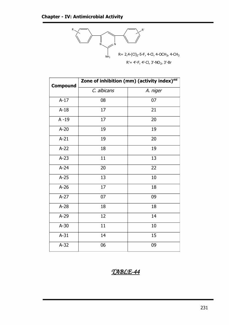
Chapter - IV: Antimicrobial Activity
N N
NH2
R R'
R= 2,4-(Cl)2-5-F, 4-Cl, 4-OCH3, 4-CH3
R'= 4'-F, 4'-Cl, 3'-NO2, 3'-Br
TABLE-44
CompoundZone of inhibition (mm) (activity index)std
C. albicans A. niger
A-17 08 07
A-18 17 21
A -19 17 20
A-20 19 19
A-21 19 20
A-22 18 19
A-23 11 13
A-24 20 22
A-25 13 10
A-26 17 18
A-27 07 09
A-28 18 18
A-29 12 14
A-30 11 10
A-31 14 15
A-32 06 09
231
Chapter - IV: Antimicrobial Activity
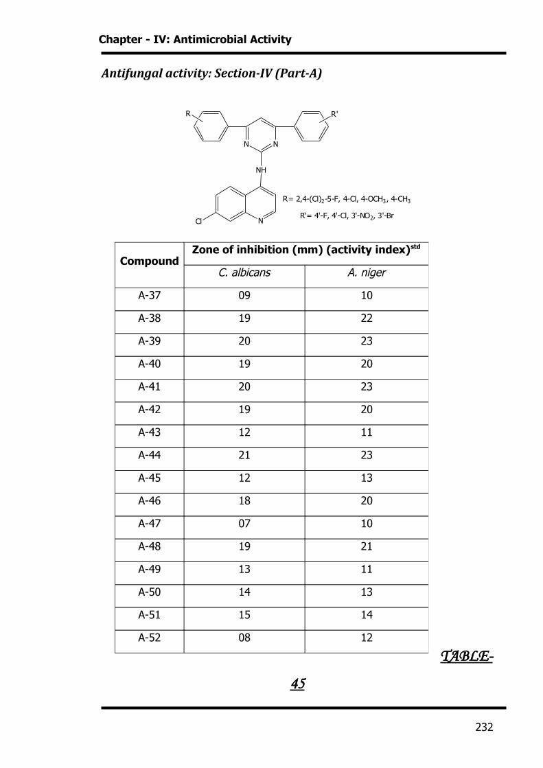
Antifungal activity: Section-IV (Part-A)
N N
NH
R R'
NCl
R= 2,4-(Cl)2-5-F, 4-Cl, 4-OCH3, 4-CH3
R'= 4'-F, 4'-Cl, 3'-NO2, 3'-Br
TABLE-
45
CompoundZone of inhibition (mm) (activity index)std
C. albicans A. niger
A-37 09 10
A-38 19 22
A-39 20 23
A-40 19 20
A-41 20 23
A-42 19 20
A-43 12 11
A-44 21 23
A-45 12 13
A-46 18 20
A-47 07 10
A-48 19 21
A-49 13 11
A-50 14 13
A-51 15 14
A-52 08 12
232
Chapter - IV: Antimicrobial Activity
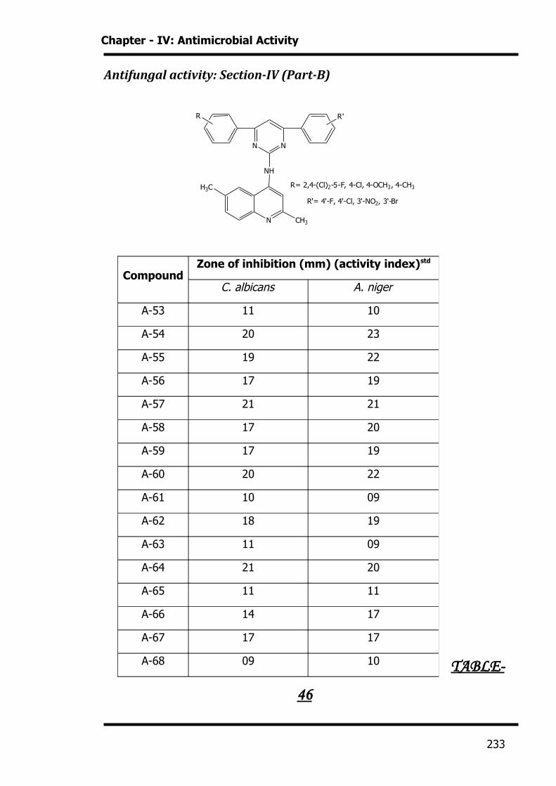
Antifungal activity: Section-IV (Part-B)
N N
NH
R R'
N
R= 2,4-(Cl)2-5-F, 4-Cl, 4-OCH3, 4-CH3
R'= 4'-F, 4'-Cl, 3'-NO2, 3'-Br
CH3
H3C
TABLE-
46
CompoundZone of inhibition (mm) (activity index)std
C. albicans A. niger
A-53 11 10
A-54 20 23
A-55 19 22
A-56 17 19
A-57 21 21
A-58 17 20
A-59 17 19
A-60 20 22
A-61 10 09
A-62 18 19
A-63 11 09
A-64 21 20
A-65 11 11
A-66 14 17
A-67 17 17
A-68 09 10
233
Chapter - IV: Antimicrobial Activity
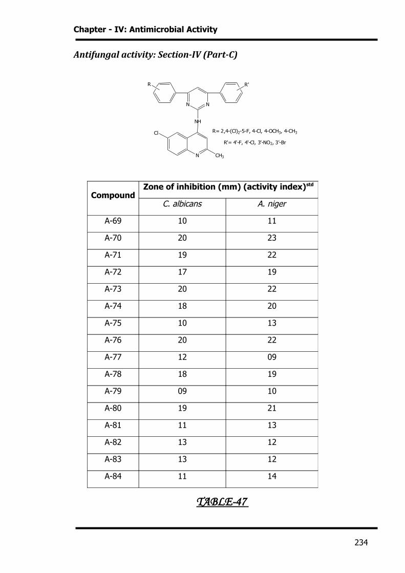
Antifungal activity: Section-IV (Part-C)
N N
NH
R R'
N
R= 2,4-(Cl)2-5-F, 4-Cl, 4-OCH3, 4-CH3
R'= 4'-F, 4'-Cl, 3'-NO2, 3'-Br
CH3
Cl
TABLE-47
CompoundZone of inhibition (mm) (activity index)std
C. albicans A. niger
A-69 10 11
A-70 20 23
A-71 19 22
A-72 17 19
A-73 20 22
A-74 18 20
A-75 10 13
A-76 20 22
A-77 12 09
A-78 18 19
A-79 09 10
A-80 19 21
A-81 11 13
A-82 13 12
A-83 13 12
A-84 11 14
234
Chapter - IV: Antimicrobial Activity
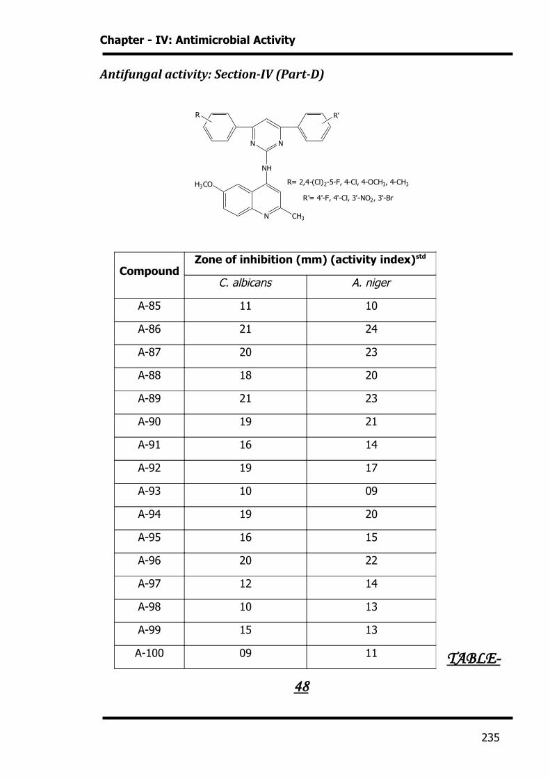
Antifungal activity: Section-IV (Part-D)
N N
NH
R R'
N
R= 2,4-(Cl)2-5-F, 4-Cl, 4-OCH3, 4-CH3
R'= 4'-F, 4'-Cl, 3'-NO2, 3'-Br
CH3
H3CO
TABLE-
48
CompoundZone of inhibition (mm) (activity index)std
C. albicans A. niger
A-85 11 10
A-86 21 24
A-87 20 23
A-88 18 20
A-89 21 23
A-90 19 21
A-91 16 14
A-92 19 17
A-93 10 09
A-94 19 20
A-95 16 15
A-96 20 22
A-97 12 14
A-98 10 13
A-99 15 13
A-100 09 11
235
Chapter - IV: Antimicrobial Activity
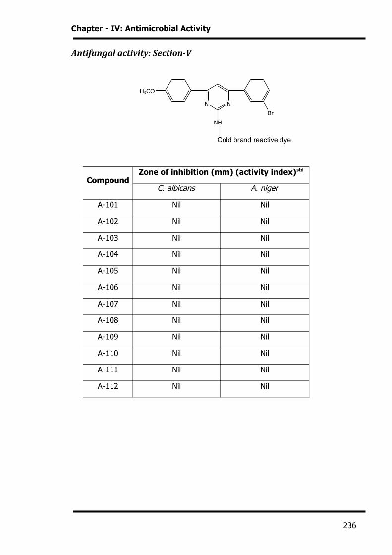
Antifungal activity: Section-V
N N
NH
Br
H3CO
Cold brand reactive dye
CompoundZone of inhibition (mm) (activity index)std
C. albicans A. niger
A-101 Nil Nil
A-102 Nil Nil
A-103 Nil Nil
A-104 Nil Nil
A-105 Nil Nil
A-106 Nil Nil
A-107 Nil Nil
A-108 Nil Nil
A-109 Nil Nil
A-110 Nil Nil
A-111 Nil Nil
A-112 Nil Nil
236
References
[1] Robert Cruickshank, Hand Book of Bacteriology, 394 (1962)
[2] K. D. Tripathi, Essentials of medical pharmacology, 625 (1994)
[3] J. W. Churchman, J. Exptl. Med., 16, 221 (1912)
[4] A. Albert, Brit. J. Exptl. Pathol., 34, 119 (1958)
[5] L. D. Gebbharadt, J. G. Bachtold, Proc. Soc. Exptl. Biol. Med., 88, 103 (1955)
[6] E. W. Stearn, A. E. Stearn, J. Bacteriol., 9, 463-479 (1924)
[7] E. Fischer, R. Muazo, J. Bacteriol., 53, 381 (1947)
[8] A. Albert, Brit. J. Expt. Pathol., 35, 75 (1954)
[9] A. H. Bakett, J. Pharm. Pharmacol., 10, 160 (1958)
[10] P. H. Jacobs, Fungal Diseases, 1, (1997)
[11] D. Greenwood, Antimicrob. Hemotherapy, Third Edition, 62, (1995)
[12] US 3580914, Soc. d’ Etudes de Rech., d’ Application Sci., Med.,
Microbiology Abstr., Vol. 9, No. 2, 9A, 1003 (1974)
[13] US 3592920, Stauffer Chem. Co.; Microbiology Abstr., Vol. 9, No. 2, 9A,
1009 (1974)
[14] US 3660390, Abbott Lab.; Microbiology Abstr., Vol. 9, No. 12, 9A, 7579
(1974)
[15] US 3742036, Roussel-Uclaf.; Microbiology Abstr., Vol. 10, 10A, 7281 (1975)
[16] US 3709935, Barrett P. A.; Microbiology Abstr., Vol. 10, No. 4, 10A, 2589
(1975)
236
References
[17] US 3773952; Microbiology Abstr., Vol. 11, No. 5, 11A, 3278 (1976)
[18] R. H. Khan, S. C. Bahel, Agric Biol. Chem., 40 (9), 1881-1883; Microbiology
Abstr., Vol. 12, No. 5, 12A, 3701 (1977)
[19] A. K. Srivastava, S. C. Bahel, Agric Biol. Chem., 40(4), 801-803 (1976);
Microbiology Abstr., Vol. 12, No. 4, 12A, 2955 (1977)
[20] GB 1291417, Bayer AG.; Microbiology Abstr., Vol. 12, No. 3, 12A, 2018
(1977)
[21] US 3966951, Stauffer Chem. Co.; Microbiology Abstr., Vol. 9, No. 2, 9A, 902
(1974)
[22] US 3592932, Ciba Ltd.; Microbiology Abstr., Vol. 9, No. 2, 9A, 977 (1974)
[23] US 3705903, Lilly Ind. Ltd.; Microbiology Abstr., Vol. 10, No. 3, 10A, 1636
(1975)
[24] US 3702363, Ciba-Geigy AG.; Microbiology Abstr., Vol. 10, No. 3, 10A, 1771
(1975)
[25] US 3701807, Monsanto Co.; Microbiology Abstr., Vol. 10, No. 3, 10A, 1816
(1975)
[26] GB 1313236, Merck Patent GmbH; Microbiology Abstr., Vol. 10, No. 6, 10A,
4214 (1975)
[27] GB 1313236, Bayer AG.; Microbiology Abstr., Vol. 10, No. 6, 10A, 4215
(1975)
[28] US 3661911, Monsanto Co.; Microbiology Abstr., Vol. 9, No. 12, 9A, 7577
(1974)
[29] US 3701779, Shell Oil Co.; Microbiology Abstr., Vol. 10, No. 3, 10A, 1777
(1975)
237
References
[30] US 3712894, Philips Petroleum Co.; Microbiology Abstr., Vol. 10, No. 4,
10A, 2608 (1975)
[31] US 3723439, Velsicol Chem. Corp.; Microbiology Abstr., Vol. 10, No. 6, 10A,
4065 (1975)
[32] GB 1314186, Ciba-Geigy AG.; Microbiology Abstr., Vol. 10, No. 6, 10A, 4198
(1975)
[33] US 3761526, Sandoz-Wander Inc.; Microbiology Abstr., Vol. 11, No. 3, 11A,
1616 (1976)
[34] H. Singh, L. Dhar, S. Yadav, K. N. Shukla, R. Dwivedi, J. Agric. Food. Chem.,
38, 1962-1964 (1990)
[35] Eugene D. & Weinberg, Burger’s Medicinal Chemistry & Drug Diseases, Fifth
Edition, 2, Therapeutic agents, 637 (1997)
[36] J. E. Bennet, In Goodman Gilman’s the Pharmacological Basis of
Therapeutics, Ninth Edition, 1175, (1996)
[37] C. K. Mehwala; Ph. D. Thesis, Veer Narmad South Gujarat University, Surat
(1999)
[38] J. K. Machhi; Ph. D. Thesis, Veer Narmad South Gujarat University, Surat
(2000)
[39] A. M. Joshi; Ph. D. Thesis, Saurashtra University, Rajkot (2002)
[40] V. S. Bhatt; Ph. D. Thesis, Veer Narmad South Gujarat University, Surat
(2005)
[41] A. V. Desai, Ph. D. Thesis, Veer Narmad South Gujarat University, Surat
(2007)
238
![Overview%2520 presentation[1][1]](https://static.fdocuments.us/doc/165x107/5553d7f0b4c90574028b4e2c/overview2520-presentation11.jpg)

![Outsiders%2520 facebook[1][1]](https://static.fdocuments.us/doc/165x107/555d2761d8b42ab2228b5725/outsiders2520-facebook11.jpg)


![Culminating%2520 project[1][1]](https://static.fdocuments.us/doc/165x107/5589df1ad8b42a94778b4675/culminating2520-project11.jpg)

![Noah%2520 valenzuela%2520%2528powerpointslide%2529%5b1%5d[1]](https://static.fdocuments.us/doc/165x107/58a0530e1a28ab5c1c8b485d/noah2520-valenzuela25202528powerpointslide25295b15d1.jpg)