ANTI-TUMOR STUDY OF SCOPOLETIN AND RUBBING- …
49
ANTI-TUMOR STUDY OF SCOPOLETIN AND RUBBING- MERCAPTO-NITRILE FROM NICOTIANA GLAUCA YASSER TABANA UNIVERSITI SAINS MALAYSIA 2016
Transcript of ANTI-TUMOR STUDY OF SCOPOLETIN AND RUBBING- …

ANTI-TUMOR STUDY OF SCOPOLETIN AND RUBBING-
MERCAPTO-NITRILE FROM NICOTIANA GLAUCA
YASSER TABANA
UNIVERSITI SAINS MALAYSIA
2016

ANTI-TUMOR STUDY OF SCOPOLETIN AND RUBBING-
MERCAPTO-NITRILE FROM NICOTIANA GLAUCA
By
YASSER TABANA
Thesis submitted in fulfillment of the requirements
for the degree of
Master of Science (Pharmacology)
March 2016

This thesis is dedicated to
my people in Syria who have long been suffering from the war

ii
ACKNOWLEDGEMENT
بسم الله الرحمن الرحيم
Praise be to Allah, The Most Merciful, The Most Beneficent, Who has given me
strength to complete this work.
First and foremost, I would like to express my sincere thanks and gratitude to my
supervisor, Assoc. Prof. Dr. Amin Malik Shah Bin Abdul Majid (School of Pharmaceutical
Sciences, USM), who kept guiding and supporting me during my research. This work
would not have been possible without his help and encouragement. He has always been
there to ensure the accomplishment of all tasks. I would also wish to extent my special
thanks to my co-supervisor, Dr. Aman S. Abdul Majid, who has guided me during my
study.
My deepest appreciation also goes to Dr. Mohamed Khadeer Ahamed Basheer for
training and guiding me during my stay at EMAN Laboratories. This work could not have
been possible without the invaluable help of my colleague, Dr. Loiy Elsir Ahmed Hassan,
who served as my mentor throughout my research period. My sincere thanks and gratitude
goes to all of my colleagues, especially Saad Sabbar Dahham, Fouad S. R. al-Suede,
Muhammad Asif, Suzana binti Hashim, Bassel Al-Hindi and Norshirin Idris; for their
support and assistance
I would also like to thank the Institute of Postgraduate Studies, Universiti Sains
Malaysia, for awarding me USM Postgraduate Fellowship.

iii
I would also like to sincerely thank Dr. Muhammad A. Iqbal for performing the
structural elucidation of the compounds; and Mr. Md Shamsuddin S. Khan for conducting
the molecular docking study.
I owe my greatest gratitude to my parents (Mahfooth and Fayzah). They are the
sparkling light in the dimness of my path. Without their encouragement, support, guidance
and understanding, I would not have been able to come to Malaysia and pursue my dream. I
would like to express my utmost gratitude to my gifts from Allah, with whom I have grown
and spent my childhood, my brothers and sisters, Nasser, Fatema, Mostafa, Mohammad and
Salaam for their never-ending support. Special thanks to my aunt who kept encouraging
me to purse my dream.
Last but not least, I want to dedicate my work to all of my friends and people in and
out of Syria, and those who have been forced to leave the country because of the war. I
hope that their agony will end very soon.
iv
TABLE OF CONTENTS
page
ACKNOWLEDGEMENT................................................................................................... II
TABLE OF CONTENTS .................................................................................................... IV
LIST OF FIGURES ............................................................................................................ XI
LIST OF ABBREVIATION ............................................................................................. XIV
LIST OF UNITS .............................................................................................................. XVIII
LIST OF APPENDICES ................................................................................................... XIX
ABSTRAK .......................................................................................................................... XX
ABSTRACT ...................................................................................................................... XXII
CHAPTER ONE: INTRODUCTION............................................................................ 1
1.1. Overview of cancer .......................................................................................... 1
1.2. The incidence and mortality of cancer ............................................................. 2
1.3. The differences between normal and cancer cells ............................................ 3
1.4. The molecular biology of cancer ...................................................................... 8
1.5. Apoptosis in cancer ........................................................................................ 13
1.5.2. Mechanism of apoptosis ................................................................................. 15
1.6. Colorectal carcinoma ...................................................................................... 20
1.7. Major signaling pathways in colon carcinogenesis ........................................ 21
1.7.1. Wnt /β-catenin signaling pathway .................................................................. 21
1.7.2. Notch Signaling Pathway ............................................................................... 21
1.7.3. P53 signaling pathway ................................................................................... 22
1.7.4. TGF-β Signaling Pathway .............................................................................. 23
v
1.7.5. Cell cycle (pRB/ E2F) signaling pathway ...................................................... 24
1.7.6. NF-кB signaling pathway ............................................................................... 25
1.7.7. Myc/Max signaling pathway .......................................................................... 26
1.7.8. MAPK signaling pathway .............................................................................. 26
1.8. Angiogenesis .................................................................................................. 28
1.8.1. The angiogenic switch .................................................................................... 29
1.8.2. The development of quantitative assays for angiogenesis ............................. 31
1.8.3. Role of the vascular endothelial growth factor pathway in tumor growth and
angiogenesis ................................................................................................... 32
1.8.4. The effects of hypoxia on angiogenesis: ........................................................ 35
1.8.5. History of anti-angiogenesis drugs ................................................................. 36
1.9. Natural products and cancer ........................................................................... 38
1.10. Plant literature review .................................................................................... 39
1.10.1. Solanaceae ................................................................................................ 39
1.10.2. The Nicotiana genus ................................................................................. 40
1.10.3. Nicotiana glauca ....................................................................................... 40
1.10.4. The biological activities of Nicotiana glauca ........................................... 43
1.10.5. Chemistry of the Nicotina genus .............................................................. 44
1.11. Aims and objectives ....................................................................................... 49
CHAPTER TWO: MATERIALS AND METHODS .................................................. 50
2.1. Laboratory chemicals and suppliers ............................................................... 50
2.2. Equipments and apparatus .............................................................................. 52
2.3. Plant collection and authentication ................................................................ 53
vi
2.4. Extraction and fractionation ........................................................................... 53
2.5. Isolation of the active compounds .................................................................. 54
2.6. Structure elucidation and characterization of the compounds ....................... 55
2.6.1. FT-IR Measurements ...................................................................................... 55
2.6.2. 1H NMR Measurements ................................................................................. 55
2.6.3. Mass Spectra .................................................................................................. 55
2.6.4. Thin Layer Chromatography .......................................................................... 56
2.7. High performance liquid chromatography (HPLC) analysis ......................... 56
2.7.1. Operational parameters of HPLC ................................................................... 56
2.7.2. Preparation of samples for HPLC analysis .................................................... 57
2.8. Experimental animals ..................................................................................... 57
2.9.1. Ex vivo rat aortic ring assay ............................................................................ 58
2.9.2. In vivo matrigel plug assay ............................................................................. 59
2.9.3. In vivo assessment of tumor angiogenesis in nude mouse xenograft model .. 60
2.10. Studies of molecular docking ......................................................................... 61
2.10.1. Tools for molecular docking study........................................................... 61
2.10.2. Molecular Docking Study ........................................................................ 61
2.10.3. LeadIT – FlexX ........................................................................................ 61
2.10.4. SYBYL Surflex Dock .............................................................................. 62
2.11. Cell lines and culture conditions .................................................................... 62
2.11.1. Cell Lines and Media ............................................................................... 62
2.11.2. Routine Feeding and Maintenance ........................................................... 63
2.11.3. Subculture of Cells ................................................................................... 63
2.11.4. Counting of the Cells ................................................................................ 64
vii
2.12. Studies of cytotoxic and apoptotic properties of (RMN) ............................... 64
2.12.1. Effect of the compound on cell proliferation: .......................................... 64
2.12.2. Colony formation assay: ........................................................................... 65
2.12.3. Cell migration assay ................................................................................. 66
2.12.4. Effect of the compound on Nuclear Morphology .................................... 67
2.12.5. Hanging drop spheroid assay ................................................................... 67
2.12.6. Luciferase assay to assess cancer pathways ............................................. 68
2.13. Statistical analysis .......................................................................................... 68
CHAPTER THREE: RESULTS ....................................................................................... 69
3.1. Isolation, characterization and Anti-angiogenic properties of scopoletin ...... 69
3.1.1. Plant Extraction and Isolation of Scopoletin .................................................. 69
3.1.2. Structural elucidation and characterization of Scopoletin .............................. 71
3.1.2.1. FT-IR ........................................................................................................ 71
3.1.2.2. FT-NMR ................................................................................................... 71
3.1.2.3. Gas chromatography-mass spectra (GC-MS) analysis of isolated
Scopoletin ................................................................................................. 73
3.1.2.4. Thin Layer Chromatography .................................................................... 73
3.1.2.5. HPLC analysis of Scopoletin ................................................................... 77
3.1.3. Scopoletin inhibits sprouting of microvessels in rat aortic explants .............. 78
3.1.4. Scopoletin inhibits in vivo tumor vasculature in xenograft model ................ 80
3.1.4.1. Histological findings ................................................................................ 83
3.1.5. Scopoletin inhibits matrigel induced vasculature in nude mice ..................... 86
viii
3.1.6. Scopoletin has potential binding efficiency towards ERK1, FGF2 and VEGF .
.................................................................................................................. 89
3.2. Isolation, characterization and apoptotic properties of RMN ........................ 95
3.2.1. Extraction and isolation of RMN ................................................................... 95
3.2.2. Chemistry of RMN ......................................................................................... 97
3.2.2.1. NMR spectroscopy ................................................................................... 97
3.2.2.2. GC-MS analysis ....................................................................................... 98
3.2.3. Inhibitory effect of RMN on proliferation of cancer cell lines ...................... 99
3.2.4. RMN induces nuclear condensation ............................................................. 102
3.2.5. RMN inhibits clonogenicity and tumor spheroid formation ........................ 104
3.2.6. RMN inhibits migratory and invasive properties of HCT-116 cells ............ 107
3.2.7. Effect of (RMN) on cell signaling pathways ............................................... 109
CHAPTER FOUR: DISCUSSION AND CONCLUSIONS ....................................... 111
4.1. General discussion ........................................................................................ 111
4.2. Angiogenesis properties of scopoletin ......................................................... 112
4.3. Anticancer properties of RMN ..................................................................... 115
4.4. Conclusions .................................................................................................. 119
4.5. Suggestions for Future Studies ..................................................................... 120
REFERENCES ............................................................................................................... 121
APPENDIX A: APPROVAL LETTER FROM ANIMAL ETHIC COMMITTEE ........ 143
APPENDIX B: CHARACTERIZATION OF RMN USING FT-IR, GCMS AND NMR
SPECTRAL STUDIES ................................................................................................... 144
APPENDIX C: ACADEMIC AWARDS, CONFERENCES AND PUBLICATIONS .. 146

ix
List of academic honors and awards ........................................................................... 146
List of conferences ...................................................................................................... 146
List of publications ..................................................................................................... 147
x
LIST OF TABLES
Page
Table 1.1: The differences between normal and cancer cells 5
Table 1.2 Cancer predisposition genes (Vogelstein & Kinzler, 2004) 10
Table 1.3 the differences between apoptosis and necrosis 14
Table 1.4 Stimulatory and inhibitory regulators of angiogenesis (Pandya, Dhalla, &
Santani, 2006) 30
Table 1.5: Compounds isolated from Nicotiana spp. 45
Table 3.1: Summary of the docking scores and reported binding affinities of
Scopoletin with LeadIT FlexX program. 91
Table 3.2: Summary of the docking scores and reported binding affinities of
Scopoletin with SYBYL-X Surflex program. 92
Table 3.3: IC50 (μg) values of RMN on, HCT 116, MCF7, CAP1, Ug78, EA.hy926
and CCD- 18Co cell lines assessed by MTT-assay. 100

xi
LIST OF FIGURES
Page
Figure 1.1: Forming tumor among normal cells 6
Figure 1.2: Metastasis of a cancer cell 7
Figure 1.3: Pathways of apoptosis 19
Figure 1.4: Interactions of VEGF ligands and VEGF receptors 34
Figure 1.5: Distribution of Nicotiana glauca (Florentine, 2005; Moore DM et al.,
1972). 42
Figure 3.1: Isolation of scopoletin. Schematic diagram showing the bioassay guided
sequential extraction and fractionation of N. glauca leading to isolation of
scopoletin. 70
Figure 3.2: Structural characterization of scopoletin. 74
Figure 3.3: Intermolecular H-bonding and mass fragmentation pattern in scopoletin. 75
Figure 3.4: TLC analysis of different extracts of N. glauca stems. 76
Figure 3.5:. HPLC analysis of scopoletin in the n-hexane extract of N. glauca. 77
Figure 3.6: Anti-angiogenic effect of scopoletin. Photomicrographic images show
inhibition of sprouting of microvessels in rat aortic explants. 79
Figure 3.7: Illustration of the tumor growth profile in the respective test groups. 81
Figure 3.8: In vivo antitumor and anti-angiogenic activities of scopoletin determined
using a human tumor xenograft model in athymic nude mice bearing
HCT-116 tumors at day 28 post-inoculation. 82
Figure 3.9: Visualization of ligands and protein interaction profile. Surface
visualization of proteins and active site residue interactions of protein and
hydrophobic interactions are shown in the green region. 90
Figure 3.10: Graphical representation of the estimated binding activity (A) and ligand
efficiency (B) of Scopoletin and Suramin with ERK1, FGF2, and VEGF
in LeadIT. 93
Figure 3.11: Graphical abstract of the activity of scopoletin. 94

xii
Figure 3.12: Isolation of RMN: Schematic diagram showing bio-assay guided
sequential extraction and fractionation of N. glauca leading to isolation of
Rubbing-Mercapto-Nitrile (RMN). 96
Figure 3.13: The structure and IUPAC name of the isolated compound (RMN) 98
Figure 3.14: Effect of RMN on cellular morphology of human cancer and normal cell
lines: Photomicrograph images of cancer cell lines were taken under an
inverted phase-contrast 30 microscope at 200× magnification using a
digital camera at 48 hours after treatment with the RMN. 101
Figure 3.15: HCT-116 cells stained with Hoechst-33258: A) the photomicrographs
show HCT-116 cells stained with Hoechst-33258 after 6 and 12 hours of
treatment with RMN. 103
Figure 3.16: Antitumor aggregation effects of RMN on in vitro HCT-116 cellular
spheroids in the hanging drop assay. 105
Figure 3.17: Effect of RMN on survival of HCT-116 colonies in the colony formation
assay. 106
Figure 3.18: Effects of RMN on apoptotic signaling pathways in the HCT 116 cell
line. 109
Figure 3.19: Graphical abstract of the activity of RMN. 110
xiii
LIST OF PLATES
Page
Plate 1.1: Picture of Nicotiana glauca Graham (Bogdanovic, 2006) 42
Plate 3.1: Necrotic changes in tumor histology and evidence of the anti-angiogenic
effect of scopoletin 85
Plate 3.2: H&E stained cross-sections taken from matrigel plugs implanted
subcutaneously in representative groups of animals 88
xiv
LIST OF ABBREVIATION
ANOVA Analysis of variance
APC Adenomatous polypsis coli
ATCC American Type Culture Collection
ATP Adenosine triphosphate
Bcl-1 and 2 B-cell lymphoma 1 and 2
bFGF basic Fibroblast growth factor
BT20 Breast cancer cell line
CCD-18Co Normal human colonic fibroblast
c-myc Myelocytomatosis oncogene cellular homolog
CO2 Carbon dioxide
Conc Concentration
3D Three-dimensional
DMEM Dulbecco's modified eagle medium
DMSO Dimethyl sulphoxide
DNA Deoxyribonucleic acid
EC50 Concentration that provides 50% inhibition
xv
ECM Endothelial cell medium
e.g. For example
etc. Et cetera, it means “and other things”
FTIR Fourier transform infrared spectrometry
GC-MS Gas chromatography mass spectrometry
1 H-NMR Hydrogen Nuclear Magnetic Resonance
HCT 116 Human colorectal carcinoma cell line
Hep G2 Human hepatocellular carcinoma cell line
HIFBS Heat-inactivated fetal bovine serum
HUVECs Human umbilical vein endothelial cell line
HIFs Hypoxia inducible factors
i.e. That means
IC50 Half-maximal inhibitory concentration
IL Interleukin
IR Infrared
LD50 Lethal dose of 50% of the tested animals
MCF 7 Human hormone sensitive and invasive breast
cancer cell line
xvi
MDA-MB-231 Human hormone resistant breast cancer cell line
MDA-MB-468 Breast cancer cell line
MEM Minimum essential medium
MIC Minimal inhibitory concentration
MTT 3-(4,5-Dimethylthiazol-2-yl)-2,5diphenyl
tetrazolium bromide
mRNA Messenger Ribonucleic acid
N. glauca Nicotiana glauca
NO Nitric oxide
p53 Tumour suppressor protein 53
PBS Phosphate buffer saline
PC-3 Human prostate cancer cell line
PDGF Platelet-derived growth factor
PE Plating efficiency
ROS Reactive oxygen species
RMN Rubbing-Mercapto-Nitrile
RPMI-1640 Roswell Park Memorial Institute medium
RT Retention time

xvii
SD Standard deviation
SF Surviving fraction
SPSS Statistical Package for the Social Sciences
TGF-β Transforming growth factor-β
TNF Tumour necrosis factor
US Ultrasound
USA United States of America
USM Universiti Sains Malaysia
UV Ultraviolet
VEGF Vascular endothelial growth factor
VEGFRs Vascular endothelial growth factor receptors
WHO World health organization

xviii
LIST OF UNITS
Cm Centimetre
g Gram
h Hour
kg Kilogram
mg Milligram
ml Millilitre
mm Millimetre
mm3 Cubic millimetre
min Minute
U Unit
Μg Microgram
Μl Micro litter
μM Micro Molar
μm Micron

xix
LIST OF APPENDICES
Page
Appendix A: Approval letter from animal ethic committee 143
Appendix B: Characterization of RMN using FT-IR, GCMS and NMR spectral 144
studies
Figure S1 (A); FT-IR spectroscopy
Figure S1 (B&C); NMR spectroscopy
Figure S1 (D); GC-MS analysis
Appendix C: Academic awards, conferences and publications 146

xx
KAJIAN ANTI-TUMOR SCOPOLETIN DAN RUBBING-MERCAPTO-NITRILE
DARI NICOTIANA GLAUCA
ABSTRAK
Nicotiana glauca (Solanaceae) telah digunakan secara tradisional untuk merawat
jangkitan dan kanser. Tumbuhan ini dilaporkan kaya dengan komponen–komponen yang
baik untuk kesihatan. Dalam kajian ini, dua komponen anti-tumor daripada ekstrak n-
heksana N. glauca telah berjaya diasingkan, iaitu scopoletin dan satu komponen baru,
Rubbing-Mercapto-Nitrile (RMN). Pengasingan scopoletin berpandukan aktiviti anti-
angiogenesis telah dijalankan menggunakan ekstrak daripada batang N. glauca. Aktiviti
anti-angiogenesis scopoletin dikaji menggunakan model–model angiogenesis secara ex vivo
dan in vivo. Keberkesanan anti-tumorigenik scopoletin dikaji menggunakan xenograf tumor
kolorektal manusia pada mencit nude atimik. Scopoletin menyebabkan perencatan
signifikan dalam percambahan salur mikro pada eksplan aortik tikus dengan nilai kepekatan
perencatan 50% adalah 34 µg/ml. Scopoletin (100 dan 200 mg/kg) merencat dengan
berkesan vaskularisasi plak matrigel yang diimplan pada mencit nude, masing–masing
sebanyak 59.72% dan 89.4%. Dalam model xenograf tumor, scopolectin menunjukkan
potensi perencatan pertumbuhan tumor (34.2% dan 94.7%, masing–masing pada kepekatan
100 dan 200 mg/kg). Kajian histologi tumor menunjukkan penurunan drastik dalam proses
vaskularisasi. Secara in silico, scopoletin menunjukkan afiniti ligan yang kuat dan daya
ikatan yang tinggi terhadap faktor–faktor angiogenik berikut: protin kinase (ERK1), faktor
pertumbuhan endothelial vascular-A (VEGF-A), dan faktor pertumbuhan fibroblast-2
(FGF-2). Seterusnya, kompaun baru yang dikenali sebagai Rubbing-Mercapto-Nitrile
(RMN) juga telah diasingkan dari N. glauca dan dikaji dengan lebih lanjut. RMN
xxi
mempunyai kesan sitotoksik yang poten dan terpilih terhadap sel–sel kanser kolorektal
HCT-116. Kompaun ini meransang proses apoptosis di dalam tisu neoplastik dengan nilai
indeks terpilih yang tinggi (SI = 4.3). Keadaan ini menyebabkan berlakunya kondensasi
nuklear, pereputan kromatin, dan kerosakan pada membran mitokondria. RMN juga
menyebabkan penurunan pengawalaturan laluan isyarat bagi TGF dan HIF dan kenaikan
pengawalaturan pada laluan–laluan isyarat WNT, NOTCH, NF-κB, ERK, P53, dan JNK
secara in vitro. Secara keseluruhannya, dua komponen anti-kanser telah diasingkan dan
dikenalpasti daripada N. glauca dan aktiviti anti-tumor scopoletin telah disahkan secara ex
vivo dan in vivo dan aktiviti RMN telah dipastikan secara in vitro beserta dengan target
molekular.

xxii
ANTI-TUMOR STUDY OF SCOPOLETIN AND RUBBING-MERCAPTO-NITRILE
FROM NICOTIANA GLAUCA
ABSTRACT
Nicotiana glauca (Solanaceae) has been traditionally used as a folk remedy to treat
infections and cancer. It has been reported as a rich source of beneficial phytochemicals. In
the present study, two anti-tumor compounds derived from n-hexane extract of N. glauca
namely scopoletin and, a novel compound, Rubbing-Mercapto-Nitrile (RMN) have been
isolated. Antiangiogenesis-guided isolation of scopoletin was conducted using an extract
from the stem of N. glauca. The anti-angiogenic activity of scopoletin was investigated
using ex vivo and in vivo angiogenesis models. The antitumorigenic efficacy of scopoletin
was studied in human colorectal tumor xenografts using athymic nude mice. Scopoletin
caused significant suppression of microvessel sprouting in rat aortic explants with IC50 of
34 µg/ml. Scopoletin (100 and 200 mg/kg) strongly inhibited (59.72% and 89.4%,
respectively) vascularization in matrigel plugs implanted in nude mice. In a tumor
xenograft model, scopoletin showed potent inhibition of tumor growth (34.2% and 94.7%
at 100 and 200 mg/kg, respectively). Tumor histology reveals a drastic decline of
vascularization. In silico studies suggest that scopoletin strongly inhibit protein kinase
(ERK1), vascular endothelial growth factor A (VEGF-A), and fibroblast growth factor 2
(FGF-2). A novel anti-cancer compound Rubbing-Mercapto-Nitrile (RMN) was also
isolated from N. glauca and characterized. RMN has potent but selective cytotoxic activity
towards HCT-116 colorectal cancer cells. The compound stimulates strong apoptosis in the
neoplastic tissue with a high selective index (SI = 4.3). It causes nuclear condensation,
chromatin degradation, and damage to the mitochondrial membrane. RMN also causes
xxiii
down-regulation of the TGF and HIF signaling pathways, and up-regulation of the WNT,
NOTCH, NF-κB, ERK, P53, and JNK signaling pathways in vitro. Overall, in this study,
two anticancer compounds were isolated and identified from N. glauca and the antitumor
activity of Scopoletin was verified ex vivo and in vivo and the RMN activity was confirmed
in vitro with their molecular targets identified.
1
CHAPTER ONE: INTRODUCTION
1.1.Overview of cancer
Over 200 types of cancer that harm the human body have been recognized. Cancer is
the second leading cause of death worldwide, surpassed only by heart disease. It is reported
in all ages; and one in three people may contract this disease in his or her lifetime.
Unfortunately, this ratio is expected to increase to 55% by 2020 (Cerveira & Bizarro,
2012). Cancer is characterized by excessive and uncontrolled cellular growth followed by
local tissue invasion and tumor metastasis (Chabner, 2006). A tumor may be comprised of
either normal cells (benign, hyperplasia) or abnormal cells (dysplasia, malignant), probably
due to genetic mutation. Tumorigenesis occurs when there is dysregulation in more than six
cellular pathways, leading to prolonged angiogenesis, irresponsiveness to anti-growth
signals, evasion of programmed cell death, continuous growth independently of
proliferation signals, lack of apoptosis, and metastasis. A variety of factors may lead to an
increase in the risk of cancer. Some may be genetically linked, resulting in irregular control
of cell-cycle checkpoints, hyper-activation of oncogenes, and inhibition of tumor-
suppressing genes. Environmental factors comprise absence of physical activity, oxidative
stress, radiation, smoking, and obesity (Hanahan & Weinberg, 2000; Johnson et al., 1996)
The main methods of cancer treatment currently applied are: surgery, chemotherapy and
radiotherapy. Traditionally, cancer is diagnosed using available medical imaging
techniques and biochemical screening tests (Shewach & Kuchta, 2009).
2
1.2. The incidence and mortality of cancer
Cancer has become one of the most common health problems in the world. In 2012,
more than 14 million new cases were reported and about 8.2% resulted in death (Ferlay et
al., 2014). The highest incidence of cancer among men is that of prostate cancer, which
accounts for 25% of all newly diagnosed cancer cases in men. On the other hand, breast
cancer represents 27% of all newly diagnosed cancer cases in women and is followed by
lung and colon cancers respectively (A. Jemal et al., 2009). In the developed countries,
including North America, Japan, the European countries, Russia, Australia and New
Zealand, the number of new cases is less than that reported in developing countries of lower
resources (5,600,000 vs. 7,100,000 cases respectively) (Boffetta et al., 2014). In addition ,
the cancer survival rate tends to be lower in the developing countries; and that is most
likely because of lacking educational facilities and financial resources, limited access to
health services, and the kind of dietary intake (Jemal et al., 2011). The World Health
Organization (WHO) has estimated that the number of people diagnosed with cancer will
rise in 2030 by 21.4 million.
Based on the latest Health Facts released by the Malaysian Ministry of Health
(MoH) in 2013, cancer is one of the top-ten causes of hospitalization and one of the top-
five causes of death in Malaysia. The incidence of cancer increased from the annual 32,000
new cases in 2008 to about 37,000 in 2012. Mortality due to cancer stood at 20,100 deaths
in 2008, and has increased to 21,700 deaths in 2012.

3
1.3.The differences between normal and cancer cells
What makes a cancer cell different is its abnormal behavior and inability to react
properly under a whole set of conditions (Iyer et al., 2009). Cancer cells have many distinct
characteristics compared with the normal ones, some of which are well understood,
whereas others are still under research. These differences pave the way for researchers to
discover new treatments which target cancer cells and spare the normal ones (Polyak &
Weinberg, 2009). First, normal cells are controlled by the tumor suppressor genes, which
control the growth of cells by sending death signals when the cells are damaged or too old;
and any mutation in these genes may result in an uncontrolled cancer cell growth. Second, a
normal cell responds to signals from the neighboring cells to stop its growth when such is
needed. In contrast, malignant cells ignore these signals and move outward from the focus
of the origin of the neoplasm (Abercrombie & Ambrose, 1962). Third, cancer is an age-
related disease in organisms with renewable tissues; and it is widely acceptable that age-
related degenerative diseases generally trigger a loss of cell function. As a result, a cancer
cell does not perform the function compared with a normal cell. For example, in leukemia,
the cancerous white cells are not functional and cannot treat an infection despite the very
high count of white blood cells. And that also applies to cancerous thyroid cells, which
cannot produce thyroid hormones like the normal ones (Campisi & Yaswen, 2009). Fourth,
cancer cells are not limited by oxidative metabolism as they produce most of their energy in
the absence of oxygen, unlike normal cells, which require oxygen to complete the krebs
cycle (Moreno-Sanchez et al., 2007). Fifth, normal cell populations are mortal, which
distinguishes them from cancerous cells which have an immortality property i.e., they don’t
have a life span (Hayflick, 2000). Researchers addressed the causes and mechanisms
underlying this phenomenon and they concluded that the limitations in the growth cycles of

4
normal cells are attributable to the length of their telomeres (long strands at the end of the
DNA). As a cell divides multiple times, the telomeres get shorter and shorter till the cell
dies. However, in the event of cancer, the cells renew their telomeres so that they can
continue to divide endlessly–basically becoming immortal (Hayflick, 2000). Sixth, cancer
cells are “undifferentiated” i.e., they don’t have the ability to differentiate into specialized
mature cells, whereas normal cells are “well-differentiated” (Edge & Compton, 2010). The
term ‘differentiation’ is also used to provide information for cancer assessment. It is an
indicator that provides information on the speed of growth and the spreading of a tumor.
Hence, the ‘degree of differentiation’ is associated with a tumor’s developmental process,
whereby it gives information on cancer progression and aggressiveness. The closer to
normal (differentiated) a cancer cell appears, the lower the grade is, and the less aggressive
the tumor is considered (Jögi et al., 2012). Cancer cells are characterized by genomic
instability, which is proportional to the number of chromosomal abnormalities resulting
from mutations in the DNA-repair genes. The greater this number is, the higher the stage of
cancer should be (Negrini et al., 2010). Other contributing factors are the decrease and
increase in the oligonucleotide repeats present in microsatellite sequences (Leach et al.,
1993), and the increase in the number of base-pair mutations (Al-Tassan et al., 2002; Fishel
et al., 1993). On the other hand, normal cells have intact, normal DNA; and a normal
number of chromosomes (Negrini et al., 2010). Finally, cancer cells have the ability to
spread from a primary site (the origin of the cancer) to a distant organ in a process called
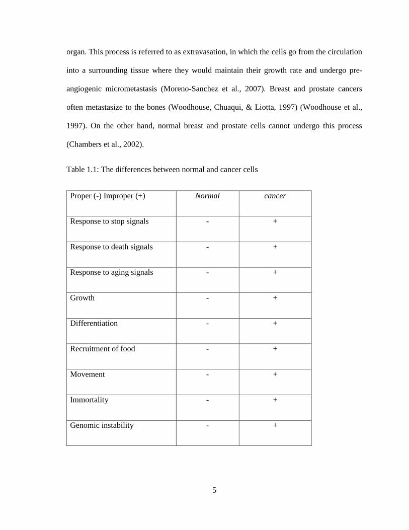
“Metastasis”, which consists of many distinct sequential steps (Table 1.1) (Figure 1.2)
(Judah Folkman, 2002b) summarized as follow: A cancer cell is shed from the primary
tumor into the body’s circulatory or lymphatic systems, a phenomenon known as
intravasation. The cells must then survive in the circulation till they are arrested in an
5
organ. This process is referred to as extravasation, in which the cells go from the circulation
into a surrounding tissue where they would maintain their growth rate and undergo pre-
angiogenic micrometastasis (Moreno-Sanchez et al., 2007). Breast and prostate cancers
often metastasize to the bones (Woodhouse, Chuaqui, & Liotta, 1997) (Woodhouse et al.,
1997). On the other hand, normal breast and prostate cells cannot undergo this process
(Chambers et al., 2002).
Table 1.1: The differences between normal and cancer cells
Proper (-) Improper (+) Normal cancer
Response to stop signals - +
Response to death signals - +
Response to aging signals - +
Growth - +
Differentiation - +
Recruitment of food - +
Movement - +
Immortality - +
Genomic instability - +

6
Figure 1.1: Forming tumor among normal cells

7
Figure 1.2: Metastasis of a cancer cell

8
1.4.The molecular biology of cancer
Cancer, in essence, is a heterogeneous disease caused by the accumulation of many
somatic mutations. Each single gene defect “causes” cancer, because each mutation drives a
wave of cellular proliferation, tumor size exaggeration, disorganization and malignancy (B.
Vogelstein & Kinzler, 1993). The majority of these mutations are acquired and “not
inherited”. They casually arise as consequences to damage at the genome level. Such
damage could result from an endogenous process, like a mistake occurring during DNA
replication; an attack by free radicals produced during metabolism; or the instability of
certain DNA bases. It might also involve exogenous agents, such as UV and ionizing
radiations, or chemical carcinogens (Bertram, 2000). Tumors show different behaviors in
different tissues. For example ,cancer cells of the pancreas are more aggressive than those
in the prostate (Pedraza-Fariña, 2006). The mechanism through which cells transform to
cancer is incompletely understood. However, it is assumed that it implicates many factors
which disturb the balance between cell proliferation and cell death, leading to abnormal cell
differentiation and proliferation. A cancer is believed to take place through the activation of
oncogenes, or the downregultion of the tumor-suppressor genes; which are linked to signal
transduction pathways (Cerveira & Bizarro, 2012). Oncogene activation from Proto-
oncogenes could be the result of gene amplification or chromosomal translocations. It
promotes cell growth and proliferation analogous to a car which still moves forward even
after the driver has removed his foot off the pedal. Tumor-suppressor genes work in the
opposite direction as they attenuate cell growth processes. Inactivation of these genes could
be the result of missense mutations, mutations resulting in truncated proteins, or epigenetic
silencing. It is analogous to having dysfunctional brakes in an automobile preventing the
car from stopping even when the driver tries to engage them (Vogelstein & Kinzler, 2004).
9
Five major pathways must be activated/inactivated in the process of cell genesis for
a normal cell to transform into a cancer cell (Bertram, 2000), contributing to the
development of independence from growth stimulatory signals; development of a refractory
state to growth inhibitory signals; development of resistance to programmed cell death,
apoptosis; development of an infinite proliferative capacity i.e., overcoming cellular
senescence; and development of an angiogenic potential i.e., the capacity to form new
blood vessels and capillaries.

10
Table 1.2 Cancer predisposition genes (Vogelstein & Kinzler, 2004)
Gene synonym(s) Syndrome Hereditary
pattern Pathway
Major heredity
tumor types
Tumor-suppressor genes
APC FAP Dominant APC Colon, thyroidal,
stomach, intestinal
AXIN2 Attenuated
polyposis Dominant APC Colon
CDH1 (E-cadherin)
Familial
gastric
carcinoma
Dominant APC Stomach
GPC3
Simpson-
Golabi-Behmel
syndrome
X-linked APC Embryonal
CYLD
Familial
cylindromatosi
s
Dominant APOP Pilotrichomas
EXT1,2
Hereditary
multiple
exostoses
Dominant GLI Bone
PTCH Gorlin
syndrome Dominant GLI
Skin,
medulloblastoma
SUFU
Medulloblasto-
ma
predisposition
Dominant GLI
Skin,
medulloblastoma
FH Hereditary
leiomyomatosis Dominant HIF1
Leiomyomas
SDHB, C, D Familial
paraganglioma Dominant HIF1
Paragangliomas,
pheochromocytoma
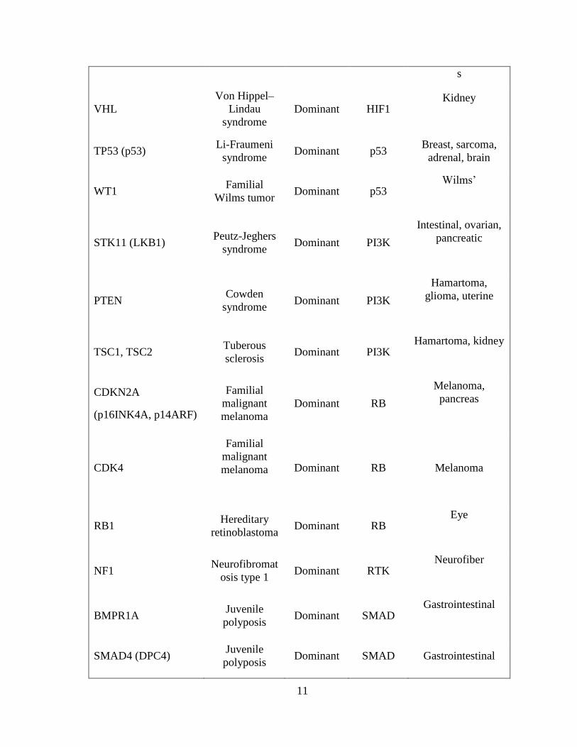
11
s
VHL
Von Hippel–
Lindau
syndrome
Dominant HIF1 Kidney
TP53 (p53) Li-Fraumeni
syndrome Dominant p53
Breast, sarcoma,
adrenal, brain
WT1 Familial
Wilms tumor Dominant p53
Wilms’
STK11 (LKB1) Peutz-Jeghers
syndrome Dominant PI3K
Intestinal, ovarian,
pancreatic
PTEN Cowden
syndrome Dominant PI3K
Hamartoma,
glioma, uterine
TSC1, TSC2 Tuberous
sclerosis Dominant PI3K
Hamartoma, kidney
CDKN2A
(p16INK4A, p14ARF)
Familial
malignant
melanoma
Dominant RB
Melanoma,
pancreas
CDK4
Familial
malignant
melanoma
Dominant RB Melanoma
RB1 Hereditary
retinoblastoma Dominant RB
Eye
NF1 Neurofibromat
osis type 1 Dominant RTK
Neurofiber
BMPR1A Juvenile
polyposis Dominant SMAD
Gastrointestinal
SMAD4 (DPC4) Juvenile
polyposis Dominant SMAD Gastrointestinal
12
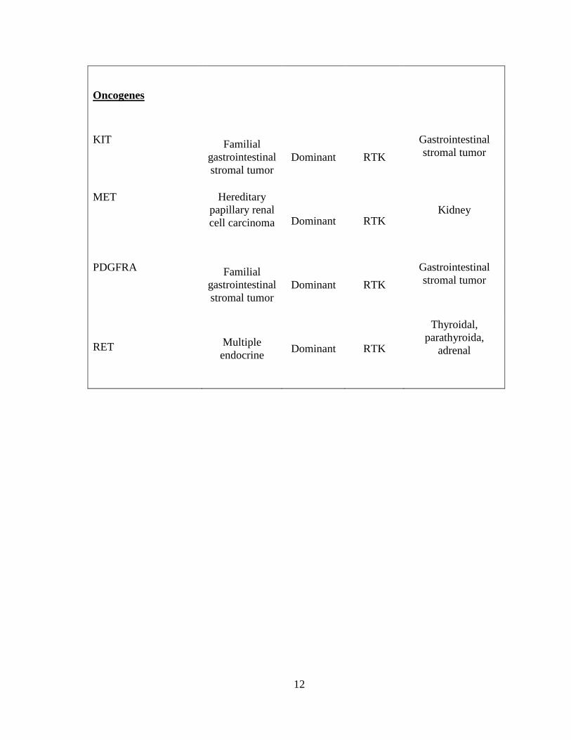
Oncogenes
KIT Familial
gastrointestinal
stromal tumor
Dominant RTK
Gastrointestinal
stromal tumor
MET Hereditary
papillary renal
cell carcinoma
Dominant RTK Kidney
PDGFRA Familial
gastrointestinal
stromal tumor
Dominant RTK
Gastrointestinal
stromal tumor
RET
Multiple
endocrine Dominant RTK
Thyroidal,
parathyroida,
adrenal
13
1.5.Apoptosis in cancer
Apoptosis, programmed cell death, is a physiological process of cellular suicide that is
integral to tissue homeostasis and is more feasibly observable during embryonic
development, whereas at which stage more than 10 billion cells are made in the human
body every day to balance those dying of apoptosis (Abud, 2004). It is characterized by a
number of cellular morphological changes, such as cell shrinkage, membrane blebbing,
organelle relocalization, nuclear fragmentation, chromatin condensation, and the
production of the ‘apoptotic bodies’, which are membrane-enclosed particles containing
intracellular material (Kerr et al., 1972). Another pathway of cell death is “necrosis”, which
lacks the features of apoptosis and autophagy. It could be differentiated from apoptosis by
certain changes in cellular mechanisms and morphological characteristics, including
dysfunctional organelles, collapsing mitochondria, ATP depletion, cellular swelling, and
extreme cellular disintegration, all of which end up forming an inflammation in the cells.
This is in contrast with apoptosis, which is not accompanied by any inflammation (Gerl &
Vaux, 2005; Kerr et al., 1972).
Triggering cells by apoptotic inducers, such as viral infections, bacterial toxins, free
radicals, death ligands (like Fas), glucocorticoids, chemotherapeutics, radiation therapy,
heat shock, growth factor withdrawal and/or irreparable DNA damage; makes many
distinctive changes in the cells (Elmore, 2007). One of the distinguished families of
proteins is called caspases. It plays a significant role in the initial stages of apoptosis. It
breaks down the main components required for normal cellular functions, like the proteins
which shape the cytoskeleton and the DNA repair enzymes. In addition, caspases induce
the degradation enzymes, like DNases, leading to DNA cleavage in the nucleus (Song &
Steller, 1999). The apoptotic process promotes different morphological changes that are

14
specific to the apoptotic cells. Mainly, cells shrink due to the cleavage of actin and laminin
in the cytoskeleton, and the nuclei take on a "horse-shoe" shape because of the breakdown
of their chromatin contents. As shrinking continues, the cells package themselves to
become easy victims for removal by macrophages. Changes in the apoptotic cells’
membranes are responsible for triggering the macrophagal response (Ghatage et al., 2012)
Table 1.3 the differences between apoptosis and necrosis
Apoptosis Necrosis
Physiologic, regulated
Cell shrinkage
Chromatin condensation
Preservation of Intracellular organelles
Membrane blebbing
apoptotic bodies
Organized chromatin
Digestion to small fragments (‘DNA ladders’)
Pathologic, unregulated
Cell swelling
Irregular chromatin
Clumping, dysfunction and destruction of organelles
Disruption of cellular membranes
Non-specific and random degradation of DNA
(‘DNA smears’)
15
1.5.1. Caspase family regulates apoptosis:
Caspases, which are cysteine-dependent, aspartate-specific proteases, are a family
of enzymes that is vital for the initiation and execution of apoptosis (Earnshaw, Martins, &
Kaufmann, 1999). They remain in their inactive proenzyme forms in most healthy cells;
however, caspases’ action is irreversible once activated by death signals; because the
protease cascade starts the initiation process, whereas the single-chain procaspases are
driven into a breakdown mechanism at specific aspartic acid residues, whereby the
inhibitory N-terminal pro-domain is removed to form the active proteases. Apoptotic
caspases are divided into two types: the first one comprises the initiators, or “the upstream
caspases”. They usually come with long pro-domains (>90 amino acid); and start the
cascade such as caspase-2,-8,-9,-10. The second one comprises the effectors, or “the
downstream caspases”. They come with short pro-domains, containing 20-30 residues, and
play a major role in the cleavage processes that disassemble the cell. An example is
caspase-3,-6,-7 (Thornberry & Lazebnik, 1998). Hence, the activation of an effector
caspase, like caspase-3 or caspase-7, is achieved by an initiator caspase, such as caspase-9.
However, this process requires many components, such as apoptosome, which is a protein
complex responsible for the activation of caspase-9. The latter then activates caspase-3,
leading to changes in morphology and biology in the apoptotic cell (Ghatage et al., 2012).
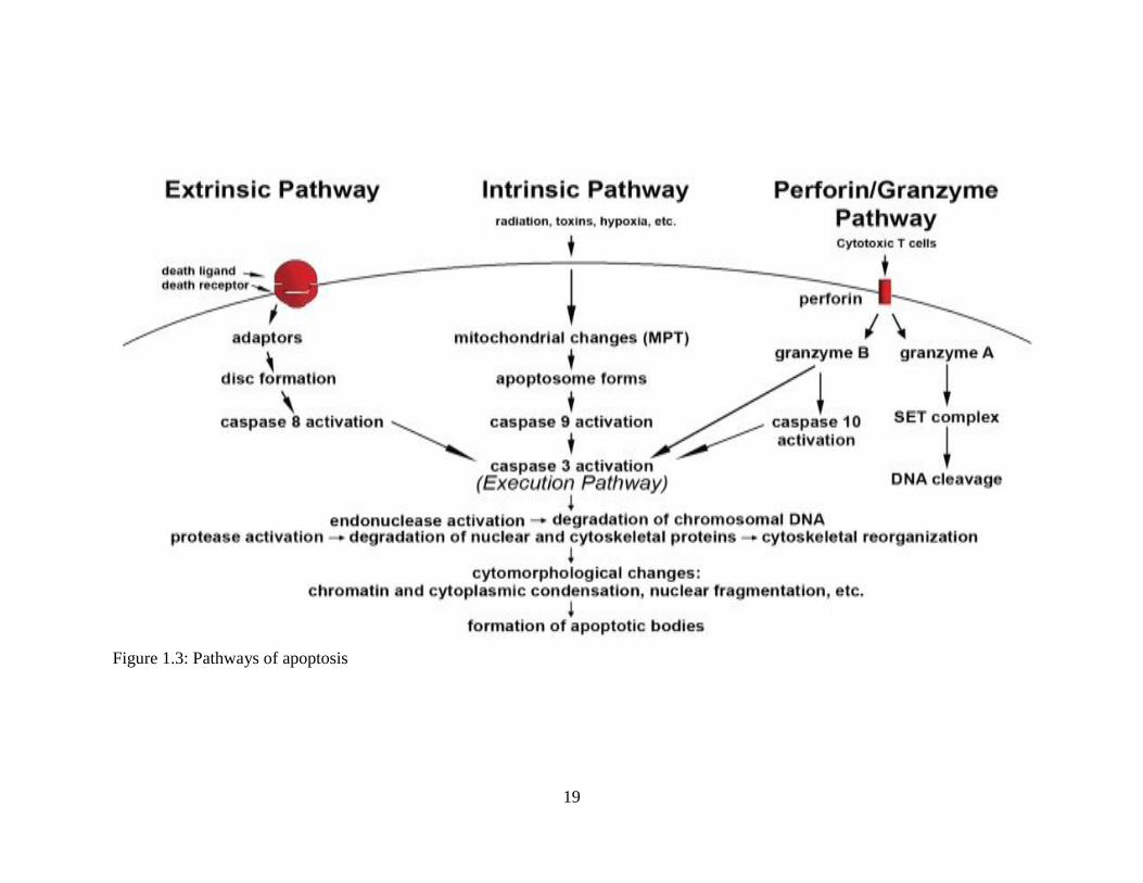
1.5.2. Mechanism of apoptosis
The mechanisms of apoptosis are highly complex and advanced processes. They
make an energy-dependent cascade of molecular events. There are two main pathways of
apoptosis: the intrinsic or the mitochondria-initiated apoptosis pathway, and the extrinsic or
16
death-receptor initiated apoptosis pathway (Igney & Krammer, 2002). However, there is an
additional pathway, referred to as the perforin/granzyme pathway, which could induce
programmed cell death via either granzyme B or granzyme A. All of the intrinsic, extrinsic,
and granzyme B pathways lead to the execution pathway, which starts with the cleavage of
caspase-3, leading to the morphological changes associated with apoptosis. On the other
hand, the granzyme A pathway activates a parallel caspase-independent cell death pathway
via single-strand DNA damage (Martinvalet et al., 2005).
The intrinsic pathway acts directly on targets within the cell. It may be induced by
many triggers, like DNA damage, hyperthermia, radiation, free radicals, hypoxia and/or a
viral infection (Abud, 2004), all of which could cause changes in the inner mitochondrial
membrane and lead to the release of pro-apoptotic proteins, such as cytochrome C,
Smac/DIABLO, AIF, Endo G and HtrA2/Omi, from the intermembrane space into the
cytosol. Consequently, activation of caspase-3 starts and the execution pathway begins. The
Bcl2 families play a primary role in mediating and controlling this process. The Bcl2
family has 4 homologous (BH) domains, which might either be anti-apoptotic or pro-
apoptotic. The pro-apoptotic proteins in the cell include Cl-10, Bax, Bak, Bid, Bad, Bim,
Bik, and Blk, which may be localized at different parts of the cell. The anti-apoptotic
members of the Bcl2 family are Bcl2 and Bclxl. Bcl2 are localized completely within the
intracellular membranes and in the cytosol (Ghatage et al., 2012). These protein are key in
determining whether the cell must perform apoptosis or not as they regulate the release of
cytochrome C from the mitochondria in a process labeled as mitochondrial outer membrane
permeabilization (MOMP).This is accomplished by an interaction between pro-apoptotic
Bcl2 and membrane pores and channels which might be accompanied by a loss of
mitochondrial membrane potential (Saelens et al., 2004). When the pro-apoptotic Bcl2 units
17
are released from the inter-membrane spaces into the cytosol, the main caspase-dependent
group, which comprises cytochrome c, Smac/DIABLO and HtrA2/Omi, works to induce
the caspase cascade. It binds to and activates Apaf-1 and procaspse-9, leading to the
formation of the apoptosomes, which result in the activation of caspase-9 and the effectors,
caspase-3 and caspase-7; and this marks the end of the apoptotic pathway (Fulda &
Debatin, 2004).
The extrinsic Pathway is a rapid mechanism that results in apoptosis. It is a caspase-
dependent process which requires transmembrane receptor-mediated interactions, like the
reactions between the ligands corresponding to the death receptors, including FasL/FasR,
TNF-α/TNFR1, Apo3L/DR3, Apo2L/DR4 and Apo2L/DR5 (Ashkenazi & Dixit, 1998).
After a ligand and a death receptor bind, the receptor changes its conformation (Abud,
2004) allowing them to interact with specific intracellular adaptor proteins, leading to
changes in the “death domain”; which is made up of an extracellular domain and a
cytoplasmic domain, and plays an essential role in transmitting the death signal from the
surface of the cell to intracellular signaling pathways (Ashkenazi & Dixit, 1998). For
example, TRADD, a TNFR-associated death domain, recruits many apoptotic proteins to
the receptor, at which point, the protein complex formed is often called the Death-Inducing
Signaling Complex (DISC). DISC results in the autocatalytic activation of caspase-8, and
the initiation of apoptosis. Alternatively, FADD, a FAS-associated death domain, could
form a link with TRADD, which would lead to the cleavage of pro-caspase 8, and the
induction of apoptosis (Kischkel et al., 1995; Wajant, 2002).
The perforin/granzyme pathway is an extrinsic pathway used by cytotoxic T
lymphocytes (CTLs) to kill specific cells, such as tumor and virus-infected cells (Brunner et

18
al., 2003). Its mechanism is based on the release of perforin, proteases granzyme A and
granzyme B. The perforin units form transmembrane pore proteins in the attacked cells’
membranes (Trapani & Smyth, 2002). Thus, perforin proteins enable the entry of granzyme
A and granzyme B, which are considered the main components inside the granules.
Granzyme B units activate pro-caspase-10 and caspase-3, which results in the induction of
apoptosis (Sakahira et al., 1998). On the other hand, granzyme A, also released by T cells,
induces apoptosis via a caspase-independent mechanism as it indirectly activates a tumor
suppressor gene product, an effect which is called DNA nicking. The expression of this
product, known as DNAse NM23-H1, is diminished in cancer cells (Fan et al., 2003). The
activation of NM23-H1 occurs by cleavage of the proteins which inhibits NM23-H1 inside
of the SET complex. Hence, the activation process promotes the release of NM23-H1,
causing DNA fragmentation (Lieberman & Fan, 2003)
19
Figure 1.3: Pathways of apoptosis
20
1.6.Colorectal carcinoma
Colorectal carcinoma (CRC) is the term used to describe tumor formation in the large
intestine, colon and/or the rectal area. It has the third highest incidence rate among all
cancer types globally, preceded only by lung and breast cancers; and it is considered to be
one of the leading causes of death among patients of both sexes. The risk rises after the age
of 50. However, it could be managed if detected early. (Gustin & Brenner, 2002).
Specialists have divided colorectal cancer into four main stages in terms of the
morbidity of the disease: Dukes’ A, B, C and D. This pathological staging system has been
in use for more than 50 years. The first stage, Dukes' A, refers to a condition whereby
contiguous lesions contiguous exist only in the innermost lining bowel wall of the colon or
the rectum, without reaching the muscularis. Dukes’ B refers to cases in which the cancer
penetrates the muscularis. Dukes' C describes conditions whereby the cancer has spread to
the lymph nodes around the bowel area. Dukes' D stage is the most dangerous and death-
leading phase as cancer would have spread to other organs in the body, such as the lungs
and/or the spleen (Dukes, 1932).
Colon cancer is a multi-step disease. It occurs as a consequence of a series of
pathological changes that transform normal epithelial cells of the colon into invasive
carcinoma. Several studies have shown that the multi-step process of colon cancer is
accompanied by certain mutations affecting the adenomatous polypsis coli (APC) gene and
cyclooxygenase-2 (COX-2), as well as K-ras mutations (caused by Kirsten-rat sarcoma
virus), loss of the 18q21 gene, microsatellite instability, mutations in the transforming
growth factor-β II receptor (TGFβR2), and stabilization and translocation of β-catenin
(Kanwar et al., 2010; Kobayashi et al., 2000)
21
1.7.Major signaling pathways in colon carcinogenesis
1.7.1. Wnt /β-catenin signaling pathway
Wnt (Wingless and INT-1) – which refers to a large family of secretory
glycoproteins – signaling pathway has a significant role in embryogenesis, cell-to-cell
signaling, and adult tissue homeostasis (Clevers, 2006). Wnt/β-catenin, which is considered
the most important Wnt pathway, regulates the transcription of cofactor β-catenin,
and, therefore, enhances the expression of its respective gene (Logan & Nusse, 2004).
Abnormal Wnt/β-catenin signaling is a primary factor in the development of
tumorigenesis, degenerative disorders, osteoporosis and aging (Moon et al., 2004). It is
well known that over 90% of colon cancer cases result from the accumulation of Wnt and
β-catenin due to mutations of the APC tumor suppressor gene, or oncogenic mutations of β-
catenin (Giles et al., 2003; Luu et al., 2004). An increase in the levels of β-catenin, a key
component of the Wnt signaling pathway, has been observed in many types of human
cancer, such as prostate cancer, colon cancer, melanoma, and breast cancer (Chien et al.,
2009; Luu et al., 2004). The Wnt /β-catenin complexes promote the expression of many
oncogenes, like the c-Myc, cyclin D1 and MMP genes, which are essential for tumor
angiogenesis and cancer development (Dihlmann & Doeberitz, 2005). Thus, compounds
which target the Wnt signaling pathway and downregulate it could make for a potential
treatment of cancer (Luu et al., 2004; Tetsu & McCormick, 1999).
1.7.2. Notch Signaling Pathway
Notch signaling plays a fundamental role in the development of normal cells and
tissues, like those of the cardiovascular system, central nervous system, and endocrine
system. Its role is also implicated in bone development and tissue renewal (Allenspach et
22
al., 2007). In addition, Notch is considered as a conserved cell signaling mechanism. It is
associated with different effects on cellular processes, such as cell fate determination,
differentiation, cell proliferation, programmed cell death (apoptosis), adhesion, epithelial-
mesenchymal transition, migration, and angiogenesis. Regulation of these processes can
become faulty in Notch-mediated pathological situations (Artavanis-Tsakonas, Rand, &
Lake, 1999; Bolós, Grego-Bessa, & de la Pompa, 2007). In mammals, there are four Notch
proteins (Notch 1–4) and one Notch membrane-bound type 1 receptor with one
transmembrane domain (Baron, 2003; Bolós et al., 2007). Preclinical and clinical studies
have attributed a pro-oncogenic function to Notch signaling in many solid tumors (Rizzo et
al., 2008). An increase in the rate of Notch pathway signaling is associated with many kinds
of cancers, such as melanoma and lung, breast, pancreas, renal and colon cancers (Collins
et al., 2004; Strizzi et al., 2009).
An in vivo study has demonstrated strong synergies between Notch and Wnt signals
in colon cancer that result in the formation of intestinal adenomas and, more significantly,
colorectal tumor (Fre et al., 2009; Hayward et al., 2008). Since Notch pathway is
complicated, a better understanding of its interactions with other pathways is required for
future therapeutic applications (Bolós et al., 2007; Rizzo et al., 2008).
1.7.3. P53 signaling pathway
The p53 tumor suppressor gene was identified in 1979. It performs an essential role
in the activation of apoptosis and cell arrest pathways. It has been labeled as "the guardian
of the genome" because of its role in preventing genome mutation (Fridman & Lowe, 2003;
Read & Strachan, 1999) Activation of p53 by DNA damage, aberrant oncogene expression
or hypoxia causes an induction in cell-cycle checkpoints, DNA repair, cellular senescence,
23
and apoptosis. Hence, disruption of this pathway or a mutation in p53 may end in
checkpoint defects, genomic instability, inappropriate survival, cancer, neurodegeneration,
ischemia, cholestasis and/or atherosclerosis (Amaral et al., 2010). Many factors make p53
gene mutation very prevalent in human malignancies. First of all, a single base change in
the coding sequence results in a dysfunctioning P53. Secondly, a single abnormal p53 allele
or allele loss can alter the phenotype of the p53 proteins. Thirdly, since p53 plays a role in
multiple pathways, defects in its coding gene make the cell’s defenses completely
diminished against carcinogenesis (Bellamy, 1997).
It is well-established that any mutation in the p53 tumor suppressor gene causes its
function to be lost, leading to profound proliferation of cancer cells and chemoresistance
(Beroud & Soussi, 2003; Hussain & Harris, 1998; Wallace-Brodeur & Lowe, 1999). On the
other hand, hyper-activation of p53 product has been associated with many different
diseases, like arthritis, multiple sclerosis, and neuropathies (Mattson et al., 2001; Wosik et
al., 2003). Moreover, in vivo studies have suggested that acute p53 activation is the
mechanism behind the side effects of cancer chemotherapy (Komarova & Gudkov, 2001;
Tyner et al., 2002).
1.7.4. TGF-β Signaling Pathway
TGFβ and its receptors are extensively expressed in all tissues; and TGFβ signaling
has a dual role in human diseases (Massagué, 1998). In cancer, TGFβ acts as both a tumor
suppressor and an oncogene which promotes tumor metastasis (Katz et al., 2013). Its tumor
suppressive effects, which are observed in the early stages of cancer and in normal cells,
include an inhibition of cell proliferation, an induction of apoptosis, and an inhibition of
cell immortalization. In contrast, the tumor promoting effects of TGFβ include invasion,

24
migration, cell adhesion, tumor metastasis and an induction of Epithelial-Mesenchymal
Transition (EMT), which has been observed in invasive and aggressive tumors (Massagué,
1998; Xu et al., 2009) . Furthermore, as a tumor progresses and increases in size, it secretes
a huge amount of autocrine TGFβ, which gets released into the surrounding area. The
increased levels of TGFβ affect the tumor and the vicinity, and promote
immunosuppression and angiogenesis, inhibit cell adhesion, and promote the metastatic
processes (Gorsch et al., 1992). Using antibodies to target the TGF-β proteins or receptors
has shown a promising anticancer effect. However, the dual role of TGFβ translates into
potential side effects for any novel compounds targeting this pathway (Massagué, 2008).
1.7.5. Cell cycle (pRB/ E2F) signaling pathway
The main element in this pathway is the retinoblastoma tumor suppressor protein
(pRB), whose role is to induce apoptosis and inhibit cell cycle progression (Robert A
Weinberg, 1995). pRB stops premature G1/S transitions by physical interaction with
several transcription factors, namely E2F, which is vital for the activation of the S-phase
genes (Bartek et al., 1996). It is believed that mutations in the Rb gene are present in the
vast majority of human cancer conditions (Hunter, 2002). For example, such mutations
have been detected in cases of osteosarcoma, small cell lung carcinoma, breast carcinoma
and other cancer types (Nevins, 2001). In addition, a downregulated E2F activity has been
reported in human tumor cases, and has been attributed to many mechanisms, such as a
functional loss of pRB; amplification of cyclin D, a cyclin-dependent kinase inhibitor that
inhibits the phosphorylation of pRB, which promotes the phosphorylation of pRB; a loss of
p16; and an expression of E7, the human papillomavirus (HPV) oncoprotein, which
disrupts the pRB–E2F complexes (Sherr & McCormick, 2002).