Anti-malaria studies on Azadirachta indica leaves in...
14
© 2018 Journal of Pharmacy & Pharmacognosy Research, 6 (3), 191-204, 2018 ISSN 0719-4250 http://jppres.com/jppres Original Article | Artículo Original _____________________________________ Therapeutic effects of Azadirachta indica A.Juss. leaves in malaria- induced male Wistar rats [Efectos terapéuticos de hojas de Azadirachta indica A.Juss. en ratas Wistar machos infectadas con malaria] Ngozi K. Achi 1* , Chimaraoke Onyeabo 1 , Daniel A. Nnate 2* , Chima A. Ekeleme-Egedigwe 3 , Igwe K. Kalu 4 , Ikedichim C. Chibundu 1 , Grace C. Wokoma 1 1 Department of Biochemistry, College of Natural Sciences, Michael Okpara University of Agriculture, Umudike, PMB 7267, Umuahia, Abia State, Nigeria. 2 Department of Biochemistry, Faculty of Biological and Physical Sciences, Abia State University, Uturu, Abia State, Nigeria. 3 Department of Chemistry/Biochemistry, Faculty of Science, Federal University Ndufu-Alike, Ikwo, PMB 1010 Abakaliki, Ebonyi State, Nigeria. 4 Department of Veterinary Biochemistry and Pharmacology, Faculty of Veterinary Medicine, Michael Okpara University of Agriculture, Umudike, Abia State, Nigeria. *E-mail: [email protected], [email protected] Abstract Resumen Context: Azadirachta indica has long been used in herbal or folk medicine as a remedy for the treatment of malaria and the administration of herbal preparations has raised concerns on their toxicity. Aims: To determine the phytochemical content of A. indica and its therapeutic effect on indices of clinical importance in malaria-induced male Wistar rats. Methods: Plant material was extracted with ethanol, and the lethal dose (LD50) on the rats was determined before the study. Normal and Plasmodium berghei infected rats were divided into eight groups of five rats each with groups 1 and 2 serving as normal and disease control respectively. Lumartem was administered twice daily at oral therapeutic doses of artemether/lumefantrine (2/12 mg/kg) and plant extract at 300 and 500 mg/kg body weight. After 5 days of treatment, all the animals were sacrificed according to their groups for the experimental analysis. Results: The plant extract was considered safe with LD50 > 5000 mg/kg body weight. Quantitative phytochemical studies showed a high concentration of alkaloids, tannin, and terpenoids. Treatment with both extracts of A. indica and lumartem in malaria-infected rats showed a slight reduction in triglycerides while total cholesterol, HDL, and LDL levels increased significantly (p < 0.05). Increase in body weight of rats treated with A. indica was dependent on the concentration of extract administered. Treatment of malaria with the extract and lumartem resulted in a slight restoration of the hematological values. Conclusions: This study shows that both Azadirachta indica and lumartem was practically safe and well tolerated. Contexto: Azadirachta indica se ha utilizado durante mucho tiempo en la medicina herbal para el tratamiento de la malaria y la administración de preparados de hierbas ha generado preocupaciones sobre su toxicidad. Objetivos: Determinar el contenido fitoquímico de A. indica y su efecto terapéutico en ratas Wistar machos infectadas con malaria. Métodos: El material vegetal se extrajo con etanol y la dosis letal (LD50) en las ratas se determinó antes del estudio. Las ratas infectadas normales y de Plasmodium berghei se dividieron en ocho grupos de cinco ratas cada uno, con los grupos 1 y 2 como normales y control de la enfermedad, respectivamente. Lumartem se administró oralmente dos veces al día a dosis terapéuticas de arteméter/lumefantrina (2/12 mg/kg) y extracto de planta a 300 y 500 mg/kg de peso corporal. Después de 5 días de tratamiento, todos los animales fueron sacrificados de acuerdo con sus grupos para el análisis experimental. Resultados: El extracto de la planta se consideró seguro con LD50 > 5000 mg/kg. Los estudios fitoquímicos cuantitativos mostraron una alta concentración de alcaloides, taninos y terpenoides. El tratamiento con ambos extractos de A. indica y lumartem en ratas infectadas con malaria mostró una ligera reducción en los triglicéridos, mientras que los niveles de colesterol total, HDL y LDL aumentaron significativamente (p <0.05). El aumento en el peso corporal de ratas tratadas con A. indica dependió de la concentración de extracto administrado. El tratamiento de la malaria con el extracto y lumartem dio como resultado una ligera restauración de los valores hematológicos. Conclusiones: Este estudio muestra que tanto Azadirachta indica como lumartem fueron prácticamente seguros y bien tolerados. Keywords: anti-malaria; Azadirachta indica; lethal dose; lumartem; phytochemical; Plasmodium. Palabras Clave: anti-malaria; Azadirachta indica; dosis letal; fitoquímica; lumartem; Plasmodium. ARTICLE INFO Received: August 16, 2017. Received in revised form: February 2, 2018. Accepted: February 12, 2018. Available Online: March 22, 2018. Declaration of interests: The authors declare no conflict of interest. Funding: The authors confirm that the project has not funding or grants.
Transcript of Anti-malaria studies on Azadirachta indica leaves in...

© 2018 Journal of Pharmacy & Pharmacognosy Research, 6 (3), 191-204, 2018 ISSN 0719-4250
http://jppres.com/jppres
Original Article | Artículo Original
_____________________________________
Therapeutic effects of Azadirachta indica A.Juss. leaves in malaria-induced male Wistar rats
[Efectos terapéuticos de hojas de Azadirachta indica A.Juss. en ratas Wistar machos infectadas con malaria]
Ngozi K. Achi1*, Chimaraoke Onyeabo1, Daniel A. Nnate2*, Chima A. Ekeleme-Egedigwe3, Igwe K. Kalu4, Ikedichim C. Chibundu1, Grace C. Wokoma1
1Department of Biochemistry, College of Natural Sciences, Michael Okpara University of Agriculture, Umudike, PMB 7267, Umuahia, Abia State, Nigeria. 2Department of Biochemistry, Faculty of Biological and Physical Sciences, Abia State University, Uturu, Abia State, Nigeria.
3Department of Chemistry/Biochemistry, Faculty of Science, Federal University Ndufu-Alike, Ikwo, PMB 1010 Abakaliki, Ebonyi State, Nigeria. 4Department of Veterinary Biochemistry and Pharmacology, Faculty of Veterinary Medicine, Michael Okpara University of Agriculture, Umudike, Abia State, Nigeria.
*E-mail: [email protected], [email protected]
Abstract Resumen
Context: Azadirachta indica has long been used in herbal or folk medicine as a remedy for the treatment of malaria and the administration of herbal preparations has raised concerns on their toxicity.
Aims: To determine the phytochemical content of A. indica and its therapeutic effect on indices of clinical importance in malaria-induced male Wistar rats.
Methods: Plant material was extracted with ethanol, and the lethal dose (LD50) on the rats was determined before the study. Normal and Plasmodium berghei infected rats were divided into eight groups of five rats each with groups 1 and 2 serving as normal and disease control respectively. Lumartem was administered twice daily at oral therapeutic doses of artemether/lumefantrine (2/12 mg/kg) and plant extract at 300 and 500 mg/kg body weight. After 5 days of treatment, all the animals were sacrificed according to their groups for the experimental analysis.
Results: The plant extract was considered safe with LD50 > 5000 mg/kg body weight. Quantitative phytochemical studies showed a high concentration of alkaloids, tannin, and terpenoids. Treatment with both extracts of A. indica and lumartem in malaria-infected rats showed a slight reduction in triglycerides while total cholesterol, HDL, and LDL levels increased significantly (p < 0.05). Increase in body weight of rats treated with A. indica was dependent on the concentration of extract administered. Treatment of malaria with the extract and lumartem resulted in a slight restoration of the hematological values.
Conclusions: This study shows that both Azadirachta indica and lumartem was practically safe and well tolerated.
Contexto: Azadirachta indica se ha utilizado durante mucho tiempo en la medicina herbal para el tratamiento de la malaria y la administración de preparados de hierbas ha generado preocupaciones sobre su toxicidad.
Objetivos: Determinar el contenido fitoquímico de A. indica y su efecto terapéutico en ratas Wistar machos infectadas con malaria.
Métodos: El material vegetal se extrajo con etanol y la dosis letal (LD50) en las ratas se determinó antes del estudio. Las ratas infectadas normales y de Plasmodium berghei se dividieron en ocho grupos de cinco ratas cada uno, con los grupos 1 y 2 como normales y control de la enfermedad, respectivamente. Lumartem se administró oralmente dos veces al día a dosis terapéuticas de arteméter/lumefantrina (2/12 mg/kg) y extracto de planta a 300 y 500 mg/kg de peso corporal. Después de 5 días de tratamiento, todos los animales fueron sacrificados de acuerdo con sus grupos para el análisis experimental.
Resultados: El extracto de la planta se consideró seguro con LD50 > 5000 mg/kg. Los estudios fitoquímicos cuantitativos mostraron una alta concentración de alcaloides, taninos y terpenoides. El tratamiento con ambos extractos de A. indica y lumartem en ratas infectadas con malaria mostró una ligera reducción en los triglicéridos, mientras que los niveles de colesterol total, HDL y LDL aumentaron significativamente (p <0.05). El aumento en el peso corporal de ratas tratadas con A. indica dependió de la concentración de extracto administrado. El tratamiento de la malaria con el extracto y lumartem dio como resultado una ligera restauración de los valores hematológicos.
Conclusiones: Este estudio muestra que tanto Azadirachta indica como lumartem fueron prácticamente seguros y bien tolerados.
Keywords: anti-malaria; Azadirachta indica; lethal dose; lumartem; phytochemical; Plasmodium.
Palabras Clave: anti-malaria; Azadirachta indica; dosis letal; fitoquímica; lumartem; Plasmodium.
ARTICLE INFO Received: August 16, 2017. Received in revised form: February 2, 2018. Accepted: February 12, 2018. Available Online: March 22, 2018. Declaration of interests: The authors declare no conflict of interest. Funding: The authors confirm that the project has not funding or grants.
Achi et al. Anti-malaria studies on Azadirachta indica leaves in rats
http://jppres.com/jppres J Pharm Pharmacogn Res (2018) 6(3): 192
INTRODUCTION
Azadirachta indica A.Juss. (Meliaceae) known by its common name as Neem tree is native to Asian countries. The people of India have long regarded the neem tree as a remedy for sickness (Tiwari et al.,
2014). With each part of either its leaves, bark, stem or root having medicinal properties, a large number of biologically active compounds have been isolated from the plant (Biswas et al., 2002). It has also been reported that the leaf extract is effective as analgesic, anthelminthic, antibacterial, antifungal, antihyperglycemic, anti-inflammatory, antiviral, antimalarial, antipyretic, insecticidal, hypercho-lesteremic and hypoglycemic agents (Parotta, 2001;
Maragathavalli et al., 2012). A. indica has been extensive-ly reported as being effective in the treatment of malaria caused by various strains of Plasmodium parasite even those resistant to traditional anti-malaria (Nwafor et al., 2003). The use of herbal medications in the treatment of malaria is popular in many parts of Africa and Asia where malaria in-festation is endemic. The plant is well identified for its high antioxidant activity; its active molecules could also be chemically manipulated into effective drugs, which can be wildly used in the treatment of certain ailments (Bamidele et al., 2017). Chemical compounds in plants mediate their effect on the human body through processes identical with compounds in conventional drugs. Thus herbal medicines do not differ greatly from conventional drugs regarding how they work (Vickers et al., 2001). This enables herbal medicines to have beneficial pharmacology but also gives them the same potential as conventional pharmaceutical drugs to cause harmful side effects (Ajero and Mbagwu, 2005).
Malaria is a tropical disease that poses serious problems on human being especially in tropical countries where the environment provides condu-cive ground for the parasite to thrive. Malaria is re-sponsible for at least 750,000 deaths a year, mostly in young children in Africa (Greenwood et al., 2012;
WHO, 2013). It is a disease of global importance that results in 300-600 million cases annually, and an estimated 2.2 billion people are at risk of infection (Singh et al., 2010). Children under five years and preg-nant women are particularly vulnerable to the dis-ease due to their weaker immune systems (WHO,
2000; Lamb, 2012). An estimated 438,000 people died of malaria in 2015, with over 90% of these deaths occurring in sub-Saharan Africa, and nearly all of the others occurring in South-East Asia and South America (WHO, 2015). Treatment of malaria with Af-rican herbs and medicines has been in existence long before the arrival of western drugs (Ngarivhume
et al., 2015). The neem tree appears to have originated in India and South East Asia and is now spread throughout tropical and subtropical regions like Africa hence the popularity in the treatment of ma-laria.
Lumartem is a fixed-dose Artemisinin Combina-tion Therapy of artemether and lumefantrine, with each tablet containing 20 mg artemether and 120 mg lumefantrine. Artemether is a fat-soluble arte-misinin derivative (Nosten and Brasseur, 2002). Artemisinin is a natural product derived from the Chinese medicinal plant Artemisia annua L., known as qinghao in China and as sweet wormwood in Eu-rope and America (Lefèvre and Thomsen, 1999). Lu-mefantrine, previously known as benflumetol, is a synthetic dibutyl aminoethanol substituted fluo-rene racemate and is structurally related to quinine (Anyasor et al., 2013). Artemether acts quickly to rapid-ly reduce the affliction caused by the parasite and also the resolution of clinical symptoms by interfer-ing with parasite transport proteins, disruption of mitochondrial function, inhibition of angiogenesis and modulation of host immune function (Anyasor et
al., 2013). On the other hand, lumefantrine prevents recrudescence, acts slowly and serves as a longer-acting agent to eliminate remaining parasites (Ezzet
et al., 1998). This combination makes it highly effective especially in resistant strains of P. falcipa-rum (Lefèvre and Thomsen, 1999). Both artemether and lumefantrine are well absorbed after oral admin-istration. Administration with fat-containing foods improves bioavailability by more than 2-fold for ar-temether and up to 16-fold for lumefantrine in adults (Djimdé and Lefèvre, 2009).
Many anti-malarial drugs like quinine and arte-misinin were isolated from plants, and because of the increased resistance of the parasite to estab-lished drugs, there is need to investigate more chemical compounds within traditional plants. The use of plant extracts without a standard dosage

Achi et al. Anti-malaria studies on Azadirachta indica leaves in rats
http://jppres.com/jppres J Pharm Pharmacogn Res (2018) 6(3): 193
coupled with non-availability of adequate scientific studies on their safety has raised concerns about their toxicity (Achi and Ohaeri, 2012). Understanding the biochemical effects and safety of these medici-nal plants on its users is also very important be-cause limited toxicological information on these plants has been a source of fear for its users. Animal models have become a useful tool in the study of many anti-malarial drugs. The rodent parasite Plasmodium berghei is effective in testing and moni-toring the efficacy of anti-malarial drugs against human malaria parasite (Langhorne et al., 2011). However, in this study, the authors were not concerned about the activity of A. indica against P. berghei malaria parasite on murine models as several studies have reported a positive result (Obih
and Makinde, 1985; Priyanka et al., 2013) while others reported no activity against the rodent parasite (Rochanakij et al., 1985; Farahna et al., 2010). The study is aimed at giving a better understanding of A. indica in the treatment of malaria by comparing its effect with a synthetic anti-malarial drug lumartem on the body weights, lipid and hematological parame-ters in malaria-infected and normal Wistar rats, which will serve as a baseline for further studies in humans
MATERIAL AND METHODS
Collection and identification of plant materials
Fresh leaves of Azadirachta indica were carefully collected with hands covered in gloves from the school premises of the Michael Okpara University of Agriculture, Umudike (5°28′50.957″ N and 7°32′ 45.921″ E). The plant was identified by Mr. Ibe K. Ndukwe of the Department of Forestry and Envi-ronmental Management, Michael Okpara Universi-ty of Agriculture, Umudike, Abia State, Nigeria. A voucher specimen with number MOUAU/1631529 was kept at the herbarium of the same department for future reference.
Drug and chemicals
Lumaterm (artemether 20 mg + lumefantrin 120 mg) was purchased from a Pharmacy in Umuahia, Abia state, Nigeria. All other chemicals used are of analytical grade.
Preparation of plant extract
The leaves of Azadirachta indica were sorted, washed thoroughly and air dried on a laboratory slab for four weeks at room temperature (25 – 270C). The dry leaves were pulverized into a fine powder using an electric blender (Corona – Ref, 121, Landers). One thousand grams of the powdered leaves was extracted with 1500 mL of 88% ethanol for 48 h in a stopped bottle with occasional stirring at room temperature. It was then sieved using a muslin cloth and filtered using Whatman No. 1 fil-ter paper (Sigma, Aldrich). This process was repeated three times. The extract was concentrated under reduced pressure at 40°C for four days using a water bath (HH-W420). Various concentrations were prepared from the resultant crude extract, dis-solved in distilled water, and administered orally to determine the LD50 and the treatment of animals.
Quantitative determination of phytochemical constituents of leaves of Azadirachta indica
Determination of tannin
An analytical method for quantitative determination of tannin was done according to Harborne (1973) with modification by dissolving 25 g of sodium tungstate (Na2WO4) in 30 mL of distilled water to form Folin-Denis reagent. To the reagent prepared above, 5 g of phosphomolybdic acid (H3PMo12O40) and 20 mL of orthophosphoric acid (H3PO4) were added. Ninety minutes reflux of the mixture was carried out, cooled, and diluted to 400 mL with distilled water. Five grams of powder (sample) in a conical flask was added to 100 mL of distilled water. This was boiled gently for 1 h on an electric hot plate and filtered using number 42 (125 mm) Whatman filter paper in a 100 mL volu-metric flask. Addition of 5.0 mL Folin-Denis reagent and 10 mL of saturated Na2CO3 solution into 50 mL of distilled water and 10 mL of diluted extract (ali-quot volume) was carried out after being pipetted into a 100 mL conical flask for color development. The solution was allowed to stand for 45 min in a water bath at a temperature of 25°C after thorough agitation. With the aid of a Spectrum Lab 23 A spec-trophotometer optical density (brand, city, compa-ny) was measured at 700 nm and compared on a

Achi et al. Anti-malaria studies on Azadirachta indica leaves in rats
http://jppres.com/jppres J Pharm Pharmacogn Res (2018) 6(3): 194
standard tannic acid curve. The following equation (1) was used in the calculation.
𝑇𝑎𝑛𝑖𝑐 𝑎𝑐𝑖𝑑 =𝐶 ∗ 100𝑉
𝐴𝑙𝑖𝑞𝑢𝑜𝑡 𝑣𝑜𝑙𝑢𝑚𝑒 ∗ 𝑤𝑒𝑖𝑔ℎ𝑡 𝑜𝑓 𝑠𝑎𝑚𝑝𝑙𝑒
(1)
Where: C was concentration of tannic acid read off the graph V was the volume of extract
Determination of alkaloids
Quantitative determination of alkaloid was ac-cording to the methodology by Harborne (1973). Ex-actly, 200 mL of 10% acetic acid in ethanol was add-ed to the powder sample of leaves of A. indica (5.0 g) in a 500 mL beaker and allowed to stand for 3 hours. The extract was concentrated in a water bath to one-quarter of the original volume followed by addition of 15 drops of concentrated ammonium hydroxide dropwise to the extract until the precipitation was complete immediately after filtration. After 3 h of mixture sedimentation, the supernatant was discarded, and the precipitates were washed with 20 mL of 0.1 M of ammonium hy-droxide and then filtered using Gem filter paper (12.5 cm). Using electronic weighing balance Model B-218 (B-218 Series, Jiangsu, China), the residue was dried in an oven, and the percentage of alkaloid was expressed mathematically as equation (2).
%𝐴𝑙𝑘𝑎𝑙𝑜𝑖𝑑 = 100𝑊𝑒𝑖𝑔ℎ𝑡 𝑜𝑓 𝑎𝑙𝑘𝑎𝑙𝑜𝑖𝑑
𝑊𝑒𝑖𝑔ℎ𝑡 𝑜𝑓 𝑠𝑎𝑚𝑝𝑙𝑒
(2)
Determination of flavonoids
Flavonoid determination was by the method reported by Boham and Kocipai (1994). Exactly 50 mL of 80% aqueous methanol added was added to 5.0 g of sample in a 250 mL beaker, covered, and allowed to stand for 24 h at room temperature. Af-ter discarding the supernatant, the residue was re-extracted (three times) with the same volume of ethanol. Whatman filter paper number 42 (125 mm) was used to filter the whole solution of the sample. Each sample filtrate was later transferred into a crucible and evaporated to dryness over a water bath. The content in the crucible was cooled in a desiccator and weighed until constant weight was obtained. The percentage of flavonoid was calculated as equation (3):
%𝐹𝑙𝑎𝑣𝑜𝑛𝑜𝑖𝑑 = 100𝑊𝑒𝑖𝑔ℎ𝑡 𝑜𝑓 𝑓𝑙𝑎𝑣𝑜𝑛𝑜𝑖𝑑
𝑊𝑒𝑖𝑔ℎ𝑡 𝑜𝑓 𝑠𝑎𝑚𝑝𝑙𝑒
(3)
Determination of saponin
Saponin quantitative determination was carried out using the method reported by Obadoni and Ochuko (2002). Exactly, 100 mL of 20% aqueous eth-anol was added to 5.0 g of the powder sample of A. indica leaves in a 500 mL conical flask. The mixture was heated over a hot water bath for 4 h with con-tinuous stirring at a temperature of 55°C. The resi-due of the mixture was re-extracted with another 100 mL of 20% aqueous ethanol after filtration and heated for 4 h at a constant temperature of 55°C with constant stirring. The combined extract was evaporated to 40 mL over a water bath at 90°C. Twenty mL of diethyl ether was added to the con-centrate in a 250 mL separator funnel and vigorous-ly agitated from which the aqueous layer was recovered while the ether layer was discarded. This purification process was repeated twice. 60 mL of n-butanol was added and extracted twice with 10 mL of 5% sodium chloride. After discarding the sodium chloride layer, the remaining solution was heated in a water bath for 30 min, after which the solution was transferred into a crucible and was dried in an oven to a constant weight. The saponin content was calculated as equation (4).
%𝑆𝑎𝑝𝑜𝑛𝑖𝑛 = 100𝑊𝑒𝑖𝑔ℎ𝑡 𝑜𝑓 𝑠𝑎𝑝𝑜𝑛𝑖𝑛
𝑊𝑒𝑖𝑔ℎ𝑡 𝑜𝑓 𝑠𝑎𝑚𝑝𝑙𝑒
(4)
Determination of cyanogenic glycoside
Cyanogenic glycoside (CG) quantitative deter-mination methodology used in this research is that by Harbone (1973) with modifications. Five grams of the sample was weighed into a 250 mL round bot-tom flask and about 200 mL of distilled water was added and allowed to stand for 3 h for autolysis to occur. Full distillation was carried out in a 250 mL conical flask containing 20 mL of 2.5% NaOH (so-dium hydroxide) in the sample after adding an anti-foaming agent (tannic acid). Cyanogenic glycoside (100 mL), 8 mL of 6 M NH4OH (ammonium hydrox-ide), and 2 mL of 5% KI (potassium iodide) were added to the distillate(s), mixed, and titrated with 0.02 M AgNO3 (silver nitrate) using a microburette against a black background. Turbidity which was
Achi et al. Anti-malaria studies on Azadirachta indica leaves in rats
http://jppres.com/jppres J Pharm Pharmacogn Res (2018) 6(3): 195
continuous indicates the end point. The content of cyanogenic glycoside in the sample was calculated as equation (5).
𝐶𝐺 (𝑚𝑔/100 𝑔) = 100𝑇𝑖𝑡𝑟𝑒 𝑣𝑎𝑙𝑢𝑒 𝑚𝐿 ∗ 1.8 ∗ 𝑒𝑥𝑡𝑟𝑎𝑐𝑡 𝑣𝑜𝑙𝑢𝑚𝑒
𝐴𝑙𝑖𝑞𝑢𝑜𝑡 𝑣𝑜𝑙𝑢𝑚𝑒 𝑚𝐿 ∗ 𝑠𝑎𝑚𝑝𝑙𝑒 𝑤𝑒𝑖𝑔ℎ𝑡 (𝑔)
(5)
Determination of percentage steroids
Into a thimble connected to a Soxhlet extractor chamber with a pre-weighed flat bottom was added to the dry sample powder (5.0 g) and connected to a condenser. Petroleum ether (100 mL) enough to cause a reflux was added to the flask and the lipid from the wood sample was extracted for 3 h by heating on an electric hot plate at 50°C. The extractant (petroleum ether) was distilled off and the lipid recovered by cooling the flask in a desicca-tor and its value calculated by reweighed flask and content (AOAC, 1984, Khan et al., 2011). The calculated percentage of lipid content as equation (6).
%𝐿𝑖𝑝𝑖𝑑 = 100𝑊𝑒𝑖𝑔ℎ𝑡 𝑜𝑓 𝑙𝑖𝑝𝑖𝑑
𝑊𝑒𝑖𝑔ℎ𝑡 𝑜𝑓 𝑠𝑎𝑚𝑝𝑙𝑒
(6)
Determination of phenols
Defatting of 5.0 g of the sample was carried out for 2 h in 100 mL of ether using a Soxhlet apparatus. The defatted sample was boiled for 15 min with 50 mL of ether for the extraction of the phenolic components. Exactly 10 mL of distilled water, 2 mL of 0.1 N ammonium hydroxide solution, and 5 mL of concentrated amyl alcohol were also added to 5 mL of the extract and left to react for 30 min for color development. The optical density was measured at 505 nm. Tannic acid (0.20 g) was dissolving in dis-tilled water and diluted to 200 mL mark (1 mg/mL) in preparation for phenol standard curve. Varying concentrations (0.2 – 1.0 mg/mL) of the standard tannic acid solution were pipetted into five differ-ent test tubes to which 2 mL of NH3OH, 5 mL of amyl alcohol, and 10 mL of water were added. The solution was made up to 100 mL volume and left to react for 30 min for color development. The optical density was determined at 505 nm (Edeoga et al., 2005).
Acute toxicity study
Healthy male Swiss mice weighing 18 - 25 g were purchased from the Department of Veterinary, Uni-versity of Nigeria, Nsukka (UNN). The LD50 of the ethanol leaf extract of A. indica was determined us-ing the method as described by Chinedu et al., (2013). After 2 weeks of acclimatization, the mice were starved overnight for acute toxicity test. The study was done in two stages. In the first stage, 4 groups of 1 mouse each were orally administered 50, 200, 400 and 800 mg/kg of the extract. The mice were observed for signs of toxicity and mortality for the first 1 h and then at regular interval for 24 h. In the second stage, 3 groups of 1 mouse each were administered 1000, 1500 and 2000 mg/kg of the ex-tract and observed for 1 h post-administration and then periodically for 24 h. For the third stage, 3 groups of 1 mouse each were administered 3000, 4000 and 5000 mg/kg of the extract and observed for 1 h post-administration and then every 2 h for 24 h. There was no mortality at 5000 mg/kg and a con-firmatory test was carried-out on two mice, which confirmed the test results. The doses of 300 and 500 mg/kg extract were selected for the study.
Experimental animals
Forty healthy male Wistar rats weighing be-tween (110 – 125 g) were also purchased from the Department of Veterinary, University of Nigeria, Nsukka (UNN). Ethical approval was obtained from the Director, board of experimental animal research, College of Veterinary Medicine, Michael Okpara University of Agriculture, Umudike, Abia State, Nigeria, which was in line with the guidelines for the care and use of laboratory animals as given by the National Institute of Health (NRC, 1985). The rats were acclimatized to their diets and water in standard laboratory cages for 14 days and main-tained under good laboratory practice (12-h light and dark cycle, uniform temperature of 25 - 28°C) with free access to food (Pfizer feeds, Kaduna) and water ad libitum.

Achi et al. Anti-malaria studies on Azadirachta indica leaves in rats
http://jppres.com/jppres J Pharm Pharmacogn Res (2018) 6(3): 196
Induction of malaria parasite
Plasmodium berghei (NK65) used for the study was obtained from National Institute for Medical Research (NIMR), Lagos, Nigeria. The parasites were preserved by continuous intraperitoneal re-introduction in healthy Wistar albino mice (18 - 25 g) after 2 days. The inoculum was prepared as de-scribed by Akuodor et al. (2015). Blood was collected from the donor mouse by cardiac puncture. The parasitized erythrocyte was diluted in normal saline such that the blood suspension contains 1 x 10⁷ P. berghei parasitized erythrocytes. Each rat was intraperitoneally infected with 0.2 mL of the stand-ard inoculum.
Experimental procedure
Two days after inoculation, both normal and in-fected rats were randomly divided into 8 groups of 5 rats each. The ethanol extract of A. indica and lumartem separately were dissolved in distilled wa-ter and administered orally with the aid of an oral cannula to the experimental rats. Lumartem was administered twice daily at oral therapeutic doses of artemether (2 mg/kg body weight) and lumefan-trine (12 mg/kg body weight). These doses were equivalent to the therapeutic doses of artemether-lumefantrine (20/120 mg/10 kg). The rats were treated according to their groups (Table 1). The dis-ease control (Group 2) received 1 mL of distilled wa-ter. Treatment was continued daily according to their groups for 5 days in mg/kg body weights.
The body weights of the rats were recorded on a daily basis in order to ascertain the new amount of the extract to be administered according to their body weight.
At the end of the 5th day, the changes in body weights of the rats were calculated using the equa-tion (7).
%𝐶ℎ𝑎𝑛𝑔𝑒 𝑖𝑛 𝑡ℎ𝑒 𝑏𝑜𝑑𝑦 𝑤𝑒𝑖𝑔ℎ𝑡𝑠 = 100𝐹𝑖𝑛𝑎𝑙 𝑏𝑜𝑑𝑦 𝑤𝑒𝑖𝑔ℎ𝑡 − 𝑖𝑛𝑖𝑡𝑖𝑎𝑙 𝑏𝑜𝑑𝑦 𝑤𝑒𝑖𝑔ℎ𝑡
𝐹𝑖𝑛𝑎𝑙 𝑏𝑜𝑑𝑦 𝑤𝑒𝑖𝑔ℎ𝑡
(7)
Experimental animals were sacrificed on the 6th
day following the method as described by Achi et al. (2017). Blood sample for lipid analysis was collected by cardiac puncture into plain tubes and allowed to clot. Sera were harvested from the clot-
ted blood samples following centrifugation at 3000 rpm for 20 min. Analysis of lipid profile was carried out using Randox diagnostic kits (Randox Laborato-ries, Diamond Road Crumlin, Co. Antrim, United Kingdom). Serum total cholesterol, triacylglycerol and high-density lipoprotein (HDL) were estimated following the methods described by Allain et al., (1974) and Tietz (1995). LDL-cholesterol levels of the rats were calculated using the method of Frie-dewald et al. (1972).
Table 1. Experimental grouping of animals and treatment doses.
Group Oral administration
Group 1 (normal control) Distilled water 1 mL/kg body weight
Group 2 (disease control) Distilled water 1 mL/kg body weight
Group 3 (normal) Extract 300 mg/kg body weight
Group 4 (normal) Extract 500 mg/kg body weight
Group 5 (normal) Lumartem (artemether 2 mg/kg + lumefantrine 12 mg/kg)
Group 6 (malaria infected) Extract 300 mg/kg body weight
Group 7 (malaria infected) Extract 500 mg/kg body weight
Group 8 (malaria infected) Lumartem (artemether 2 mg/kg + lumefantrine 12 mg/kg)
Erythrocyte count, total leucocyte count, throm-
bocyte number concentration, erythrocyte volume fraction and hemoglobin concentration were car-ried out by the method as described by Ochei and Kolhatkar (2008). Blood was collected in a tube con-taining 10% EDTA dipotassium salt, as an anti-coagulant (2 mg/mL of blood). Hematological pa-rameters were calculated using the equations (8-11) bellow.
(8)
Where: RBC was red blood cells Total erythrocyte count (1012/L) Conversion factor (1012/L= 106/mL)
𝑇𝑜𝑡𝑎𝑙 𝑙𝑒𝑢𝑐𝑜𝑐𝑦𝑡𝑒 𝑐𝑜𝑢𝑛𝑡 =𝑁𝑢𝑚𝑏𝑒𝑟 𝑜𝑓 𝑊𝐵𝐶 𝑐𝑜𝑢𝑛𝑡𝑒𝑑
𝑉𝑜𝑙𝑢𝑚𝑒 𝑐𝑜𝑢𝑛𝑡𝑒𝑑 𝑥 𝑑𝑖𝑙𝑢𝑡𝑖𝑜𝑛 𝑓𝑎𝑐𝑡𝑜𝑟 𝑥 𝑐𝑜𝑛𝑣𝑒𝑟𝑠𝑖𝑜𝑛 𝑓𝑎𝑐𝑡𝑜𝑟
(9)
Where: WBC was white blood cell count Total leucocyte count (109/L) Conversion factor (109/L = 103/mL)

Achi et al. Anti-malaria studies on Azadirachta indica leaves in rats
http://jppres.com/jppres J Pharm Pharmacogn Res (2018) 6(3): 197
𝑃𝑙𝑎𝑡𝑒𝑙𝑒𝑡 𝑐𝑜𝑢𝑛𝑡 =𝑁𝑢𝑚𝑏𝑒𝑟 𝑜𝑓 𝑝𝑙𝑎𝑡𝑒𝑙𝑒𝑡 𝑐𝑜𝑢𝑛𝑡𝑒𝑑
𝑉𝑜𝑙𝑢𝑚𝑒 𝑐𝑜𝑢𝑛𝑡𝑒𝑑 𝑥 𝑑𝑖𝑙𝑢𝑡𝑖𝑜𝑛 𝑓𝑎𝑐𝑡𝑜𝑟 𝑥 𝑐𝑜𝑛𝑣𝑒𝑟𝑠𝑖𝑜𝑛 𝑓𝑎𝑐𝑡𝑜𝑟
(10) Where: Volume counted = 0.1 mm x 4 mm2 = 0.4 mm3 Platelet count (109/L) Conversion factor (109/L= 103/mL)
𝑃𝐶𝑉% = 100𝑃𝑎𝑐𝑘𝑒𝑑 𝑅𝐵𝐶 𝑐𝑜𝑙𝑢𝑚𝑛 ℎ𝑒𝑖𝑔ℎ𝑡
𝑇𝑜𝑡𝑎𝑙 𝑏𝑙𝑜𝑜𝑑 𝑣𝑜𝑙𝑢𝑚𝑒 ℎ𝑒𝑖𝑔ℎ𝑡
(11)
Where: PCV was packed cell volume or hematocrit RBC was red blood cells
The hemoglobin (Hb) concentration was deter-mined, using fully automated hematology analyzer (BSysmex® Automated Analyser KX-2IN, Sysmex Corporation, Kobe, Japan) and results expressed in g/L (Stott and Lewis, 1995).
Statistical analysis
The experimental results were performed in triplicate, and the results were expressed as mean ± standard deviation. Data were analyzed using SPSS Statistics version 21, and one-way analysis of vari-ance (ANOVA) was used for comparison of means. Differences between means were considered to be significant when p< 0.05.
RESULTS
Quantitative phytochemical studies of the plant extract showed a high concentration of alkaloids, tannin, and terpenoids; moderate phenol and fla-vonoid with lower glycoside contents (Table 2). This is in line with previous studies, which reported that alkaloids and terpenoids impact the antimalar-ial properties on most medicinal plants (Bray et al.,
1990; Saxena et al., 2003). No symptoms similar to P. berghei infection was
observed three days after infection. Two deaths in the disease control group and none in the treat-ment groups was recorded on the 4th day of treat-ment, i.e., day 6 of inoculation with P. berghei. At the end of the study, i.e., day five of treatment, in-fected rats were still alive as no further death was recorded. Serum lipid profile of the rats investigat-ed in this study is shown in Figure 1. There was a non-significant difference (p > 0.05) in cholesterol level of normal rats and those treated with 300
mg/kg body weight. However, administration of 500 mg/kg body weight showed a significant de-crease in total cholesterol, triglycerides, and LDL while HDL levels increased significantly compared to normal control (Fig. 1). Administration of lumartem on normal rats led to a reduction of total cholesterol, LDL, HDL and increased triglycerides. Treatment with both extracts of A. indica and lumartem in malaria-infected rats showed a slight reduction in triglycerides while total cholesterol, HDL, and LDL levels increased significantly. There was no significant difference (p > 0.05) between HDL levels of malaria-infected rats treated with lumartem and the plant extracts. Administering 500 mg/kg body weight of the A. indica to malaria-infected rats in Group 7 slightly increased the total cholesterol and LDL levels compared to Groups 6 and 8. The study also recorded a significant (p<0.05) increase in triglycerides in the disease con-trol (Group 2) when compared with experimental groups and the control.
Table 2. Percentage phytochemical constituents of ethanol extract of Azadirachta indica leaves.
Phytochemical Yield (%)
Tannins 2.63 ± 0.07
Soluble CHO 0.54 ± 0.03
Flavonoid 1.94 ± 0.06
Glycoside 0.35 ± 0.06
Phenol 1.48 ± 0.02
Saponins 1.20 ± 0.04
Cyanogenic glucosides 0.04 ± 0.03
Alkaloid 3.21 ± 0.07
Terpenoid 2.57 ± 0.05
Steroids 0.68 ± 0.10
Results expressed as mean ± standard deviations of triplicate analysis.
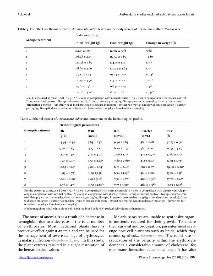
The body weights of the rats investigated in this study are shown in Table 3. There was a significant (p < 0.05) weight difference in all groups compared to normal control. The disease control experienced a greater weight loss. Normal rats treated with A. indica experienced an increase in weight while ad-ministration of lumartem led to a decrease in weight when compared to normal control. The same trend was also observed in malaria infected

Achi et al. Anti-malaria studies on Azadirachta indica leaves in rats
http://jppres.com/jppres J Pharm Pharmacogn Res (2018) 6(3): 198
rats when compared to disease control. Increase in body weight of rats treated with A. indica was de-pendent on the concentration of extract adminis-tered.
The results of hematological parameters of the rats investigated in this study are shown in Table 4. A reduction in all hematological values was ob-served in normal rats after the administration of extracts and lumartem compared to normal control. Malaria infection gave rise to a reduction of Hb, RBC, PCV, and platelets while an increase in WBC was observed. Treatment of malaria with extracts and lumartem resulted in a slight restoration of the hematological values. An inverse proportionality was observed between WBC and platelets in all treatment groups except for lumartem (Group 5), which recorded the highest number of platelets.
DISCUSSION
Malarial is characterized by anemia resulting from the destruction of infected as well as uninfect-ed RBCs. This massive destruction leads to a de-crease in erythroid precursors and erythropoiesis inhibition, usually resulting in the death of the pa-tient (Coronado et al., 2014). In this experiment, an in-oculum size of 107 parasitized erythrocytes was used, which resulted in less than 50% death in the disease control. During the erythrocytic cycle of P. falciparum from the trophozoite stage to a schizont, about 80% of the host-cell hemoglobin is ingested and is degraded by the developing trophozoite. Anemia is a preventable cause of death in malaria-infected children under five years and pregnant women (WHO, 2006).
Figure 1. Lipid profile of animals administered extract of Azadirachta indica and lumartem.
Results expressed as mean ± SD (n = 3). *significant values at p < 0.05 relative to group 1, # not significant at p > 0.05 relative to group 1.
Group 1: (normal control); Group 2: disease control; Group 3: extract 300 mg/kg; Group 4: extract 500 mg/kg; Group 5: lumartem (artemether 2 mg/kg + lumefantrine 12 mg/kg); Group 6: disease induction + extract 300 mg/kg; Group 7: disease induction + extract 500 mg/kg; Group 8: disease induction + lumartem (artemether 2 mg/kg + lumefantrine 12 mg/kg).
Achi et al. Anti-malaria studies on Azadirachta indica leaves in rats
http://jppres.com/jppres J Pharm Pharmacogn Res (2018) 6(3): 199
Table 3. The effect of ethanol extract of Azadirachta indica leaves on the body weight of normal male albino Wistar rats.
Group/treatment
Body weight (g)
Initial weight (g) Final weight (g) Change in weight (%)
1 114.15 ± 1.05 121.02 ± 3.36 5.68
2 116.78 ± 4.13 112.50 ± 1.85 -3.80
3 122.48 ± 1.85 124.91 ± 1.12 1.95a
4 118.60 ± 2.30 120.42 ± 2.63 1.51a
5 113.22 ± 1.84 111.83 ± 3.00 -1.24b
6 120.52 ± 3.76 123.00 ± 2.22 2.02c
7 115.61 ± 1.36 118.34 ± 2.41 2.31c
8 124.20 ± 3.00 121.11 ± 1.17 -2.55d
Results expressed as mean ± SD (n = 3). a, bP < 0.05 in comparison with normal control; c, dp < 0.05 in comparison with disease control. Group 1: (normal control); Group 2: disease control; Group 3: extract 300 mg/kg; Group 4: extract 500 mg/kg; Group 5: lumartem (artemether 2 mg/kg + lumefantrine 12 mg/kg); Group 6: disease induction + extract 300 mg/kg; Group 7: disease induction + extract 500 mg/kg; Group 8: disease induction + lumartem (artemether 2 mg/kg + lumefantrine 12 mg/kg).
Table 4. Ethanol extract of Azadirachta indica and lumertem on the hematological profile.
Group/treatment
Hematological parameters
Hb
(g/L)
WBC
(109/L)
RBC
(1012/L)
Platelet
(109/L)
PCV
(%)
1 14.95 ± 0.39 7.65 ± 2.57 9.40 ± 1.63 581 ± 0.08 42.27± 0.96
2 9.02 ± 0.59 13.17 ± 2.58 6.20 ± 2.33 361 ± 1.00 25.35 ± 3.53
3 12.13 ± 1.32a 7.30 ± 3.10c 7.06 ± 1.51a 473 ± 0.77a 37.67 ± 1.15a
3 11.23 ± 0.45a 6.53 ± 2.28a 7.80 ± 2.60a 433 ± 0.10a 32.01 ± 1.73a
5 10.83 ± 1.29a 9.00 ± 1.64a 6.61 ± 2.40a 610 ± 0.87c 29.00 ± 2.20a
6 11.93 ± 0.73b 11.90 ±3.31b 6.74 ± 1.19d 421 ± 0.60b 35.62 ± 1.51b
7 10.01 ± 0.90b 9.41 ± 5.10b 7.30 ± 1.87b 480 ± 0.39b 27.77 ± 2.28d
8 9.76 ± 1.43d 10.53 ±4.66b 7.17 ± 3.00b 466 ± 0.38b 15.73 ± 1.65b
Results expressed as mean ± SD (n = 3). aP < 0.05 in comparison with normal control; bp < 0.05 in comparison with disease control; cp > 0.05 in comparison with normal control; dp > 0.05 in comparison with disease control. Group 1: (normal control); Group 2: disease con-trol; Group 3: extract 300 mg/kg; Group 4: extract 500 mg/kg; Group 5: lumartem (artemether 2 mg/kg + lumefantrine 12 mg/kg); Group 6: disease induction + extract 300 mg/kg; Group 7: disease induction + extract 500 mg/kg; Group 8: disease induction + lumartem (ar-temether 2 mg/kg + lumefantrine 12 mg/kg).
Hb: hemoglobin; WBC: white blood cell; RBC: red blood cell; PCV: packed cell volume or hematocrit.
The onset of anemia is as a result of a decrease in hemoglobin due to a decrease in the total number of erythrocytes. Most medicinal plants have a protective effect against anemia and can be used for the management of severe damage of erythrocytes in malaria infection (Onyeabo et al., 2017). In this study, the plant extracts resulted in a slight restoration of the hematological values.
Malaria parasites are unable to synthesize organ-ic nutrients required for their growth. To ensure their survival and propagation, parasites must scav-enge host cell nutrients such as lipids, which they cannot synthesize (Sherman, 1979). The rapid rate of replication of the parasite within the erythrocyte demands a considerable amount of cholesterol for membrane formation (Visser et al., 2013). It has also

Achi et al. Anti-malaria studies on Azadirachta indica leaves in rats
http://jppres.com/jppres J Pharm Pharmacogn Res (2018) 6(3): 200
been reported that these host lipids have also been implicated in the formation of hemozoin in vivo (Bendrat et al., 1995; Fitch et al., 1999). The hem, ferripro-toporphyrin IX (hematin) that is released upon di-gestion of the globin chains is lethal to the parasite and is converted into a crystalline substance called hemozoin (HZ) that is harmless to the parasite (Bray
et al., 2005). Since the transformation of heme into hemozoin is an essential process for the survival of the malaria parasite, this molecule has become a target for antimalarial drugs (Monti et al., 1999).
Another phytochemical with antiplasmodial activity in the plant material is azadirachtin a tetra-nortriterpenoid (Jones et al., 1994). Limonoids like gedunin and meldenina isolated from medicinal plants have also been demonstrated to impact an-timalarial activity on A. indica (MacKinnon et al., 1997). However, gedunin did not inhibit P. berghei in mice (Odetola and Bassir., 1986; Bray et al., 1990) while meldenin was very much active against Plasmodium falcipa-rum (Joshi et al., 1998). Gedunin has been proposed to be responsible for the alkylation of hem and specific proteins, which leads to the death of malaria para-site (Arnason et al., 2004). Azadirachtin, on the other hand, inhibits the formation of mobile microgam-etes in vitro (Jones et al., 1994).
A. indica has been reported to be effective against P. falciparum in humans (El-Tahir et al., 1999;
Alshawsh et al., 2009; Adebayo and Krettli, 2011). The di-lemmas with A. indica on rodent models is because the causative agent of malaria infection in humans do not cause infection in rodents while P. berghei only causes infection in murine models but not in humans (Farahna et al., 2010). However, arteme-ther/lumefantrine seems to be effective against both human and rodent parasites (Abolaji et al., 2012;
Otuechere et al., 2012). Previous studies showed that Wistar rats inoculated with 107 parasitized red blood cells by intraperitoneal route showed 25% mortality, with a reduction in parasitemia and complete eradication of parasitemia around the 18th day of post inoculation (Dow et al. 1999; Pedroni et al.,
2006). This could be responsible for the survival of the experimental animals after inoculation with P. berghei parasite. Therefore, despite the high dose of extract administered, it is not certain that the sur-vival of experimental animals was as a result of the anti-plasmodial activity of A. indica.
The significant increase in the level of HDL-cholesterol and reduction of LDL-cholesterol in the groups administered with A. indica compared to normal control showed that the extract could be of great importance in the treatment of hyperlipidem-ia and cardiovascular diseases. Studies have shown that high concentrations of HDL have a protective effect against cardiovascular diseases such as is-chemic stroke and myocardial infarction (Sacco et al.,
2001). The result obtained in this study shows that ethanol extracts of A. indica and lumartem are fa-vorable against atherogenesis, because of the ability to maintain a normal level of TAG and cholesterol.
Artemether has a rapid schizontocidal action (Lefèvre and Thomsen, 1999). It is primarily metabolized by cytochrome P450 (CYP) 3A4/5 but also by CYP-2B6, CYP2C9, and CYP2C19. Metabolism through CYP3A4 produces an active metabolite, dihydroar-temisinin that contributes significantly to its anti-malarial activity (Cousin et al., 2008). The endoperox-ide bond (O–O) is crucial for expression of antipar-asitic activity. Its activation involves an initial chemical decomposition induced by ferrous hem Fe(II) produced from 1-electron reduction of oxi-dized ferric hematin within the malaria parasite and is responsible for the bioactivation of the endoper-oxide bridge to potentially toxic free radicals in the food vacuole of the parasite (Bray et al., 2005). The oxy radical initially produced subsequently rearranges into carbon-centered radical intermediates (O’Neill
and Posner, 2004), which gives off a high-valent iron-oxo species to produce a stable end-product (Posner
et al., 1995). Nnate and Achi (2016) have reported that the radical complex formed by the reaction of Fe(II) with oxygen is suitable for the formation of another radical intermediate in the presence of a radical electron. Free-radical mediated autoxidation of membrane lipids is triggered by abstraction of a weakly bonded allylic hydrogen by some strong oxidant, which might be the hydroxyl radical (·OH), the perhydroxyl radical (HO2·), a chelated iron-oxygen complex such as ferryl (Fe+ +O) or perferryl (Fe+ +O2) compounds (Girotti, 1985).
A Fenton-like reaction has also been proposed where the highly reactive endoperoxide moiety in artemisinins undergo homolytic cleavage to form hydroperoxide, which is then degraded to produce reactive oxygen species and carbon-centered radical

Achi et al. Anti-malaria studies on Azadirachta indica leaves in rats
http://jppres.com/jppres J Pharm Pharmacogn Res (2018) 6(3): 201
molecules that modify the proteins of malaria para-sites (Haynes et al., 1999). The endoperoxide bond is crucial for expression of antiparasitic activity, which is mediated through this reactive species produced.
Lumefantrine is metabolized primarily by CYP3A4 and then undergoes glucuronidation (Hi-
etala et al., 2010; Mwesigwa et al., 2010). The mechanism of action of lumefantrine is equivalent to quinine, mefloquine, and halofantrine. Lumefantrine inhibits the formation of non-toxic hemozoin crys-tals by interfering with hematin, the toxic interme-diate step produced during hemoglobin break-down. Accumulation of hematin generates free rad-icals, which destroys the membranes by a lipid pe-roxidation mechanism and results in parasite death (Byakika-Kibwika et al., 2010).
Interaction of lipid solubilized hem with arte-misinin followed by ferrous-mediated generation of oxyl and carbon radicals makes these reactive in-termediates available to the allylic hydrogens of un-saturated lipid bilayers. Hydrogen abstraction and allylic carbon radical formation with subsequent triplet ground state oxygen capture result ultimate-ly in the formation of lipid hydroperoxides. Studies by Berman and Adams (1997) demonstrated that ar-temisinin causes an increase in hem-mediated lipid membrane damage. This is in agreement with re-cent studies on artemether-lumefantrine and ar-tesunate-amodiaquine combination (Otuechere et al.,
2012). The decrease in weight of infected rats could be due to the breakdown of hemoglobin and lipids by malaria parasite; while that observed in groups treated with lumartem was as a result of the de-struction of lipid membranes. The induction of ma-laria parasite in this study did not affect lipid pro-files of rats. A similar trend was also recorded by Abolaji et al. (2012) without P. berghei infection in the rats.
The action of ROS on lipids leads to the activa-tion of blood platelets (Sener et al., 2005). Thus, ad-ministration of lumartem caused an elevation in lipid peroxidation and activation of blood platelet. This enhances platelet-leukocyte aggregate for-mation and triggers inflammatory responses (Graff et
al., 2001) as a result of increased leucocyte formation. The elevated leucocyte and platelets observed in the group treated with lumartem were as a result of lipid peroxidation. The administration of A. indica
in normal rats reduced platelet count. Tohti et al. (2006) have a previously reported a decrease in platelet count as a result of the presence of phyto-chemicals such as saponins and cardiac glycosides malaria infection. Changes in WBC and platelets in P. falciparum malaria infection has also been reported by Guerra et al. (2008) and Karanikas et al. (2004).
CONCLUSIONS
Azadirachta indica leaf extract was practically safe, non-toxic and well tolerated at the adminis-tered doses. The extract of Azadirachta indica dis-played a considerable anti-malaria property by hav-ing a higher efficacy and could be a template for short-term anti-malaria drug development.
CONFLICT OF INTEREST
The authors declare no conflict of interest.
ACKNOWLEDGMENT
The authors confirm that the project has no funding or grants.
REFERENCES
Abolaji AO, Eteng MU, Omonua O, Adenrele Y (2012) Influence of coadministration of artemether and lumefantrine on selected plasma biochemical and erythrocyte oxidative stress indices in female Wistar rats. Hum Exp Toxicol 32(2): 206–215.
Achi NK, Ohaeri OC (2012) Acute and subacute toxicity studies of Sansevieria liberica aqueous leaf extracts. Pharma Sci Monit 3(4): 1938–1951.
Achi NK, Ohaeri OC, Ijeh II, Eleazu C (2017) Modulation of the lipid profile and insulin levels of streptozotocin induced diabetic rats by ethanol extract of Cnidoscolus aconitifolius leaves and some fractions: Effect on the oral glucose tolerance of normoglycemic rats. Biomed Pharmacother 86: 562–569.
Adebayo JO, Krettli AU (2011) Potential antimalarials from Nigerian plants: a review. J Ethnopharmacol 133: 289–302.
Ajero CMU, Mbagwu FN (2005) Advances in Biotechnology (Biological weapons and Phytomedicine). Owerri, Nigeria: Megasoft Publishers. PDF Report, pp. 34–40.
Akuodor GC, Ajoku GA, Ezeunala MN, Chilaka KC, Asika EC (2015) Antimalarial potential of the ethanolic leaf extract of Pseudocedrala kotschyi. J Acute Dis 4(1): 23–27.
Allain CC, Poon LS, Chan CS, Richmond W, Fu PC (1974) Enzymatic determination of total serum cholesterol. Clin Chem 20(4): 470–475.
Alshawsh MA, Mothana RA, Al-Shamahy HA, Alsllami SF, Lindequist U (2009) Assessment of antimalarial activity

Achi et al. Anti-malaria studies on Azadirachta indica leaves in rats
http://jppres.com/jppres J Pharm Pharmacogn Res (2018) 6(3): 202
against Plasmodium falciparum and phytochemical screening of some Yemeni medicinal plant. Evid Based Complement Alternat Med 6: 453–456.
Anyasor GN, Odunaike OM, Olorunsogo OO (2013) Induction and uncoupling of rat liver mitochondria by oral administered coartemether. Am J Biochem Mol Biol 3: 110–118.
AOAC (1984) Official Methods of Analysis. Washington, DC, USA: Association of Official Analytical Chemists, 14th edition.
Arnason JT, Guillet G, Durst T (2004). Phytochemical diversity of insect defenses in tropical and temperate plant families. In: Carde, R.T., Millar, J.G. (Eds.), Advances in Insect Chemical Ecology. Cambridge, UK: Cambridge University Press, pp. 1–20.
Bamidele A, Bamidele AP, Nnate DA (2017) Evaluation of antioxidant potentials of the methanolic leaf extracts of vegetables, fruits and medicinal plants commonly consumed in Kaduna state, Nigeria. J Med Plants Stud 5(1): 388–393.
Bendrat K, Berger BJ, Cerami A (1995) Haem polymerization in malaria. Nature 378: 138–139.
Berman PA, Adams PA (1997) Artemisinin enhances heme-catalysed oxidation of lipid membranes. Free Rad Biol Med 22: 1283–1288.
Biswas K, Chattopadhyay I, Banerjee RK, Bandyopadhyay U (2002) Biological activities and medicinal properties of Neem (Azadirachta indica). Curr Sci 82: 1336–1345.
Boham BA, Kocipai RA (1994) Flavonoids and condensed tannins from leaves of Hawaiian Vaccinium vaticulatum and V. calycinium. Pac Sci 48: 458–463.
Bray DH, Warhurst DC, Connolly JD, O’Neill MJ, Phillipson JD (1990) Plants as sources of antimalarial drugs. Part 7. Activity of some species of Meliaceae and their constituent limonoids. Phytother Res 4: 29–35.
Bray PG, Ward SA, O’Neill PM (2005) Quinolines and artemisinin: Chemistry, biology and history. Curr Top Microbiol Immunol 295: 3–38.
Byakika-Kibwika P, Lamorde M, Mayanja-Kizza H, Merry C, Colebunders B, van Geertruyden JP (2010) Update on the efficacy, effectiveness and safety of artemether-lumefantrine combination therapy for treatment of uncomplicated malaria. Ther Clin Risk Manag 6: 11–20.
Chinedu E, Arome D, Ameh FS (2013) A new method for determining acute toxicity in animal models. Toxicol Intern 20(3): 224–226.
Coronado LM, Nadovich CT, Spadafora C (2014) Malarial hemozoin: From target to tool. Biochim Biophys Acta 1840(6): 2032–2041.
Cousin M, Kummerer S, Lefevre G, Marrast AC, Stein D, Weaver M (2008) Coartem (artemether-lumefantrine) tablets for the treatment of malaria in patients with acute uncomplicated infections due to Plasmodium falciparum or mixed infections including ip. falciparum. Novartis, Africa, pp. 14–20.
Djimdé A, Lefèvre G (2009) Understanding the pharmacokinetics of Coartem. Malar J 8(suppl.1): S4.
Dow GS, Reynoldson JA, Thompson RCA (1999) Plasmodium berghei: a new rat model for assessment of blood
schizonticidal activity. Exp Parasitol 93: 92–94. Edeoga HO, Okwu DE, Mbaebie BO (2005) Phytochemical
constituents of some Nigerian medicinal plants. Afr J Biotechnol 4(7): 685–688.
El-Tahir A, Satti GM, Khalil SA (1999) Antiplasmodial activity of selected Sudanese medicinal plants with emphasis on Maytenus senegalensis (Lam). Exell J Ethnopharmacol 64: 227–233.
Ezzet F, Mull R, Karbwang J (1998) Population pharmacokinetics and therapeutic response of CGP 56697 (artemether + benflumetol) in patients. Br J Clin Pharmacol 46(6): 553–561.
Farahna M, Bedri S, Khalid S, Idris M, Pillai CR, Khalil EA (2010) Anti-plasmodial effects of Azadirachta indica in experimental cerebral malaria: Apoptosis of cerebellar Purkinje cells of mice as a marker. N Am J Med Sci 2(11): 518–525.
Fitch CD, Cai GZ, Chen YF, Shoemaker JD (1999) Involvement of lipids in ferriprotoporphyrin IX polymerization in malaria. Biochim Biophys Acta 1454: 31–37.
Friedewald WT, Levy RI, Fredrickson DS (1972) Estimation of the concentration of low density lipoprotein cholesterol in plasma: with-out use of the preparative ultracentrifuge. Clin Chem 18: 499–505.
Girotti AW (1985) Mechanisms of lipid peroxidation. J Free Rad Biol Med 1: 87–95.
Graff J, Andries D, Elsner M, Westrup, D, Bassus S, Franz N, Klinkhardt U, Harder S (2001) Platelet CD62 expression and PDGFAB secretion in patients undergoing PTCA and treatment with abciximab. Br J Clin Pharmacol 51(6): 577–582.
Greenwood D, Slack RC, Barer MR, Irving WL (2012) Medical Microbiology: A Guide to Microbial Infections: Pathogenesis, Immunity, Laboratory Diagnosis and Control. China: Elsevier Health Sciences. p. 642.
Guerra CA, Gikandi PW, Tatem AJ, Noor AM, Smith DL, Hay SI, Snow RW (2008) The limits and intensity of Plasmodium falciparum transmission: implications for malaria control and elimination worldwide. PLoS Med 5(2): 38.
Harborne JB (1973) Phytochemical Methods: A Guide to Modern Techniques of Plant Analysis, London, UK: Chapman and Hall, pp. 49–188.
Haynes RK, Pai HHO, Voerste A (1999) Ring opening of artemisinin (qinghaosu) and dihydroartemisinin and interception of the open hydroperoxides with formation of N-oxides—A chemical model for antimalarial mode of action. Tetrahedron Lett 40: 4715–4718.
Hietala SF, Martensson A, Ngasala B, Dahlstrom S, Lindegardh N, Annerberg A, Premji Z, Farnert A, Gil P, Bjorkman A, Ashton M (2010) Population pharmacokinetics and pharmacodynamics of artemether and lumefantrine during combination treatment in children with uncomplicated falciparum malaria in Tanzania. Antimicrob Agents Chemother 54: 4780–4788.
Jones IW, Denholm AA, Ley SV, Lovell H, Wood A, Sinden RE (1994) Sexual development of malaria parasites is inhibited in vitro by the neem extract azadirachtin, and its semi-synthetic analogues. FEMS Microbiol Lett 120: 267–273.

Achi et al. Anti-malaria studies on Azadirachta indica leaves in rats
http://jppres.com/jppres J Pharm Pharmacogn Res (2018) 6(3): 203
Joshi SP, Rojatkar SR, Nagasampagi BA (1998) Antimalarial activity of neem (Azadirachta indica). J Med Aromat Plant Sci 20: 1000–1004.
Karanikas G, Zedwitz-Liebenstein K, Edherr K, Schuetz M (2004) Platelet kinetics and scintigraphic imaging in thrombocytopenic malaria patients. Thromb Haemost 91(3): 553–557.
Khan AM, Qureshi RA, Ullah F, Gilani SA, Nosheen A, Sahreen S,Laghari MK, Laghari MY, Rehman SU, Hussain I, Murad W (2011) Phytochemical analysis of selected medicinal plants of Margalla Hills and surroundings. J Med Plants Res 5(25): 6017–6023.
Lamb TJ (2012) Immunity to Parasitic Infection. 1st ed. New Delhi, India. pp. 91–104.
Langhorne J, Buffet P, Galinski M, Good M, Harty J, Leroy D, Mota MM, Pasini E, Renia L, Riley E, Stins M, Duffy P (2011) The relevance of non-human primate and rodent malaria models for humans. Malar J 10: 23.
Lefèvre G, Thomsen MS (1999) Clinical pharmacokinetics of artemether and lumefantrine (Riamet®). Clin Drug Invest 18: 467–480.
MacKinnon S, Durst T, Arnason JT, Angerhofer C, Pezutto J, Sanchez-Vindas PE, Poveds LJ, Gbeassor M (1997) Antimalarial activity of tropical Meliaceae extracts and gedunin derivatives. J Nat Prod 60: 336–341.
Maragathavalli S, Brindha S, Kaviyarasi NS, Annadurai, B, Gangwar SK (2012) Antimicrobial activity in leaf extract of Neem (Azadirachta indica Linn). Int J Sci Nature 3: 110–113.
Monti D, Vodopivec B, Basilico N, Olliaro P, Taramelli D (1999) A novel endogenous antimalarial: Fe(II)-protoporphyrin IX alpha (heme) inhibits hematin polymerization to beta-hematin (malaria pigment) and kills malaria parasites. Biochemistry 38: 8858–8863.
Mwesigwa J, Parikh S, McGee B, German P, Drysdale T, Kalyango NJ, Clark DT, Dorsey G, Lindegardh N, Annerberg A, Rosenthal PJ, Kamya MR, Aweeka F (2010) Pharmacokinetics of artemether -lumefantrine and artesunate and amodiaquine in Children in Kampala, Uganda. Antimicrob Agents Chemother 54: 52–59.
Ngarivhume T, Vant Klooster CIEA, De Jong JTVM, Van der Westhuizen JH (2015) Medicinal plants used by traditional healers for the treatment of malaria in the Chipinge district in Zimbabwe. J Ethnopharmacol 159: 224–237.
Nnate DA, Achi NK (2016) Nitrate metabolism: a curse or blessing to humanity? J Sci Res Rep 11(4): 1–19.
Nosten F, Brasseur P (2002) Combination therapy for malaria: the way forward? Drugs 62(9): 1315–1329.
NRC (1985) Guide for the Care and Use of Laboratory Animals, National Institute of Health, Bethesda, MD: National Research Council. pp. 8523.
Nwafor SV, Akah PA, Okoli CO, Onyirioha KC, Nwosu CS (2003) Interaction between chloroquine sulphate and aqueous extract of Azadirachta indica A. Juss (Meliaceae) in rabbit. Acta Pharm 52: 305–311.
O’Neill PM, Posner GH (2004) A medicinal chemistry perspective on artemisinin and related endoperoxides. J Med Chem 47: 2945–2964.
Obadoni BO, Ochuko PO (2002) Phytochemical studies and comparative efficacy of the crude extracts of some
haemostatic plants in Edo and Delta States of Nigeria. Global J Pure Appl Sci 8(2): 203–208.
Obih PO, Makinde JM (1985) Effect of Azadirachta indica on Plasmodium berghei berghei in mice. Afr J Med Medic Sci 14: 51–54.
Ochei J, Kolhatkar A (2008) Medical Laboratory Sciences; Theory and Practice. New Delhi: Tata McGraw-Hill Publishing Co. Ltd., pp. 321–324.
Odetola AA, Bassir O (1986) In: The state of medicinal plants in Nigeria. Sofowora A. Editor. Ibadan, Nigeria: University Press. pp. 275–284.
Onyeabo C, Achi NK, Ekeleme-Egedigwe CA, Ebere CU, Okoro CK (2017) Haematological and biochemical studies on Justicia carnea leaves extract in phenylhydrazine induced-anemia in albino rats. Acta Sci Pol Technol Aliment 16(2): 217–230.
Otuechere CA, Edewor G, Kale OE, Ekor M (2012) Subacute therapeutic dosing of artemether-lumefantrine and artesunate-amodiaquine combination preserves plasma cholesterol, renal antioxidant status and organ weights in rats. Malar Res Treat 2012: 1–5, article ID 257986.
Parotta JA (2001) Healing plants of Peninsular India. CABI Publishing. pp. 495–496.
Pedroni HC, Bettoni CC, Spalding SM, Costa TD (2006) Plasmodium berghei: Development of an irreversible experimental malaria model in Wistar rats. Exp Parasitol 113: 193–196.
Posner GH, Cumming JN, Ploypradith P, Chang HO (1995) Evidence for Fe(Iv)=O in the molecular mechanism of action of the trioxane antimalarial artemisinin. J Am Chem Soc 117: 5885–5886.
Priyanka J, Hingorani L, Nilima K (2013) Pharmacodynamic evaluation for antiplasmodial activity of Holarrhena antidysenterica (Kutaja) and Azadirachta indica (Neem) in Plasmodium berghei infected mice model. Asian Pacific J Trop Med 6(7): 520–524.
Rochanakij S, Thebtaranonth Y, Yenjai C, Nimbolide YY (1985) A constituent of Azadiratcha indica, inhibits Plasmodium falciparum in culture. South Asian J Trop Med Public Health 16: 66–72.
Sacco RL, Benson RT, Kargman DE, Boden-Albala B, Tuck C, Lin IF, Cheng JF, Paik MC, Shea S, Berglund (2001) High-density lipoprotein cholesterol and ischemic stroke in the elderly: The Northern Manhattan Stroke Study. JAMA 285(21): 2729–2735.
Saxena S, Pant N, Jain DC, Bhakuni RS (2003) Antimalarial agents from plant sources. Current Sci 85: 1314–1329.
Sener A, Ozsavci D, Oba R, Demirel GY, Uras F, Yardimci KT (2005) Do platelet apoptosis, activation, aggregation, lipid peroxidation and platelet–leukocyte aggregate formation occur simultaneously in hyperlipidemia? Clin Biochem 38: 1081–1087.
Sherman IW (1979) Biochemistry of Plasmodium (malarial parasites). Microbiol Rev 43: 453–495.
Singh N, Shukla MM, Shukla MK, Mehra RK, Sharma S, Bharti PK, Singh MP, Singh A, Gunasekar A (2010) Field and laboratory comparative evaluation of rapid malaria diagnostic tests versus traditional and molecular techniques in India. Malar J 9(1): 191.

Achi et al. Anti-malaria studies on Azadirachta indica leaves in rats
http://jppres.com/jppres J Pharm Pharmacogn Res (2018) 6(3): 204
Stott GJ, Lewis SM (1995) A simple and reliable method for estimating haemoglobin. Bull World Health Organ 73(3): 369–373.
Tietz NW (1995) Clinical Guide to Laboratory Tests, third ed., Philadelphia, PA: WB Saunders Company, pp. 518–519.
Tiwari R, Verma AK, Chakraborty S, Dhama K, Singh SV (2014). Neem (Azadirachta indica) and its potential for safeguarding health of animals and humans: a review. J Biol Sci 14: 110–123.
Tohti I, Tursun M, Umar A, Imin H, Moore N (2006) Aqueous extracts of Ocimum basilicumL. (sweet basil) decrease platelet aggregation induced by ADP and thrombin in vitro and rats arterio-venous shunt thrombosis in vivo. Thromb Res 118(6): 733–739.
Vickers A, Zollman C, Lee R (2001) Herbal medicine. West J Med 175(2): 125–128.
Visser BJ, Wieten RW, Nagel IM, Grobusch MP (2013) Serum
lipids and lipoproteins in malaria a systematic review and meta-analysis. Malar J 12: 442.
WHO - World Health Organization (2000) Severe falciparum malaria. Trans R Soc Trop Med Hyg 94(1): 1–90.
WHO - World Health Organization (2006). Guidelines on food fortification with micronutrients. Geneva. http://www.who.int/medical_devices/initiatives/anemia_control/en [Consulted October 17,2016].
WHO - World Health Organization (2013) WHO global malaria programme. World Malaria Report. WHO Geneva. http://www.who.int/malaria/publications/world_malaria_report_2013/en [Consulted February 8, 2017].
WHO - World Health Organization (2015) World Malaria Report. Geneva, Switzerland. http://apps.who.int/iris/bitstream/10665/200018/1/9789241565158_eng.pdf [Consulted February 8, 2017].
_________________________________________________________________________________________________________________
Author contribution:
Contribution Achi NK Onyeabo C Nnate DA Ekeleme-Egedigwe CA Kalu IK Chibundu IC Wokoma GC
Concepts or ideas X X
Design X X
Definition of intellectual content X X X
Literature search X X X
Experimental studies X X X X
Data acquisition X X
Data analysis X X
Statistical analysis X X
Manuscript preparation X X
Manuscript editing X X
Manuscript review X X X
Citation Format: Achi NK, Onyeabo C, Nnate DA, Ekeleme-Egedigwe CA, Kalu IK, Chibundu IC, Wokoma GC (2018) Therapeutic effects of Azadirachta indica A.Juss. leaves in malaria-induced male Wistar rats. J Pharm Pharmacogn Res 6(3): 191–204.