Anthony, Paul (1999) Respiratory gas carriers in plant ...
259
RESPIRATORY GAS CARRIERS IN PLANT CULTURE SYSTEMS by Paul Anthony, B.Sc. (Hons) Thesis submitted to the University of Nottingham for the degree of Doctor of Philosophy, May 1999.
Transcript of Anthony, Paul (1999) Respiratory gas carriers in plant ...
RESPIRATORY GAS CARRIERS IN
PLANT CULTURE SYSTEMS
b y
Paul Anthony, B.Sc. (Hons)
Thesis submitted to the University of Nottingham
for the degree of
Doctor of Philosophy,
May 1999.

ACKNOWLEDGEMENTS
1 would like to thank my supervisors, Drs K.C Lowe, J.B. Power and M.R. Davey,
for their constmctive advice and criticism during the completion of this Thesis and the
associated publications. I would also like to thank my industrial supervisor, Dr M.
Atherton for technical information and Dr C Washington for measurement of
interfacial tensions and help in the area of image analysis.
Thanks also go to F2 Chemicals Ltd., Preston, UK for financial support during this
research project and for gifts of perfluorodecalin.
I would also like to thank the departmental technical support staff, in particular Dave
Wilson, Trevor Jones, June Jones, Joan Jotham, Brian Case, Roy Searcy, Pat Crosby,
Ian Gilder and John Gaskin.
Finally, my heartfelt thanks go to all my colleagues in the Plant Science Division and
the School of Biological Sciences for their support and friendship, in particular Drs
Rob Marchant, Kasi Azakanandam, Tom Hartman, Matt McCabe, Andy Brown,
Heidi Triggs, Carl Edwards, Ali Jelodar, Wendy Craig and Messrs Calin Andras,
Nigel Blackball and Ms Caroline Batchelor.

CONTENTS
ACKNOWLEDGEMENTS i
TABLE OF CONTENTS ii ABSTRACT ix
ABBREVIATIONS xi
CHAPTER 1. General Introduction
1.1 Principles and current limitations of plant tissue culture 1
1.1.1 Bioreactors for the large-scale cultivation of plant cells 8
1.2 Effects of the gaseous atmosphere on in vitro plant 14
growth and development
1.2.1 Beneficial effects of oxygen enrichment of the culture 18
environment
1.2.2 Beneficial effects of carbon dioxide enrichment of the 19
environment
1.3 Comparison of plant and animal cell culture systems 22
1.4 Perfluorochemicals (PFCs) and cell biotechnology 24
1.4.1 Introduction 24
1.4.2 Properties of PFC liquids 24
1.4.3 Biomedical applications of PFCs 26
1.4.4 Applications of PFCs to cell biotechnology 29
1.4.4.1 Biocompatibility of PFCs in fermenter systems 30
1.4.4.2 Animal cell culture 32
1.4.4.3 PFCs and carbon dioxide 32
1.4.4.4 PFCs and physiological oxygen monitoring 33
1.4.4.5 Culture of cells at PFC-aqueous interfaces 34
1.4.4.6 Fluorocarbon polymers in cell cultures 36
1.5 Haemoglobin-based oxygen carriers 36
and cell biotechnology
1.5.1 Recent history 36
1.5.2 Haemoglobin in cell biotechnology 39
1.6 Thesis objectives 40
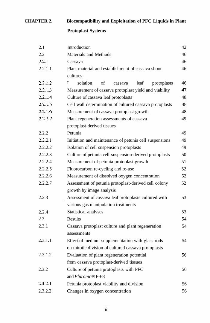
CHAPTER 2. Biocompatibility and Exploitation of PFC Liquids in Plant
Protoplast Systems
2.1 Introduction 42
2.2 Materials and Methods 46
2.2.1 Cassava 46
2.2.1.1 Plant material and establishment of cassava shoot 46
cultures
2.2.1.2 I solation of cassava leaf protoplasts 46
2.2.1.3 Measurement of cassava protoplast yield and viability 47
2.2.1.4 Culture of cassava leaf protoplasts 48
2.2.1.5 Cell wall determination of cultured cassava protoplasts 48
2.2.1.6 Measurement of cassava protoplast growth 48
2.2.1.7 Plant regeneration assessments of cassava 49
protoplast-derived tissues
2.2.2 Petunia 49
2.2.2.1 Initiation and maintenance of petunia cell suspensions 49
2.2.2.2 Isolation of cell suspension protoplasts 49
2.2.2.3 Culture of petunia cell suspension-derived protoplasts 50
2.2.2.4 Measurement of petunia protoplast growth 51
2.2.2.5 Fluorocarbon re-cycling and re-use 52
2.2.2.6 Measurement of dissolved oxygen concentration 52
2.2.2.7 Assessment of petunia protoplast-derived cell colony 52
growth by image analysis
2.2.3 Assessment of cassava leaf protoplasts cultured with 53
various gas manipulation treatments
Statistical analyses 53
Results 54
Cassava protoplast culture and plant regeneration 54
assessments
Effect of medium supplementation with glass rods 54
on mitotic division of cultured cassava protoplasts
Evaluation of plant regeneration potential 56
from cassava protoplast-derived tissues
2.3.2 Culture of petunia protoplasts with PFC 56
and Pluronic® F-68
2.3.2.1 Petunia protoplast viability and division 56
2.3.2.2 Changes in oxygen concentration 56
ni
2.2.4
2.3
2.3.1
2.3.1.1
2.3.1.2

2.3.2.3 Growth of petunia protoplasts with perfluorodecalin 57
2.3.2.4 Growth of petunia protoplasts with perfluorodecalin 60
and Pluronic F-68
2.3.2.5 Image analysis of petunia protoplast-derived cell 60
colony growth
2.3.3 Effects of various gas-manipulation treatments 63
on mitotic division of cultured cassava protoplasts
2.4 Discussion 65
CHAPTER 3. Biocompatibility of Haemoglobin-Based Respiratory Gas
Carriers in Plant Protoplast Systems
3.1 Introduction 71
3.2 Materials and Methods 72
3.2.1 Culture of petunia suspension cells on medium 72
supplemented with Erythrogen™
3.2.2 Isolation and culture of petunia protoplasts 73
3.2.3 Supplementation of protoplast culture medium with 73
Erythrogen™ alone or with oxygen or carbon monoxide
3.2.4 Supplementation of protoplast culture medium with 74
Pluronic® F-68
3.2.5 Measurement of protoplast viability, growth 74
and biomass
3.2.6 Measurement of pH and osmotic pressure in culture 75
medium
3.2.7 Comparison of media supplementation with Erythrogen™ 75
and/or PFC
3.2.7.1 Initiation and maintenance of passion fmit cell 75
suspensions
3.2.7.2 Isolation of protoplasts from cell suspensions of passion 75
fmit and petunia
3.2.7.3 Culture of passion fmit and petunia protoplasts with 76
Erythrogen™ and/or PFC
3.2.7.4 Measurement of IPE 76
3.2.8 Medium supplementation with fresh and stored 76
Erythrogen™ solutions
3.2.9 Statistical analyses 76
IV
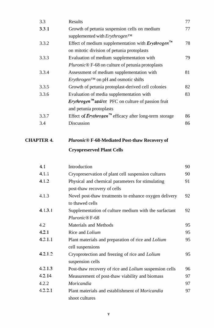
3.3 Results 77
3.3.1 Growth of petunia suspension cells on medium 77
supplemented with Erythrogen™
3.3.2 Effect of medium supplementation with Eryr/irogen™ 78
on mitotic division of petunia protoplasts
3.3.3 Evaluation of medium supplementation with 79
Pluronic® F-68 on culture of petunia protoplasts
3.3.4 Assessment of medium supplementation with 81
Erythrogen™ on pH and osmotic shifts
3.3.5 Growth of petunia protoplast-derived cell colonies 82
3.3.6 Evaluation of media supplementation with 83
Erythrogen™and/or PFC on culture of passion fruit
and petunia protoplasts
3.3.7 Effect of fry^/irogen™ efficacy after long-term storage 86
3.4 Discussion 86
CHAPTER 4. Pluronic® F-68-Mediated Post-thaw Recovery of
Cryopreserved Plant Cells
4.1 Introduction 90
4.1.1 Cryopreservation of plant cell suspension cultures 90
4.1.2 Physical and chemical parameters for stimulating 91
post-thaw recovery of cells
4.1.3 Novel post-thaw treatments to enhance oxygen delivery 92
to thawed cells
4.1.3.1 Supplementation of culture medium with the surfactant 92
Pluronic® F-68
4.2 Materials and Methods 95
4.2.1 Rice and Lolium 95
4.2.1.1 Plant materials and preparation of rice and Lolium 95
cell suspensions
4.2.1.2 Cryoprotection and freezing of rice and Lolium 95
suspension cells
4.2.1.3 Post-thaw recovery of rice and Lolium suspension cells 96
4.2.14 Measurement of post-thaw viability and biomass 97
4.2.2 Moricandia 97
4.2.2.1 Plant materials and establishment of Moricandia 97
shoot cultures

4.2.2.2 Callus initiation and generation of Moricandia 98
cell suspensions
4.2.2.3 Growth of Moricandia cell suspensions 98
4.2.2.4 Preparation of Mor/canrf/a cells for cryopreservation 99
4.2.2.5 Post-thaw recovery of Moricandia cryopreserved cells 99
4.2.2.6 Re-initiation of Moricandia cell suspensions from 100
cryopreserved cells and isolation of protoplasts
4.3 Statistical analyses 101
4.4 Results 101
4.4.1 Cryopreservation of rice and Lolium cells 101
4.4.1.1 Post-thaw viability of rice and Lolium cells 101
4.4.1.2 Post-thaw viability of rice and Lolium cells using 103
TTC buffer supplemented with Pluronic F-68
4.4.1.3 Post-thaw growth of rice and Lolium cells 103
4.4.1.4 Supplementation of the cryoprotectant mixture with 106
Pluronic® F-68 prior to cryopreservation of rice
and Lolium cells
4.4.2 CryopreservationofMor/ca«£//a cells 106
4.4.2.1 Comparison of cryoprotectant effectiveness for 106
cryopreservation of Moricarulia cells
4.4.2.2 Effect of Pluronic® F-68 on recovery of cryopreserved 107
Moricandia cells
4.4.2.3 Re-establishment of Monca/tJ/fli cell suspensions and 109
subsequent isolation of suspension-derived protoplasts
4.5 Discussion 110
CHAPTER 5. PFC-Mediated Post-thaw Recovery of Cryopreserved
Rice Cells
5.1 Introduction 114
5.1.1 Oxygen free radicals and plant antioxidant systems 114
5.1.2 Respiratory imbalances during post-thaw recovery of 116
cryopreserved cells
5.2 Materials and Methods 118
5.2.1 Plant materials and preparation of cell suspensions 118
5.2.2 Cryopreservation and post-thaw recovery 118
5.2.3 Measurement of post-thaw viability and biomass 120
VI

5.2.4 Re-initiationof cell suspension cultures 120
5.2.5 Isolation and culture of protoplasts from re-established 120
cell suspension cultures
5.2.6 Plant regeneration from protoplast-derived callus 121
5.2.7 Cytological analysis of protoplast-derived plants 122
5.3 Statistical analyses 123
5.4 Results 123
5.4.1 Assessment of PFC medium supplementation on 123
post-thaw cell viability
5.4.2 Supplementation of medium with PFC and/or 124
Pluronic® F-68 on post-thaw cell viability
5.4.3 Post-thaw growth of cryopreserved cells recovered in 126
the presence of PFC and/or Pluronic F-68
5.4.4 Re-establishment of cell suspension cultures 127
5.4.5 Assessment of protoplast viability, yield, plating 128
efficiency and plant regeneration
5.4.6 Cytological analysis of regenerated plants 130
5.5 Discussion 130
CHAPTER 6. General Discussion
6.1 Introduction 134
6.2 Protoplast culture systems 134
6.3 Synergistic effects of PFCs and surfactants as protoplast 136
culture medium supplements
6.4 Image analysis assessments of cell growth 136
6.5 Comparison of PFCs with other physical and chemical 137
options for enhancing oxygen supply
6.6 Pluronic® F-68 and plant cell cryopreservation 138
6.7 PFCs and post-thaw recovery of cryopreserved rice 139
cells
6.8 Synergistic effects of PFCs and surfactants on post-thaw 140
recovery of cryopreserved rice cells
6.9 Concluding remarks 142
Vll

APPENDICES
1
11
III
Enzyme formulation
Media formulations
Publications
144
145
154
155
Vlll

ABSTRACT
A cmcial pre-requisite in genetically manipulating higher plants involves systems for
culturing plant protoplasts and cells under static conditions with an adequate oxygen
supply. This is especially the case for cells from cryopreservation, where respiratory
perturbations are known to occur during early post-thaw recovery. Therefore, studies
were undertaken to assess the potential, and actual, beneficial effects involving culture
of cells at an interface between inert, oxygen-gassed perfluorocarbon (PFC) liquid
overlaid with liquid or semi-solidified media supplemented with or without the non-
ionic surfactant, Pluronic F-68. Assessments were also made to compare the
efficacy of PFC supplementation with other physical (medium implanted with glass
rods to increase the surface area available for gaseous exchange) and chemical
(haemoglobin; Hb) options, both alone and in combination, for gaseous manipulation
of plant protoplast cultures.
Investigations involving novel PFC-mediated oxygen delivery to cultured protoplasts
were carried out on a broad range of plant species, which included Petunia hybrida (a
herbaceous species) and Passiflora giberti (a woody species), as model systems,
together with cassava (Manihot esculenta) a relatively recalcitrant species in tissue
culture. Studies revealed enhanced protoplast initial plating efficiencies (IPEs) as
measured by increased mitotic division, thereby demonstrating no short-term
detrimental effects of exposure to PFC. Similarly, supplementation of culture media
with Hb, at 1:50 (v/v), increased the mean IPEs of both Petunia and Passiflora
protoplasts over that of untreated controls. Additionally, supplementation of aqueous
medium with 0.01% (w/v) Pluronic® F-68 not only lowered interfacial tension, but
further enhanced mitotic activity over that stimulated by both oxygenated PFC and
Hb.
IX

In the context of cryopreservation, media supplementation with Pluronic F-68, at
0.01-1.0% (w/v), significantly improved the post-thaw viability and growth of
embryogenic suspension cells of the rice (Oryza sativa L.) cultivars Taipei 309 and
Tarom, together with non-embryogenic cells of Lolium multiflorum and Moricandia
arvensis. Moreover, a more pronounced synergistic effect in terms of viability and
growth was observed for Taipei 309 cells when 0.01% (w/v) Pluronic F-68 was
evaluated in conjunction with oxygenated PFC. Plants regenerated from such
cryopreserved cells were morphologically normal with expected chromosome
complements (2n = 2x = 24), thus confirming the long-term biocompatibility of PFCs,
with no adverse effect up on cellular totipotency.
These results indicate, for the first time, that both oxygenated PFC and Hb provide
options for enhancing cellular oxygen supply to cultured eukaryotic cells in vitro.
However, the recoverability and, hence, recyclability of PFCs make them a
commercially more attractive option, despite the high initial investment cost. Overall,
PFC-facilitated improvements in cell culture technology will have increasingly
important biotechnological implications in the context of plant micropropagation,
somatic hybridisation, transgenic plant production and commercial exploitation of
these technologies.
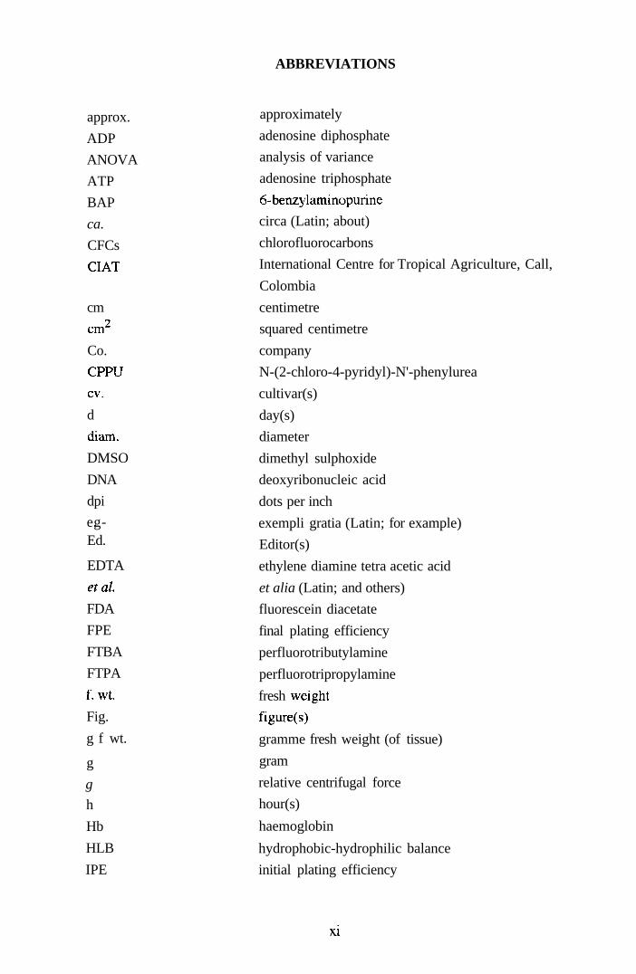
ABBREVIATIONS
approx.
ADP
ANOVA
ATP
BAP
ca.
CFCs
CIAT
cm
cm
Co.
CPPU
cv.
d
dizim.
DMSO
DNA
dpi
eg-Ed.
EDTA
etal
FDA
FPE
FTBA
FTPA
f wt.
Fig.
g f wt.
g
g
h
Hb
HLB
IPE
approximately
adenosine diphosphate
analysis of variance
adenosine triphosphate
6-benzylaminopurine
circa (Latin; about)
chlorofluorocarbons
International Centre for Tropical Agriculture, Call,
Colombia
centimetre
squared centimetre
company
N-(2-chloro-4-pyridyl)-N'-phenylurea
cultivar(s)
day(s)
diameter
dimethyl sulphoxide
deoxyribonucleic acid
dots per inch
exempli gratia (Latin; for example)
Editor(s)
ethylene diamine tetra acetic acid
et alia (Latin; and others)
fluorescein diacetate
final plating efficiency
perfluorotributylamine
perfluorotripropylamine
fresh weight
figure(s)
gramme fresh weight (of tissue)
gram
relative centrifugal force
hour(s)
haemoglobin
hydrophobic-hydrophilic balance
initial plating efficiency
XI

kJ
I
Ltd.
m
M
MES
mg
min
ml
mm
mOsmol kg"l
MS
n
NAA
NAD
NADP
nm
NMR
NPR
P
PCV
per se
PFC
pH
POE
POP
PPP
PTFE
r.p.m.
SCV
s.d.
s.e.m.
SOD
spp.
STP
TBA
TCA
kilojoules
litre(s)
Limited Company
metre
molar
2- [morpholinojethanesulphonic acid
milligramme(s)
minute(s)
millilitre(s)
millimetre(s)
milliosmole per kilogramme
Murashige and Skoog (1962) basal medium
number of observations
a-naphthaleneacetic acid
nicotinamide adenine dinucleotide
nicotinamide adenine dinucleotide phosphate
nanometre
nuclear magnetic resonance
net photosynthetic rate
probability
packed cell volume
Latin; as such
perfluorocarbon(s)
hydrogen potential; logarithm of the reciprocal of
hydrogen ion concentration.
i.e.logio(l/[Fr])or-logio[H+]
polyoxyethylene
polyoxypropylene
pentose phosphate pathway
polytetrafl uoroethylene
revolutions per minute
settled cell volume
standard deviation
standard error of the mean
superoxide dismutase
species
standard temperature and pressure
thiobarbituric acid
tricarboxylicacid pathway
XI1

TTC triphenyl tetrazolium chloride
UK United Kingdom
UM Uchimiya and Murashige (1974) medium
USA United States of America
UV ultra-violet (light)
v/v volume to volume
w/v weight to volume
% percent
°C degrees Celsius
< less than
= equals
fAl microlitre
pim micrometre(s)
;<M micromoles
2,4-D dichlorophenoxyacetic acid
2,3-DPG diphosphoglycerate
Xlll

CHAPTER ONE: GENERAL INTRODUCTION
1.1 Principles and current limitations of plant tissue culture
Plant tissue culture comprises a set of in vitro techniques, pre and post culture
methods and strategies that collectively have been exploited to create genetic
variability from which crop species can be improved (Brown and Thorpe, 1995).
Tissue culture approaches to crop improvement cover a wide range of protocols
involving the growth, under aseptic conditions, of axenic (organism-free) whole
plant organs such as shoots or embryos or alternatively the culture of dedifferentiated
calli or single cells and protoplasts as suspensions. Additionally, specialised tissue
culture techniques and the availability of gametic cells allow for the production of
haploid (and dihaploid) plants, rescue of immature or Fl hybrid embryos,
transformation for the production of transgenic plants, somatic hybridisation based
predominantly on interspecific protoplast fusion and the production of synthetic
seeds using encapsulated somatic embryos (Martin et al, 1998). The induction and
sustainability of plant regeneration (totipotency) is the overriding consideration
behind such applied technologies. Despite many recent and significant advances in
genetic manipulation methodologies and outcomes, particularly in such areas of
transgenic plant research and allied molecular biological and recombinant DNA
techniques, there still remains a number of practical constraints at the tissue culture
underpin technology level.
Micropropagation, although arguably the least sophisticated technique, remains the
widest applied use of plant tissue culture technology. Over the last three decades it
has become possible to regenerate plants from a wide range of explants/specialised
cells of plant species including monocotyledonous species which were until recentiy
generally considered recalcitrant to the cultural strategies that had worked efficientiy
with dicotyledonous species (Maheshwari et al, 1995; Buiteveld et al, 1998). The
1 Chapter one

obvious (practical) exploitation of micropropagation (starting with organised
tissue/organ explants) include rapid multiplication of elite stock whereby plants can
be maintained pathogen-free (Kozai et al, 1997) along with the creation of gene
banks for the storage of endangered plant germplasm (Martin et al, 1998).
There are three recognised strategies currently employed for micropropagation: (1)
enhancing axillary shoot-bud development; (2) production of adventitious shoots;
and (3) the induction of somatic embryogenesis often from an intermediate
callus/cell phase. In the latter process, somatic cells develop through a series of
characteristic and discrete embryogenic stages (reflecting zygotic embryo
developmental pathways) to give whole plants but without gametic cell fusion (De
Klerk et al., 1997; Dey et al, 1998). This is in contrast to the alternative pathway,
organogenesis, whereby somatic cells under appropriate synergistic media hormonal
conditions (cytokinin/auxin ratios) generate shoots and/or adventive roots in a
sequential way. Axillary-bud proliferation for most species produces the least
number of cloned individuals and is simply regulated by the number of axillary buds
introduced into culture. However, this approach has become the most important and
widely practiced (commercially applied) propagation method since genetic and
hence phenotypic stability can usually be preserved (Karp, 1995).
In the case of the organogenic and somatic embryogenic developmental pathways,
differentiated stmctures arise either directly from the input explant or via a
(preferably short-lived) callus phase. The latter often leading to genetic variation,
collectively termed somaclonal variation (Karp, 1995; Collin and Edwards, 1998).
The genetic and phenotypic alterations associated with this poorly understood
phenomenon include changes in ploidy status and chromosomal rearrangements,
such as loss of whole chromosomes, chromosome arms and chromosome segments
and changes at the DNA level, such as transposition and deletion of DNA sequences.
Whilst this in the context of generating genetic novelty offers prospects for the
2 Chapter one

recovery of regenerants with desirable (now or reinforced) traits such as alterations
in foliage pigmentation (Franzone et al, 1996), plant vigour and size, leaf shape and
flower morphology and floral amendments (Nikova et al, 1997), there remains the
possibility of concomitant biochemical changes, such as loss of secondary product
biosynthesis (Sevon et al., 1997) or changes in characteristics such as disease
resistance/tolerance and overall fertility. For example, Claxton et al. (1998) found
that 40% of a population of 833 tissue culture-regenerated Rorippa nasturtium-
aquaticum (watercress) plants were susceptible, as measured by the number of
diseased roots, to the fungal pathogen Spongospora subterranea f. sp. nasturtii . In
contrast, only 33% of 500 control cuttings of R. nasturtium-aquaticum were found to
be susceptible to disease, as measured using the same disease index. The detection
of somaclonal variants is relatively simple when these alterations are associated with
phenotypic changes. Phenotypic assessment though is not always reliable since, a
regular phenotype is not a guarantee of no changes at the genotype level.
Furthermore, a given genotype may be inherentiy stable, but phenotypic changes
may occur, that are temporal in nature and thus are not transmitted through the
sexual cycle to the next (seed) generation. Clearly these issues are of less relevance
to vegetatively propagated crops and ornamentals. These developmental changes are
frequentiy epigenetic (Lambe et al., 1997). Under certain conditions or after
prolonged periods of culture some callus cells acquire an ability to grow in the
continued absence of auxin and/or cytokinin. In most cases, this process of
habituation is reversible and the callus retains totipotency albeit for a finite period of
time. For example, Hoffman and Hoffmanntsay (1994) reported that carrot (Daucus
carota L.) protoplasts isolated from an habituated cell suspension could di\'ide and
regenerate to plants without the presence of exogenously supplied growth regulators.
A further phenotypic feature observed in micropropagated plants is a tendency to
produce fasciated (multi-crowned) regenerants (Tang and Knap, 1998). This
phenomenon is often cultivar-specific and can be moderated through the
3 Chapter one

manipulation of the growth regulators during multiplication. The persistence of an
altered growth habit ex vitro, sometimes through one or two vegetative propagation-
generations again suggests that a transient physiological change has occurred during
culture rather than a persistent, stable genetic change.
Somaclonal variation occurs more frequently via dedifferentiated culture such as
callus and suspension cultures and is typically less common in cultures where
organised plant stmctures, such as pre-existing meristems are maintained. Factors
that may influence the occurrence of such genetic variation have been attributed to
source of explant and pre-existing variability, micropropagation cycle, duration of
culture period and types and concentrations of growth regulators used in particular
synthetic auxins such as 2,4-dichlorophenoxyacetic acid [2,4-D; Bregitzer et al,
(1995)]. However, such genetic variation can be circumvented by the use of
cryopreservation (see Chapters Four and Five of this Thesis). Under conditions of
storage in liquid nitrogen (-196 °C), there is complete cessation of all cellular
metabolism. Consequentiy, no cell division, cell degeneration, or genetic changes
can take place with time (Grout, 1995). Additionally, cryopreservation negates the
need to re-initiate and characterise new cell lines and also provides a constant supply
of totipotent, genetically-uniform cells. Moreover, there is no possibility of loss due
to bacterial or fungal contamination and significant savings can be made in terms of
labour input, culture media, culture vessels and space in controlled-environment
growth chambers.
Genetic variation seen in clonal regenerants can also be attributed to the natural
variation already present within the donor plant. These somatic mutations will be
passed on and perhaps accentuated through any intermediate callus phase and may
ultimately be expressed in the regenerant phenotype. Furthermore, since
adventitious buds can arise from single cells within the donor parent, any somatic
mutations present will be passed directly and rapidly to the apical meristem of the
4 Chapter one

adventitious bud and thus will be incorporated in all cells of the regenerated plant
(Collin and Edwards, 1998).
Apart from the emergence of somaclonal variation within cell cultures another
frequent problem is that of lack (or decline) in expression of totipotency
(Hoxtermann, 1997). The recovery of plants from in vitro cultures is most readily
achieved using organised input tissues which retain developmental competence.
However, with dedifferentiated tissues of some species/genotypes, such as barley
[Hordeum vulgare L.; Bregitzer et al, (1995)] and rice [Oryza sativa L.; Utomo et
al, (1996)] and many monocotyledonous species there remains an inability to
express totipotency which obviously precludes plant regeneration potential. This
may also be tme for such cultures maintained in a dedifferentiated state on culture
medium often containing high physiological levels of auxin leading to a gradual loss
in the ability to re-differentiate over progressive sub-cultures (Kubalakova et al,
1996). In many species, most notably the aforementioned group, in vitro culture of
cells/tissues in a dedifferentiated state is accompanied by a progression from
compact and nodular meristematic callus to friable, vitrified callus concomitant with
a reduction of emergent meristematic zones. Furthermore, the relative proportion of
such friable calli/tissues increases with progressive sub-culture and such tissues are
only capable of rhizogenesis at best (Lambe et al, 1997). The apparent (permanent)
loss of totipotency has been correlated to irreversible genetic alterations, such as
single gene mutations, chromosome breakages and ploidy changes. However,
Lambe et al (1997) have speculated that the progressive loss of totipotency during
callus culture may be due, in part, to progressive methylation of genes (gene
silencing) specifically relevant to cell differentiation during dedifferentiated cell
division. This leads to the continuous elimination (by preferential selection pressure)
of cells capable of sustained differentiation. Furthermore, there exists specific
developmental pathways of regeneration for species within a family. Organogenesis
is the characteristic pathway for members of the Solanaceae, such as sweet pepper
5 Chapter one

[Capsicum annuum L.; Franck-Duchenne et al, (1998)]. In contrast, somatic
embryogenesis is the preferred route for members of the Umbelliferae, such as carrot
[Daucus carota L.; McCabe et al, (1997)]. Additionally, some species within a
genus can be regenerated from somatic tissues according to standard protocols,
whilst other taxonomically closely related species may be unresponsive (Litz and
Gray, 1992). For example, Bencheikh and Gallais (1996) found that somatic
embryogenesis occurred in lines of pea (Pisum sativum L.), whereas lines of Pisum
arvense L. (wild-type pea) were unresponsive. Moreover, regeneration may be
limited to a number of cultivars within a species. In a separate study, Vandoome et
al (1995) evaluated 16 cultivars of pea and found that for example Stehgolt, Maro
and Progreta (commercial cultivars) showed the highest tendency to form somatic
embryos, whilst Alaska, Rondo and Ascona were non-competent in the context of
embryo production.
Hyperhydricity or vitrification is an additional complication for certain species (and
tissue types) when maintained in vitro. This physiological disfunction results in
excessive hydration of tissues, low lignification of cells and hence reduced
mechanical strength of tissue culture-generated regenerants (Ueno et al, 1998).
Such pleuits are glossy and translucent in appearance and are usually difficult to
proliferate ex vitro and can not be successfully established during acclimation.
Vitrification is causally related to several culture/environmental regimes, notably
excess cytokinin levels in medium (Lu, 1993), high levels of ammonium ions
(Brand, 1993), form and type of gelling agents employed (Zimmerman et al., 1995)
and for certain genera ethylene production (Righetti, 1996). Use of increased agar
concentration as gelling agent may alleviate this condition by reducing water
availability and the uptake of cytokinins; this may though have an adverse effect on
growth rate (Ghashghaie et al, 1991). Raising the cytokinin concentration restored
multiplication rates of in vitro cultured globe artichoke [Cynara scolymus', Debergh,
(1983)]. Ventilation of the culture vessel can similarly be influential on culture
6 Chapter one
development, since it permits diffusion of water vapour and ethjiene from the
immediate culture atmosphere (Armstrong et al, 1997). Production of this latter
gaseous by-product occurs in vitro as a result of tissue wounding during explanting
or on sub-culture, together with the cellular metabolism of methionine. Ethylene is a
well known plant hormone that inhibits growth, differentiation and in some cases
senescence of in vitro cultured plants, cells and tissues at concentrations as low as
0.01 pi 1 (Kumar et al., 1998). Morphogenic responses of cultured cells of several
Brassica species have been shown to be highly sensitive to ethylene (Pua, 1993),
whilst many monocotyledonous species are less affected by the gas. Similarly,
Lakshmanan et al. (1997) found that shoot bud regeneration was delayed, but not
totally inhibited by ethylene produced from cultured leaf explants of mangosteen
(Garcinia mangostana L.). Generally, such differences in morphogenic responses
between monocotyledonous and dicotyledonous plant species can, in part, be
attributed to the fact that ethylene plays a key role in the fmit ripening process of
many dicotyledonous plant species and cells of these species will be responsive to its
hormonal effect. In contriist, ethylene is not essential for such a process in many
monocotyledonous species and thus cells of the latter have not been conditioned to
be responsive. Moreover, ethylene can interact with other in vitro plant hormones
acting, either synergistically or inhibitory, on the critical threshold values responsible
for determining morphogenic responses.
The establishment of in vitro plant cultures per se involves the physical removal of
tissues from the donor and following establishment (in vitro) there is mounting
evidence that the requisite transition from autotrophic to heterotrophic [sugar-
containing (carbon source)-based medium] metabolism, together with the induction
of major developmental pathways caused by altered (compared to endogenous
levels) hormonal sti-ess, may be expected to promote oxidative stress within the
tissues (Benson and Roubelakis-Angelakis, 1994). Indeed, the high physiological
concentrations of auxins that are essential for the induction and maintenance of
7 Chapter one

dedifferentiated cells, callus and isolated protoplasts can increase cellular uptake of
oxygen (Chkanikov et al, 1990), this in turn leads to the activation of an alternative
respiratory pathway (Sluse and Jarmuszkiewicz, 1998) such as the mitochondrial
respiratory pathway that branches at the ubiquone pool (Fig. 1.1) and consists of an
alternative oxidase encoded by the nuclear gene Aoxl. This pathway does not permit
oxidative phosphorylation of adenosine diphosphate (ADP) and is thus not energy
conserving. Altemative oxidase activity is influenced by stress stimuli such as cold,
oxidative stress, pathogen attack (in whole plants) and by factors constricting flow
through the cytochrome pathway (Fig. 1.1) of respiration (Vanlerberghe and
Mcintosh, 1997).
1.1.1 Bioreactors for the large-scale cultivation of plant cells
The biotechnological exploitation of plant cell cultures for the production of useful
phytochemicals has been studied intensively over the past 30 years (Toivonen,
1993). Such cell and tissue cultures have been used for the synthesis and production
of pharmaceuticals [for example, paclitaxel in cell cultures of Taxus yunnanensis',
Pandey, (1998)], insecticides [production of the quinone, plumbagin in cell
suspension cultures of Drosophyllum lusitanicum; Nalalka et al, (1996)], flavours
[alkylcysteinesulphoxides in tissue cultures of Allium species; Mellouki et al,
(1996)] and pigments [alkannin production in cell suspension cultures of Alkanna
tinctoria', Urbanek et al, (1996)]. However, most often plant cell cultures fail to
produce, qualitatively or quantitatively, the desired substances found in the intact
parent plant from which they were established (Domenburg and Knorr, 1996).
Additionally, problems associated with cultural instabilities and the lack of a basic
knowledge of the key biosynthetic pathways and their genetic base responsible for
the production of plant (secondary) metabolites have severely limited progress.
Consequentiy, efforts have been directed towards increasing product yields based on
the selection for high-producing cell lines. For example, Zhao et al. (1998) observed
that calli of Saussurea medusa could be distinguished into two distinct cell types,
8 Chapter one

one being a faint yellow in colour and the other red; the former was identified by
spectrophotometric analysis to produce 2.5 and 3.9 times more flavanoid and
jaceosidin respectively than the latter line. In addition, medium optimization and
culture conditions have important roles in the large-scale culture of plant cells. In
this respect, Huang and Chen (1998) recentiy demonstrated that 3,4-
dihydroxyphenylalanine production in cell suspension cultures of Stizolobium
hassjoo could be increased four-fold by simply altering the concentrations of the
carbon and nitrogen sources, inorganic nutrients, trace elements and organic
supplements in the basal culture medium.
Plant cells have been grown in a wide variety of bioreactors. Initieilly it was
considered that conventional stirred tank reactors would be unsuitable for the growth
of plant cells due to the high shear forces produced. Consequentiy, approaches were
re-directed at air-driven reactors with comparatively low levels of cell shear. Sterile
air is forced up through the cell suspension thereby mixing the cells and maintaining
them as a suspension. In most batch cell suspension and organ cultures, maximum
secondary product accumulation was observed typicdly to occur towards the end of
the exponential growth phase. For example, cocoa (Theobroma cacao) cell
suspension cultures were found to produce caffeine and theobromine concomitant
with log phase growth (Gumey et al, 1992). Similarly, the highest rate of thiophene
production in hairy root (transformed) cultures of Tagetes patula occurred during
late exponential growth, when fresh weight increase (biomass) of the root cultures
were maximal (Arroo et al, 1995).
Many secondary metabolites are produced by cell cultures that are not undergoing
mitosis. These secondary plant products show non-associated growth product
kinetics and thus optimization of product formation is dependent on extending the
stationary phase of growth responsible for product biosynthesis (Domenborg and
Knorr, 1996). However, in order to achieve maximum production, it is necessary to
9 Chapter one

accumulate a large initial biomass followed by a period in which growth is inhibited.
For example, in the case of shikonin production by Lithospermum erythrorhizon
cultures, a two-stage batch process was used, whereby cells were cultured in a liquid
medium favouring rapid cell growth, followed by a second stage of nutrient-
limitation coupled with a high sucrose concentration to promote secondary
metabolism (Su, 1995). This finding lead to the concept of immobilization whereby
cells could be maintained continuously for a period of several weeks under limiting
growth conditions (Collin and Edwards, 1998). Cells were enclosed in an inert
material, usually encapsulated as calcium alginate beads, or grown within the spaces
of porous foam blocks. Cell-to-cell contact is maintained, throughout, in a culture
medium, usually with reduced phosphate to limit adenosine triphoshate (ATP)
biosynthesis and thus cell growth (Su, 1995), reduced nitrate and growth regulator
levels, together with a high sucrose concentration. The medium was circulated
throughout the bioreactor so that secondary products released into the medium can
be removed, thereby avoiding any feedback inhibition. Bioreactors of this type can
be of the Eurlift configuration or simply be a flat-bed where the cells are positionally
fixed and medium circulated through the bed.
In spite of all the positive advantages immobilization offers there still remain
problems with low productivity (Choi et al., 1995), poor secretion of the
accumulated product into the surrounding medium (Domenburg and Knorr, 1995)
and genetic instability of the cells (Zhong et al, 1995). To date, the technological
feasibilities of large-scale plant cell culture remains confined to shikonin production
from cell suspensions of L. erythrorhizon, berberine from cell cultures of Coptis
japonica and ginseng together with saponin from suspensions of Panax ginseng
(Domenburg and Knorr, 1995; Su, 1995). However, these latter processes utilised
freely suspended cells in conventionally stirred tank reactors and it is still generally
accepted that no immobilized plant cell process are yet, commercially viable (Collin
and Edwards, 1998).
10 Chapter one

Differentiated tissues such as roots, shoots and somatic embryos exhibit an increased
capacity for secondary metabolite production and are inherentiy more genetically
stable than dedifferentiated cells. In this regard, large-scale hairy root culture
technologies have recentiy been developed (Wysokinska and Chmiel, 1997). In
contrast to plant cell suspension cultures, hairy root cultures do not require the
addition of phytohormones for growth and are often characterised by high
biosynthetic capacity. Furthermore, novel moieties, such as the indole alkaloid
anthraserpine, in Catharanthus trichophyllus have been found in transformed roots
and do not occur in the corresponding intact plant (Wysokinska and Chmiel, 1997).
Hairy roots are heterotrophic, so oxygen plays a critical role in their respiratory
metabolism. Respiration in living cells is the oxidation of organic substrates usually
glucose by specific metabolic pathways. Such substrates are partially oxidized to
carbon dioxide concomitant with the release of free energy in the form of oxidative
phosphorylation of ADP. There are two pathways available for the respiratory
oxidation of glucose: glycolysis (Plaxton, 1996) and the pentose phosphate pathway
[PPP; Mamushina et al, (1997)] (Fig 1.1). In the former case, glucose is
enzymatically converted to pymvate concomitant with substrate phosphorylation
leading to ATP synthesis. Pymvate is decarboxylated and the remaining acetyl
moiety linked with coenzyme A to produce acetyl-CoA which enters the
tricarboxylic acid cycle [TCA; Ernes and Neuhaus, (1997)] resulting in progressive
oxidation of substiates to yield carbon dioxide and pairs of hydrogen atoms which,
ultimately, react with oxygen to produce water. The combined actions of glycolysis
and TCA result in the complete oxidation of glucose.
In contrast, the PPP does not result in the complete oxidation of glucose and is used
by plant cells to produce the pentose sugar, ribulose-5-phosphate, which can be used
for the biosynthesis of nucleic acids. This oxidati\e decarboxylation produces
carbon dioxide and pairs of hydrogen atoms which are accepted by the coenzyme
11 Chapter one

COz
nicotinamide adenine dinucleotide phosphate (NADP) to provide cellular reducing
power in the form of NADPH. Thus, the end fate of glucose-6-phosphate (the
substrate starting point of both glycolysis and PPP) is dependent on the cellular
requirements of ATP, NADP/NADPH and intermediate products of both pathways,
although glycolysis is the predominant pathway. However, both pathways ultimately
merge to enter the cytochrome system or the altemative oxidase pathway (see
Section 1.1).
Fig. 1.1 Biochemical pathways leading to electron transport routes in higher
plant respiration
Intermediates (aromatic compounds, pentoses)
ATP
^ Cyt pathway - • O2
NADH- ^ ° ^
r ^ Alternative - • O2 Glucose - > - G-6-P — • Glycolysis - > - Pyr - • TCA cycle — ^ pathway
"* ~ ^ ^ ATP t V
Fermentation Intermediates " ^ (amino acids)
CO2
Modified from van der Plas (1984). ATP, adenosine triphosphate; CO2, carbon
dioxide; CoQ, coenzyme Q; Cyt, cytochrome; G-6-P, glucose-6-phosphate; NADH,
nicotinamide adenine dinucleotide (reduced form); NADPH, nicotinamide adenine
dinucleotide phosphate (reduced form); O2, oxygen; PP, pentose phosphate; Pyr,
pymvate; TCA, tricarboxylic acid cycle.
The cytochrome system consists of a cytochrome chain (cytchromes a, b and c) along
which electrons are passed to molecular oxygen which acts as a terminal acceptor to
form water. Cytochromes are protein molecules with a haem prosthetic group, each
having an iron atom at its centre. As electrons pass along the cytochrome chain, the
iron is reduced to the ferrous state (Fe "*") and reoxidized to the ferric state (Fe^"'') by
receiving an electron from the preceding member of the chain. Concomitantiy, the
12 Chapter one

pairs of hydrogen atoms derived from the TCA cycle, except for the succinate
oxidation step of the cycle, are used to reduce the coenzymes NAD or NADP
according to the reaction:
NAD (P)+ -I- 2 (H) -> NAD(P) H -I- H+
Oxidized coenzyme Reduced coenzyme
Upon reduction the coenzymes takes up one hydrogen atom and one electron from
the remaining hydrogen atom to leave a free proton (H"*"). Subsequently, the
electrons are passed from the coenzymes to molecular oxygen \ ia the cytochrome
chain. Such passage of electrons along the chain provides the energy to facilitate the
oxidative phosphorylation of ADP to form ATP and thus provide cellular energy.
The resultant charged oxygen atom is then able to react with the free hydrogen
proton and the proton derived from the reoxidation of the reduced coenzyme, to form
water.
Provision of an adequate oxygen supply to cells (in vitro or in vivo) inside hairy root
clumps can be impeded by external boundary layers and is exacerbated by the high
external oxygen critical levels of 90-100% air saturation required for the growth of
hairy roots (Yu and Doran, 1994). Kim and Yoo (1993) investigated the effects of
agitation and aeration on the high density growth of cartot hairy roots and found that
high hydrodynamic stiess inhibited root growth, whilst a high volumetric oxygen
transfer resulted in higher specific growth rate. Oxygen supply has also been shown
to affect differentiation of embryogenic cell suspensions. High dissolved oxygen
concentrations, similar to 60% air saturation, favoured undifferentiated biomass
production of transformed Eschoscholtzia californica cell suspensions at the expense
of somatic embryo differentiation. In contrast, too low a dissolved oxygen
concenti-ation (similar to 5-10%) inhibited biomass and somatic embryo production
(Archambault et al, 1994). Similariy, Jay et al. (1992) reported that a dissolved
oxygen concentration of 10% inhibited cartot somatic embryo production by up to
13 Chapter one

75% compared to cultures maintained with a dissolved oxygen concentration of
100%.
Novel approaches to supply cultured hairy roots with a sustainable oxygen supply
have thus demanded novel approaches in bioreactor concept and design to provide
for optimal root growth and product extraction. These have involved growing
transformed roots suspended above porous membrane tubing, as a supplementary
aeration device, within the bioreactor (Kanokwaree and Doran, 1998). These
workers reported that the use of combined air sparging and membrane tubing
aeration in a gas-driven bioreactor increased biomass levels of Atropa belladonna
hairy roots by 32-65% compared to sparging only with air or oxygen-enriched air
delivered at the same total gas flow rate (0.6 l min).
1.2 Effects of the gaseous atmosphere on in vitro plant growth and development
In vitro cultures are routinely maintained in semi-sealed vessels, such as glass jars
and Petri dishes, which unintentionally restricts gaseous exchange between the
culture vessel atmosphere and the external atmosphere. Growth and development of
plants and tissues is not only dependent on medium composition but is affected by
the gaseous environment within the vessel, which may differ significantiy in terms of
carbon dioxide and oxygen levels from the outside air, allowing accumulation of
gaseous components such as carbon dioxide (and ethylene) produced by the plEints
and ongoing metabolism of the culture media components (Blazkova et al, 1989). A
culture medium giving optimal results in one type of culture vessel (and sealing) may
fail to do so in another vessel. This indicates that the culture medium, the gas phase
composition and the plants/tissues interact with each other in a complex and often
poorly understood manner to form more or less favourable conditions for growth and
differentiation. Factors that influence the accumulation of gases include the type of
tissue (photosynthetic or non-photosynthetic) and amount of viable tissue biomass
present, since any increase in tissue fresh weight will lead to increased levels of
14 Chapter one

photosynthesis and respiratory gaseous products. Additionally, the physical sealing
properties of the container, composition of the culture medium, together with aspects
of the macroclimate such as temperature and light intensity and quality, the latter two
being particularly important in the photosynthetic rates of chlorophyllous tissues
grown under photoautotrophic conditions, will all contribute to the composition of
the gaseous environment. Therefore, manipulation or optimization of plant growth
and development may be possible by controlling the gaseous composition in tissue
culture vessels (Buddendorf-Joosten and Weltering, 1994). However, such
manipulations are limited in that they do not permit regulation of the composition of
the gaseous atmosphere within the culture vessel and hence do not provide a constant
gaseous environment for plant growth and development. Culture chambers, with
increased atmospheric carbon dioxide, have been used in the micropropagation of
cultured shoots (Section 1.2.2), but this requires a gas-tight (sealed) environment.
Alternatively gas mixtures, of known comjxDsition, can be passed through linked
culture vessels, but this often introduces contaminants and can reduce humidity
(Wardrop et al, 1997). The use of perfluorochemical (PFC) media supplements for
controlled respiratory gas delivery to in vitro cultures provides a better option in
avoiding such difficulties and will be discussed more fully in Section 1.4.
The Earths atmosphere contains about 21% (v/v) oxygen and this is more than
adequate to sustain plant respiration at as high a rate as other (limiting) factors will
permit. However, this is not the case for in vitro tissue cultures where
chlorophyllous (shoots, explants, plantiets) and non-chlorophyllous (calli, somatic
embryos, hairy roots) tissues can be grown under heterotrophic, photoautotrophic
(use of a sugar-free medium) or a combination of these conditions, photomixotrophy.
For example, shoot cultures of cauliflower [Brassica oleracea', Kanechi et ah,
(1998)], gardenia [Gardenia jasminoides Ellis; Serret et al, (1996)] potato [Solanum
tuberosum L.; Kozai et al, (1995)] and cell suspension cultures of
mesembryanthemum [Mesembryanthemum crystallinum; Willenbrink and
15 Chapter one
Husemann, (1995)] have all been grown successfully under photoautotrophic
conditions. In contrast, callus cultures of Coleonema album (Reil and Berger, 1997),
Digitalis lanata (Haussmann et al., 1997), Petroselinum crispum (Reil and Berger,
1996) and shoot cultures of tobacco [Nicotiana tabacum L.; Ticha et al, (1998)]
have demonstrated a preference for growth under photomixotrophic conditions.
In chlorophyllous cell/tissue culture, carbon dioxide concentrations within the
culture vessel are altered due to respiration and photosynthesis. During the dark
period, when photosynthesis is switched off, carbon dioxide concentrations increase
due to respiratory metabolism of sugar, using the glycolytic and TCA pathways (Fig.
1.1). However, in the illuminated photoperiod, respiration can also proceed via a
light dependent pathway which utilises the fixed carbon products of photosynthesis
as substrates (Gillon and Griffiths, 1997). Indeed, under normal atmospheric
concentrations of oxygen (21% v/v) and carbon dioxide (0.03% v/v), plants can lose
up to half of their photosynthate via this pathway. The biological significance of this
pathway is still unclear, but it is generally thought that it serves to protect the
chloroplast pigments against photo-oxidation under conditions of high temperature
and light intensity in which, more energy may be absorbed than can be used in
carbon dioxide fixation. In contrast, under in vitro conditions, this respiratory
pathway is suppressed by carbon dioxide concentrations much above 0.1% (v/v),
which can be carried over from the preceding dark period, thus allowing
photosynthesis to operate more efficientiy. Moreover, photosynthesis proceeds at
much higher rates than respiration, and thus the overall carbon dioxide
concentiations within culture vessels decrease during the photoperiod. For example,
Serret et al. (1997) reported that in the dark period, carbon dioxide concentrations
within culture tubes containing axenic shoot cultures of gardenia (Gardenia
jasminoides) accumulated to levels of ca. 0.09% (v/v) in loosely-closed tubes, whilst
in tightiy-closed tubes, the carbon dioxide accumulated to concentrations of 0.3-
16 Chapter one

0.8% (v/v). However, this carbon dioxide concentration was found to decrease to
0.01% (v/v) within 4 h of the initiation of the light period.
Despite the decrease in carbon dioxide concentration that occurs during the
illuminated photoperiod, an overall increase in carbon dioxide concentration, over
time, is often observed, to levels (< 1% v/v) that are generally considered toxic to
plants maintained in vivo. Concomitantiy, with such increases in carbon dioxide
concentration within the vessels, there is usually an expected decrease in oxygen
levels (Buddenddorf-Joosten and Woltering, 1994). Jackson et al. (1991) reported
that oxygen concentrations in the gas phase of culture vessels, containing explants of
Ficus lyrata, decreased to approximately 4% (v/v) in association with an increase of
carbon dioxide to 30% (v/v). Furthermore, these workers proposed that such oxygen
shortage was likely to have imposed anaerobic respiration within explant tissues,
leading to fermentative metabolism in which carbonaceous sources are utilised as
electron acceptors, resulting in lactic acid and ethanol accumulation. Similarly,
changes in the gaseous composition of the vessel headspace were also observed in
four cultivars of cherry (Prunus avium) over a 30 day culture period (Righetti and
Pacini, 1992). In the case of two cultivars Victoria and Casavecchia, a large oxygen
decrease was accompanied by increases in both carbon dioxide and ethylene to 19%
(v/v) and 5 pi F , respectively. Such plants displayed leaf yellowing and tissue
softening which was cortelated to photosynthetic incapability and respiratory stress
as measured by acetaldehyde and ethanol production by the tissues. However, with
Bigarreau Moreau and Bigarreau Burlat, there was a slight increase in oxygen
concentration to 22-24% (v/v) and a decrease in carbon dioxide. This suggested that
the function cind efficacy of the metabolic pathways differed between genotypes
within the same species.
17 Chapter one

1.2.1 Beneficial effects of oxygen enrichment of the culture environment
Oxygen availability during in vitro culture of plant cells and tissues is known to be a
growth limiting factor in a number of cells/tissues and several species, such as root
tips of maize [Zea mays L.; Saglio et al., (1984)] and rape protoplasts [Brassica
napus L.; Brandt, (1991)], particularly when such cells and tissues are cultured
statically in liquid medium. The growth of non-photosynthetic cells and tissues
under aerobic conditions is dependent on the provision of an adequate supply of
oxygen for the production of ATP through oxidative phosphorylation (Van der Plas
and Wagner, 1986). Growth and respiration of dedifferentiated tissues may be
restricted due to the lack of gaseous exchange within the cell mass and has been
demonstrated in a number of species including Catharanthus roseus (Tate and
Payne, 1991), wheat [Triticum aestivum L. cultivar Banks; Adkins, (1992)] and rice
[Oryza sativa L. cultivars 1R42, Khao Dawk Mali 105, FR13A and Kurkaruppan;
Adkins et al, (1993)]. In the latter study, manipulation of the culture atmosphere
using sterile air was shown to increase cell biomass of all four rice cultivars by 60%
in comparison to cells exposed to an enriched carbon dioxide (8% v/v) atmosphere.
Similzirly, Van der Plas and Wagner (1986) reported that this problem of impaired
oxygen diffusion to cells could be overcome by culturing (potato) calli of cultivar
Bintje in a 70% (v/v) oxygen atmosphere. This approach of the use of an enriched
atmosphere has also been exploited to improve somatic embryo production in alfalfa
[Medicago sativa L. clone RA-3; Stuart et al., (1987)] and in wheat (Carman, 1988).
Studies have also demonstrated that the mitotic activity of protoplasts of rice cultivar
Taipei 309, tomato (Lycopersicon esculentum Mill, cultivar Santa Clara) and jute
(Corchorus olitorius variety D154) could be enhanced by culture in an oxygen-
enriched atmosphere (d'Utra Vaz et al, 1992). Additionally, plant regeneration
frequencies of rice protoplast-derived colonies could also be improved by exposure
to a high oxygen atmosphere. Shoot regeneration efficiency of rice suspension cells
was also improved threefold, when cells were cultured in a bioreactor with a 40%
18 Chapter one

(v/v) oxygen atmosphere in comparison to cultures supplied with air alone
(Okamoto, 1996).
Recentiy, attention has focused on the regulation of oxygen supply to cultured cells
and tissues of species that produce commercially important secondary metabolites.
For example, production of the alkaloid, ajmalicine, was more than five times higher
in cell suspensions of Catharanthus roseus cultured with a dissolved oxygen
concentration of 85% (v/v) air saturation as opposed to cultures grown with an
oxygen concentration of 15% (v/v) (Schlatmann et al., 1994). A more pronounced
effect of oxygen regulation on biosynthetic pathways was observed in high density
root cultures of Duboisia myoporoides that produced both nicotine and tropane
alkaloids. Duboisia roots cultured in air produced equal amounts of both alkaloids.
However, when roots were cultured in pure oxygen, the metabolic flux to tropane
alkaloids increased, whilst that to nicotine alkaloids simultaneously decreased
(Yukimune et al, 1994). The tropane alkaloids hyoscyamine and scopolamine are
important compounds that are used as spasmolytics and anaesthetics, whereas
nicotine is toxic and can be considered an undesirable by-product. Therefore,
suppressing nicotine production is highly desirable in the context of industrial
production and downstream processing of tropane alkaloids.
1.2.2 Beneficial effects of carbon dioxide enrichment of the culture environment
Recent research has revealed that cauliflower shoot cultures [Brassica oleracea',
Kanechi et al, (1998)], tobacco [Nicotiana tabacum L.; Ticha, (1996)] and
regenerating rice calli (Seko and Nishimura, 1996) had outstanding photosynthetic
ability and sometimes grew better under photoautotrophic than hetero- or
photomixotrophic conditions when the culture environment was properly controlled
to maximise photosynthesis (Kozai et al, 1997). Carbon dioxide levels in the
gaseous environment of such in vitro grown plants decreased sharply during the
photoperiod and thus may have reached the compensation point within a few hours,
19 Chapter one

thereby limiting the net photosynthetic rate (NPR). Thus, carbon dioxide enrichment
under relatively high light intensities (150-200 pmol m s ) can be used to promote
photosynthesis and thus growth of photoautotrophically cultured plants in vitro .
Carbon dioxide enrichment of in vitro plant cultures can be mediated by placing
culture vessels in a growth chamber with an elevated carbon dioxide concentration
(Ticha, 1996). This approach relies on passive diffusion of gas into the vessels and
results in an increased carbon dioxide concentration in the culture atmosphere. An
altemative method requires flushing gas mixtures (of known composition) through
the culture vessels in a continuous flow system of forced ventilation (DolcetSanjuan
et al, 1997) or by injection of gas directiy into the vessel at regular intervals
(Infante et al, 1989). The beneficial effects of carbon dioxide enrichment on plant
cell cultures are shown in Table 1.1.
A further advantage of carbon dioxide enrichment during culture is that it serves to
promote net photosynthesis and prepare the plant for ex vitro acclimation (Kanechi et
al, 1998). In many cases this can improve plant growth and survived after transfer
from in vitro culture to soil, since it circumvents the stress exerted when plants are
deprived of sucrose, which was previously available. Furthermore, carbon dioxide
enrichment may significantiy shorten the acclimation period (Desjardins et al,
1990). For example, Laforge et al. (1991) reported that in vitro treatment with a
carbon dioxide enriched atmosphere enhanced the performance of raspberry (Rubus
idaeus L.) micropropagated plantiets and reduced the acclimation period by 2 weeks.
20 Chapter one

Table 1.1 Beneficial effects of carbon dioxide enrichment
on in vitro plant growth
Species Response Reference
Actinidia deliciosa
Brassica oleracea
Chrysanthemum
(unspecified species)
Dianthus caryophyilus
Eucalyptus camaldulensis
Nicotiana tabacum
Pistacia vera, P. attartica
X inttegerrima
Rosa hybrida
Solanum tuberosum
Vitis vinifera
Increased NPR
Increased NPR ,
Promoted acclimation
Increased growth
(dry weight)
Increased growth
Increased growth.
Increased NPR ,
Promoted accUmation
Increased growth
(dry weight)
Increased growth.
Promoted acchmation
Increased chlorophyll content.
Inhibition of senescence
Increased growth
(fresh weight)
Increased growth.
Promotion of adult morphology
Infante era/. (1989)
Kanechi cf a/. (1998)
Cuelloeffl/. (1992)
Solarova and
Pospisilova{1997)
Kirdmanee et al.
(1995)
Ticha (19%)
Solarova and
Pospisilova (1997)
Parfitt and Almehdi
(1994)
Woltering (1990)
Buddendorf-Joosten
and Woltering
(19%)
Fomioux and Bessis
(1993)
NPR- Net photosynthetic rate
Additionally, a carbon dioxide enriched atmosphere may serve to inhibit in vitro
ethylene production and accumulation within culture vessels. This gaseous by
product is known to adversely affect growth and development in many plant species
(Kevers et al, 1992). For example, with shoot cultures of Ficus lyrata, ethylene
accumulation was accompanied by a decrease in leaf area and increased callus
formation at the expense of shoot proliferation (Buddendorf-Joosten and Woltering,
21 Chapter one

1994). In contrast, in some in vitro cultures, where carbon dioxide enrichment has
beneficial effects on plant growth, ethylene concentrations can also increase without
any detrimental effect on culture development (Figueira and Janick, 1994).
1.3 Comparison of plant and animal ceil culture systems
Animal cell cultures are becoming increasingly useful as tools for the production of
highly valued biological products, such as viral vaccines, hormones, growth factors,
enzymes and monoclonal antibodies (Wu, 1995). The range of cultured animal cells
extends to fibroblasts, endocrine cells (e.g. pancreatic and pituitary cells),
melanocytes, and many different types of tumour cells. As in cultured plant cell
systems, cells can be cultured as free cell suspensions or interactively with a matrix.
Culture is dependent on a number of factors, such as nutritional requirements like
semm or calcium ions, hormones and cell density, all of which can affect cell
differentiation and cell proliferation, often adversely (Freshney, 1992). Additionally,
dissolved oxygen tension and oxygen uptake rates are critical parameters in such
cultures (Palomares and Ramirez, 1996).
Unlike cultured plant cells, animal cells lack the presence of a protective cell wall
and are generally more sensitive to fluid mechanical stresses (Ncimdev and Dunlop,
1995). Interestingly, the latter workers demonstrated that plant protoplasts behaved
analogously to animal cells when sheared under defined conditions. Although
cinimal cells can be cultured statically as supported monolayers, thereby eliminating
fluid mechanical stresses, cell suspension growth in mechanically agitated and
aerated bioreactors is more suitable for large-scale production. Sparging is the most
convenient method of supplying oxygen to the culture medium in large scale animal
cell bioreactors, but unfortunately this can damage cells through cell-bubble
interactions and bubble mpture at the free air/liquid surface (Michaels et al, 1996).
In both animal and plant suspension cultures, sparging can often lead to foaming
which necessitates the need for the addition of antifoaming agents. Such additives
22 Chapter one

can reduce surface tension and act as an additional barrier to gaseous transfer by
concentration of molecules at the gas-liquid interface. In animal cell systems,
surface active polymers, such as Pluronic F68 have been used to stabilise surface
foams, thereby reducing the exposure of cells to damaging bubble mpture and
coalescence (Zhang et al, 1992; Nienow et al., 1996). Cells are further protected
against shear stress and bubble damage by hydrophobic binding of the surface active
molecules to the cell membranes conferring increased membrane stability (Wu,
1996). In contrast, plant cell systems are more tolerant of such stresses and this
negates the need for the addition of surfactants to the culture medium in this context.
Therefore, the use of spargers is only limited to animal cells that are unaffected by
the presence of bubbles or the addition of surfactants (Moreira et al, 1995).
Although most plant and animal cells have lower oxygen requirements compared
with microbial systems, oxygen demand increases significantiy with scale-up and
higher cell densities. In animal cell systems, monoclonal antibody production rate
has been found to be a strong function of the viable cell density, increasing with
raised cell density (Banik and Heath, 1995). Similarly, in large-scale plant cell
suspension cultures, maximum production of secondary metabolites occurs towards
the end of the exponential growth phase, which also cortelates with the highest cell
density.
Immobilization of plant and animal cells for use in bioreactors avoids the problem of
shear stress and enables cells to be cultured at high densities. In addition, exchange
of culture medium is simplified and extracellular products are easily recovered on a
continuous basis (Tyler et al, 1995). However, cells are dependent on diffusion of
nutrients and transport limitations can cause nutrient concentrations and oxygen
transfer to decrease towards the centre of the immobilized cell layer (Riley et al,
1997).
23 Chapter one

In order to overcome such oxygen limitations in animal cell culture, efforts have
focused on the use of media supplements that are capable of acting as oxygen
carriers. These novel approaches have utilised perfluorocarbon and haemoglobin-
based technologies and are discussed in Sections 1.4 and 1.5, respectively.
1.4 Perfluorochemicals (PFCs) and cell biotechnology
1.4.1 Introduction
Perfluorochemical liquids have properties, especially high gas solubility, which
make these compounds extremely useful in medicine and biotechnology. Such
properties are potentially particularly relevant to plant tissue culture since limited
respiratory and photosynthetic gas supply can adversely effect plant cell and tissue
growth during culture. PFC-facilitated improvements in cell culture technology will
have increasingly important biotechnological implications in the context of plant
micropropagation, somatic hybridisation, transgenic plant production and
commercial exploitation of these technologies.
1.4.2 Properties of PFC liquids
PFCs are linear, cyclic or polycyclic hydrocarbons in which hydrogen atoms have
been replaced, in general, with fluorine. Some commonly used compounds, such as
perfluorotripropylamine (FTPA) or periluorooctyl bromide (perflubron), also contain
residual nitrogen or bromine atoms. PFCs are available as a wide range of molecular
configurations as shown in Fig. 1.2 and have exceptional chemical and thermal
stability, endowed by the strength of the carbon-fluorine bond (ca. 480 kJ moF ).
This carbon-fluorine interaction results in the intramolecular carbon atoms being
sterically protected by the surrounding water excluding fluorine atoms (Riess and Le
Blanc, 1982). Hence, the hydrophobic nature of PFC liquids renders them
immiscible and insoluble in aqueous systems. Consequentiy, they should not be
confused with volatile chlorofluorocarbons (CFCs) that can release highly-reactive
chlorine atoms into the stratosphere and which damage the Earth's ozone layer.
24 Chapter one

Fig. 1.2 Examples of the chemical structure of PFCs
Perfluorodecalin (CioF^g)
CF2CF2CF2CF3
I CF3CF2CF2CF2 - N - CF2CF2CF2CF3
Perfluorotributylamine (C12F27N)
CF3(CF2)6CF2Br
Perfluorooctylbromide (CgFi7Br)
PFCs were first produced during the Second World War in the search for materials
that were resistant to attack by the highly reactive uranium isotopes being produced
for the first atomic bomb (Lowe, 1997). The perfluorocarbon manufacturing process
can be performed through a number of industrial methodologies. Fluorination of
hydrocarbons was originally carried out by either an electrochemical fluorination
reaction (Simons et al, 1949) or by treating vapourised hydrocarbons with high
valency metal fluorides, such as cobalt trifluoride. Today, this latter process has
been further refined and involves introducing hydrocarbons and fluorine gas
simultaneously into a fluidised bed reactor filled with cobalt fluoride (May, 1997).
The resulting PFCs are invariably mixtures of unpredictable composition, due to
incomplete fluorination, and require a purification step to remove all traces of
hydrocarbons, hydrofluorocarbons and hydrogen fluoride.
25 Chapter one
Table 1.2 Solubility of respiratory gases in PFC liquids.
Respiratory Gas Solubility^
N2 24 - 32
Oo 35-44
COo 123-203
'values are mmol 1 at Standard Temperature (0°C) and Pressure (1.013 bar) (STP).
An unique attribute of PFC liquids is that they dissolve large volumes of respiratory
gases (Table 1.2), gas solubility increasing linearly with partial pressure
approximating to Henry's Law. The gas solubilities in PFC liquids decrease CO2 »
O2 > CO > N2 > H2 > He, in accord with the decrease in molecular volume of the
solute. Gas molecules are thought to occupy 'molecular cavities' within liquid PFCs
without chemical reactions being involved.
1.4.3 Biomedical applications of PFCs
The ability of PFC liquids to dissolve respiratory gases has inevitably attracted the
interest from clinicians and biotechnologists over the past three decades. This
included detailed assessments of PFCs for improving gaseous supply in vivo. One of
the early and classical biological studies with PFCs was the report of Clark and
Gollan (1966), whereby mice could survive for extended periods by "breathing"
when immersed in an oxygenated PFC liquid. Subsequentiy, wide-ranging research
has investigated the role of PFCs and their emulsions, as respiratory gas-carrying
fluids for intravascular and respiratory applications (Lowe, 1997; Scott et al, 1997;
Frietsch et al, 1998; Sedova et al, 1998). PFC emulsions with small droplet
diameters (ca. 0.2 f4m diameter or less) were essential for intravascular use, because
PFC liquids themselves are immiscible with aqueous systems, including blood.
Furthermore, when PFCs are used as pure chemicals, for intravascular injection.
26 Chapter one

these dense water-immiscible liquids may cause circulatory abnormalities and
embolisms (Waschke et al, 1997).
PFC emulsion formulations for intravascular use contain a number of additional
compounds, primarily a PFC, but up to 90% (w/v) and a surfactant, such as the
polyoxyethylene-polyoxypropylene co-polymer, Pluronic F68 (poloxamer 188),
lecithins or a fluorinated surfactant (Riess, 1994; Riess and Weers, 1996; Krafft and
Riess, 1998) which are emulsified in an isotonic solution. Emulsification can be
performed by ultrasonication which can lead to PFC degradation and fluoride ion
release (Riess and Le Blanc, 1982) or by the more favoured process involving high
pressure homogenisation (Riess and Le Blanc, 1982). Recent progress in
emulsification techniques have incorporated egg yolk phospholipids as a replacement
for surfactants, which were unstable and had adverse side effects when used as
injectable oxygen carriers (Krafft and Riess, 1998). Consequentiy, it is not
surprising that PFC emulsions have been described as blood substitutes, red cell
substitutes (Goodnough et al, 1998; Fratantoni, 1999; Winslow, 1999) and oxygen-
carrying macromolecules (Minato and Nose, 1992; Nose, 1998). Blood substitutes
are also convenient for on-site rescue of trauma victims, support during transfer to
hospitals and providing necessary transfusions to patients who refuse blood for
religious reasons (May, 1997).
Furthermore, PFCs are not metabolized by the body and are excreted via the lungs,
urine and faeces. They are minimally absorbed by the lungs when inhaled and are
almost exclusively excreted by the lungs, either primarily or after passage through
the reticuloendothelial system, through exhalation (Sakas et al., 1996). The body
retention time and rate of excretion is primarily dependent on the molecular weight
of the specific PFC. However, for artificial oxygen carriers to become clinically
acceptable, a number of criteria must be fulfilled and these are summarised in Table
1.3.
27 Chapter one

Table 1.3 Desirable characteristics of PFCs for biomedical application
Efficacy under physiological conditions
Sterility and no disease transmission
No toxicity and adverse dmg reactions
No immunogenicity
Physiological pH, osmolarity and oncontic pressure
Sufficient intravasular retention time
Prolonged shelf life and ease of storage
Universal compatibility
Reasonable costs
^Modified from Waschke et al (1997)
PFCs are not only confined in their use as red blood cell substitutes and have been
exploited in many other medical fields (Table 1.4). For example, PFC based
emulsions have been used in the perfusion and preservation of isolated tissues and
organs, prior to transplantation (Voiglio et al, 1996). In cancer therapy, PFC
emulsions have been employed to overcome hypoxia, which can protect cancer cells,
within tumours, to the cytotoxic effects of radiotherapy and some chemotherapies
(Teicher, 1995). PFCs can also replace eye fluid during surgery since they are
injectable, immiscible in water and are optically clear (Ong et al, 1993). PFCs are a
diverse (liquids, waxes, solids) group of compounds which have properties that make
them extremely relevant to cutting edge research and development in medicine and
cell biotechnology.
28 Chapter one
Table 1.4 Applications of PFCs in biomedical sciences
Perfluorochemical Primary application Reference
Perfluorodichlorooctane
Perfluorotributylamine
Huosol
Perfluorooctylbromide
Periluorooctane
Periluorophenanthrene
Perfluoroperhydrophenanthrene
Perfluorodecalin
Fluosol
Perfluoroalkylethene
Perfluorooctylbromide
Perfluorooctylbromide
Blood substitute
Myocardial protection
against tissue ischemia
Tumour treatment
Ventilatory support
Vitreoretinal surgery
Vitreoretinal surgery
Vitreoretinal surgery
Vitreous humour substitute
during surgery
Organ transplantation
Contrast agent in
radiographic diagnosis
Immimohistochemical
labelling of cultured dorsal
root gangUon ceUs
Blood substitute
Rosoffeffl/. (1998)
Scheuleera/. (1997)
Campbell et al.
(1990)
Evans effl/. (1993)
Leonard (1998)
Moreira ef a/. (1997)
Wilson effl/. (1992)
Batman and Cekic
(1998)
MalchiodiAlbedi et
al. (1998)
Frietsch era/. (1998)
Sanchez era/. (1994)
Mellerefa/. (1998)
Hableretaf.(1998) 1 Modified from Sakas et al. (1996)
1.4.4 Applications of PFCs to cell biotechnology
PFCs are valuable in both animal and microbial cell culture systems for regulating
gas supply, leading to improved growth (King et al., 1989; Van Sonsbeek et al,
1993). In addition, such use of PFCs can also decrease mechanical damage to cells
caused by conventional aeration through sparging or stirring. This is significant, not
only in basic research, but, more importantiy, in those commercially-orientated
laboratories which utilise large-volume fermentation vessels. The principal benefits
of employing PFC liquids in cell culture systems are given in Table 1.5.
29 Chapter one

Table 1.5 Benefits of PFCs in cell culture systems
Chemically and biologically inert
Ease of sterihsation
Recoverable and recycleable
Respiratory gas solubility
Scavengers of gaseous cellular products
Interface and physical support for cell division
1.4.4.1 Biocompatability of PFCs in fermenter systems
PFCs have been exploited in microbial culture systems to increase oxygen transfer
under fermentation conditions (King et al, 1989). In one study employing a
bioreactor, PFC liquid (FC-40; 3M Company, USA) coalescing at the bottom of the
culture vessel was re-cycled through an external oxygenater and returned to the
container (Van Sonsbeek et al, 1993). More recentiy, Jia et al. (1997) demonstrated
that the addition of 3% (v/v) PFC to the fermentation broth of cultured yeast cells,
Saccharomyces cerevisiae increased cell growth by 20% above that of fermentation
in the absence of the oxygen vector. PFCs have also been emulsified into droplets,
(ca. 20-50 }im in diameter), by mechanical agitation within the fermenter, thus
increasing the surface area for gaseous exchange (McMillan and Wang, 1987). PFC
emulsions of increasing disperse phase fraction, have also been utilised to increase
the oxygen transfer coefficient in bioreactor cultures of Escherichia coli (Ju et al.,
1991; Martin, 1995).
Recentiy, Elibol and Mavituna (1995; 1997) described the use of perfluorodecalin
(S)
(Flutec PP6; Fl Chemicals Ltd., Preston, UK) as an oxygen carrier in cultures of
Streptomyces coelicolor, a fungus synthesising the antiobiotic, actinorhodin.
Significantly, biomass and actinorhodin production were both enhanced when the
PFC was added up to 50% (v/v) to shake-flask cultiires. Emulsifying the PFC witii
the polyoxyethylene-polyoxypropylene co-polymer surfactant, Pluronic® F-68
30 Chapter one

(poloxamer 188), also resulted in a significant increase in antiobiotic production,
without affecting microbial growth. In a similar study with the carbon dioxide-fixing
microbe, Alcaligenes eutrophus, Yamamoto et al (1994) used a mass transfer model
to evaluate growth in the presence of perfluorotributylamine (FTBA). This study
demonstrated a linear relationship between the volume of added PFC to a stirred
bioreactor containing a fixed volume of inoculated liquid culture medium and the
diameter of the aqueous medium droplets under agitated conditions (400 r.p.m.). As
the volume of PFC was increased, there was a concomitant decrease in the size of the
aqueous droplets, thereby increasing the PFC/medium interfacial area available for
oxygen transfer. Additionally, the size of the gas bubbles was also found to decrease
with increased PFC addition, furthering the gaseous exchange rate between the gas
bubbles and the PFC phase. The beneficial effect of these increased oxygen transfer
rates was reflected by an enhanced microbial growth rate.
Novel applications of PFC emulsions include the use of human immunoglobulin G
(IgG) immobilized poly(vinyl alcohol)-stabilized liquid perfluorocarbon droplets to
selectively identify and remove subpopulations of cells from an heterogeneous
culture (McCreath and Chase, 1996). This affinity emulsion was exploited to rapidly
adsorb Staphylococcus aureus cells from a mixed suspension culture of
Saccharomyces cerevisae and 5. aureus cells. Such an approach offers an alternative
to conventional immunological methods which utilise antibodies or other recognition
molecules immobilized to magnetic polystyrene beads.
A further and innovative use of PFC emulsions involves supplementation for cultures
of Pseudomonas denitrificans. This bacterium reduces nitrogen oxides to nitrogen,
the former are gaseous industrial waste products, responsible for acid rain and
stratospheric ozone damage (Turick and Bulmer, 1998). In this study, nitious oxide
reduction and nitrogen production increased 2-3 fold when bacteria were cultured in
31 Chapter one

the presence of a 10% (v/v) PFC emulsion, concomitant with increases in bacterial
growth rates.
1.4.4.2 Animal cell cultures
Several studies have utilised PFCs to facilitate oxygen supply to cultured animal
cells. For example, Cho and Wang (1988) used perfluoro-(methyldecalin) (Flutec
PPll; F2 Chemicals Ltd.) to oxygenate mouse hybridoma cells, whilst Ju and
Armiger (Ju and Armiger, 1992) employed emulsified Fluorinert FC-40 (3M
Company Ltd.) to enhance oxygen supply to monoclonal antibody-producing
hybridoma cells cultured in surface-aerated, rotating tubes. In the latter paper, it was
reported that an increase in the density of viable cells occurted when the culture
medium wats supplemented with 10% (v/v) of emulsion. In bioreactor cultures of the
same cells, there was also evidence for protection of cells against mechanical
damage by emulsion. Unfortunately, the surfactant used to prepare the emulsions
was not specified, making it difficult to determine the specific component
responsible for such cell protection. However, several commonly-used surfactants,
particularly Pluronic F-68, are known to protect insect and mammalian cells in
culture, probably through the interaction of such compounds with components of
cytoplasmic membranes (Lowe et al., 1993). It is feasible that similar, surfactant-
mediated cytoprotection operated in the study of Ju and Armiger (1992).
1.4.4.3 PFCs and carbon dioxide
PFCs have been used to supply carbon dioxide to the gangrene-causing bacterium,
Clostridium perfringens (Ceschlin et al, 1985). This suggested possible applications
for PFCs in anaerobic, as well as aerobic, culture systems. The chemical and
biological inertness of PFC liquids, coupled with their high gas solubility (Table
1.2), have made them ideal compounds for evaluating gas transfer dynamics in
cultures of obligate anaerobic organisms. In contrast, PFC emulsions have also been
employed to facilitate removal of carbon dioxide from the fermentation broth of the
32 Chapter one

strict anaerobic bacterium, Clostridium acetobutylicum (Percheron et al, 1995).
This latter study utilised the anaerobic bacterium, as a model system, since anaerobic
conditions produce significant levels of carbon dioxide partial pressure without being
disturbed by oxygen supply. Such use of PFC emulsions could now be applied to
aerobic fermentations, since carbon dioxide accumulation in these systems can cause
enzyme inhibition and cell lysis.
Recently, perfluorodecalin (Flutec PP6; F2 Chemicals Ltd.) has been used to
regulate the supply of carbon dioxide to axenic shoots of Rosa chinensis (Wardrop et
al, 1997b). The culture of shoots in semi-solid medium overlaying carbon dioxide-
gassed PFC increased shoot biomass and root production. The growth of
adventitious roots induced on cultured rose shoots was often inhibited by the
production of phenolic compounds, but, interestingly, PFC treatment reduced
significantiy the accumulation of these compounds. Furthermore, PFCs have been
employed in biological systems as scavengers of toxic gaseous by-products, such as
ethylene (Lowe et al, 1998) and reactive oxygen species (Bekyarova et al., 1996;
Rossman et al, 1997).
1.4.4.4 PFCs and physiological oxygen monitoring
Perfluorinated alkyl amines, such as FTPA, can be detected readily by ' ^ nuclear
magnetic resonance (NMR) spectroscopy and their ^ F relaxation rates (l/Tj) vary
linearly with oxygen concentration (Thomas et al, 1994; Shukla et al, 1995).
McGovem et al. (1993) assessed the oxygen status of tumour cells immobilised in a
gel matrix incorporating FTPA and compared their findings with theoretical
predictions. They concluded that PFCs were valuable, not only as oxygen carriers,
but also as physiological oxygen sensors.
33 Chapter one

1.4.4.5 Culture of cells at PFC-aqueous interfaces
Mammalian cells, including human foreskin fibroblasts, hepatocytes,
retinoblastomas and malignant melanoma (A375) cells have been cultured for
extended periods at the interface formed between PFC liquid and aqueous medium
(Keese and Giaever, 1983; Nabih et al, 1989; Shnyra et al, 1990; Terada et al,
1992, Mathy-Hartert et al, 1997). Such an interface provides support for anchorage-
dependent cell growth, additional to any alterations in oxygen supply. Recentiy, this
approach has been extended to the culture of murine hybridoma cells which
synthesise IgG antibody (Lowe et al, unpublished results, 1996).
In some two-phase, medium-PFC systems, the cultured cells did not grow directiy on
the PFC, but on a layer of protein that formed spontaneously at the interface that
formed between the two fractions. The protein forms a rigid substrate to withstand
the forces exerted by the cytoskeletons of the spreading cells. The formation of such
a protein monolayer is believed to be stimulated by residual hydrogen-containing
impurities present in commercial grade PFCs. The production of a protein
monolayer suitable for cell growth can also be induced by the addition of small
quantities (ca. 2 }i% m l ) of pentafluoro-benzoyl chloride to alumina-purified PFCs
(Keese and Giaever, 1983, 1991). Similariy, Rappaport et al (1996) reported that
anchorage-dependent HeLa cells could be cultured as monolayers, with more than 19
layers of cells, when overlaid onto oxygen-saturated perfluorodecalin. This
represented a significant improvement on the conventional use of gas permeable
membranes and hollow fibres perfused with oxygenated medium. In the latter cases,
cells could only be grown as monolayers to a depth of 2-3 cell layers.
Sanfilippo et al. (1988) reported that both oncogene-transformed and non-
transformed mammalian fibroblast cells proliferated on a PFC-aqueous interface,
provided that appropriate growth regulators were included in the culture medium.
These authors emphasised that the two-phase culture system may be superior to
34 Chapter one

conventional approaches, such as the culture of cells in soft-agar media [0.3% (w/v)]
for monitoring the growth of anchorage-independent cells because of the ease of cell
plating, the ability to recover cells and secreted products from the PFC-aqueous
interface and upper aqueous phase, respectively. An additional advantage of this
two-phase system is the shorter growth period required (3-4 days) compared with 10-
14 days for routine soft-agar assays.
An extensive investigation of the morphology and proliferation of dermal skin
fibroblasts, cultured at interfaces formed between various PFC liquids and aqueous
culture medium, was subsequentiy performed by Sparrow and co-workers (Sparrow
et al, 1990). Again, cell growth was significantiy improved in the presence of
commercial grade PFCs, compared with alumina-purified preparations. While PFCs
containing trace impurities may be preferable as support/gas regulating systems for
cultured animal cells, paradoxically, such materials may be unsuitable for in vivo
use.
Moussy et al. (1990) used the fluorocarbon, FC721 (3M Company Ltd.), to study the
factors affecting the force of detachment of human umbilical cord endothelial cells
from various substrates. Subsequentiy, Keese and Giaever (1991) extended this
work to evaluate the relationships between cell spreading and substrate mechanics by
utilising FTBA-aqueous culture medium interfaces coated with protein films. In a
more comprehensive investigation, Ando et al (1991) reported that endothelial cells
grew more readily on PFC-medium interfaces coated with collagen or fibrinogen,
rather than laminin, fibrinonectin or gelatin. These studies emphasised the
usefulness of PFC-supplemented aqueous systems for studying aspects of cell-matrix
interactions.
35 Chapter one

1.4.4.6 Fluorocarbon polymers in cell cultures
Several studies have demonstrated that fluorocarbon polymers have beneficial effects
in both animal and plant cell culture systems. Schneider et al (1995) described the
use of polytetrafluoroethylene (PTFE; Teflon™) membranes, which possess high gas
permeability, for oxygenating animal hybridoma cells. These membranes reduced
significantiy, the mass transfer surface (surface area for bubble-free oxygen diffusion
to culture media), compared to aeration with silicone tubing. In related experiments,
Fukui and Tanaka (1995) reported that culture vessels made from fluorocarbon
polymer films (Neoflon PFA , Daikin Industries, Japan), also possessing high gas
permeability, could be used to culture cells of Nicotiana tabacum and Lithospermum
erythrohizon cells without the use of potentially damaging agitation.
1.5 Haemoglobin-based oxygen carriers and cell biotechnology
1.5.1 Recent history
Haemoglobin (Hb) exists widely in organisms ranging from prokaryotes to
eukaryotes. It has been found in a number of plant species, most notably
dicotyledonous leguminous plants, such as cowpea [Vigna unguiculata; Artedondo-
Peter et al., (1997)], broad bean [Vicia faba', Fmhling et al, (1997)] and the
actinorhizal plant Alnus glutinosa (Suharjo and Tjepkema, 1995) engaged in
symbiotic relationships with nitrogen-fixing bacteria. In legumes, the symbiotic Hbs
function by facilitating the flow of oxygen necessary for the energy-generating
respiration by the nitrogen-fixing bacteria, yet at sufficiently low concentrations that
do not inactivate the oxygen-intolerant nitrogen reducing enzyme complexes (Nie
and Hill, 1997). Plant haemoglobins have a common genetic origin with animal and
microbial haemoglobins (Appleby, 1992). This was confirmed when protein and
gene stmcture studies demonstrated conclusively that this Hb, now referted to as
leghaemoglobin, was indeed a member of the broader animal haemoglobin family
and is one of the most-cited examples of horizontal gene transmission, possibly via a
viral vector, some 1500 million years ago (Appleby et al, 1990).
36 Chapter one
Native human Hb consists of a protein component (globin) comprising four
polypeptide chains. This tetrameric molecule is composed of two dimers of a and |3-
subunits. An a subunit binds strongly to a p subunit, creating a dimer; the two a-p
dimers then bind weakly to each other resulting in the tetramer. Each sub-unit is
associated with a porphyrin derivative (heme) containing one atom of ferrous iron
(Fe2+). Each iron atom can rapidly and reversibly bind with one oxygen molecule,
but the reaction is oxygenation rather than oxidation, since the redox status of the
iron remains unchanged (Azhakanandam et al, 1997), each molecule of Hb can thus
bind to four molecules of oxygen.
When Hb is removed from red blood cells, there is a tendency for the Hb molecule to
dissociate into di- and monomer subunits. Additionally, the ferrous atom becomes
susceptible to oxidation and a proportion of the haemoglobin undergoes conversion
to the ferric state. This conversion results in the formation of a small quantity of
methaemoglobin, a form of Hb that does not bind oxygen and also has a reduced
solubility in water. The same problems can arise with Hb. To be effective as an
oxygen carrier, Hb must contain 23 diphosphoglycerate (2,3-DPG), which is only
present in red blood cells (Nucci and Abuchowski, 1998). This compound acts under
conditions of low oxygen tensions to decrease the oxygen affinity of the Hb
molecule by cross linking the two p chains of the tetramer, thereby allowing the Hb
molecule to release its oxygen. Such loss of 2,3-DPG from extracellular Hb
preparations results in Hb acquiring a much higher oxygen affinity and the oxygen
remains firmly bound to the Hb molecule. In comparison to intracellular Hb, the P50
(partial pressure of oxygen resulting in a 50% Hb saturation) of free Hb is reduced
from 26-28 to 12-15 mmHg. Another reason for the increased oxygen affinity of
acellular Hb is the pH-dependency of oxygen binding [Bohr effect; Schubert, (1996)]
since the pH inside the red blood cell is lower than in plasma. This reduced P50
impairs the releeise of adequate amounts of oxygen to tissues (Waschke et al, 1997).
37 Chapter one

For example, at a P50 of 10 mmHg less than 10% of the bound oxygen is released
from the Hb molecule (Scott et al, 1997).
To overcome such difficulties associated with the use of free Hb, as in the context of
animal cell culture and for this Thesis, plant cell culture as related to media
supplementation, a number of chemical modifications have been found to be
effective, these include cross linking alpha-alpha, beta-beta or alpha-beta chains to
decrease the oxygen affinity of the Hb molecule (Jones et al., 1996); polymerization
of Hb molecules with glutaraldehyde (Nishide et al, 1997); or conjugation to
macromolecules such as polyethyleneglycol (Alayash and Cashon, 1995). The latter
two approaches also serve to produce polymeric haemoglobins which do not
dissociate into dimers and monomers.
A further modification specifically for use as a blood substitute involves the
microencapsulation of Hb in liposomes which results in the formation of artificial
red blood cells (Tsuchida, 1994; Sakai et al, 1996). The advantages of such a
system are that the Hb remains tetrameric, does not dissociate into dimers and
monomers as is the case for free Hb, and is partially protected from the external
environment. However, encapsulated Hb is difficult to remove from the mammalian
circulatory system and adverse side effects and immune responses may be induced
by the use of liposomal membranes.
Outdated human blood units, and animal and genetically engineered recombinant
DNA technologies provide sources of Hb solutions for clinical investigation. Bovine
haemoglobin offers several advantages over human blood as a source of Hb in that
the oxygen affinity of bovine Hb is not governed by 2,3-DPG but by chloride ions
which are found in abundance in human plasma. This would also make these Hbs a
better option for evaluation in plant systems (see this Thesis) since there is no
38 Chapter one

shortage of such ions in plant cells. Thus, in the case of extracellular bovine Hb,
chemical modifications only have to serve to prevent dissociation of the tetramer.
1.5.2 Haemoglobin in cell biotechnology
In comparison with PFC technologies, haemoglobin-based methodologies have
received littie attention in the context of use as an artificial oxygen carrier in animal,
microbial and certainly (see this Thesis) plant cell culture systems. However, there
are a few reports that have demonstrated that Hb-supplementation of culture media
can be beneficial to animal cell growth. For example, Shi et al. (1998) reported that
monoclonal antibody production was increased when hybridoma cells were cultured
in a bioreactor containing culture medium supplemented with four different Hb
preparations. Interestingly, in this study it was found that the inclusion of one of the
commercially available Hb preparations was superior to the use of PFC in respect to
protein production. In a separate study using cultured (unspecified) insect cells it
was found that cell growth, over a lOOh culture period, could be increased by 100%
in medium containing 6g 1 Hb in comparison to cells grown in medium lacking Hb
(details given in European Patent Office, Rijswijk, The Netherlands; Patent US94,
03116).
In some microbial cell cultures, such as Pseudomonas fragi (Champomier-Verges et
al, 1996) and Vibrio anguillarum (Mazoy and Lemos, 1996), supplementation of the
agar media with heme compounds such as hemin and Hb have been shown to
beneficially affect bacterial vimlence (Daniel et al, 1998). Such compounds have
been primarily incorporated as altemative sources of metabolic iron. Indeed, globin
derived from Hb contains all the essential amino acids for bacterial growth and has
been used to replace the usual protein source such as tryptone in agar culture media
(Hazarika, 1994). However, their use as respiratory gas carriers in microbial cell
culture was impaired by the proteolytic activity of the bacteria. Therefore, it was not
surprising that microbial cell culture systems were employed based on PFC
39 Chapter one

technologies as altemative respiratory gas carriers since these compounds cannot be
metabolized by the bacteria.
1.6 Thesis objectives
The ultimate focus of this Thesis was to evaluate and exploit, in the context of
improved tissue culture competence for a range of plant species, the gas solubility
characteristics primarily of PFC liquids for technology options where oxygen
availability is actually or (potentially) a growth-limiting factor. This will include an
assessment of a diverse range of both monocotyledonous and dicotyledonous
genotypes, both over short-term and longer-term culture periods to confirm no
possible detrimental effects of PFC on cell genetic status. Against this backdrop
such proving this technology will be applicable to plant genetic manipulation p^r se.
Such PFC-facilitated improvements in regulation of respiratory gas supply to
cultured cells, tissues and organs will have increasingly important biotechnological
implication in the context of plant micropropagation, somatic hybridisation and
transgenic plant production.
The specific objectives of the studies presented in this Thesis are summarised as
follows:
1) To assess the biocompatibility of PFC liquids, both alone and in conjunction
with surfactants, as media supplements, by investigating the effects of exposure to
PFC for plant protoplast technology options. Both these supplementation options
will be assessed, since they have been successfully exploited, both alone or in
combination, in a number of animal cell culture and fermenter systems. This study
will also include assessments of cell viability, protoplast cell wall regeneration and
mitotic index of protoplast-derived cells isolated from both leaves and cell
suspensions. The species of choice will focus on Petunia hybrida as a convenient
model system and Manihot esculenta as a relatively recalcitrant genotype in tissue
culture.
40 Chapter one

2) To assess PFC liquids as media supplements in comparison with other
respiratory-gas modification options for cultured plant protoplasts and their cellular
derivatives. This will also address the appropriateness of these compounds as
physical supports for the interfacial growth of plant cells.
3) To investigate the potential use of haemoglobin as an altemative (to PFC)
respiratory gas carrier for plant cell culture and to compare the efficacies of both
haemoglobin and PFC as discrete media supplements.
4) To exploit the gas solubility properties of PFC liquids, both alone and in
combination with surfactants, for improving the post-thaw recovery of cryopreserved
plant cells. The latter technology provides a constant supply of competent cells for
which some cmcial areas of plant biotechnology are totally dependent. This will be
potentially particularly beneficial in reducing the time frame for the re-establishment
of cell suspension cultures and hence, the return and availability of cells for
biotechnology programmes. Such studies will also evaluate both non-embryogenic
cells of Moricandia arvensis and embryogenic cells of Oryza sativa, these being a
representative of both dicotyledonous and monocotyledonous plant species,
respectively. In the context of cryopreservation, PFCs will also be assessed for
recovered cell biomass considerations and more cmcially plant regeneration as a
measure of the efficacy of the process.
The results presented in this Thesis are based on a number of publications which
have been included in the Appendices for cross reference.
41 Chapter one

CHAPTER TWO: BIOCOMPATIBILITY AND EXPLOITATION OF PFC
LIQUIDS IN PLANT PROTOPLAST CULTURE SYSTEMS
2.1 Introduction
Cassava (Manihot esculenta Crantz.), a perennial shmb of the family Euphorbiaceae,
is cultivated throughout the lowland tropics. It is an important source of calories,
ranking fourth following rice, maize and sugarcane. In contrast to other tropical
crops, cassava has suffered from a lack of fundamental research at the tissue culture
level. However, genetic manipulation technologies should permit the improvement
of cassava with respect to useful agronomic traits via genetic recombination
involving nominally sexually-incompatible genotypes (Mathews et al, 1993). One
key pre-requisite for the application of such technologies is the development of an
efficient protoplast-to-plant system and, for somatic hybridisation, the subsequent
establishment of an effective selection strategy for the recovery of hybrid products.
The recalcitrance of cassava as an actively dividing protoplast system in vitro is well
documented (Byrne, 1984), and there is, to date, only one, non-reproducible report
on plant regeneration from cassava protoplasts (Shahin and Shepard, 1980). Until a
reproducible protoplast culture and shoot regeneration protocol has been established,
the tme potential of protoplast fusion and/or transformation via direct DNA delivery
into protoplaists for the genetic improvement of cassava cannot be realised.
Preliminary studies using cassava leaf protoplasts cultured in liquid medium,
overlaying a basal agar-solidified medium, demonstrated that sustained mitotic
division only occurred at the periphery of the Petri dishes (Anthony et al, 1994a).
Here the depth of the liquid medium was at its minimum due to meniscus effects and
it is probable that this allowed for greater gaseous exchange between the cultured
protoplasts and the atmosphere within the Petri dish. Previous studies have also
demonstrated that the depth of the liquid culture medium within Petri dishes
42 Chapter two

containing statically cultured protoplasts can significantiy influence the initial plating
efficiency (IPE). For example, Brandt (1991) using hypocotyl-derived protoplasts of
rape (Brassica napus L. cv. Omega) found that the IPE could be increased over 20-
fold by decreasing the amount of liquid protoplast suspension plated per Petri dish
from 2 mm in depth to 1 mm. Thus a study was undertaken to increase the number
of menisci within Petri dishes, containing cultured cassava leaf protoplasts, by
insertion of short glass rods embedded perpendicularly within, and protmding from,
the agarose layer.
Oxygen deprivation is known to reduce plant cell growth through inhibitory effects
on the production of respiratory ATP (Van der Plas and Wagner, 1986). In contrast,
high oxygen availability Ccin preferentially enhance the growth of non-embryogenic
callus cells from plants such as wheat (Carman, 1988). Additionally, studies have
shown that the growth of tomato, jute and rice protoplasts can be improved by
culture in oxygen-enriched atmospheres (d'Utra Vaz et al., 1992). The IPE of the
protoplasts assessed were increased from 0% to 23 ± 1.3% in tomato, from 10 ±
0.7% to 45 ± 1.9% in jute and from 0.71 ± 0.09% to 1.46 ± 0.01% in rice. Plant
regeneration was also significantiy increased in rice protoplast-derived calli which
had been exposed to an oxygen-enriched environment from 2% of the calli
exhibiting shoot regeneration to 14%. Similarly, the earlier study of Brandt (1991)
also demonstrated that the IPE of cultured rape protoplasts could be further increased
by 125% over the optimised reduced volume of protoplast suspension plated, by
increasing the oxygen concentration of the gas phase within the dish, from normal
atmospheric conditions of 20% to 30% (v/v). There is thus a strong case for
investigating the growth and differentiation of plant protoplasts, cells and tissues
when cultured in the presence of media supplements which, in principle, can
facilitate improved oxygen transfer.
43 Chapter two

PFCs are inert, organic compounds which can dissolve substantial volumes of
respiratory gases and have been used for enhancing oxygen supply both in vivo and
in vitro (King et al, 1989; Lowe, 1992, 1993). PFCs have been used, for example,
in fermenter studies (Junker et al, 1990; Ju et al, 1991) or hybridoma cell cultures
(Ju and Armiger, 1992) to increase oxygen transfer rates. Mammalian cells,
including fibroblasts and retinoblastoma cells, have also been cultured successfully
at the interface formed between PFC liquid and aqueous media (Giaever and Keese,
1983; Nabih et al, 1989). Since there appears to have been no cortesponding
assessment of the beneficial effects of using PFCs in plant cell cultures, the present
study was performed to investigate the growth of isolated protoplasts in such a
culture system, using cell suspension-derived protoplasts of albino Petunia hybrida
as a convenient model for these assessments. This approach could have significant
biotechnological application, not only in the context of plant genetic manipulation
where the efficacy of protoplast conversion to plants is paramount for successful
somatic hybridisation and transgenic plant production, but also in the production of
phytochemicals from cultured plant cells and tissues.
In the present study perfluorodecalin was selected as the PFC of choice as a plant
cell culture supplement (Anthony et al, 1994a, b, 1995b; Lowe et al, 1995) since
the biocompatibility of this PFC has been widely assessed for use in both
mammalian and microbial cell systems. The perfluorodecalin liquids PP5 and PP6
used in this project were provided by F2 Chemicals Ltd., Preston, UK and are
hereafter known as Flutec fluids. The latter are well suited to plant cell culture and
have a boiling point of 142°C which enables sterilization to be performed by
autoclaving. Moreover the gas solubility of these PFCs (24.4 ml of oxygen gas per
100 g of liquid) is such that problems of volatility are avoided, since as the gas
solubility of the PFC increases, the boiling point decreases and the vapour pressure
increases. For this reason other Flutec liquids with higher gas solubilities and
44 Chapter two

hence lower boiling points were unsuitable for use in plant cell culture due to their
volatility.
The growth of cells in semi-solid media is commonly assessed simply by visual
comparison of areas of coverage of culture plates. Whilst this approach may be
adequate if major variations exist between individual treatments, its power of
discrimination is inadequate for cultures which show relatively littie difference.
Under these circumstances, therefore, it is valuable to develop techniques providing
a non-invasive and hence, non-destmctive, quantitative assessment of cell growth
within the confines of a culture vessel.
Computer-based image analysis techniques are extremely valuable in making such
measurements but they do not yet appear to have been widely employed for this
purpose. This approach enables accurate measurement, not only of total area
covered, but also of more sophisticated variables, such as individual cell colony
number, colony area and fractal stmcture. Image analysis has been used, for
example, to characterise tiie growth of Streptomyces tendae (Reichl et al., 1992), to
assess the growth of mammalian cells on microcarriers (Pons et al, 1992), and to
follow the growth of plant somatic embryos cultured in a continuous loop bioreactor
(Hartell et al, 1992). However, there appear to be no reports to date of the use of
this approach to assess growth characteristics and morphology of plant protoplast-
derived cell colonies on agar-solidified media. Therefore, in the present
experiments, image analysis has been used to assess the growth of protoplast-derived
cell colonies (of P. hybrida as a convenient model) in the presence of oxygenated
perfluorocarbon and Pluronic F-68, either or both of which can promote cell
growth (Lowe et al, 1993; Anthony et al, 1994a, b).
45 Chapter two

2.2 Materials and Methods
2.2.1 Cassava
2.2.1.1 Plant material and establishment of cassava shoot cultures
Axenic shoot cultures of Manihot esculenta Crantz. cv. M. Thai 8 were supplied by
the International Centre for Tropical Agriculture (CIAT), Call, Colombia. Shoots of
ca. 2.0 cm in height were routinely maintained on 50 ml aliquots of MS medium
(Murashige and Skoog, 1962) lacking growth regulators and made semi-solid with
0.8% (w/v) agar (Difco-Bacto, Detroit, USA; 3 explants per 175 cm glass jar,
Beatson Clarke and Co. Ltd., Rotherham, UK). Cultures were maintained under a 12
h photoperiod (19.5 |<mol m s'^, daylight fluorescent tubes. Cool Light; Thorn EMI
Ltd., Hayes, UK) at 28 + 2°C and sub-cultured at 28-35 d intervals.
2.2.1.2 Isolation of cassava leaf protoplasts
The second and third fully expanded leaves (from the apex) of axenic shoot cultures,
taken 21 d after sub-culture, were used as a source of protoplasts . Leaves were
sliced transversely into 1.0 mm strips and plasmolysed by immersion for 1 h in 10 ml
of modified CPW salts solution (Frearson et al, 1973) containing 1.48 g l
CaCl2.2H20 supplemented with 9% (w/v) mannitol (CPW9M solution; Appendix II),
pH 5.8. The plasmolysis solution was replaced with an enzyme mixture (10 ml of
enzyme solution; g.fwt of tissue) which consisted of 1.0% (w/v) Hemicellulase
(Sigma, Poole, UK), 0.4%; (w/v) Cellulase RS (Yakult Honsha Co., Nishinimiya
Hyogo, Japan), 0.1% (w/v) Pectolyase Y23 (Seishim Pharmaceutical, Tokyo, Japan)
and 5.0 mM 2-[N-morpholino]ethane sulfonic acid (MES) in CPW9M solution, pH
5.8 (designated ENZCl). The enzyme solution was pre-filtered through a 0.2 pim
pore size filter nitrocellulose 47 mm diam. membrane filter (Whatman, Maidstone,
UK) to remove insoluble impurities prior to filter-sterilisation through a sterile
microbial filter (0.2 j4m pore size, 30 mm diam.; Minisart NML, Sartorius AG,
Gottingen, Germany). The enzyme solution was stored in aliquots of 30 ml at -20°C
and was defrosted at room temperature before use. Incubation was carried out in the
46 Chapter two
dark (25 ± 2°C) on a horizontal shaker (40 r.p.m.) for 16 h in 9.0 cm diam. Petri
dishes.
The digested tissues were filtered through a 30 pm pore size nylon sieve (Wilson
Sieves, Nottingham, UK) into a 9 cm Petri dish to remove undigested cells. The
protoplast-enzyme mixture was transferred, using a Pasteur pipette, into 16 ml
screw-capped round bottom glass centrifuge tubes (Coming Ltd., Stone, UK) and
pelleted by centrifugation at 80 x ^ for 7 min in a bench centrifuge (Centaur 1, MSE,
R.W. Jennings Co. Ltd., Nottingham, UK). The protoplast pellet was washed twice
in CPW9M solution by repeated resuspension and centrifugation and resuspended in
a known volume of CPW9M solution for counting and viability assessments.
2.2.1.3 Measurement of cassava protoplast yield and viability
Protoplasts suspended in CPW9M solution were counted using a modified Fuchs-
Rosenthal double-chambered haemocytometer with a depth of 0.2 mm (Scientific
Laboratory Supplies Ltd., Nottingham, UK) using a Nikon Diaphot TMD inverted
microscope. Protoplast yield was calculated and expressed as the number of
protoplasts g f wt. of leaf tissue.
Protoplast viability was determined by uptake and cleavage of fluorescein diacetate
(FDA), as described previously (Widholm, 1972). An FDA stock solution (100;.<1 of
3 mg mf FDA in acetone) was added to 16 ml of CPW9M solution, prior to
viability assessments. An aliquot (200 pi) of this solution was mixed with an equal
volume of the protoplast suspension and incubated for 2-3 min at room temperature.
Protoplasts were examined under ultra-violet (UV) illumination using a Nikon
Diaphot TMD inverted microscope with high pressure mercury vapour lamp HBO
100 W/2, a Bl HTC exciter filter IF 420-485 nm, dichromic mirror DM510 and
eyepiece absorption filter 570. Viable protoplasts labelled with FDA were visible
under UV light and exhibited yellow-green fluorescence.
47 Chapter two

2.2.1.4 Culture of cassava leaf protoplasts
Protoplasts were cultured in the dark (28 + 2°C) at a density of 4.0 x 10^ ml" in 1.0
ml aliquots of liquid, MS medium lacking NH4NO3, but supplemented with 3%
(w/v) sucrose, 2.12 g I NaN03, 2.0 mg l^ a-naphthaleneacetic acid (NAA), 0.5 mg
r^ 6-benzyladenine (BAP) and 9% (w/v) mannitol, pH 5.8 (designated MSP19M-N
medium). One ml aliquots of this liquid medium, with protoplasts, was laid over 1.0
ml volumes of B5 medium (designated 1B5CNK; Misawa et al, 1982) made semi
solid with 0.6% (w/v) Sea Plaque agarose (FMC BioProducts, Rockland, USA), pH
5.8, in 3.5 cm Petri dishes (A/S Nunc, Kamstmp, Denmark). Four autoclaved glass
rods (each 6.0 mm diam. x 8.0 mm length) were placed end-on equidistantiy within
each Petri dish prior to the addition of the semi-solid 1B5CNK medium layer, with
the immobilised rods protmding from the agarose medium (Anthony et al, 1995a,
b). Control protoplasts were cultured under identical conditions, but in dishes
without glass rods.
2.2.1.5 Cell wall determination of cultured cassava protoplasts
Cell wall regeneration was determined after 2-3 d of culture by use of a commercial
fluorescent brightener (Sigma). An aliquot of stock solution (100 pi of 10 mg ml
in CPW9M solution) was mixed with an equal volume of the protoplast suspension.
The protoplasts were examined under UV illumination after 2-3 min incubation at
room temperature using a Nikon Diaphot TMD inverted microscope with high
pressure mercury vapour lamp HBO 100 W/2, fitted with a 350 nm exciter filter and
a GG4-I-2E barrier filter. The presence of a cell wall was indicated by an intense blue
fluorescence.
2.2.1.6 Measurement of cassava protoplast growth
Growth responses were assessed in terms of initial (IPE; day 25) and final (FPE; day
50) plating efficiencies. IPE was defined as the number of protoplast-derived cells
48 Chapter two
which had undergone at least one mitotic division; FPE as the number of protoplast-
derived cell colonies.
2.2.1.7 Plant regeneration assessments of cassava protoplast-derived tissues
Protoplast-derived cell colonies (n = 1(X)) were transferted after 50 d to MS medium
lacking NaN03 and mannitol, but supplemented with 3.0% (w/v) sucrose, 1.65 g 1"
NH4NO3, 2.0 mg r^ NAA and 0.5 mg 1' BAP (designated MSPl) and made semi
solid with 0.4% (w/v) agarose (Sigma Type 1) for further proliferation (20 ml per 9
cm Petri dish). Cultures were maintained under the same growth conditions as for
axenic shoots (Section 2.2.1.1). One-month-old protoplast-derived calh (ca. 1.0 cm
in diam.) were transferred to MS medium of the same composition as MSPl, but
with NAA reduced to 0.2 mg 1 and 1.2 mg 1" N-(2-chloro-4-pyridyl)-N'-phenylurea
(CPPU) replacing BAP. At this stage, protoplast-derived calli were placed on the
surface of 10 ml aliquots of medium contained in 5.0 cm Petri dishes (4 calli/dish)
and cultured under the same conditions as described in Section 2.2.1.1.
2.2.2 Petunia
2.2.2.1 Initiation and maintenance of petunia cell suspensions
Cell suspensions of Petunia hybrida cv. Comanche were provided by Dr. J.B. Power,
University of Nottingham, Nottingham, UK. Cells were maintained in liquid UM
medium (Uchimiya and Murashige, 1974; Appendix II) in 250 ml Erlenmeyer flasks
with shaking (90 r.p.m.) at 24°C + 2°C in the light under Cool Light fluorescent tubes
(Thorn EMI Ltd.; 25 ;<mol m s'^; 16 h photoperiod). Suspensions were routinely
sub-cultured every 7 d by transferring 0.5 ml of settied cells to 50 ml aliquots of
fresh medium.
2.2.2.2 Isolation of petunia cell suspension protoplasts
Protoplasts were isolated enzymatically from cell suspensions taken 3-4 d after sub
culture. Cells were harvested using a nylon sieve of 64 pim pore size and a pre-
49 Chapter two

weighed aliquot of Ig f wt. of cells was incubated in 20 ml of enzyme solution as
described in Section 2.2.1.2. The enzyme solution (designated ENZ223) consisted of
2% (w/v) Rhozyme HP 150 (Genencor, Reading, UK), 2% (w/v) Meicelase (Meiji
Seika Kaisha Ltd., Tokyo, Japan), 0.03% (w/v) Macerozyme RIO (Yakult Honsha
Co. Ltd.) and modified CPW salts solution (Section 2.2.1.2) supplemented with 13%
(w/v) mannitol (CPW13M solution; Appendix 11), pH 5.8. The enzyme solution was
filter sterilized and stored as in Section 2.2.1.2.
Following incubation, the protoplasts suspension was filtered through a 64 pm pore
size nylon sieve into a 9 cm Petri dish to remove undigested cells. The protoplast-
enzyme mixture w£is transferred into 16 ml glass centrifuge tubes and pelleted by
centrifugation at 80 x g for 7 min in a bench centrifuge. The protoplast pellets were
washed twice in CPW13M solution as described in Section 2.2.1.2, prior to counting
and viability eissessments.
2.2.2.3 Culture of petunia cell suspension-derived protoplasts
Protoplasts of albino P. hybrida were cultured in the dark (24 ± 2°C) at a plating
density of 2.0 x 10 ml in 2.0 ml aliquots of liquid KM8P culture medium based on
the formulation of Kao and Michayluk (1975), as modified by Gilmour et al. (1989),
and supplemented with 0.1 mg l 2,4-dichlorophenoxyacetic acid (2,4-D) and 0.2
mg 1 zeatin (Appendix II). The medium containing protoplasts was placed in 30 ml
screw-capped glass bottles, either alone, or over 6.0 ml of sterile perfluorodecalin
(Flutec PP5; Plate 2.2a, b). The perfluorodecalin was saturated with 100% oxygen
by bubbling for 15 min (10 mbar) before use; in some controls, the perfluorodecalin
was left unsaturated. It was necessary to maintain a PFC liquid layer at least 5 mm
deep in the culture vessels, in order to obtain a stable interface for culture purposes.
In some treatments, the aqueous culture medium was supplemented with 0.01%
(w/v) of a commercial-grade of the non-ionic, co-polymer surfactant, Pluronic F-68
50 Chapter two

(BASF, Wyandotte, USA). This concentration of Pluronic was selected on the basis
of previous studies of growth-stimulating effects of this compound on S. dulcamara
protoplasts (Kumar et al, 1992; Lowe et al, 1993). It was sufficient to reduce the
perfluorodecalin-water interfacial tension by approximately 40%, so as to facilitate
maximum contact of protoplasts with the interface. Interfacial tensions between
perfluorodecalin and Pluronic F-68 solutions were measured using a conventional
Wilhelmy Plate apparatus attached to Cahn microbalance; it was necessary to use a
plate made from PTFE (15 x 15 x 0.8 mm) since the conventional glass or platinum
plates are not wetted by the perfluorocarbon liquid.
The experimental design consisted of a 2 x 2 x 2 matrix experiment (total 8 groups).
The treatment groups were:-
(a) with and without oxygen bubbling for 15 min at 10 m bar;
(b) with and without supplementation of KM8P medium with 0.01% (w/v)
Pluronic® F-68.
(c) with and without perfluorodecalin.
At 10 and 17 d, the spent KM8P medium (0.25 ml) was replaced with an equal
volume of KM8 medium [of reduced osmotic pressure and prepared to the
formulation of medium 8 of Kao and Michayluk (1975) with the modifications of
Gilmour et al, (1989); Appendix II]. The total population of cell colonies was
transferred, on day 40, to the surface of 10 ml of agar-solidified (0.6% w/v; Sigma)
UM medium in 5.5 cm Petri dishes. Cultures were incubated in the dark at 25 ± 2°C.
2.2.2.4 Measurement of petunia protoplast growth
Protoplast growth, as determined by IPE was assessed after 10 d. A minimum of 500
protoplasts per treatment were counted; each treatment was replicated 5 times.
51 Chapter two

2.2.2.5 Fluorocarbon re-cycling and re-use
Perfluorodecalin was recovered for re-use by centrifugation of the culture medium
(2000 X g for 15 min). The PFC pellet was shaken with alkaline permanganate (1.0 g
1 in IM sodium hydroxide) and repeatedly rinsed (minimum 5 times) with distilled,
deionised water prior to re-use and resterilisation by autoclaving (12 rc , 20 min).
2.2.2.6 Measurement of dissolved oxygen concentration
Oxygen concentrations in perfluorodecalin or culture medium were measured using a
Jenway 9015 Dissolved Oxygen Meter with a POM102 probe (Scientific Laboratory
Supplies Ltd.). The instmment was calibrated to measure oxygen concentration in
ml/1 using oxygen-saturated perfluorodecalin or aqueous medium as standards. The
saturation solubilities of oxygen in these media are 423 ml/1 and 25 ml/1 respectively
at STP (Riess and Le Blanc, 1982). The overall coefficient of variation was 5.2%.
2.2.2.7 Assessment of petunia protoplast-derived cell colony growth by image
analysis
On day 68 post protoplast isolation, the Petri dishes were photographed onto
Fujichrome 64T colour film (Fuji Photo Film Co. Ltd., Minato-ku, Tokyo, Japan)
using a Nikon 601 automatic 35 mm camera with a 55 mm macro-lens (1 sec
exposure at fl6); the dishes were illuminated by bounced lights. The film was
processed (E6 method) into slides and printed onto Fuji reversal printing paper.
Prints were scanned using an Apple document scanner connected to an Apple
Macintosh llci computer. The colour images were imported into the image analysis
package (NIH Image, Version 1.44) at a resolution of 150 dpi. Protoplast-derived
colonies were selected by thresholding. This identified two populations; those
showing discrete colony formation and growth, and those in which colony formation
was insignificant. The latter were visible only as single pixels on the image, and
were eliminated from the thresholded picture using a single cycle of erosion-dilation.
This technique (Serta, 1972) erases single point objects whilst maintaining the size
52 Chapter two

of all larger selected areas, representing the actively dividing protoplast-derived
colonies. The main source of error in this process is the selection of the thresholding
limits identifying the colonies. The mean variation between such readings was
assessed by making multiple, separate measurements.
2.2.3 Assessment of cassava leaf protoplasts cultured with various respiratory
gas-manipulation treatments
Cassava cv. M. Thai 8 leaf protoplasts were isolated and cultured as in Sections
2.2.1.2 and 2.2.1.4, respectively. All dishes (with and without glass rods) were
transferred into 7.0 cm height sealed glass chambers containing a 1.0 cm thick layer
of 0.8% (w/v) aqueous agar solution (d'Utra Vaz et al, 1992). For some treatments,
the chamber was gassed for 1 min with 100% gaseous oxygen (10 mbar). In a
further series of treatments, 0.75 ml of oxygenated (10 mbar, 15 min)
perfluorodecalin (Flutec PP5) was introduced into the Petri dishes which settied
beneath the aqueous culture medium containing the protoplasts. This produced a
three-layer culture system viz. agar, perfluorodecalin and aqueous phase. Each
treatment was replicated 3 times.
For all treatments, protoplasts were cultured for up to 25 d under the conditions
described in Section 2.2.1.4. The IPE was assessed by light microscopy and a
minimum of 100 protoplasts were counted per Petri dish.
2.2.4 Statistical analyses
Means and either standard deviations (s.d.) or standard errors (s.e.m.) were used
throughout, with statistical significance between mean values assessed using a non-
parametric Moods median test to determine significant differences and Mann
Whitney f/-tests to identify significant treatments when analysing protoplast plating
efficiency (Snedecor and Cochran, 1989). A probability of P < 0.05 was considered
significant.
53 Chapter two

2.3 Results
2.3.1 Cassava protoplast culture and plant regeneration assessments
2.3.1.1 Effect of medium supplementation with glass rods on mitotic division of
cultured cassava protoplasts
Juvenile leaves, taken from axenic shoot cultures, provided high protoplast yields of
1.95 X 10 g.fwt (Plate 2. la). Protoplasts were 10-45 pm in diam. with peripherally-
aligned chloroplasts, and had a mean viability of 85 ± 2%. Cell wall formation
commenced after 1-2 d of culture, with a concomitant increase in the volume and
cytoplasmic content of the protoplasts. During the first 25 d of culture, a 6-fold
increase (P < 0.05) in the number of protoplasts entering their first mitotic division,
as assessed by IPE, occurred in dishes with glass rods (Plate 2.1b, c). In contrast,
only 1.5 ± 0.4% of the protoplasts had undergone mitosis in the control cultures
(Table 2.1). In the latter, cell division occurred only where freshly-isolated
protoplasts, suspended in the liquid phase, were in contact with the meniscus formed
where the culture medium touched the sides of the Petri dishes. In control cultures,
mitosis was not sustained and significant numbers of cell colonies were not formed
(Plate 2.10- However, when glass rods were introduced into the two-phase culture
system, the extent of the liquid meniscus was increased and cell colonies developed
in close association with the glass rods. Protoplasts cultured with glass rods
proliferated into dense colony aggregates by day 40 (Plate 2. If, g).
Table 2.1 Mean initial (IPE) and final (FPE) plating efficiencies of cassava (M.
esculenta cv. M. Thai 8) leaf protoplasts following culture for 50 d in the
presence of glass rods compared with controls lacking glass rods.
Treatment IPE (%) (25 d) FPE (%) (50 d)
Control (no glass rods) 1.5+0.4 No colonies
Glass rods 8.9 ±0.7* 1.0 ±0.2
Values are mean ± s.e.m. (n = 5); *P < 0.05
54 Chapter two

Plate 2.1 Culture of cassava (Manihot esculenta cv. M. Thai 8) leaf protoplasts
and plant regeneration assessments of protoplast-derived tissues
(a) Freshly isolated cassava leaf protoplasts in CPW9M solution (x200), (b) Leaf
protoplasts of cassava cultured in the dark, 15 d after isolation in liquid MSP19M-N
medium (x200), (c, d) Protoplasts undergoing first mitotic division in liquid
MSP19M-N medium after 20 d and repeated mitosis after 30 d culture in the dark
(xlOO, x200 respectively), (e) Micro-colony (35 d old) derived from leaf protoplasts
of cassava (x 100), (0 Petri dishes in which cassava leaf protoplasts (40 d old) were
cultured in the light: (left) liquid medium over agarose-solidified medium (control),
(right) liquid over agarose-solidified medium with glass rods (gr) protmding from
the underlying agarose layer (xO.85), (g) higher magnification of (0 showing glass
rods surrounded by densely packed protoplast-derived cell colonies (x2.8), (h)
Protoplast-derived callus of cassava, (120 d old after protoplast isolation) cultured on
MSPl medium and maintained in the light undergoing rhizogenesis (x3), (i) Cassava
protoplast-derived callus (125 d old after protoplast isolation), cultured in the light
on MS medium containing CPPU showing typical nodular appearance (x2.8).
55 Chapter two


2.3.1.2 Evaluation of plant regeneration potential from cassava protoplast-
derived tissues
Proliferating multicellular colonies, when transferred to semi-solid MSPl medium
after 50 d of culture, developed rapidly to produce friable callus (2-3 cm diam.) by
day 90. Such callus typically developed 1-2 roots per tissue portion after a further 21
d of culture (Plate 2.1h). Calli transferred to semi-sohd MS medium containing
CPPU, developed a nodular appearance within 30 d (Plate 2.1i). Such nodular
callus, when sectioned, displayed an epidermal layer and distinguishable cortex and
vascular tissues, with an internal organisation similar to that of cassava tubers.
Meristematic regions were absent.
2.3.2 Culture of petunia protoplasts with PFC and Pluronic® F-68
2.3.2.1 Petunia protoplast viability and division
The mean viability of protoplasts was 95 ± 1% (n = 3) immediately following
enzymatic isolation. Protoplasts divided normally at the interface that formed
between perfluorodecalin and the aqueous culture medium (Plate 2.2c, d), with no
significant loss in viability over the culture period. However, in some cases, there
was a tendency for protoplasts to aggregate concomitantly with cell wall
regeneration, but this was minimalised in media supplemented with 0.01% (w/v)
Pluronic® F-68.
2.3.2.2 Changes in oxygen concentration
The mean (± s.d.) oxygen concentration in oxygenated perfluorodecalin at the start of
the experiment was 423 ± 4 ml/1, while that of the aqueous phase was 8.7 ±0.1 ml/l
(Fig. 2.1). The mean oxygen concentration in the aqueous medium increased to a
maximum of 25.0 ± 0.6 ml/l after 24 h, then declined slowly to 22.0 ± 0.4 ml/1 after
3 d. There was a concurrent decrease in the mean concentration of oxygen in the
underiying perfluorodecalin phase, to 340 ± 10 ml/l after 3 d (Fig. 2.1).
56 Chapter two

Fig. 2.1 Changes in mean (± s.d.) oxygen concentrations (ml/l) in liquid KM8P
culture medium (open symbols) overlaying oxygenated perfluorodecalin (solid
symbols) during a 3 d culture period at 25 C.
30 =
3
S
Duration of Culture Period (days)
The oxygen meaisurements, both in the perfluorodecalin and culture medium phases,
were restricted to the first 3 d of culture since the protoplasts of P. hybrida will have
regenerated a new cell wall and entered first mitotic division during this period. No
significant difference was observed in the oxygen concentrations in a parallel series
of experiments lacking the Pluronic F-68 medium component.
2.3.2.3 Growth of petunia protoplasts with perfluorodecalin.
The growth of protoplasts was significantiy enhanced when they were cultured at the
interface between aqueous KM8P medium and oxygenated perfluorodecalin. For
example, tiie mean IPE after 10 d was 9.7 ± 0.7 % (n = 5) compared to 7.1 ± 0.4%
for control protoplasts cultured in unsupplemented medium (P < 0.05). In contrast,
there were no significant changes in protoplast growth when oxygen alone, or non-
oxygenated perfluorodecalin were used in separate control experiments. Figure 2.2
57 Chapter two
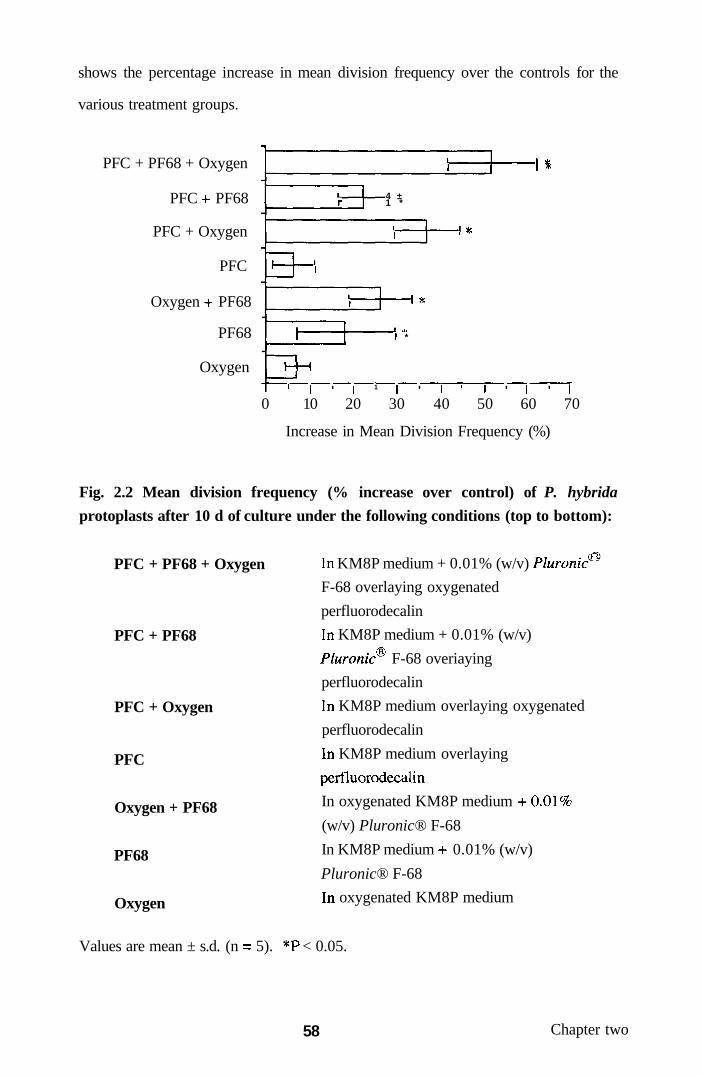
shows the percentage increase in mean division frequency over the controls for the
various treatment groups.
PFC + PF68 + Oxygen
PFC -h PF68
PFC + Oxygen
PFC
Oxygen -i- PF68
PF68
Oxygen
f 4 ± 1 *
1
1
1
-
-
1 1
1 1 • ( •
1 *
1—1
1 1 ' 1 1 1 '
1
1 '1-
1 • 1
1 -1-1 ^
' 1 ' 1 0 10 20 30 40 50 60 70
Increase in Mean Division Frequency (%)
Fig. 2.2 Mean division frequency (% increase over control) of P. hybrida
protoplasts after 10 d of culture under the following conditions (top to bottom):
PFC + PF68 + Oxygen
PFC + PF68
PFC + Oxygen
PFC
Oxygen + PF68
PF68
Oxygen
In KM8P medium + 0.01% (w/v) Pluronic^
F-68 overlaying oxygenated
perfluorodecalin
In KM8P medium + 0.01% (w/v)
Pluronic F-68 overiaying
perfluorodecalin
In KM8P medium overlaying oxygenated
perfluorodecalin
In KM8P medium overlaying
jDcrfluorodecalin
In oxygenated KM8P medium -1-0.01%
(w/v) Pluronic® F-68
In KM8P medium 4- 0.01% (w/v)
Pluronic® F-68
In oxygenated KM8P medium
Values are mean ± s.d. (n = 5). *P < 0.05.
58 Chapter two

Plate 2.2 Culture of Petunia hybrida protoplasts at a PFC/aqueous medium
interface
(a) Schematic diagram of the culture system, (b) Suspension cell-derived protoplasts
of albino P. hybrida cv. Comanche maintained in the dark at the interface formed
between oxygenated perfluorodecalin and liquid KM8P culture medium (x3), (c)
Freshly isolated P. hybrida protoplasts in CPW13M solution prior to culture at the
PFC/KM8P media interfaces (x200), (d) Protoplast-derived cells, some of which are
dividing (arrow), harvested from the interface after 10 d of culture in the dark (x200),
(e) Protoplast-derived cell micro-colonies of P. hybrida after 40 d culture under dark
conditions in liquid KM8P/KM8 medium overlaying oxygenated perfluorodecalin;
such micro-colonies could be converted back to discrete calli and eventually cell
suspension cultures (xlOO).
59 Chapter two


2.3.2.4 Growth of petunia protoplasts with perfluorodecalin and Pluronic F-68
Supplementation of the aqueous culture medium with 0.01% (w/v) Pluronic F-68
increased protoplast growth, such that the mean division frequency after 10 d (8.4 ±
1.0%) was significantiy greater (P < 0.05) than control (Fig. 2.2). In contrast, no
further increase in growth over controls occurred when the Pluronic F-68-
supplemented culture media were either gassed with oxygen alone or layered over
non-gassed perfluorodecalin. Addition of 0.01% (w/v) Pluronic F-68 to KM8P
medium overlaying oxygenated perfluorodecalin produced the greatest increase in
mean protoplast growth after 10 d (10.8 ± 1.1%; Fig. 2.2). This led to normal
protoplast-derived cell colony formation by 40 d, thereby confirming that repeated
mitotic division in protoplasts was not impaired by culture in the presence of both
oxygenated perfluorodecalin and surfactant (Plate 2.2e). Samples of protoplast-
derived colonies could be returned by pipetting to the growth conditions/culture
medium as routinely used for the original cell suspension, thereby completing the
cultural cycle.
2.3.2.5 Image analysis of petunia protoplast-derived cell colony growth
Plate 2.3; Figs. 1-4 show typical protoplast-derived colony formation after 40 d of
culture with the various treatments. A naked eye evaluation of plates showed major
differences in protoplast growth, with untreated controls characteristically showing
typically small, non-proliferated colonies. In contrast, the treated protoplast cultures
show extensive regions of cell growth and colony formation, the latter leading, in
some cases, to intergrowth of discrete cell colonies somewhat equivalent to artificial
tissue production.
60 Chapter two
Plate 2.3 Culture and development of Petunia hybrida cell suspension
protoplast-derived colonies from PFC/aqueous media interfaces and with
Pluronic® F-68
Protoplast-derived cell colonies of P. hybrida cultured in the light on agar-solidified
UM medium showing progressive mitotic activity leading to higher micro-colony
throughput; development recorded 68 d after protoplast isolation. Protoplasts were
initially cultured in the dark for 40 d under the following conditions prior to transfer
to UM medium at day 40: (la) KM8P/KM8 (3:1 v/v) liquid medium, (lb)
Oxygenated (100% oxygen; 10 mbar, 15 min) KM8P/KM8 liquid medium, (2a)
KM8P/KM8 liquid medium + 0.01%? (w/v) Pluronic® F-68, (2b) Oxygenated
KM8P/KM8 hquid medium -i- 0.01% (w/v) Pluronic® F-68, (3a) KM8P/KM8 liquid
medium overlaying non-oxygenated perfluorodecalin, (3b) KM8P/KM8 liquid
medium overlaying oxygenated (100% oxygen; 10 mbar, 15 min) perfluorodecalin,
(4a) KM8P/KM8 liquid medium -i- 0.01%) (w/v) Pluronic® F-68 overiaying non-
oxygenated perfluorodecalin, (4b) KM8P/KM8 liquid medium + 0.01% (w/v)
Pluronic F-68 overlaying oxygenated perfluorodecalin. (la-4b inclusive, xO.9).
61 Chapter two


Fig. 2.3 Percentage of plate area covered by P. hybrida protoplast-derived cell
colonies
601
50
•o Ol
Ol
> ©
U C3
< iJ
^-t
Pla
40
^o
20
^
10
• Controls • Perfluorocarbon Treated
la 3a CONTROL
lb 3b 2a 4a 2b 4b OXYGEN PLURONIC F68 PLURONIC F68+ O2
Treatment Group
The percentage area covered (Fig. 2.3) and the mean area per colony (Fig. 2.4) varied
from 2.8 ± 0.9% and 0.36 ± 0.11 mm respectively for untreated controls to 58.5 ±
2.1% and 12.5 ± 0.5 mm for colonies derived from protoplasts which had
previously been cultured on oxygenated perfluorodecalin in the presence of 0.01%
(\\'l\)Pluronic F-68. The percentage coverage of the plate for each PFC-treated
group was significantiy greater than that for the corresponding group not treated with
PFC (P < 0.001 for all groups). In addition, oxygenation increased the percentage
coverage significantiy (P < 0.001) for all treatment groups except for oxygenation of
aqueous culture medium alone. It is also noteworthy that colony area tended to
remain below approx. 4 mm in all treatments, with the exception of the groups
receiving PFC, oxygen and Pluronic® F-68. These colonies had a mean area of 12.5
mm , an increase of 35-fold over the untreated controls (P < 0.001).
62 Chapter two
Fig. 2.4 Mean area (mm ) per colony of P. hybrida protoplast-derived cell
colonies
151
E B
c o "o U 0)
Cu, w
c CS Ol
10
• Controls n Perfluorocarbon Treated
l a 3a CONTROL
l b 3b 2a 4a 2b 4b OXYGEN PLURONIC F68 PLURONIC F68 + ©2
Treatment Group
2.3.3 Effect of various respiratory gas-manipulation treatments on mitotic
division of cultured cassava protoplasts
The yield of cassa\'a leaf protoplasts was 6.9 ± 0.9 x 10 g f wt., with a mean
viability of 74 ± 4%. During the first 25 d of culture, a 68% increase (P < 0.05) in
the mean number of protoplasts entering their first mitotic division, as assessed by
IPE, occurred in dishes with glass rods compared to their untreated controls under a
normal gaseous atmosphere (Fig. 2.5, treatments A, B). Culture of protoplasts under
an oxygen-enriched atmosphere, but without glass rods, increased their IPE 2-fold (P
< 0.05) after 25 d, when compared to protoplasts cultured in the absence of glass
rods under normal atmosphere conditions (Fig. 2.5 treatments A, C). There was no
further elevation (or suppression) of IPE when protoplasts were cultured under an
increased oxygen atmosphere and in the presence of rods (Fig. 2.5, treatments C, D).
63 Chapter two

Fig. 2.5 Mean IPE of cassava leaf protoplasts after 25 d of culture in response to
respiratory gas-manipulation treatments.
CL,
(V
Treatment
Protoplasts cultured m: A) medium without glass rods in a normal atmosphere
(control), B) with glass rods in a normal atmosphere, C) without glass rods but with
an oxygen-enriched atmosphere, D) with glass rods and an oxygen-enriched
atmosphere, E) without glass rods and with non-oxygenated PFC, F) with glass rods
and non-oxygenated PFC, G) without glass rods and with oxygenated PFC or H)
with glass rods and oxygenated PFC. Vertical bars represent s.e.m. (n = 3
throughout).
The mean IPE values following culture of protoplasts with oxygenated
perfluorodecalin interjacent layer, either without or with glass rods, were
significantiy greater (P < 0.05) than those of the respective control cultures (Fig. 2.5,
treatments A, G; B, H, respectively). Indeed, the mean IPE of protoplasts cultured
with oxygenated perfluorodecalin in the presence of glass rods (5.8 ± 0.2%; n = 3)
was over 2-fold greater than in the controls (P < 0.05) lacking glass rods (Fig. 2.5,
treatments H, A). There were no significant differences between IPEs of cassava
64 Chapter t\\ o

protoplasts when cultured under an increased oxygen atmosphere, or with
oxygenated perfluorodecalin, irrespective of the presence or absence of glass rods
(Fig. 2.5, treatments C, G; D, H, respectively). In addition, the mean IPEs of
protoplasts following culture with non-oxygenated perfluorodecalin, with or without
glass rods, were significantiy different (P < 0.05) from their respective controls; an
increase in IPE was observed in the absence of rods (Fig. 2.5, treatments, E, A),
whereas a reduced IPE occurred in the presence of rods (Fig. 2.5, treatments F, B).
Interestingly, protoplast aggregation or the accumulation of phenolic products did
not occur in cultures containing glass rods and oxygenated PFC.
2.4 Discussion
The present experiments demonstrate that high yields of cassava protoplasts can be
routinely obtained from leaves of cultured shoots. These isolated protoplasts can be
induced to undergo sustained division to form multicellular colonies when partially
embedded glass rods are included in the two-phase culture system. Such use of glass
rods, to increase the number of liquid : glass/plastic contact points, provides a simple
method for ensuring a sustainable plating efficiency of cassava leaf protoplasts
which, hitherto, has not been obtained with other approaches (Byrne, 1984).
The novel system described here probably facilitates a greater gaseous exchange
between the liquid phase, containing the protoplsists, and the immediate atmosphere
above the medium in the dish. In this regard, previous studies have shown that
oxygen-enriched atmospheres also enhance the plating efficiency of cultured
protoplasts of rape (Brandt, 1991), jute, rice and tomato (d'Utra Vaz et al, 1992).
Consequentiy, it will be of interest, in future work, to assess whether combinations
of these physic2il and chemical approaches to gaseous manipulation can enhance
mitotic division and cell colony formation in other recalcitrant protoplast systems,
including protoplasts which have been modified genetically. In cassava, such studies
should also involve the use of a broader range of growth regulators in the medium
65 Chapter two
ultimately to promote plant regeneration from protoplast-derived tissues.
Additionally, it may be feasible to regenerate plants directiy from protoplasts
through somatic embryogenesis without an intervening callus phase. In this respect,
embryogenic cell suspensions may be useful source material for the isolation of
protoplasts.
To date, studies have shown adenine-type cytokinins to be ineffective growth
regulators for inducing differentiation of cassava protoplast-derived callus (Anthony
et al, unpublished). In the present study, the substituted pyridyl phenylurea
compound, CPPU, was evaluated, since this synthetic cytokinin is effective at very
low concentrations on a wide range of species (Fellman et al, 1987) and induces
somatic embryogenesis in grape (Matsuta and Hirabayashi, 1989). The induction, by
CPPU, of nodular and tuber-like stmctures in protoplast-derived callus of cassava,
indicates that the use of this phenylurea derivative may, ultimately, result in the
differentiation of shoots in the cv. M. Thai 8 tested in these experiments and in other
cassava genotypes previously considered to be recalcitrant to differentiation when
exposed to adenine-type cytokinins. A focus of future studies to further improve
protoplast growth and differentiation in this biotechnologically-important plant will
be to optimise not only gas supply, but also growth regulator-induced differentiation.
The results of this study also demonstrate, for the first time, that plant protoplasts
will grow normally at the interface between perfluorodecalin and an aqueous culture
medium. Thus, the technology previously employed in animal cell cultures (Giaever
and Keese, 1983; Nabih et al., 1989) can be successfully adapted for plant systems.
The finding that division and colony formation from protoplast-derived cells of P.
hybrida were not affected by oxygen or perfluorodecalin alone, but only by their
subsequent combination, demonstrates the dual efficacy of PFCs for both oxygen
storage and effective delivery to cells.
66 Chapter two

Furthermore, the present study also shows that supplementation with Pluronic F-68
of the aqueous culture medium overlaying the perfluorodecalin liquid reduces
aggregation of protoplasts associated with the onset of cell wall regeneration,
probably through surface tension effects. This is consistent with previous findings
that Pluronic F-68 reduced clumping in insect cell cultures (Murhammer and
Goochee, 1990). This is additional to any growth-enhancing effects of this
compound, as reported previously for cultured Solanum dulcamara protoplasts
(YMmaretal, 1992; Lowe etal, 1993).
The finding that Pluronic F-68 further enhanced the growth of protoplasts over the
increase promoted by oxygenated perfluorodecalin, suggests that these agents
promote protoplast division through separate mechanisms. While there is
speculation that Pluronics stimulate growth by enhancing cellular uptake of nutrients
and/or growth regulators (Lowe et al., 1993), PFCs most probably act by facilitating
oxygen transfer directiy into individual cells. This is supported by the present
finding that the perfluorodecalin phase acted as an oxygen reservoir, progressively
transferring the gas into the aqueous medium and, hence, to the protoplasts.
However, the possibility that Pluronic F-68 can itself facilitate cellular oxygen
uptake cannot be discounted. Alternatively, the likelihood that the PFC may have
facilitated protoplast and cell division by removing some unspecified growth
regulator from the aqueous medium also cannot be discounted. In this context, it has
been suggested previously (King et al, 1989) that one possible application of PFCs
in cell culture systems could be as scavengers of toxic gaseous by-products of cell
metabolism.
The image analysis technique employed in this investigation provided a precise and
convenient method for assessing protoplast responses to perfluorodecalin and
Pluronic F-68. This approach should be valuable in the quantitative assessment of
cell growth in a wide range of prokaryotic and eukaryotic cell culture systems.
67 Chapter two

Image analysis technology will provide an important adjunct to measuring and
discriminating accurately protoplast (and cell) growth responses. Traditionally, these
have involved assessments of IPE and FPE (Latif et al, 1993) based on sampling
after several days and weeks of culture resf)ectively. Image analysis provides,
particularly for FPE determinations, an altemative option. This eliminates the
inevitable task of identifying discrete colonies which, more often than not, have
grown together, making the value of FPE assessments somewhat problematical.
The use of PFC as an oxygenating agent is especially advantageous in static cultures
of protoplasts in which conventional aeration through agitation (e.g. stirring) cannot
be used because of the tendency to promote cell damage. Further advantages of
using PFCs as a culture medium support include their inertness, ease of sterilisation
by autoclaving, and re-cycleability (King et al, 1989), which offsets the relatively
high initial cost of these materials. PFCs are especially useful in illuminated
cultures, since they have refractive indices similar to water (Riess and Le Blanc,
1982). One improvement of the present system would be to "recharge" the
perfluorodecalin with oxygen during culture, thereby maximising the potential for
oxygen delivery to growing protoplasts. In addition to facilitating oxygen transfer, it
has been emphasised previously (King et al, 1989) that PFC-supplemented systems
could be valuable for supplying other gases (e.g. carbon dioxide) to anaerobic
cultures.
The present experiments further demonstrate that the use of oxygenated
perfluorodecalin for enhancing oxygenation of cultured cassava protoplasts is
superior to the inclusion, in semi-solid medium, of partially-embedded glass rods,
which increase the surface area for gaseous exchange, thus enhancing protoplast
division frequency. The present approach extends previous preliminary work which
demonstrated that protoplasts of cassava would undergo sustained division when
cultured in the presence of glass rods (Anthony et al., 1995a).
68 Chapter two

The present finding that the increase in division frequency of cassava protoplasts
could not be further enhanced by the inclusion of glass rods in cultures supplemented
with oxygenated perfluorodecalin suggests that there was no synergism between
these oxygenation options. This contrasts with previous work using protoplasts of P.
hybrida where supplementation of aqueous medium overlaying oxygenated
perfluorodecalin with 0.01%o (w/v) of the co-polymer surfactant, Pluronic® F-68,
promoted a further increase in division frequency (Anthony et al, 1994a). An
extension of the present investigation with cultured cassava protoplasts would be to
assess the beneficial effects of supplementing culture medium with Pluronic F-68,
which may stimulate protoplast growth by enhancing the uptake of nutrients and/or
protect against mechanical damage (Lowe et al, 1993).
Woody plants, such as cassava, are generally considered to have relatively poor
protoplast plating efficiencies (Ochatt and Power, 1992). The use of oxygenated
perfluorodecalin alone, as shown in the present investigation, may go some way
towards the enhanced throughput of protoplast-derived multicellular colonies and, in
turn, whole plant regeneration.
Previous studies on the culture of protoplasts from woody plants have emphasised
that one frequentiy encountered problem is that of phenolic oxidation caused by
aggregation of cells, especially during the initial stages of culture (Ochatt, 1990;
Ochatt and Power, 1992). While some success has been achieved in overcoming this
problem through the culture of protoplasts as monolayers on polyester discs within
the aqueous medium (Russell and McCown, 1986, 1988), the use of oxygenated
perfluorodecalin, as in the present study, to prevent protoplast clumping and phenolic
oxidation, provides an altemative option. Further studies are needed to evaluate this
approach using protoplasts and cells from other woody plant species.
69 Chapter two

The use of oxygenated PFC in plant cell cultures could provide a handling strategy
for culturing small numbers of protoplasts (e.g. flow-sorted heterokaryons) in media
droplets on liquid-PFC interfaces. A more efficient protoplast-to-plant system would
have implications for the transformation efficacy of protoplasts using direct DNA
delivery technologies. In the context of somatic hybridization, one of the cmcial
stages is the conversion of heterokaryons to somatic hybrid cells. Thus, an improved
plating efficiency, as indicated by the present use of oxygenated PFC, is likely to
lead to a greater throughput of putative hybrid material and will have profound
implications for plant cell biotechnology and genetic manipulation.
70 Chapter two
CHAPTER THREE: BIOCOMPATABILITY OF HAEMOGLOBIN-BASED
RESPIRATORY GAS CARRIERS IN PLANT PROTOPLAST CULTURE
3.1 Introduction
Isolated plant protoplasts are the source material for the genetic manipulation of
plants by interspecific and intergeneric somatic hybridisation through protoplast
fusion, transformation by direct DNA uptake and the exposure of somaclonal
variation (Blackball et al, 1994; Dixon 1994). One cmcial requirement for
successful protoplast culture and subsequent plant regeneration from protoplast-
derived tissues is the provision of an adequate and sustainable oxygen supply.
Previous approaches to enhance respiratory gas exchange for cultured protoplasts
have included the use of oxygen-enriched atmospheres (d'Utra Vaz et al, 1992),
inert, respiratory gas-dissolving PFC liquids (Anthony et al, 1994a, b, 1995b; Lowe
1997, 1998) and liquid-over-agar media-matrices implanted with glass rods to
increase the surface area and gaseous exchange of the liquid culture phase (Anthony
etal, 1995a).
An additional and novel approach to further facilitate oxygen supply, which has not
been evaluated previously for plant cell systems, is the supplementation of aqueous
culture media with a commercial haemoglobin (Hb) solution (Erythrogen™).
Previous research has focused on the assessment of native and, to a greater extent,
chemically-modified haemoglobins as vehicles for respiratory gas transport in intact
animals and isolated, perfused organs (Zuck and Riess, 1994; Tsuchida, 1995;
Winslow et al, 1995). The most widely studied molecules are of human or bovine
origins, although recombinant haemoglobins, expressed in Escherichia coli
(Hoffman et al, 1990) and transgenic mice (Behringer et al, 1989) and pigs
(Swanson et al, 1992), are now available. Haemoglobins liberated from animal
erythrocytes are normally treated with pyridoxal phosphate or glutaraldehyde to
71 Chapter three

reduce their oxygen binding to acceptable physiological levels (Zuck and Riess
1994; Tsuchida, 1995). One advantage of using haemoglobin as a respiratory gas-
carrying culture medium supplement compared with PFC liquids is that the latter are
immiscible in aqueous systems (King et al., 1989; Anthony et al, 1994a; Lowe et
al, 1995). However, paradoxically, this enables PFCs to be recovered readily and to
be recycled.
The present investigation has employed cell suspension-derived protoplasts of albino
P. hybrida cv. Comanche, as a model system to assess the effects on mitotic division
of protoplasts and their cellular derivatives of improved gas manipulation through
supplementation of the culture medium with a commercially-available, stabilised
bovine haemoglobin solution, Erythrogen™. The effects of haemoglobin were also
evaluated alongside the co-polymer surfactant, Pluronic F-68, which is commonly
employed as a cytoprotectant for cultured animal cells (Goldblum et al, 1990;
Papoutsakis, 1991; Handa-Corrigan et al, 1992). This surfactant has been shown
previously to promote growth in cultured plant cells, tissues and organs (Lowe et al,
1993, 1994).
Furthermore, to date, the relative merits of Hb compared to PFCs for stimulating the
mitotic division of plant protoplasts and protoplast-derived cells have not been
assessed. Consequentiy, the present investigation has also made a direct comparison
of these options, alone and in combination, using cell suspension-derived protoplasts
of Passiflora giberti and Petunia as model systems.
3.2 Materials and Methods
3.2.1 Culture of petunia suspension cells on medium supplemented with
Erythrogen'™
Suspension cells of P. hybrida cv. Comanche were harvested, 5 d after sub-culture,
onto a 45 pm. pore size nylon mesh. Aliquots of 0.5 g f.wt. of cells were placed onto
72 Chapter three

a 5.5 cm diam. Whatman No. 1 filter paper disk (Whatman International Ltd.,
Maidstone, UK) overlaying 20 ml aliquots of agar-solidified UM medium contained
in 9 cm Petri dishes. In some treatments the UM medium was supplemented with
Erythrogen™ (Biorelease Corporation, Salem, USA, obtained from TCS Biologicals,
Botolph Claydon, UK), a stabilised bovine haemoglobin solution (103 g 1" pH
7.42), to final concentrations of 1:50, 1:100 or 1:500 (v/v). Following addition of Hb
solution at 1:50 (v/v), the resultant pH of the culture medium was found to have
increased from 5.8 to 6.1. Therefore, the pH of the UM medium used for other
treatments was adjusted, prior to sterilization, to take account of this pH shift. Petri
dishes were maintained in the dark at 25 ± 2°C for 15 d, prior to growth assessments.
3.2.2 Isolation and culture of petunia protoplasts
Protoplasts of P. hybrida were isolated enzymatically from a well characterised and
established cell suspension, as described in Section 2.2.2.2. Protoplasts were
cultured in tiie dark (25 ± 2°C) at a final plating density of 2.0 x 10^ ml" in 1.5 ml
aliquots of liquid KM8P culture medium. The latter medium, containing protoplasts,
was dispensed into 3.5 cm diam. Petri dishes (Nunc). All experimental treatments
were replicated 5 times.
3.2.3 Supplementation of protoplast culture medium with Erythrogen^'* alone or
with oxygen or carbon monoxide
Erythrogen™ was added to KM8P medium to final concentrations of 1:50, 1:100 or
1:5(X) (v/v). For the former concentration, a further series of replicates were
established whereby the medium was gassed with 1(X)% oxygen (10 mbar, 15 min)
or 100% carbon monoxide (P/No: 850203; Phase Separations, Deeside, UK; 20 s),
immediately prior to the addition of protoplasts.
73 Chapter three

3.2.4 Supplementation of protoplast culture medium with Pluronic® F-68
In an additional assessment using non-gassed medium containing 1:50 (v/v)
Erythrogen™, the aqueous culture medium was also supplemented with 0.01, 0.1 or
1.0% (w/v) of Pluronic F-68 (Sigma). These concentrations of Pluronic were
based on those shown previously to stimulate the growth in culture of cell
suspension-derived protoplasts of S. dulcamara (Kumar et al, 1992). All
experiments using Erythrogen™, alone or in conjunction with other treatments,
utilised protoplasts of the same population, in turn isolated from the same
homogenous cell suspensions. Dishes were sealed with Nescofilm (Bando
Chemical, Kobe, Japan) and maintained under static conditions.
For all treatments, including controls, spent KM8P medium (0.25 ml per dish) was
removed after 9 and 15 d and replaced, on both occasions, with an equal volume of
KM8 medium lacking Erythrogen™. After 45 d of culture, the total population of
protoplast-derived cell colonies from one randomly selected replicate 3.5 cm Petri
dish was transferred to the surface of 6 ml of agar-solidified (0.6% w/v) UM medium
contained in 5.5 cm diam. Petri dishes. Cell colonies/microcalli were further
incubated in the dark at 25 ± 2°C for 35 d in order to confirm no adverse influence of
the treatments on long-term cell viability.
3.2.5 Measurement of protoplast viability, growth and biomass
The viability of freshly isolated protoplasts was determined by uptake and cleavage
of FDA, prior to counting as described in Section 2.2.1.3. Protoplast growth and
mitotic activity was assessed after 9 d, by the IPE, as defined in Section 2.2.1.6. A
minimum of 100 protoplasts per Petri dish were scored; each treatment was repeated
5 times. The biomeiss of protoplast-derived cells was recorded as fresh weight Eifter
80 d of culture with the various treatments.
74 Chapter three
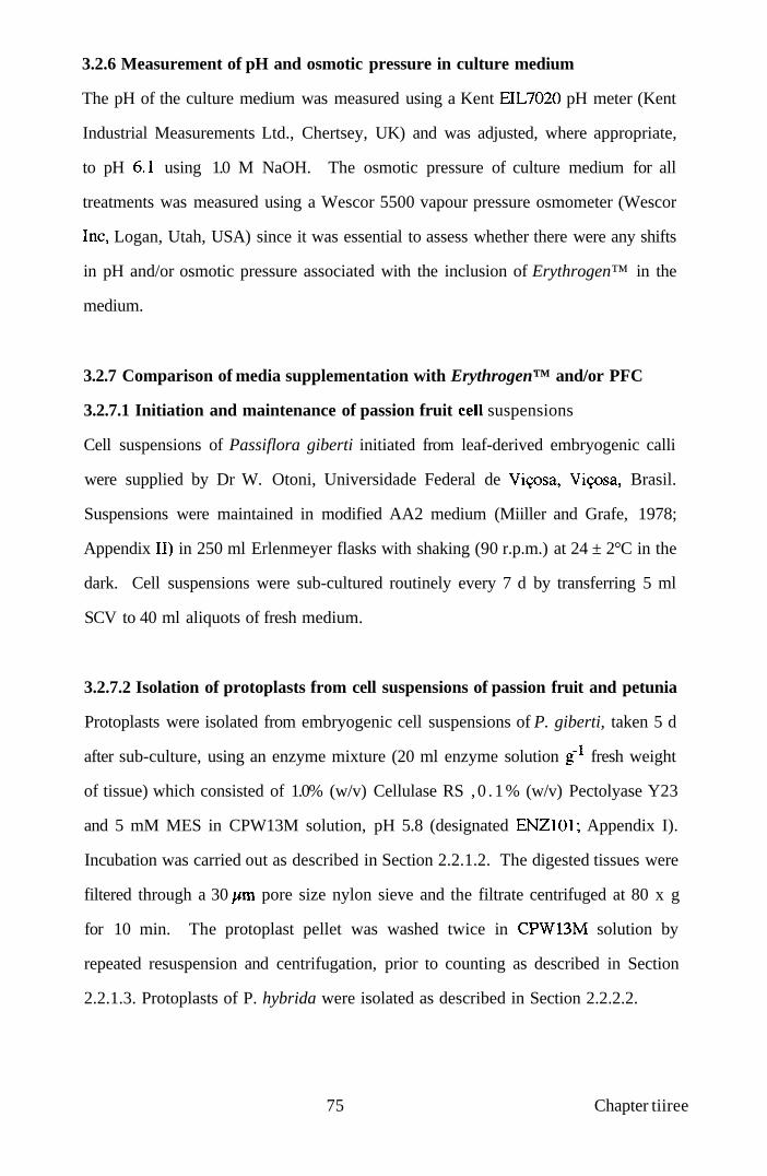
3.2.6 Measurement of pH and osmotic pressure in culture medium
The pH of the culture medium was measured using a Kent EIL7020 pH meter (Kent
Industrial Measurements Ltd., Chertsey, UK) and was adjusted, where appropriate,
to pH 6.1 using 1.0 M NaOH. The osmotic pressure of culture medium for all
treatments was measured using a Wescor 5500 vapour pressure osmometer (Wescor
Inc, Logan, Utah, USA) since it was essential to assess whether there were any shifts
in pH and/or osmotic pressure associated with the inclusion of Erythrogen™ in the
medium.
3.2.7 Comparison of media supplementation with Erythrogen™ and/or PFC
3.2.7.1 Initiation and maintenance of passion fruit cell suspensions
Cell suspensions of Passiflora giberti initiated from leaf-derived embryogenic calli
were supplied by Dr W. Otoni, Universidade Federal de Vi90sa, Vi9osa, Brasil.
Suspensions were maintained in modified AA2 medium (Miiller and Grafe, 1978;
Appendix II) in 250 ml Erlenmeyer flasks with shaking (90 r.p.m.) at 24 ± 2°C in the
dark. Cell suspensions were sub-cultured routinely every 7 d by transferring 5 ml
SCV to 40 ml aliquots of fresh medium.
3.2.7.2 Isolation of protoplasts from cell suspensions of passion fruit and petunia
Protoplasts were isolated from embryogenic cell suspensions of P. giberti, taken 5 d
after sub-culture, using an enzyme mixture (20 ml enzyme solution g fresh weight
of tissue) which consisted of 1.0% (w/v) Cellulase RS , 0 .1% (w/v) Pectolyase Y23
and 5 mM MES in CPW13M solution, pH 5.8 (designated ENZlOl; Appendix I).
Incubation was carried out as described in Section 2.2.1.2. The digested tissues were
filtered through a 30 pm pore size nylon sieve and the filtrate centrifuged at 80 x g
for 10 min. The protoplast pellet was washed twice in CPW13M solution by
repeated resuspension and centrifugation, prior to counting as described in Section
2.2.1.3. Protoplasts of P. hybrida were isolated as described in Section 2.2.2.2.
75 Chapter tiiree

3.2.7.3 Culture of passion fruit and petunia protoplasts with Erythrogen'^'*
and/or PFC
Protoplasts of P. giberti were cultured, for 6 d, at a density of 1.0 x 10^ ml"^ in liquid
KPR medium (Thompson et al., 1986; Appendix II) and those of P. hybrida, for 10
d, at a density of 2.0 x 10 ml'^ in liquid KM8P medium. Protoplast suspensions (2
ml aliquots) were placed in 30 ml glass bottles, either alone, or overlaying 6 ml
aliquots of oxygenated (10 mbar, 15 min) perfluorodecalin (Flutec® PP6). In a
further series of treatments, the media were supplemented with 1:50 (v/v) of
Erythrogen™ solution. This concentration of Hb was selected on the basis of
previous studies, which demonstrated the stimulatory effects of this molecule on the
mitotic division of cell suspension-derived protoplasts of P. hybrida (Anthony et al,
1997a, b). Protoplasts were cultured at 25 ± 2°C in the dark.
3.2.7.4 Measurement of IPE
After 6 d (Passiflora) or 10 d (Petunia) of culture with oxygenated PFC or
Erythrogen™, the IPE of protoplasts was measured as described in Section 2.2.1.3.
The differential time scale for measurement of IPE between the two culture systems
reflected the inherent differences in mitotic cycle times of Passiflora and Petunia.
3.2.8 Medium supplementation with fresh and stored Erythrogen^'* solutions
In the present experiments, the Hb solution was evaluated within 3 months of
purchase. In a separate assessment, protoplasts of P. hybrida were cultured in liquid
KM8P medium supplemented with 1:50 (v/v) of Erythrogen™ that had been stored
for ca 30 months at 4°C prior to use (designated stored-Hb). Protoplasts were
isolated and cultured as described in Sections 2.2.2.2 and 3.2.2, respectively.
3.2.9 Statistical analyses
Means and standard errors (s.e.m.) were used throughout, with statistical significance
between mean values assessed using a non-parametric Moods median test to
76 Chapter three

determine significant differences and Mann Whitney I/-tests to identify significant
treatments when analysing protoplast plating efficiency. A conventional parametric
one-way ANOVA was used to assess pH and osmotic pressure shifts (Snedecor and
Cochran, 1989). A probability of P < 0.05 was considered significant.
3.3 Results
3.3.1 Growth of petunia suspension cells on medium supplemented with
Erythrogen'^'*
The addition of Hb to agar-solidified UM medium resulted in a change of medium
colouration from pale yellow to red. Interestingly, when suspension cells of P.
hybrida (Plate 3. la) were placed onto filter paper disks, overlaying Hb supplemented
medium, such cells also exhibited a colour change from white to red after 3-4 d of
culture (Plate 3.1b). This did not appear to inhibit cell growth and after a further 7 d
of culture actively growing cells had resumed normal colouration. Indeed, the mean
fresh weights of cells after 15 d culture on medium supplemented with 1:50 (6.71 ±
0.21 g; n = 10) and 1:100 (v/v) of Erythrogen™ (6.92 ± 0.25 g; n = 9) were
significantiy (P < 0.05) greater than the mean fresh weight of control cells cultured in
the absence of Erythrogen™ (5.33 ± 0.22 g, n = 10; Table 3.1).
Table 3.1 Mean fresh weight and dry weight of Petunia suspension cells after 15
d culture on medium with Erythrogen'^'*
Concentration of Erythrogen™ (v/v) f wt. (g) dry wt. (g)
0 (control) 5.33 ± 0.22 0.15 ± 0.007
1:50 6.71 ±0.21* 0.19 ±0.004*
1: 100 6.92 ± 0.25* 0.18 ± 0.006*
1:500 5.32 ± 0.22 0.14 ±0.008
Values are mean ± s.e.m.; n = 9-10 throughout. *P < 0.05 compared to control mean
value.
77 Chapter three

However, there was no significant difference between the former treatments.
Similarly, the mean dry weights of such cells were also significantly (P < 0.05)
greater than cells cultured on medium lacking Erythrogen'^^ (Table 3.1). In addition
there was no significant difference between the mean dry weights of cells cultured on
medium with 1:50 and 1:100 (v/v) of Erythrogen™ (Table 3.1).
3.3.2 Efiect of medium supplemention with Erythrogen™ on mitotic division of
cultured petunia protoplasts
The yield of petunia protoplasts was 6.24 ± 0.45 x 10 g.f.wt of suspension cells,
with a mean viability of 94 ± 1% (n = 3). Culture of protoplasts in KM8P medium
supplemented with 1:50 (v/v) of Erythrogen™ stimulated protoplast division (Plate
3.1c), with an increase in mean IPE of 64% (P < 0.05) over control, after 9 d (Fig.
3.1). In contrast, the mean IPE values after 9 d in medium supplemented with 1:100
or 1:500 (v/v) of Erythrogen™ were not significantly different to control (Fig. 3.1).
For cultures supplemented with 1:50 (v/v) Erythrogen™ and gassed with oxygen, the
mean (± s.e.m.) IPE (18.1 ± 1.5%, n = 5) was significantiy (P < 0.05) greater than
control (11.3 ± 0.4%, n = 5), but was not significantiy different from cultures
containing the same, but ungassed, quantity of Erythrogen™ (18.5 ± 0.8%, n = 5;
Fig. 3.1). Similarly, for cultures containing 1:50 (v/v) Erythrogen™, but gassed with
carbon monoxide, the mean IPE after 9 d (13.1 ± 1.2%, n = 5) was also not
significantiy different to control (Fig. 3.1).
78 Chapter three
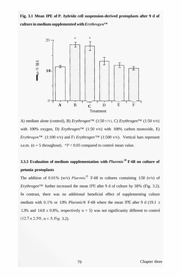
Fig. 3.1 Mean IPE of P. hybrida cell suspension-derived protoplasts after 9 d of
culture in medium supplemented with Erythrogen'^'*
OH
I
20 n
10-
0-A
^ i
B —r-C
— I —
D
-t
— I —
E
Treatment
- 1 —
F
A) medium alone (control), B) Erythrogen™ (1:50 \7v), C) Erythrogen™ (1:50 v/v)
with 100% oxygen, D) Erythrogen™ (1:50 v/v) with 100% carbon monoxide, E)
Erythrogen'^^ (1:100 v/v) and F) Erythrogen'™ (1:500 v/v). Vertical bars represent
s.e.m. (n = 5 throughout). *P < 0.05 compared to control mean value.
3.3.3 Evaluation of medium supplementation with Pluronic F-68 on culture of
petunia protoplasts
The addition of 0.01% (w/v) Pluronic F-68 to cultures containing 1:50 (v/v) of
Erythrogen™ further increased the mean IPE after 9 d of culture by 38% (Fig. 3.2).
In contrast, there was no additional beneficial effect of supplementing culture
medium with 0.1% or 1.0% Pluronic® F-68 where the mean IPE after 9 d (19.1 ±
1.3% and 14.8 ± 0.8%, respectively n = 5) was not significantiy different to control
(12.7 ± 2.5%, n = 5; Fig. 3.2).
79 Chapter three
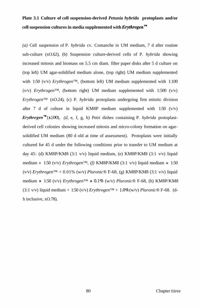
Plate 3.1 Culture of cell suspension-derived Petunia hybrida protoplasts and/or
cell suspension cultures in media supplemented with Erythrogen^'*
(a) Cell suspension of P. hybrida cv. Comanche in UM medium, 7 d after routine
sub-culture (xO.62), (b) Suspension culture-derived cells of P. hybrida showing
increased mitosis and biomass on 5.5 cm diam. filter paper disks after 5 d culture on
(top left) UM agar-solidified medium alone, (top right) UM medium supplemented
with 1:50 (v/v) Erythrogen™, (bottom left) UM medium supplemented with 1:100
(v/v) Erythrogen™, (bottom right) UM medium supplemented with 1:500 (v/v)
Erythrogen™ (xO.24), (c) P. hybrida protoplasts undergoing first mitotic division
after 7 d of culture in liquid KM8P medium supplemented with 1:50 (v/v)
Erythrogen™(xlOO), (d, e, f, g, h) Petri dishes containing P. hybrida protoplast-
derived cell colonies showing increased mitosis and micro-colony formation on agar-
solidified UM medium (80 d old at time of assessment). Protoplasts were initially
cultured for 45 d under the following conditions prior to transfer to UM medium at
day 45: (d) KM8P/KM8 (3:1 v/v) liquid medium, (e) KM8P/KM8 (3:1 v/v) liquid
medium -i- 1:50 (v/v) Erythrogen™, (f) KM8P/KM8 (3:1 v/v) liquid medium -i- 1:50
(v/v) Erythrogen™ + 0.01% (w/v) Pluronic® F-68, (g) KM8P/KM8 (3:1 v/v) liquid
medium -i- 1:50 (v/v) Erythrogen™ -1-0.1% (w/v) Pluronic® F-68, (h) KM8P/KM8
(3:1 v/v) liquid medium + 1:50 (v/v) Erythrogen™ + \.0% (w/v) Pluronic® F-68. (d-
h inclusive, xO.78).
80 Chapter tiiree


Fig. 3.2 Mean IPE of P. hybrida cell suspension-derived protoplasts after 9 d of
culture in medium supplemented with Erythrogen'™ and Pluronic® F-68
OH
c
B C D
Treatment
A) medium alone (control), B) Erythrogen™ (1:50 v/v), C) Erythrogen™ (1:50 v/v)
with 0.01%) (w/v) Pluronic® F-68, D) Erythrogen™ (1:50 v/v) with 0.1% (w/v)
Pluronic® F-68, E) Erythrogen™ (1:50 v/v) with 1.0% (w/v) Pluronic® F-68.
Vertical bars represent s.e.m. (n = 5 throughout). *P < 0.05 compared to control
mean value.
3.3.4 Assessment of medium supplementation with Erythrogen'^** on pH and
osmotic pressure shifts
Supplementation of culture medium with 1:50 and 1:100 (v/v) Erythrogen™, had a
significant effect on pH but not osmotic pressure (Table 3.2). Moreover, gassing of
culture medium containing 1:50 (v/v) Erythrogen™ with oxygen or carbon
monoxide had no significant effect on mean pH (6.1 throughout) or osmotic pressure
(764 ± 7.5 mOsmol kg and 774 ±3.1 mOsmol kg , respectively), compared to
ungassed medium with Erythrogen™ (756 ± 8.4 mOsmol kg ). Furthermore, light
81 Chapter three
microscopy revealed no marked morphological or cytological alterations of
protoplasts or protoplast-derived cells in response to any of the culture treatments.
Table 3.2 Effect of medium supplementation with Erythrogen^** on pH and
osmotic pressure
Concentration of Erythrogen™ (v/v) pH osmoticum
(mOsmol kg" )
0 (control)
1:50
1: 100
1:500
5.73 ± 0.07
6.13 ±0.03*
6.03 ± 0.03*
5.73 ± 0.07
736 ± 3.8
727 ±1.0
724 ± 3.0
736 ± 2.0
Values are mean ± s.e.m.; n = 3 throughout. *P < 0.05 compared to control mean
value.
3.3.5 Growth of petunia protoplast-derived cell colonies
Culture of protoplast-derived cells for 45 d with 1:50 (\7v) ungassed Erythrogen'^'*
produced a 2-fold increase (P < 0.01) in biomass, as reflected by mean fresh weight
after a further 35 d of culture on medium lacking Erythrogen'^'* (1.9 + 0.1 g, n = 5,
Table 3.3; Plate 3.1e) compared to control (1.0 ± 0.1 g, n = 5, Table 3.3; Plate 3. Id).
Culture of protoplast-derived cells for the same period in the presence of 1:50 (v/v)
Erythrogen™ and 0.01% (w/v) Pluronic F-68 produced a further significant (P <
0.01) increase in mean fresh weight (2.6 ±0.1 g, n = 5, Table 3.3; Plate 3.If) in
comparison to control (Table 3.3) and all other treatments. In contrast, there was no
additional beneficial effect of supplementing medium containing 1:50 (\/v)
Erythrogen™ with 0.1 or 1.0%; (w/v) Pluronic® F-68 (Table 3.3; Plate 3.1g, h)
82 Chapter three

Table 3.3 Mean fresh weight (g) of P. hybrida protoplast-derived colonies
cultured with 1: 50 (v/v) Erythrogen™ and Pluronic® F-68
Treatment Mean f. wt. (g)
control (medium alone) 1.0 ± 0.11
control (Erythrogen™ alone) 1.9 ± 0.12*
Pluronic® F-68 0.01 % (w/v) 2.6 ± 0.11 *
Pluronic® F-68 0.1 % (w/v) 1.2 ± 0.17
Pluronic® F-68 1.0% (w/v) 1.0±0.11
Values are mean ± s.e.m.; n = 5 throughout. *P < 0.05 compared to control (medium
alone) mean value.
3.3.6 Evaluation of media supplemention with Erythrogen"^** and/or PFC on
culture of passion fruit and petunia protoplasts
The yields of P. giberti and P. hybrida protoplasts were 5.83 ± 0.23 x 10^ g. f. wt
and 2.81 ± 0.15 X 10^ g. f. wt (n = 3 throughout), with mean viabilities of 78 ± 3% (n
= 3) and 80 ± 2% (n = 3), respectively. The mean IPE of P. giberti protoplasts with
oxygenated PFC (20.3 ± 0.6%; n = 5) was significantiy (P < 0.05) greater than
control (10.4 ± 0.3%, n = 5; Fig. 3.3). A similar, but more pronounced, increase (P <
0.05) in mean IPE of Passiflora protoplasts was observed when the medium was
supplemented solely with Erythrogen™ (22.9 ± 1.0%, n = 5; Fig 3.3).
83 Chapter three

Fig. 3.3 Mean IPE of P. giberti protoplasts cultured for 6 d in medium
supplemented with oxygenated PFC and Erythrogen™
i
A) medium alone (control), B) medium supplemented with 1:50 (v/v) Erythrogen™,
C) medium overiaying oxygenated PFC or D) medium supplemented with 1:50 (v/v)
Erythrogen'^'* and overlaying oxygenated PFC. Vertical bars represent s.e.m. (n = 5
throughout). *P < 0.05 compared to control mean value.
There was no significant difference in mean IPE values for Passiflora between the
oxygenated PFC and Erythrogen™ treatments (Fig. 3.3). When the latter two
treatments were combined, the mean IPE (18.3 ± 0.8%; n = 5) was also significantiy
(P < 0.05) greater than that in untreated controls (Fig. 3.3). Interestingly, there was
no significant difference between the mean IPE of the combined treatments and that
of protoplasts cultured in the presence of oxygenated PFC alone. However, the mean
IPE of protoplasts cultured with oxygenated PFC and Erythrogen'^'* in combination
was 20%) lower (P < 0.05) than that with Erythrogen™ alone (Fig. 3.3).
84 Chapter three

Fig. 3.4 Mean IPE of P. hybrida protoplasts cultured for 10 d in medium
supplemented with oxygenated PFC and Erythrogen™
oi.
^
A) medium alone (control), B) medium supplemented with 1:50 (v/v) Erythrogen™,
C) medium overlaying oxygenated PFC or D) medium supplemented with 1:50 (v/v)
Erythrogen™ and overlaying oxygenated PFC. Vertical bars represent s.e.m. (n = 5
throughout). *P < 0.05 compared to control mean value.
The mean IPE of P. hybrida protoplasts cultured with oxygenated PFC after 10 d of
culture (20.3 ± 1.5%; n = 5 throughout) was significantiy (P < 0.05) greater than
control (9.6 ± 0.6%; Fig 3.4). In addition, the mean IPE of Petunia protoplasts
cultured in medium containing Erythrogen'^'* was 17.3 ± 1.1% compared to 9.6 ±
0.6% in control (P < 0.05). As for Passiflora, no significant difference in IPE was
observed between the oxygenated PFC and Erythrogen'^'* treatments, despite the
time-scale difference in the IPE assessments. When the PFC and Hb treatments were
combined, the mean IPE (23.0 ± 0.9%; n = 5) was also significantiy (P < 0.05)
greater than that in untreated controls or with Erythrogen™ alone (Fig 3.4). This
contrasted with Passiflora, where Hb promoted the greatest increase in IPE.
However, as with P. giberti, there was no significant difference between the mean
85 Chapter three

IPE of protoplasts cultured with oxygenated PFC and Erythrogen™, in combination,
and with oxygenated PFC alone.
3.3.7 Effect of Erythrogen^** efficacy after long-term storage
In a separate experiment comparing the effects of medium supplementation with
fresh- or stored-Hb, the mean IPE of Petunia protoplasts after 10 d, in the former
case (26.7 ± 1.5%; n = 5 throughouO was significantly greater than with the stored-
Hb (18.4 ± 0.9%). However, the results for both of these treatments were
significantiy greater (P < 0.05) than for the untreated control (11.5 ± 1.0%; Table
3.4).
Table 3.4 Mean IPE of P. hybrida cell suspension-derived protoplasts cultured
for 10 d in medium supplemented with 1:50 (v/v) of fresh- or stored-Hb.
Treatment Mean % IPE of protoplasts
(lOd)
Control 11.5 ±1.0
Fresh-Hb 26.7 ±1.5*
Stored-Hb 18.4 ±0.9*
Values are mean ± s.e.m.; n = 5 throughout. *P < 0.05 compared to control mean
value.
3.4 Discussion
The results of these experiments demonstrate, for the first time, that supplementation
of medium with Erythrogen™ at 1:50 and 1:100 (v/v) dilutions, optimally promoted
mitotic division of P. hybrida suspension-derived cells over a 15 d culture period, as
measured by increases in both mean cell fresh weight and dry weight. Similarly,
supplementation of medium with 1:50 (v/v) Erythrogen™ enhanced mitotic division
in protoplasts of P. hybrida over a 9 d culture period. Pluronic F-68 at 0.01%
86 Chapter three

(w/v) had a synergistic effect by further enhancing protoplast division. This study
shows that Erythrogen™, with or without Pluronic F-68, provides a further option
for enhancing the growth of cultured plant protoplasts and protoplast-derived cells
under static and, perhaps, agitated conditions. This may be especially valuable for
facilitating the in vitro biosynthesis of phytochemicals, since related studies using
cultured animal cells have shown Erythrogen™ not only to stimulate cell division,
but also to promote the production of recombinant protein (Goffe et al, 1994; Shi et
al, 1998). This technology could be readily scaled-up to fermenter systems and may
be effective in stimulating growth of cultured plant tissues and organs, such as
Agrobacterium rhizogenes-transformed roots, which are now employed extensively
as a source of economically-important plant products (Toivonen, 1993).
The finding that Pluronic F-68 further enhanced the growth of Petunia protoplasts
over that induced by Erythrogen™ alone, suggests that these media supplements
stimulate protoplast division through separate mechanisms. Erythrogen™ is
believed to "trap" oxygen from air/medium interfaces and to facilitate delivery of this
gas directiy to cells, concomitantiy improving growth (Goffe et al, 1994). In
addition to alterations in oxygen supply per se, it is clearly possible that growth-
enhancement produced by supplementing medium with Erythrogen™ may be part-
driven by, for example, subtle changes in iron availability to cells, mild buffering of
medium pH or the uptake of amino acids from hydrolysed haemoglobin. Clearly,
these possibilities, individually or collectively, could be the focus of future studies
with eukaryotic cells.
The range of concentrations of Pluronic F-68 used in the present study were
selected on the basis of previous work, which demonstrated the growth-stimulating
effects, in culture, of this agent on isolated protoplasts of Solanum dulcamara
(Kumar et al, 1992). Interestingly, the optimum concentration of 0.01% (w/v) of the
surfactant in the present experiments was identical to that found to act synergistically
87 Chapter tiiree

with the oxygenated PFC liquid, Flutec PP5 , in promoting mitosis in P. hybrida
protoplasts in a static, 2-phase culture system (Anthony et al, 1994a).
Studies using animal cells have shown that Pluronic F-68 is adsorbed onto
cytoplasmic membranes, conferring increased resistance to mechanical damage
(Goldblum et al, 1990; Handa-Corrigan et al, 1992; Lowe et al, 1993, 1994).
However, the possibility that Pluronic® F-68 also promotes the uptake of nutrients,
growth regulators or oxygen into cultured protoplasts cannot be discounted and
should form a basis for future studies.
This study further demonstrates that both oxygenated PFC and Erythrogen™ provide
options for enhancing mitotic division, as reflected by increased IPE, of Passiflora
protoplasts and protoplast-derived cells in vitro. Interestingly, the study revealed
some variations in the effectiveness of the separate or combined treatments. For P.
giberti, Erythrogen™ promoted the greatest increase in IPE, whilst for P. hybrida,
the combined oxygenated PFC and Hb treatments were most effective. Whilst
further studies are required, these experiments suggest that the effectiveness of these
options for manipulating respiratory gas supply in vitro may be species specific.
However, the possibility also exists, at least for Passiflora, that a combined treatment
of Hb and PFC served to suppress mitotic division, through possible oxygen toxicity
effects. It may well be that for Petunia, such a threshold was not reached.
Furthermore, it will be interesting to determine whether haemoglobin, in
combination with Pluronic F-68 in aqueous culture medium overlaying oxygenated
PFC, will promote protoplast growth over and above the stimulation already
achieved by chemical and physical parameters (Anthony et al, 1994a, 1995b,
1997a). Future studies should evaluate these strategies in other plant culture systems
and determine whether they can be more broadly applied to cultured eukaryotic cells
in general. Clearly, such studies should incorporate more detailed assessments,
88 Chapter tiiree

including measurements of protein biosynthesis, of changes associated with cell
growth. This will identify the most effective system for sustaining division of
protoplast-derived cells prior to plant regeneration, which will be cmcial in
maximising plant throughput, especially of genetically-manipulated material.
A further important objective of this study was to assess whether there was any
change in the efficacy of the Hb solution following long-term (>1 year) storage at
4°C prior to incorporation into culture medium. The results demonstrate that, whilst
there was some diminution in the effectiveness of stored Erythrogen™ to stimulate
mitotic of cultured Petunia protoplasts, there remained, nevertheless, a significant
benefit in supplementing culture medium with the stored preparation. In contrast, for
PFCs which are chemically inert (Lowe et al, 1998), their ability to facilitate
respiratory gas supply in cell culture systems is undiminished by long-term storage.
Thus, the recoverability and recycleability of PFCs, may make them a commercially
more attractive option compared to haemoglobin solutions, despite a relatively high
investment cost of PFC liquids.
89 Chapter tiiree

CHAPTER FOUR: PLURONIC® F-68-MEDIATED POST-THAW RECOVERY OF CRYOPRESERVED PLANT CELLS
4.1 Introduction
4.1.1 Cryopreservation of plant cell suspension cultures
V The storage of cells at ultra-low temperatures in liquid nitrogen is a procedure which
has assumed an increasingly important role in underpinning many aspects of
biotechnology.^De-differentiated plant cells maintained as suspension cultures in
liquid medium have been successfully cryopreserved and recovered in a range of
agronomically-important crops, including asparagus [Asparagus officinalis L.;
Nishizawa et al, (1992)], banana [Musa sapientum L.; Panis et al, (1990)], barley
[Hordeum vulgare L.; Fretz et al, (1992)], bramble [Rubus fruticosus L.; Lett and
Schmitt, (1992)], grape [Vitis vinifera L.; Dussert et al., (1991)], millet [Setaria
italica L.; Lu and Sun, (1991)], orange [Citrus deliciosa L.; Aguilar et al, (1993)],
pine [Pinus caribea L.; Laine et al, (1992)], rice [Oryza sativa L.; Meijer et al,
(1991)], mbber [Hevea brasiliensis L.; Veisseire etal., (1993)] and soybean [Glycine
max (L.) Men-.; Luo and Widholm, (1997)].
Embryogenic cells cultured as suspensions in liquid medium are used routinely as
source materials for the isolation of totipotent protoplasts. The latter have been
exploited extensively in plant genetic manipulation, particularly in the case of
cereals such as rice (Blackball et al, 1994). Such embryogenic cell cultures also
provide an altemative to immature zygotic embryo-derived tissues for the production
of fertile, transgeiuc rice plants following gene delivery by biolistics (Sivamani et
al, 1996). Additionally, embryogenic suspension-cultured cells are also amenable
to Agrobacterium-mediiated transformation (Hiei et al, 1994). In general, the
establishment and maintenance of embryogenic cell suspensions of rice, particularly
those of Indica rices, presents technical difficulties, with the morphogenic
90 Chapter four

competence of suspensions usually declining with successive sub-culture over
prolonged periods (Abe and Futsuhara, 1991). Such changes are often accompanied
by the production of morphologically abnormal, infertile plants (Rajasekaran, 1996),
particularly from cells of long-established suspensions. Such loss of totipotency is
not unique to rice suspension cultures.
Cryopreservation is now exploited as a routine procedure for the long-term storage
of biological materials at ultra-low temperatures (Grout, 1995). This technique
circumvents the loss of totipotency in plant systems and eliminates or reduces
genetic perturbations, together with alterations in secondary product biosynthesis in
some cell systems, frequentiy associated with extended culture of suspensions at
physiologically normal temperatures (usually about 25°C). Cryopreservation also
negates the labour-intensive requirement to re-initiate periodically and to
characterise, in terms of their growth and totipotency, new cell lines. Indeed, storage
at ultra-low temperature provides a readily available and constant supply of
morphogenetically competent cells (Lynch et al, 1994).
4.1.2 Physical and chemical parameters for stimulating post-thaw recovery of
cells
Eariy cryopreservation studies with rice cell suspensions involved assessments of the
freezing and thawing conditions that permit the successful recovery of viable cells
and their subsequent growth in culture (Lynch et al, 1994). Previously, Meijer et al
(1991) reported that supporting post-thawed rice cells on filter paper disks, as
opposed to placing cells directiy on the surface of the culture medium employed
during cell recovery, resulted in significant increases in cell regrowth. Subsequentiy,
Lynch et al. (1994) also found that the duration of post-thaw culture prior to the
transfer to new medium of filter disks supporting the thawed cells was an important
consideration, with the correct timing of the transfer decreasing the time required for
91 Chapter four

the re-establishment of new cell suspensions. Additionally, these workers noted that
the recovery of rice cells from freezing in 9 cm diam. Petri dishes was superior
compared to their culture in 5.5 cm diam. vessels. Presumably, in the larger dishes,
the greater volume of culture medium assisted the rehydration of cells and the
diffusion away from the cells of the potentially toxic cryoprotectant, dimethyl
sulphoxide (DMSO), together with any oxidation products from the cells.
Interestingly, the inclusion of activated charcoal in the cell recovery medium has
been demonstrated to have beneficial effects on the post-thaw recovery of
suspension cultured cells of grape (Dussert et al, 1991), presumably through the
absorption of toxic cellular waste products.
4.1.3 Novel post-thaw treatments to enhance oxygen delivery to thawed cells
Novel treatments which focus specifically on enhancing oxygen delivery to cells, are
important in maximising the {X)st-thaw recovery of plant cells. In recent years,
several approaches have been evaluated, including the use of surfactants (Anthony et
al, 1996, 1997c; Craig et al, 1997), perfluorochemical liquids (Anthony et al,
1997c) and modified haemoglobin (Azhakanandam et al, 1998) as supplements to
the culture medium used during the post-thaw recovery phase of cell growth. Cell
suspensions of rice have been exploited as a "model" system in several of these
investigations, because of the importance of rice as a major cereal crop.
4.1.3.1 Supplementation of culture medium with the surfactant Pluronic® F-68
The non-ionic, polyoxyethylene (POE)-polyoxypropylene (POP) surfactant,
Pluronic F-68 (Fig 4.1), has been employed extensively as a low cost, non-toxic,
cell-protecting agent in both animal (Papoutsakis, 1991; Handa-Corrigan et al,
1992) and plant (Lowe et al, 1993, 1994) culture systems. Pluronic® F-68 acts as a
foam-stabilising agent in agitated/aerated cultures and is believed to protect cells
against fluid-mechanical damage (Handa-Corrigan et al, 1989). Pluronic® F-68 is
92 Chapter four

adsorbed onto cell surfaces and studies with both cultured insect cells (Murhammer
and Goochee, 1988, 1990; Goldblum et al, 1990) and mammalian hybridomas
(Zhang et al, 1992) have demonstrated that interaction of the surfactant with
cytoplasmic membranes can increase the resistance of cells to shear forces, albeit
over differing time periods depending on the cell type.
Fig. 4.1 General chemical structure of Pluronic® F-68^
CHs
I
HO(CH2CH20)a (CHCH20)b (CH2CH20)cH
Polyoxyethylene Polyoxypropylene Polyoxyethylene block block block
Lowe et al (1993). Average values of a, b, and c = 75, 30, and 75, respectively.
Approx. molecular weight = 8350
The cytoprotectant properties of Pluronic F-68 make this compound a strong and
obvious candidate for use in plant cell cryopreservation protocols. Early studies
showed that the Pluronics could prevent haemolysis of human red blood cells in
response to freeze-thawing procedures (Glauser and Talbot, 1956), and subsequent
studies demonstrated that Pluronic F-68 was an effective cryoprotectant of cultured
Chinese Hamster cells (Ashwood-Smith et al, 1973). Surprisingly, there has been
no evaluation of the effects of using Pluronic® F-68 with plant cells, either as a
cryoprotectant per se or post-thaw cytoprotecting agent, despite the increasing
interest in cryopreservation for conserving agronomically-important and endangered
plant germplasms (Benson, 1994). Consequentiy, the present investigation has
employed embryogenic cells of Japonica rices, together with non-embryogenic cells
of Lolium multiflorum (Italian ryegrass), to evaluate the potential beneficial effects
of Pluronic® F-68, either as a cell-protectant during freezing or as a specific culture
93 Chapter four

medium supplement, in enhancing post-thaw cellular growth following short-term
(30 d, Oryza sativa cv. Tarom, Lolium) or long-term (3 years, O. sativa cv. Taipei
309) cryopreservation.
In a further separate assessment cryopreservation protocols were evaluated for non-
embryogenic cell suspension cultures of Moricandia arvensis (L.) DC. (2n=2x=28),
within the sub-tribe Moricandiinae of the Brassicaceae. This dicotyledonous plant
species is a wild relative of crop Brassicas, with an intermediate C3 and C4 photo-
synthetic/photorespiratory system (Holaday et al, 1981) giving an efficient
recapture of photorespiration-released carbon dioxide. This accounts for a low
carbon dioxide compensation point for this plant (McVetty et al, 1989). Cell
suspensions were initiated as a source of protoplasts for somatic hybridisation and
cybrid production with Brassica napus, to incorporate, ultimately, the C3-C4
metabolic pathway into oilseed rape. To this end, such suspension cultures required
the development of long-term preservation protocols and subsequent post-thaw
handling strategies. Cryopreservation for the Brassicaceae is restricted to isolated
microspores (Chen and Beversdorf 1992a, 1992b) and microspore-derived embryos
(Bajaj, 1983; Uragami et al, 1993) of Brassica rmpus, together with cell suspensions
of Arabidopsis thaliana (Ford, 1990; Ribeiro et al, 1996). Therefore, an assessment
was carried out to develop a cryopreservation protocol for Moricarulia suspension
cells since, to date, no freeze-thaw handling strategy exists. A further extension of
the work was to determine any beneficial growth effects of supplementing the post-
thaw recovery medium with Pluronic F-68. These cells being representative of a
dicotyledonous plant species.
94 Chapter four

4.2 Materials and Methods
4.2.1 Rice and Lolium
4.2.1.1 Plant materials and preparation of rice and Lolium cell suspensions
Cell suspensions of O. sativa L. cv. Taipei 309 and O. sativa L. cv. Tarom were
initiated from embryogenic calli derived from mature seed scutella (Finch et al.,
1991). Cell suspensions of Lolium multiflorum were supplied by Dr E. Guiderdoni,
IRAT/CIRAD, Montpellier, France. Cell suspensions were maintained in AA2
medium [cv. Taipei 309, Abdullah et al., (1986); Appendix II], R2 medium [cv.
Tarom, Ohira et al, (1973); Appendix II] and N6 medium [Lolium, Chu et al,
(1975); Appendix II] in 100 ml Erlenmeyer flasks with shaking (120 r.p.m., 2.5 cm
throw) at 28 ± TC in the dark. Rice cell suspensions (both cvs.) were sub-cultured
routinely every 7 d by transferring 1 ml of settled cells with 7 ml of spent medium to
22 ml aliquots of fresh medium; Lolium cell suspensions were similarly sub-cultured
by transfer of settied cells (3 ml) and 7 ml of spent medium to 40 ml aliquots of fresh
medium. Prior to cryopreservation, cells were cultured for 3-4 d in their respective
liquid medium supplemented with 60.0 g 1" mannitol.
4.2.1.2 Cryoprotection and freezing of rice and Lolium suspension cells
The cryopreservation protocol was based on that of Meijer et al. (1991). Cells were
harvested onto a nylon mesh (45 pm pore size) and placed into 2 cm polypropylene
vials (Sarstedt, Leicester, UK) with approximately 0.2 g fresh weight of cells per
vial. Approximately 0.75 ml of a cryoprotectant mixture [46.0 g 1" glycerol, 39.0 g
r^ DMSO, 342.0 g r^ sucrose, 5.0 g 1" proline] was added to each vial. The
cryoprotectant mixture was prepared in the liquid culture medium appropriate for
each species and the pH adjusted to 5.8, prior to filter sterilization (0.2 pm pore size;
Sartorius). All cryoprotectants were of Analar grade except DMSO, which was
spectroscopically pure.
95 Chapter four

Cells were cryoprotected for 1 h on iced water, vials containing the cells were
transferted to aluminium canes, and the cells were frozen at a controlled rate (-TC
min" ) from 0 to -35°C and held at this temperature for 35 min in a programmable
freezer (Planer Cryo 10 Series, Planer Biomed, Sunbury-on-Thames, UK), prior to
storage in liquid nitrogen at -196°C. Cells of cv. Tarom and Lolium were stored for
30 d, whereas those of cv. Taipei 309 were cryopreserved for 3 years.
4.2.1.3 Post-thaw recovery of cryopreserved rice and Lolium suspension cells
Cells of both species (two cvs. for rice) were thawed by plunging the vials into
sterile water at 45°C; excess cryoprotectants were removed under axenic conditions
from the cells using a disposable Pasteur pipette. The cells were placed onto two
superimposed 5.5 cm diam. Whatman No. 1 filter paper discs overlying 20 ml
aliquots of the appropriate culture medium. The latter was semi-solidified with 0.4%
(w/v) SeaKem Le agarose (FMC BioProducts). In some treatments, the medium was
supplemented with 0.01, 0.1 or 0.2% (w/v) of Pluronic® F-68 (Sigma). These
concentiations of Pluronic were selected on the basis of previous studies
demonstrating the stimulatory effects of this compound on the growth in culture of
protoplasts isolated from cell suspensions of Solanum dulcamara (Kumar et al,
1992). In an additional assessment, the cryoprotectant mixture for both species was
also supplemented with the aforementioned concentrations of Pluronic F-68 and
cells cryopreserved for 14d in liquid nitrogen.
Cells were cultured for all treatments in the dark for 3 d at 28 ± TC prior to transfer
of the upper filter disk containing the cells to the respective fresh media. Cells were
cultured for a further 24 h prior to viability assessments and, where appropriate, for a
further 26 d, under the same conditions, for biomass determinations. Each
experiment was replicated up to 20 times, except where surfactant was incorporated
into the cryoprotectant, where n = 7.
96 Chapter four

4.2.1.4 Measurement of post-thaw viability and biomass
The post-thaw viability and metabolic capacity of cells were assessed by the
reduction of triphenyl tetrazolium chloride (TTC; Steponkus and Lamphear, 1967).
Three millilitre aliquots of TTC buffer, consisting of 0.6% (w/v) TTC in 0.05M
Na2HP04-KH2P04 buffer containing 0.05% (v/v) Tween 80 (Sigma, pH 7.4), was
added to 50 mg of cells contained in 16.0 ml screw-capped centrifuge tubes. Cells
were incubated at 28 ± TC for 16-18 h in the dark, washed once with distilled water,
and extracted with 7.0 ml of 95% (v/v) ethanol in a boiling water bath for 5 min.
Supematants were cooled, and each was adjusted to 10.0 ml with 95% (v/v) ethanol.
The absorbance was measured at 490 nm using a Lambdabio spectrophotometer
(Perkin Elmer Ltd., Beaconsfield, UK). The same protocol was also employed for
unfrozen cells 4d following sub-culture. In some assessments, using both cv. Taipei
309 and Lolium cells, the incubation buffer was supplemented with 0.01 or 0.1%
(w/v) Pluronic F-68, respectively, as a control for any effects of the surfactant on
TTC reduction. The fresh weight of thawed cells was recorded to determine biomass
(Lynch etal, 1994).
4.2.2 Moricandia
4.2.2.1 Plant material and establishment of Moricandia shoot cultures
Seeds of Moricandia arvensis (L.) DC were provided by R.J. Mathias. Seeds were
surface sterilised with 15 % (v/v) "Domestos" bleach solution (Lever Industrial Ltd.,
Runcom, UK.) for 15 min, followed by three washes with sterile tap water. Seeds
were germinated in 175 ml capacity glass jars (10 seeds per jar) each containing 50
ml of MS semi-solid medium with 30 g 1 sucrose and 0.8 % (w/v) agar, but lacking
growth regulators (designated MSO). These, and all subsequent cultures used in this
study, were maintained under a 16 h photoperiod (21.45 /<mol m s , Cool-White
fluorescent tubes; Thorn EMI Ltd.) at 22 ± 2°C, unless otherwise stated. Seed-
97 Chapter four
derived shoots were maintained by transfer, every 28 d, of shoot tips (upper 3 nodes)
to fresh medium (4 shoots/jar).
4.2.2.2 Callus initiation and generation of Moricandia cell suspensions
Callus initiation and cell suspension establishment was carried out following
modification of published procedures (Toriyama et al, 1987). Stem intemodes of
cultured shoots (28 d after sub-culture) were cut into 0.5 cm lengths and placed
longitudinally in 9 cm Petri dishes each containing 25 ml of MS-based medium
(designated MSDl; Appendix II) with 1.0 mg V^ 2,4-D and semi-solidified with
0.4% (w/v) Sea Kem Le agarose. Callus resulting from stem sections was used to
produce cell suspensions, the latter being initiated by transferring approx. 0.8 g f wt.
portions of rapidly growing, friable 4-6 week-old callus to 8 ml aliquots of liquid
MSDl medium in 50 ml Erlenmeyer flasks, on a rotary shaker (90 r.p.m.). After 7-
10 d, the contents of each flask were transferted to 100 ml flasks to which 10 ml of
liquid MSDl medium weis added. Those cultures surviving 7-10 d of culture had an
additional 10 ml of medium added and became stocks for the subsequent initiation of
fast-growing cell suspensions in 250 ml flasks. This was effected by transfer of 1.5
ml packed cell volume (PCV) of cells, with 8.5 ml of partially-spent medium, to a
250 ml flask containing 30 ml of fresh medium (40 ml total culture volume).
Thereafter, cell suspensions were sub-cultured every 7 d by transfer of 1.5 ml PCV,
as already described.
4.2.2.3 Growth of Moricandia cell suspensions
A known volume of cells (1.5 ml PCV) in 8.5 ml of partially-spent medium was
transferted to a 250 ml flask, with a graduated side-arm, the flask containing 30 ml
of fresh medium. Cell growth was measured daily, over 15 d, by allowing 10 ml of
suspension to settie into the graduated side-arm. The settled cell volume (SCV) was
expressed as the volume (ml) of cells in 10 ml of culture. Growth curves were
98 Chapter four

plotted as a mean of 3 separate assessments; 5 readings were recorded at each
sampling time. The morphology and viability as measured by FDA staining (see
Section 2.2.1.3 for details) of 3-4 month-old (established) suspensions was assessed
every 7 d.
4.2.2.4 Preparation of Moricandia cells for cryopreservation
Cell suspensions were partially dehydrated, 7 d after transfer and prior to
cryopreservation, by replacing spent MSDl medium with fresh MSDl medium
supplemented with 6 % (w/v) mannitol. The suspensions were cultured for a further
7 d, after which suspensions were sieved (45 pm) and the spent medium discarded.
Cells (0.25 ml PCV) were transferred to 2,0 cm capacity polypropylene vials and
freshly prepared filter-sterilised cryoprotectant added (0.75 ml). Two cryoprotectant
mixtures, cryoprotectant A (46.0 g l'^ glycerol, 39.0 g l" DMSO, 342.0 g f ^ sucrose)
and cryoprotectant B (92.0 g 1" glycerol, 78.0 g 1" DMSO, 342.0 g 1' sucrose) as
modified from Lynch et al (1994), both based on MSDl medium, were assessed.
Chilled vials were loaded onto aluminium canes and frozen as described in Section
4.2.1.2, prior to storage in liquid nitrogen.
4.2.2.5 Post-thaw recovery of Moricandia cryopreserved cells
Cells cryopreserved for a minimum of 7 d were recovered from liquid nitrogen by
plunging the vials into sterile water at 45°C (10 min). Cells were transferted onto
two layers of sterile filter paper (No.l, 5.5 cm; Whatman) overlaying 25 ml aliquots
of MSDl medium semi-solidified with 0.8% (w/v) agar, contained in 9 cm Petri
dishes. MSDl medium contained Pluronic® F-68, at 0, 0.01, 0.1 or 1.0 % (w/v).
The contents of one vial were placed in each Petri dish. Cells were cultured for 3 d
(dark, 22 ± 2°C), prior to viability assessments, using the TTC assay, as described in
Section 4.2.1.4. The remaining cells were transferred, attached to the uppermost
filter paper, to fresh MSDl medium witii Pluronic® F-68, as appropriate. Cells were
99 Chapter four

sub-cultured every 28 d. Callus f wt. was recorded 28 d and 70 d post-thawing for a
minimum of 10 replicate plates for each treatment.
4.2.2.6 Re-initiation of Moricandia cell suspensions from cryopreserved cells
and isolation of protoplasts
Callus from M. arvensis cells cultured for 80-100 d post-thawing, was used to re
initiate cell suspensions in MSDl liquid medium. Re-initiated suspensions were
maintained alongside non-cryopreserved suspensions of the same cell line. Growth
curves were plotted for re-established and control suspensions.
Protoplasts were isolated from suspension cells 4 d after sub-culture and 125-130 d
after re-initiation of suspensions following modified protocols (Glimelius, 1984).
Cells were harvested on a 100 pm mesh nylon sieve and the cells (10-12 ml SCV)
incubated (14-16 h; 22 ± 2°C) in a 250 ml flask containing 40 ml of enzyme solution.
The latter consisted of 0.03 % (w/v) Macerozyme RIO, 2.0 % (w/v) Meicelase (Meiji
Seika Kaisha Ltd.,Tokyo, Japan) and 2.0 % (w/v) Rhozyme HP150 (Rohm and Haas
Co., Philadelphia, USA) in CPW13M solution at pH 5.7. Digestion was carried out
in the dark on a rotary shaker (40 r.p.m.).
After incubation, the cmde digests were passed through a 100 pm nylon mesh to
remove undigested cell colonies and spontaneous fusion bodies, and the filtrate
collected in 9 cm diam. Petri dishes. W5 salts solution (Menczel et al, 1981), at
approx. one-third volume of the original enzyme-protoplast mixture, was added to
the protoplast suspension prior to transfer to 16 ml screw-capped tubes and
centrifuging (7 min; 100 x g). The enzyme supernatant was removed, the protoplast
pellets resuspended in W5 salts solution (16 ml) and centrifuged. Protoplast pellets
were resuspended in W5 solution (2 ml) and purified by layering onto a 21 % (w/v)
sucrose solution in 16 ml screw-capped tubes and centrifuging (5 min; 150 x g).
100 Chapter four

Intact protoplasts forming a layer at the W5 solution-21 % (w/v) sucrose solution
interface, were collected by Pasteur pipette and transferred to 16 ml tubes.
Protoplasts were washed twice by resuspension in W5 solution with centrifugation
(7 min; 100 x g) and finally resuspended in W5 solution prior to yield and viability
assessments as described in Section 2.2.1.3. A minimum of 500 cells were counted
per culture.
4.3 Statistical analyses
Means and standard errors (s.e.m.) were used throughout; statistical significance
between mean values was assessed using a conventional one-way ANOVA and
identified using the Tukey-HSD test (Snedecor and Cochran, 1989). A probability
of P < 0.05 was considered significant.
4.4 Results
4.4.1 Cryopreservation of rice and Lolium cells
4.4.1.1 Post-thaw viability of cryopreserved rice and Lolium cells
The mean absorbance of unfrozen cells, as measured by TTC reduction, was 1.60 ±
0.11% (n = 10) for O. sativa cv. Tarom and 0.54 ± 0.04%) (n = 10) for Lolium. In the
case of O. sativa cv. Taipei 309, the mean cell absorbance, prior to long-term (3
year) cryopreservation, was 0.96 ± 0.05% (n = 10).
Supplementation of culture medium for the cv. Taipei 309 with Pluronic® F-68 at
0.01% (w/v) significantiy increased the mean post-thaw cell absorbance following
TTC reduction to more than 2-fold greater (P < 0.05) than that in untreated controls
(Fig. 4.2). A similar, but less pronounced effect also occurted with 0.1% (w/v)
surfactant (Fig. 4.2). In contrast, there was no cortesponding increase in absorbance
in these rice cells when 0.2% (w/v) Pluronic was incorporated into the culture
medium.
101 Chapter four

00
s g
s
it Xi
O CL
4>
s
JQ
C ^O *5 u 3
u H H M) B
• ^
O
0^
B
o Vi
Xi
<
5
a
E 2 as
> o
a
I
ON O en 'S a
'3
>
S S
U
H - CQ
l-H
r d d
•5f
"T"
d
•55 - u
pq
)l
00
d d
"T"
d d
u
* w- - OQ
r - - 00
d
I ' I ' I ' vo -^ ci d d d
(luu 06t7) souBqjosqB UBSIAI
3
> o o
00 I
o 3
a o
o c o U
V cu X •X-
o d V
B
s ^
>
C _ o "cS > (U (»
J D
o o n
c
e
S 13 >
d
d
u ^' r-H
O d CQ
o
<
2 3
Q ^ c o o
u s o (U ;-! a
i
102 Chapter four

In the case of O. sativa cv. Tarom, supplementation of medium with Pluronic® F-68
had a consistently greater stimulatory effect on the mean post-thaw cell absorbance
compared with that of untreated controls (Fig. 4.2). The most pronounced increase
occurted with 0.1% (w/v) surfactant, which promoted a 4-fold increase (P < 0.05) in
absorbance over the control. Supplementation of medium for Tarom cells with
0.01% (w/v) or 0.2% (w/v) Pluronic increased the mean cell absorbances by 2-fold
(P < 0.05) and 3-fold (P < 0.05), respectively (Fig. 4.2).
A significant (P < 0.05) increase in mean post-thaw cell absorbance was also
recorded when Pluronic® F-68 at 0.01% (w/v) was added to the culture medium for
Lolium cells, although the mean maximum increase over control of 31% (Fig. 4.2)
was markedly lower than that observed when the same concentration of surfactant
was used with cells of O. sativa cv. Taipei 309 (140%) or cv. Tarom (95%).
4.4.1.2 Post-thaw viability of cryopreserved rice and Lolium cells using TTC
buffer supplemented with Pluronic® F-68
No significant difference was observed, using post-thawed cv. Taipei 309 cells,
between the mean cell absorbance in the TTC assay (0.62 ± 0.05, n = 15) and in the
same assay using buffer supplemented with 0.01% (w/v) Pluronic F-68 (0.61 ±
0.05, n = 15). Comparable results were also obtained with post-thawed Lolium cells
using 0.1% (w/v) surfactant (control, 0.41 ± 0.03, n = 15; Pluronic 0.39 ± 0.04, n =
5).
4.4.1.3 Post-thaw growth of cryopreserved rice and Lolium cells
Addition of Pluronic® F-68 to culture medium also fostered an increase in biomass,
as measured by cell fresh weight following 30 d of post-thaw culture, in both O.
sativa cv. Taipei 309 and Lolium, with significant (P < 0.05, P < 0.01) increases in
the presence of 0.01% (w/v) of the surfactant in both cases (Table 4.1, 4.2). The
KB Chapter four
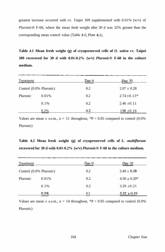
greatest increase occurred with cv. Taipei 309 supplemented with 0.01% (w/v) of
Pluronic® F-68, where the mean fresh weight after 30 d was 32% greater than the
corresponding mean control value (Table 4.1; Plate 4.1).
Table 4.1 Mean fresh weight (g) of cryopreserved cells of O. sativa cv. Taipei
309 recovered for 30 d with 0.01-0.2% (w/v) Pluronic® F-68 in the culture
medium.
Treatment DayO Day 30
Control (0.0% Pluronic) 0.2 2.07 ± 0.28
Pluronic 0.01% 0.2 2.74 ±0.13*
0.1% 0.2 2.46 ±0.11
0.2% 02 1.98 ±0.16
Values are mean ± s.e.m., n = 11 throughout, *P < 0.05 compared to control (0.0%
Pluronic)
Table 4.2 Mean fresh weight (g) of cryopreserved cells of L. multiflorum
recovered for 30 d with 0.01-0.2% (w/v) Pluronic® F-68 in the culture medium.
Treatment DayO Day 30
Control (0.0% Pluronic) 0.2 3.49 ± 0.18
Pluronic 0.01% 0.2 4.50 + 0.20*
0.1% 0.2 3.29 ±0.21
02% 02 3.32 ± 0 1 9
Values are mean ± s.e.m., n = 14 throughout, *P < 0.05 compared to control (0.0%
Pluronic)
104 Chapter four
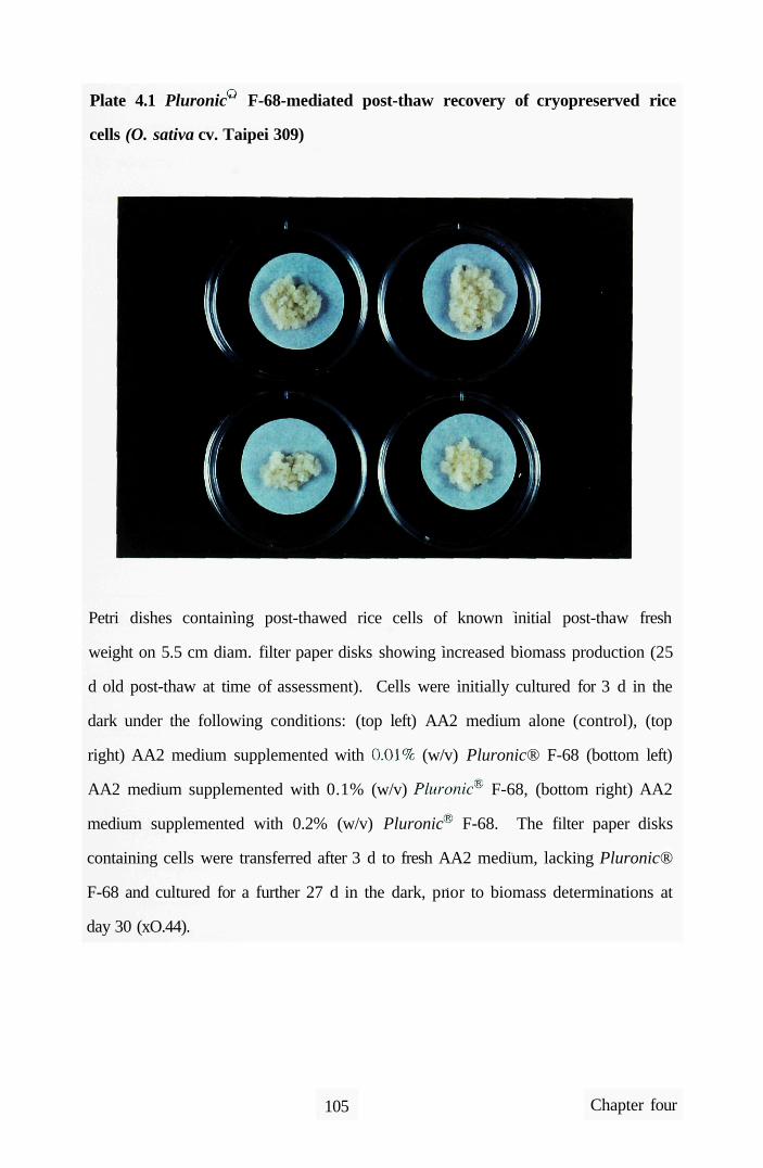
Plate 4.1 Pluronic'' F-68-mediated post-thaw recovery of cryopreserved rice
cells (O. sativa cv. Taipei 309)
Petri dishes containing post-thawed rice cells of known initial post-thaw fresh
weight on 5.5 cm diam. filter paper disks showing increased biomass production (25
d old post-thaw at time of assessment). Cells were initially cultured for 3 d in the
dark under the following conditions: (top left) AA2 medium alone (control), (top
right) AA2 medium supplemented with 0.01% (w/v) Pluronic® F-68 (bottom left)
AA2 medium supplemented with 0.1% (w/v) Pluronic F-68, (bottom right) AA2
medium supplemented with 0.2% (w/v) Pluronic F-68. The filter paper disks
containing cells were transferred after 3 d to fresh AA2 medium, lacking Pluronic®
F-68 and cultured for a further 27 d in the dark, pnor to biomass determinations at
day 30 (xO.44).
105 Chapter four

4.4.1.4 Supplementation of the cryoprotectant mixture with Pluronic® F-68
prior to cryopreservation of rice and Lolium cells
The addition of 0.01-0.2% (w/v) of Pluronic® F-68 to the cryoprotectant mixture,
both before and during freezing, did not affect the post-thaw viability of cells from
either rice cultivar or Lolium. For example, the mean absorbance of Lolium cells at
4 d following thawing in the presence of 0.01% (w/v) Pluronic (0.74 ± 0.04, n = 7)
was not significantiy different to control lacking surfactant (0.70 ± 0.04, n = 7). A
similar pattern was observed with both rice cvs. (Table 4.3).
Table 4.3 Mean post-thaw absorbance of cells of O. sativa cvs. Taipei 309 and
Tarom and L. multiflorum cryopreserved with 0.01-0.2% (w/v) Pluronic® F-68
in the cryoprotectant mixture
Treatment
0%) (control)
001%
oi%
02%
0. sativa
cv. Taipei 309
0.68 ± O02
067 ± O07
0.74 ± 0.06
0.74 ± 0.04
Absorbance (490 nm)
O. sativa
cv. Tarom
0.89 ± O20
0.94 ± 0 2 1
1.00 ± 0 2 0
0.93 ± 0 1 7
L. multiforum
0.70 ± 0 1 0
0.74±Oll
0.73 ±017
0.76 ± 0 1 8
Values are means ± s.e.m., n = 7 throughout.
4.4.2 Cryopreservation of Moricandia cells
4.4.2.1 Comparison of cryoprotectant effectiveness for cryopreservation of
Moricandia cells
The viability of unfrozen cells, as determined by mean cell absorbance following
TTC reduction, was 19.0 ± 0.8, whereas the viability, of partially dehydrated, non-
cryopreserved cells at day 3 was 13.2 ± 0.5 (n = 10 throughout). In the case of
frozen cells, there was no significant difference at 3d post-recovery, in the mean
106 Chapter four

absorbance of cells cryopreserved with cryoprotectant A (3.5 ± 0.4; n = 10) or
cryprotectant B (2.8 ± 0.2; n = 10). All cells were orange-brown in colour following
3d post-thaw culture.
There was no significant difference in the mean cell viability and the mean f. wt. of
cells at 28 d post-thawing of cells treated with cryoprotectants A or B (Table 4.4).
However, the mean f. wt. of cells treated with cryoprotectant B, after 70 d of culture
was 12 % greater (P < 0.05) than that of cells treated with cryoprotectant A (Table
4.4). There was also a significant (P < 0.05) increase in f wt. of cells at 70 d
compared to 28 d with cryoprotectant B. In addition, cells treated with
cryoprotectant B were friable and chlorophyllous in appearance by 70 d of post-thaw
culture, compared to those cryopreserved with cryoprotectant A. The latter remained
similar in appearance and colour to freshly thawed cells.
Table 4.4 Effect of cryoprotectant on viability and fresh weight (mg) of
Moricandia cells 28 and 70 d post-thawing
28 d post-thawing 70 d post-thawing
Cell viability Callus f wt. Callus f wt.
Treatment
Cryoprotectant A
Cryoprotectant B
(Absorbance)
1.08 ± 0 0 9
1.45 ± 0 2 1
(mg)
120.4 ± 15.3
129.2 ± 8.2
(mg)
139.1 ±6.9
156.4 ± 4.6*
Values are mean ± s.e.m. (n = 10 throughout), * P < 0.05 compared to cortesponding
mean value for Cryoprotectant A.
4.4.2.2 Effect of Pluronic® F-68 on recovery of cryopreserved Moricandia cells
Cell viability was comparable at all concentrations of Pluronic tested, except at 0.1%
(w/v) after 3 d of culture when viability was increased significantiy (Table 4.5).
107 Chapter four
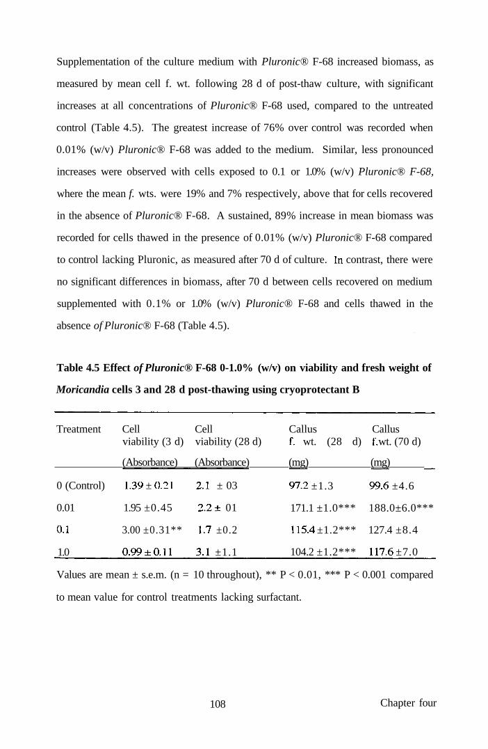
Supplementation of the culture medium with Pluronic® F-68 increased biomass, as
measured by mean cell f. wt. following 28 d of post-thaw culture, with significant
increases at all concentrations of Pluronic® F-68 used, compared to the untreated
control (Table 4.5). The greatest increase of 76% over control was recorded when
0.01% (w/v) Pluronic® F-68 was added to the medium. Similar, less pronounced
increases were observed with cells exposed to 0.1 or 1.0% (w/v) Pluronic® F-68,
where the mean f. wts. were 19% and 7% respectively, above that for cells recovered
in the absence of Pluronic® F-68. A sustained, 89% increase in mean biomass was
recorded for cells thawed in the presence of 0.01% (w/v) Pluronic® F-68 compared
to control lacking Pluronic, as measured after 70 d of culture. In contrast, there were
no significant differences in biomass, after 70 d between cells recovered on medium
supplemented with 0.1% or 1.0% (w/v) Pluronic® F-68 and cells thawed in the
absence of Pluronic® F-68 (Table 4.5).
Table 4.5 Effect of Pluronic® F-68 0-1.0% (w/v) on viability and fresh weight of
Moricandia cells 3 and 28 d post-thawing using cryoprotectant B
Treatment Cell Cell Callus Callus viability (3 d) viability (28 d) f wt. (28 d) f wt. (70 d)
(Absorbance) (Absorbance) (mg) (mg)
0 (Control) 1.39 ± 0.21 2.1 ± 03
0.01 1.95 ±0.45 2.2+ 01
01 3.00 ±0.31** 1.7 ±0.2
1.0 0 .99±Oll 3.1 ±1.1
97.2 ±1.3 99.6 ±4.6
171.1 ±1.0*** 188.0±6.0***
115.4 ±1.2*** 127.4 ±8.4
104.2 ±1.2*** 117.6 ±7.0
Values are mean ± s.e.m. (n = 10 throughout), ** P < 0.01, *** P < 0.001 compared
to mean value for control treatments lacking surfactant.
108 Chapter four

Fig. 4.3 Growth curves of suspension cultures of Moricandia arvensis, in MSDl
medium, initiated from cryopreserved (cryo) and non-cryopreserved (non-cryo)
cells.
2.5
E
o > u J)
CO
1.5
1 i^
0.5
-0— non-cryo
-A—cryo
5 7 9 11 13 15
Days post-subculture
4.4.2.3 Re-establishment of Moricandia cell suspensions and subsequent
isolation of suspension-derived protoplasts
Cells of the suspensions re-established from cryopreserved cells were
morphologically similar to cells of the original line. The growth pattern of
suspensions re-initiated from cryopreserved cells was very similar to that of the non-
cryopreserved contiol as shown in Fig. 4.3. In addition, the mean viability (58.3 ±
2.7 %) and yield (5.7 ± 0.6 x 10 ) of protoplasts from re-established suspensions
109 Chapter four

were not significantly different (P < 0.05) to the corresponding values for control,
non-cryopreserved suspensions of the same line (55.2 ± 4.4 % and 6.2 ± 0.8 x 10 ,
respectively).
4.5 Discussion
The results of these experiments demonstrate that supplementation of the culture
medium with Pluronic® F-68 increases the post-thaw metabolism and growth of
cryopreserved rice and Lolium cells. While it is generally accepted that reduction of
TTC can be used as an indicator of cell viability and metabolism (Benson et al,
1992), the present study also includes data on biomass increases to support the
conclusion of the growth-enhancing effects of Pluronic® F-68. It is probable that the
present observations reflect a combination of increased cell survival post-thawing
coupled, perhaps, with a growth-stimulating effect of the surfactant in plant culture
systems, as reported previously (Lowe et al, 1993). Assessment of TTC reduction
was made 4 days post-thaw since previous related work indicated that, at this time,
the TTC assay provides optimal information on cell metabolic capacity (Lynch et al,
1994).
The results of the present experiments also demonstrate that Moricandia cell
suspensions can be cryopreserved and that cryoprotectant mixture B, consisting of
DMSO, glycerol and sucrose, each at 1.0 M, was optimal for successful post-thaw
recovery. This study extends the earlier investigation which demonstrated that
supplementation of medium with Pluronic® F-68 enhanced post-thaw growth of rice
and Lolium suspension cells. The observation that such use of Pluronic F-68 with
Moricandia enhances post-thaw division leading to increased biomass production,
implies that this relatively low-cost strategy can be readily extended to plant systems
in general. The M. arvensis cell suspensions recovered from cryogenic storage
maintained growth characteristics and levels of protoplast yield and viability
110 Chapter four

comparable to the original, unfrozen cultures. However, M. arvensis cells were slow
to recover their normal growth rates after thawing, compared to cryopreserved cells
of rice where growth can be readily re-established (Lynch et al, 1994). This may be
due to the nature of the original non-embryogenic Moricandia cell suspensions,
since other workers (Lynch et al, 1994) observed differential responses of
embryogenic and non-embryogenic rice cells to cryopreservation, with embryogenic
cells exhibiting significantiy faster growth rates than non-embryogenic cells after
thawing.
An important finding from this investigation was that the optimum concentration of
surfactant, which increased cell growth, differed between the two rice cvs. and
between O. sativa cv. Tarom and Lolium. The present observations, showing that
there are both species- and cultivar-specific responses to Pluronic® F-68, are
consistent with previous observations using Chrysanthemum morifolium, in which
the optimum concentration of surfactant that stimulated adventive shoot regeneration
from cultured leaf explants differed by an order of magnitude between cvs. (Khehra
et al, 1995). Differences in tissue and organ responsiveness to the growth-promoting
effects of Pluronic® F-68 have also been observed in Solanum dulcamara (Kumar et
al, 1992), Corchorus capsular is (Khatun et al, 1993), Hypericum perforatum
(Bmtovska et al, 1994) and Populus spp. (lordan-Costache et al, 1995). For
example, culture of C. capsularis cotyledons with attached petioles in the presence
of up to 0.5% (w/v) Pluronic® F-68 increased shoot production, with no further
stimulation at higher concentrations (Khatun et al, 1993). In contrast, maximum
growth of transformed roots of Solanum dulcamara occurted with 0.01% (w/v)
Pluronic® F-68 and that of leaf-derived callus with 0.1% (w/v) of the surfactant
(Khatun et al., 1993). The present approach should be precision tuned for individual
species to optimise the concentration of Pluronic® F-68 eliciting maximum recovery
following cryopreservation.
111 Chapter four

Studies using animal cells have shown that Pluronic® F-68 adsorbs onto cytoplasmic
membranes, conferring increased resistance to mechanical damage (Handa-Corrigan
et al, 1992; Lowe et al., 1993, 1994). The Pluronic polyols have hydrophobic POP
cores, which are believed to become embedded in the phospholipid membranes of
cells, leaving their hydrophilic, POE tails outside. This would have the effect of
reducing the interfacial tension of the cells and sterically hindering adhesive
interaction between molecules on the cell surfaces. Such reduction in adhesive
interaction would prevent cell-to-cell contact and, thus, further reduce mechanical
damage.
Pluronic F-68 may also promote the increased uptake of nutrients, growth
regulators or oxygen into cells during the post-thaw period. Indeed, related
experiments using animal cells cultured under static conditions have shown that
concentrations of Pluronic F-68 comparable to those used in the present
investigation, stimulated both 2-deoxyglucose uptake and cellular amino acid
incorporation (Cawrse et al, 1991). In addition, related evidence from studies with
(E)
yeast has demonstrated that Pluronic F-68, at concentrations comparable to those
used here, enhanced the uptake of FDA and antibiotics (King et al, 1993; Bassetti
and Tramper, 1995). Any increase in nutrient uptake promoted by Pluronic would
be expected to alter metabolic flux, allowing biochemical pathways to operate more
efficientiy, particularly under the stress conditions of early post-thaw recovery. In
this regard, previous work has shown respiratory impairment occurs during the early
post-thaw period (Cella et al, 1982, Benson et al, 1992), and it is possible that
Pluronic® F-68 assists in overcoming such perturbations. The adsorption of
Pluronic molecules onto the cytoplasmic membranes of post-thawed plant cells may
also reduce cellular damage which is known to occur during rehydration when the
DMSO cryoprotectant is removed progressively from the system (Benson and
Withers, 1987).
112 Chapter four

Further studies are required to determine the mechanism(s) by which surfactants,
such as Pluronic® F-68, can facilitate post-thaw survival and growth of plant cells.
One focus of such work should be to investigate the effects of Pluronic® F-68 on
respiratory gas dynamics, since related work has provided evidence that this
compound can alter oxygen transport in agitated, sparged bioreactors (Murhammer
and Pfalzgraf, 1992). There is also speculation that Pluronic® F-68 may influence
the release of carbon dioxide from cells (Lowe et al, 1993) or inhibit ethylene
production in a manner comparable to that promoted by Triton X-100 (Sauerbrey et
al, 1988). Interestingly, experiments have demonstrated that inert, oxygen-
dissolving perfluorochemical liquids, emulsified with Pluronic® F-68, were effective
in prolonging the fertilising capability of turkey spermatozoa stored at lowered
temperature (Thurston et al., 1993). Whilst the specific effects of Pluronic were not
evaluated, it is possible that the surfactant may have contributed to cell survival,
either through cytoprotection or, perhaps, by enhancing oxygen and/or nutrient
uptake. This earlier work of Thurston et al. (1993), together with the results
reported in the present study, indicate that the mechanism(s) whereby Pluronic® ¥-
68 facilitates survival of both animal and plant cells warrants further detailed study.
The present data strongly suggest that Pluronic® F-68, which is relatively
inexpensive and commercially available, could be incorporated routinely into post-
thaw culture media, to increase plant cell recovery and growth during the jxjst-thaw
handling procedures. It will be interesting to extend these studies by evaluating the
effects of media supplementation with surfactants with different physico-chemical
properties to elucidate the underlying mechanism for their beneficial effects. Such
experiments should take account of previous investigations demonstrating that
growth enhancement by surfactants in cultured jute tissues was related to the
hydrophobic-hydrophilic balance (HLB) of the compound (Khatun et al, 1993).
113 Chapter four

CHAPTER FIVE: PFC-MEDIATED POST-THAW RECOVERY
OF CRYOPRESERVED RICE CELLS
5.1 Introduction
5.1.1 Oxygen free radicals and plant antioxidant systems
Whilst the provision of an adequate oxygen supply is essential for cellular
respiratory metabolism (see Section 1.1), the reductive environment of the cellular
milieu provides ample opportunities for oxygen to undergo unscheduled reduction
leading to the production of reactive intermediates (Davies, 1995). Reduced oxygen
species, such as the superoxide and hydroxyl radicals have unpaired electrons in their
orbital shells, and it is this instability that causes them to be reactive species as they
attempt to restore normal electron pairing. In this respect, such radicals can react
with other molecules to produce another new radical. For example, polyunsaturated
fatty acids which are important constituents of cell membranes are particularly
susceptible to interaction with hydroxyl radicals and this, in turn, leads to the
production of lipid peroxy radicals and lipid hydroperoxides that are also reactive
(Barclay and McKersie, 1994). In plant systems, as well as being produced as a
result of normal cellular metabolism, oxygen free radicals are produced in response
to a number of environmental stresses, such as chilling (Shen et al, 1997), in vitro
culture (Benson et al, 1997), pollution, (Hippeli and Elstner, 1996), and fungal
infection (von-Tiedemann, 1997). However, plants have evolved a wide range of
mechanisms to contend with this problem which comprises of a variety of
antioxidant molecules such as glutathione (Vanacker et al, 1998), ascorbic acid
(vitamin C; Wheeler et al, 1998) and a-tocopherol (vitamin E; Navarilzzo et al,
1997) together with detoxifying enzyme systems (Gille and Sigler, 1995).
Superoxide (O^ is the first radical generated by the reduction of molecular oxygen.
Under certain conditions of physiological pH, this molecule will slowly dismutate to
114 Chapter five

produce hydrogen peroxide, not itself a radical, but a weak oxidizing agent which
can react catalytically with transition metal ions (Haber-Weiss reaction now called
the Fenton reaction; Fig. 5.1) to liberate the highly reactive hydroxyl radical (OH;
Fuller era/., 1988).
Fig. 5.1 Summary of the Haber-Weiss (Fenton) reaction
H2O2 + 0-2" (Fe^+) -> OH' + O2 + OH
This dismutation of the superoxide radical can also be brought about more rapidly by
the action of a group of enzymes known as superoxide dismutases (SOD; Fig. 5.2).
The latter are mainly located within the chloroplasts of photosynthetic tissues, since
these sites are the most susceptible to toxic radicals, whereas in non-chlorophyllous
tissues they are distributed throughout the cytoplasm and mitochondrial membranes.
Thus, SOD is an extremely important cellular defence enzyme, since it directiy
controls the availability of substrates for the Fenton reaction by removal of the
superoxide radical.
Fig. 5.2 Reaction catalysed by SOD
20-2' + 2H^-> H2O2 + O2
The resultant hydrogen peroxide can be cytotoxic if allowed to accumulate and can
re-enter the Fenton reaction to produce hydroxyl radicals. To this end, plants have
evolved a catalase enzyme system to dissipate any potential hydrogen peroxide-
mediated hydroxyl formation (Fig. 5.3). However, not all of the hydrogen peroxide
is metabolised by catalase and plants also contain a second detoxifying enzyme
system consisting of several different peroxidases. Unlike SOD and catalase,
peroxidases require a substrate for the catalysis of hydrogen peroxide. The nature of
the plant substrates still remains unclear but a general scheme has been elucidated
from work with spinach chloroplasts and is shown in Fig. 5.3. Moreover,
115 Chapter five
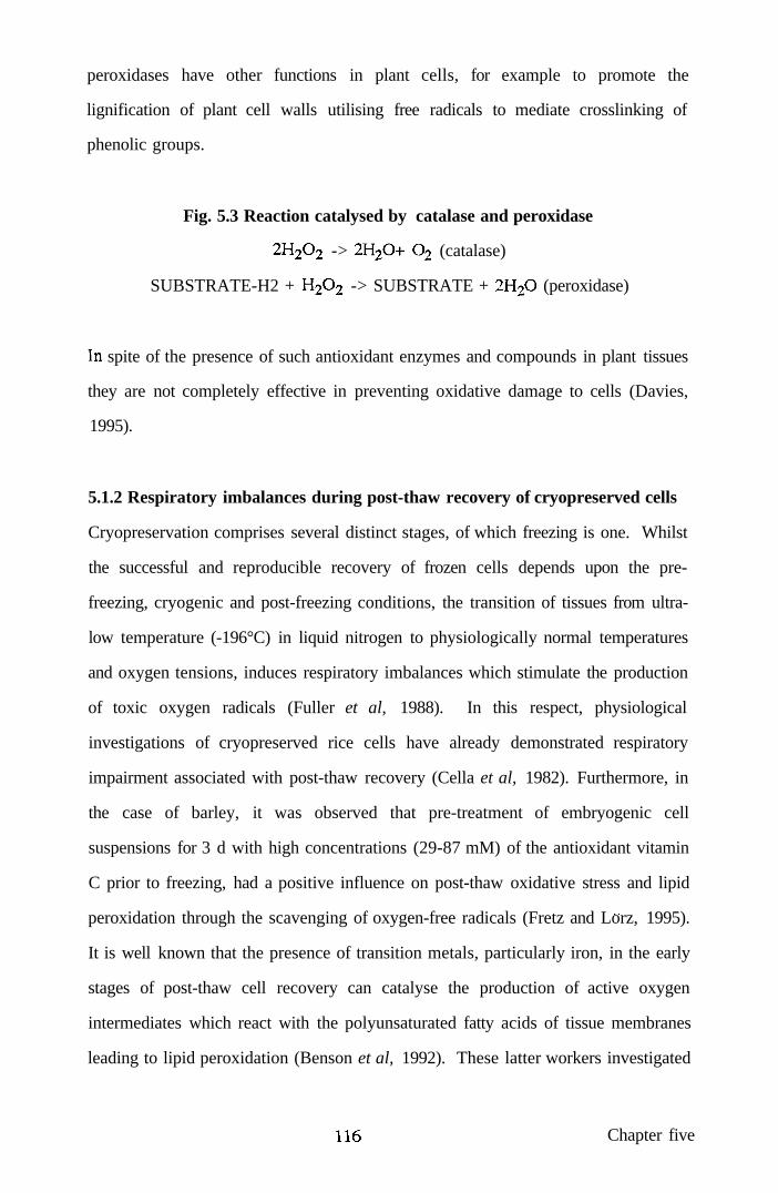
peroxidases have other functions in plant cells, for example to promote the
lignification of plant cell walls utilising free radicals to mediate crosslinking of
phenolic groups.
Fig. 5.3 Reaction catalysed by catalase and peroxidase
2H2O2 -> 2H2O+ O2 (catalase)
SUBSTRATE-H2 + H2O2 -> SUBSTRATE + 2H2O (peroxidase)
In spite of the presence of such antioxidant enzymes and compounds in plant tissues
they are not completely effective in preventing oxidative damage to cells (Davies,
1995).
5.1.2 Respiratory imbalances during post-thaw recovery of cryopreserved cells
Cryopreservation comprises several distinct stages, of which freezing is one. Whilst
the successful and reproducible recovery of frozen cells depends upon the pre-
freezing, cryogenic and post-freezing conditions, the transition of tissues from ultra-
low temperature (-196°C) in liquid nitrogen to physiologically normal temperatures
and oxygen tensions, induces respiratory imbalances which stimulate the production
of toxic oxygen radicals (Fuller et al, 1988). In this respect, physiological
investigations of cryopreserved rice cells have already demonstrated respiratory
impairment associated with post-thaw recovery (Cella et al, 1982). Furthermore, in
the case of barley, it was observed that pre-treatment of embryogenic cell
suspensions for 3 d with high concentrations (29-87 mM) of the antioxidant vitamin
C prior to freezing, had a positive influence on post-thaw oxidative stress and lipid
peroxidation through the scavenging of oxygen-free radicals (Fretz and Lorz, 1995).
It is well known that the presence of transition metals, particularly iron, in the early
stages of post-thaw cell recovery can catalyse the production of active oxygen
intermediates which react with the polyunsaturated fatty acids of tissue membranes
leading to lipid peroxidation (Benson et al, 1992). These latter workers investigated
116 Chapter five

supplementation of the post-thaw recovery medium of rice cells with the iron
chelating dmg, desferrioxamine, which had been used previously to reduce oxidative
stress in mammalian tissues subjected to low temperatures (Benson et al, 1995).
However, although positive and beneficial effects were observed in terms of cell
viability and cellular regrowth of rice cells, there was no significant reduction in lipid
peroxidation as assessed by thiobarbituric acid (TBA) production (Benson et al,
1992).
A novel approach for enhancing oxygen supply to post-thaw cryopreserved cells is
the use of chemically-inert, oxygen-carrying PFC liquids. These compounds
dissolve substantial volumes of key respiratory gases and have been studied in, for
example, emulsified form as vehicles for oxygen transport in vivo (Lowe, 1994;
Lowe et al, 1995). PFC liquids have also been used to routinely facilitate oxygen
supply to cultured plant protoplasts and protoplast-derived cells at normal
temperatures (25°C) using standard tissue culture practices (Anthony et al, 1994a;
Lowe et al., 1995a, b, 1997). In one investigation, supplementation of an aqueous
culture medium overlaying the PFC with a low concentration (ca. 0.01% w/v) of the
non-ionic, polyoxyethylene (POP) - polyoxypropylene (POE) surfactant, Pluronic
F-68 further enhanced mitotic division of protoplast-derived cells at the PFC-
medium interface (Anthony et al, 1994a; Lowe et al., 1995a).
Pluronic F-68 has been widely used as a cytoprotectant and growth-promoting
additive to animal cell and microbial cultures (Lowe et al, 1993, 1994). It has also
been evaluated as a cryoprotectant for Chinese hamster cells (Ashwood-Smith et al,
1973), but there have been relatively few comparable studies with plant cells.
Recentiy, Anthony et al (1996) reported that the incorporation of Pluronic F-68
into the recovery medium enhanced the growth of cells of both rice (Oryza sativa)
and Lolium multiflorum. It was speculated, from these experiments, that the
beneficial effects Pluronic® F-68 were primarily mediated through changes in
117 Chapter five

oxygen flux. Therefore, a strong case can be made for evaluating the post-thaw
growth of plant cells cultured in the presence of oxygen-gassed PFC liquid, in
combination with Pluronic F-68. An objective of the study was to extend earlier
findings that mitotic division of cultured protoplasts and protoplast-derived cells can
be enhanced by elevated oxygen atmospheres (d'Utra Vaz et al, 1992).
In the present investigation, embryogenic (totipotent) suspension cells of rice were
used to assess the potential beneficial effects of oxygenated perfluorodecalin, alone
and in combination with Pluronic F-68, on post-thaw growth following long-term
(ca. 3 years) cryopreservation.
5.2 Materials and methods
5.2.1 Plant materials and preparation of cell suspensions
Embryogenic calli were induced from mature seed scutella of the Japonica rice, O.
sativa L. cv. Taipei 309 (Finch et al, 1991) and such tissues were pooled and used to
initiate cell suspensions. The latter were maintained as described in Section 4.2.1.1.
Suspensions were split into two populations. One population was maintained by
regular sub-culture for a minimum period of 3 years; the other population was
cryopreserved. In the latter case, cells were cultured, immediately before
cryopreservation, for 3-4 d in liquid AA2 medium supplemented with 60.0 g 1"
mannitol.
5.2.2 Cryopreservation and post-thaw recovery
The cryopreservation procedure was based on that of Lynch et al (1994). Cells were
pooled and harvested from mannitol-supplemented AA2 medium onto a nylon mesh
(45 pm pore size) before being frozen as described in Section 4.2.1.2. Vials were
stored for 3 years in liquid nitrogen at -196°C.
118 Chapter five

Cells were thawed by plunging the frozen vials into sterile water at 45^C; excess
cryoprotectant was removed, under axenic conditions, from the cells using a Pasteur
pipette. The cells from individual vials were placed onto the top of two
superimposed 2.5-cm diam. Whatman No. 1 filter paper disks overlaying 5.0 ml
aliquots of AA2 medium made semi-solid with 0.4%o (w/v) Sea Kem Le agarose
contained in 100 ml capacity screw-capped glass jars (Beatson Clarke and Co. Ltd.)
(Treatment A). In some treatments, the AA2 culture medium was supplemented
with 0.01% (w/v) Pluronic® F-68 (Sigma; Treatment B), this concentration of
Pluronic being selected based on previous studies which confirmed the beneficial
effects of this compound on post-thaw growth of cells of O. sativa and L.
multiflorum (Anthony et al, 1996). In a further series of treatments, the cells were
placed on semi-solidified AA2 medium overlaying 20.0 ml aliquots of oxygenated
(10 mbar, 15 min) perfluorodecalin (Flutec® PP6; Treatment C). Additionally, the
AA2 medium overlaying oxygenated perfluorodecalin, was supplemented with
001% (w/v) Pluronic® F-68 (Treatment D).
Cells of all treatments were cultured in the dark for 3 d at 28 ± l^C prior to transfer
of the upper filter disk, supporting the cells, to fresh medium lacking Pluronic® F-68
and/or overlaying ungassed perfluorodecalin, as appropriate. Cells were cultured for
a further 24 h prior to viability assessments, followed by another 26 d, under the
same conditions, before biomass determinations. Each of the treatments consisted of
20 replicates of cells of the same cell line taken from 20 individual vials.
In order to assess the feasibility of recovering cryopreserved cells on a smaller scale,
the post crypreservation cell handling protocol was further modified, using 24 well
Petri dishes (CostarLtd., High Wycombe, UK). Each well contained 2.0 ml aliquots
of oxygenated perfluorodecalin and 1.0 ml of semi-solidified AA2 medium, with the
filter papers (1.3 cm diam.; Whatman No.l), overlaid with 50 mg fresh weight of
cells. The 50 mg cell aliquots used were each taken from 20 individual vials.
119 Chapter five

5.2.3 Measurement of post-thaw viability and biomass
The reduction of TTC was used to assess the post-thaw viability of cells based on a
modification of the method of Steponkus and Lamphear (1967) as described in
Section 4.2.1.4. Biomass was recorded by determining the fresh weight of thawed
cells (Lynch era/., 1994).
5.2.4 Re-initiation of cell suspension cultures
Five cell suspensions were re-established from each of the treatments by removing
the cells from the surface of 5 randomly selected filter disks, after 30 d of post-thaw
culture, and placing into 22 ml aliquots of AA2 liquid medium in 100 ml Erienmeyer
flasks. Suspensions were sub-cultured thereafter every 7 d for 28 d by removing all
the spent medium and replacing with the equivalent volume of fresh AA2 liquid
medium. Subsequentiy, suspensions were maintained as described earlier in Section
4.2.1.1.
5.2.5 Isolation and culture of protoplasts from re-established cell suspension
cultures
After 8 passages, cell suspensions were used for protoplasts isolations as described
by Jain et al. (1995). Cells were harvested onto a nylon mesh (45 pm pore size) and
a pre-weighed aliquot of 1 g f wt. of cells was incubated in 20 ml of enzyme
solution as described in Section 2.2.1.2. The enzyme solution (designated ENZlOl;
Appendix I) which consisted of 1% (w/v) Cellulase RS, 0.1% Pectolyase Y23 and
5mM MES in CPW13M solution, pH 5.8, was filter sterilized and frozen as
described in Section 2.2.1.2. The latter was thawed and diluted with CPW13M (1:3),
prior to overnight incubation.
Following digestion, the protoplasts were washed, counted and the viabilities
assessed as described in Sections 2.2.1.2 and 2.2.1.3, respectively. Protoplasts were
suspended in liquid KPR medium (Abdullah et al, 1986; Appendix II) and their
120 Chapter five

density adjusted to give an overall plating density of 5.0 x 10^ protoplasts mF^
Protoplasts were spread uniformly in single 200 pi aliquots over the surface of 47
mm diam., 0.2 pm pore size nitrocellulose nitrate filter membranes (Whatman
Intemational Ltd.) using a sterile plastic loop (Bibby-Sterilin Ltd.) to cover approx.
90% of the surface area of each membrane. Prior to spreading of the protoplasts on
the membranes, the latter were placed on the surface of 20 ml aliquots of KPR
medium, semi-solidified with 0.8% (w/v) Sea Plaque agarose and containing
suspension cells of Lolium multiflorum as a nurse system in 9 cm diam. Petri dishes.
Cell suspension cultures of L. multiflorum were maintained as described in Section
4.2.1.1. The nurse cells were harvested 3 d after sub-culture and spread at a density
of 5 ml PCV in 100 ml aliquots of semi-solidified KPR medium. Individual
membranes were placed on the surface of the medium, after a further 24 h, prior to
dispensing the protoplasts. Cultures were incubated in the dark at 28 ± 2° for 14 d.
Thereafter, the membranes containing protoplast-derived cells were transferted to 20
ml aliquots of LS2.5 medium (Linsmaier and Skooge, 1965; Appendix II) made
semi-solid with 0.4% (w/v) Sea Kem Le agarose contained in 9 cm Petri dishes.
Cultures were maintained in the dark as described earlier for a further 14 d.
5.2.6 Plant regeneration from protoplast-derived callus
Protoplast-derived colonies were transferred from the membranes to 20 ml aliquots
of MSKN medium, the latter being modified from the formulation of Jain et al
(1995), and contained in 9 cm Petri dishes (25 colonies/dish). Modified MSKN
medium contained maltose [5% (w/v)] and was semi-solidified with 1% (w/v) Sea
Kem Le agarose. Cell colonies were cultured in the dark at 26 ± 1°C for 10 d, prior
to transfer to the same medium, but with the agarose concentration reduced to 0.4%
2 1
(w/v). Cultures were transferted to the light (55 p mol m s ,16 h photoperiod;
Cool White fluorescent tubes) and regenerating shoots (75 per treatment) were
detached from individual calli after 40 d and transferred to 175 ml capacity screw-
capped glass jars, each containing 50 ml of MS based medium supplemented with
121 Chapter five

1.5 mg r ' NAA, 5%o (w/v) sucrose (designated MSN1.5 medium) and semi-
solidified with 0.2% (w/v) Phytagel (Sigma). Cultures were maintained under a 16 h
photoperiod, as described previously.
5.2.7 Cytological analysis of protoplast-derived plants
Cytogenetic studies were based on the chromosome spreading technique developed
by Andras et al. (1999). Actively growing roots (1 cm in length) were harvested
from protoplast-derived in vitro rice plants (Treatments A-D), after 21 d of transfer
to MSN1.5 medium (Section 5.2.6) and immersed in 1 pm trifluralin (Dow
AgroSciences Ltd., Hitchin, UK) (1 mM stock solution in DMSO diluted to 1 pm in
reverse-osmosis water) for 4 h at room temperature. Subsequentiy roots were fixed
in ethanol: glacial acetic acid [3:1 (v/v)] and stored at 4°C for 16 h. Fixed roots were
washed 3 times in reverse-osmosis water. Root tips (50; approx. 1 mm in length)
were excised and transferred to 40 pi aliquots of reverse-osmosis water contained in
3.5 cm Petri dishes. The water was removed and replaced with an equal volume of
IN HCl whereupon root tips were incubated in a humid chamber at 37°C for 15 min.
Following incubation, the HCl was removed and root tips equilibrated (1 min) in an
equal volume of TE buffer (10 mM Tris-HCl, 1 mM EDTA; Sigma), pH 8.0. The
latter was replaced with initially 10-20 pi of an enzyme mixture [4 % (w/v) Cellulase
RIO, 1%) (w/v) Pectolyase Y23] in citrate buffer (0.0IM citric acid, O.OIM sodium
citrate; pH 4.6) and the root tips gently macerated with a brass rod prior to the
enzyme solution being increased to a final volume of 200 p\. The material was
incubated in a humid chamber at 37°C for 1 h, prior to transfer to a 1.5 ml capacity
Eppendorf tube (Scientific Laboratory Supplies Ltd.). Incubation was terminated by
the addition of 200 pi of reverse-osmosis water and the digested cells were
centrifuged (300 x ^ , 10 min) in a MSE Micro Centaur centrifuge (Jennings Co.
Ltd.). The supernatant was discarded and the pellet resuspended in 400 pi of
reverse-osmosis water, prior to centrifugation and resuspension in 10 pi of reverse-
122 Chapter five
osmosis water. The latter was replaced with 400 pi of freshly prepared methanol:
glacial acetic acid [4:1 (v/v)] and centrifuged as described eariier. The pellet was
resuspended in 50 pi of the methanol: glacial acetic acid mixture whereupon aliquots
(8 pi) of the resulting suspension were dropped onto individual microscope slides
from a height of 10-15 cm and allowed to air dry.
Chromosomes were stained with 15 pi of La Cour acetic orcein solution (R.A.
Lambe Ltd., London, UK; Yong, 1997) and a cover slip applied which was sealed
with mbber solution (Dunlop Adhesives, Birmingham, UK), prior to examination on
a Nikon Microphot SA microscope.
5.3 Statistical analyses
Means and standard errors (s.e.m.) were used throughout; statistical significance
between mean values was assessed, as appropriate using a conventional one-way
ANOVA coupled with a Tukey-HSD post test and Student's t test (Snedecor and
Cochran, 1989). A probability of P < 0.05 Wcis considered significant.
5.4 Results
5.4.1 Assessment of PFC medium supplementation on post-thaw cell viability
The mean absorbance, as an indicator of cell viability, of unfrozen suspension cells
was 0.96 ± 0.05 (n = 10) prior to long-term (3 year) cryopreservation. The mean
absorbance of cryopreserved cells following recovery in the presence of oxygenated
perfluorodecalin (Treatment C; 0.45 ± 0.07; n = 20; Plate 5. la) was significantiy (P <
0.05) greater than for the mean of the (control) treatment which lacked
perfluorodecalin (Treatment A; 0.35 ± 0.08; n = 20; Plate 5.1a). Similar differences
in post-thaw viabilities were also observed (P < 0.05) in the reduced-scale series of
experiments when cells were recovered using 24-well Petri dishes (control: 1.17 ±
O06, n = 20; oxygenated PFC: 1.45 ± O07, n = 20; P < 0.05).
123 Chapter five

5.4.2 Supplementation of medium with PFC and/or Pluronic F-68 on post-
thaw cell viability
Fig. 5.4 shows the mean absorbance when cryopreserved rice cells were recovered in
the presence of 0.01% (w/v) Pluronic F-68, oxygenated perfluorodecalin or
oxygenated PFC combined with Pluronic F-68. The mean absorbance of cells
recovered in the presence of 0.01% (w/v) Pluronic® F-68 (Treatment B; 1.27 ± 0.04,
n = 20) or oxygenated PFC (Treatment C; 1.13 ± 0.03, n = 20) was significantiy
greater (P < 0.05) than on semi-solid AA2 medium alone (Treatment A; 0.93 ± 0.03,
n = 20; Fig. 1).
Fig. 5.4 Mean absorbance of cryopreserved rice cells recovered for 4 d in the
presence of various medium supplements
S c o
%
i Xi u. O [/>
x>
CS
(D
(A) medium alone (control), (B) medium with Pluronic® F-68 (0.01% w/v), (C)
medium underiayed with oxygenated PFC, and (D) a combination of treatments (B)
and (C). Vertical bars represent s.e.m. (n = 20 throughouO- *P < 0.05.
124 Chapter five
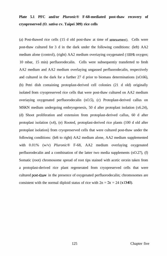
Plate 5.1 PFC and/or Pluronic® F-68-mediated post-thaw recovery of
cryopreserved (O. sativa cv. Taipei 309) rice cells
(a) Post-thawed rice cells (15 d old post-thaw at time of assessmenO- Cells were
post-thaw cultured for 3 d in the dark under the following conditions: (left) AA2
medium alone (control), (right) AA2 medium overiaying oxygenated (100% oxygen;
10 mbar, 15 min) perfluorodecalin. Cells were subsequentiy transferted to fresh
AA2 medium and AA2 medium overlaying ungassed perfluorodecalin, respectively
and cultured in the dark for a further 27 d prior to biomass determinations (xO.66),
(b) Petri dish containing protoplast-derived cell colonies (21 d old) originally
isolated from cryopreserved rice cells that were post-thaw cultured on AA2 medium
overiaying oxygenated perfluorodecalin (xO.5), (c) Protoplast-derived callus on
MSKN medium undergoing embryogenesis, 50 d after protoplast isolation (x6.24),
(d) Shoot proliferation and extension from protoplast-derived callus, 60 d after
protoplast isolation (x4), (e) Rooted, protoplast-derived rice plants (100 d old after
protoplast isolation) from cryopreserved cells that were cultured post-thaw under the
following conditions: (left to right) AA2 medium alone, AA2 medium supplemented
with 0.01% (w/v) Pluronic® F-68, AA2 medium overlaying oxygenated
perfluorodecalin and a combination of the latter two media supplements (xO.27), (f)
Somatic (root) chromosome spread of root tips stained with acetic orcein taken from
a protoplast-derived rice plant regenerated from cryopreserved cells that were
cultured j)ost-thaw in the presence of oxygenated perfluorodecalin; chromosomes are
consistent with the normal diploid status of rice with 2n = 2x = 24 (xl340).
125 Chapter five

A further synergistic increase (P < 0.05) in mean absorbance was also observed
when Pluronic F-68 and perfluorodecalin were used in combination (Treatment D;
1.46 ± 0.08, n = 20), compared to all other treatments. No significant differences
were observed between Treatment B (0.01% (w/v) Pluronic® F-68) and Treatment C
(oxygenated perfluorodecalin).
5.4.3 Post-thaw growth of cryopreserved cells recovered in the presence of PFC
and/or Pluronic® F-68
The recovery of cells with 0.01% (w/v) Pluronic® F-68 (Treatment B) or oxygenated
perfluorodecalin (Treatment C) promoted sustained mitotic division, since biomass,
measured as increase in fresh weight at 30 d post-thaw, was elevated by 21% and
38% (P < 0.05), respectively, compared to the biomass of cells in unsupplemented
medium (Treatment A; Fig. 5.5). When PFC and Pluronic® F-68 treatments were
combined (Treatment D), a significant (P < 0.05) increase in biomass was also
observed, that was greater than for Treatment B, but not significantiy different to that
found for Treatment C (Fig. 5.5).
126 Chapter five

Fig. 5.5 Mean fresh weight (g) of cryopreserved rice cells recovered after 30 d
in the presence of various medium supplements
I n
0 8 -
00
t 0.6-J3
0.4-
0.2-
0 C
Treatment
D
(A) medium alone (control), (B) medium with Pluronic® F-68 (0.01% w/v), (C)
medium underiayed with oxygenated PFC, and (D) a combination of treatments (B)
and (C). Vertical bars represent s.e.m. (n = 20 throughout). *P < 0.05.
5.4.4 Re-establishment of cell suspension cultures
Cell suspensions (n = 5), re-initiated simultaneously from cells recovered from all
treatments, ultimately exhibited growth rates after 35 d of culture comparable to
those of unfrozen suspensions maintained by regular sub-culture every 7 d for 3
years, this time being equivalent to the cryopreservation period.
127 Chapter five

5.4.5 Assessment of protoplast viability, yield, plating efficiency and plant
regeneration
There were no significant differences, in terms of yields, viabilities, plating
efficiencies (Plate 5. lb) and plant regeneration frequencies (Table 5.1; Plate 5. Ic, d),
of protoplasts isolated from cryopreserved cells exposed to the experimental
treatments A, B, C, and D. Additionally, there were no significant differences in
terms of protoplast yield (4.0 x 10^ g'^ f.wt), mean protoplast viability (89 ± 2%, n =
3) and the mean number of protoplast-derived calli regenerating shoots (44 ± 4%, n =
100) between cells subjected to cryopreservation followed by treatments A-D and
unfrozen suspension cells assessed prior to their 3 year cryopreservation procedure.
Protoplast isolation was not attempted from suspensions maintained continously for
3 years without cryopreservation, since the suspensions had lost their totipotency, as
expected by the end of this culture period.
128 Chapter five

B O
1 > km 9i W V ia
a o b.
E o ,u « M
"O V b.
> o w u CA B o
*5 B
CA 3 CA
"S w
rice
CM
O (A tg _« "a o ' t i '
o
om p
r
cS B
tio
n fc. V B V &c V
ntr
n "a -a B
CA
.s^
cenc
B a M)
,B
lat
a. l-H
w> •
Tab
le ^
-o V >> A
" b «; "O B S
s 3
'"B V
E ,0-m^
u ..*' .
^ l-H O d
00 ^ Eb
1 >.« a. JS • ^
J S 3
-3 9i E
« V .
^^ o b.
' ( i '
B O
v B
"rt
E 3
•a E ^ < s _ ^
CM O
9i
B V
b.
a
•t.^
^ U
-o B
J B
E -4-) A ii b.
'W CM O B
a B
2 E o CQ
g •o B
u b O. "O V
ts B i) OD >>
c« <U
'5 o o
^
0/1
c cS
c <u 0 0 <u
u. (U
c. CS TD
^ c cS
T3 O r
r-r i +1 r 00 ro C D H
o I - c
o fa is 1) 2 ^
OH . S
cS
"a.
05 CS
"a. o
•4—t 2 a,
^
• a o CO
cS
CS ^
O
CS
• > > "
O I.-,
2 3 0-
c s CS
o d •H
r4
00
+1 CO
o d +1 o\ ri
m ri +1 o
o +1 r-CI
m u
r-ci +1
CO
o d +1 C)
- H
m +1
75.0
6
a\ C l
+1
76.5
5
C l
.- +1
79.8
6
m C )
+1
76.8
9 >n r II
c •o
ON II
c
o 00 o +1 o 00 CO
in oo o +1
CI 'si-
in 00 o -H
C l CO
in
CI
o o •H
o in
CO
CO
cS
i C/5
+1 e CS <u S c <u czi
2i
a Vi (U 3 Is >
129 Chapter five

5.4.6 Cytological analysis of regenerated plants
Plants regenerated from cryopreserved cells that were post-thau- cultured with
Treatments A-D were morphologically normal (Plate 5.1e), with expected diploid
chromosome complements (2n = 2x = 24; Plate 5. If).
5.5 Discussion
The present study utilised the same cell suspension for all treatments in order to
eliminate any somaclonal effects. Lack of somaclonal variation was confirmed by
there being no significant differences in terms of plant regeneration potential of
cryopreserved cells and unfrozen cells of the same suspension as assessed 3 years
earlier.
These experiments show that the post-thaw viability and growth of cryopreserved
rice cells per se is increased in the presence of oxygenated perfluorodecalin either
alone, or in combination with Pluronic® F-68. Additionally, the culture of rice
protoplasts and of their cell derivatives at the interface between oxygen-geissed PFC
overlaid with liquid or agarose-solidified culture medium also enhances mitotic
division and, in totipotent systems, stimulates shoot formation (Wardrop et al.,
1997). This confirms a genuine growth enhancement of this treatment, rather than a
marginal effect on cells recovered from cryopreservation. PFC is believed to act as a
reservoir for oxygen which diffuses into the aqueous medium/cell phase during
initial culture. This is supported by changes in oxygen tension in the medium
(Anthony et al, 1994a). It is probable, therefore, that the increased and sustainable
post-thaw growth of rice cells was also due to an enhanced oxygen supply provided
by the PFC. Indirect evidence for the diffusion of oxygen from the PFC to aqueous
culture medium, comes from related observations with suspension-derived
protoplasts of Salpiglossis sinuata, in which an increase in intracellular superoxide
dismutase (SOD) occurred after 3 days culture (Lowe et al, 1997). This, in turn,
was consistent with studies using Mycobacterium spp., in which SOD was elevated
130 Chapter five

dunng culture of the bacterium in perfluorodecalin-supplemented medium (Popkova
et al, 1988). Lipid peroxidation and protein degradation can occur in the eariy
stages of post-thaw recovery (Fuller et al, 1988; Benson et al, 1995). Increased
SOD biosynthesis associated with culture of protoplasts and protoplast-derived cells
with oxygenated PFC may protect cells not only against a supplemented oxygen
supply, but also against oxygen radicals generated by impaired oxygen flux during
thawing. Further studies should determine the eariy time-course of changes in SOD
and other oxygen-sensitive enzymes during cell recovery.
The present study has extended eariier investigations which demonstrated that
culture medium containing Pluronic® F-68 increased post-thaw growth of
cryopreserved rice cells (Anthony et al, 1996). Pluronic® F-68 adsorbs onto cell
membranes, increasing resistance to mechanical damage (Handa-Corrigan et al,
1992; Lowe et al, 1993, 1994). Pluronics have hydrophobic POP cores, which are
believed to become incorporated into membranes, leaving their hydrophilic, POE
tails outside the cell. This would lower the interfacial tension by sterically hindering
adhesive interactions between molecules on cell surfaces and, in turn, minimise cell-
to-cell contacts, thereby reducing mechanical dismption. However, it is possible that
the responses to Pluronic could differ between plant cells with intact walls, and
animal cells or plant protoplasts, which have naked plasma membranes, the latter
albeit for a finite period of time when in culture.
As discussed previously (Anthony et al, 1996), Pluronic F-68 may increase the
uptake of nutrients, growth regulators or respiratory gases into cells during post-
thawing. Studies with animal cells have demonstrated that Pluronic F-68 at
concentrations comparable to those used in the present investigation, stimulated 2-
deoxyglucose uptake and incorporation of amino acids (Cawrse et al, 1991).
Changes in nutrient uptake, promoted by Pluronic, would be expected to alter
metabolic flux, allowing biochemical pathways to operate more efficientiy,
131 Chapter five

especially under the stress of initial post-thaw recovery (Anthony et al, 1996).
Additional studies are required, since the composition of the culture medium may
influence the growth of cells during the recovery phase. For example, uptake of
ammonium ions is known to inhibit the growth of thawed rice cells (Kuriyama et al,
1989), although in the present stiidy, the cell recovery medium lacked ammonium
ions. Adsorption of Pluronic molecules onto post-thawed plant cells may also reduce
cellular damage which can occur during rehydration when the DMSO cryoprotectant
is removed progressively from the system (Benson and Withers, 1987) and thus help
to preserve, in the short term, a stable celhmedium density equilibrium cmcial to the
re-establishment of maximal mitotic activity.
Interestingly, whilst Pluronic stimulated post-thaw growth of cryopreserved rice
cells, there was no further growth enhancement when the surfactant was added to the
culture medium overiaying oxygenated PFC. This contrasted with earlier
observations using protoplasts of P. hybrida, where Pluronic acted in an additive
manner with oxygenated perfluorodecalin to increase the plating efficiency by 57%
above control (Anthony et al, 1994a). Such differences may relate to the fact that
the latter study involved protoplasts of cell suspension-derived cells that had not
been cryopreserved. Nevertheless, these results demonstrate that, in terms of simply
promoting post-thaw growth of rice cells based on biomass assessments, oxygenated
perfluorodecalin alone is superior for inclusion in cryopreservation protocols
compared to Pluronic F-68, either alone or in combination with PFC.
PFCs may also be useful as gas delivery vehicles for multicellular explants in the
context of cryopreservation. For example, the conversion of cryopreserved apical
meristems to intact plants may well be facilitated by oxygen-gassed PFC, to promote
initial post-cryopreservation survival, and by carbon dioxide-gassed PFC to stimulate
rooting and ex vitro acclimation (Wardrop et al, 1997). Such applications should
take account of related studies in which PFCs and their emulsions facilitated survival
132 Chapter five

of fish semen during low temperature storage (McNiven et al, 1993) and promoted
hypothermic preservation of mammalian organs, (Umshihara et al, 1994).
The present results indicate that oxygenated PFC, together with Pluronic® F-68
should be incorporated routinely into post-thaw culture media and handling
strategies, to maximise plant cell viability and recovery. A further advantage of
using PFCs in such systems is that they are easily recoverable and recycleable,
thereby providing a cost effective underpin to germplasm storage technologies
(Lowe et al, 1997). Moreover, the present demonstration that PFCs can be exploited
in small-scale culture systems, makes their routine use economically feasible.
J33 Chapter five

CHAPTER SIX: GENERAL DISCUSSION
6.1 Introduction
Oxygen deprivation can reduce the growth of plant cells in vitro tiirough inhibition
of the production of respiratory ATP (Van der Plas and Wagner, 1986). Conversely,
high oxygen availability preferentially enhances the growth of plant cells,
particularly non-embryogenic callus cells of wheat (Carman, 1988). Mitotic division
of plant protoplasts isolated enzymatically from cells and tissues of several species,
can be enhanced by culture in an oxygen-enriched atmosphere (d'Utra Vaz et al,
1992). Similariy, the production of commercially-important secondary metabolites
can be increased when plant cell suspensions are exposed to high dissolved oxygen
concentrations (Schlatmann et al, 1994). Furthermore, gassing of bioreactors with >
40% (v/v) oxygen increases the shoot regeneration efficiency of rice (Oryza sativa)
cells cultured in bioreactors (Okamoto et al, 1996). A novel approach for enhancing
oxygen supply to cultured plant cells is the use of chemically-inert,
perfluorochemical (PFC) liquids. PFCs are linear, cyclic or bicyclic hydrocarbons in
which most or all hydrogen atoms have been replaced with fluorine. PFC liquids
typically have specific gravities about twice that of water (Lowe et al, 1998). One
of the most significant attributes of such PFC liquids is that they dissolve substantial
volumes of oxygen and other respiratory gases and have been studied in, for
example, emulsified form as vehicles for oxygen transport in vivo (Lowe et al, 1994,
1995). In principle, PFCs offer a more convenient approach to facilitate respiratory
gas transfer in plant cell cultures than direct gassing or sparging (Lowe et al, 1997)
and it is this property that has been exploited in the present studies.
6.2 Protoplast culture systems
Plant protoplasts, of various species including those of petunia (Petunia hybrida),
cassava (Manihot esculenta), passion fmit (Passiflora giberti) have been
successfully cultured for extended periods at an interface formed between
134 Chapter six
oxygenated perfluorodecalin (Cj^F^^) and the appropriate aqueous culture medium.
In petunia, which has been studied extensively as a model system (Anthony et al,
1994a), protoplasts synthesised new cell walls, divided normally, and had a mean
viability exceeding 92% at the end of the culture period. Their mean plating
efficiency (ability to produce cell colonies) was elevated by 37% following culture at
such a perfluorodecalin/medium interface. In comparison, growth was similar to that
in contiol (medium alone) when protoplasts were cultured in medium overlaying
non-gassed perfluorodecalin, or in oxygenated medium not overlying
perfluorodecalin. Whilst these latter studies did not demonstrate plant regeneration
from PFC-treated protoplast-derived cells, since the protoplast systems employed
were either originally non-totipotent (M. esculenta, P. hybrida) or had lost
totipotency (P. giberti ) at the time of assessment. However, subsequent related
studies have shown that phenotypically normal, fertile rice (O. sativa cv. Taipei 309)
plants could be regenerated from protoplasts cultured for up to 34 days with
oxygenated PFC, confirming that there were no adverse effects of this novel culture
regime on the expression of totipotency (Wardrop et al, 1996). Moreover, these
workers found that culture of rice protoplasts with oxygenated PFC actually led to an
increase in plant regeneration frequency by 12% over untreated controls cultured in
the absence of PFC.
Whilst littie attention has focused on the molecular responses of cells cultured in the
presence of oxygenated PFC, Wardrop et al. (1997a) reported significant increases in
intracellular SOD activity in protoplasts of Salpiglossis sinuata after 3 days of
culture in aqueous medium overiaying a commercial grade of oxygenated
perfluorodecalin (Flutec® PP6), compared to protoplasts cultured witii non-
oxygenated PFC. These authors speculated that such an increase in SOD would help
to protect cells from oxidative damage arising from free radicals generated through
prolonged exposure to the increased oxygen supplied by the PFC.
135 Chapter six

6.3 Synergistic effects of PFCs and surfactants as protoplast culture medium
supplements
A further refinement of the PFC-aqueous medium system exploited in the present
studies was to supplement the aqueous component with low concentrations (typically
0.01% w/v) of the non-ionic surfactant, Pluronic® F-68. The surfactant lowered the
interfacial tension and also possibly acted as a cell protecting/growth-enhancing
agent (Lowe et al, 1993), especially as the protoplasts synthesised new cell walls
and entered mitotic division. The concentration of Pluronic® F-68 selected for these
experiments was based on eariier studies of the growth-stimulating effects of this
compound on Solanum dulcamara protoplasts (Kumar et al, 1992; Lowe et al,
1993).
The addition of Pluronic F-68 to the aqueous culture medium was sufficient to
reduce the PFC-water interfacial tension by approximately 40%, thus facilitating
maximum contact of protoplasts with the interface. Such use of Pluronic® F-68
would also minimise any protein-stripping effects of the PFC surface indicated in
previous investigations with animal cells (Ju et al, 1991). Significantiy, the mean
plating efficiency of petunia protoplasts at the end of the culture period was
increased by 52% in the presence of oxygenated PFC overlaid by aqueous culture
medium supplemented with Pluronic F-68 at 0.01% (w/v). Future studies should
evaluate the extent to which other, related non-ionic surfactants such as Tween and
Triton XICK), may have similar beneficial effects to Pluronic F-68.
6.4 Image analysis assessments of cell growth
Computer-based image analysis techniques have been extensively employed for non-
invasive/non-destmctive assessments of the growth of cells within culture vessels.
This approach enables accurate measurement, not only of the total area occupied by
cells, but also of more specific parameters, such as the number of individual cell
colonies, colony area and fractal stmcture. Image analysis has been used in the
136 Chapter six

present studies to assess the growth of cell suspension-derived protoplasts of P.
hybrida, again as a convenient model, following culture at a PFC/aqueous medium
interface. The mean area of protoplast-derived cell colonies after 68 days of growth
was increased 35-fold in the presence of oxygenated perfluorodecalin and aqueous
culture media supplemented with 0.01% (w/v) Pluronic® F-68 (Anthony et al,
1994b). Image analysis has also been used to monitor shoot regeneration from rice
protoplasts following their culture in the presence of oxygenated PFC (Wardrop et
al, 1996). A further extension of this technology could also be readily exploited to
monitor the growth of protoplast-derived cells following PFC-regulated delivery of
other gases, such as carbon dioxide, to cultured cells, tissues and organs (Lowe et al,
1997).
6.5 Comparison of PFCs with other physical and chemical options for
enhancing oxygen supply
Whilst PFC liquids and surfactants can improve culture conditions for prokaryotic or
eukaryotic cells, their benefits must be evaluated alongside other physical or
chemical options for regulating respiratory gases. In this respect, Anthony et al
(1995b) demonstrated that glass rods inserted vertically into agarose-solidified
culture medium enhanced the division of cassava leaif mesophyll protoplasts plated in
an overlying liquid layer. The glass rods and their associated menisci were believed
to increase the surface area for gaseous exchange. Interestingly, these studies also
demonstrated that the use of an underlying oxygenated perfluorodecalin (Flutec
PP5) layer was, in fact, superior to the use of glass rods for enhancing the plating
efficiency of cassava leaf protoplasts. In a separate assessment, the mitotic division
of petunia protoplasts was enhanced significantiy by supplementing aqueous culture
medium with 1:50 (v/v) of a commercial stabilised bovine haemoglobin preparation,
Erythrogen™ (Anthony et al 1997b). This result was consistent with findings from,
related studies by Azhakanandam et al (1997) which similariy demonstrated that
culture media supplementation with 1:50 (v/v) Erythrogen™ promoted mitotic
137 Chapter six
division, cell colony formation and, importantly, plant regeneration from rice cv.
Taipei 309 protoplasts. Additionally, protoplast studies have also focused on the
extent to which haemoglobin, as a respiratory gas carrier, may act synergistically
with PFCs for gas delivery to cell cultures (Anthony et al, 1997a). An interesting
finding from this work was that the effectiveness of these separate or combined
treatments may be species-specific. In the case of P. giberti protoplasts,
Erythrogen™ promoted the greatest increase in mean plating efficiency, whereas for
P. hybrida protoplasts, a combination of oxygen-saturated PFC and Erythrogen™
was the most effective. Future studies should evaluate the beneficial effects of these
technologies on other recalcitrant protoplast systems.
6.6 Pluronic® F-68 and plant cell cryopreservation
Early studies showed that supplementation of medium with Pluronics could prevent
haemolysis of human red blood cells in response to freeze-thawing procedures
(Glauser and Talbot, 1956). Subsequent investigations demonstrated that Pluronic®
F-68 was an effective cryoprotectant for culmred Chinese Hamster cells (Ashwood-
Smith et al, 1973). Surprisingly, the effects had not been evaluated of using
Pluronic F-68 with plant cells, either as a cryoprotectant per se, or as a post-thaw
cytoprotecting agent, despite the increasing interest in cryopreservation for
conserving agronomically-important and endangered plant germplasms (Benson,
1994). Consequentiy, the present studies demonstrated that media supplementation
with Pluronic® F-68 significantiy enhanced post-thaw cellular growth following
cryopreservation of both embryogenic and non-embryogenic suspension-cultured
cells of the Japonica rices Oryza sativa cvs. Taipei 309 and Tarom, together with
non-embryogenic cells of Lolium multiflorum (Anthony et al, 1996) and Moricandia
arvensis (Craig et al, 1997). It is noteworthy that there was no measurable
beneficial effect of adding Pluronic® F-68 to the cryoprotectant before freezing of
rice and Lolium cells.
138 Chapter six

An important finding from the present studies was that the optimum concentration of
Pluronic® F-68, which increased cell growth, differed between the two rice cultivars
and between the rice cv. Tarom and the non-embryogenic cells of Lolium.
Consequently, there were both species and cultivar-specific responses to Pluronic®
F-68. These responses of cryopreserved cells were consistent with previous
observations using cells and tissues not exposed to ultra-low temperatures, as in
Chrysanthemum morifolium, in which the optimum concentration of surfactant
which stimulated adventive shoot regeneration from cultured leaf explants differed
by an order of magnitude between cultivars (Khehra et al, 1995). Indeed,
differences in the responsiveness of tissues and organs to the growth-promoting
effects of Pluronic® F-68 have also been observed in Solanum dulcamara (Kumar et
al, 1992), Corchorus capsularis (Khatun et al, 1993), Hypericum perforatum
(Brutovskdetal, 1994) and Populus spp. (lordan-Costache etal, 1995).
In plant cells, Pluronic F-68 may promote the increased uptake of nutrients, growth
regulators or oxygen into cells during the post-thaw culture period. Any increase in
nutrient uptake promoted by Pluronic would be expected to alter metabolic flux,
allowing biochemical pathways to operate more efficientiy, particularly under the
stress conditions of early post-thaw recovery. The adsorption of Pluronic molecules
onto the cytoplasmic membranes of post-thawed plant cells may also have reduced
any cellular damage which is known to occur during rehydration when the DMSO in
the cryoprotectant is removed progressively from the system (Benson et al, 1987).
However, further studies are needed to determine the precise mechanism of action of
Pluronic® F-68 as a cytoprotectant.
6.7 PFCs and post-thaw recovery of cryopreserved rice cells
Previous work using cryopreserved rice cells, as a model system, focused on the
optimisation of the post-thaw conditions for the successful recovery of suspension
cells from long-term storage in liquid nitrogen (Lynch et al, 1994). However, these
139 Chapter six

studies did not address the basic requirement of providing thawed cells with an
adequate supply of oxygen, particulariy during the eariy and cmcial stages of post-
thaw recovery. It is at this critical stage that oxidative stress conditions arise leading
to the production of toxic oxygen radicals, which are well known to cause lipid
peroxidation of the cells (Fuller et al, 1988; Benson et al, 1995). This, in turn, can
lead to rancidification of adjacent cells which, in some cases, may result in
unsuccessful recovery of cells from cryopreservation, through insufficient viable
cells being available to enter sustained mitotic division.
Embryogenic suspension cells of rice cv. Taipei 309 were used to assess the potential
beneficial effects of oxygenated perfluorodecalin (Flutec® PP6) on the post-thaw
viability, following long-term cryopreservation in liquid nitrogen. Mean cell
viability, as assessed by TTC reduction at 4 days after tiiawing was increased by
20% over the control by oxygenated perfluorodecalin in 100 ml glass jars, and,
similarly, by 24% when the cells were recovered on a smaller scale in 24-well Petri
dishes. Related studies using protoplasts isolated from unfrozen cell suspensions of
the rice cv. Taipei 309 showed that mitotic division was enhanced during culture of
the protoplasts at the interface between oxygen-gassed PFC overlaid with liquid or
agarose-solidified culture medium. Related work has showed that shoot formation
was also stimulated in this totipotent cell system (Wardrop et al, 1996). Overall,
these results confirmed a genuine growth enhancement of PFC treatment, rather than
a marginal effect on cells recovered from cryopreservation.
6.8 Synergistic effects of PFCs and surfactants on post-thaw recovery of
cryopreserved rice cells
Interestingly, experiments with animal cells have demonstrated that PFC liquids,
emulsified with Pluronic® F-68, were effective in prolonging the fertilising
capability of turkey spermatozoa stored at 4°C (Thurston et al, 1993). In assessing
any synergistic effect of PFC and surfactants in plant systems, PFC both alone and in
140 Chapter six

combination with 0.01 (w/v) Pluronic® F-68 was used to supplement the medium
used for the recovery of cryopreserved rice cells of the cv. Taipei 309. The results of
such experiments did, indeed, demonstrate a synergistic effect of such compounds.
For example, a 21% increase in post-thaw viability over controls was observed with
oxygenated perfluorodecalin alone, with a 36% increase in post-thaw viability when
cells were exposed to medium supplemented with 0.01% (w/v) Pluronic® F-68
alone. However, a more pronounced, synergistic increase in viability of up to 57%
over control occurted witii rice cells recovered in tiie presence of both oxygenated
perfluorodecalin and 0.01% (w/v) Pluronic® F-68 (Anthony et al, 1996). Similariy,
perfluorodecalin and Pluronic® F-68 treatments, either alone or in combination, also
promoted an increase in biomass, when measured as fresh weight gain 30 days after
thawing of rice cells, to a maximum of 38% above the control value in the case of
PFC alone and when oxygen-gassed perfluorodecalin was used in combination with
Pluronic F-68. Interestingly, when the PFC and Pluronic® F-68 treatments were
combined, the increase in biomass was greater than that of cells recovered in the
presence of Pluronic F-68 alone, but not significantiy different from that with PFC
alone. In addition, there were no significant differences, in terms of yields,
viabilities, plating efficiencies and plant regeneration frequencies of protoplasts
isolated from cryopreserved cells exposed to the various experimental treatments.
Similarly, there were no significant differences in terms of protoplast yield, viability
and the mean number of protoplast-derived calli regenerating shoots between cells
subjected to cryopreservation followed by the various treatments and unfrozen
suspension cells assessed prior to their 3 year cryopreservation procedure. Moreover,
plants regenerated from cryopreserved rice cells were morphologically normal, with
expected diploid chromosome complements (2n = 2x = 24) thus demonstrating no
long-term detrimental effects of exposure to PFC.
To date, the results with plant cells indicate that the use of oxygenated PFC
underiying culture medium, combined with the inclusion of a surfactant, such as
141 Chapter six

(So
Pluronic F-68 in the medium itself, may be useful if incorporated routinely into
post-thaw culture media and handling strategies, to maximise plant cell viability
during culture after recovery from cryopreservation. However, once cells have
recovered from the freezing process, there is no additional advantage of including
both oxygen-gassed PFC and surfactant in the culture medium, since the presence of
oxygenated PFC alone is adequate to maximise biomass production, at least in rice
cells. The use of PFC and surfactant options may be especially relevant in post-thaw
handling strategies for cells of those plant species which, normally, respond poorly
to conventional recoveiy procedures following storage at ultra-low temperatures.
Moreover, the fact that PFCs can be exploited in small-scale culture systems, makes
their routine use economically feasible.
PFCs may also be useful as gas delivery vehicles for multicellular explants in the
context of cryopreservation. For example, the conversion following thawing of
cryopreserved apical meristems to intact plants may be facilitated by oxygen-gassed
PFC, to promote initial post-cryopreservation survival, followed by exposure of
meristem-derived shoots to carbon dioxide-gassed PFC to stimulate rooting and ex
vitro acclimation (Wardrop et al, 1997b). Such applications should take account of
related studies in animal systems, in which PFCs and their emulsions facilitated
survival of fish semen during low temperature storage (McNiven et al, 1993) and
promoted hypothermic preservation of mammalian organs (Umshihara et al, 1994).
6.9 Concluding remarks
PFCs clearly have multi-faceted applications in both prokaryotic and eukaryotic cell
culture systems. Their principal physical property is to facilitate respiratory gas
supply which, in turn, regulates biomass production and allied yields of
commercially-important cellular secondary products. The maintenance of
protoplasts at the interfacial boundary between PFC liquids and aqueous culture
media is convenient for regulating gas supply and provides a novel support option
142 Chapter six

from which cells can be readily aspirated. The chemical inertness of PFCs has the
advantage that such compounds can be readily recovered and hence, re-cycled, thus
negating a relatively high initial investment cost. Thus, PFC-facilitated
improvements that have been demonstrated in both animal and microbial cell culture
technologies can clearly be extended to many fields of plant biotechnology and will
complement their progressive exploitation in medicine.
143 Chapter six

APPENDICES
APPENDIX I - Enzyme formulations
Composition of Enzyme ENZCl
Component Concentration (mg f*)
Hemicellulase 10000
Cellulase RS 4000
Pectolyase Y23 1000
MES 11000
pH5.8
ENZCl was made up in CPW9M solution and filter sterilized by passing through a
0.2 pm pore size membrane.
Composition of Enzyme ENZ223
Component Concentration (mg \ )
Rhozyme 20000
Meicelase 20000
Macerozyme RIO 300
pH5.8
ENZ223 was made up in CPW13M solution and filter sterilized by passing through a
0.2 pm pore size membrane.
Composition of Enzyme ENZlOl
Component Concentration (mg I' )
Cellulase RS 10000
Pectolyase Y23 1000
MES 11000
pH5.8
ENZlOl was made up in CPW13M solution and filter sterilized by passing through a
0.2 pm pore size membrane.
144 Appendices

APPENDIX II - Media formulations
Composition of MS medium (Murashige and Skoog, 1962)
Component Concentration (mg f*)
CaCl2 2H2O NH4NO3
KNO3
Kl
C0CI2.6H2O
KH2PO4
H3BO3
NaMo04
MgS04.7H20
MnS04.H20
CUSO4.5H2O
ZnS04.7H20
FeS04.7H20
Na2EDTA
Glycine
Inositol
Nicotinic acid
Pyridoxine HCl
Thiamine HCl
Sucrose
pH5.8
440
1650 1900
0.83 0.025
170
6.2
025
370
22 3
0.025
8.6
27.85
37.25
2.0
100
0 5
0 5
0 1
30000
The above components, with the exception of sucrose, are contained in the dried
preparation marketed by Sigma, UK. Medium was made up in reverse osmosis
water and autoclaved at 1 2 r c for 20 min.
145 Appendices
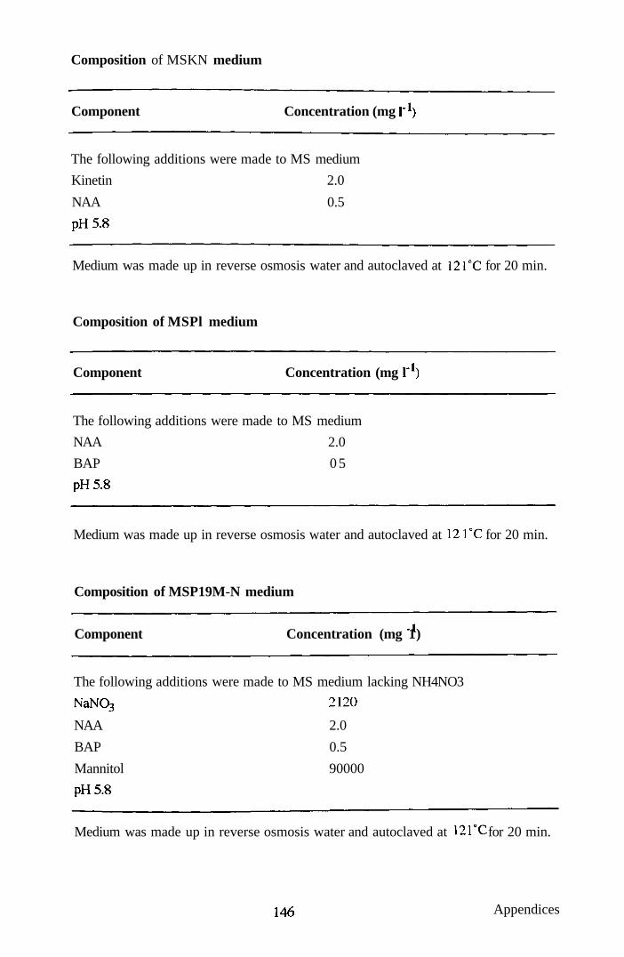
Composition of MSKN medium
Component Concentration (mg 1" )
The following additions were made to MS medium
Kinetin 2.0
NAA 0.5
pH5.8
Medium was made up in reverse osmosis water and autoclaved at 12 TC for 20 min.
Composition of MSPl medium
Component Concentration (mg f*)
The following additions were made to MS medium
NAA 2.0
BAP 0 5
pH5.8
Medium was made up in reverse osmosis water and autoclaved at 12 TC for 20 min.
Composition of MSP19M-N medium
Component Concentration (mg 1)
The following additions were made to MS medium lacking NH4NO3
NaNp3 2120
NAA 2.0
BAP 0.5
Mannitol 90000
pH5.8
Medium was made up in reverse osmosis water and autoclaved at 1 2 r c for 20 min.
146 Appendices

Composition of MSDl medium
Component Concentration (mg l'^)
The following additions were made to MS medium
2,4-D 1.0
pH5.8
Medium was made up in reverse osmosis water and autoclaved at 12rC for 20 min.
Composition of LS 2.5 medium (Linsmaier and Skoog, 1965)
Component I-IN Concentration (mg \ )
CaCl2 2H2O
NH4NO3
KNO3
KI
C0CI2.6H2O
KH2PO4
H3BO3
NaMo04
MgS04.7H20
MnS04.H20
CUSO4.5H2O
ZnS04.7H20
FeS04.7H20
Na2EDTA
Inositol
Thiamine HCl
Sucrose
2,4-D
pH5.8
440
1650 1900
0.83
0.025
170
6.2
0.25
370
22.3
0.025
8.6
27.85
37.25
100
1.0
30000
2.5
Medium was made up in reverse osmosis water and autoclaved at 1 2 r c for 20 min.
147 Appendices

Composition of N6 medium (Chu et al., 1975)
Component -IN Concentration (mg \ )
CaCl2.2H20
(NH4)S04
KNO3
KH2PO4
H3BO3
MgS04.7H20
ZnS04.7H20
MnS044H20
FeS04.7H20
Na2EDTA
Glycine
Nicotinic acid
Pyridoxine HCl
Thiamine HCl
2,4-D
Sucrose
pH5.8
166
463
2830
400
1.6
185
1.5
3.3
24.9
26.1
2.0
0 5
0 5
1.0
2.0
30000
Medium was made up in reverse osmosis water and autoclaved at 1 2 r c for 20 min.
Composition of UM medium (Uchimiya and Murashige, 1974)
Component Concentration (mg Y^)
The following additions were made to MS medium
2,4-D 2.0
Kinetin 0.25
Thiamine HCl 9.91
Pyridoxine HCl 9.5
Nicotinic acid 4.5
Casein hydrolysate 2000
pH5.8
Medium was made up in reverse osmosis water and autoclaved at 1 2 r c for 20 min.
148 Appendices

Composition of AA2 medium^ (Muller and Grafe, 1978) and R2 medium (Ohira
etal., 1973)
Component AA2 medium Concentration (mg r )
R2 medium
CaCl2.2H20 KH2PO4 MgS04 7H2O KQ NaH2P04.2H20 KNO3 (NH4)2S04 KI H3BO3 MnS04.4H20 NaMo04.2 H2O ZnS04.7H20 CUSO4.5H2O Coa2.6H20 FeS04.7H20 Na2EDTA NaFeEDTA Myo-inositol Nicotinic acid Pyridoxine HCl Thiamine HCl Glycine L-Glutamine L-Aspartic acid L-Arginine L-Proline 2,4-D Gibberellic acid Kinetin Sucrose Maltose pH
440 170 370 2940
0.83 6.2 22.3 0.25 8.6 0.025 0.025 27.85 37.25
100 0.5 0.1 0.5 75 877 266 228
2.0 0.1 0.2 20000
5.8
150 170 270
310 4040 330
0.5 0.5 0.05 0.5 0.05
2.5 too
0.5 0.1 2.0
560 1.0
30000 5.8
Modified AA2 medium contains ICX) mg 1 myo-inositol, 0.5 mg l" thiamine HCl,
7.5 mg r^ L-glycine and 2.0 mg 1" 2,4-D.
AA2 medium was made up in reverse osmosis water and filter sterilized by passing
through a 0.2 pm pore size membrane.
R2 medium was made up in reverse osmosis water and autoclaved at 12 TC for 20
min.
149 Appendices

Composition of CPW salts solution^ (Frearson etal, 1973)
Component Concentration (mg \'^)
KH2PO4 27.2
KNO3 101
CaCl22H20 44O
MgS04.7H20 246
Kl 016 CUSO4.5H2O 0.025
^Modified CPW salts solution contains 1480 mg 1"' CaCl2 2H2O
To prepare: CPW9M add 9% (w/v) mannitol
CPW13M add 13% (w/v) mannitol
CPW21S add 21% (w/v) sucrose
For all CPW solutions; pH 5.8. Solutions were made up in reverse osmosis water
and autoclaved at 1 2 r c for 20 min.
Composition of W5 solution (Menczel etal., 1981)
Component Concentration (mg F )
NaCl 9000
CaCl22H20 18370
KCl 373
Glucose 901
pH5.6
W5 solution was made up in reverse osmosis water and autoclaved at 1 2 r c for 20
min.
150 Appendices

Composition of 1B5CNK medium (Misawa et al, 1982)
Component 1-1 Concentration (mg Y )
CaCl2 2H2O (NH4)2S04
KNO3
KI C0CI2.6H2O
NaH2P04.2H20
H3BO3
NaMo04
MgS04.7H20
MnS04.4H20
CUSO4.5H2O
ZnS04.7H20
FeS04.7H20
Na2EDTA
Myo-inositol
Nicotinic acid
Pyridoxine HCl
Thiamine HCl
Casamino acids
Kinetin
NAA
2,4-D
Sucrose
Glucose
pH5.8
150
134 2500
0.75
O025
150
3.0
025
250
10
0.025
2.0
27.85
37.25
100
1.0
1.0
100
2000
0.25
1.0
1.0
20000
61250
Medium was made up in reverse osmosis water and autoclaved at 1 2 r c for 20 min.
151 Appendices

Composition of KM8P and KM8 media^ (Kao and Michaluk, 1975) and K8
medium (Kao, 1977)
Component
NH4NC)3 KNO3 CaCl2.2H20 MgS04.7H20 KH2PO4 Sequestrene 330 Fe KI H3BO3 MnS04.4H20 ZnS04.7H20 NaMo04.2 H2O CUSO4.5H2O Coa2.6H20 Myo-inositol Nicotinamide Pyridoxine HCl Thiamine HQ D-Ca Pantothenate Folic acid Abscisic acid Biotin Choline chloride Riboflavin Ascorbic acid Vitamin A Vitamin D5
Vitamin B 22 Na pyruvate Citric acid Malic acid Fumaric acid Fructose Ribose Xylose Mannose Rhamnose Cellobiose Sorbitol Mannitol Vitamin free Casamino acids Coconut milk 2.4-D Zeatin NAA Sucrose Glucose pH
KM8P
600 1900 600 300 170 28 0.75 3.0 10.0 2.0 0.25 0.025 0.025
too 1.0 1.0 1.0 1.0 0.4 0.02 0.01 1.0 0.2 2.0 0.01 0.01 0.02 20 40 40 40 250 250 250 250 250 250 250 250
250
20mir^ 0.2 0.5 1.0 250 100000 5.8
Concentration (mg 1' )
KM8
600 1900 600 300 170 28 0.75 3.0 10.0 2.0 0.25 0.025 0.025 100 1.0 1.0 1.0 1.0 0.4 0.02 0.01 1.0 0.2 2.0 0.01 0.01 0.02 20 40 40 40 250 250 250 250 250 250 250 250
250 20mir^ 0.1 0.2 1.0 20000 10000 5.8
K8P
600 1900 600 300 170 28 0.75 3.0 10.0 2.0 0.25 0.025 0.025
too 1.0 1.0 1.0 0.5 0.2 0.01 0.005 0.5 0.1 1.0 0.005 0.005 0.01 5 10 10 10 125 125 125 125 125 125 125 125
125 lOmir^ 0.2 0.5 1.0 250 100000 5.8
152 Appendices

^Modified KM8P and KM8 media (Gilmour et al, 1989) based on the formulation
of Kao and Michaluk, 1975
Media were made up in reverse osmosis water and filter sterilized by passing
through a 0.2 pm pore size membrane.
Composition of KPR medium (Abdullah et al, 1986)
Component Concentration (mg Y^)
The following additions were made to KP8 medium
2,4-D 0.3
pH5.8
Medium was made up in reverse osmosis water and filter sterilized by passing
through a 0.2 pm pore size membrane.
253 Appendices
APPENDIX III - Publications
1 ^ Appendices

Plant Cell Reports © Springer-Verlag 1994
Synergistic enhancement of protoplast growth by oxygenated perfluorocarbon and Pluronic F-68
p. Anthony S M.R. Davey \ J.B. PowerS C. Washington^, and K.C. Lowe^
' Departments of Life Science and ^ Pharmaceutical Sciences, tJniversity of Nottingham, University Parle, Nottingham NG7 2RD, LJK
Received 1 September 1993/Revised version received 16 December 1993 - Communicated by H. Lorz
Summary. Cell suspension-derived protoplasts of albino Petunia hybrida were grown for 10 d at the interface between aqueous culture medium (KM8P) and an oxygenated (10 mbar for 15 min) perfluorocarbon liquid, perfluorodecalin. Protoplasts synthesised new cell walls and divided normally at the perfluorodecalin/culture medium interface, with a mean viability after 10 d of > 92.0%. The mean plating efficiency of protoplasts was elevated by 37% (P<0.05) following culture at the perfluorodecalin/medium interface, but was unaltered by perfluorodecalin or oxygen separately. The mean plating efficiency of protoplasts cultured at the interface was further increased to a maximium of 52% above control, in the presence of oxygenated perfluorodecalin and KM8P medium supplemented with the non-ionic, co-polymer surfactant, Pluronic F-68 atO.01% (w/v). These findings demonstrate the effectiveness of oxygenated perfluorodecalin forpromoting protoplast growth, by facilitating oxygen delivery. The finding that Pluronic F-68 further increased the plating efficiency of protoplasts cultured at the perfiuoro-carbon/aqueous interface suggests that these agents improve growth through separate, but cumulative, mechanisms.
Key words: Petunia hybrida, protoplasts, oxygen delivery, perfluorochemicals, Pluronic F-68, surfactant, cell division.
Abbreviations: ATP, adenosine triphosphate; PFCs, perfluorochemicals; STP, standard temperature and pressure.
Introduction
Oxygen deprivation is known to reduce plant cell growth through inhibitory effects on the production of respiratory ATP (Van der Plas and Wagner 1986). In contrast, high oxygen availability can preferentially enhance the growth of non-embryogenic callus cells from plants such as wheat (Carman 1987). Additionally, recent studies have shown that the growth of tomato, rice and jute protoplasts can be improvedbyculture in oxygen-enriched atmospheres(d'Utra
Wazetal. 1992). There is thus a strong case for investigating the growth and differentiation of plant protoplasts, cells and tissues when cultured in the presence of media supplements which, in principle, can facilitate improved oxygen transfer.
PFCs are inert, organic compounds which can dissolve substantial volumes of respiratory gases and have been used for enhancing oxygen supply both in vivo and in vitro (King el al. 1989; Lowe. 1992, 1993). PFCs have been used, for example, in fermenter studies (Junker et al. 1990; Ju et al. 1991) or hybridoma cell cultures (Ju and Armiger 1992) to increase oxygen transfer rates. Mammalian cells, including fibroblasts and retinoblastoma cells, have also been cultured successfully at the interface formed between PFC liquid and aqueous media (Giaever and Keese 1983; Nabih el al. 1989). Since there appears to have been no corresponding assessment of the beneficial effects of using PFCs in plant cell cultures, the present study was performed to investigate the growth of isolated protoplasts in such a culture system, using cell suspension-derived protoplasts of albino Petunia hybrida as a convenient model for these assessments. This approach could have significant biotechnological application, not only in the context of plant genetic manipulation where the efficacy of protoplast conversion to plants is paramount for successful somatic hybridization and transgenic plant production, but also in the production of phytochemicals from cultured plant cells and tissues.
Materials and methods
Protoplast isolation and culture systems. Protoplasts of albino Petunia hybrida cv. Comanche were isolated enzymatically from cell suspensions (Powercf a/. 1990) and were cultured in the dark (25°C) at a final plating density of 2.0 X lO'/ml in 2.0 ml aliquots of liquid KM8P culture medium (Gilmour cf a/. 1989). The medium containing protoplasts was placed in 30 ml screw-capped glass bottles, either
Correspondence to: K. C. lx)we

252
Figure 1. (a) Schematic drawing of the culture system; (b) Suspension cell-derived protoplasts of albino P. hybrida cv. Comanche growing at the interface formed between oxygenated perfluorodecalin and KM8P culture medium (width of bottle = 27 mm); (c) Freshly isolated P. hybrida protoplasts (x 100); (d) Protoplast-derived cells, some of which are dividing (arrow), harvested from the interface after 10 d of culture (x 100); (e) Protoplast-derived cell colonies oiP. hybrida after 64 d of culture in KMBP medium overiaying oxygenated perfluorodecalin (x 50).

253
alone, or over 6.0 ml of sterile perfluorodecalin (Flutec PP5; BNFL Fluorochemicals Ltd. Preston, U.K.; Fig. la,b). The perfluorodecalin was saturated with 100% oxygen by bubbling for 15 min. (10 mbar) before use; in some controls, the perfluorodecalin was left unsaturated. It was necessary to maintain a PFC liquid layer at least 5 mm deep in the culture vessels, in order to obtain a stable interface for culture purposes.
In some treatments, the aqueous culture medium was supplemented with 0.01% (w/v) of a commercial-grade of the non-ionic, co-polymer surfactant, Pluronic F-68 (BASF, Wyandotte, U.S.A.). This concentfation of Pluronic was selected on the basis of previous studies of growth-stimulating effects of this compound on S. dulcamara protoplasts (Kumar et al. 1992; Lowe et al. 1993). It was sufficient to reduce the perfluorodecalin-water interfacial tension by approximately 40%, so as to facilitate maximum contact of protoplasts with the interface (Fig. 2). Interfacial tensions between perfluorodecalin and Pluronic F-68 solutions were measured using a conventional Wilhelmy Plate apparatus attached to Cahn microbalance; it was necessary to use a plate made from PTFE (15 x 15 x 0.8 mm) since the conventional glass or platinum plates are not wetted by the perfluorocarbon liquid.
Measurement of protoplast viability. Protoplast viability was determined by uptake of fluorescein diacetate, as described previously (Widholm 1972).
Measurement of protoplast growth. Protoplast growth, as determined by the division frequency (measured as the number of protoplasts that had regenerated a new cell wall and had undergone at least first mitotic division), was assessed after 10 d. A minimum of 500 protoplasts per treatment were counted; each experiment was repeated 5 times.
Measurement of dissolved oxygen concentration. Oxygen concentrations in perfluorodecalin or culture medium were measured using a Jenway 9015 Dissolved Oxygen Meter with a POM102 probe (Scientific Laboratory Supplies Ltd., Nottingham, U.K.). The instrument was calibrated to measure oxygen concen-tration in ml/l using oxygen-saturated perfluorodecalin or aqueous medium as standards. The saturation solubilities of oxygen in these media are 423 ml/ 1 and25 ml/l respectively at STP (Riess and Le Blanc, 1982). The overall coefficient of variation was 5.2%.
Fluorocarbon re-cycling and re-use. Perfluorodecalin was recovered for re-use by centrifugation of the culture medium (2000 g; 15 min ). The PFC pellet was shaken with alkaline permanganate (1.0 gA in IM sodium hydroxide) and repeatedly rinsed (minimum 5 times) with distilled, deionised water prior to re-use and re-sterilisation by autoclaving (121°C. 20 min).
c o V} C * » .-.
*-?
u
40
30
20
10 0.0 0.2 0.4 0.6 0.8
Pluronic F-68 concentration (%w/v)
Figure 2. Interfacial tension (mN/m) of a perfluorodecalin-water interface as a function of concentration of Pluronic F-68 (% w/v) in the aqueous phase.
Statistical methods. Statistical analyses were performed according to Snedecor and Cochran (1989). Means and either standard deviations (s.d.) or standard errors (s.e.m.) were used throughout and statistical significance between mean values was assessed using a conventional Student's (-test. A probability of P<0.05 was considered significant.
Results
Changes in oxygen concentration. The mean (+ s.e.m.) oxygen concentration in oxygenated perfluorodecalin at the start of the experiment was 423 + 4 ml/l, while that of the aqueous phase was 8.7 + 0.1 ml/I (Fig. 3). The mean oxygen concentration in the aqueous medium increased to a maximum of 25.0 ± 0.6 ml/l after 24 h, then declined slowly to 22.0 + 0.4 mlA after 3 d. There was a concurrent decrease in the mean concentration of oxygen in the underlying perfluorodecalin phase, to 340 + 10 ml/l after 3 d (Fig. 3). The oxygen measurements, both in the perfluorodecalin and culture medium phases, were restricted to the first 3 d of culture since the protoplasts of P. hybrida
£B
Duration of Culture Period (days)
Figure 3. Changes in mean (+ s.d.) oxygen concentrations (ml/l) in liquid KM8P culture medium (open symbols) overlaying oxygenated perfluorodecalin (solid symbols)

254
PFC + PF68 + Oxygen
PFC + PF68
PFC + Oxygen
PFC
Oxygen + PF68
PF68
Oxygen H 10 20 30 40 50 60
Increase In Mean Division Frequency (%)
70
Figure 4. Mean division frequency (% increase over conu-ol) of P. hybrida protoplasts after 10 d of culture under the following conditions (top to bottom):
PFC + PF68 + Oxygen PFC + PF68 PFC + Oxygen PFC Oxygen + PF68 PF68 Oxygen
In KM8P medium + 0.01% (w/v) Pluronic F-68 overlaying oxygenated perfluorodecalin In KM8P medium + 0.01% (w/v) Pluronic F-68 overiaying perfluorodecalin In KM8P medium overlaying oxygenated perfluorodecalin In KM8P medium overlaying perfluorodecalin In oxygenated KM8P medium + 0.01% (w/v) Pluronic F-68 In KM8P medium + 0.01% (w/v) Pluronic F-68 In oxygenated KM8P medium
will have regenerated a new cell wall and entered fust mitotic division during this period. No significant difference was observed in the oxygen concentrations in a parallel series of experiments lacking the Pluronic F-68 medium component.
Protoplast viabdiiy and division. The mean viability of protoplasts was 95 + 1% (n = 3) immediately following enzymatic isolation. Protoplasts divided normally at the interface that formed between perfluorodecalin and the aqueous culture medium (Fig. lc,d), with no significant loss in viability over the culture {jeriod. However, in some cases, there was a tendency for protoplasts to aggregate concomi-tandy with cell wall regeneration, but this was minimalised in media with 0.01% (w/v) Pluronic F-68.
Growth of protoplasts with perfluorodecalin. The growth of protoplasts was significantly enhanced when they were cultured at the interface between aqueous KM8P medium and oxygenated perfluorodecalin. For example, the mean division frequency (plating efficiency) after 10 d was 9.7 + 0.7 % (n = 5) compared to 7.1 + 0.4% for conu-ol protoplasts cultured in unsupplemented medium (P<0.05). In conuast, there were no significant changes in protoplast growth when oxygen alone, or non-oxygenated perfluorodecalin were
used in separate control experiments. Figure 4 shows the percentage increase in mean division frequency over the controls for the various treatment groups.
Growth of protoplasts with perfluorodecalin and Pluronic F-68. Supplementation of the aqueous culture medium with 0.01 % (w/v) Pluronic F-68 increased protoplast growth, such that the mean division frequency after lOd (8.4 + 1.0%) was significantly greater (P<0.05) than control (Fig. 4). In conu^ast, no further increase in growth over controls occurred when die Pluronic F-68-supplemented ctilture media were either gassed with oxygen alone or layered over non-gassed perfluoro-decalin. Addition of 0.01% (w/v) Pluronic F-68 to KM8P medium overlaying oxygenated perfluorodecalin produced the greatest increase in mean protoplast growth after 10 d (10.8 +1.1%; Fig. 4). This led to normal protoplast-derived cell colony formation by 64d, thereby confirming that repeated mitotic division in protoplasts was not impaired by culture in the presence of both oxygenated perfluorodecalin and surfactant (Fig. le). Samples of protoplast-derived colonies could be returned by pipetting to the growth conditions/culture medium as routinely used for the original cell susp)ension, thereby completing the cultural cycle.

255
Discussion
The present experiments demonstrate, for the first time, that plant protoplasts will grow normally at the interface between perfluorodecalin and an aqueous culture medium. Thus, the technology previously employed in animal cell cultures (Giaever and Keese, 1983; Nabih et al. 1989) can be successfully adapted for plant systems. The finding thatdivision and colony formation from protoplast-derived cells of P. hybrida were not affected by oxygen or perfluorodecalin alone.butonly by theirsubsequentcombination,demonstrates the dual efficacy of PFCs for both oxygen storage and effective delivery to cells.
The present study also shows that supplementation with Pluronic F-68 of the aqueous culture medium overlaying the perfluorodecalin liquid reduces aggregation of protoplasts associated with the onset of cell wall regeneration, probably through surface tension effects. This is consistent with previous findings that Pluronic F-68 reduced clumping in insect cell cultures (Murhammer and Goochee 1990). This is additional to any growth-enhancing effects of this compound, as reported previously for cultured Solanum dulcamara protoplasts (Kumar et al. 1992; Lowe et al. 1993).
The finding that Pluronic F-68 further enhanced the growth of protoplasts over the increase promoted by oxygenated perfluorodecalin, suggests that these agents promote protoplast division through separate mechanisms. While there is speculation that Pluronics stimulate growth by enhancing cellular uptake of nutrients and/or growth regulators (Lowe et al. 1993), PFCs most probably act by facilitating oxygen transfer directly into individual cells. This is supported by the present finding that the perfluorodecalin phase acted as an oxygen reservoir, progressively transferring the gas into the aqueous medium and, hence, to the protoplasts. However, the possibility that Pluronic F-68 can itself facilitate cellular oxygen uptake cannot be discounted.
The use of PFC as an oxygenating agent is especially advantageous in static cultures of protoplasts in which conventional aeration through agitation (e.g. stirring) cannot be used because of the tendency to promote cell damage. Further advantages of using PFCs as a culture medium support include their inertness, ease of sterilisation by autoclaving, and re-cycleability (Kingcfa/. 1989), which offsets the relatively high initial cost of these materials. PFCs are especially useful in illuminated cultures, since they have refractive indices similar to water (Riess and Le Blanc, 1982). One improvement of the present system would be to "recharge" the perfluorodecalin with oxygen during culture, thereby maximising the potential for oxygen delivery to growing protoplasts. In addition to facilitating oxygen transfer, it has been proposed (King et al. 1989) that PFC-supplemented systems could be valuable for supplying other gases (e.g. carbon dioxide) to anaerobic cultures.
The present technology could provide a handling strategy for
culturing small numbers of protoplasts (e.g. flow-sorted heterokaryons) in media droplets on a liquid-PFC interface. A more efficient protoplast-to-plant system will have implications for the transformation efficacy of protoplasts using direct DNA delivery systems. In the context of somatic hybridization, one of the crucial stages is the conversion of heterokaryons to somatic hybrid cells. Thus, an improved plating efficiency, as indicated by the present approach, is likely to lead to a greater throughput of putative hybrid material and will have profound implications for plant cell biotechnology and genetic manipulation.
Acknowledgements
PA was supported by The Rockefeller Foundation. The authors thank BNFL Fluorochemicals Ltd, UK, for gifts of perfluorodecalin.
References
Carman JG (1987) Planta 175:417-424 d' UU-a Vaz FB, Slamet IH. Khatun A, Cocking EC,
Power JB (1992) Plant Cell Rep 11:416-418 Giaever I, Keese CR (1983) Proc Nad Acad Sci. USA
80: 219-222 Gilmour DM, Golds TJ, Davey MR (1989) In: Bajaj
YPS (ed) Biotechnology in agriculture and forestry. Vol 8, Plant protoplasts and genetic engineering I. Springer-Verlag, Heidelberg, pp 370-388
Ju L-K, Armiger WB (1992) BioTechniques 12:258-263 Ju L-K, Lee JF, Armiger WB (1991) Biotechnol Prog 7:
323-329 Junker BH, Halton TA, Wang DIC (1990) Biotechnol
Bioeng 35: 578-585 King AT, Mulligan BJ, Lowe KC (1989) Bio/Technol 7:
1037-1042 Kumar V, Laouar L, Davey MR, Mulligan BJ, Lowe,
KC (1992) J Exp Bot43:487-493 Lowe KC (1992) Clin Hemor 12:141-156 Lowe KC (1993) Vase Med Rev 4: 31-48 Lowe KC, Davey MR, Power JB, Mulligan BJ (1993)
Agro-food-industry Hi-Tech 4:9-13 Murhammer DW, Goochee CF (1990) Biotechnol Prog 6:
142-148 Nabih M, Peymann GA, Clark LC, Hoffman RE, Miceli M, Abou-Steit M, Tawakoi M, Liu KR (1989) Ophthalmol
Surg 20:286-293 Power JB, Davey MR, McLellan M, Wilson D (1990)
Biotechnol Education 1:115-124 Riess JG, Le Blanc M (1982) Pure Appl Chem 54:2383-
2406 Snedecor GW, Cochran WG (1989) Statistical Methods,
8th Edition. Ames, Iowa State University Press Van der Plas LHW. Wagner MJ (1986) Plant Cell Tiss Org
Cult 7:217-225 Widholm J (1972) Stain Technol 47: 186-194

© 1994 Kluwer Academic Publisriers. Printed in ttie Neirierlands.
Image analysis assessments of perfluorocarbon- and surfactant-enhanced protoplast division
p. Anthony, M.R. Davey, J.B. Power, C. Washington^ & K.C. Lowe* Department of Life Science & ^Pharmaceutical Sciences, University of Nottingham, University Park, Nottingham NG7 2RD, UK (* requests for offprints)
Received 24 December 1993; accepted in revised form 6 May 1994
Key words: cell culture, image analysis. Petunia hybrida, protoplast, perfluorocarbon, surfactant
Abstract
Image analysis has been used to assess the growth of cell suspension-derived protoplasts of Petunia hybrida cv. Comanche at an interface between aqueous culture medium (KM8P), supplemented with 0.01% (w/v) Pluronic F-68, and oxygenated (10 mbar; 10 min) perfluorodecalin. Protoplasts synthesised a new cell wall and entered normal mitotic division which was sustainable to the cell colony/callus stage. This process was accentuated by the collective and additive effects of oxygen, perfluorodecalin and surfactant media supplements. The mean area (mm') of protoplast-derived cell colonies after 68 days of growth was increased 35 fold over control (media alone) in the presence of these combined treatments. The new cultural regime, leading to improved cell throughput from protoplasts, is discussed primarily in relation to the role of perfluorodecalin as a gas carrier and possible effects of Pluronic F-68 in stimulating cellular uptake of nutrients and/or growth regulators. Image analysis provides a novel and accurate approach to quantifying cell growth responses.
Abbreviations: dpi - dots per inch, FPE - final plating efficiency, IPE - initial plating efficiency, KM - Kao & Michayluk (1975), PFC - Perfluorocarbon, UM - Uchimiya & Murashige (1974)
Introduction
Perfluorocarbon (PFCs) liquids can dissolve substantial volumes of respiratory gases, which makes them attractive for regulating gas supply in cell culture systems. This approach has been used to improve oxygen supply in microbial and animal cell cultures (King et al. 1989; Rols & Goma 1989; Junker et al. 1990; Ju et al. 1991; Ju & Armiger 1992). In addition, recent experiments have shown that plant protoplast growth is enhanced at the interface between oxygenated perfluorodecalin and aqueous medium supplemented with the non-ionic surfactant, Pluronic F-68 (Anthony et al. 1994). Such use of PFCs to facilitate cellular gas exchange, coupled with surfactant supplements which may enhance nutrient uptake (Lowe et al. 1993), could have profound implications in plant cell biotechnology. One pre-requisite to wider applications of this novel technology, is the assessment of the beneficial effects
of such agents, not only on mitotic division of single cells (or protoplasts), but also on their multi-cellular derivatives.
The growth of cells in semi-solid media is commonly assessed simply by visual comparison of areas of coverage of culture plates. Whilst this approach may be adequate if major variations exist between individual treatments, its power of discrimination is inadequate for cultures which show relatively little difference. Under these circumstances, therefore, it is valuable to develop techniques providing a non-invasive and hence, non-destructive, quantitative assessment of cell growth within the confines of a culture vessel.
Computer-based image analysis techniques are extremely valuable in making such measurements but they do not yet appear to have been widely employed for this purpose. This approach enables accurate measurement, not only of total area covered, but also of more sophisticated variables, such as individual

40
cell colony number, colony area and fractal structure. Image analysis has been used, for example, to characterise the growth of Streptomyces tendae (Reichl et al. 1992), to assess the growth of mammalian cells on microcarriers (Pons et al. 1992), and to follow the growth of plant somatic embryos cultured in a continuous loop bioreactor (Harrell et al. 1992). However, there appear to be no reports to date of the use of this approach to assess growth characteristics and morphology of plant protoplast-derived cell colonies on agar-solidified media. Therefore, in the present experiments, image analysis has been used to assess the growth of protoplast-derived cell colonies (of Petunia hybrida as a convenient model) in the presence of oxygenated perfluorocarbon and Pluronic F-68, either or both of which can promote cell growth (Lowe et al. 1993; Anthony et al. 1994).
Materials and methods
Protoplast isolation, culture systems and experimental design
Protoplasts of albino Petunia hybrida cv. Comanche were isolated enzymatically from cell suspensions (Power et al. 1990) and were cultured in the dark (25°C) at a plating density of 2.0 x lO' ml" ' in 2.0 ml aliquots of liquid KM8P culture medium [the protoplast culture medium of Kao & Michayluk (1975) as modified by Gilmour et al. (1989)]. Protoplast viability was assessed by cleavage of fluorescein diacetate (Widholm 1972). The medium containing protoplasts was placed in 30 ml screw-capped glass bottles, either alone, or over 6.0 ml of sterile perfluorodecalin (Flutec®PP5; BNFL Fluorochemicals Ltd, Preston, UK). It was necessary to maintain a PFC liquid layer at least 5 mm deep in the culture vessels, in order to obtain a stable interface with the aqueous medium for culture evaluation purposes.
The experimental design consisted of a 2 x 2 x 2 matrix experiment (total 8 groups). The treatment groups were:-
- with and without oxygen bubbling for 15 min at 10 mbar;
- with and without supplementation of KM8P medium with 0.01 % (w/v) of commercial-grade Pluronic F-68 (BASF, Wyandotte, U.S.A.). This concentration of surfactant was sufficient to reduce the perfluorodecalin-water interfacial tension by approximately 40%, so as to facilitate maximum
contact of protoplasts with the interface (Anthony et al. 1994);
- with and without perfluorodecalin. At 10 and 17 days, spent KMBP medium (0.25 ml)
was replaced with an equal volume of KM8 medium [of reduced osmotic pressure and prepared to the formulation of medium 8 of Kao & Michayluk (1975) with the modifications of Gilmour et al. (1989)]. The total population of cell colonies was transferred, on day 40, to the surface of lO.O ml of agar-solidified (0.6% w/v; Sigma) UM medium (Uchimiya & Murashige 1974) in 5.5 cm diameter Petri dishes. Cultures were incubated in the dark at 25 ± 2°C.
Assessment of protoplast-derived cell colony growth by image analysis
On day 68 post protoplast isolation, the Petri dishes were photographed onto Fujichrome 64T colour film using a Nikon 601 automatic 35 mm camera with a 55 nun macro-lens (1 s exposure at fl6); the dishes were illuminated by bounced lights. The film was processed (E6 method) into slides and printed onto Fuji reversal printing paper (Fuji, Japan). Prints were scanned using an Apple document scanner connected to an Apple Macintosh Ilci computer. The colour images were imported into the image analysis package (NIH Image, Version 1.44) at a resolution of 150 dpi. Protoplast-derived colonies were selected by thresholding. This identified two populations; those showing discrete colony formation and growth, and those in which colony formation was insignificant. The latter were visible only as single pixels on the image, and were eliminated from the thresholded picture using a single cycle of erosion-dilation. This technique (Ser-ra 1972) erases single point objects whilst maintaining the size of all larger selected areas, representing the actively dividing protoplast-derived colonies. The main source of error in this process is the selection of the thresholding limits identifying the colonies. The mean variation between such readings was assessed by making multiple, separate measurements.
Statistical methods
Statistical analyses were performed according to Snedecor & Cochran (1989). Means and standard deviations (s.d.) were used throughout and statistical significance between mean values was assessed using a conventional Student's t-test. A probability of p < 0.05 was considered significant.

41
Figs. 1-4. Protoplast-derived cell colonies of P. hybrida after 68 days of culture under the following conditions: (1 a) KM8P/KM8 medium, (I b) Oxygenated KM8P/KM8 medium, (2 a) ICM8P/KM8 medium + 0.01 % (w/v) Pluronic F-68, (2 b) Oxygenated KM8P/KM8 medium + 0.01 % (w/v) Pluronic F-68, (3 a) KM8P/KM8 medium overlaying perfluorodecalin (3 b) KM8P/KM8 medium overlaying oxygenated perfluorodecalin, (4 a) KM8P/KM8 medium + 0.01% (w/v) Pluronic F-68 overlaying perfluorodecalin, (4 b) KM8P/KM8 medium + 0.01% (w/v) Pluronic F-68 overlaying oxygenated perfluorodecalin. (Petri dish diameter = 5.5 cm).
Results
Protoplast viability and division
Immediately following enzymatic isolation, the mean viability of protoplasts was 95 ± 1% (n = 3). Protoplasts divided normally at the interface that formed between perfluorodecalin and the aqueous culture medium, with no significant loss in viability over the culture period. However, in some cases, there was a tendency for protoplasts to aggregate concomitantly with cell wall regeneration, but this was minimalised in media supplemented with 0.01% (w/v) Pluronic F-68.
Protoplast-derived cell colony growth
Figs 1 ^ show typical protoplast-derived colony formation after 68 d of culture with the various treatments. A naked eye evaluation of plates showed major differences in protoplast growth, with untreated controls characteristically showing typically small, non-proliferated colonies. In contrast, the treated protoplast cultures show extensive regions of cell growth and colony formation, the latter leading, in some cas
es, to intergrowth of discrete cell colonies somewhat equivalent to artificial tissue production.
The percentage area covered (Fig. 5a) and the mean area per colony (Fig. 5b) varied from 2.8 ± 0.9% and 0.36 ±0.11 mm- respectively for untreated controls to 58.5 ± 2.1% and 12.5 ± 0.5 mm^ for colonies derived from protoplasts which had previously been cultured on oxygenated perfluorodecalin in the presence of 0.01% Pluronic F-68. The percentage coverage of the plate for each PFC-treated group was significantly greater than that for the corresponding group not treated with PFC (p < 0.001 for all groups). In addition, oxygenation increased the percentage coverage significantly {p < O.OOl) for all treatment groups except for oxygenation of aqueous culture medium alone. It is also noteworthy that colony area tended to remain below approximately 4 mm^ in all treatments, with the exception of the groups receiving PFC, oxygen and Pluronic F-68. These colonies had a mean area of 12.5 mm , an increase of 35 fold over the untreated controls (/?< O.OOl).

42
i 40
}• 4a PLURONIC F68
2b 4b P L O R O N I C F « 8 » 0 2 OXVGEN PLURONECF68 PLURONIC F«8 • O j CONTROL OXVCEN PLURONICF68 PLURONIC F i l ^ O j
Treatmcnl Group Treatment Group
fig. 5. Image analysis assessments of Figs 1-4:- (A) Percentage of plate area covered by P. Ixybrida protoplast-derived cell colonies. (B) Mean area (mm^) per colony of P. hybrida protoplast-derived cell colonies.
Discussion and conclusions
These results demonstrate a clear, beneficial effect of supplementing protoplast cultures with oxygenated perfluorodecalin and Pluronic F-68. This study further shows, for the first time, that the subsequent growth of protoplast-derived cell colonies of P. hybrida and, by implication, other plant species, can be significantly enhanced in the presence of these agents. The present experiments provide further evidence of a synergistic effect of perfluorodecalin and Pluronic F-68 in enhancing plant protoplast division leading to more rapid cell proliferation. There is speculation that PFCs most probably act by facilitating oxygen transfer directly to individual cells, whilst Pluronic F-68 may enhance cellular uptake of nutrients and/or growth regulators through effects on membrane permeability (Lowe et al. 1993; Anthony et al. 1994). Alternatively, the possibility that the PFC may have facilitated protoplast and cell division by removing some unspecified growth regulator from the aqueous medium cannot be discounted. In this context, it has been suggested previously (King et al. 1989) that one possible application of PFCs in cell culture systems could be as scavengers of toxic gaseous by-products of cell metabolism.
The image analysis technique employed in this investigation provided a precise and convenient method for assessing protoplast responses to perfluorodecalin and Pluronic F-68. This approach should be valuable in the quantitative assessment of cell growth in a wide range of prokaryotic and eukaryotic cell culture systems. Image analysis technology will provide an important adjunct to measuring and discriminating accurately protoplast (and cell) growth responses. Tra
ditionally, these have involved assessments of IPE and FPE (Latif et al. 1993) based on sampling after several days and weeks of culture respectively. Image analysis provides, particularly for FPE determinations, an alternative option. This eliminates the inevitable task of identifying discrete colonies which, more often than not, have grown together, making the value of FPE assessments somewhat problematical.
Acknowledgements
PA was supported by The Rockefeller Foundation. The authors are grateful to BNFL Fluorochemicals Ltd, Preston, UK, for gifts of perfluorodecalin and to Mr B V Case for photographic assistance.
References
Anthony P, Davey MR, Power JB, Washington C & Lxjwe KC (1994) Synergistic enhancement of protoplast growth by oxygenated per-fluorocaitjon and Pluronic F-68. Plant Cell Rep. 13 : 251-255
Gilmour DM, Golds TJ & Davey MR (1989) Medicago protoplasts: fusion, culture and plant regeneration. In: Bajaj YPS (Ed) Biotechnology in Agriculture and Forestry, Plant Protoplasts and Genetic Engineering 1, Vol 8 (pp 370-388). Springer-Verlag, Heidelberg
Harrell RC, Bieniek M & Canlliffe DJ (1992) Noninvasive evaluation of somatic embryogenesis, Biotechnol. Bioeng. 39: 378-383
Ju I^K & Armiger WB (1992) Use of perfluorocarbon emulsions in cell culture. BioTechniques 12: 258-263
Ju L-K, Lee JF & Armiger WB (1991) Enhancing oxygen transfer in bioreactors by perfluorocarbon emulsions. Biotechnol. Prog. 7: 323- 329

43
Junker BH, Hatton TA & Wang DIC (1990) Oxygen transfer enhancement in aqueous/perfluorocarbon fermentation systems. I. Experimental observaUons. Biotechnol, Bioeng. 35: 578-585
Kao KN & Michayluk MR (1975) Nutritional requirements for growth of Vicia hajaslana cells and protoplasts at a very tow population density in liquid media. Planta 126: 105-110
King AT, Mulligan BJ & I^we KC (1989) Perfluorochemicals and cell culture. Bio/Technol. 7: 1037-1042
l^tif M, Mumtaz N, Davey MR & Power JB (1993) Plant regeneration from protoplasts isolated from cell suspension cultures of the wild tomato, Lycopersicon chilense Dun. Plant Cell Tiss. Org. Cult. 32; 311-317
Lowe KC, Davey MR, Power JB & Mulligan BJ (1993) Surfactant supplements in plant culture systems. Agro-food-industry HiTech 4: 9-13
Pons M-N, Wagner A, Vivier H & Marc A (1992) Application of quantitative image analysis to a mammalian cell line grown on microcaniers. Biotechnol. Bioeng. 40: 187-193
Power JB, Davey MR, McLellan M & Wilson D (1990) Isolation, culture and fusion of protoplasts - 2. Fusion of protoplasts. Biotechnol, Educat. 1: 115-124
Reichl U, King R & Gilles ED (1992) Characterisation of pellet morphology during submerged growth of Streptomyces tendae by image analysis. Biotechnol, Bioeng. 39: 164^170
Rols JL & Goma G (1989) Enhancement of oxygen transfer rates in fermentation using oxygen-vectors. Biotech. Adv 7: 1-14
Serra J (1972) Stereology and structuring elements. J. Micros. 95: 93-103
Snedecor GW & Cochran WG (1989) Statistical Methods, 8th Edi-Uon. Ames, Iowa State University Press
Uchimiya H & Murashige T (1974) Evaluation of parameters in the isolation of viable protoplasts from cultured tobacco cells. Plant Physiol. 54: 936-944
Widholm J (1972) The use of FDA and phenosafranine for determining viability of cultured plant cells. Stain, Technol, 47: 186-194

Plant Cell. Tissue and Organ Culture 42: 299-302, 1995. © 1995 Kluwer Academic Publishers. Printed in the Netherlands. 299
Research Note
An improved protocol for the culture of cassava leaf protoplasts
P. Anthony, M.R. Davey*, J.B. Power & K.C. Lowe Department of Life Science, University of Nottingham, University Park, Nottingham NG7 2RD, UK ('requests for offprints)
Received 19 December 1994; accepted in revised form 4 July 1995
Key words: Manihot esculenta, two-phase culture system, glass rods, plating efficiency
Abstract
Viable protoplasts (yield > 1.9 x lO'' g" ' fresh weight; mean viability 85 ± 2%, n = 5) were isolated from leaves of axenic shoot cultures of Manihot esculenta Crantz. cv. M. Thai 8. Protoplasts were cultured for up to 50 days in liquid, ammonium-free MS medium, overiaying agarose-solidified 35 medium with short glass rods embedded perpendicularly within, and protruding from, the agarose layer Control protoplasts were cultured identically, but without glass rods. Sustained protoplast division was observed only in the presence of glass rods, where the initial plating efficiency was almost 6-fold greater than control (p < 0.05). The mean final plating efficiency of treated cultures was 1.0 ± 0.2% while, in contrast, significant colony formation was not observed in controls.
Abbreviations: BA - 6-benzyladenine, CPPU - N-(2-chloro-4-pyridyl)-N'-phenylurea, MES - 2[N-morpholinojethane sulphonic acid, MS - Murashige & Skoog (1962), NAA - a-naphthaleneacetic acid, IPE - initial plating efficiency, FPE - final plating efficiency
Cassava (Manihot esculenta Crantz.), a perennial shrub of the family Euphorbiaceae, is cultivated throughout the lowland tropics. It is an important source of calories, ranking fourth following rice, maize and sugarcane. In contrast to other tropical crops, cassava has suffered from a lack of fundamental research at the tissue culture level. However, genetic manipulation technologies should permit the improvement of cassava with respect to useful agronomic traits via genetic recombination involving nominally sexually-incompatible genotypes (Mathews et al. 1993). One key pre-requisite for the application of such technologies is the development of an efficient protoplast-to-plant system and, for somatic hybridisation, the subsequent establishment of an effective selection strategy for the recovery of hybrid products. The recalcitrance of cassava as an actively dividing protoplast system in vitro is well documented (Byrne 1984), and there is, to date, only one, non-reproducible report on plant regeneration from cassava protoplasts (Shahin & Shepard 1980). Until a reproducible protoplast culture and
shoot regeneration protocol has been established, the true potential of protoplast fusion and/or transformation via direct DNA delivery into protoplasts for the genetic improvement of cassava cannot be realised. This paper describes a protocol for inducing sustained division of cassava leaf protoplasts using a novel, simple, glass rod-medium system.
Axenic shoot cultures of Manihot esculenta cv. M. Thai 8 were supplied by the Intemational Centre for Tropical Agriculture (CIAT), Cali, Colombia. Shoots of ca. 2.0 cm in height were routinely maintained on 50 ml aliquots of MS medium with 87.6 mM sucrose, lacking growth regulators and made semi-solid with 0.8% (w/v) agar (Difco-Bacto, USA; 3 explants per 175 cm^ glass jar). Cultures were kept under a 12 h photoperiod (19.5 /imol m"" s" ' , daylight fluorescent tubes, Coolight; Thorn EMI Ltd., UK) at 28 ± 2 °C and sub-cultured at 28-35 day intervals.
The second and third fully expanded leaves (from the apex) of axenic shoot cultures were used as a source of protoplasts. Leaves were sliced transversely into l.O

300
Table 1. Mean initial (IPE) and final (FPE) plating efficiencies of cassava (M. esculenta cv. M. Thai 8) leaf protoplasts following culture for 50 days in the presence of glass rods compared with controls lacking glass rods.
Conuol (no glass rods)
Glass rods
Values are mean ± s.e.m. (n
IPE (%) (25 days)
1.5 ±0 .4
8.9 ± 0.7*
= 5); 'p < 0.05
FPE (%) (50 days)
No colonies
1.0 ±0 .2
mm strips and plasmolysed by immersion for 1 h in 10 ml CPW salts solution (Frearson et al. 1973) containing 0.5 M mannitol (designated CPW9M), pH 5.8. The plasmolysis solution was replaced with an enzyme mixture (10 ml enzyme solution g~' fresh weight of tissue) which consisted of 1.0% (w/v) Hemicellulase (Sigma, UK), 0.4% (w/v) Cellulase RS (Yakult Honsha Co., Japan), 0.1% (w/v) Pectolyase Y23 (Seishim Pharmaceutical, Japan) and 5.0 mM MES in CPW9M solution, pH 5.8. Incubation was carried out in the dark (25 ± 2 °C) on a shaker (40 rpm) for 16 h in 9.0 cm diameter Petri dishes. The digested tissues were filtered through a nylon sieve (30 /im pore size; Wilson Sieves, UK) and the filtrate centrifuged (80 x g; 10 min). The protoplast pellet was washed twice in CPW9M solution by repeated resuspension and centrifugation. Protoplast viability was determined by uptake of fluorescein diacetate and the diameter of isolated protoplasts was measured by light microscopy using a stage micrometer.
Protoplasts were cultured in the dark (28 ± 2 °C) at a density of 4.0 x 10 ml"' in 1.0 ml aliquots of liquid, MS medium lacking NH4NO3, but supplemented with 87.6 mM sucrose, 25 mM NaNOa, 10.7 pM NAA, 2.22 MM BA and 0.5 M mannitol, pH 5.8 (designated MSP19M-N medium). One ml aliquots of this liquid medium, with protoplasts, was laid over 1.0 ml volumes of B5 medium (designated 1B5CNK; Misawa etal 1982) made semi-solid with 0.6% (w/v) Sea Plaque agarose (FMC Corporation, USA), pH 5.8, in 3.5 cm Petri dishes (Nunc, Denmark). Four autoclaved glass rods (each 6.0 mm diameter x 8.0 mm length) were placed end-on equidistantiy within each Petri dish prior to the addition of the semi-solid 1B5CNK medium layer, with the immobilised rods protruding from the agarose medium. Control protoplasts were cultured under identical conditions, but in dishes without glass rods.
Cell wall regeneration was determined after 3 days, by mixing equal volumes (50 p\) of 0.1% (w/v) Cal-cofluor White (Sigma) in CPW9M solution with cultured protoplasts suspended in MSPI9M-N medium and viewing under UV illumination. Growth responses were assessed in terms of initial (IPE; day 25) and final (FPE; day 50) plating efficiencies. IPE was defined as the numlier of protoplast-derived cells which had undergone at least one mitotic division; FPE as the number of protoplast-derived cell colonies. The latter (n = 100) were transferred after 50 days to MS medium lacking NaN03 and mannitol, but supplemented with 87.6 mM sucrose, 20.6 mM NUtNOs, 10.7 pU NAA and 2.22 pM BA (designated MSPl) and made semisolid with 0.4% (w/v) agarose (Sigma Type 1) for further proliferation (20 ml per 9 cm Petri dish). Cultures were maintained under the same growth conditions as for axenic shoots. One-month-old protoplast-derived calli (ca. 1.0 cm in diameter) were transferred to MS medium of the same composition as MSPl, but with NAA reduced to 0.107 ^M and 4.8 pM CPPU replacing BA. At this stage, protoplast-derived calli were placed on the surface of 10 ml aliquots of medium contained in 5.0 cm Petri dishes (4 calli/dish) and cultured as above.
Means and standard errors (s.e.m.) were used throughout; statistical significance between mean values was assessed using a conventional Student's t-test. A probability of p < 0.05 was considered significant.
Juvenile leaves, taken from axenic shoot cultures, provided high protoplast yields of 1.95 x 10' g~' fresh weight. Protoplasts were 10-25 /im in diameter with peripherally-aligned chloroplasts, and had a mean viability of 85 ± 2%. Cell wall formation commenced after 1-2 days of culture, with a concomitant increase in the volume and cytoplasmic content of the protoplasts. During the first 25 days of culture, a 6-fold increase (p < 0.05) in the number of protoplasts entering their first mitotic division, as assessed by IPE, occurred in dishes with glass rods. In contrast, only 1.5 ± 0.4% of the protoplasts had undergone mitosis in the control cultures (Table 1). In the latter, cell division occurted only where freshly-isolated protoplasts, suspended in the liquid phase, were in contact with the meniscus formed where the culture medium touched the sides of the Petri dishes. In control cultures, mitosis was not sustained and significant numbers of cell colonies were not formed (Fig. I A). However, when glass rods were introduced into the two-phase culture system, the extent of the liquid meniscus was increased and cell

301
Fig. I. Petri dishes (3.5 cm diameter) in which leaf protoplasts of M. esculenta cv M. Thai 8 were cultured for 40 days m (a) Mquid medium over agarose-solidified medium (control), (b) Uquid over agarose-solidified medium with glass rods (gr) protruding from the underlying agarose layer, and (c) higher magnification of part of (b) showing glass rods surrounded by protoplast-denved cell colonies.
colonies developed in close association with the glass rods. Protoplasts cultured with glass rods proliferated into dense colony aggregates by day 40 (Figs. IB, IC).
Proliferating multicellular colonies, when transferred to semi-solid MSPl medium after 50 days of culture, developed rapidly to produce friable callus (2-3 cm diameter) by day 90. Such callus typically developed 1-2 roots per tissue portion after a further 21 days of culture. Calli transferred to semi-solid MS medium containing CPPU, developed a nodular appearance within 30 days. Such nodular callus, when sectioned, displayed an epidermal layer and distinguishable cortex and vascular tissues, with an internal organisation similar to that of cassava tubers. Meristematic regions were absent.
The present experiments demonstrate that high yields of cassava protoplasts can be routinely obtained from leaves of cultured shoots. These isolated protoplasts can be induced to undergo sustained division to form multicellular colonies when partially embedded glass rods are included in the two-phase culture system. Such use of glass rods, to increase the number of liquid:glass/plastic contact points, provides a simple method for ensuring a sustainable plating efficiency of cassava leaf protoplasts which, hitherto, has not been obtained with other approaches (Byrne 1984).
The novel system described here probably facilitates a greater gaseous exchange between the liquid phase, containing the protoplasts, and the immediate atmosphere above the medium in the dish. In this regard, previous studies have shown that oxygen-enriched atmospheres also enhance the plating efficiency of cultured protoplasts of jute, rice and tomato
(d'Utra Vaz et al. 1992). Furthermore, the division of protoplasts from cell suspensions of albino Petunia hybrida is stimulated by culture of the protoplasts at the interface between oxygenated perfluorocarbon and an overiaying aqueous culture medium (Anthony et al. 1994). Consequently, it will be of interest, in future work, to assess whether combinations of these physical and chemical approaches to gaseous manipulation can further enhance protoplast division and cell colony formation in cassava, including protoplasts which have been modified genetically. In cassava, such studies should also involve the use of a broader range of growth regulators in the medium ultimately to promote plant regeneration from protoplast-derived tissues. Additionally, it may be feasible to regenerate plants directly from protoplasts through somatic embryogenesis without an intervening callus phase. In this respect, embryogenic cell suspensions may be useful source material for the isolation of protoplasts.
To date, studies have shown adenine-type cytokinins to be ineffective growth regulators for inducing differentiation of protoplast-derived callus (Anthony et al. unpublished). In the present study, the substituted pyridyl phenylurea compound, CPPU, was evaluated, since this synthetic cytokinin is effective at very low concentrations on a wide range of species (Fellman etal. 1987) and induces somatic embryogenesis in grape (Matsuta & Hirabayashi 1989). Theinduc-tion, by CPPU, of nodular and tuber-like structures in protoplast-derived callus of cassava, indicates that the use of this phenylurea derivative may, ultimately, result in the differentiation of shoots in the cv. M. Thai 8 tested in these experiments and in other cassava genotypes previously considered to be recalcitrant to differen-

302
tiation when exposed to adenine-type cytokinins. A focus of future studies to further improve protoplast growth and differentiation in this biotechnologically-important plant will be to optimise not only gas supply, but also growth regulator-induced differentiation.
Acknowledgements
PA. was supported by the Rockefeller Foundation. The authors thank Dr. W. Roca (CIAT Colombia) and Professor G.G. Henshaw (University of Bath, UK) for plant materials.
References
Anthony P, Davey MR, Power JB, Washington C & Lowe KC (1994) Synergistic enhancement of protoplast growth by oxygenated per-fluorocartwn and Pluronic F-68. Plant Cell Rep. 13: 251-255
Byrne DH (1984) Breeding cassava. In: Janick, J. (Ed) Plant Breeding Reviews, Vol 2 (pp 73-134). AVI, Westport
d'Utra Vaz FB, Slamet IH, Khatun A, Cocking EC & Power JB (1992) Protoplast culture in high molecular oxygen atmospheres. Plant Cell Rep. 11:416-418
Fellman CD. Read PE & Hosier MA (1987) Effects of Thidi-azuron and CPPU on meristem formation and shoot proliferation. HortSci. 22: 1197-1200
Frearson EM, Power JB & Cocking EC (1973) The isolation, culture and regeneration o( Petunia leaf protoplasts. Dev. Biol. 33: 130-137
Mathews H, Schopke C, Carcamo R, Chavarriago P, Fauquel C & Beachy, RN (1993) Improvement of somatic embryogenesis and plant recovery in cassava. Plant Cell Rep. 12: 328-333
Matsuta N & Hirabayashi H (1989) Embryogenic cell lines from somatic embryos of grape {Vuis vinifera L) . Plant Cell Rep. 7: 684-^87
Misawa N, Yamano Y, Ohyama K & Komano T (1982) Protoplast culture of Euphorbia tirucalli and oil body formation in the cells. In: Fujiwara A (Ed) Plant Tissue Culture (pp 607-608). Maruzen, Tokyo
Murashige T & Skoog F (1962) A revised medium for rapid growth and bioassays with tobacco tissue cultures. Physiol. Plant. 56: 473^97
Shahin EA & Shepard JF (1980) Cassava mesophyll protoplasts: isolation, proliferation and shoot formation. Plant Sci. Lett. 17: 459-465
BIOTECHNOLOGY TECHNIQUES Volume 9 No.II (Nov.1995) p.777-782 Received 16th August
STRATEGIES FOR PROMOtiNG blVISION OF CULTURED PLANT PROTOPLASTS BENEFICIAL EFFECTS OF OXYGENATED PERFLUOROCARBON
Paul Anthony, Kenneth C. Lowe*, Michael R. Davey and J. Brian Power
Plant Generic Manipulation Group, Department of Life Science, University of Nottingham, Nottingham NG7 2RD, U.K.
SUMMARY
Assessments have been made of physical and chemical options, alone and in combinarion, for gaseous manipulation of cassava (Manihot esculenta Crantz.) leaf protoplast cultures. Protoplasts were cultured for 25 d in liquid medium at an initial plating density of 4 x 10^ ml"' overlying (1) agar-solidified medium (control), (2) agar medium under an oxygen-enriched (10 mbar, I min) atmosphere, (3) agar medium supplemented with perfluorodecalin (Flutec® PP5), or (4) agar medium supplemehted with oxygenated (10 mbar, 15 min) perfluorodecalin. Similar experimental treatments were also set lip, with glass rods embedded in the agar medium. The mean initial plating efficiency (IPE) of protoplasts following culture with oxygenated perfluorodecalin without glass rods (6.7 ± 0.6%; n i= 3) was over 2-fol(i greater (P < 0.05) than that of control cultures (2.6 ± 0.2%; n = 3). The mean IPE of protoplasts cultured with oxygenated perfluorodecalin in the presence of glass rods (5.8 ± 0.2%; n = 3) was also over 2-fold greater (P < 0.05) than controls. There was no significant difference between the IPE of protoplasts cultured under an increased oxygen atmdsphere or with oxygenated perfluorodecalin, irrespective of the presence of glass rods.
INTRODUCTION
A key pre-requisite for the application of plant cell culture biotechnology to plant improvement through genetic engineering is, ultimately, the development of efficient protoplast-to-plant regeneration systems, based on a maximisation of sustainable mitotic division. Previous approaches for enhancing oxygen supply to cultured protoplasts include the use of oxygen-enriched atmospheres (d'Utra Vaz et al.i 1992), or H^uid-over-agar medium implanted With glass rods to increase the surface area of the liquid layer to promote respiratory gas exchange (Anthony et al., 1995). A further and novel innovation has been the use of oxygenated perfluorocarbon (PFC) liquid to facilitate respiratory gas transfer to plant protoplasts and cells cultured under static conditions (Anthony et al, 1994a,b). PFCs are inert, organic compounds which can dissolve large volumes of respiratory gaseSi They have been used for enhancing oxygen supply both in vivo and in vitro (King et al. 1989; Lowe, 1994; Lowe et al, 1995). The extent to which PFCs per se will be of routine use in plant cell biotechnology will depend upon their ability to facilitate improved and sustainable conversion of protoplasts to cells, whose mitotic activity and totipotency leads, ultimately, to plant regeneration. The relative efficieiicy of PFCs must be compared with other options, such as the use of oxygen-enriched atmospheres (d'Utra Vaz ef al., 1992), or increased mediuhi surface area to promote gaseous exchange.
"Author to whom all correspondence should be addressed
777

The present investigation employed mesophyll protoplasts of cassava (Manihot esculenta Crantz.) to assess the effects of improved gas manipulation methods on the mitotic division of protoplast-derived cells. Attention was focused on cassava, since protoplasts of this species, like those of several other major crop plants, are still regarded as relatively recalcitrant to culture when compared to well resolved, ubiquitous plant models, such as tobacco.
MATERIALS AND METHODS
Plant material and establishment of cultures
Axenic shoots (Stamp & Henshaw, 1987) of Manihot esculenta Crantz. cv. M. Thai 8 [supplied by the International Centre for Tropical Agriculture (CIAT), Cali, Colombia] were cultured (3 explants per 175 cm^ glass jar) on 50 ml aliquots of MS medium (Murashige & Skoog, 1962) lacking growth regulators and made semisolid with 0.8% (w/v) agar (Difco-Bacto, Detroit, Michigan, USA), pH 5.8. Cultures were maintained under a 12 h photoperiod (19.5 \imol m" s"', Coolight daylight fluorescent tubes; Thorn EMI Ltd., Ruislip, UK) at 28 ± 2"C and sub-cultured every 28-35 d by transferring 2.0 cm apical sections to fresh medium.
Protoplast isolation
Second and third fully expanded leaves (from the apex) of axenic cultured shoots were used as a source of protoplasts (1.0 g f.wt. per isolation). Leaves were sliced transversely into 1.0 mm strips and plasmolysed, by immersion for 1 h, in 10 ml CPW salts solurion (Frearson et at., 1973) containing 0.5 M mannitol (designated CPW9M solution), pH 5.8, in 9.0 cm diameter Petri dishes. The plasmolysis solution was replaced with an enzyme mixture (10 ml enzyme solution g" f. wt. of tissue) which consisted of 1.0% (w/v) Hemicellulase (Sigma, UK), 0.4% (w/v) Cellulase RS (Yakult Honsha Co., Nishinomiya, Japan), 0.1% (w/v) Pectolyase Y23 (Seishim Pharmaceutical, Tokyo, Japan) and 5.0 mM 2-2-N-[mopholino] ethane sulphonic acid in CPW9M solution, pH 5.8. Incubation was in the dark (25 ± 2"C) on a rotary shaker (40 r.p.m.) for 16 h in 9.0 cm diameter Petri dishes sealed with Nescofilm (Bando Chemical Ind., Kobe, Japan). The digested tissues were passed through a nylon sieve (30 iim pore size; Wilson Sieves, Nottingham, UK) and the filtrate centrifuged (80 x g; 10 min). The protoplast pellet was washed twice in CPW9M solution by repeated resuspension and centrifugation. Protoplast viability was determined by uptake and cleavage of fluorescein diacetate (Widholm, 1972), prior to counting.
Culture of protoplasts
Protoplasts were cultured in the dark (28 ± 2'C) at a final plaring density of 4.0 X lO' ml"' in 0.75 ml aliquots of liquid MS medium which lacked NH4NO3, but which was supplemented with 87.6 mM sucrose, 25 mM NaN03, 10.7 tiM a-naphthaleneacetic acid, 2.22 jiM 6-benzyladenine and 0.5 M mannitol, pH 5.8 (designated MSP19M-N medium). Aliquots (0.75 ml) of protoplast siispension were laid over 0.75 ml volumes of B5 medium (designated 1B5CNK; Misawa et at., 1982) semi-solidified with 0.6% (w/v) Sea Plaque agarose (FMC Corporation, Rockland, USA), pH 5.8, in the bottom halves of 3.5 cm Petri dishes (Nunc, Kamstrup, Denmark) (treatment A, control). The Petri dish lids were not used. All dishes were transferred into 7.0 cm high glass chambers with lids. Each chamber contained a 1.0 cm thick layer of 0.8% (w/v) aqueous agar solution (d'Utra Vaz et al., 1992). Some chambers were gassed (flushed) for 1 min with 100% gaseous oxygen (10 mbar) prior to sealing to retain an oxygen-enriched atmosphere
778

(treatment C). A further modification was the addition of 1.0 ml aliquots of non-oxygenated or oxygenated (10 mbar, 15 min) perfluorodecalin (Flutec® PP5, BNFL Fluorochemicals Ltd., Preston, UK) into the Petri dishes (treatments E, G, respectively). This produced a triple, discontinuous layered culture system (agar medium, overlaid by perfluorodecalin and aqueous medium phases). Experimental treatments were also established with the inclusion of four autoclaved glass rods (each 6.0 mm diameter x 8.0 mm length) placed end-on equidistantiy within each Petri dish prior to the addition of the agar-solidified IB5CNK medium layer. The immobilised rods protruded from the agarose medium (Anthony et al., 1995) (Ueatments B, D, F, H). Each treatment, with and without glass rods, was replicated 3 times.
Protoplasts were cultured for up to 25 d under the above conditions. All treatments utilised protoplasts from the same protoplast population. The IPE, defined as the number of protoplast-derived cells that had undergone at least one mitotic division, was assessed by light microscopy; a minimum of 100 protoplasts were scored per Petri dish.
Statistical analyses
Means and standard errors (s.e.m.) were used throughout; statistical significance between mean values was assessed using a conventional Student's t-test (Snedecor & Cochran, 1989). A probability of P < 0.05 was considered significant .
RESULTS
The yield of cassava leaf protoplasts was 6.9 ± 0.9 x 10* g.f.wt, with a mean viability of 74 ± 4%. During the first 25 d of culture, a 68% increase (P < 0.001) in the mean number of protoplasts entering their first mitotic division, as assessed by IPE, occurred in dishes with glass rods compared to their untreated controls under a normal gaseous atmosphere (Fig. 1, treatments A, B). Culture of protoplasts under an oxygen enriched-atmosphere, but without glass rods, increased their IPE 2-fold (P < 0.05) after 25 d, when compared to protoplasts cultured in the absence of glass rods under normal atmosphere conditions (Fig. 1, treatments A, C). There was no further elevation (or suppression) of IPE when protoplasts were cultured under ah increased oxygen atmosphere and in the presence of rods (Fig. 1, treatments C, D). The mean IPE values following culture of protoplasts with the oxygenated perfluorodecalin interjacent layer, either without or with glass rods, were significarttly greater (P < 0.05) than those of the respective control cultures (Fig. I, treatments A, G; B, H, respectively). Indeed, the mean IPE of protoplasts cultured with oxygenated perfluorodecalin in the presence of glass rods (5.8 ± 0.2%; n = 3) wais over 2-fold greater than in the controls (P < 0.05) lacking glass rods (Fig. 1, treatments H, A). There were no significant differences between IPEs of cassava protoplasts when cultured under an increased oxygen atmosphere, or with oxygenated perfluorodecalin, irrespective of the presence or absence of glass rods (Fig. 1, treatments, C, G; D, H, respectively). In addition, the mean IPEs of protoplasts following culture with non-oxygenated perfluorodecalin, with or without glass rods, were significantly different (P < 0.05) from their respective controls; an increase in IPE was observed in the absence of rods (Fig. I, treatments E, A), whereas a reduced IPE occurred in the presence of rods (Fig. 1, treatments F, B). Interestingly, protoplast aggregation or the accumulation of phenolic products did not occur in cultures containing glass rods and oxygenated PFC.
779
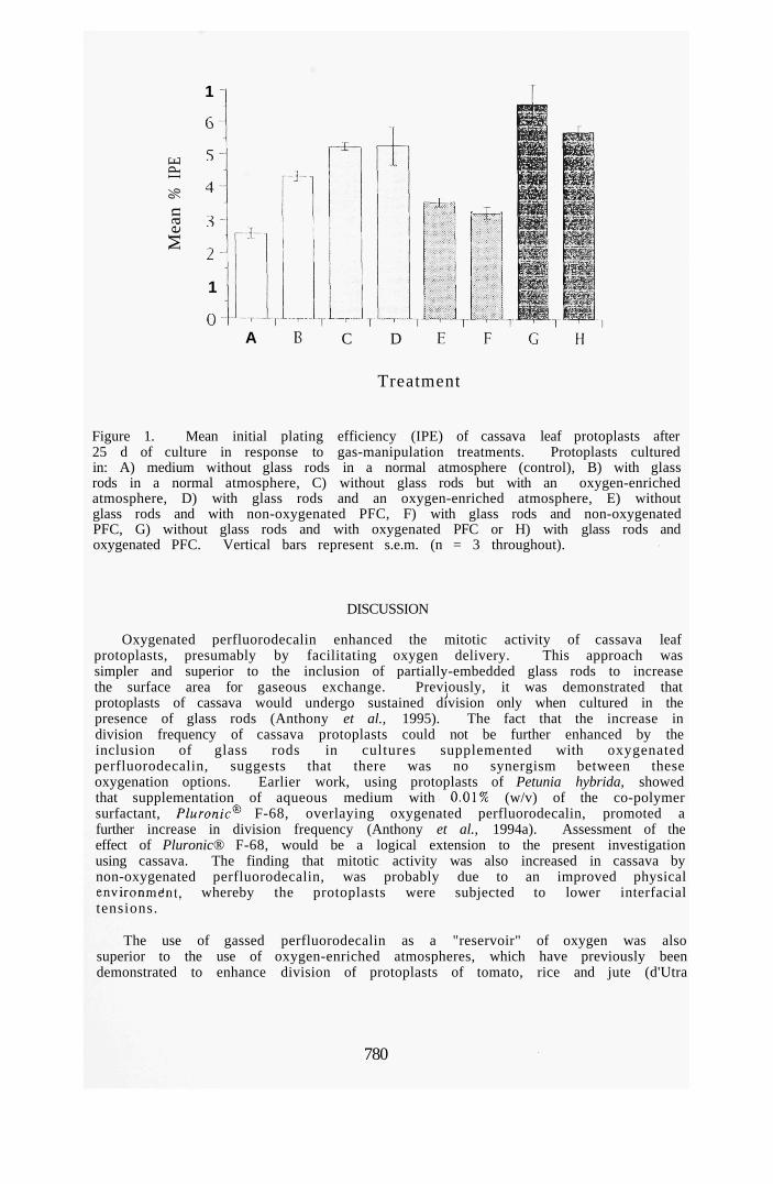
% I
PE
Mea
n
1 -
6
5
4
3
2
1 -
0 -
A:
A C D
...^.,
Treatment
Figure 1. Mean initial plating efficiency (IPE) of cassava leaf protoplasts after 25 d of culture in response to gas-manipulation treatments. Protoplasts cultured in: A) medium without glass rods in a normal atmosphere (control), B) with glass rods in a normal atmosphere, C) without glass rods but with an oxygen-enriched atmosphere, D) with glass rods and an oxygen-enriched atmosphere, E) without glass rods and with non-oxygenated PFC, F) with glass rods and non-oxygenated PFC, G) without glass rods and with oxygenated PFC or H) with glass rods and oxygenated PFC. Vertical bars represent s.e.m. (n = 3 throughout).
DISCUSSION
Oxygenated perfluorodecalin enhanced the mitotic activity of cassava leaf protoplasts, presumably by facilitating oxygen delivery. This approach was simpler and superior to the inclusion of partially-embedded glass rods to increase the surface area for gaseous exchange. Previously, it was demonstrated that protoplasts of cassava would undergo sustained division only when cultured in the presence of glass rods (Anthony et al., 1995). The fact that the increase in division frequency of cassava protoplasts could not be further enhanced by the inclusion of glass rods in cultures supplemented with oxygenated perfluorodecalin, suggests that there was no synergism between these oxygenation options. Earlier work, using protoplasts of Petunia hybrida, showed that supplementation of aqueous medium with 0.01% (w/v) of the co-polymer surfactant, Pluronic F-68, overlaying oxygenated perfluorodecalin, promoted a further increase in division frequency (Anthony et al., 1994a). Assessment of the effect of Pluronic® F-68, would be a logical extension to the present investigation using cassava. The finding that mitotic activity was also increased in cassava by non-oxygenated perfluorodecalin, was probably due to an improved physical environmeint, whereby the protoplasts were subjected to lower interfacial tensions.
The use of gassed perfluorodecalin as a "reservoir" of oxygen was also superior to the use of oxygen-enriched atmospheres, which have previously been demonstrated to enhance division of protoplasts of tomato, rice and jute (d'Utra
780

Vaz et al., 1992). Arguably, the use of perfluorodecalin is a more convenient and less hazardous option for oxygen delivery than the use of oxygen atmospheres. Supplementation of aqueous culture medium with oxygenated perfluorodecalin liquid, which is readily recoverable, sterilisable and, hence, recycleable (Anthony et al., 1994a; Lowe et al., 1995), clearly provides a convenient approach for facilitating respiratory gas supply not only to protoplasts of cassava, but also to those of other plant species. Despite the high initial cost of the PFC liquid, this approach should have wide applications in plant cell biotechnology. As emphasised previously (Anthony et al., 1994a), a particular advantage of the use of PFC as an oxygenating agent is in static cultures of protoplasts (or cells) where conventional aeration through agitation (e.g. stirring) cannot be employed because of the tendency to cause cell damage.
Woody plants, such as cassava, are generally considered to have relatively poor protoplast plating efficiencies (Ochatt & Power, 1990). The use of oxygenated perfluorodecalin alone, as shown in the present investigation, may go some way towards the enhanced throughput of protoplast-derived multicellular colonies and, in turn, whole plant regeneration.
A difficulty frequently encountered during the early stages of protoplast culture, particularly of woody species, is that of phenolic Oxidation accentuated by the aggregation of protoplast-derived cells (Ochatt, 1989; Ochatt & Power, 1990). While some success has been achieved in overcoming this fundamental problem through the culture of protoplasts, for example as monolayers on polyester discs interspersed within the aqueous medium (Russell & McCown, 1986, 1988), the use of oxygenated perfluorodecalin, as in the present study, to prevent protoplast clumping and phenolic oxidation, provides an additional, convenient approach. One further refinement of the present system would be to "recharge" the PFC with oxygen during culture, since previous studies have demonstrated that oxygen-saturated PFC becomes depleted of oxygen after 3 days during culture of P. hybrida protoplasts (Anthony et a/., 1994a). This would maximise and extend oxygen delivery to protoplast-derived cells.
Oxygenated PFC layers, droplets or emulsions, as media adjuncts, will now provide a baseline for the culture of limited numbers or individual protoplasts (e.g. flow-sorted heterokaryons or micromanipulated cells), since such supplementation enhances division potential. Likewise, the availability of efficient protoplast-to-plant systems has implications in terms of their transformation efficacy via protoplasts using direct D N A delivery technologies (Davey et al., 1989). Additionally, in the context of somatic hybridisation, the conversion of heterokaryons (intergeneric and interspecific protoplast fusion products) to somatic hybrid cells will be facilitated by improved plating efficiencies.
ACKNOWLEDGEMENTS
PA was supported by BNFL Fluorochemicals Ltd, Preston, UK, to whom the authors are also grateful for gifts of perfluorodecalin.
REFERENCES
Anthony, P., Davey, M.R., Power, J.B., Washington, C. and Lowe, K.C. (1994a) Plant Cell Rep. 13, 251-255.
Anthony, P., Davey, M.R., Power, J.B., Washington, C. and Lowe, K.C. (1994b) Plant Cell Tiss. Org. Cult. 38, 39-43.
781

Anthony, P., Davey, M.R., Power, J.B. and Lowe, K.C. (1995) Plant Cell Tiss. Org. Cult. (in press).
Davey, M.R., Rech, E.L. and Mulligan, B.J. (1989) Plant Molec. Biol. 13, 273-285. d'Utra Vaz, F.B., Slamet, I.H., KhaUin, A., Cocking, E.C. and Power, J.B. (1992) Plant
Cell Rep. 11, 416-418. Frearson, E.M., Power, J.B. and Cocking, E.C. (1973) Dev. Biol. 33, 130-137. King, A.T., Mulligan, B.J. and Lowe, K.C. (1989) Bio/Technol. 7, 1037-1042. Lowe, K.C. (1994) Vase. Med. Rev. 5, 31-48. Lowe, K.C, Davey, M.R. and Power, J.B. (1995) In: Banks, R.E. (Ed.) Fluorine in
Agriculture (pp. 157-164). Shrewsbury: Rapra. Misawa, N., Yamano, Y., Ohyama, K. and Komano, T. (1982) In: Fujiwara, A. (Ed.)
Plant Tissue Culture (pp. 607-608) Maruzen: Tokyo. Murashige, T. and Skoog, F. (1962) Physiol. Plant. 56, 473-497. Ochatt, S.J. (1990) Acta Hort. 280, 215-226. Ochatt, S.J. and Power. J.B. (1990) In: Fowler, M.W. and Warren, G.S. (Eds.) Plant
Biotechnology, Supplement 2 (pp. 99-129). Oxford: Pergamon Press. Russell, J.A. and McCowan, B.H. (1986) Plant Sci. 46, 133-142. Russell, J.A. and McCowan, B.H. (1988) Plant Cell Rep. 1, 59-62. Snedecor, G.W. and Cochran, W.G. (1989) Statistical Methods, 8th Edn. Ames: Iowa
State University Press. Stamp, J.A. and Henshaw, G.G. (1987) Ann. Bot. 59, 445-450. Widholm, J. (1972) Stain Technol. 47, 186-194.
782

CRYOBiouQOY 33 , 5 0 8 - 5 1 4 (1996)
ARTICLE NO. 0054
Pluronit F-68 Increases the Post-thaw Growth of Cryopreserveci Plant Cells
P. ANTHONY, N. B. JELODAR, K. C. LOWE,' J. B. POWER, AND M. R. DAVEY Plant Genetic Manipulation Group, Department of Life Science, University of Nottingham,
University Park, Nottingham NG7 2RD, United Kingdom
The beneficial effects of supplementing culture media with Pluronic F-68 (0.01-0.2% w/v), on post-thaw growth following cryopreservation of suspension-cultured cells of Oryza sativa cvs. Taipei 309 and Tarom (Japonica rices), together with Lolium multiflorum (Italian ryegrass), were studied. The mean absorbance, following triphenyltetrazolium chloride (TTC) reduction, of Taipei 309 cells 4 days after thawing was increased twofold over conuol by 0.01% (w/v) Pluronic F-68 (P < 0.01) and almost twofold by 0.1% (w/v) surfactant. A more pronounced, fourfold increase (P < 0.001) in post-thaw absorbance occurred with Tarom cells exposed to 0.1% (w/v) Pluronic F-68; cell absorbance was elevated threefold with 0.2% (w/v) Pluronic (P < 0.001) and twofold (P < 0.001) by 0.01% (w/v) surfactant. TTC reducdon by Lolium cells was elevated 31% (/» < 0.01) with 0.01% (w/v) surfactant. Pluronic F-68 (0.01% w/v) also promoted sustained mitotic activity since biomass, measured 30 days post-thawing, was increased to a maximum of 32% above control (P < 0.05), in both Taipei 309 and Lolium. There was no measurable beneficial effect of adding Pluronic F-68 to the cryoprotectant before freezing. These results, which demonstrate a marked cytoprotection of Pluronic F-68 on plant cells recovered from cryostorage, suggest that this surfactant is a useful additive agent in post-thaw handling strategies, c 1996 Academic Press, inc.
The nonionic polyoxyethylene (POE)-poly-oxypropylene (POP) surfactant Pluronic F-68 (Poloxamer 188) has been employed extensively as a low-cost, nontoxic, cell-protecting agent in both animal (14, 27) and plant (19, 20) culttire systems. Pluronic F-68 acts as a foam-stabilizing agent in agitated/aerated cultures and is believed to protect cells against fluid-mechanical damage (13). Pluronic F-68 is adsorbed onto cell surfaces, and studies with both cultured insect cells (24, 23, 12) and mammalian hybridomas (32) have demonstrated that interaction of the surfactant with cytoplasmic membranes can increase the resistance of cells to shear forces, albeit over differing time periods depending on the cell type.
The cytoprotectant properties of Pluronic F-68 make this compound a strong and obvious candidate for use in plant cell cryopreservation protocols. Early studies showed that the Pluronics could prevent hemolysis of human red blood cells in response to freeze-thawing procedures
Received April 25, 1996; accepted July 8, 1996. ' To whom correspondence should be addressed.
(11), and subsequent studies demonstrated that Pluronic F-68 was an effective cryoprotectant of cultured Chinese hannster cells (2). Surprisingly, there has been no evaluation of the effects of using Pluronic F-68 with plant cells, either as a cryoprotectant per se or a post-thaw cytoprotect-uig agent, despite the increasing interest in cryopreservation for conserving agronomically important and endangered plant germplasms (3). Consequently, the present investigation has employed embryogenic cells of Japonica rices, together with nonembryogenic cells of Lolium, to evaluate the potential beneficial effects of Pltu?o-nic F-68, either as a cell protectant during freezing or as a specific culture medium supplement, in enhancing post-thaw cellular growth following short-term (30 days, Oryza sativa cv. Tarom, Lolium) or long-term (3 years, O. sativa cv. Taipei 309) cryopreservation.
MATERIALS AND METHODS
Plant Materials and Preparation of Cell Suspensions
Cell suspensions of O. sativa L. cv. Taipei 309 and O. sativa L. cv. Tarom were initiated
0011-2240/96 $18.00 Copyright O 1996 by Academic Press, Inc. All rights of reproduction in any fonn reserved.
508

PLURONIC AND CRYOPRESERVED PLANT CELLS 509
from embryogenic calli derived from mature seed scutella (10). Cell suspensions of Lolium multiflorum were supplied by Dr. E. Guiderdoni, IRAT/CIRAD, Montpellier, France. Cell suspensions were maintained in AA2 medium [cv. Taipei 309 (1), R2 medium (cv. Tarom) (26), and N6 medium (Lolium) (9)] in 100-ml Erlenmeyer flasks with shaking (120 rpm, 2.5-cm throw) at 28 ± TC in the dark. Rice cell suspensions (both varieties) were subcultured routinely every 7 days by transferring 1 ml of settled cells with 7 ml of spent medium to 22-nil aliquots of fresh medium; Lolium cell suspensions were similarly subcultured by transfer of settled cells (3 ml) and 7 ml of spent medium to 40-ml aliquots of fresh medium. Prior to cryopreservation, cells were cultured for 3 -4 days in their respective liquid medium supplemented with 60.0 g liter"' mannitol.
Cryopreservation and Post-thaw Recovery
The cryopreservation protocol was based on that of Meijer et al. (22). Cells were harvested onto a nylon mesh (45-/im pore size) and placed into 2-cm^ polypropylene vials (Sarstedt, Leicester, UK) with approximately 0.2 g fresh weight of cells per vial. Approximately 0.75 ml of a cryoprotectant mixture [46.0 g liter"' glycerol, 39.0 g liter"' dimethyl sulfoxide (MezSO), 342.0 g liter"' sucrose, 5.0 g liter"' proline] was added to each vial. The cryoprotectant mixture was prepared in the liquid culture medium appropriate for each species and the pH adjusted to 5.8, prior to filter sterilization (0.2-/im pore size; Sartorius, Gottingen, Germany). All cryoprotectants were of Analar grade except Me^SO, which was spectroscopically pure.
Cells were cryoprotected for I h on iced water, vials containing the cells were transferred to aluminum canes, and the cells were frozen at a controlled rate ( - T C min"') from 0 to -35°C and held at this temperature for 35 min in a programmable freezer (Planer Cryo 10 Series, Planer Biomed, Sunbury-on-Thames, UK), prior to storage in liquid nitrogen at - 196°C. Cells of cv. Tarom and Lolium
were stored for 30 days, whereas those of cv. Taipei 309 were cryopreserved for 3 years.
Cells of both species (two cultivars for rice) were thawed by plunging the vials into sterile water at 45°C; excess cryoprotectant was removed under axenic conditions from the cells using a disposable Pasteur pipet. The cells were placed onto two superimposed 5.5-cm-diameter Whatman No. 1 filter paper disks overlying 20-ml aliquots of the appropriate culture medium. The latter was semisolidified with 0.4% (w/v) Sea Kem Le agarose (FMC Corp., Rockland, ME). In some treatments, the medium was supplemented with 0.01, O.I, or 0.2% (w/v) Pluronic F-68 (Sigma, Poole, UK). These concentrations of Pluronic were selected on the basis of previous studies demonstrating the stimulatory effects of this compound on the growth in culture of protoplasts isolated from cell suspensions of Solanum dulcamara (18). In an additional assessment, the cryoprotectant mixture for both species was also supplemented with the aforementioned concentrations of Pluronic F-68 and cells cryopreserved for 14 days in liquid nitrogen.
CeUs were cultured for all treatments in the dark for 3 days at 28 ± TC prior to transfer of the upper filter disk containing the cells to the respective fresh media. Cells were cultured for a fiirther 24 h prior to viabiUty assessments and, where appropriate, for a fiirther 26 days, under the same conditions, for biomass determinations. Each experiment was repUcated up to 20 times, except where surfactant was incorporated into the cryoprotectant, where n = 1.
Measurement of Post-thaw Viability and Biomass
The post-thaw viability and metabolic capacity of cells were assessed by the reduction of triphenyltetrazolium chloride (TTC) (30). Three-milliliter aliquots of TTC buffer, consisting of 0.6% (w/v) TTC in 0.05 M Na2HP04-KH2P04 buffer containing 0.05% (v/v) Tween 80 (Sigma, pH 7.4), was added to 50 mg of cells contained in 16.0-ml screw-capped centrifuge tubes (Coming, Coming, NY). Cells were incubated at 28 ± TC for

510 ANTHONY ET AL.
16-18 h in the dark, washed once with distilled water, and extracted with 7.0 ml of 95% (v/v) ethanol in a boiling water bath for 5 min. Supematants were cooled, and each was adjusted to lO.O ml with 95% (v/v) ethanol. The absorbance was measured at 490 nm. The same protocol was also employed for unfrozen cells 4 days following subculture.
In some assessments, using both cv. Taipei 309 and Lolium cells, the incubation buffer was supplemented with 0.01 or 0.1% (w/v) Pluronic F-68, respectively, as a control for any effects of the surfactant on TTC reduction. The fresh weight of thawed cells was recorded to determine biomass (21).
Statistical Armlyses
Means and standard errors (SEM) were used throughout; statistical significance between mean values was assessed using a conventional analysis of variance (ANOVA) and Student's t test, as appropriate (29). A probability P less than 0.05 was considered significant.
RESULTS
The mean absorbance of unfrozen cells, as measured by TTC reduction, was 1.60 ± 0.11% (n = 10) for O. sativa cv. Tarom and 0.54 ± 0.04% (n = 10) tor Lolium. In the case of O. sativa cv. Taipei 309, the mean cell absorbance, prior to long-term (3-year) cryopreservation, was 0.96 ± 0.05% (n = 10).
Supplementation of culture medium for the cv. Taipei 309 with Pluronic F-68 at 0.01% (w/v) significantiy increased the mean post-thaw cell absorbance following TTC reduction to more than twofold greater (P < 0.01) than that in untreated controls (Fig. 1). A similar, but less pronounced effect also occurred with 0.1% (w/v) surfactant (Fig. 1). In conti-ast, there was no corresponding increase in absorbance in these rice cells when 0.2% (w/v) Pluronic was incorporated into the culture medium.
In tiie case of O. sativa cv. Tarom, supplementation of medium with Pluronic F-68 had a consistently greater stimulatory effect on tiie
mean post-thaw cell absorbance compared with that of untreated controls (Fig. I). The most pronounced increase occurred with 0.1% (w/v) surfactant, which promoted a fourfold increase (P < 0.001) in absorbance over the control. Supplementation of medium for Tarom cells with 0.01% (w/v) or 0.2% (w/v) Pluronic increased the mean cell absorbances by twofold (P < 0.001) and tiireefold (P < 0.001), respectively (Fig. 1).
A significant (P < 0.01) increase in mean post-thaw cell absorbance was also recorded when Pluronic F-68 at 0.01% (w/v) was added to the culture medium for Lolium cells, although the mean maximum increase over control of 31% (Fig. 1) was markedly lower than that observed when the same concentration of surfactant was used with cells of O. sativa cv. Taipei 309 (140%) or cv. Tarom (95%).
No significant difference was observed, using post-thawed cv. Taipei 309 cells, between the mean cell absorbance in the TTC assay (0.62 ± 0.05, n = 15) and in the same assay using buffer supplemented with 0.01% (w/v) Pluronic F-68 (0.61 ± 0.05, n = 15). Comparable results were also obtained with post-thawed Lolium cells using 0.1% (w/v) surfactant (control, 0.41 ± 0.03, n = 15; Pluronic, 0.39 ± 0.04. n = 5).
Addition of Pluronic F-68 to culture medium also fostered an increase in biomass, as measured by cell fresh weight following 30 days of post-thaw culture, in both O. sativa cv. Taipei 309 and Lolium, witii significant (P < 0.05, P < 0.01) increases in the presence of 0.01% (w/v) surfactant in both cases (Table 1). The greatest increase occurred with cv. Taipei 309 supplemented with 0.01% (w/v) Pluronic F-68, where the mean fresh weight after 30 days was 32% greater than the corresponding mean control value (Table 1).
The addition of 0.01-0.2% (w/v) Pluronic F-68 to the cryoprotectant mixture, both before and during freezing, did not affect the post-thaw viability of cells from either rice cultivar or Lolium. For example, the mean absorbance of Lolium cells 4 days following thawing in die presence of 0.01% (w/v) Pluro-

E c
I
2
PLtJRONlC AND CRYOPRESERVED PLANT CELLS
Oryza sativa cv. Taipei 309 Oiyza sativa cv. Tarom Lolium multiflorum
511
1.2
1
0.8-1
I 0.6-i
0.4-
0.2-
0-
1-1
0.8-
0.6-
0.4
0.2
0
i-0.6-
0.4-
0.2-
Concentration of Pluronic® F-68 in cell recovery medium
FIG. I. Absorbance, following TTC reduction, by plant cells post-thawed in the presence of Pluronic F-68: (A) 0%, (B) 0.01%, (C) 0.1%, (D) 0.2% (w/v). Values are means of 20 observations; vertical bars represent SEMs. *P < 0.05, **P < 0.01, ***P < 0.001 compared with mean control (0% Pluronic).
nic (0.74 ± 0.04, n = 7) was not significantly different from that of control lacking surfactant (0.70 ± 0.04, n = 7). A similar pattern was observed with both rice cultivars (data
TABLE 1 Mean Fresh Weight of Cryopreserved Cells of Oryza
sativa cv. Taipei 309 and Lolium multiflorum Recovered after 30 Days with 0.01-0.2% (w/v) Pluronic F-68 in the Culture Medium
Treatment Mean fresh weight
on day 30 (g)
Oryza sativa cv. Taipei 309 Control (0% Pluronic) Pluronic
0.01% 0.1% 0.2%
Lolium multiflorum Control (0% Pluronic) Pluronic
0.01% 0.1% 0.2%
2.07 ± 0.28
2.74 ± 0.13* 2.46 ±0.11 L98 ± 0.16
3.49 ± 0.18
4.50 ± 0.20** 3.29 ± 0.21 3.32 ± 0.19
Note. Values are means ± sem, n = 11-14 throughout. For all treatments, ca. 0.2 g of cells was used as starting material (Day 0).
* P < 0.05, **P < 0.01 compared with control (0% Pluronic).
not shown). Cells recovered of both rice cultivars and Lolium had a dark, wet appearance, during tiie first few days of culture, following their initial recovery from cryopreservation. By day 10 of culture, however, cell color and friability were the same as for unfrozen controls.
Cell suspensions (n = 9) reinitiated from recovered cells of O. sativa cv. Tarom in the presence of 0.01% (w/v) Pluronic F-68 showed no difference (after 28 days) in mean absorbance following TTC reduction compared witii conti-ol (1.04 ± 0.16 and 0.87 ± 0.60, respectively). Additionally, their overall growth rates were comparable to those of controls and tiiey were routinely subcultured in accord with previously established protocols. Interestingly, these cell lines gave viable protoplast yields approximately twice those of controls (data not shown).
DISCUSSION
The results of these experiments demonstrate tiiat supplementation of the culture medium with Pluronic F-68 increases the post-thaw metabolism and growth of cryopreserved rice and Lolium cells. While it is generally accepted tiiat reduction of TTC can be used as an indicator of cell viability and metabo-

512 ANTTiONY ET AL.
lism (5), the present study also includes data on biomass increases to support the conclusion of growth-enhancing effects of Pluronic F-68. It is probable that the present observations reflect a combination of increased cell survival post-thawing coupled, perhaps, with a growth-stimulating effect of the surfactant in plant culture systems, as reported previously (19). Assessment of TTC reduction was made 4 days post-thaw since previous related work indicated tiiat, at tiiis time, the TTC assay provides optimal information on cell metabolic capacity (21).
An important finding from this investigation was that the optimum concentration of surfactant, which increased cell growth, differed between the two rice cultivars and between O. sativa cv. Tarom and Lolium. The present observations, showing that there are both species- and cultivar-specific responses to Pluronic F-68, are consistent with previous observations using Chrysanthemum morifolium, in which the optimum concentration of surfactant that stimulated adventive shoot regeneration from cultured leaf explants differed by an order of magnitude between cultivars (17). Differences in tissue and organ responsiveness to the growth-promoting effects of Pluronic F-68 have also been observed in Solanum dulcarrujra (18), Corchorus capsularis (16), Hypericum perforatum (6), and Populus spp. (15).
Studies using animal cells have shown that Pluronic F-68 adsorbs onto cytoplasmic membranes, conferring increased resistance to mechanical damage (14, 19, 20). The Pluronic polyols have hydrophobic TPOP cores, which are believed to become embedded in the phospholipid membranes of cells, leaving their hydrophilic POE tails outside. This would have the effect of reducing the interfacial tension of the cells and sterically hindering adhesive interaction between molecules on the cell surfaces. Such reduction in adhesive interaction would prevent cell-to-cell contact and, thus, further reduce mechanical damage.
Pluronic F-68 may also promote the increased uptake of nutrients, growtii regulators.
or oxygen into cells during the post-thaw period. Indeed, related experiments using animal cells cultured under static conditions have shown that concentrations of Pluronic F-68 comparable to those used in the present investigation stimulated both 2-deoxyglucose uptake and cellular amino acid incorporation (7). Any increase in nutrient uptake promoted by Pluronic would be expected to alter metabolic flux, allowing biochemical pathways to operate more efficientiy, particularly under the stress conditions of early post-thaw recovery. In this regard, previous work has shown that respiratory impairment occurs during the early post-thaw period (5, 8), and it is possible that Pluronic F-68 assists in overcoming such perturbations. The adsorption of Pluronic molecules onto the cytoplasmic membranes of post-thawed plant cells may also reduce cellular damage which is known to occur during rehydration when the MeiSO cryoprotectant is removed progressively from the system (4).
Further studies are required to determine the mechanism(s) by which surfactants, such as Pluronic F-68, can faciUtate post-thaw survival and growth of plant cells. One focus of such work should be to investigate the effects of Pluronic F-68 on respiratory gas dynamics, since related work has provided evidence that this compound can alter oxygen transport in agitated, sparged bioreactors (25). There is also speculation that Pluronic F-68 may influence the release of carbon dioxide from cells (19) or inhibit ethylene production in a manner comparable to that promoted by Triton X-100 (28). Interestingly, experiments have demonstrated that inert, oxygen-dissolving perfluorochemical liquids, emulsified with Pluronic F-68, were effective in prolonging the fertilizing capability of turkey spermatozoa stored at lowered temperature (31). While the specific effects of Pluronic were hot evaluated, it is possible that the surfactant may have contributed to cell survival, either through cytoprotection or, perhaps, by enhancing oxygen and/or nutrient uptake. This earlier work of Thurston et al. (31), together with the results reported in the present paper, indicate that the

PLURONIC AJMD CRYOPRESERVED PLANT CELLS 513
mechanism(s) whereby Pluronic F-68 facilitates survival of both animal and plant cells warrants further detailed study.
The present data imply that Pluronic F-68, which is relatively inexpensive and commercially available, could be incorporated routinely into post-thaw culture media, to increase plant cell recovery and growth during post-thaw handling procedures.
ACKNOWLEDGMENTS
P.A. was supported by BNFL Fluorochenucals Ltd., Preston, United Kingdom, and N.B.J, by the Government of Iran.
REFERENCES
1. Abdullah, R., Thompson, J. A., and Cocking, E. C. Efficient plant regeneration from rice protoplasts through somatic embryogenesis. Bio/Technology 4,1087-1090(1986).
2. Ashwood-Smith, M. J., Voss, W. A. G., and Warby, C. Cryoprotection of mammalian cells in tissue culture with Pluronic polyols. Cryobiology 10, 502-504 (1973).
3. Benson, E. E. Cryopreservation. In "Plant Cell Culture: A Practical Approach" (R. A. Dixon and R. A. Gonzales, Eds.), 2nd. ed., pp. 147-167. Oxford Univ. Press, Oxford, 1994.
4. Benson, E. E., and Withers, L. A. Gas chromatographic analysis of volatile hydrocarbon production by cryopreserved plant tissue cultures: a nondestructive method for assessing stability. Cryo-Utters 8, 35-46 (1987).
5. Benson, E. E., Lynch, P. T., and Jones, J. The detection of lipid peroxidation products in cryoprotected and frozen rice cells: Consequences for post-thaw survival. Plant Sci. 85, 107-114 (1992).
6. BrutovsW, R., Cell4rovS, E., Davey, M. R., Power, J. B., and Lowe, K. C. Stimulation of multiple shoot regeneration from seedling leaves of Hypericum perforatum L. by Pluronic F-68. Acta Biotechnol. 4,371-377(1994).
7. Cawrse, N.. de Pomerai, D. L, and Lowe, K. C. Effects of Pluronic F-68 on 2-deoxyglucose uptake and amino acid incorporation into chick embryonic fibroblasts in vitro. Biomed. Sci. 2, 180-182 (1991).
8. Cella, R., Columbo, R., Galli, M. G., Nielson, E., RoUo, F., and Sala, F. Freeze-preservation of rice cells: A physiological study of freeie-thawed cells. Physiol. Plant. 55, 279-284 (1982).
9. Chu, C. C, Wang, C. S., Sun, C. S., Hsu, C, Yin, K. C, Chu, C. Y., and Bi, F. Y. Establishment of an efficient medium for another culture of rice through comparative experiments on the nitrogen sources. Sci. Sin. 18, 659-668 (1975).
10. Finch. R. P., Lynch, P. T., Jotham, J. P., and Cocking, E. C. Isolation, culture and fusion of rice protoplasts. In "Biotechnology in Agriculture and Forestry" (Y. P. S. Bajaj, Ed.), Vol. 14. pp. 251-268. Springer-Verlag, Heidelberg, 1991.
11. Glauser, S. C, and Talbot, T. R. Some studies on freezing and thawing of human erythrocytes. Am. J. Med Sci. 231, 75-81 (1956).
12. Goldblum, S., Bae, Y-K., Hink, W. F., and Chalmers, J. Protective effect of methylcellulose and other polymers on insect cells subjected to laminar shear stress. Biotechnol. Prog. 6, 383-390 (1990).
13. Handa-Corrigan, A., Emery, A. N., and Spier, R. E. Effect of gas-liquid interfaces on the growth of suspended animal cells: Mechanisms of cell damage by bubbles. Enzyme M'lcrob. Technol. 11, 230-235 (1989).
14. Handa-Corrigan, A., Zhang, S., and Brydges, R. Surface-active agents in animal cell cultures. In "Animal Cell Biotechnology" (R. E. Spier, and J. B. Griffiths, Eds.), Vol. 5, pp. 279-301. Academic Press, I^ndon, 1992.
15. lordan-Costache, M., lx)we, K. C, Davey, M. R., and Power, J. B. Improved micropropagation of Populus spp. by Pluronic F-68. Plant Growth Regal. 17, 233-239 (1995).
16. Khatun, A., t^ouar, L., Davey, M. R., Mulligan, B. J., and Lowe, K. C. Effects of Pluronic F-68 on shoot regeneration from cultured jute cotyledons and on growth of transformed roots. Plant Cell Tissue Org. Cult. 34, 133-140 (1993).
17. Khehra, M., Lowe, K. C, Davey, M. R., and Power, J. B. An improved micropropagation system for Chrysanthemum based on Pluronic F-68-supple-mented media. Plant Cell Tissue Org. Cult. 41, 87-90 (1995).
18. Kumar, V., Laouar, L., Davey, M. R., Mulligan, B. J., and Lowe, K. C. Pluronic F-68 stimulates growth of Solanum dulcamara in culture. J. Exp. Bot. 43, 487-493 (1992).
19. Lowe, K. C, Davey, M. R., Power, J. B., and Mulligan, B. J. Surfactant supplements in plant culture systems. Agro-food-Ind. Hi-Tech. 4 , 9 -13 (1993).
20. tvowe, K. C, Davey, M. R., I-aouar, L., Khatun, A., Ribeiro, R. C. S., Power, J. B., and Mulligan, B. J. Surfactant stimulation of growth in cultured plant cells, tissues and organs. In "Physiology, Growth and Development of Plants in Culture" (P. J. Lums-den, J. R. Nicholas, and W. J. Davies, Eds.), pp. 234-244. Kluwer, Dordrecht, 1994.
21. Lynch, P. T., Benson, E. E., Jones, J., Cocking, E. C, Power, J. B., and Davey, M. R. Rice cell cryopreservation: The influence of culture methods and the embryogenic potential of cell suspensions on post-thaw recovery. Plant Sci. 98, 185-192 (1994).
22. Meijer, E. G. M., van Iren, F., Schrijnemakers, E.,

514 ANTHONY ET AL.
Hensgens, L. A. M., van Zijderveld, M., and Schilperoort, R. A. Retention of the capacity to produce plants from protoplasts in cryopreserved cell lines of rice (Oryza sativa L.). Plant Cell Rep. 10, 171-174(1991).
23. Murhammer, D. W., and Goochee, C. F. Scale-up of insect cell cultures: Protective effects of Pluronic F-68. Bio/Technology 6, 1411-1418 (1988).
24. Murhammer, D. W., and Goochee, C. F. Structural features on non-ionic polyglycol polymer molecules responsible for the protective effect in sparged animal cell bioreactors. Biotechnol. Prog. 6, 142-148(1990).
25. Murhammer, D. W., and Pfalzgraf, E. C. Effects of Pluronic F-68 on oxygen transport in an agitated, sparged bioreactor. Biotechnol. Technol. 6, 199-202 (1992).
26. Ohira, K., Ojima, K., and Fujiwara, A. Studies on the nutrition of rice cell culture. I. A simple defined medium for rapid growth in suspension culture. Plant Cell Physiol. 14, 1013-1121 (1973).
27. Papoutsakis, E. T. Media additives for protecting freely suspended animal cells against agitation and aeration damage. Trends Biotechnol. 9, 427-437 (1991).
28. Sauerbrey, E., Grossmann, K., and Jung, J. Ethylene production by sunflower cell suspensions. Plant Physiol. 87,510-513(1988).
29. Snedecor, G. W., and Cochran, W. G. "Statistical Methods," 8th ed. Iowa State Univ. Press, Ames, 1989.
30. Steponkus, P. L., and Lamphear, F. O. Refinement of the triphenyl tetrazolium chloride method of determining cold injury. Plant Physiol. 42, 1423-1426(1967).
31. Thurston, R. J., Rogoff, M. S., Scott, T. R., and Kora, N. Effects of perfluorochemical diluent additives on fertilizing capacity of turkey semen. Poultry Sci. 72, 598-602 (1993).
32. Zhang, Z., Al-Rubeai, M., and Thomas, C. R. Effect of Pluronic F-68 on the mechanical properties of mammalian cells. Enzyme Microb. Technol. 14, 980-983 (1992).

Biotabnology TKbniquts, Vol 11, No 8, August 1997, pp. iSl-i84
Enhanced mitotic division of cultured Passiflora and Petunia protoplasts by oxygenated perfluorocarbon and haemoglobin Paul Anthony, Michael R. Davey, J. Brian Power and Kenneth C. Lowe Department of Life Science, University of Nottingham, Nottingham NG7 2RD, U.K.
Cell suspension-derived protoplasts were cultured in (A) liquid medium, (B) medium overlaying oxygenated perfluorodecalin (PFC), (C) medium containing 1:50 (v.v) of haemoglobin solution (Erythrogen"*), or (D) medium with 1:50 (v:v) Erythrogen™ overlaying oxygenated PFC. In Passiflora, mitotic division of protoplasts was increased (P<0.05) by all treatments, with Erythrogen™ being the most effective (120% increase over control). For Petunia, treatment D Induced maximum mitosis (140% over control), whilst Erythrogen™ alone produced a less pronounced (80% over control) increase.
introduction A key requirement iti the genetic manipulation and biotechnological exploitation of higher plants is the culture of protoplasts (wall-less cells) and cells under static conditions with adequate respiratory gas supply. Previous approaches for enhancing Oj stipply to cultured protoplasts include the use of Oj-enriched atmospheres (d'Utra Vaz et al., 1992) and liquid-over-agar medium implanted with glass rods to increase the surface area of the liquid layer (Anthony tt al., 1995). Mitotic division of protoplasts can also be enhanced by culture with Oj-gassed perfluortKarbons (PFCs), inert, highly fluorinated organic liquids that dissolve large volumes of respiratory gases (Anthony et al., 1994; Lowe et al., 1997).
Related experiments have shown that supplementation of aqueous culture medium with low concentrations (ca. 2.0 g |-') of a commercial bovine haemoglobin (Hb) solution (Anthony et al., 1997) enhances"division of cell suspension-derived protoplasts o( Petunia hybrida in vitro. The present study extends these observations with direct comparisons of oxygenated PFC and Hb, alone and in combination, using Passiflora (a woody species) and Petunia (a herbaceous species) as model protoplast systems.
Materials and methods Plant materials and protoplast culture Protoplasts were isolated from embryogenic cell suspensions of Passiflora giberti using an enzyme mixture
© 1997 Chapman & Hall
(20 ml enzyme solution g"' fresh wt. cells) which consisted of 1.0% (w/v) Cellulase RS (Yakult Honsha Co., Nishinomiya Hyogo, Japan), 0.1% (w/v) Pectolyase Y23 (Seishim Pharmaceutical, Tokyo, Japan) and 5 mM 2-[N-morpholino]ethane sulphonic acid in CPW salts solution (Frearson tt al., 1973). The latter contained 27.2 mg/l KHjPO^, 101 mg/1 KNOj, 246 mg/1 MgClj-7HjO. 0.16 mg/1 Kl, 0.025 mg/1 CuSO^-5HjO, 1480 mg/1 CaClj-2H20 and 130 g/1 mannitol (CPW13M), pH 5.8. Protoplasts of an albino mutant of Petunia hybrida cv. Comanche were isolated from cell suspensions using the same enzyme solution, but at half enzyme strength. Incubation was performed in the dark (25 ± 2"'C) on a horizontal rotary shaker (40 rpm) for 16 h in 9.0 cm diam Petri dishi:s. The digested cells were filtered through 30 nm and 64 p.m pore size nylon sieves (Wilson Sieves, Nottingham, U.K.) for Passiflora and Petunia protoplasts, respectively. The filtrates were centrifuged (80 X g; 10 ntin) and the protoplast pellets washed twice in CPW13M solution by repeated resuspension and centrifugation. Protoplast viability was determined by the uptake and cleavage of fluorescein diacetate (Widholm, 1972), ptiot to counting and plating.
Culture of protoplasts with PFC and/or haemoglobin Protoplasts of P. giberti were cultured, for 6 d , at 1.0 X 10' ml-' in liquid KPR medium (Thompson ef al., 1986) and those of P. hybrida, for 10 d. at 2.0 X 10' ml-' in liquid KM8P medium based on the
Bitttcbiuhgy Ttchitiijuts . Vtl 11 • N« 8 • 1997 581

p. Anthony et al.
fotmulation of Kao and Michayluk (1975), as modified by Gilmour et al. (1989). Protoplast suspensions (2 ml aliquots) were placed in 30 ml glass bottles, either alone, or overlaying 6 ml aliquots of oxygenated (10 mbar, 15 min) perfluorodecalin (Flutec'^ PP6, BNFL Fluorochemicals, Springfields, Preston, U.K.). In some treatments, the media were supplemented with 1:50 (v;v) of the commercial bovine Hb solution, Erythrogen'^" (Biorelease Corporation, Salem, U.S.A.). This concentration of Hb was selected on the basis of previous studies, which demonstrated a stimulatory effect of Hb on mitotic division of cell suspension-derived protoplasts of P. hybrida (Anthony tt al, 1997). Following the addition of Hb solution, the resultant pH of the medium was 6.1 and, therefore, all comparative assessments utilised media at this value. Protoplasts were cultured at 25 ± 2°C in the dark.
Measurement of initial plating efficiency After 6 d (Passiflora) or 10 d (Petunia) of culture with oxygenated PFC or Erythrof,en'", an initial plating efficiency, defined as the percentage of protoplasts originally plated that had regenerated a new cell wall and undergone one or more mitotic divisions, was measured. The differential time scale for measurement of initial plating effeciency between the two culture systems reflected the inherent differences in mitotic cycle times of Passiflora and Petunia.
Statistical analyses Means and standard errors (s.e.m.) were used throughout, with statistical significance between mean values assessed using a non-parametric Mann-Whitney U test (Snedecor and Cochran, 1989). A probability of P < 0.05 was considered significant.
Results Yields of P. giberti and P. hybrida protoplasts were 5.83 ± 0.23 X 10* g-' f. wt. and 2.81 t 0.15 X 10* g-' f. wt. (n = 3 throughout), with mean viabilities of 78 ± 3% (n = 3) and 80 ± 2% (n = 3), respectively. The mean initial plating efficiency of P. giberti protoplasts with oxygenated PFC (20.3 ± 0.6%; n = 5) was significantly (P < 0.05) greater than control (10.4 ± 0.3%; n = 5; Fig. 1). A similar, but more pronounced, increase (P < 0.05) in mean initial plating efficiency of Passiflora protoplasts was observed when the medium was supplemented solely with Erythrogen"" (22.9 ± 1.0%; n = 5; Fig. 1). Tliere was no significant difference in mean initial plating efficiency values between the oxygenated PFC and Erythrogen'" treatments (Fig. 1). When the latter two treatments were combined, the mean initial plating efficiency (18.3 ± 0.8%; n = 5) was also
30n
§20-d OS
Si -^10-
0-A B C
Treatment
Figure 1 Mean initial plating efficiency (IPE) of Passiflora giberti protoplasts cultured lor 6d In A) KM8P medium alone (control), B) medium supplemented with 1:50 (v:v) Erythrogen'", C) medium overlaying oxygenated PFC or D) medium supplemented with 1:50 (v:v) Erythrogen™ and overiaying oxygenated perfluorocarbon. Vettical bars represent s.e.m. (n = 5 throughout). *P < 0.05 compared to control mean value.
significantly (P < 0.05) greater than in untreated controls (Fig. I). Interestingly, there was no significant difference between the mean initial plating efficiency with the combined treatments and that of protoplasts cultured in the presence of oxygenated PFC alone. However, the mean initial plating efficiency of protoplasts cultured with oxygenated PFC and Erythrogen'^'*, in combination, was 20% lower (P < 0.05) than with Erythrogtn™ alone (Fig. 1).
The mean initial plating efficiency of P. hybrida protoplasts cultured with oxygenated PFC after 10 d of culture (20.3 ± 1.5%; n = 5 throughout) was significantly (P < 0.05) greater than control (9.6 ± 0.6%; Fig. 2). In addition, the mean initial plating efficiency of Petunia protoplasts cultured in medium containing Erythrogtn'" was 17.3 ± 1.1% compared to 9-6 ± 0.6% in control (P < 0.05). As for Passiflora, no significant difference in mean initial plating efficiency was observed between the oxygenated PFC and Erythrogtn^" treatments, despite the time-scale difference in the initial plating efficiency assessments. When the PFC and Hb treatments were combined, the mean initial plating efficiency (23.0 ± 0.9%; n = 5) was also significantly (P < 0.05) greater than that in untreated controls or with Erythrogtn^ alone (Fig. 2). This contrasted with Passiflora, where Hb promoted the greatest increase
582 Bittechnology Ttchniquei -Vol II No 8 • 1997

Enhanced mitotic division ofcidtured Passiflora and Petunia protoplasts by oxygenated perfluorocarbon and haemoglobin
B C
Treatment
Figure 2 Mean Initial plating efficiency (IPE) of Petunia hybrida protoplasts cultured for 10 d in A) KI\/I8P medium alone (control), B) medium supplemented with 1:50 (v;v) Erythrogen™, C) medium overlaying oxygenated PFC or D) medium supplemented with 1:50 (v:v) Erythrogen™ and overiaying oxygenated perfluorocarbon. Vertical bars represent s.e.m. (n = 5 throughout). *P < 0.05 compared to control mean value.
in mean initial plating efficiency. However, as with P. giberti, there was no significant difference between the mean initial plating efficiency with oxygenated PFC and Erythrogen"", in combination, and with oxygenated PFC alone.
Discussion The results of these experiments demonstrate that oxygenated perfluorodecalin and Eiythrogen'" provide options for enhancing mitotic division, as reflected by increased initial plating efficiency, of both Passiflora and Petunia protoplasts and their respective protoplast-derived cells in vitro. Interestingly, the study revealed some variations in the effectiveness of the separate or combined treatments. In the case of P. giberti, Erylhrogtn"" promoted the greatest increase in mean initial plating efficiency, whilst for P. hybrida, the combined oxygenated PFC and Hb treatment was the most effective. Whilst further studies are" required, these experiments suggest that the effectiveness of these options for manipulating respiratory gas supply in vitro may be species-specific. However, the jjossibility also exists, at least for Passiflora, that the combined tteat-ment of Hb and PFC served to suppress mitotic division, through possible Oj toxicity effects. It may well be that for the chosen Pttitnia system, such a threshold was not reached.
Previously, oxygenated PFC has been shown to effectively enhance mitotic division of cultured higher plant protoplasts of several species, including those of P. hybrida (Anthony tt al., 1994), Manihot esculenta (cassava; Anthony et al, 1995) and Oryza sativa (rice) (Wardrop tt al., 1996). In the latter study, morphologically and cytologically normal plants were regenerated from protoplasts cultured in the presence of oxygenated PFC. Whilst clear evidence of changes in Oj flux between the PFC phase and the overlying aqueous medium has been obtained, at least during the first 48-72 h of culture (Anthony tt al., 1994), it is not inconceivable that such a liquid-liquid interface provides a more optimised physical micro-environment conducive for cell division per st. This would be consistent with studies in which mammalian cells were successfully cultured for extended periods at such interfaces (King tt al, 1989; Lowe tt al, 1997).
The finding that supplementation of culture medium with Erythrogen''" can stimulate protoplast division was consistent with previous studies using animal cells, in which Erythrogen"" not only enhanced mitotic division, but also stimulated production of recombinant protein (Goffe et al., 1994). It is speculated that, in the present experiments, Eiythrogen''" captured dissolved O^ from air-medium interfaces, thereby facilitating delivery of gas to protoplast-derived cells. This hypothesis would support claims from the earlier investigations with animal cells.
Future studies should evaluate these growth-enhancing strategies in other plant culture systems and determine whether they can be applied more broadly to the culture of eukaryotic cells. Whilst Hb is a convenient option for enhancing cellular O^ supply in vitro, currently available commercial formulations have limited shelf-life. Thus, the recoverability and hence, recycleability of PF(^ may make them a commercially more attractive and routine option, despite the high initial investment cost.
Acknowledgements P.A. was supported by BNFL Fluorochemicals Ltd., U.K. The authors acknowledge Biorelease Corporation, U.S.A., for technical information.
References Abdullali, R., Tliompson, J.A. and Cocking, E.C. (1986)
Biorrechnol. 4, 1087-1090. Antliony, P., Davey, M.R., Power, J.B., Washington, C. and
Lowe, K.C. (1994) Plant Cell Rep. 13, 251-255. Anthony, P., Lowe, K.C, Davey, M.R. and Power, J.B. (1995)
Biottchntl. Ticbniqiits 11, 777-782.
DiotcchncUgy Ttchniquei • Vtl 11 • Nt 8 • 1997 583

p. Anthony et al.
Anthony, P., Lowe. K.C. Power. J.U. and Davey, MR. (1997) Plant Cell Rep. (in press),
d' Ucca Vaz, F.B., Slamet, I.H.. Kliacun, A., Cocking, E.C. and Power. J.B. (1992) Plant Ctll Rtp. 11, 416-418.
Frearson, E.M., Power, J.B. and Cocking, EC. (1973) Om Biol. 33. 130-137.
Gilmour, D.M.. Golds. TJ. and Davey. M.R. (1989) In: Bajaj, Y.P.S. (Ed.) Biotechnology in Ponslry and Agriculture, Vol. 8, plant Protoplaits and Genetic Engineering I (pp. 370-388). Heidelberg: Springer-Verlag.
Goffe, R.A., Hguyen, A. and Shi, J.Y. (1994) An. Celli, Blood Suks., Immob. Biotechnol. 22: A18.
Kao, K.N. and Michayluk, M.R. (1975) Plania 126, 105-110. King, A.T., Mulligan, B.J. and Lowe, K.C. (1989) BiorTtcbnol.
7, 1037-1042. Lowe, K.C, Anthony, P., Wardrop, J., Davey, M.R. and Power,
J.B. (1997) An. Cells, Blood Subs., Immoh. Biotechnol. (in press). Snedecor, G.W. and Cochran, W.G. (1989) Statistical Methods,
8th Edn. Ames: Iowa State University Press. Tliompson, J.A., Abdullah, R. and Cocking, E.C. (1986) Plant
Sci. 47, 179-183. Wardrop, } . , Lowe, K.C, Power, J.B. and Davey, M.R. (1996)
/ Biotechnol 50, 377-393. Widholm, J. (1972) Stain Ttchnol. 47, 186-194.
Received 2 May 1997; Revisions requested 28 May 1997;
Revisions received 4 June 1997 Accepted 4 June 1997
584 Biotechnelegy Ttchniquts • Vol 11 • Ho B • 1997
Plant Cell Reports (lyy/) w: a-io > Springer-Verlag 1997
P. Anthony • K. C. Lowe • M. R. Davey • J. B. Power
Strategies for promoting division of cultured plant protoplasts: synergistic beneficial effects of haemoglobin {Erythrogen) and Pluronic F-68
Received: 24 May 1996 / Revision received: 12 July 1996 / Accepted: 15 August 1996
Abstract Novel approaches, involving supplementation of aqueous culture medium with haemoglobin solution (Erythrogen), in the presence or absence of the copolymer surfactant, Pluronic F-68, have been evaluated to facilitate cellular oxygen availability to promote mitotic division. Cell-suspension-derived protoplasts of albino Petunia hybrida cv. Comanche were cultured for up to 45 days in KM8P medium containing 1:50-1:500 (vol:vol) Erythrogen. The mean initial protoplast plating efficiency after 9 days with 1:50 Erythrogen (18.5%) was significantly greater (P<0.05) than in untreated controls (11.3%). Supplementation of culture medium with 1:50 Erythrogen, together with 0.01% (wt/vol) Pluronic F-68, increased the mean plating efficiency after 9 days (24.4%) by 92% (P<0.05) over the control (12.7%). These treatments also produced increases in biomass of protoplast-derived cells up to 2.5-fold greater than control (P<0.01) over 80 days. Gassing the medium, containing 1:50 Erythrogen, with carbon monoxide abolished the increase in plating efficiency. There was no additional benefit of gassing Eryth-rogen-supplemented medium with 100% oxygen. The synergistic, beneficial effect of Erythrogen and Pluronic F-68 on protoplast division Jias implications for plant biotechnology utilising protoplasts.
Key words P^tuniff hybrida • Oxygen delivery • Erythrpgen • HaemQg)pl)iii • pluronic F-68
Abbreviations IPE fnifial protoplast plating efficiency • PFC Perfluorochemical
Communicated by H. Lxirz P. Anthony • K. C. Lowe (a) • M. R. Davey • J. B. Power Plant Genetic Manipulation Group, Department of Life Science, University of Nottingham, University Park, Nottingham, NG7 2RD, UK
Introduction
Isolated plant protoplasts are the source material for the genetic manipulation of plants by interspecific and intergeneric somatic hybridisation through protoplast fusion, transformation by direct DNA uptake and the exposure of somaclonal variation (Blackball et al. 1994; Dixon 1994). One crucial requirement for successful protoplast culture and subsequent plant regeneration from protoplast-derived tissues is the provision of an adequate and sustainable oxygen supply. Previous approaches to enhance respiratory gas exchange for cultured protoplasts have included the use of oxygen-enriched atmospheres (d'Utra Vaz et al. 1992), inert, respiratory gas-dissolving perfluorochemical (PFC) liquids (Anthony et al. 1994a, b, 1995b) and liquid-over-agar media matrices implanted with glass rods to increase the surface area and gaseous exchange of the liquid culture phase (Anthony et al. 1995a).
An additional and novel approach to further facilitate oxygen supply, which has not been evaluated previously for plant cell systems, is the supplementation of aqueous culture media with a commercial haemoglobin solution (Erythrogen). Previous research has focused on the assessment of native and, to a greater extent, chemically modified haemoglobins as vehicles for respiratory gas transport in intact animals and isolated, perfused organs (Zuck and Riess 1994; Tsuchida 1995; Winslow et al. 1995). The most widely studied molecules are of human or bovine origin, although recombinant haemoglobins, expressed in Escherichia coli (Hoffman et al. 1990), transgenic mice (Behringer et al. 1989) and pigs (Swan-son et al. 1992), are now available. Haemoglobins liberated from animal erythrocytes are normally treated with pyridoxal phosphate or glutaraldehyde to reduce their oxygen binding to acceptable physiological levels (Zuck and Riess 1994; Tsuchida 1995). One advantage of using haemoglobin as a respiratory-gas-carrying culture medium supplement compared with PFC liquids is that the latter are immiscible in aqueous systems (King et al. 1989; Anthony et al. 1994a; Lowe et al. 1995). However,

14
paradoxically, this enables PFCs to be recovered readily and to be recycled.
The present investigation has employed cell-suspension-derived protoplasts of albino Petunia hybrida cv. Comanche, as a model system to assess the effects on mitotic division of protoplasts and their cellular derivatives of improved gas manipulation through supplementation of the culture medium with a commercially available, stabilised bovine haemoglobin solution, Erythrogen. The effects of haemoglobin were also evaluated alongside the copolymer surfactant, Pluronic F-68, which is commonly employed as a cytoprotectant for cultured animal cells (Goldblum et al. 1990; Papoutsakis 1991; Handa-Corrigan et al. 1992). This surfactant has been shown previously to promote growth in cultured plant cells, tissues and organs (Lowe et al. 1993, 1994).
the total population of protoplast-derived cell colonies from one randomly selected replicate 3.5-cm Petri dish was transferred to the surface of 6 ml of agar-solidified (0.6% wt/vol; Sigma) UM medium (Uchimiya and Murashige 1974) contained in 5.5-cm-diameter Petri dishes. Cell colonies/microcalli were further incubated in the dark at 25±2°C for 35 days in order to confirm no adverse influence of the treatments on long-term cell viability.
Measurement of protoplast viability, growth and biomass
The viability of freshly isolated protoplasts was determined by uptake and cleavage of fluorescein diacetate (Widholm 1972), prior to counting. Protoplast growth and mitotic activity was assessed after 9 days, by the initial plating efficiency (IPE), defined as the percentage of protoplasts originally plated that had regenerated a new cell wall and undergone at least their first mitotic division. A minimum of 100 protoplasts per Petri dish were scored; each treatment was repeated five times. The biomass of protoplast-derived cells was recorded as fresh weight after 80 days of culture with the various treatments.
Materials and methods
Isolation and culture of protoplasts
Protoplasts of an albino accession of Petunia hybrida cv. Comanche were isolated enzymatically from a well-characlerised and established cell suspension, as reported previously (Power et al. 1990). Protoplasts were cultured in the dark (25±2°C) at a final plating density of 2.0x10' ml"' in 1.5-ml aliquots of liquid KM8P culture medium (osmotic pressure 755 mmol kg"') based on the formulation of Kao and Michayluk (1975), as modified by Gilmour et al. (1989), and supplemented with 0.1 mg 1"' 2,4-dichlorophenoxyacetic acid and 0.2 mg I"' zeatin. The latter medium, containing protoplasts, was dispensed into 3.5-cm-diameter plastic Petri dishes (Nunc, Roskilde, Denmark). All experimental treatments were replicated five times.
Supplementation of medium with haemoglobin alone or with oxygen or carbon monoxide
Erythrogen (Biorelease Corporation, Salem, Mass., obtained from TCS Biologicals, Botolph Claydon, UK), a stabilised bovine haemoglobin solution (103 g I"', pH 7.42), was added to KM8P medium to final concentrations of 1:50, 1:100 or 1:500 (vol:vol). For the former concentration, a further series of replicates were established whereby the medium was gassed with 100% oxygen (10 mbar, 15 min) or 100% carbon monoxide (No: 850203; Phase Separations, Deeside, UK; 20 s), immediately prior to the addition of protoplasts.
Supplementation of medium with haemoglobin and Pluronic F-68
In an additional assessment using non-gassed medium containing 1:50 (vol: vol) Erythrogen, the aqueous culture medium was also supplemented with 0.01 %, 0.1 % or 1.0% (wt/vol) of Pluronic F-68 (Sigma, Poole, UK). These concentrations of Pluronic were based on those shown previously to stimulate the growth in culture of cell suspension-derived protoplasts of Solanum dulcamara (Kumar et al. 1992). All experiments using Erythrogen, alone or in conjunction with other treatments, utilised protoplasts of the same population, in turn isolated from the same homogenous cell suspensions. Dishes were sealed with Nescofilm (Bando Chemical, Kobe, Japan) and maintained under static conditions.
For all treatments, including controls, spent KM8P medium (0.25 ml per dish) was removed after 9 and 15 days and replaced, on both occasions, with an equal volume of KM8 mediym of reduced osmotic pressure (220 mmol kg"'), based on the formulation of medium 8 of Kao and Michayluk (1975), as modified by Gilmour et al. (1989). This medium lacked Erythrogen. After 45 days of culture.
Measurement of pH and osmotic pressure in the culture medium
The pH of the culture medium was measured using a Kent EIL7020 pH meter and was adjusted, where appropriate, to pH 6.1 using 1.0 M NaOH. The osmotic pressure of the culture medium for all treatments was measured using a Wescor 5500 vapour pressure osmometer, since it was essential to assess whether there were any shifts in pH and/or osmotic pressure associated with the inclusion of Erythrogen in the medium.
Statistical analyses
Means and standard errors (SE) were used throughout; statistical significance between mean values was assessed using a parametric Moods median test and a Mann-Whitney U-test (Snedecor and Cochran 1989). A probability of/'<0.05 was considered significant.
Results
The yield of Petunia protoplasts was 6.24±0.45xl0^ per gram fresh weight of suspension cells, with a mean viability of 94±1% (n=3). Culture of protoplasts in KM8P medium supplemented with 1:50 (vol:vol) Erythrogen stimulated protoplast division, with an increase in mean IPE of 64% (P<0.05) over the control after 9 days (Fig. 1). In contrast, the mean IPE values after 9 days in medium supplemented with 1:100 or 1:500 (vol:vol) of Erythrogen were not significantly different to the control (Fig. 1).
For cultures supplemented with 1:50 (vol: vol) Erythrogen and gassed with oxygen, the mean (±SE) IPE (18.1±1.5%, n=5) was significantly (P<0.05) greater than the control (11.3±0.4%, n=5), but was not significantly different from cultures containing the same, but ungassed, quantity of Erythrogen (18.5±0.8%, n=5'. Fig. 1). Similarly, for cultures containing 1:50 (vol: vol) Erythrogen, but gassed with carbon monoxide, the mean IPE after 9 days (13.1 ±1.2%, «=5) was also not significantly different to the control (Fig. 1).
The addition of 0.01% (wt/vol) Pluronic F-68 to cultures containing 1:50 (vol:vol) Erythrogen further increased the mean IPE after 9 days of culture by 38%

15
20
10
4t
a * ;
30-1
m '3?
ill
IS
C D E F Treatment
Fig. 1 Mean IPEof P^ru;iiacell-su.spension-derivedprotopla,sts after 9 days of culture in medium alone (control; A), Erythrogen (1:50 vol:vol), B) Erythrogen (1:50) with 100% oxygen (C), Erythrogen (1:50) with 100% carbon monoxide (D). Erythrogen (1:100; E) and Erythrogen (1:500; F). Vertical bars represent SE (n=5 throughout); *P<0.05 compared to control mean value
B C D Treatment
Fig, 2 Mean IPE of/ prHfi/a cell-suspension-derived protoplast-s after 9 days of culture in medium alone (control. A), Erythrogen (1:50 vol:vol; B), Erythrogen (1:50) with 0.01% Pluronic F-68 (O Erythrogen (1:50) with 0.1% Pluronic F-68 (£») and Erythrogen (1:50) with 1.0% (wt/vol) Pluronic F-68 (£). Verticalfeari represent SE(n=5 throughout); */'<0.05 compared to control mean value
(Fig. 2). In contrast, there was no additional beneficial effect of supplementing culture medium with 0.1 % or 1.0% Pluronic F-68 where the mean IPE after 9 days (19.1 ±1.3% and 14.8±0.8%, respectively, n=5) was not significantly different to the control (12.7 +2.5%, n = 5; Fig. 2). Supplementation of culture medium with Erythrogen, had no significant effects on pH or osmotic pressure. Similarly, gassing the culture medium containing 1:50 (vol:vol) Erythrogen with oxygen or carbon monoxide had no significant effect on mean pH(6.1 throughout) or osmotic pressure (764±7.5 mmol kg""' and 774+3.1 mmol kg"', respectively), compared to ungassed medium with Erythrogen (756±8.4 mmol kg"'). Furthermore, light microscopy revealed no marked morphological or cytological alterations of protoplasts or protoplast-derived cells in response to any of the culture treatments.
Culture of protoplast-derived cells for 80 days with 1:50 (vohvol) ungassed Erythrogen produced a two-fold increase (P<0.01) in biomass, as reflected by mean fresh weight (1.9±0.1 g, n=5) compared to the control (1.0+0.1 g, n=5). Culture of protoplast-derived cells for the same period in the presence of 1:50 (vol:vol) Erythrogen and 0.01% (wt/vol) Pluronic F-68 produced a further significant (P<0.01) increase in mean fresh weight (2.6+0.1 g, n=5).
Discussion
The results of these experiments demonstrate, for the first time, that supplementation of medium with Erythrogen at a 1:50 (vol:vol) dilution, optimally promoted mitotic division in protoplasts of P. hybrida over a 9-day culture period. Pluronic F-68 at 0.01% (wt/vol) had a synergistic effect by further enhancing protoplast division. This study
shows that Erythrogen, with or without Pluronic F-68, provides a further option for enhancing the growth of cultured plant protoplasts and protoplast-derived cells under static and, perhaps, agitated conditions. This may be especially valuable for facilitating the in vitro biosynthesis of phytochemicals, since related studies using cultured animal cells have shown Erythrogen not only to stimulate cell division, but also to promote the production of recombinant protein (Goffe etal. 1994). This technology could be readily scaled up to fermenter systems and may be effective in stimulating growth of cultured plant tissues and organs, such as Agrobacterium rhizogenes-transformed roots, which are now employed extensively as a source of economically important plant products (Toivonen 1993).
The finding that Pluronic F-68 further enhanced the growth of Petunia protoplasts over that induced by Erythrogen alone suggests that these media supplements stimulate protoplast division through separate mechanisms. Erythrogen is believed to "trap" oxygen from air/medium interfaces and to facilitate delivery of this gas directly to cells, concomitantly improving growth (Goffe et al. 1994). The range of concentrations of Pluronic F-68 used in the present study were selected on the basis of previous work, which demonstrated the growth-stimulating effects, in culture, of this agent on isolated protoplasts of 5. dulcamara (Kumar et al. 1992). Interestingly, the optimum concentration of 0.01% (wt/vol) of the surfactant in the present experiments was identical to that found to act synergistically with the oxygenated PFC liquid, Flutec PP5 (BNFL Fluorochemicals, UK), in promoting mitosis in P. hybrida protoplasts in a static, two-phase culture system (Anthony et al. 1994a).
Studies using animal cells have shown that Pluronic F-68 is adsorbed onto cytoplasmic membranes, conferring increased resistance to mechanical damage (Goldblum et al. 1990; Handa-Corrigan et al. 1992; Lowe et al. 1993,

16
1994). However, the possibility that Pluronic F-68 also promotes the uptake of nutrients, growth regulators or oxygen into cultured protoplasts cannot be discounted and should form a basis for future studies. Furthermore, it will be interesting to determine whether haemoglobin, in combination with Pluronic F-68 in aqueous culture medium overlaying oxygenated PFC, will promote protoplast growth over and above the stimulation already achieved by chemical and physical parameters (Anthony et al. 1994a, b, 1995a, b). Clearly, such studies should incorporate more detailed assessments, including measurements of protein biosynthesis, of changes associated with cell growth. This will identify the most effective system for sustaining division of protoplast-derived cells prior to plant regeneration, which will be crucial in maximising plant throughput, especially of genetically manipulated material.
Acknowledgements P. A. was supported by BNFL Fluorochemicals Ltd, Preston, UK. The authors thank R. B. Reeves, Biorelease Corporation, Bedford, Mass., USA, for technical information.
References
Anthony P, Davey MR, Power JB, Washington C, Lowe KC (1994a) Synergistic enhancement of protoplast growth by oxygenated perfluorocarbon and Pluronic F-68. Plant Cell Rep 13:251-255
Anthony P, Davey MR, Power JB, Washington C, Lowe KC (1994b) Image analysis assessments of perfluorocarbon- and surfactant-enhanced protoplast division. Plant Cell Tissue Organ Cult 38:39^3
Anthony P, Davey MR, Power JB, Lowe KC (1995a) An improved protocol for the culture of cassava leaf protoplasts. Plant Cell Tissue Organ Cult 42:299-302
Anthony P, Lowe KC, Davey MR, Power JB (1995b) Strategies for promoting division of cultured plant protoplasts: beneficial effects of oxygenated perfluorocarbon. Biotechnol Tech 11.777-782
Behringer RR, Ryan JM, Reilly P, Asakura T, Palmiter RD, Brinster RL, Townes TM (1989) Synthesis of functional human hemoglobin in transgenic mice. Science 245:971-973
Blackball NW, Davey MR, Power JB (1994) Fusion and selection of somatic hybrids. In: Dixon RA, Gonzales RA (eds) Plant cell culture, a practical approach, 2nd edn. Oxford University Press, Oxford, pp 41-^8
Dixon RA (1994) Transient gene expression and stable transformation. In; Dixon RA, Gonzales RA (eds) Plant cell culture, a practical approach, 2nd. Edn. Oxford University Press, Oxford, UK, pp 49-55
d'Utra Vaz FB, Slamet IH, Khatun A, Cocking EC, Power JB (1992) Protoplast culture in high molecular oxygen atmospheres. Plant Cell Rep 11:416-418
Gilmour DM, Golds TJ, Davey MR (1989) Medicago protoplasts: fusion, culture and plant regeneration. In: Bajaj YPS (ed) Plant protoplasts and genetic engineering I. Biotechnology in agriculture and forestry, vol 8. Springer, Berlin, Heidelberg, New York, pp 370-388
Goffe RA, Hguyen A, Shi JY (1994) Media additives for high performance cell culture in hollow fiber bioreactors. Art Cells Blood Substitutes Immob Biotechnol 22:A18
Goldblum S, Bae Y-K, Hink WF, Chalmers J (1990) Protective effect of methylcellulose and other polymers on insect cells subjected to laminar shear stress. Biotechnol Prog 6:383-390
Handa-Corrigan A, Zhang S, Brydges R (1992) Surface-active agents in animal cell cultures. In: Spier RE, Griffiths JB (eds) Animal cell biotechnology, vol 5. Academic Press, London, pp 279-301
Hoffman SJ, Ixioker DL, Roehrich JM, Cozart PE, Durfee SL, Te-desco JL. Stetler GL (l990) Expression of fully functional tetrameric human hemoglobin in Escherichia coli. Proc Natl Acad Sci USA 87:8521-8525
Kao KN, Michayluk MR (1975) Nutritional requirements for growth of Vicia hajastana cells and protoplasts at a very low population density in liquid media. Planta 126:105-110
King AT, Mulligan BJ, Lowe KC (1989) Perfluorochemicals and cell culture. Bio/Technology 7:1037-1042
Kumar V, Laouar L, Davey MR, Mulligan BJ, Lowe KC (1992) Pluronic F-68 stimulates growth of Solanum dulcamara in culture. J Exp Bot 43:487-493
Lowe KC, Davey MR, Power JB, Mulligan B J (1993) Surfactant supplements in plant culture systems. Agro-Food-Ind Hi-Tech 4:9-13
Lowe KC, Davey MR, Laouar L, Khatun A, Ribeiro RCS, Power JB, Mulligan BJ (1994) Surfactant stimulation of growth in cultured plant cells, tissues and organs. In; Lumsden P, Nicholas JR, Davies WJ (eds) Physiology, growth and development of plants in culture. Kluwer, Dordrecht, pp 234-244
Lowe KC, Davey MR, Power JB (1995) Perfluorochemicals and plant biotechnology. In: Banks RE (ed) Fluorine in agriculture. Rapra, Shrewsbury, pp 157-164
Papoutsakis ET (1991) Media additives for protecting freely suspended animal cells against agitation and aeration damage. Trends Biotechnol 9:427^37
Power JB, Davey MR, McLellan M, Wilson D (1990) Isolation, culture and fusion of protoplasts. 2. Fusion of protoplasts. J Biol Edu 1:173-177
Snedecor GW, Cochran WG (1989) Statistical methods, 8th edn. Iowa State University Press, Ames
Swanson ME, Martin MJ, O'Donnel JK, Hoover K, Lago W, Huntress V, Parsons CT, Pinkert CA, Pilder S, Logan JS (1992) Production of functional human hemoglobin in transgenic swine. Bio/Technology 10:557-559
Toivonen L (1993) Utilization of hairy root cultures for production of secondary metabolites. Biotechnol Prog 9:12-20
Tsuchida E (ed) (1995) Artificial red cells - materials, performances and clinical study as blood substitutes. Wiley, Chichester
Uchimiya H, Murashige T (1974) Evaluation of parameters in the isolation of viable protoplasts from cultured tobacco cells. Plant Physiol 54:936-944
Widholm J (1972) The use of FDA and phenosafranine for determining viability of cultured plant cells. Stain Technol 47:186-194
Winslow RM, Vandegriff KD, Intaglietta M (eds) (1995) Blood substitutes - physiological basis of efficacy. Birkhauser, Boston
Zuck TF, Riess JG (1994) The current status of injectable oxygen carriers. Crit Rev Clin Lab Sci 31:295-324

ARTICLE NO. CY972041
Synergistic Enliancement of tiie Postthaw Growth of CryopreservecJ Rice Cells by Oxygenated Perfluorocarbon and Pluronic F-68
p. Anthony, K.C. Lowe,' J.B. Power, and M.R. Davey Plant Genetic Manipulation Group. Department of Life Science, University of Nottingham,
Nottingham NG7 2RD, United Kingdom
The beneficial effects were assessed of supplementing culture medium with oxygenated perfluorocarbon, both alone and in combination with 0.01 (w/v) Pluronic F-68, on the postthaw viability, following cryopreservation, of suspension cultured cells of the Japonica rice, Oryza sativa cv. Taipei 309. The mean viability, as assessed by triphenyl tetrazolium chloride reduction, of cells at 4 days after thawing was increased 20% over control by oxygenated perfluorodecalin (P < 0.05) in 100-ml glass jars and similarly by 24% (P < 0.01) when recovered on a smaller scale in 24-well petri dishes. In a separate assessment, a 21% increase above control treatments (P < 0.05) in postthaw viability was also observed with oxygenated perfluorodecalin. A similar, 36% increase (P < 0.05) in postthaw viability occurred with cells exposed to 0.01% (w/v) Pluronic F-68 alone. A more pronounced, synergistic increase in viability, up to 57% over control (P < 0.05), occurred with cells recovered in the presence of both oxygenated perfluorodecalin and 0.01% (w/v) Pluronic F-68. No significant difference was observed between Pluronic F-68 and oxygenated perfluorodecalin treatments. Both the perfluorodecalin and the Pluronic F-68 treatments alone and in combination also promoted an increase in biomass, measured as fresh weight gain 30 days after thawing, to a maximum of 38% above control (P < 0.05). These results demonstrate the marked cytoprotectant effects of oxygenated perfluorodecalin and Pluronic F-68, both alone and/or in combination, for plant cells recovered from cryostorage. Such options offer altemative postthaw handling strategies to cells of those plant species which, normally, respond poorly to conventional recovery procedures. © 1997 Academic Press
Key Words: cryopreservation; oxygenated perfluorocarbon; Pluronic F-68; plant cell culture; Oryza sativa: postthaw growth recovery.
Cryopreservation is a routine procedure for temperatures and oxygen tensions induces res-the stable, long-term storage of biological tis- piratory imbalances which stimulate the pro-sues at ultralow temperatures (12). This tech- duction of toxic oxygen radicals (11). In this nique circumvents the loss, in plant systems, respect, physiological investigations of cryo-of totipotency and genetic pertubations and preserved rice cells have already demonstrated alterations in secondary product biosynthesis respiratory impairment associated with post-normally associated with extended culture of thaw recovery (8). plant cell suspensions at physiologically nor- A novel approach for enhancing oxygen mal temperatures (usually about 25°C). Cryo- supply to postthaw cryopreserved cells is the preservation comprises several distinct stages, use of chemically inert, oxygen-carrying per-of which freezing is one. While the successful fluorochemical (PFC) liquids. These com-and reproducible recovery of frozen cells de- pounds dissolve substantial volumes of key pends upon prefreeze, cryogenic, and post- respiratory gases and have been studied in, freeze conditions, the transition of tissues be- for example, emulsified form as vehicles for tween ultralow and physiologically normal oxygen transport in vivo (16, 20). PFC liquids
have also been used to routinely facilitate oxygen supply to cultured plant protoplasts and protoplast-derived cells at normal tempera-
Received March 10, 1997; accepted June 14, 1997 ^ ^ ^ (25°C) using Standard tissue culture ' To whom correspondence should be addressed at De- ,.-, ,rv ^iix x • ••„„»•
partment of Life Science, University of Nottingham, Uni- Prac t ices (2, 1 9 - 2 1 ) . In one inves t iga t ion , versity Park, Nottingham NG7 2RD, UK. supplementation of an aqueous culture me-
201 0011-2240/97 $25.00 Copyright © 1997 by Academic Press All righls of reproduction in any form reserved.

202 ANTHONY ET AL.
dium overlaying the PFC with a low concentration (ca. 0.01% w/v) of the nonionic, polyoxyethylene (POP)-polyoxypropylene (POE) surfactant Pluronic F-68 (Poloxamer 188) further enhanced mitotic division of protoplast-derived cells at the PFC-medium interface (2, 19).
Pluronic F-68 has been widely used as a cytoprotectant and growth-promoting additive to animal cell and microbial cultures (17, 18). It has also been evaluated as a cryoprotectant for Chinese hamster cells (4), but there have been relatively few comparable studies with plant cells. Recently, Anthony et al. (3) reported that the incorporation of Pluronic F-68 into the recovery medium enhanced the growth of cells of both rice (Oryza sativa) and Lolium multiflorum. It was speculated, from these experiments, that the beneficial effects of Pluronic F-68 were primarily mediated through changes in oxygen flux. Therefore, a strong case can be made for evaluating the postthaw growth of plant cells cultured in the presence of oxygen-gassed PFC liquid, in combination with Pluronic F-68. An objective of the study was to extend earlier findings that mitotic division of cultured protoplasts and protoplast-derived cells can be enhanced by elevated oxygen atmospheres (9).
In the present investigation, embryogenic (totipotent) suspension cells of rice were used to assess the potential beneficial effects of oxygenated perfluorodecalin, alone and in combination with Pluronic F-68, on postthaw growth following long-term (ca. 3 years) cryopreservation.
MATERIALS AND METHODS
Plant Materials and Preparation of Cell Suspensions
Embryogenic calli were induced from mature seed scutella of the Japonica rice, O. sativa L. cv. Taipei 309 (10), and such tissues were pooled and used to initiate cell suspensions. The latter were maintained in 29-ml aliquots of AA2 liquid medium (1) in 100-ml Erlenmeyer flasks with shaking (120 rpm, 2.5-cm throw) at 28 ± TC in the dark. Cell sus
pensions were subcultured every 7 days by transferring 1 ml of settled cells, in 7 ml of spent medium, to 22-ml aliquots of fresh medium. Suspensions were split into two populations. One population was maintained by regular subculture for a minimum period of 3 years; the other population was cryopreserved. In the latter case, cells were cultured, immediately before cryopreservation, for 3-4 days in liquid AA2 medium supplemented with 60.0 g liter"' mannitol.
Cryopreservation and Postthaw Recovery
The cryopreservation procedure was based on that of Lynch et al. (22). Cells were pooled and harvested from mannitol-supplemented AA2 medium onto a nylon mesh (45-^m pore size) before being placed into 2-cm^ polypropylene vials (Sarstedt, Leicester, UK; 0.2 g fresh weight of cells per vial). To each vial was added ca. 0.75 ml of a cryoprotectant mixture composed of AA2 liquid medium containing 46.0 g liter"' glycerol, 39.0 g liter"' dimethyl sulfoxide (MejSO), 342.0 g liter"' sucrose, and 5.0 g liter"' proline, at pH 5.8. The cryoprotectant mixture was filter sterilized (0.2-/xm pore size; Sartorius, Gottingen, Germany) prior to use. All cryoprotectant components were of Analar grade except McjSO, which was spectroscopically pure.
Vials containing cells in cryoprotectant were incubated for 1 h on iced water. Subsequently, the vials with their contents were transferred to aluminium canes, cooled from 0 to -35°C at - r C min"', and held at this temperature for 35 min in a programmable freezer (Planer Cryo 10 Series, Planer Biomed, Sunbury-on-Thames, UK). Vials were stored for 3 years in liquid nitrogen at -196°C.
Cells were thawed by plunging the frozen vials into sterile water at 45°C; excess cryoprotectant was removed, under axenic conditions, from the cells using a Pasteur pipet. The cells from individual vials were placed onto the top of two superimposed 2.5-cm-diameter Whatman No. 1 filter paper disks overlaying 5.0-ml aliquots of AA2 medium made semisolid

PERFLUOROCARBON AND PLURONIC EFFECTS ON CRYOPRESERVED PLANT CELLS 2 0 3
with 0.4% (w/v) SeaKem LE agarose (FMC Corporation, Rockland, ME, U.S.A.) contained in 100-ml capacity screw-capped glass jars (Beatson Clark and Co. Ltd., Rotherham, UK) (Treatment A). In some treatments, the AA2 culture medium was supplemented with 0.01% (w/v) Pluronic F-68 (Sigma, Poole, UK; Treatment B), this concentration of Pluronic being selected based on previous studies which confirmed the beneficial effects of this compound on postthaw growth of cells of O. sativa and L. multiflorum (3). In a further series of treatments, the cells were placed on semisolidified AA2 medium overlaying 20.0-ml aliquots of oxygenated (10 mbar, 15 min) perfluorodecalin (Rutec PP6; BNFL Fluorochemicals Ltd., Preston, UK; Treatment C). Additionally, the AA2 medium overlaying oxygenated perfluorodecalin was supplemented with 0.01% (w/v) Pluronic F-68 (Treatment D).
Cells of all treatments were cultured in the dark for 3 days at 28 ± 1°C prior to transfer of the upper filter disk, supporting the cells, to fresh medium lacking Pluronic F-68 and/ or overlaying ungassed perfluorodecalin, as appropriate. Cells were cultured for an additional 24 h prior to viability assessments, followed by another 26 days, under the same conditions, before biomass determinations. Each of the treatments consisted of 20 replicates of cells of the same cell line taken from 20 individual vials.
In order to assess the feasability of recovering cryopreserved cells on a smaller scale, the postcryopreservation cell handling protocol was further modified, using 24-well petri dishes (Costar Ltd., High Wycombe, UK). Each well contained 2.0-ml aliquots of oxygenated perfluorodecalin and 1.0 ml of semi-solidified AA2 medium, with the filter papers (1.3-cm-diameter; Whatman No. 1), overlaid with 50 mg fresh weight of cells. The 50-mg cell aliquots used were each taken from 20 individual vials.
Measurement of Postthaw Viability and Biomass The reduction of triphenyl tetrazolium chlo
ride (TTC) was used to assess the postthaw
viability of ceils based on a modification of the method of Steponkus and Lamphear (26). The wetting agent used in the original assay was replaced with 0.05% (v/v) Tween 80 (Sigma) and the absorbance was read spectro-photometrically at 490 nm and not at 530 nm. Biomass was recorded by determining the fresh weight of thawed cells (22).
Reinitiation of Cell Suspensions and Subsequent Isolation of Protoplasts
Five cell suspensions were reestablished from each of the treatments by removing the cells from the surface of five randomly selected filter disks, after 30 days of postthaw culture, and placing into 22-ml aliquots of AA2 liquid medium in 100-ml Erlenmeyer flasks. Suspensions were subcultured thereafter every 7 days for 28 days by removing all the spent medium and replacing with the equivalent volume of fresh AA2 liquid medium. Subsequently, suspensions were maintained as described earlier. After eight passages, protoplasts were isolated enzymatically and cultured in the presence of L. multiflorum nurse cells, as described by Jain et al. (14). Protoplast-derived colonies were transferred from the membranes to 20-ml aliquots of MSKN medium, the latter being modified from the formulation of Jain et al. (14), and contained in 9-cm-diameter petri dishes (25 colonies/dish). Modified MSKN medium contained maltose [5% (w/v)] and was semisolidified with 1% (w/v) SeaKem LE agarose. Cell colonies were cultured in the dark at 26 ± l°C for 10 days, prior to transfer to the same medium, but with the agarose concentration reduced to 0.4% (w/v). Cultures were transferred to the light (55 /xE m'^ s"'; 16 h photoperiod; Cool White fluorescent tubes; Thorn EMI Ltd., Hayes, UK) and regenerating shoots (75 per treatment) were detached from individual calli after 40 days and transferred to 175-ml capacity screw-capped glass jars, each containing 50 ml of MS-based medium supplemented with 1.5 mg hter"' a-naphthaleneacetic acid, 5% (w/v) sucrose, and semi-solidified with 0.2% (w/v) Phytagel (Sigma).

204 ANTHONY ET AL.
Cultures were maintained under a 16-h photoperiod, as described previously.
Statistical Analyses
Means and standard errors (SEM) were used throughout. Statistical significance between mean values was assessed, as appropriate, using a conventional ANOVA and Student's t test (25). A probability of P less than 0.05 was considered significant.
RESULTS
The mean absorbance, as an indicator of cell viability, of unfrozen suspension cells was 0.96 ± 0.05 (n = 10) pnor to long-term (3-year) cryopreservation. The mean absorbance of cryopreserved cells following recovery in the presence of oxygenated perfluorodecalin (Treatment C; 0.45 ± 0.07; n = 20) was significantly (P < 0.05) greater than for the mean of the (control) treatment which lacked perfluorodecalin (Treatment A; 0.35 ± 0.08; n = 20). Similar differences in postthaw viabilities were also observed (P < 0.05) in the reduced-scale series of experiments when cells were recovered using 24-well petri dishes (control: 1.17 ± 0.06, n = 20; oxygenated PFC: 1.45 ± 0.07, n = 20; P < 0.05).
Figure 1 shows the mean absorbance when cryopreserved rice cells were recovered in the presence of 0.01% (w/v) Pluronic F-68, oxygenated perfluorodecalin, or oxygenated PFC combined with Pluronic F-68. The mean absorbance of cells recovered in the presence of 0.01% (w/v) Pluronic F-68 (Treatments; 1.27 ± 0.04, n = 20) or oxygenated PFC (Treatment C; 1.13 ± 0.03, n = 20) was significantly greater (P < 0.05) than on semisolid AA2 medium alone (Treatment A; 0.93 ± 0.03, n = 20; Fig. I). A further synergistic increase (P < 0.05) in mean absorbance was also observed when Pluronic F-68 and perfluorodecalin were used in combination (Treatment D; 1.46 ± 0.08, n = 20), compared to all other treatments. No significant differences were observed between Treatment B (0.01% (w/v) Pluronic F-68) and Treatment C (oxygenated perfluorodecalin).
Treatment
FIG. 1. Mean viability of cryopreserved rice cells recovered for 4 days in the presence of (A) medium alone (control), (B) medium with Pluronic F-68 (0.01% w/v), (C) medium underiayed with oxygenated PFC, and (D) a combmalion of Treatments (B) and (C). Vertical bars represent SEM (n = 20 throughout). *P < 0.05.
The recovery of cells with 0.01% (w/v) Pluronic F-68 (Treatment B) or oxygenated perfluorodecalin (Treatment C) promoted sustained mitotic division, since biomass, measured as increase in fresh weight at 30 days postthaw, was elevated by 21 and 38% (P < 0.05), respectively, compared to the biomass of cells in unsupplemented medium (Treatment A; Fig. 2). When PFC and Pluronic F-68 treatments were combined (Treatment D), a significant (P < 0.05) increase in biomass was also observed that was greater than for Treatment B, but not significantly different from that found for Treatment C (Fig. 2).
Cell suspensions (n = 5), reinitiated simultaneously from cells recovered from all treatments, ultimately exhibited growth rates after 35 days of culture comparable to those of unfrozen suspensions maintained by regular subculture every 7 days for 3 years, this time being equivalent to the cryopreservation period.
There were no significant differences, in terms of yields, viabilities, plating efficiencies and plant regeneration frequencies (Table 1), of protoplasts isolated from cryopreserved

PERFLUOROCARBON AND PLURONIC EFFECTS ON CRYOPRESERVED PLANT CELLS 2 0 5
Treatment
FIG. 2. Mean fresh weight (g) of cryopreserved rice cells recovered after 30 days in the presence of (A) medium alone (control), (B) medium with Pluronic F-68 (0.01% w/v), (C) medium underiayed with oxygenated PFC, and (D) a combination of Treatments (B) and (C). Vertical bars represent SEM (n = 20 throughout). *P < 0.05.
cells exposed to the experimental Treatments A, B, C, and D. Additionally, there were no significant differences in terms of protoplast yield (4.0 X 10* g"' f.wt.), mean protoplast viability (89 ± 2%, n = 3) and the mean number of protoplast-derived calli regenerating
shoots (44 ± 4%, /J = 100) between cells subjected to cryopreservation followed by Treatments A - D and unfrozen suspension cells assessed prior to their 3-year cryopreservation procedure. Protoplast isolation was not attempted from suspensions maintained continously for 3 years without cryopreservation, since the suspensions had lost their totipotency, as expected by the end of this culture period. Plants regenerated from cryopreserved cells were morphologically normal, with expected diploid chromosome complements (2n = 2x = 24).
DISCUSSION
The present study utilized the same cell suspension for all treatments in order to eliminate any somaclonal effects. Lack of somaclonal variation was confirmed by there being no significant differences in terms of plant regeneration potential of cryopreserved cells and unfrozen cells of the same suspension as assessed 3 years earlier.
These experiments show that the postthaw viability and growth of cryopreserved rice cells per se is increased in the presence of oxygenated perfluorodecalin either alone or in combination with Pluronic F-68. Additionally, the culture of rice protoplasts and of their cell
TABLE 1 Plating Efficiencies and Plant Regeneration from Protoplasts of Rice Cell Suspensions Recovered from Cryopreserva
tion in the Presence of (A) Medium Alone (Control), (B) Medium with Pluronic F-68 (0.01% w/v), (C) Medium Underiayed with Oxygenated PFC, and (D) a Combination of Treatments (B) and (C).
Treatment Protoplast yield" (g fwt. X 10'*)
Protoplast viability' (%)
Protoplast plating
efficiency' (%) after 30 days
% of colonies regenerating plants'*
after 70 days
Control Pluronic F-68 PFC PFC/Pluronic F-68
3.80 ± 0.80 4.27 ± 0.85 5.32 ± 0.85 5,07 ± 0.02
75.06 ± 3.1 76.55 ± 2.9 79.86 ± 1.2 76.89 ± 2.3
1.25 ± 0.03 1.29 ± 0.05 1.27 ± 0.04 1.27 i 0.06
38.7 ± 2.7 41.3 ± 4.8 40.0 ± 2.3 37.3 ± 2,7
Note. Values represent mean -n = 3. "fl = 3.
'n = 9. •'n = 15.
SEM.
IDT ANTHONY ET AL,
derivatives at the interface between oxygen-gassed PFC overlaid with liquid or agarose-solidified culture medium also enhances mitotic division and, in totipotent systems, stimulates shoot formation (2, 28). This confirms a genuine growth enhancement of this treatment, rather than a marginal effect on cells recovered from cryopreservation. PFC is believed to act as a reservoir for oxygen which diffuses into the aqueous medium/cell phase during initial culture. This is supported by changes in oxygen tension in the medium (2). It is probable, therefore, that the increased and sustainable postthaw growth of rice cells was also due to an enhanced oxygen supply provided by the PFC. Indirect evidence for the diffusion of oxygen from the PFC to aqueous culture medium comes from related observations with suspension-derived protoplasts of Salpiglossis sinuata, in which an increase in intracellular superoxide dismutase (SOD) occurred after 3 days culture (21). This, in turn, was consistent with studies using Mycobacterium spp., in which SOD was elevated during culture of the bacterium in perfluorodecalin-supplemented medium (24). Lipid peroxidation and protein degradation can occur in the early stages of postthaw recovery (6, 11). Increased SOD biosynthesis associated with culture of protoplasts and protoplast-derived cells with oxygenated PFC may protect cells not only against a supplemented oxygen supply, but also against oxygen radicals generated by impaired oxygen flux during thawing. Further studies should determine the early time-course of changes in SOD and other oxygen-sensitive enzymes during cell recovery.
The present study has extended earlier investigations which demonstrated that culture medium containing Pluronic F-68 increased postthaw growth of cryopreserved rice cells (3). Pluronic F-68 adsorbs onto cell membranes, increasing resistance to mechanical damage (13, 17, 18). Pluronics have hydrophobic POP cores, which are believed to become incorporated into membranes, leaving their hydrophilic, POE tails outside the cell. This would lower the interfacial tension by
sterically hindering adhesive interactions between molecules on cell surfaces and, in turn, minimize cell-to-cell contacts, thereby reducing mechanical disruption. However, it is possible that the responses to Pluronic could differ between plant cells with intact walls and animal cells or plant protoplasts, which have naked plasma membranes, the latter albeit for a finite period of time when in culture.
As discussed previously (3), Pluronic F-68 may increase the uptake of nutrients, growth regulators, or respiratory gases into cells during postthawing. Studies with animal cells have demonstrated that Pluronic F-68 at concentrations comparable to those used in the present investigation stimulated 2-deoxyglucose uptake and incorporation of amino acids (7). Changes in nutrient uptake, promoted by Pluronic, would be expected to alter metabolic flux, allowing biochemical pathways to operate more efficiently, especially under the stress of initial postthaw recovery (3). Additional studies are required, since the composition of the culture medium may influence the growth of cells during the recovery phase. For example, uptake of ammonium ions is known to inhibit the growth of thawed rice cells (15), although in the present study, the cell recovery medium lacked ammonium ions. Adsorption of Pluronic molecules onto postthawed plant cells may also reduce cellular damage which can occur during rehydration when the Me2SO cryoprotectant is removed progressively from the system (5) and thus help to preserve, in the short term, a stable celhmedium density equilibrium crucial to the reestablishment of maximal mitotic activity.
Interestingly, while Pluronic stimulated postthaw growth of cryopreserved rice cells, there was no further growth enhancement when the surfactant was added to the culture medium overlaying oxygenated PFC. This contrasted with earlier observations using protoplasts of Petunia hybrida, where Pluronic acted synergistically with oxygenated perflu-orodecaUn to increase the plating efficiency by 57% above control (2). Such differences may relate to the fact that the latter study in-
PERFLUOROCARBON AND PLURONIC EFFECTS ON CRYOPRESERVED PLANT CELLS 207
volved protoplasts of cell suspension-derived cells that had not been cryopreserved. Nevertheless, these results demonstrate that in terms of simply promoting postthaw growth of rice cells based on biomass assessments, oxygenated perfluorodecalin alone is superior for inclusion in cryopreservation protocols compared to Pluronic F-68, either alone or in combination with PFC.
PFCs may also be useful as gas delivery vehicles for multicellular explants in the context of cryopreservation. For example, the conversion of cryopreserved apical meristems to intact plants may well be facilitated by oxygen-gassed PFC, to promote initial postcryopreservation survival, and by carbon dioxide-gassed PFC to stimulate rooting and ex vitro acclimation (29). Such applications should take account of related studies in which PFCs and their emulsions facilitated survival of fish semen during low temperature storage (23) and promoted hypothennic preservation of mammalian organs (27).
The present results indicate that oxygenated PFC, together with Pluronic F-68 should be incorporated routinely into postthaw culture media and handling strategies, to maximize plant cell viability and recovery. A further advantage of using PFCs in such systems is that they are easily recoverable and recycleable, thereby providing a cost-effective underpin to germplasm storage technologies (21). Moreover, the present demonstration that PFCs can be exploited in small-scale culture systems makes their routine use economically feasible.
ACKNOWLEDGMENT
PA was supported by BNFL Fluorochemicals Ltd., Preston, United Kingdom.
REFERENCES
1. Abdullah, R., Cocking, E. C, and Thompson, J. A. Efficient plant regeneration from rice protoplasts through somatic embryogenesis. Bio/Technology 4, 1087-1090(1986).
2. Anthony, P., Davey, M. R., Power, J. B., Washington, C, and Lowe, K. C. Synergistic enhancement of protoplast growth by oxygenated perfluorocarbon and Pluronic F-68. Plant Cell Rep. 13, 251-255 (1994).
3. Anthony, P., Jelodar, N. B., Lowe, K. C, Power, J. B., and Davey, M. R. Pluronic F-68 increases the postthaw growth of cryopreserved plant cells. Cryobiology 33, 508-514 (1996).
4. Ashwood-Smith, M. J., Voss, W. A. G., and Warby, C. Cryoprotection of mammalian cells in tissue culture with Pluronic polyols. Cryobiology 10, 502-504 (1973).
5. Benson, E. E., and Withers, L. A. Gas chromatographic analysis of volatile hydrocarbon production by cryopreserved plant tissue cultures: A nondestructive method for assessing stability. Cryo-Utt. 8, 35-46 (1987).
6. Benson, E. E., Lynch, P. T., and Jones, J. The use of the iron chelating agent desferrioxamine in rice cell cryopreservation; A novel approach for improving recovery. Plant Sci. 110,249-258 (1995).
7. Cawrse, N., de Pomerai, D. I., and Lowe, K. C. Effects of Pluronic F-68 on 2-deoxyglucose uptake and amino acid incorporation into chick embryonic fibroblasts in vitro. Biomed. Sci 2, 180-182 (1991).
8. Cella, R., Columbo, R., Galli, M. G., Nielson, E., RoUo, F., and Sala, F. Freeze-preservation of rice cells: A physiological study of freeze-thawed cells. Physiol. Plant. 55, 279-284 (1982).
9. d'Utra Vaz, F. B., Slamet, I. H., Khatun, A., Cocking, E. C, and Power, J. B. Protoplast culture in high molecular oxygen atmospheres. Plant Cell Rep. 11,416-418(1992).
10. Finch, R. P., Lynch, P. T., Jotham, J. P., and Cocking, E. C. Isolation, culture and fusion of rice protoplasts. In ' 'Biotechnology in Agriculture and Forestry" (Y. P. S. Bajaj, Ed.), Vol. 14, pp. 251-268. Springer-Verlag, Heidelberg, 1991.
11. Fuller, B. J., Gower, J. D., and Green, C. J. Free radical damage and organ preservation: fact or fiction. Cryobiology 25, 377-393 (1988).
12. Grout, B. W. W. Introduction to the in vitro preservation of plant cells, tissues and organs. In "Genetic Preservation of Plant Cells in Vitro" (B. W. W. Grout, Ed.), pp. 1-19. Springer-Verlag, Heidelberg, 1995.
13. Handa-Corrigan, A., Zhang, S., and Brydges, R. Surface-active agents in animal cell cultures. In "Animal Cell Biotechnology" (R. E. Spier and J .B. Griffiths, Eds.), Vol. 5, pp. 279-301. Academic Press, London, 1992.
14. Jain, R. K., Khehra, G. S., U e , S-H., Blackhall, N. W., Marchant, R., Davey, M. R., Power, J. B., Cocking, E. C, and Gosal, S. S. An improved procedure for plant regeneration from indica and japonica rice protoplasts. Plant Cell Rep. 14, 515-519 (1995).
15. Kuriyama, A., Watanabe, K., Ueno, S., and Mitsuda, H. Inhibitory effect of ammonium ion on recovery
zus ANTHONY ET AL.
of cryopreserved rice cells. Plant Sci. 64, 231 -235 (1989).
16. Lowe, K.C. Perfluorochemicals in vascular medicine. Vase. Med Rev. 5, 15-32 (1994).
17. Lowe, K. C, Davey, M. R., Power, J. B., and Mulligan, B. J. Surfactant supplements in plant culture systems. Agro-Food-Ind. Hi-Tech. 4 ,9 -13 (1993).
18. Lowe, K. C, Davey, M. R., Laouar, L., Khatun, A., Ribeiro, R. C. S., Power, J. B., and Mulligan, B. J. Surfactant stimulation of growth in cultured plant cells, tissues and organs. In "Physiology, Growth and Development of Plants in Culture" (P. J. Lumsden, J. R. Nicholas, and W. J. Davies, Eds.), pp. 234-244. Kluwer, Dordrecht, 1994.
19. Lowe, K. C, Anthony, P., Davey, M. R., Power, J. B., and Washington, C. Enhanced protoplast growth at the interface between oxygenated fluorocarbon liquid and aqueous culture medium supplemented with Pluronic F-68. Art Cells, Blood Subs., Immob. Biotech. 23, 417-422 (1995).
20. Lowe, K. C, Davey, M. R., and Power, J. B. Perfluorochemicals and plant biotechnology. In "Fluorine in Agriculture" (R. E. Banks, Ed.), pp. 157-164. Shrewsbury, Rapra, 1995.
21. Lowe, K. C, Anthony, P., Wardrop, J., Davey, M. R., and Power, J. B. Perfluorochemicals and cell biotechnology. Art. Cells. Blood Subs, Immob. Biotech. 25, 261-274 (1997).
22. Lynch, P. T., Benson, E. E., Jones, J., Cocking, E. C, Power, J. B., and Davey, M. R. Rice cell cryopreservation: The influence of culture methods and the
embryogenic potential of cell suspensions on postthaw recovery. Plant Sci. 98, 185-192 (1994).
23. McNiven, M. A., Gallant, R. K., and Richardson, G. F. Fresh storage of rainbow trout (Oncorhyn-chus mykiss) semen using a nonaqueous medium. Aquaculture 109, 71-82 (1993).
24. Popkova, N. I., Yushchenko, A. A., Yurkiv, V. A., and Irtuganova, O. A. Functional characteristics of the antioxidative system of mycobacteria grown on media modified by perfluorodecalin. Bull. Exp. Biol. Med 105, 215-217 (1988).
25. Snedecor, G. W., and Cochran, W.G. "Sutistical Methods," 8th ed. Iowa State Univ. Press, Ames, 1989.
26. Steponkus, P. L., and Lamphear, F. O. Refinement of the triphenyl tetrazolium chloride method of determining cold injury. Plant Physiol. 42, 1423-1426 (1967).
27. Umshihara, T., Sumimoto, K., Ikeda, M., Hong, H-Q., Fukuda, Y., and Dohi, K. A comparative study of two-layer cold storage with perfluorochemical alone and University of Wisconsin solution for rat pancreas preservation. Transplantation 57, 1684-1686(1994).
28. Wardrop, J., Lowe, K. C, Davey, M. R., and Power, J. B. Perfluorochemicals and plant biotechnology: An improved protocol for protoplast culttire and plant regeneration in rice. /. Biotechnol. SO, 4 7 -54 (1996).
29. Wardrop, J., Lowe, K. C, Davey, M. R., Marchant, R., and Power, J. B. Carbon dioxide-gassed fluorocarbon enhances micropropagation of rose (Rosa chinensis Jacq.). Plant Cell Rep. 16, in press (1997).

Cryo-Letlers 18, 201-208(1997) Published by Cryo-Letlers, 7, Wootton Way, Cambridge CB3 9LX. U.K
CRYOPRESERVATION OF MORICANDIA ARVENSIS (L.) DC (BRASSICACEAE) CELL SUSPENSIONS: BENEFICIAL EFFECTS OF
PLURONIC F-68
W Craig', P. Anthony', CM. 0'Neill^ R.J. Mathias-. J.B. Power', K.C. Lowe' and M.R. Davey''
1 Plant Research Group. Department of Life Science, University of N o t t i n g h a m . University Park, Nottingham NG7 2RD. UK
2 John Irmes Centre, Norwich Research Park, Colney, Norwich NR4 7UJ, UK
* To whom correspondence should be addressed
Summarj' Cell suspension cultures oiMoricandia arvensis, a photosynthetic C -C^ intermediate within the Brassicaceae, were recovered from cryogenic storage. Post-thaw cell viability, determined by triphenyltetrazolium chloride reduction and cell re-growth, was influenced by composition of the ci7oprotectant and, positively, by incorporation into the post-thaw medium of 0.01% (w/v) of the non-ionic surfactant, Pluronic F-68. The growth characteristics of re-estabiished suspension cultures were similar to those of non-cryopreserved suspensions. Re-established suspensions also released viable protoplasts in yields comparable to the same non-cryopreserved cell suspensions.
Key words Brassicaceae, cell suspensions, cryopreservation, Moricandia arvensis, protoplasts, Pluronic F-68
Introduction Moricandia arvensis (L.) DC. (2n=2x=28), within the sub-tribe Moricandiinae of the Brassicaceae, is a wild relative of crop Brassicas, with an intermediate Cj and C^ photo-syntheUc/photorespiratory system (1) giving an efficient recapture of photorespiration-released carbon dioxide. This accounts for a low carbon dioxide compensation point for this plant (2). Cell suspensions were initiated as a source of protoplasts for somatic hybridisation and cybrid production with Brassica napus, to incorporate, uhimately, the C3-C4 metabolic pathway into oilseed rape. To this end, suspension cultures require the development of long-tenn preservation protocols to negate the requirement for periodic re-initiation, and to maximise mitotic competence.
Cryopreservation tecliniques have been developed and employed for cell suspensions of numerous species (3, 4), with demonstrable plant regeneration post-thaw (5), thus supplying competent target cells and, indirectly, protoplasts for plant genetic manipulation (6, 7). The risk of spontaneous and undesirable genetic change (somaclonal variation) inherent in cultured, dedifferentiated cells is thus minimised. Cryopreservation for the Brassicaceae xs restricted to isolated microspores (8,9) and microspore-derived embryos (10, \\)oi Brassica napus. together with cell suspensions oi Arabidopsis thaliana (12, 13).
Tissue culture studies using Solanum dulcamara (14) and Corchorus capsularis (15). have demonstrated that low concentrations of the non-ionic, co-polymer surfactant Pluronic F-68
201

(Poloxamer 188), stimulate both growth and differentiation of cultured tissues and organs. It has been proposed that such effects of Pluronic F-68 are mediated, in part, through increased plasma membrane permeability to nutrients and/or growth regulators (16). Post-thaw sub-culture regimes have been shown to influence significantly the regrowth of thawed cells (6, 17). In this respecL supplementation of the recovery medium with 0.01 - 0.2% (w/v) Pluronic F-68 enhanced the post-thaw viability of embryogenic cells of the Japonica rice cvs. Taipei 309 and Tarom (18). The possibility exists that this relatively inexpensive, commercially available surfactant could be more widely applicable to freeze-thaw programmes, including non-embryogenic cells, such as those of Moricandia arvensis. Therefore. Pluronic F-68 was incorporated into the post-thaw recover)' protocols for this species. This paper reports (i) the establishment of a reproducible cryopreservation protocol for M. arvensis cell suspensions, and (ii) assesses the effect of incorporation of Pluronic F-68 into the posl-thaw cell recover)' medium.
Materials and methods Plant maleria! Seeds of Moricandia arvensis (L.) DC were provided by R.J. Mathias. Seeds were surface sterilised with 15 % (v/v) "Domestos" bleach solution (Lever IndusU-ial Ltd., Runcom, UK.) for 15 min, followed by three washes with sterile tap water. Seeds were germinated in 175 ml capacil}' glass jars (Beatson Clarke and Co. Ltd., Rotherham, UK; 10 seeds per jar) each containing 50 ml of semi-solid medium consisting of Murashige and Skoog (MS) salts and vitamins (19) with 30 g T' sucrose and 0.8 % (w/v) agar (Sigma, Poole, UK), but lacking growth regulators (designated MSO). These, and all subsequent cultures used in this study, were maintained under a 16 h light/8 h dark photoperiod (21.45 ^iEm'^s"', Cool-White fluorescent tubes; Thorn EMI Ltd.. Hayes, UK) at 22 ± 2''C, unless otherwise stated. Seed-derived shoots were maintained by transfer, ever,' 28 days, of shoot tips (upper 3 nodes) to fresh medium (4 shoots/jar).
Callus initiation and generation of cell suspensions Callus initiation and cell suspension establisliment was carried out following modification of published procedures (20). Stem intemodes of cultured shoots (28 days after subculture) were cut into 0.5 cm lengths and placed longitudinally in 9 cm Petii dishes (Bibby-Sterilin, Stone, UK) each containing 25 ml of MS-based medium (designated MSDl) with 1.0 mg 1"' 2,4-dichlorophenoxyacelic acid (2.4-D) and semi-solidified with 0.4% (w/v) Sea Kem Le agarose (FMC Bioproducts, Rockland. ME 04841, USA). Callus resulting from stem sections was used to produce cell suspensions, the latter being initiated by transferring approx. 0.8 g f. wt. portions of rapidly growing, friable 4-6 week-old callus to 8 ml aliquots of liquid MSDl medium in 50 ml Erlenmeyer flasks, on a rotar>' shaker (90 rpm). After 7-10 days, the contents of each flask were tiansfeired to 100 ml flasks to which 10 ml of liquid MSDl medium were added. Those cultures surviving 7-10 days of culture had an additional 10 ml of medium added and became slocks for the subsequent initiation of fast-growing cell suspensions in 250 ml flasks. This was effected by transfer of 1.5 ml packed cell volume (PCV) of cells, with 8.5 ml of partially-spent medium, to a 250 ml flask conlaining 30 ml of fresh medium (40 ml total culture volume). Thereafter, cell suspensions were subcultured eveo' 7 days by transfer of 1.5 ml PCV. as already described.
Gronih of cell si/.-ipensions A known volume of cells (1.5 ml PCV) in 8.5 ml of partially-spent medium was transferred to a 250 ml flask with a graduated side-arm. the flask containing 30 ml of fresh medium. Cell grouth was measured dailv. over 15 days, by allowing 10 ml of suspension to settle into the
202

graduated side-eum. The settled cell volume (SCV) was expressed as the voltime (ml) of cells in 10 ml of culture. Growth curves were plotted as a mean of 3 separate assessments; 5 readings were recorded at each sampling time. The morphology and viability [fluorescein diacetate (FDA) staining (21)] of 3-4 month-old (established) suspensions was assessed every 7 days.
Preparation of cells for cryopreservation Seven days after transfer and prior to cryopreservation, the spent MSDl medium was replaced with fresh MSDl medium supplemented with 6 % (w/v) mannitol. The suspensions were cultured for a further 7 days, after which suspensions were sieved (45 ^m) and the spent medium discarded. Cells (0.25 ml PCV) were transferred to 2.0 ml capacity polypropylene vials (Sarstedt Ltd., Leicester, UK) and freshly prepared filter-sterilised cryoprotectant added (0.75 ml). Two cryoprotectant mixtures, cryoprotectemt A (0.5 M dimethylsulphoxide [DMSO], 0.5 M glycerol, 1.0 M sucrose) and cryoprotectant B (DMSO, glycerol and sucrose each at 1.0 M) as modified from [6], both based on MSDl medium, were assessed. Chilled vials (ice, I h), were loaded onto aluminium canes and transferred to a programmable freezer (Cryo 10 Series; Planer Biomed, Sunbury-on-Thames, UK). Cells were cooled at 1 °C min'' to a terminal temperature of-35°C and held (35 min) at this temperature prior to storage (-196°C) in liquid nitrogen, ice nucleation was allowed to initiate spontaneously.
Post-thaw recovery of cryopreserved cells Cells cryopreserved for a minimum of 7 days were recovered from liquid nitrogen by plunging the vials into sterile water at 45 °C (10 min). After thawing, excess cryoprotectant was removed and the cells transferred onto two layers of sterile filter paper (No.l, 5.5 cm; Whatman, Maidstone, UK) overlaying 25 ml aliquots of MSDl medium semi-solidified with 0.8% (w/v) agar, contained in 9 cm Petii dishes. MSDl medium contained Pluronic F-68 (Sigma) at 0,0.01, 0.1 or 1.0 % (w/v). The contents of one vial were placed in each Petri dish. Cells were cultured for 3 days (dark, 22 ± 2°C). A sample of cells (50 mg f. wt.) was removed from each Petri dish for triphenyltetrazolium chloride (TTC) viability assessment. The remaining cells were transferred, attached to the uppermost filter paper, to fresh MSDl medium with Pluronic F-68, as appropriate. Cells were sub-cultured every 28 days. Callus f. wt. was recorded 28 days and 70 days post-thawing for a minimum of 10 replicate plates for each treatment.
TTC assay Active dehydrogenases in viable cells reduce TTC to a red formazan product which is measured spectrophotometrically. Three ml of 0.6 % (w/v) TTC in 0.05 M Na^HPO, - KHjPO,, 0.05 % (v/v) Tween 80 (Sigma), pH 7.4, was added to cells harvested 3 and 28 days after thawing (50 mg f. wt.) in 16 ml glass, screw-capped centrifuge tubes (Coming Glass Works, New York, USA) and the cells incubated (dark, 28 °C) for 16-18 h. To each tube was added 95 % ethanol (10 ml) and the tetrazolium salt extracted (100°C, 10 min). Tube contents were adjusted to 7 ml by addition of 95 % (v/v) ethanol; the absorbance of each supernatant was measured (OD^^) Viabilitj' was expressed as OD 90 g'' f- wt- Assays were performed on a minimum of 10 samples per treatment in each experiment; each experiment was repeated twice.
Re-initiation of cell suspensions from cryopreserved cells and isolation of protoplasts Callus from M arvensis cells cultured for 80-100 days post-thawing, was used to re-initiate cell suspensions in MSDl liquid medium. Re-initiated suspensions were maintained alongside non-cryopreserved suspensions of the same cell line. Growth curves were plotted for re-established and control suspensions.
203

Protoplasts were isolated from suspension cells 4 days after sub-culture and 125-130 days after re-initiation of suspensions following modified protocols (22). Cells were harvested on a 100 am mesh nylon sieve (Wilson Sieves, Hucknall, Nottingham, UK) and the cells (10-12 ml SCV) incubated (14-16 h; 22 ± 2°C) in a 250 ml flask containing 40 ml of enzyme solution. The latter consisted of 0.03 % (w/v) Macerozyme RIO (Yakult Honsha Co. Ltd., Nishinomiya, Japan), 2.0 % (w/v) Meicelase (Meiji Seika Kaisha Ltd.,Tokyo, Japan) and 2.0 % (w/v) Rhozyme HP 150 (Rohm and Haas Co., Philadelphia, USA) in CPW salts solution (23) at pH 5.7, in the dark on a rotary shaker (40 rpm).
After incubation, the crude digests were passed through a 100 iim nylon mesh to remove undigested cell colonies and spontaneous fusion bodies, and the filtrate collected in 9 cm diameter Petri dishes. W5 salts solution (24). at approx. one-third volume of the original enzyme-protoplast mixture, was added to the protoplast suspension prior to transfer to 16 ml screw-capped tubes and centrifuging (7 min: 100 x g). The enzyme supernatant was removed, the protoplast pellets resuspended in W5 salts solution (16 ml) and centrifuged. Protoplast pellets were resuspended in W5 solution (2 ml) and purified by layering onto a 21 % (w/v) sucrose solution in 16 ml screw'-capped tubes and centrifuging (5 min; 150 x g). Intact protoplasts forming a layer al the W5 solution-21 % (w/v) sucrose solution interface, were collected by Pasteur pipette and transferred to 16 ml tubes. Protoplasts were washed twice by resuspension in W5 solution with centrifugation (7 min; 100 x g) and finally resuspended in W5 solution prior to yield and viability assessments. Protoplast yield was expressed as the total number of protoplasts isolated g' f .wt. of suspension culture. Cell viability was determined using FDA staining (21) and expressed as the percentage of viable cells in a minimum of 500 cells per culture.
Slaiislical analyses Means and standard errors (s.e.m.) were used throughout; statistical significance between mean values was assessed using a conventional analysis of variance (ANOVA) and Student's (test, as , appropriate (25). A probability Q{ P < 0.05 was considered significant.
Results Comparison of cryoproieclant effectiveness The viability of unfrozen cells, as delemiined by mean cell absorbance following TTC reduction, was 19.0 ± 0.8. whereas the viability, of non-cryopreserved cells pre-cultured in mannitol enriched medium for 3 days was 13.2 ± 0.5 (n = 10 (liroughoul). In the case of frozen cells, tliere was no significant difference al 3 days post-recovery, in the mean absorbance of cells cryopreserved with cryoprotectant A (3.5 ± 0.4; n = 10) or cryprotectant B (2.8 ± 0.2: n = 10). All cells were orange-brown in colour following 3 days post-thaw culture.
There was no significanl difference in the mean cell viability and the mean f. wt. of cells at 28 days posl-(hawing of cells treated with ctyoprolectanls A-or B (Table I). However, the mean f. wL of cells treated with cryoprotectant B, after 70 days of culture was 12 % greater (P ^ 0.05) than thai of cells Healed with ciyoproleclanl A (Table I). There was also a significant (P < 0.05) increase in f. wt. of cells at 70 days compared to 28 days with cryoprotectant B. In addition, cells ireaied with ciyoproleclanl B were friable and chlorophyllous in appearance by 70 days of post-Ihnu ciillure. compaicd lo lho.se cryopreserved with ciyoproleclaiil A. The latter remained similar in appcniancc nnti tohnir lo (rcshl\' Ihnwed cells.
204

Effect of Pluronic F-68 on recovery of cryopreserved cells Cell viability was comparable at all concentrations of Pluronic tested, except at 0.1% (w/v) after 3 days of cultiu-e when viability was increased significantly (Table 2). Supplementation of the culture medium with Pluronic F-68 increased biomass, as measured by mean cell f. wL following 28 days of post-thaw culture, with significant increases at all concentrations of Pluronic F-68 tjsed, compared to the untreated control (Table 2). The greatest increase of 76% over control was recorded when 0.01% (w/v) Pluronic F-68 was added to the medium. Similar, less pronounced increases were observed with cells exposed to 0.1 or 1.0% (w/v) Pluronic F-68, where the mean f. wts. were 19% and 7% respectively, above that for cells recovered in the absence of Pluronic F-68. A sustained, 89% increase in mean biomass was recorded for cells thawed in the presence of 0.01% (w/v) Pluronic F-68 compared to control lacking Pluronic, as measured after 70 days of culture. In contrast, there were no significant differences in biomass, after 70 days between cells recovered on medium supplemented with 0.1% or 1.0% (w/v) Pluronic F-68 and cells thawed in the absence of Pluronic F-68 (data not shown).
Table 1. Effect of cryoprotectant on viability and fresh weight of Moricandia arvensis cells 28 and 70 days post-thawing.
28 days post-thawing 70 tiays post-thawing
Treatment
Cryoprotectant A
Cryoprotectant B
Cell viability (Absorbance)
1.08 ±0.09
1.45 ±0.21
Callus f.wt. (mg)
120.4 ±15.3
129.2 ± 8.2
Callus f.wt (mg)
139.1 ±6.9
156.4 ± 4.6*
Values are mean ± s.e.m. (n = 10 throughout), * P •< 0.05 compared to corresponding mean value for Cryoprotectant A.
Table 2. Effect of Pluronic F-68 on viability and fresh weight of Moricandia arvensis cells (frozen using cryoprotectant B) at 3 and 28 days post-thawing.
Surfactant cone. Cell viability (3 days) Cell viability (28 days) Callus f.wt. (28 days)
(% w/v Pluronic F-68) (Absorbance) (Absorbance) (mg)
0
0.01
0.1
1.0
1.39 ±0.21
1.95 ± 0.45
3.00 ±0.31**
0.99 ±0.11
2.1 ± 0.3
2.2 ±0.1
1.7 ±0.2
3.1 ±1.1
97.2 ± 1.3
171.1 ± 1.0***
115.4 ±1.2**'
104.2 ± 1.2**'
Values are mean ± s.e.m. (n = 10 throughout), ** P < 0.01, **• P < 0.001 compared to mean value for control ti-eatments lacking surfactant.
205

Re-establishment of cell suspensions and subsequent isolation of suspension-derived protoplasts Cells of the suspensions re-established from cells frozen with cryoprotectant B and recovered under optimal conditions were morphologically similar to cells of the original line. The growth pattern of suspensions re-initiated from cryopreserved cells was not significantly different (P > 0.05) from that of the non-cryopreserved control (Fig. 1). In addition, the mean viability (58.3 ± 2.7 %) and yield (5.7 ± 0.6 x 10') of protoplasts from re-established suspensions were not significantly different (P > 0.05) to the corresponding values for control, non-cryopreserved suspensions of the same line (55.2 ± 4.4 % and 6.2 ± 0.8 x 10*, respectively).
Fig. 1. Growth curves of suspension cultures of Moricandia arvensis, in MSDl medium, initiated from cryopreserved (cryo; cryoprotectant B) and non-cryopreserved (non-cryo) cells.
E _D O >
o • D t> ti «
0.5
-0—non-cryo
-k—cryo
3 5 7 9 11 13
Days post-subculture
15
Discu.ssion The results of the present experiments demonstrate that Moricandia cell suspensions can be cr\'opresei^'ed and thai cn'oproiectanl mixture B. consisting of DMSO, glycerol and sucrose, each at 1.0 M. was superior to cryoprotectant mixture A for successful post-thaw recover,'. Tliis study extends an eariier investigation (18) which demonstrated that supplementation of medium with
206

Pluronic F-68 enhanced post-thaw growth of rice suspension cells. The observation that such use of Pluronic F-68 with Moricandia enhances post-thaw division leading to increased biomass production, implies that this relatively low-cost strategy can be readily extended lo plant systems in general. The M. arvensis cell suspensions recovered from cryogenic storage maintained growth characteristics and levels of protoplast yield and viability comparable to the original, unfrozen cultures. However, M. arvensis cd\s were slow to recover their normal growth rates after thawing, compared to cryopreserved cells of rice, where growth can be readily reestablished (6). This may be due to the nature of the original non-embryogenic Moricandia cell suspensions, since other workers (6) observed differential responses of embryogenic and non-embryogenic rice cells to cryopreservation, •with embryogenic cells exhibiting significantly faster growth rates than non-embryogenic cells after thawing. Further studies should examine the effects of initiation of ice crystals by seeding, since previous studies have demonstrated that the regrowth after thawing of cell suspension cultures of Musa could be enhanced by manual initiation of ice nucleation (26).
The present investigation was consistent with previous studies using Pluronic F-68 as a growth-enhancing medium supplement, where differences in response were seen not only between cells, tissues and organs from the same plant species, but also between species of different genera (16, 27). For example, culture of C capsularis cotyledons with attached petioles in the presence of up to 0.5% (w/v) Pluronic F-68 increased shoot production, with no further stimulation at higher concentrations (15). In conti-ast, maximum growth of transformed roots of Solanum dulcamara occuned with 0.01% (w/v) Pluronic F-68 and that of leaf-derived callus with 0.1% (w/v) of the surfactant (15). The present approach should be precision tuned for individual species to optimise the concentration of Pluronic F-68 eliciting maximum recovery following cryopreservation. Whilst the precise mechanisms underpinning the effects of Pluronic F-68 on the grovvth of plant protoplasts, cells, tissues and organs have not been detenmined, the effects may be mediated through increases in cell membrane permeability to nutrients and/or growth regulators (16, 27, 28). Indeed, related evidence from studies with yeast has demonstrated that Pluronic F-68, at concentrations comparable to those used here, enhanced the uptake of FDA and antibiotics (29, 30).
It will be interesting to extend these studies by evaluating the effects of media supplementation with surfactants with different physico-chemical properties to elucidate the underlying mechanism for their beneficial effects. Such experiments should take account of previous investigations demonsti-ating that growth enhancement by surfactants in cultured jute tissues was related to the hydrophobic-hydrophilic balance (HLB) of the compound (15).
Acknowledgements WC was supported by a grant from the Biotechnology and Biological Sciences Research Council Link Scheme (Grant LR 42/A00372) to MRD, JB? and RJM. PA was supported by BNFL Fluorochemicals Ltd., Preston, UK.
References 1. A.S. Holaday, Y.J. Shieth, K.W. Lee and R. Chollet, Biochem. Biophys. Acta. 637, 334-
341 (1981). 2. P.B.E. McVetty, R.B. Austin and CL. Morgan, Ann. Bot. 64, 87-94 (1989). 3. E.E. Benson, in "Plant Cell Culture. A Practical Approach", R.A. Dixon and R.A.
Gonzales eds. IRL Press, Oxford University Press, Oxford, 1994, pp. 147-167. 4. L.A. Withers, in "Cryopreservation of Plant Cells and Organs", K.K. Kartha ed. CRC
207

Press, Inc., Boca Raton, Florida, USA, 1985, pp. 243-268. 5. B.W.W. Grout, in "Genetic Preservation of Plant Cells In Vitro ", B. Grout ed., Springer-
Veriag, Beriin, Heidelberg, New York, 1995, pp. 1-19. 6. P.T. Lynch, E.E. Benson, J. Jones, E.C. Cocking, J.B. Power and M.R. Davey, Plant Sci.
98,185-192(1994). 7. R.D. Shillito, G.K. Carswell, CM. Johnson, J.J. Maio and G.T. Harms, Bio/Technol. 7,
581-587(1989). 8. J.L. Chen and W.D. Beversdorf, Plant Breed. 108, 324-327 (1992). 9. J.L. Chen and W.D. Beversdorf, Plant Cell, Tiss. Org. Cult. 31, 141-149 (1992). 10. Y.P.S. Bajaj, Curr. Sci. 52, 484-486 (1983). 11. A. Uragami, M.O. Lucas, J. Ralambosoa, M. Renard and J. Dereuddre, Cryo-Letters 14,
83-90(1993). 12. K.G. Ford, Plant Cell Rep. 8, 534-537 (1990). 13. R.CS. Ribeiro, Z. Jekkel, B.J. Mulligan, E.C. Cocking, J.B. Power, M.R. Davey and P.T.
Lynch, Plant Sci. 115, 115-121 (1996). 14. V. Kumar, L. Laouar, M.R. Davey, B.J. Mulligan and K.C. Lowe, Plant Cell Rep. 10,52-
54(1992). 15. A. Khatun, L. Laouar, M.R. Davey, J.B. Power, B.J. Mulligan and K.C. Lowe, Plant
Cell, Tiss. Org. Cult. 34, 133-140 (1993). 16. K.C. Lowe, M.R. Davey, J.B. Power and B.J. Mulligan, Agro-food hid. Hi-Tech. 4, 9-13
(1993). 17. T. Yamada, A. Sakai, T. Matsumura and S. Higuchi, Plant Sci. 73, 111-116 (1991). 18. P. Anthony, N.B. Jelodar, K.C. Lowe, J.B. Power and M.R. Davey, Cryobiol. 33, 508-
514(1996). 19. T. Murashige and F. Skoog, Physiol. Plant. 15, 473-497 (1962). 20. K. Toriyama, K. Hinata and T. Kameya, Plant Sci. 48, 123-128 (1987). 21. J.M. Widholm, Stain Technol. 47, 189-194 (1972). 22. K. Glimelius, Physiol. Plant. 61, 38-44 (1984). 23. E.M. Frearson, J.B. Power and E.C Cocking, Dev. Biol. 33, 130-137 (1973). 24. L. Menczel, F. Nagy, Z.R. Kiss and P. Maliga, Theor. Appl. Genet. 59, 191-195 (1981). 25. G.W. Snedecor and W.G. Cochran, Statistical Methods, 8th Editn. Iowa State Univ.
Press, Ames, 1989. 26. B.J. Panis, L.A. Withers and E.A.L. De Langhe, Cryo-Letters 11, 337-350 (1990). 27. K.C. Lowe, M.R. Davey, L. Laouar, A. Khatun, R.CS. Ribeiro, J.B. Power and B.J.
Mulligan, in "Physiolog)', Growth and Development of Plants in Culture", P.J. Lumsden, J.R. Nicholas and W.J. Davies eds. Kluwer Academic Publishers, Dordrecht, The Netheriands, 1994 pp. 234-244.
28. L. Bassetti and J. Tramper, Enzyme Microb. Technol. 17, 353-358 (1995). 29. A.T. King, M.R. Davey, I.R. Mellor, B.J. Mulligan and K.C. Lowe, Enzyme Microb.
Technol. 13, 148-153 (1991). 30. L. Laouar, K.C. Lowe and B.J. Mulligan, Enzyme Microb. Technol. 18, 433-438 (1996).
208

REFERENCES
Abdullah, R., Thompson, J.A. and Cocking, E.C. (1986). Efficient plant
regeneration from rice protoplasts through somatic embryogenesis.
Bio/Technology. 4: 1087-1090.
Abe, T., and Futsuhara, Y. (1991). Regeneration of rice plants from suspension
cultures. In: Biotechnology in Agriculture and Forestry, Vol. 14. Bajaj, Y.P.S.
(Ed.), Springer-Veriag, Heidelberg, pp. 38-46.
Adkins, S.W. (1992). Cereal callus cultures: Control of headspace gases can
optimise the conditions for callus proliferation. Australian Journal of Botany.
40: 737-749.
Adkins, S.W., Kunanuvatchaidach, R., Gray, S.J. and Adkins, A.L. (1993).
Effect of ethylene and culture environment on rice callus proliferation. Journal
of Experimental Botany. 44: 1829-1835.
Aguilar, M.E., Engelmann, F. and Michauxferriere, N. (1993). Cryopreservation
of cell-suspensions of Citrus deliciosa Tan and histological study. Cryo-
letters. 14: 217-228.
Alayash, A.I. and Cashon, R.E. (1995). Hemoglobin and free radicals-
implications for the development of a safe blood substitute. Molecular
Medicine Today. 1: 122-127.
Ando, J., Aibelda, S.M. and Levine, E.M. (1991). Culture of human adult
endothelial cells on liquid-liquid interfaces: a new approach to the study of
cell-matrix interactions. In Vitro Cellular Developmental Biology. 27A: 525-
532.
Andras, S.C, Marshall, J.A., Hartman, T.P.V., Marchant, R., Davey, M.R. and
Power, J.B. (1999). High quality preservation of Solanum and Lycopersicon
chromosomes: optimised drop-spreading technique. Chromosome Research,
(submitted).
155 References

Anthony, P., Davey, M.R., Power, J.B., Washington, C. and Lowe, K.C. (1994a).
Synergistic enhancement of protoplast growth by oxygenated perfluorocarbon
and Pluronic F-68. Plant Cell Repori;s. 13: 251-255.
Anthony, P., Davey, M.R., Power, J.B., Washington, C. and Lowe, K.C. (1994b).
Image analysis assessments of perfluorocarbon- and surfactant-enhanced
protoplast division. Plant Cell, Tissue and Organ Culture. 38: 39-43.
Anthony, P., Davey, M.R., Power, J.B. and Lowe, K.C. (1995a). An improved
protocol for the culture of cassava leaf protoplasts. Plant Cell, Tissue and
Organ Culture. 42: 299-302.
Anthony, P., Lowe, K.C, Davey, M.R. and Power, J.B. (1995b). Strategies for
promoting division of cultured plant protoplasts: beneficial effects of
oxygenated perfluorocarbon. Biotechnology Techniques. 9: 777-782.
Anthony, P., Jelodar, N.B., Lowe, K.C, Power, J.B. and Davey, M.R. (1996).
Pluronic F-68 increases the post-thaw growth of cryopreserved plant cells.
Cryobiology. 33: 508-514.
Anthony, P., Davey, M.R., Power, J.B. and Lowe, K.C. (1997a). Enhanced
mitotic division of cultured Passiflora and Petunia protoplasts by oxygenated
perfluorocarbon and haemoglobin. Biotechnology Techniques. 11:581-584.
Anthony, P., Lowe, K.C, Power, J.B. and Davey, M.R. (1997b). Strategies for
promoting division of cultured plant protoplasts: synergistic beneficial effects
of haemoglobin (Erythrogen™) and Pluronic® F-68. Plant Cell Reports. 17:
13-16.
Anthony, P., Lowe, K.C, Power, J.B. and Davey, M.R. (1997c). Synergistic
enhancement of the post-thaw growth of cryopreserved rice cells by
oxygenated perfluorocarbon and pluronic F-68. Cryobiology. 35: 201-208.
Appleby, CA. (1992). The origin and function of haemoglobin in plants. Science
Progress. 76: 365-398.
Appleby, C.A., Dennis, E.S. and Peacock, W.J. (1990). A primaeval origin for
plant and animal haemoglobins ? Australian Systematic Botany. 3: 81-89.
156 References

Archambault, J., Williams, R.D., Lavoie, L., Pepin, M.F. and Chavarie, C
(1994). Production of somatic embryos in a helical ribbon impeller bioreactor.
Biotechnology and Bioengineering. 44: 930-943.
Armstrong, J., Lemos, E.E.P., Zobayed, S.M.A., Justin, S.H.F.W. and
Armstrong, W. (1997). A humidity-induced convective throughflow
ventilation system benefits Annona squamosa L. explants and coconut calloid.
Annals of Botany. 79: 31-40.
Arredondo-Peter, R., Moran, J.F., Sarath, G., Luan, P. and Klucas, R.V. (1997).
Molecular cloning of the cowpea leghemoglobin II gene and expression of its
cDNA in Escherichia coli- purification and characterization of the recombinant
protein. Plant Physiology. 114: 493-500.
Arroo, R.R.J., Develi, A., Meijers, H., Vandewesterlo, E., Kemp, A.K., Croes,
A.F. and Wullems, G.J. (1995). Effect of exogenous auxin on root
morphology and secondary metabolism in Tagetes patula hairy root cultures.
Physiologica Plantarum. 93: 233-240.
Ashwood-Smith, M.J., Voss, W.A.G. and Warby, C (1973). Cryoprotection of
mammalian cells in tissue culture with Pluronic polyols. Cryobiology. 10:
502-504.
Azhakanandam, K., Lowe, K.C, Power, J.B. and Davey, M.R. (1997).
Hemoglobin (Erythrogen™ )-enhz.nc&i mitotic division and plant regeneration
from cultured rice protoplasts (Oryza sativa L.). Enzyme and Microbial
Technology. 21:572-577.
Azhakanandam, K., Anthony, P., Davey, M.R., Power, J.B. and Lowe, K.C
(1998). Haemoglobin (Erythrogen™) improves post-thaw growth of
cryopreserved rice cells. Cryo-Letters. 19: 189-196.
Bajaj, Y.P.S. (1983). Regeneration of plants from pollen-embryos of Arachis,
Brassica and Triticum sps cryopreserved for one year. Current Science. 52:
484-486.
Banik, G.G. and Heath, CA. (1995). Hybridoma growth and antibody-production
as a function of cell-density and specific growth-rate in perfusion culture.
Biotechnology and Bioengineering. 48: 289-300.
157 References

Barclay, K.D. and McKersie, B.D. (1994). Peroxidation reactions in plant
membranes- effects of free fatty-acids. Lipids. 29: 877-882.
Batman, C and Cekic, O. (1998). Effects of the long-term use of
perfluoroperhydrophenanthrene on the retina. Ophthalmic Surgery and Lasers.
29: 144-146.
Behringer, R.R., Ryan, J.M., Reilly, P., Asakura, T., Palmiter, R.D., Brinster,
R.L. and Townes, T.M. (1989). Synthesis of functional human hemoglobin
in transgenic mice. Science. 245: 971-973.
Bekyarova, G., Yankova, T. and Galunska, B. (1996). Increased antioxidant
capacity, suppression of free radical damage and erythrocyte aggrerability after
combined application of alpha-tocopherol and FC-43 perfluorocarbon
emulsion in early postbum period in rats. Artificial Cells, Blood Substitutes,
and Immobilization Biotechology. 24: 629-641.
Bencheikh, M. and Gallais, A. (1996). Study of the variation in the somatic
embryogenesis ability of some pea lines (Pisum sativum L. and Pisum arvense
L.). Euphytica. 90; 251-256.
Benson, E.E. (1994). Cryopreservation. In: Plant Cell Culture. A Practical
Approach, 2nd. Edn. Dixon, R.A. and Gonzales, R.A. (Eds.), Oxford
University Press, Oxford, pp. 147-167.
Benson, E.E. and Withers, L.A. (1987). Gas chromatographic analysis of volatile
hydrocarbon production by crypreserved plant tissue cultures: a non
destructive method for assessing stability. Cryo-Letters 8: 35-46.
Benson, E.E. and Roubelakis-Angelakis, K.A. (1994). Oxidative stress in
recalcitrant tissue cultures of grapevine. Free Radical Biology and Medicine.
16: 355-362.
Benson, E.E., Lynch, P.T. and Jones, J. (1995). The use of the iron chelating
agent desferrioxamine in rice cell cryopreservation: a novel approach for
improving recovery. Plant Science. 110: 249-258.
158 References

Benson, E.E., Magill, W.J. and Bremner, D.H. (1997). Free radical processes in
plant tissue culture: Implications for plant biotechnology programmes.
Phyton-Annales Rei Botanicae. 37: 31-38.
Blackhall, N.W., Davey, M.R. and Power, J.B. (1994). Fusion and selection of
somatic hybrids. In: Plant Cell Culture. A Practical Approach, 2nd. Edn.
Dixon, R.A. and Gonzales, R.A. (Eds.), Oxford University Press, Oxford, pp.
41-48.
Blazkova, A., Ullman, J., Josefusova, Z. and Machackova, I. (1989). The
influence of the gaseous phase on growth of plants in vitro - the effect of
different types of stoppers. Acta Horticulturae. 251: 209-214.
Brand, M.H. (1993). Agar and ammonium-nitrate influence hyperhydricity, tissue
nitrate and total content of serviceberry (Amelanchier arborea) shoots in vitro.
Plant Cell, Tissue and Organ Culture. 35: 203-209.
Brandt, K. (1991). A method for estimating oxygen availability of stationary plant
cell cultures in liquid medium. Plant Cell, Tissue and Organ Culture. 26: 195-
201.
Bregitzer, P., Campbell, R.D. and Wu, Y. (1995). Plant regeneration from bariey
callus: Effect of 2,4-dichlorophenoxyacetic acid and phenylacetic acid. Plant
Cell, Tissue and Organ Culture. 43: 229-235.
Brown, D.C.W. and Thorpe, T.A. (1995). Crop improvement through tissue
culture. World Journal of Microbiology and Biotechnology. 11: 409-415.
Brutovska, R., Cellarova, E., Davey, M.R., Power, J.B. and Lowe, K.C. (1994).
Stimulation of multiple shoot regeneration from seedling leaves of Hypericum
perforatum L. by Pluronic F-68. Acta Biotechnoligica. 4: 371-377.
Buddendorf-Joosten, J.M.C and Woltering, E.J. (1994). Components of the
gaseous environment and their effects on plant growth and development in
vitro. Plant Growth Regulation. 15: 1-16.
Buddendorf-Joosten, J.M.C. and Woltering, E.J. (1996). Controlling the gaseous
composition in vitro- description of a flow system and effects of different
159 References

gaseous components on in vitro growth of potato plantiets. Scientia
Horticulturae. 65: 11-23.
Buiteveld, J., Suo, Y., van Lookeren Campagne, M.M. and Creemers-Molenaar, J. (1998). Production and characterization of somatic hybrid plants between
leek (Allium ampeloprasum L.) and onion (Allium cepa L.). Theoretical and
Applied Genetics. 96: 765-775.
Byrne, D.H. (1984). Breeding cassava. In: Plant Breeding Reviews, Vol. 2. Janick,
J. (Ed.), AVI, Westport. pp. 73-134.
Campbell, B.H., Janjan, N.A., Byhardt, R.W. and Toohill, R.J. (1990).
Treatinent-related toxicities with Fluosol-DA 20% infusion during radiation in
advanced head and neck malignancies. Laryngoscope. 100: 237-239.
Carman, J.G. (1988). Improved somatic embryogenesis in wheat by partial
simulation of the in-ovulo oxygen, growth-regulator and dessication
environments. Planta. 175: 417-424.
Cawrse, N., de Pomerai, D.I. and Lowe, K.C. (1991). Effects of Pluronic F-68 on
2-deoxyglucose uptake and amino acid incorporation into chick embryonic
fibroblasts in vitro. Biomedical Science. 2: 180-182
Cella, R., Columbo, R., Galli, M.G., Nielson, E., Rollo, F. and Sala, F. (1982).
Freeze-preservation of rice cells: A physiological study of freeze-thawed cells.
Physiologia Plantarum. 55: 279-284.
Ceschlin, C, Malet-Martino, M.C, Michel, G. and Lattes, A. (1985).
Optimization of anaerobes growth with a perfluorinated oil. Comptes rendus
de 1 Academic des Sciences Serie Ill-Sciences de la Vie-Life Sciences. 18:
669-672.
Champomier-Verges, M.C, Stintzi, A. and Meyer, J.M. (1996). Acquisition of
iron by the non-siderophore-producing Pseudomonas fragi. Microbiology-
UK. 142: 1191-1199.
Chen, J.L. and Beversdorf, W.D. (1992a). Production of spontaneous diploid lines
from isolated microspores following cryopreservation in spring rapeseed
(Brassica napus L.). Plant Breeding. 108: 324-327.
160 References

Chen, J.L. and Beversdorf, W.D. (1992b). Cryopreservation of isolated
microspores of spring rapeseed (Brassica napus L.) for in vitro embryo
production. Plant Cell, Tissue and Organ Culture. 31: 141-149.
Chkanikov, D.I., Mikityuk, O.D. and Rakitin, V.Y. (1990). Activation of the
alternate respiratory pathway in pea-seedlings under the influence of 2,4-D.
Soviet Plant Physiology. 37: 652-655.
Cho, M.H. and Wang, S.S. (1988). Enhancement of oxygen transfer m hybridoma
cell culture by using a perfluorocarbon as an oxygen carrier. Biotechnology
Letters. 10: 855-860.
Choi, H.J., Tao, B.Y. and Okos, M.R. (1995). Enhancement of secondary
metabolite production by immobilized Gossypium arboreum cells.
Biotechnology Progress. 11: 306-311.
Chu, C C , Wang, CS., Sun, C.S., Hsu, C, Yin, K.C, Chu, CY. and Bi, F.Y.
(1975). Establishment of an efficient medium for anther culture of nee
through comparative experiments on the nitrogen sources. Scientia Sinica. 18:
659-668.
Clark, L.C and Gollan, F. (1966). Survival of mammals breathing organic liquids
equilibrated with oxygen at atmospheric pressure. Science. 152: 1755-1756.
Claxton, J.R., Arnold, D.L., Clarkson, J.M. and Blakesley, D. (1998). The
regeneration and screening of watercress somaclones for resistance to
Spongospora subterranea f. sp. nasturtii and measurement of somaclonal
variation. Plant Cell, Tissue and Organ Culture. 52: 155-164.
Collin, H.A. and Edwards, S. (1998). Introduction to Biotechniques. Plant Cell
Culture. Rikwood, D. and Howe, C. (Eds.), BIOS Scientific Publishers Ltd.,
Oxford.
Craig, W., Anthony, P., O'Neill, CM., Mathias, R.J., Power, J.B., Lowe, K.C.
and Davey, M.R. (1997). Cryopreservation of Moricandia arvensis (L.) DC
(Brassicaceae) cell suspensions: Beneficial effects of pluronic F-68. Cryo-
Letters. 18: 201-208.
161 References

Cuello, J.L., Walker, P.N. and Heuser, CW. (1990). Controlled in vitro
environment for stage II micropropagation of chrysanthemum. Transactions of
the American Society of Agricultural Engineers. 35: 1079-1083.
Daniel, C, Bissinger, M.C. and Courcol, R.J. (1998). Effects of host iron
transport compounds on growth kinetics and outer-membrane protein
expression of Bi/o/j^i/a wfldwor?/i/a. Anaerobe. 4: 103-109.
Davies, K.J.A. (1995). Oxidative stress: The paradox of aerobic life. Biochemical
Society Symposium. 61: 1-31.
Debergh, P.C (1983). Effects of agar brand and concentration on the tissue-culture
medium. Physiologia Plantarum. 59: 270-276.
De Klerk, G.J., Arnholdt-Schmitt, B., Lieberei, R. and Neumann, K.H. (1997).
Regeneration of roots, shoots and embryos: physiological, biochemical and
molecular aspects. Biologia Plantarum. 39: 53-66.
Desjardins, Y., Gosselin, A. and Lamarre, M. (1990). Growth of transplants and
in vitro cultured clones of asparagus in response to CO2 enrichment and
supplemental lighting. Journal of the American Society for Horticultural
Science. 115:364-368.
Dey, M., Kalia, S., Ghosh, S. and Guha-Mukherjee, S. (1998). Biochemical and
molecular basis of differentiation in plant tissue culture. Current Science. 74:
591-596.
Dixon, R.A. (1994). Transient gene expression and stable transformation. In: Plant
Cell Culture. A Practical Approach, 2nd. Edn. Dixon, R.A. and Gonzales, R.A.
(Eds.), Oxford University Press, Oxford, pp. 49-55.
Dolcet-Sanjuan, R., Claveria, E. and Huerta, A. (1997). Androgenesis in
Capsicum annuum L. - Effects of carbohydrate and carbon dioxide enrichment.
Journal of the American Society for Horticultural Science. 122: 468-475.
Domenburg, H. and Knorr, D. (1995). Strategies for the improvement of
secondary metabolite production in plant cell cultures. Enzyme and Microbial
Technology. 17: 674-684.
162 References

Domenburg, H. and Knorr, D. (1996). Generation of colors and flavors in plant
cell and tissue cultures. Critical Reviews in Plant Sciences. 15: 141-168.
Dussert, S., Mauro, M.C, Deloire, A., Hamon, S. and Engelmann, F. (1991).
Cryopreservation of grape embryogenic cell-suspensions: 1 Influence of the
pretreatment, freezing and thawing conditions. Cryo-letters. 12: 287-298.
d'Utra Vaz, F.B., Slamet, I.H., Khatun, A., Cocking, E.C. and Power, J.B.
(1992). Protoplast culture in high molecular oxygen atmospheres. Plant Cell
Reports. 11:416-418.
Elibol, M. and Mavituna, F. (1995). Effect of perfluorodecalin as an oxygen
carrier on actinorhodin production by Streptomyces coelicolor. Applied
Microbiology and Biotechnology. 43: 206-210.
Elibol, M. and Mavituna, F. (1997). Characteristics of antibiotic production in a
multiphase system. Process Biochemistry. 32: 417-422.
Ernes, M.J. and Neuhaus, H.E. (1997). Metabolism and transport in non-
photosynthetic plasmids. Journal of Experimental Botany. 48: 1995-2005.
Evans, R.G., Kimler, B.F., Morantz, R.A. and Batnitzky, S. (1993). Lack of
complications in long-term survivors after treatment with Ruosol® and oxygen
as an adjuvant to radiation-therapy for high grade brain-tumours. Intemational
Journal of Radiation Oncology Biology Physics. 26: 649-652.
Fellman, CD., Read, P.E. and Hosier, M.A. (1987). Effects of Thidiazuron and
CPPU on meristem formation and shoot proliferation. HortScience. 22: 1197-
1200.
Figueira, A. and Janick, J. (1994). Optimizing carbon dioxide and light levels
during in vitro culture of Theobroma cacao. Journal of the American Society
for Horticultural Science. 119: 865-871.
Finch, R.P., Lynch, P.T., Jotham, J.P. and Cocking, E.C. (1991). Isolation,
culture and fusion of rice protoplasts. In: Biotechnology in Agriculture and
Forestry, Vol. 14. Bajaj, Y.P.S. (Ed.), Springer-Veriag, Heidelberg, pp. 251-
268.
163 References

Ford, K.G. (1990). Plant regeneration from Arabidopsis thaliana protoplasts. Plant
Cell Reports. 8: 534-537.
Fournioux, J.C and Bessis, R. (1993). Use of carbon dioxide enrichment to obtain
adult morphology of grapevine in vitro. Plant Cell, Tissue and Organ Culture.
33: 51-57.
Fratantoni, J.C. (1999). Red blood cell substitutes: Evolution of approaches to
demonstrating efficacy. Artificial Cells, Blood Substitutes, and
Immobilization Biotechology. 27: 1-9.
Franck-Duchenne, M., Wang, Y.W., BenTahar, S. and Beachy, R.N. (1998). In
vitro stem elongation of sweet pepper in media containing 24-epi-brassinolide.
Plant Cell, Tissue and Organ Culture. 53: 79-84.
Franzone, P.M., Suarez, E.Y., Favret, E.A., Rios, R.D. and Paleo, A.H.D. (1996).
Somaclonal variation in three Argentinean varieties of Triticum aestivum with
different karyotypic instability. Plant Breeding. 115: 89-93.
Frearson, E.M., Power, J.B. and Cocking, E.C. (1973). The isolation, culture and
regeneration of Petunia leaf protoplasts. Developmental Biology. 33: 130-137.
Freshney, R.I. (1992). Introduction to basic principles. In: Animal Cell Culture. A
Practical Approach, 2nd. Edn. Freshney, R.I. (Ed.), Oxford University Press,
Oxford, pp. 1-14.
Fretz, A., Jahne, A. and Lorz, H. (1992). Cryopreservation of embryogenic
suspension-cultures of barley (Hordeum vulgare L.). Botanica Acta. 105: 140-
145.
Frietsch, T., Lenz, C and Waschke, K.F. (1998). Artificial oxygen carriers.
European Journal of Anaesthesiology. 15: 571-584.
Fmhling, M., Roussel, H., Gianinazzi-Pearson, V., Puhler, A. and Perlick, A.M.
(1997). The Vicia faba leghemoglobin gene VfLh29 is induced in root nodules
and in roots colonized by the arbuscular mycorrhizal fungus Glomus
fasciculatum. Molecular Plant-Microbe Interactions. 10: 124-131.
164 References

Fuller, B.J., Gower, J.D. and Green, CJ. (1988). Free radical damage and organ
preservation: factor fiction. Cryobiology. 25: 377-393.
Fukui, H. and Tanaka, M. (1995). An envelope-shaped film culture vessel for
plant suspension cultures and metabolite production without agitation. Plant
Cell, Tissue and Organ Culture. 41: 17-21.
Ghashghaie, J., Brenckmann, F. and Saugier, B. (1991). Effects of agar
concenti-ation on water status and growth of rose plants cultured in vitro.
Physiologia Plantarum. 82: 73-78.
Gille, G. and Sigler, K. (1995). Oxidative stress and living cells. Folia
Microbiologics 40: 131-152.
Gillon, J.S. and Griffiths, H. (1997). The influence of (photo)respiration on carbon
isotope discrimination in plants. Plant Cell and Environment. 20: 1217-1230.
Gilmour, D.M., Golds, T.J. and Davey, M.R. (1989). Medicago protoplasts:
fusion, culture and plant regeneration. In: Biotechnology in Agriculture and
Forestry, Vol. 8, Plant Protoplasts and Genetic Engineering I. Bajaj, Y.P.S.
(Ed.), Springer-Veriag, Heidelberg, pp. 370-388.
Glauser, S.C and Talbot, T.R. (1956). Some studies on freezing and thawing of
human erythrocytes. American Journal of Medical Sciences. 231: 75-81.
Glimelius, K. (1984). High growth-rate and regeneration capacity of hypocotyl
protoplasts in some Brassicaceae. Physiologia Plantarum. 61: 38-44.
Goffe, R.A., Hguyen, A. and Shi, J.Y. (1994). Media additives for high
performance cell culture in hollow fiber bioreactors. Artificial Cells, Blood
Substitutes, and Immobilization Biotechnology. 22: A18.
Goldblum, S., Bae, Y-K., Hink, W.F. and Chalmers, J. (1990). Protective effect
of methylcellulose and other polymers on insect cells subjected to laminar
shear stress. Biotechnology Progress. 6: 383-390.
Goodnough, L.T., Scott, M.G. and Monk, T.G. (1999). Oxygen carriers as blood
substitutes- Past, present, and future. Clinical Orthopaedics and Related
Research. 357:89-100.
165 References

Grout, B.W.W. (1995). Introduction to the in vitro preservation of plant cells,
tissues and organs. In: Genetic Preservation of Plant Cells in vitro. Grout,
B.W.W. (Ed.), Springer-Veriag, Heidelberg, pp. 1-19.
Gurney, K.A., Evans, L.V. and Robinson, D.S. (1992). Purine alkaloid production
and accumulation in cocoa callus and suspension-cultures. Journal of
Experimental Botany. 43: 769-775.
Habler, O.P., Kleen, M.S., Hutter, J.W., Podtschaske, A.H., Tiede, M.,
Kemming, G.I., Welte, M.V., Corso, CO., Keipert, P.E., Faithfull, N.S.
and Messmer, K.F.W. (1998). Hemodilution and intravenous perflubron
emulsion as an altemative to blood transfusion: effects on tissue oxygenation
during profound hemodilution in anesthetized dogs. Transfusion. 38: 145-155.
Handa-Corrigan, A., Emery, A.N. and Spier, R.E. (1989). Effect of gas-liquid
interfaces on the growth of suspended animal cells; mechanisms of cell
damage by bubbles. Enzyme and Microbrobial Technology. 11: 230-235.
Handa-Corrigan, A., Zhang, S. and Brydges, R. (1992). Surface-active agents in
animal cell cultures. In: Animal Cell Biotechnology, Vol. 5. Spier, R.E. and
Griffiths, J.B. (Eds.), Academic Press, London, pp. 279-301.
Harrell, R.C, Bieniek, M. and Cantliffe, D.J. (1992). Noninvasive evaluation of
somatic embryogenesis. Biotechnology and Bioengineering. 39: 378-383.
Haussmaann, W., Kreis, W., Stuhlemmer, U. and Reinhard, E. (1997). Effects
of various pregnanes and two 23-nor-5-cholenic acids on cardenolide
accumulation in cell and organ cultures of Digitalis lanata. Planta Medica. 63:
446-453.
Hazarika, M. (1994). Use of globin protein in microbiological media for
enumeration of aerobic viable cell counts. Joumal of Food Science and
Technology-Mysore. 31: 418-419.
Hiei, Y., Ohta, S., Komari, T. and Kumashiro, T. (1994). Efficient
transformation of rice (Oryza sativa L.) mediated by Agrobacterium and
sequence analysis of the boundaries of the T-DNA. The Plant Joumal. 6: 271-
182.
166 References

Hippeli, S. and Elstner, E.F. (1996). Mechanisms of oxygen activation during
plant stress: Biochemical effects of air pollutants. Joumal of Plant Physiology.
148: 249-257.
Hoffman, S.J., Looker, D.L., Roehrich, J.M., Cozart, P.E., Durfee, S.L.,
Tedesco, J.L. and Stetler, G.L. (1990). Expression of fully functional
tetrameric human hemoglobin in Escherichia coli. Proceedings of the National
Academy of Sciences of the United States of America. 87: 8521-8525.
Hoffmann, F. and Hoffmanntsay, S.S. (1994). Growth regulator-free plant-
regeneration and habituated cell-suspensions from carrot protoplasts.
Differentiation. 57: 1-5.
Holaday, A.S., Shieh, Y.J., Lee, K.W. and Chollet, R, (1981). Anatomical,
ultrastmctural and enzymic studies of leaves of Moricandia arvensis, a C3-C4
intermediate species. Biochimica et Biophysica Acta. 637: 334-341.
Hoxtermann, E. (1997). Cellular 'elementary organisms' in vitro. The eariy vision
of Gottlieb Haberlandt and its realization. Physiologia Plantamm. 100: 716-
728.
Huang, S.Y. and Chen, S.Y. (1998). Efficient L-DOPA production by Stizolobium
hassjoo cell culture in a two stage configuration. Joumal of Biotechnology.
62: 95-103.
Infante, R., Magnanini, E. and Righetti, B. (1989). The role of light and CO2 in
optimizing the conditions for shoot proliferation of Actinidia deliciosa in vitro.
Physiologia Plantamm. 77: 191-195.
lordan-Costache, M., Lowe, K.C, Davey, M.R. and Power, J.B. (1995).
Improved micropropagation of Populus spp. by Pluronic F-68. Plant Growth
Regulation. 17: 233-239.
Jackson, M.B., Abbott, A.J., Belcher, A.R., Hall, K.C, Butler, R. and Cameron,
J. (1991). Ventilation in plant tissue cultures and effects of poor aeration on
ethylene and carbon dioxide accumulation, oxygen depletion and explant
development. Annals of Botany. 67: 229-237.
167 References

Jain, R.K., Khehra, G.S., Lee, S-H., Blackhall, N.W., Marchant, R., Davey,
M.R., Power, J.B., Cocking, E.C and Gosal, S.S. (1995). An improved
procedure for plant regeneration from indica and japonica rice protoplasts.
Plant Cell Reports. 14: 515-519.
Jay, v., Genestier, S. and Courduroux, J.C. (1992). Bioreactor studies on the
effect of dissolved-oxygen concentrations on growth and differentiation of
carrot (Daucus carota L.) cell cultures. Plant Cell Reports. 11: 605-608.
Jia, S., Wang, M., Kahar, P., Park, Y. and Okabe, M. (1997). Enhancement of
yeast fermentation by addition of oxygen vectors in air-lift bioreactor. Joumal
of Fermentation and Bioengineering. 84: 176-178.
Jones, R.T., Shih, D.T., Fujita, T.S., Song, Y., Xiao, H., Head, C and Kluger, R.
(1996). A doubly cross-linked human hemoglobin. Effects of cross-links
between different subunits. Joumal of Biological Chemistry. 271: 675-680.
Ju, L.-K., Lee, J.F. and Armiger, W.B. (1991). Enhancing oxygen transfer in
bioreactors by perfluorocarbon emulsions. Biotechnology Progress. 7: 323-
329.
Ju, L.-K. and Armiger, W.B. (1992). Use of perfluorocarbon emulsions in cell
culture. Bio/Techniques. 12: 258-263.
Junker, B.H., Hatton, T.A. and Wang, D.I.C (1990). Oxygen transfer
enhancement in aqueous/perfluorocarbon fermentation systems. I.
Experimental observations. Biotechnology and Bioengineering. 35: 578-585.
Kanechi, M., Ochi, M., Abe, M., Inagaki, N. and Maekawa, S. (1998). The
effects of carbon dioxide enrichment, natural ventilation, and light intensity on
growth, photosynthesis, and transpiration of cauliflower plantiets cultured in
vitro photoautotrophically and photomixotrophically. Joumal of the American
Society for Horticultural Science. 123: 176-181.
Kanokwaree, K. and Doran, P.M. (1998). Application of membrane tubing
aeration and perfluorocarbon to improved oxygen delivery to hairy root
cultures. Biotechnology Progress. 14: 479-486.
168 References

Kao, K.N. (1977). Chromosomal behaviour in somatic hybrids of soybean-
Nicotiana glauca. Molecular and General Genetics. 150: 225-230.
Kao, K.N. and Michayluk, M.R. (1975). Nutritional requirements for growth of
Vicia hajastana cells and protoplasts at a very low population density in liquid
media. Planta. 126: 105-110.
Karp, A. (1995). Somaclonal variation as a tool for crop improvement. Euphytica.
85: 295-302.
Keese, CR. and Giaever, I. (1983). Cell growth on liquid interfaces: role of
surface active compounds. Proceedings of the National Academy of Sciences
of the United States of America-Biological Sciences. 80: 5622-5626.
Keese, CR. and Giaever, I. (1991). Substrate mechanics and cell spreading.
Experimental Cell Research. 195: 528-532.
Kevers, C, Boyer, N., Courduroux, J.C. and Caspar, T. (1992). The influence of
ethylene on proliferation and growth of rose shoot cultures. Plant Cell, Tissue
and Organ Culture. 28: 175-181.
Khatun, A., Laouar, L., Davey, M.R., Mulligan, B.J. and Lowe, K.C. (1993).
Effects of Pluronic F-68 on shoot regeneration from cultured jute cotyledons
and on growth of transformed roots. Plant Cell, Tissue and Organ Culture. 34:
133-140.
Khehra, M., Lowe, K.C., Davey, M.R. and Power, J.B. (1995). An improved
micropropagation system for Chrysanthemum based on Pluronic F-68-
supplemented media. Plant Cell, Tissue and Organ Culture. 41: 87-90.
Kim, Y.H. and Yoo, Y.J. (1993). Development of a bioreactor for high-density
culture of hairy roots. Biotechnology Techniques. 7: 859-862.
King, A.T., Mulligan, B.J. and Lowe, K.C. (1989). Perfluorochemicals and cell
culture. Bio/Technology. 7: 1037-1042.
Kirdmanee, C, Kitaya, Y. and Kozai, T. (1995). Effects of CO2 enrichment and
supporting material in vitro on photoautotrophic growth of eucalyptus plantiets
169 References

in vitro and ex vitro. In Vitro Cellular and Developmental Biology. 3IP: 144-149.
Kozai, T., Watanabe, K. and Jeong, B.R. (1995). Stem elongation and growth of
Solanum tuberosum L. in vitro in response to photosynthetic flux, photoperiod
and difference in photoperiod and dark period temperatures. Scientia
Horticulturae. 64: 1-9.
Kozai, T., Kubota, C and Jeong, B.R. (1997). Environmental control for the
large-scale production of plants through in vitro techniques. Plant Cell, Tissue
and Organ Culture. 51: 49-56.
Krafft, M.P. and Riess, J.G. (1998), Highly fluorinated amphiphiles and colloidal
systems, and their applications in the biomedical field. A contribution.
Biochimie. 80: 489-514.
Kubalakova, M., Dolezel, J. and Lebeda, A. (1996). Ploidy instability of
embryogenic cucumber (Cucumis sativus L.) callus-culture. Biologia
Plantamm. 38: 475-480.
Kumar, V., Laouar, L., Davey, M.R., Mulligan, B.J. and Lowe, K.C. (1992).
Pluronic F-68 stimulates growth of Solanum dulcamara in culture. Joumal of
Experimental Botany. 43: 487-493.
Kumar, P.P., Lakshmann, P. and Thorpe, T.A. (1998). Regulation of
morphogenesis in plant tissue culture by ethylene. In Vitro Cellular and
Developmental Biology. 34P: 94-103.
Kuriyama, A., Watanabe, K., Ueno, S. and Mitsuda, H. (1989). Inhibitory effect
of ammonium ion on recovery of cryopreserved rice cells. Plant Science. 64:
231-235.
Laforge, F., Lussier, C, Desjardins, Y. and Gosselin, A. (1991). Effect of light
intensity and CO2 enrichment during in vitro rooting on the subsequent growth
of plantiets of strawberry, raspberry and asparagus in acclimatization. Scientia
Horticulturae. 47: 259-269.
170 References

Laine, E., Bade, P. and David, A. (1992). Recovery of plants from cryopreserved
embryogenic cell-suspensions of Pinus caribaea. Plant Cell Reports. 11: 295-
298.
Lakshmanan, P., Ng, S.K., Loh, CS. and Goh, CJ. (1997). Auxin, cytokinin and
ethylene differentially regulate specific developmental states associated with
shoot bud morphogenesis in leaf tissues of mangosteen (Garcinia mangostana
L.) cultured in vitro. Plant and Cell Physiology. 38: 59-64.
Lambe, P., Schie, H., Mutambel, N., Fouche, J.G., Deltour, R., Foidart, J.M.
and Caspar, T. (1997). DNA methylation as a key process in regulation of
organogenic totipotency and plant neoplastic progression. In Vitro Cellular
Developmental Biology. 33P: 155-162.
Latif, M., Mumtaz, N., Davey, M.R. and Power, J.B. (1993). Plant regeneration
from protoplasts isolated from cell suspension cultures of the wild tomato,
Lycopersicon chilense Dun. Plant Cell, Tissue and Organ Culture. 32: 311-
317.
Leonard, R.C. (1998). Liquid ventilation. Anaesthesia and Intensive Care. 26: 11-
21.
Lett, J.M. and Schmitt, P. (1992). BSA- A new cryoprotectant for suspension-
culture cells of bramble (Rubus fructicosus). Comptes rendus de 1 Academic
des Sciences Serie Ill-Sciences de la Vie-Life Sciences. 315: 453-458.
Linsmaier, E.M. and Skoog, F. (1965). Organic growth factor requirements for
tobacco tissue cultures. Physiologia Plantamm. 18: 100-127.
Litz, R.E. and Gray, D.J. (1992). Organogenesis and somatic embryogenesis. In:
Biotechnology of perennial fmit crops. Biotechnology in Agriculture, No. 8.
Hammerschlag, F.A. and Litz, R.E. (Eds.), CAB Intemational, Wallingford.
pp. 3-34.
Lowe, K.C. (1991). Perfluorochemicals: blood substitutes and beyond. Advanced
Materials. 3: 87-93.
Lowe, K.C. (1992). Perfluorochemical blood substitutes: circulatory and
biomedical applications. Clinical Hemorheology. 12: 141-156.
171 References

Lowe, K.C. (1994). Perfluorochemicals in vascular medicine. Vascular Medicine
Review. 5:31-48.
Lowe, K.C. (1997). Perfluorochemical respiratory gas carriers: applications in
medicine and biotechnology. Science Progress. 80: 169-193.
Lowe, K.C., Davey, M.R., Power, J.B. and Mulligan, B.J. (1993). Surfactant
supplements in plant culture systems. Agro-food-Industry Hi-Technology. 4:
9-13.
Lowe, K.C, Davey, M.R., Laouar, L., Khatun, A., Ribeiro, R.CS., Power, J.B.
and Mulligan, B,J. (1994). Surfactant stimulation of growth in cultured plant
cells, tissues and organs. In: Physiology, Growth and Development of Plants
in Culture. Lumsden, P., Nicholas, J.R. and Davies, W.J. (Eds.), Kluwer,
Dordrecht, pp. 234-244.
Lowe, K.C, Anthony, P., Davey, M.R., Power, J.B. and Washington, C (1995).
Enhanced protoplast growth at the interface between oxygenated fluorocarbon
liquid and aqueous culture medium supplemented with Pluronic F-68.
Artificial Cells, Blood Substitutes, and Immobilization Biotechology. 23: 417-
422.
Lowe, K.C, Davey, M.R. and Power, J.B. (1995). Perfluorochemicals and plant
biotechnology. In: Fluorine in Agriculture. Banks, R.E. (Ed.), Rapra,
Shrewsbury, pp. 157-164.
Lowe, K.C, Anthony, P., Wardrop, J., Davey, M.R. and Power, J.B. (1997).
Perfluorochemicals and cell biotechnology. Artificial Cells, Blood Substitutes,
and Immobilization Biotechnology. 25: 261-274.
Lowe, K.C, Davey, M.R. and Power, J.B. (1998). Perfluorochemicals: their
applications and benefits to cell culture. Trends in Biotechnology. 16: 272-
277.
Lu, CY. (1993). The use of thidiazuron in tissue culture. In Vitro Cellular
Developmental Biology. 29P: 92-96.
Lu, T.G. and Sun, CS. (1992). Cryopreservation of millet (Setaria italica L.).
Joumal of Plant Physiology. 139: 295-298.
172 References

Luo, X.M. and Widholm, J.M. (1997). Cryopreservation of the SB-M
photosynthetic soybean [Glycine max (L.) Merr.]. In Vitro Cellular and
Developmental Biology. 33P: 297-300.
Lynch, P.T., Benson, E.E., Jones, J., Cocking, E.C, Power, J.B. and Davey, M.R. (1994). Rice cell cryopreservation: the influence of culture methods and
the embryogenic potential of cell suspensions on post-thaw recovery. Plant
Science. 98: 185-192.
Maheshwari, N., Rajyalakshmi, K., Baweja, K., Dhir, S.K., Chowdhry, C.N. and Maheshwari, S.C. (1995). In vitro culture of wheat and genetic
transformation- retrospect and prospect. Critical Reviews in Plant Sciences.
14: 149-178.
Malchiodi-Albedi, F., Perilli, R., formisano, G., Scorcia, G. and Caiazza, S.
(1998). Perfluorodecalin modifies the pattern of cell arrangement and induces
loss of neurites in rat retinal cultures. Joumal of Biomedical Materials
Research. 41: 608-613.
Mamushina, N.S., Zubkova, E.K. and Voitsekhovskaya, O.V. (1997). Interaction
of photosynthesis and respiration in unicellular algae and C-3 higher plants.
Russian Joumal of Plant Physiology. 44: 390-400.
Martin, C, Irondino, J.M., Gonzalez-Benito, E. and Perez, C (1998). The use of
tissue culture techniques in the conservation of plant biodiversity. Agro-food-
Industry Hi-Technology. 9: 37-40.
Martin, S., Soucaiile, P. and Condoret, J.S. (1995). Bubble free gaseous transfer
in bioreactors using perfluorocarbons. Bioprocess Engineering. 13: 293-300.
Mathews, H., Schopke, C, Carcamo, R., Chavarriago, P., Fauquet, C and
Beachy, R.N. (1993). Improvement of somatic embryogenesis and plant
recovery in cassava. Plant Cell Reports. 12: 328-333.
Mathy-Hartert, M., Krafft, M.P., Deby, C, Deby-Dupont, G., Meurisse, M.,
Lamy, M. and Riess, J.G. (1997). Effects of perfluorocarbon emulsions on
cultured human endothelial cells. Artificial Cells, Blood Substitutes, and
Immobilization Biotechology. 25: 563-575.
173 References

Matsuta, N. and Hirabayashi, H. (1989). Embryogenic cell lines from somatic
embryos of grape (Vitis vinifera L.). Plant Cell Reports. 7: 684-687.
May, G. (1997). Fluid solutions. Chemistry in Britain. 33: 34-36.
Mazoy, R. and Lemos, M.L. (1996). Ferric-reductase activities in whole cells and
cell fractions of Vibrio (Listonella) anguillarum. Microbiology-UK. 142:
3187-3193.
McCabe, P.F., Valentine, T.A., Foresburg, L.S. and Pennell, R.I. (1997). Soluble
signals from cells identified at the cell wall establish a developmental pathway
in carrot. Plant Cell. 9: 2225-2241.
McCreath, G.E. and Chase, H.A. (1996). Applications of perfluorocarbon affinity
emulsions for rapid isolation of Staphylococcus aureus. Biotechnology
Progress. 12: 77-83.
McGovern, K.A., Schoeniger, J.S., Wehrle, J.P., Ng, CE. and Glickson, J.D.
(1993). Gel-entrapment of perfluorocarbons: a fluorine-19 NMR spectroscopic
method for monitoring oxygen concentration in cell perfusion systems.
Magnetic Resonance in Medicine. 29: 196-204.
McNiven, M.A., Gallant, R.K. and Richardson, G.F. (1993). Fresh storage of
Rainbow Trout (Oncorhynchus mykiss) semen using a nonaqueous medium.
Aquaculture. 109: 71-82.
McMillan, J.D. and Wang, D.I.C (1987). Enhanced oxygen transfer using oil-in-
water dispersions. Annals of the New York Academy of Sciences. 506: 569-
582.
McVetty, P.B.E., Austin, R.B. and Morgan, CL. (1989). A comparison of the
growth, photosynthesis, stomatal conductance and water-use efficiency of
Moricandia and Brassica species. Annals of Botany. 64: 87-94.
Meijer, E.G.M., van Iren, F., Schrijnemakers, E., Hensgens, L.A.M., van
Zijderveld, M. and Schilperoort, R.A. (1991). Retention of the capacity to
produce plants from protoplasts in cryopreserved cell lines of rice (Oryza
sativaL.). Plant Cell Reports. 10: 171-174.
174 References

Meller, D., Augustin, A.J., Spitznas, M., Lutz, J. and Meller, K. (1998). Effects
of different perfluorochemicals on dorsal root ganglion cells in vitro. Graefes
Archive for Clinical and Experimental Ophthalmology. 236: 182-187.
Mellouki, F., Vannereau, A. and Cosson, L. (1996). Flavor precursors in Allium
tissue cultures. Acta Botanica Gallica. 143: 131-136.
Menczel, L., Nagy, F., Kiss, Z.R. and Maliga, P. (1981). Streptomycin resistant
and sensitive somatic hybrids of Nicotiana tabacum + Nicotiana knightiana -
correlation of resistance to A . tabacum plastids. Theoretical and Applied
Genetics. 59: 191-195.
Michaels, J.D., Mallik, A.K. and Papousakis, E.T. (1996). Sparging and
agitation-induced injury of cultured cells: Do cell-to-bubble interactions in the
bulk liquid injure cells ? Biotechnology and Bioengineering. 51: 399-409.
Minato, N. and Nose, Y. (1992). Is it artificial blood? No, it should be oxygen-
carrying macromolecules. Artificial Organs. 16: 446-447.
Misawa, N., Yamano, Y., Ohyama, K. and Komano, T. (1982). Protoplast culture
of Euphorbia tirucalli and oil body formation in the cells. In: Plant Tissue
Culture. Fujiwara, A. (Ed.), Maruzen, Tokyo, pp. 607-608.
Moreira, J.L., Cruz, P.E., Santana, P.C, Feliciano, A.S., Lehmann, J. and
Carrondo, M.J.T. (1995). Influence of power input and aeration method on
mass-transfer in a laboratory-animal cell-culture vessel. Joumal of Chemical
Technology and Biotechnology. 62: 118-131.
Moreira, C.A., Uscocovich, CE. and Moreira, A.T. (1997). Experimental studies
with perfluoro-octane for hemostasis during vitreoretinal surgery. Retina- The
Joumal of Retinal and Vitreous Diseases. 17, 530-534.
Moussy, F., Neumann, A.W. and Zingg, W. (1990). The force of detachment of
endothelial cells from different solid surfaces. Transactions. American Society
for Artificial Intemal Organs. 36: M568-M572.
Miiller, A.J. and Grafe, R. (1978). Isolation and characterisation of cell lines of
Nicotiana tabacum lacking nitrate reductase. Molecular and General Genetics.
161: 67-76.
175 References

Murashige, T. and Skoog, F. (1962). A revised medium for rapid growth and
bioassays with tobacco tissue cultures. Physiologia Plantamm. 56: 473-497.
Murhammer, D.W. and Goochee, CF. (1988). Scale-up of insect cell cultures:
protective effects of Pluronic F-68. Bio/Technology. 6: 1411-1418.
Murhammer, D.W. and Goochee, CF. (1990). Stmctural features on non-ionic
polyglycol polymer molecules responsible for the protective effect in sparged
animal cell bioreactors. Biotechnology Progress. 6: 142-148.
Murhammer, D.W. and Pfalzgraf, E.C. (1992). Effects of Pluronic F-68 on
oxygen transport in an agitated, sparged bioreactor. Biotechnology
Techniques. 6: 199-202.
Nabih, M., Peymann, G.A., Clark, L.C, Hoffman, R.E., Miceli, M., Abou-Steit,
M., Tawakoi., M. and Liu, K.-R. (1989). Experimental evaluation of
periluorophenanthrene as a high specific gravity vitreous substitute: a
preliminary report. Ophthalmologic Surgery. 20: 286-293.
Nahalka, J., Blanarik, P., Gemeiner, P., Matusova, E. and Partiova, I. (1996).
Production of plumbagin by cell suspension cultures of Drosophyllum
lusitanicum Link. Joumal of Biotechnology. 49: 153-161.
Namdev, P.K. and Dunlop, E.H. (1995). Shear sensitivity of plant cells in
suspension. Applied Biochemistry and Biotechnology. 54: 109-131.
Navarilzzo, F., Quartacci, M.F. and Sgherri, C.L.M. (1997). Dessication
tolerance in higher plants related to free radical defences. Phyton-Annales Rei
Botanicae. 37: 203-214.
Nie, X. and Hill, R.D. (1997). Mitochondrial respiration and hemoglobin gene
expression in barley aleurone tissue. Plant Physiology. 114: 835-840.
Nienow, A.W., Hunt, G. and Buckland, B.C. (1996). A fluid dynamic study using
a simulated viscous, shear thinning broth of the retrofitting of large agitated
bioreactors. Biotechnology and Bioengineering. 49: 15-19.
Nikova, V., Palakarcheva, M., Pundeva, R. and Krusteva, D. (1997).
Somaclonal variationin cultured in vitro tobacco plants from the hybrid
176 References
Nicotiana gossey Domain, x A . tabacum L. Israel Joumal of Plant Sciences.
46: 35-40.
Nishide, H., Chen, X.S. and Tsuchida, E. (1997). Facilitated oxygen transport
with modified and encapsulated hemoglobins across non-flowing solution
membrane. Artificial Cells, Blood Substitutes, and Immobilization
Biotechnology. 25: 335-346.
Nishizawa, S., Sakai, A., Amano, Y. and Matsuzawa, T. (1992).
Cryopreservation of asparagus (Asparagus officinalis L.) embryogenic cells
and subsequent plant regeneration by a simple freezing method. Cryo-letters.
13: 379-388.
Nose, Y. (1998). Oxygen-carrying macromolecules: Therapeutic agents for the
treatment of hypoxia. Artificial Organs. 22: 618-622.
Nucci, M.L. and Abuchowski, A. (1998). The search for blood substitutes.
Scientific American. 278: 61-65.
Ochatt, S.J. (1990). Protoplast technology and top-fmit tree breeding. Acta
Horticulturae. 280: 215-226.
Ochatt, S.J. and Power, J.B. (1992). Plant regeneration from cultured protoplasts
of higher plants. In: Plant Biotechnology, Supplement 2. Moo-Young, M.,
Warren, G.S. and Fowler, M.W. (Eds.), Pergammon Press, Oxford, pp. 99-129.
Ohira, K., Ojima, K. and Fujiwara, A. (1973). Studies on the nutrition of rice cell
culture. I. A simple defined medium for rapid growth in suspension culture.
Plant Cell Physiology. 14: 1013-1121.
Okamoto, A., Kishine, S., Hirosawa, T. and Nakazono, A. (1996). Effect of
oxygen-enriched aeration on regeneration of rice (Oryza sativa L.) cell culture.
Plant Cell Reports. 15, 731-736.
Ong, S.G., Heng, L.K., Tan, B.B., Ang, CL. and Ang, B.C. (1993).
Perfluorocarbon liquid perfluorodecalin in vitreoretinal surgery. Annals.
Academy of Medicine (Singapore). 22: 203-205.
177 References

Palomares, L.A. and Ramirez, O.T. (1996). The effect of dissolved oxygen
tension and the utility of oxygen uptake rate in insect cell culture.
Cytotechnology. 22: 225-237.
Pandy, R.C. (1998). Prospecting for potentially new pharmaceuticals from natural
sources. Medicinal Research Reviews. 18: 333-346.
Panis, J.B., Withers, L.A. and Delanghe, E.A.L. (1990). Cryopreservation of
Musa suspension-cultures and subsequent regeneration of plants. Cryo-letters.
11:337-350.
Papoutsakis, E.T. (1991). Media additives for protecting freely suspended animal
cells against agitation and aeration damage. Trends in Biotechnology. 9: 427-
437.
Parfitt, D.E. and Almehdi, A.A. (1994). Use of high CO(2) atmosphere and
medium modifications for the successful micropropagation of pistachio.
Scientia Horticulturae. 56: 321-329.
Percheron, G., Brechignac, F., Soucaiile, Ph. and Condoret, J.-St (1995).
Carbon dioxide desorption from fermentation broth by use of oxygen vectors.
Bioprocess Engineering. 12: 11-16.
Plaxton, W.C (1996). The organization and regulation of plant glycolysis. Annual
Review of Plant Physiology and Plant Molecular Biology. 47: 185-214.
Pons, M-N., Wagner, A., Vivier, H. and Marc, A. (1992). Application of
quantitative image analysis to a mammalian cell line grown on microcarriers.
Biotechnology and Bioengineering. 40: 187-193.
Popkova, N.I., Yushchenko, A.A., Yurkiv, V.A. and Irtuganova, O.A. (1988).
Functional characteristcs of the antioxidative system of Mycobacteria grown
on media modified by perfluorodecalin. Bulletin of Experimental Biology and
Medicine. 105: 215-217.
Power, J.B., Davey, M.R., McLellan, M. and Wilson, D. (1990). Isolation, culture
and fusion of protoplasts - 2. Fusion of protoplasts. Biotechnology Education.
1: 115-124.
178 References
Pua, E.C. (1993). Cellular and molecular aspects of ethylene on plant
morphogenesis of recalcitrant brassica species in vitro. Botanical Bulletin of
Academia Sinica. 34: 191-209.
Rajasekaran, K. (1996). Regeneration of plants from cryopreserved embryogenic'
cell suspension and callus cultures of cotton (Gossypium hirsutum L.). Plant
Cell Reports. 15: 859-864.
Rappaport, C, Trujillo, E.M. and Soong, L-F. (1996). Novel oxygenation system
supports multilayer growth of HeLa cells. BioTechniques. 21: 672-677.
Reichl, U., King, R. and Gilles, E.D. (1992). Characterisation of pellet
morphology during submerged growth of Streptomyces tendae by image
analysis. Biotechnology and Bioengineering. 39: 164-170.
Reil, G. and Berger, R.G. (1996). Elicitation of volatile compounds in
photomixotrophic cell cultures of Petroselinum crispum. Plant Cell, Tissue
and Organ Culture. 46: 131-136.
Reil, G. and Berger, R.G. (1997). Variation of chlorophyll and essential oils in
photomixotrophic cell cultures of Coleonema album (Thunb). Joumal of Plant
Physiology. 150: 160-166.
Riess, J.G. (1988). Blood substitutes: where do we stand at present with the
fluorocarbon approach ? Current Surgery. 45: 365-370.
Riess, J.G. (1994). The design and development of improved fluorocarbon-based
products for use in medicine and biology. Artificial Cells, Blood Substitutes,
and Immobilization Biotechnology. 22: 215-234.
Riess, J.G. and Le Blanc, M. (1982). Solubility and transport phenomena in
perfluorochemicals relevant to blood substitution and other biomedical
applications. Pure and Applied Chemistry. 54: 2383-2406.
Riess, J.G. and Weers, J.G. (1996). Emulsions for biomedical use. Current
Opinion in Colloid and Interface Science. 1: 652-659.
179 References

Righetti, B. (1996). Chlorophyll, ethylene and biomass determination in Prunus
avium cv. Victoria cultivated in vitro under different atmospheric conditions.
Journal of Horticultural Science. 71: 249-255.
Righetti, B. and Facini, O. (1992). Headspace gas-composition in 4 Prunus avium
cultivars with differing photosynthetic capabilities. In Vitro Cellular and
Developmental Biology. 28P: 179-182.
Riley, M.R., Muzzio, F.J. and Reyes, S.C (1997). Effect of oxygen limitations on
monoclonal antibody production by immobilized hybridoma cells.
Biotechnology Progress. 13: 301-310.
Rols, J.L. and Goma, G. (1989). Enhancement of oxygen transfer rates in
fermentation using oxygen-vectors. Biotechnology Advances 7: 1-14.
Rosoff, J.D., Soltow, L.O., Vocelka, C.R., Schmer, G. (1998). A second
generation blood substitute (perfluorodichlorooctane emulsion) does not
activate complement during ex vivo circulation model of bypass. Joumal of
Cardiothoracic and vascular anesthesia. 12: 397-401.
Rossman, J.E., Caty, M.G., Rich, G.A., Karamanoukian, H.L. and Azizkhan,
R.G. (1996). Neutrophil activation and chemotaxis after in vitro treatment
with perfluorocarbon. Joumal of Pediatric Surgery. 31:1147-1150.
Russell, J.A. and McCown, B.H. (1986). Culture and regeneration of Populus leaf
protoplasts isolated from non-seedling tissue. Plant Science. 46: 133-142.
Russell, J.A. and McCown, B.H. (1988). Recovery of plants from leaf protoplasts
of hybrid-popular and aspen clones. Plant Cell Reports. 7: 59-62.
Saglio, P.H., Rancillac, M., Bruzan, F. and Pradet, A. (1984). Critical oxygen
pressure for growth and respiration of excised and intact roots. Plant
Physiology. 76: 151-154.
Sakai, H., Hamada, K., Takeoka, S., Nishide, H. and Tsuchida, E. (1996).
Physical properties of hemoglobin vesicles as red cell substitutes.
Biotechnology Progress. 12: 119-125.
180 References

Sakas, D.E., Stranjalis, G., Whittaker, K. and Whitwell, H.L. (1996).
Perfluorochemical oxygen carriers: Potential uses in neurosciences.
Cerebrovascular and Brain Metabolism Reviews. 8: 209-229.
Sanchez, V., Manfredi, A., Greiner, J. and Riess, J.G. (1994). (Perfluoroalkyl)-
ethene and 1, 2- Bis (perfluoroalkyl)- iodo- ethene and polybromo- ethene -
synthesis and preliminary evaluation as contrast agents for diagnosis. Bulletin
de la Societe Chimique de France. 131: 648-657.
Sanfilippo, B., Ciardiello, F., Salmon, D.S. and Kidwell, W.R. (1988). Growth of
cells on a perfluorocearbon-medium interface: a quantitative assay for
anchorage-independent cell growth. In Vitro Cellular Developmental Biology.
24A: 71-78.
Sauerbrey, E., Grossmann, K. and Jung, J. (1988). Ethylene production by
sunflower cell suspensions. Plant Physiology. 87: 510-513.
Scheule, A.M., Bohl, A., Heinemann, M.K., Ziemer, G. and Henze, E. (1997).
Postischemic synthesis of high energy phoshates in isolated porcine hearts
during reperfusion with 11 or 25 degrees C hypothermic perfluorocarbon
emulsion FC 43. A P-31 magnetic resonance spectroscopy study. European
Joumal of Cardio-Thoracic Surgery. 11: 746-750.
Schlatmann, J.E., Moreno, P.R.H., Vinke, J.L., Tenhoopen, H.J.G., Verpoorte,
R. and Heijnen, J.J. (1994). Effect of oxygen and nutrient limitation on
ajmalicine production and related enzyme-activities in high density cultures of
Catharanthus roseus. Biotechnology and Bioengineering. 44: 461-468.
Schneider, M., Reymond, F., Marison, I.W. and von Stockar, U. (1995). Bubble-
free oxygenation by means of hydrophobic porous membranes. Enzyme and
Microbial Technology. 17: 839-847.
Schubert, E. (1996). The theory of and experimentation into respiratory gas
exchange- Carl Ludwig and his school. Pflugers Archi-European Joumal of
Physiology. 432: 111-119.
Scott, M.G., Kucik, D.F., Goodnough, L.T. and Monk, T.G. (1997). Blood
substitutes: evolution and future applications. Clinical Chemistry. 43: 1724-
1731.
181 References

Sedova, L.A., Kochetygov, N.I., Berkos, M.V. and Pjatowskaja, N.N. (1998).
Side reactions caused by perfluorocarbon emulsions in intravenous infusion to
experimental animals. Artificial Cells, Blood Substitutes, and Immobilization
Biotechology. 26: 149-157.
Seko, Y. and Nishimura, M. (1999). Effect of CO2 and light on survival and
growth of rice regenerants grown on sugar-free medium. Plant Cell, Tissue
and Organ Culture. 46: 257-264.
Serra, J. (1972). Stereology and structuring elements. Joumal of Microscopy. 95:
93-103.
Serret, M.D., Trillas, M.L, Matas, J. and Araus, J.L. (1996). Development of
photoautotrophy and photoinhibition of Gardenia jasminoides plantiets during
micropropagation. Plant Cell, Tissue and Organ Culture. 45: 1-16.
Serret, M.D., Trillas, M.L, Matas, J. and Araus, J.L. (1997). The effect of
different closure types, light, and sucrose concentrations on carbon isotope
composition and growth of Gardenia jasminoides plantiets during
micropropagation and subsequent acclimation ex vitro. Plant Cell, Tissue and
Organ Culture. 47: 217-230.
Sevon, N., Drager, B., Hiltunen, R. and Oksman-Caldentey, K.M. (1997).
Characterization of transgenic plants derived from hairy roots of Hyoscyamus
muticus. Plant Cell Reports. 16:605-611.
Shahin, E.A. and Shepard, J.F. (1980). Cassava mesophyll protoplasts: isolation,
proliferation and shoot formation. Plant Science Letters. 17: 459-465.
Shen, B., Jensen, R.G. and Bohnert, H.J. (1997). Mannitol protects against
oxidation by hydroxyl radicals. Plant Physiology. 115: 527-532.
Shi, Y., Sardonini, CA. and Goffe, R.A. (1998). The use of oxygen carriers for
increasing the production of monoclonal antibodies from hollow fibre
bioreactors. Research in Immunology. 149: 576-587.
Shnyra, A., Bocharov, A., Bochkova, N. and Spirov, V. (1990). Large-scale
production and cultivation of hepatocytes on biosilon microcarriers. Artificial
Organs. 14: 421-428.
182 References

Shukla, H.P., Mason, R.P., Woessner, D.E. and Antich, P.P. (1995). A
comparison of three commercial perfluorocarbon emulsions as high-field '^F
NMR probes of oxygen tension and temperature. Joumal of Magnetic
Resonance. 106: 131-141.
Simons, J.H. (1949). The electrochemical process for the production of
fluorocarbons. Joumal Electrochemical Society. 95: 47-67.
Sivamani, E., Shen, P., Opalka, N., Beachy, R.N. and Fauquet, CM. (1996).
Selection of large quantities of embryogenic calli from indica rice seeds for
production of fertile transgenic plants using the biolistic method. Plant Cell
Reports. 15: 322-327.
Sluse, F.E. and Jarmuszkiewicz, W. (1998). Altemative oxidase in the branched
mitochondrial respiratory network: an overview on stmcture, function,
regulation, and role. Brazilian Joumal of Medical and Biological Research.
31: 733-747.
Solarova, J. and Pospisilova, J. (1997). Effect of carbon dioxide enrichment
during in vitro cultivation and acclimation to ex vitro conditions. Biologia
Plantamm. 39: 23-30.
Snedecor, G.W. and Cochran, W.G. (1989). Statistical Methods, 8th Edn. Iowa
State University Press, Ames.
Sparrow, J.R., Ortiz, R., MacLeish, P.R. and Chang, S. (1990). Fibroblast
behaviour at aqueous interfaces with perfluorocarbon, silicone, and
fluorosihcone liquids. Investigative Ophthalmology and Visual Science. 31:
638-646.
Steponkus, P.L. and Lamphear, F.O. (1967). Refinement of the triphenyl
tetrazolium chloride method of determining cold injury. Plant Physiology. 42:
1423-1426.
Stuart, D.A., Strickland, S.G. and Walker, K.A. (1987). Bioreactor production of
alfalfa somatic embryos. HortScience. 22: 800-802.
Su, W.W. (1995). Bioprocessing technology for plant cell suspension cultures.
Applied Biochemistry and Biotechnology. 50: 189-230.
183 References

Suharjo, U.K.J, and Tjepkema, J.D. (1995). Occurence of hemoglobin in the
nitrogen-fixing root-nodules of Alnus glutinosa. Physiologia Plantarum. 95:
247-252.
Swanson, M.E., Martin, M.J., O'Donnel, J.K., Hoover, K., Lago, W., Huntress,
v.. Parsons, C.T., Pinkert, C.A., Pilder, S. and Logan, J.S. (1992).
Production of functional human hemoglobin in transgenic mice.
Bio/Technology. 10: 557-559.
Tang, Y.H. and Knap, H.T. (1998). Fasciation mutation enhances meristematic
activity and alters pattem formation in soybean. Intemational Joumal of Plant
Sciences. 159: 249-260.
Tate, J.L. and Payne, G.F. (1991). Plant cell growth under different levels of
oxygen and carbon dioxide. Plant Cell Reports. 10: 22-25.
Teicher, B.A. (1995). An overview on oxygen carriers in cancer therapy. Artificial
Cells, Blood Substitutes, and Immobilization Biotechnology. 23: 395-405.
Terada, S., Sato, M., Katayama, R. and Shinozawa, T. (1992). Recovery of intact
membrane proteins from adherent animal cells grown in a liquid-liquid
interface. Joumal of Fermentation and Bioengineering. 74: 330-332.
Thomas, S.R., Pratt, R.G., Millard, R.W., Samaratunga, R.C, Shiferaw, Y.,
Clark, L.C. and Hoffman, R.E. (1994). Evaluation of the influence of the
aqueous phase bioconstituent environment on the F-19 Tl of perfluorocarbon
blood substitute emulsions. Joumal of Magnetic Resonance Imaging. 4: 631 -
635.
Thurston, R.J., Rogoff, M.S., Scott, T.R. and Korn, N. (1993). Effects of
perfluorochemical diluent additives on fertilizing capacity of turkey semen.
Poultry Science. 72: 598-602.
Ticha, I. (1996). Optimization of photoautotrophic tobacco in vitro culture: Effect
of suncaps closures on plantlet growth. Photosynthetica. 32: 475-479.
Ticha, I., Cap, F., Pacovska, D., Hofman, P., Haisel, D., Capkova, V. and
Schafer, C (1998). Culture on sugar medium enhances photosynthetic
184 References

capacity and high light resistance of plantiets grown in vitro. Physiologica
Plantarum. 102: 155-162.
Toivonen, L. (1993). Utilization of hairy root cultures for production of secondary
metabolites. Biotechnology Progress. 9: 12-20.
Toriyama, K., Hinata, K. and Kameya, T. (1987). Production of somatic hybrid
plants, Brassicomoricandia, through protoplast fusion between Moricandia
arvensis and Brassica oleracea. Plant Science. 48: 123-128.
Tsuchida, E. (1994). Stabilized hemoglobin vesicles. Artificial Cells, Blood
Substitutes, and Immobilization Biotechnology. 22: 467-479.
Tsuchida, E. (Ed.). (1995). Artificial Red Cells - Materials, Performances and
Clinical Study as Blood Substitutes. Wiley, Chichester.
Turick, CE. and Bulmer, D.K. (1998). Enhanced reduction of nitrous oxide by
Pseudomonas denitrificans with perfluorocarbons. Biotechnology Letters. 20:
123-125.
Tyler, R.T., Kurz, W.G.W., Paiva, N.L. and Chavadej, S. (1995). Bioreactors for
surface-immobilized cells. Plant Cell, Tissue and Organ Culture. 42: 81-90.
Uchimiya, H. and Murashige, T. (1974). Evaluation of parameters in the isolation
of viable protoplasts from cultured tobacco cells. Plant Physiology. 54: 936-
944.
Ueno, K., Cheplick, S. and Shetty, K. (1998). Reduced hyperhydricity and
enhanced growth of tissue culture-generated raspberry (Rubus sp.) clonal lines
by Pseudomonas sp. isolated from oregano. Process Biochemistry. 33: 441-
445.
Uragami, A., Lucas, M.O., Ralambosa, J., Renard, M. and Dereuddre, J. (1993).
Cryopreservation of microspore embryos of oilseed rape (Brassica napus L.)
by dehydration in air with or without alginate encapsulation. Cryo-letters. 14:
83-90.
185 References

Urbanek, H., Bergier, K., Saniewski, M. and Patykowski, J. (1996). Effect of
jasmonates and exogenous polysaccharides on production of alkannin
pigments in Alkanna tinctoria. Plant Cell Reports. 15: 637-641.
Umshihara, T., Sumimoto, K., Ikeda, M., Hong, H-Q., Fukuda, Y. and Dohi, K. (1994). A comparative study of two-layer cold storage with perfluorochemical
alone and University of Wisconsin solution for rat pancreas preservation.
Transplantation. 57: 1684-1686.
Utomo, H.S., Croughan, T.P. and Croughan, S.S. (1996). Growth and
regenerability of long-term suspension cultures of the US rice culivar Mercury.
Plant Cell, Tissue and Organ Culture. 44: 155-159.
Vanacker, H., Carver, T.W.L. and Foyer, CH. (1998). Pathogen-induced
changes in the antioxidant status of the apoplast in bariey leaves. Plant
Physiology. 117: 1103-1114.
Van der Plas, L.H.W. (1984). Respiration and morphogenesis in storage tissue. In:
The physiology and biochemistry of plant respiration. Palmer, J.M. (Ed.),
University Press, Cambridge, pp. 59-65.
Van der Plas, L.H.W. and Wagner, M.J. (1986). Effect of oxygen on growth and
respiration of potato tuber callus. Plant Cell, Tissue and Organ Culture. 7:
217-225.
Vandoorne, L.E., Marshall, G. and Kirkwood, R.C. (1995). Somatic
embryogenesis in pea (Pisum sativum L.)- Effect of explant, genotype and
culture conditions. Annals of Applied Biology. 126: 169-179.
Vanlerberghe, G. and Mcintosh, L. (1997). Altemative oxidase: from gene to
function. Annual Review of Plant Physiology and Plant Molecular Biology.
48: 703-734.
Van Sonsbeek, H.M., Beeftink, H.H. and Tramper, J. (1993). Two-liquid-phase
bioreactors. Enzyme and Microbial Technology. 15: 722-729.
Veisseire, P., Guerrier, J. and Coudret, A. (1993). Cryopreservation of
embryogenic-cell suspension of Hevea brasiliensis. Cryo-letters. 14: 295-302.
186 References

Voiglio, E.J., Zarif, L., Gorry, F.C, Krafft, M.P., Margonari, J., Martin, X.,
Riess, J. and Dubernard, J.M. (1996). Aerobic preservation of organs using
a new perflubron/lecithin emulsion stabilized by molecular dowels. Joumal of
Surgical Research. 63: 439-446.
Von-Tiedemann, A. (1997). Evidence for a primary role of active oxygen species
in induction of host cell death during infection of bean leaves with Botritus
cinerea. Physiological and Molecular Plant Pathology. 50: 151-166.
Wardrop, J., Lowe, K.C, Power, J.B. and Davey, M.R. (1996).
Perfluorochemicals and plant biotechnology: an improved protocol for
protoplast culture and plant regeneration in rice (Oryza sativa L.). Joumal of
Biotechnology. 50: 47-54.
Wardrop, J., Edwards, CM., Lowe, K.C, Davey, M.R. and Power, J.B. (1997a).
Changes in cell biochemistry in response to culture of protoplasts with
oxygenated perfluorocarbon. Artificial Cells, Blood Substitutes, and
Immobilization Biotechnology. 25: 585-589.
Wardrop, J., Lowe, K.C, Davey, M.R., Marchant, R. and Power, J.B. (1997b).
Carbon dioxide-gassed fluorocarbon enhances micropropagation of rose (Rosa
chinensis Jacq.). Plant Cell Reports. 17: 17-21.
Waschke, K.F., Quintel, M., Kerger, H. and Lenz, C (1997). Oxygen-carrying
blood substitutes. Infusionstherapie Transfusionmedizin. 24: 114-120.
Wheeler, G.L., Jones, M.A. and Smirnoff, N. (1998). The biosynthetic pathway of
vitamin C in higher plants. Nature. 393: 365-369.
Widholm, J. (1972). The use of FDA and phenosafranine for determining viability
of cultured plant cells. Stain Technology. 47: 186-194.
Willenbrink, M.E. and Husemann, W. (1995). Photoautotrophic cell suspension
cultures from Mesembryanthemum crystallinum and their response to salt
stress. Botanica Acta. 108: 497-504.
Wilson, C.A., Berkowitz, B.A. and Hatchell, D.L. (1992). Oxygen kinetics in
preretinal perfluorotributylamine. Experimental Eye Research. 55: 119-126.
187 References

Winslow, R.M., Vandegriff, K.D. and Intaglietta, M. (Eds.). (1995). Blood
Substitutes - Physiological Beisis of Efficacy. Birkhauser, Boston.
Winslow, R.M. (1999). New transfusion strategies: Red cell substitutes. Annual
Review of Medicine. 50: 337-353.
Woltering, E.J. (1990). Beneficial effects of carbon dioxide on the development of
gerbera and rose plantiets grown in vitro. Scientia Horticulturae. 44: 341-345.
Wu, J. (1995). Mechanisms of animal cell damage associated with gas bubbles and
cell protection by medium additives. Joumal of Biotechnology. 43: 81-94.
Wu, J. (1996). Insights into protective effects of medium additives on animal cells
under fluid stresses: The hydrophobic interactions. Cytotechnology. 22: 103-
109.
Wysokinska, H. and Chmiel, A. (1997). Transformed root cultures for
biotechnology. Acta Biotechnologica. 17: 131-159.
Yamamoto, S., Shiragami, N. and Unno, H. (1994). Analysis of oxygen transfer
enhancement by oxygen carrier in the autotrophic cultivation of Alcaligenes
eutrophus under low oxygen partietl pressure. Joumal of Chemical
Engineering of Japan. 27: 449-454.
Yong, K. (1997). Laboratory Manual of Plant Cytological Techniques. Royal
Botanic Gardens Edinburgh, Edinburgh, Scotland.
Yu, S. and Doran, P.M. (1994). Oxygen requirements and mass transfer in hairy-
root culture. Biotechnology and Bioengineering. 44: 880-887.
Yukimune, Y., Yamagata, H., Hara, Y. and Yamada, Y. (1994). Effects of
oxygen on nicotine and tropane alkaloid production in cultured roots of
Duboisia myoporoides. Bioscience, Biotechnology and Biochemistry. 58:
1824-1827.
Zhang, Z., Al-Rubeai, M. and Thomas, CR. (1992). Effect of Pluronic F-68 on
the mechanical properties of mammalian cells. Enzyme and Microbial
Technology. 14: 980-983.
188 References

Zhao, D.X., Qiao, CL. and Wang, Y. (1998). Cell culture and selection of high
flavanoids-producing cell lines in Saussurea medusa. Acta Botanica Sinica.
40: 515-520.
Zhong, J.J., Yu, J.T. and Yoshida, T. (1995). Recent advances in plant-cell
cultures in bioreactors. World Joumal of Microbiology and Biotechnology.
11:461-467.
Zimmerman, R.H., Bhardwaj, S.V. and Fordham, I.M. (1995). Use of starch-
gelled medium for tissue culture of some fmit crops. In Vitro Cellular
Developmental Biology. 43P: 207-213.
Zuck, T.F. and Riess, J.G. (1994). Current status of injectable oxygen carriers.
Critical Reviews in Clinical Laboratory Science. 31: 295-324.
189 References