ANovelAcetylationCycleofTranscriptionCo-activator Yes ...to identify post-translational...
11
A Novel Acetylation Cycle of Transcription Co-activator Yes-associated Protein That Is Downstream of Hippo Pathway Is Triggered in Response to S N 2 Alkylating Agents * □ S Received for publication, December 15, 2011, and in revised form, April 25, 2012 Published, JBC Papers in Press, April 27, 2012, DOI 10.1074/jbc.M111.334714 Shoji Hata ‡ , Jun Hirayama ‡ , Hiroaki Kajiho § , Kentaro Nakagawa ¶ , Yutaka Hata ¶ , Toshiaki Katada § , Makoto Furutani-Seiki , and Hiroshi Nishina ‡1 From the ‡ Department of Developmental and Regenerative Biology, Medical Research Institute, Tokyo Medical and Dental University, 1-5-45 Yushima, Bunkyo-ku, Tokyo 113-8510, Japan, the § Department of Physiological Chemistry, Graduate School of Pharmaceutical Sciences, University of Tokyo, 7-3-1, Hongo, Bunkyo-ku, Tokyo 113-0033, Japan, the ¶ Department of Medical Biochemistry, Graduate School of Medicine, Tokyo Medical and Dental University, 1-5-45 Yushima, Bunkyo-ku, Tokyo 113-8510, Japan, and the Centre for Regenerative Medicine, Department of Biology and Biochemistry, University of Bath, Claverton Down, Bath BA2 7AY, United Kingdom Background: YAP is a target molecule of the Hippo pathway. Results: YAP acetylation and deacetylation were mediated by CBP/p300 acetyltransferase and SIRT1 deacetylase, respectively. Conclusion: A YAP acetylation/deacetylation cycle is located downstream of the Hippo pathway. Significance: The discovery that YAP undergoes an acetylation cycle advances our understanding of YAP functions. Yes-associated protein (YAP) is a transcriptional co-activator that acts downstream of the Hippo signaling pathway and regu- lates multiple cellular processes. Although cytoplasmic reten- tion of YAP is known to be mediated by Hippo pathway-depen- dent phosphorylation, post-translational modifications that regulate YAP in the nucleus remain unclear. Here we report the discovery of a novel cycle of acetylation/deacetylation of nuclear YAP induced in response to S N 2 alkylating agents. We show that after treatment of cells with the S N 2 alkylating agent methyl methanesulfonate, YAP phosphorylation mediated by the Hippo pathway is markedly reduced, leading to nuclear translo- cation of YAP and its acetylation. This YAP acetylation occurs on specific and highly conserved C-terminal lysine residues and is mediated by the nuclear acetyltransferases CBP (CREB bind- ing protein) and p300. Conversely, the nuclear deacetylase SIRT1 is responsible for YAP deacetylation. Intriguingly, we found that YAP acetylation is induced specifically by S N 2 alkyl- ating agents and not by other DNA-damaging stimuli. These results identify a novel YAP acetylation cycle that occurs in the nucleus downstream of the Hippo pathway. Intriguingly, our findings also indicate that YAP acetylation is involved in responses to a specific type of DNA damage. The Yes-associated protein (YAP) 2 is a transcriptional co-ac- tivator that contributes to the regulation of multiple cellular processes by activating several transcription factors (1). Recently, YAP was shown to play an important role in organ size control and to be inhibited by the Hippo signaling pathway (2– 4). Either transgenic overexpression of YAP or knock-out of Hippo pathway genes in mouse liver results in enlargement of this organ and the eventual development of hepatic tumors (5, 6). In vitro, YAP overexpression promotes cell proliferation and induces oncogenic transformation by activating TEAD family transcription factors (7, 8). YAP also plays a critical role in maintaining stem cell pluripotency, as knockdown of YAP leads to loss of this property in embryonic stem cells, and YAP over- expression suppresses embryonic stem cell differentiation (9). In certain tumor-derived cell lines exposed to various genotoxic stimuli, YAP promotes apoptosis via the induction of pro-apo- ptotic genes controlled by the transcription factor p73 (10). The functions of YAP are regulated by multiple post-trans- lational modifications. Cell-cell contact triggers YAP inactiva- tion via phosphorylation mediated by the Hippo pathway (11). The Hippo pathway component Lats kinase phosphorylates serine 127 (Ser-127) of human YAP (hYAP), promoting its rec- ognition and cytoplasmic retention by 14-3-3 protein. In addi- tion, phosphorylation of hYAP Ser-381 by Lats primes subse- quent phosphorylation of hYAP Ser-384 by casein kinase 1, leading to hYAP ubiquitination and degradation (12). On the other hand, the genotoxic agent cisplatin induces promyelo- cytic leukemia-mediated sumoylation of YAP that prevents its ubiquitination-dependent degradation and stabilizes it (13). We previously reported that some components of the Hippo pathway are involved in responses to stress stimuli, including * This work was supported by grants from the Japan Society for the Promo- tion of Science, the Ministry of Education, Culture, Sports, Science, and Technology of Japan, and the Ministry of Health, Labor, and Welfare of Japan. □ S This article contains supplemental Table S1 and Figs. S1–S3. 1 To whom correspondence should be addressed. Tel.: 81-3-5803-4658; Fax: 81-3-5803-5829; E-mail: [email protected]. 2 The abbreviations used are: YAP, yes-associated protein; hYAP, human YAP; meYAP, medaka; TEAD, TEA domain transcription factor; Lats, large tumor suppressor; MMS, methyl methanesulfonate; CBP, cAMP-response ele- ment-binding protein (CREB)-binding protein; p300, E1A-binding protein p300; HDAC, histone deacetylase; NAM, nicotinamide; SIRT, sirtuin; DMS, dimethyl sulfate; MNNG, N-methyl-N-nitro-N-nitrosoguanidine; MNU, N-methyl-N-nitrosourea;WRN,Wernersyndromehelicase;ABH,AlkBhomo- log; ASCC3, activating signal co-integrator 1 complex subunit 3; Ab, anti- body; MTS, 3-(4,5-dimethylthiazol-2-yl)-5-(3-carboxymethoxyphenyl)-2- (4-sulfophenyl)-2H-tetrazolium. THE JOURNAL OF BIOLOGICAL CHEMISTRY VOL. 287, NO. 26, pp. 22089 –22098, June 22, 2012 © 2012 by The American Society for Biochemistry and Molecular Biology, Inc. Published in the U.S.A. JUNE 22, 2012 • VOLUME 287 • NUMBER 26 JOURNAL OF BIOLOGICAL CHEMISTRY 22089 by guest on June 1, 2020 http://www.jbc.org/ Downloaded from
Transcript of ANovelAcetylationCycleofTranscriptionCo-activator Yes ...to identify post-translational...

A Novel Acetylation Cycle of Transcription Co-activatorYes-associated Protein That Is Downstream of HippoPathway Is Triggered in Response to SN2 Alkylating Agents*□S
Received for publication, December 15, 2011, and in revised form, April 25, 2012 Published, JBC Papers in Press, April 27, 2012, DOI 10.1074/jbc.M111.334714
Shoji Hata‡, Jun Hirayama‡, Hiroaki Kajiho§, Kentaro Nakagawa¶, Yutaka Hata¶, Toshiaki Katada§,Makoto Furutani-Seiki�, and Hiroshi Nishina‡1
From the ‡Department of Developmental and Regenerative Biology, Medical Research Institute, Tokyo Medical and DentalUniversity, 1-5-45 Yushima, Bunkyo-ku, Tokyo 113-8510, Japan, the §Department of Physiological Chemistry, Graduate School ofPharmaceutical Sciences, University of Tokyo, 7-3-1, Hongo, Bunkyo-ku, Tokyo 113-0033, Japan, the ¶Department of MedicalBiochemistry, Graduate School of Medicine, Tokyo Medical and Dental University, 1-5-45 Yushima, Bunkyo-ku, Tokyo 113-8510,Japan, and the �Centre for Regenerative Medicine, Department of Biology and Biochemistry, University of Bath, Claverton Down,Bath BA2 7AY, United Kingdom
Background: YAP is a target molecule of the Hippo pathway.Results: YAP acetylation and deacetylation were mediated by CBP/p300 acetyltransferase and SIRT1 deacetylase, respectively.Conclusion: A YAP acetylation/deacetylation cycle is located downstream of the Hippo pathway.Significance: The discovery that YAP undergoes an acetylation cycle advances our understanding of YAP functions.
Yes-associated protein (YAP) is a transcriptional co-activatorthat acts downstream of the Hippo signaling pathway and regu-lates multiple cellular processes. Although cytoplasmic reten-tion of YAP is known to be mediated by Hippo pathway-depen-dent phosphorylation, post-translational modifications thatregulate YAP in the nucleus remain unclear. Here we report thediscovery of a novel cycle of acetylation/deacetylation of nuclearYAP induced in response to SN2 alkylating agents.We show thatafter treatment of cells with the SN2 alkylating agent methylmethanesulfonate, YAP phosphorylation mediated by theHippo pathway is markedly reduced, leading to nuclear translo-cation of YAP and its acetylation. This YAP acetylation occurson specific and highly conserved C-terminal lysine residues andis mediated by the nuclear acetyltransferases CBP (CREB bind-ing protein) and p300. Conversely, the nuclear deacetylaseSIRT1 is responsible for YAP deacetylation. Intriguingly, wefound that YAP acetylation is induced specifically by SN2 alkyl-ating agents and not by other DNA-damaging stimuli. Theseresults identify a novel YAP acetylation cycle that occurs in thenucleus downstream of the Hippo pathway. Intriguingly, ourfindings also indicate that YAP acetylation is involved inresponses to a specific type of DNA damage.
TheYes-associated protein (YAP)2 is a transcriptional co-ac-tivator that contributes to the regulation of multiple cellular
processes by activating several transcription factors (1).Recently, YAP was shown to play an important role in organsize control and to be inhibited by the Hippo signaling pathway(2–4). Either transgenic overexpression of YAPor knock-out ofHippo pathway genes in mouse liver results in enlargement ofthis organ and the eventual development of hepatic tumors (5,6). In vitro, YAP overexpression promotes cell proliferation andinduces oncogenic transformation by activating TEAD familytranscription factors (7, 8). YAP also plays a critical role inmaintaining stemcell pluripotency, as knockdownof YAP leadsto loss of this property in embryonic stem cells, and YAP over-expression suppresses embryonic stem cell differentiation (9).In certain tumor-derived cell lines exposed to various genotoxicstimuli, YAP promotes apoptosis via the induction of pro-apo-ptotic genes controlled by the transcription factor p73 (10).The functions of YAP are regulated by multiple post-trans-
lational modifications. Cell-cell contact triggers YAP inactiva-tion via phosphorylation mediated by the Hippo pathway (11).The Hippo pathway component Lats kinase phosphorylatesserine 127 (Ser-127) of human YAP (hYAP), promoting its rec-ognition and cytoplasmic retention by 14-3-3 protein. In addi-tion, phosphorylation of hYAP Ser-381 by Lats primes subse-quent phosphorylation of hYAP Ser-384 by casein kinase 1,leading to hYAP ubiquitination and degradation (12). On theother hand, the genotoxic agent cisplatin induces promyelo-cytic leukemia-mediated sumoylation of YAP that prevents itsubiquitination-dependent degradation and stabilizes it (13).We previously reported that some components of the Hippo
pathway are involved in responses to stress stimuli, including* This work was supported by grants from the Japan Society for the Promo-tion of Science, the Ministry of Education, Culture, Sports, Science, andTechnology of Japan, and the Ministry of Health, Labor, and Welfare ofJapan.
□S This article contains supplemental Table S1 and Figs. S1–S3.1 To whom correspondence should be addressed. Tel.: 81-3-5803-4658; Fax:
81-3-5803-5829; E-mail: [email protected] The abbreviations used are: YAP, yes-associated protein; hYAP, human YAP;
meYAP, medaka; TEAD, TEA domain transcription factor; Lats, large tumorsuppressor; MMS, methyl methanesulfonate; CBP, cAMP-response ele-
ment-binding protein (CREB)-binding protein; p300, E1A-binding proteinp300; HDAC, histone deacetylase; NAM, nicotinamide; SIRT, sirtuin; DMS,dimethyl sulfate; MNNG, N-methyl-N-nitro-N-nitrosoguanidine; MNU,N-methyl-N-nitrosourea;WRN,Wernersyndromehelicase;ABH,AlkBhomo-log; ASCC3, activating signal co-integrator 1 complex subunit 3; Ab, anti-body; MTS, 3-(4,5-dimethylthiazol-2-yl)-5-(3-carboxymethoxyphenyl)-2-(4-sulfophenyl)-2H-tetrazolium.
THE JOURNAL OF BIOLOGICAL CHEMISTRY VOL. 287, NO. 26, pp. 22089 –22098, June 22, 2012© 2012 by The American Society for Biochemistry and Molecular Biology, Inc. Published in the U.S.A.
JUNE 22, 2012 • VOLUME 287 • NUMBER 26 JOURNAL OF BIOLOGICAL CHEMISTRY 22089
by guest on June 1, 2020http://w
ww
.jbc.org/D
ownloaded from

DNA alkylating agents (14, 15). Endogenous and environmen-tal alkylating agents react with specific sites on the nucleic acidbases of DNA or RNA to generate methylated bases that arecytotoxic or mutagenic (16). The alkylating agents generatingsuch bases are classified as either SN1 or SN2, depending ontheir reaction mechanism. The distribution of the methylatedproducts on nucleic acid bases depends not only on the type ofalkylating agent but also on whether the nucleic acid targeted isin a single-stranded or double-stranded state (16, 17). SN2 alky-lating agents, but not SN1 agents, can act on single-strandedDNA and RNA to generate 1-methyladenine and 3-methylcy-tosine. However, the signal transduction events mediating cel-lular responses to damage caused by SN2 alkylating agents areundefined. In this study we show that YAP is involved in spe-cific cellular responses to SN2 alkylating agents and provide thefirst evidence that YAP can be post-translationally modified viaacetylation in the nucleus.
EXPERIMENTAL PROCEDURES
Plasmids—pCMV-FLAG-hYAP, pCMV-FLAG-TEAD4,pcDNA-VP16-hYAP, and pCS2-meYAP were constructed byinserting PCR-amplified DNA fragments into the indicatedvectors. Sequences of PCR primers are listed in supplementalTable S1. The meYAP-13KR, meYAP- 12KR/K, and hYAP-KRmutants were constructed by site-directed mutagenesis. Otherplasmids used in this study have been described elsewhere (18).Reagents—Methyl methanesulfonate (MMS), cisplatin,
doxorubicin and N-methyl-N-nitrosourea (MNU) were pur-chased from Sigma. Camptothecin, N-methyl-N-nitro-N-ni-trosoguanidine (MNNG), and dimethyl sulfate (DMS) werepurchased from Tokyo Chemical Industry. H2O2 was pur-chased fromWako Pure Chemical Industries.Antibodies—Anti-YAP antibody (Ab) has been described
previously (19). Polyclonal Abs recognizing Lys-494-acetylatedhYAP or Lys-497-acetylated hYAP were generated in rabbitsusing synthetic peptides corresponding to acetylated humanYAP (residues 489–499) (C-VLAATK(Ac)-LDKES) or acety-lated human YAP (492–502) (C-ATKLDK(Ac)-ESFLT), re-spectively. For immunizations, these peptides were conjugatedvia cysteine to maleimide-activated keyhole limpet hemocya-nin. Anti-Ac-YAP (Lys-494) and Anti-Ac-YAP (Lys-497) poly-clonal Ab were affinity-purified using the acetylated peptideand the SulfoLink Immobilization Kit for Peptides (ThermoScientific). Anti-FLAG (M2) Ab was purchased from Sigma;anti-�-actin Ab, anti-CBP (C-20) Ab, and anti-p300 (N-15) Abwere from Santa Cruz; anti-pYAP (Ser-127) Ab and anti-pan-AcK Ab were from Cell Signaling; anti-SIRT1 Ab was fromMillipore; anti-HA Ab was from Immunology ConsultantsLaboratory.Cell Culture and Transfection—For routine culture, HeLa
cells and HEK293T cells were grown in Dulbecco’s modifiedEagle’s medium (Invitrogen) supplemented with 10% fetalbovine serum (normal medium). Transfection with FuGENEHD was performed according to the manufacturer’s instruc-tions (Roche Applied Science).Retroviral Vector Infection—To generate HeLa cells stably
expressing hYAP-WT or hYAP-2KR, 293 retroviral packagingcells were transfected with empty retroviral vector or
pCLNCX-hYAP constructs. At 48 h post-transfection, retrovi-ral supernatant was supplemented with Polybrene, passedthrough a 0.22-�m filter, and used to infect HeLa cells. At 36 hpost-infection, cells were selected by adding G418 to the cul-ture medium.RNA Interference—HeLa cells were transfected with siRNA
using DharmaFECT 1 (Dharmacon Inc.) according to theman-ufacturer’s instructions. siRNAs specific for CBP (siGENOMESMART pool human CREBBP, M-003477-02), p300(siGENOME SMART pool human EP300, M-003486-04), orSIRT1 (siGENOME SMART pool human SIRT1, M-003540-01) and control siRNA (siGENOME Non-Targeting siRNAPool #1, D-001206-13), were obtained from Dharmacon Inc.MTS Assays—HeLa cells (2 � 103) were plated in a 96-well
dish in 100 �l of normal medium/well. For assessment ofMMSsensitivity, cells seeded for 24 hwere exposed for 1 h tomediumcontaining an increasing concentration (0–3mM) ofMMS.TheMMS-containing medium was then replaced with normalmedium, and cell survival was assessed using the MTS assay(Promega) at 48 h after MMS exposure.Immunoprecipitation—Immunoprecipitation and co-immu-
noprecipitation assays were performed as previously described(18) with some modifications. HeLa cells and HEK293T cellswere seeded in 10-cm dishes and treated with DNA-damagingagents or transfectedwith the appropriate expression vectors asdescribed in the figures. Cells were homogenized in immuno-precipitation buffer (150mMNaCl, 5mM EDTA, 15mMMgCl2,1%Nonidet P-40, 1mMDTT, 0.5%deoxycholic acid, and 50mM
Tris-HCl, pH 8.0) or co-immunoprecipitation binding buffer(150 mM NaCl, 1 mM EDTA, 0.5% Nonidet P-40, 1 mM EGTA,5% glycerol, and 20 mM Tris-HCl, pH 7.4) containing proteaseinhibitor mixture tablets. Homogenates were clarified by cen-trifugation for 10 min at 15,000 � g. Total protein from thesupernatant was incubated for 12 h at 4 °C with the appropriateAbs as described in the figures plus 20 �l of protein G-agarosebeads (GE Healthcare). The beads were washed 3 times withimmunoprecipitation buffer or co-immunoprecipitation washbuffer containing 1% Nonidet P-40 and boiled in SDS samplebuffer. The supernatant was fractionated by SDS-PAGE andanalyzed by standard immunoblotting.Immunofluorescence—HeLa cells were cultured on a glass
coverslip and fixed with 4% paraformaldehyde in PBS at roomtemperature. After treatment with 0.2% Triton X-100 in PBS,the cells were incubated with blocking solution (5% bovineserum albumin in TBS) before incubation with primary Abs for1 h at room temperature. Cells were washedwith PBS and incu-bated for 1 hwithAlexa 488- or 546-conjugated secondary Abs.After PBS washes, coverslips were mounted and viewed on aCarl Zeiss confocal microscope equipped with LSM510software.Luciferase Reporter Assay—HEK293T cells or HeLa cells
were plated in 12-well plates at 1.5 � 105 cells per well andincubated overnight. Cells were transfected the next day with20 ng of firefly luciferase reporter plasmid (p5xUAS-c-fos-pro-moter-Luc or 8xGT-IIC-�51LucII) and 5 ng of sea pansy lucif-erase reporter plasmid (pRL-CMV; from Promega) plus 40 ngof total of expression plasmids (as indicated in the figures). ThepcDNA vector was added as needed to make up the total
YAP Acetylation Cycle Downstream of the Hippo Pathway
22090 JOURNAL OF BIOLOGICAL CHEMISTRY VOLUME 287 • NUMBER 26 • JUNE 22, 2012
by guest on June 1, 2020http://w
ww
.jbc.org/D
ownloaded from

amount of expression plasmid DNA. At 24 h post-transfection,luciferase activities were assayed using the dual luciferasereporter assay system (Promega) according to the manufactur-er’s instructions. Firefly and sea pansy luciferase activities werequantified using a luminometer, with firefly luciferase activitynormalized for transfection efficiency based on sea pansy lucif-erase activity.Statistical Analyses—The significance of differences was
determined using two-tailed Student’s t test. Differences wereconsidered statistically significant for p� 0.05. All experimentswere performed three times with similar results.
RESULTS
MMS Treatment Induces Nuclear Translocation and Acety-lation of hYAP—To investigate the regulation of hYAP inresponse to DNA damage, we treated HeLa cells with the SN2alkylating agent MMS and examined the phosphorylation sta-tus of hYAP Ser-127. We found that hYAP Ser-127 phosphor-ylation was markedly reduced upon MMS treatment (Fig. 1A).Consistent with previous evidence showing that the inhibitionof hYAP Ser-127 phosphorylation causes hYAP to translocateto the nucleus, MMS treatment also induced hYAP nucleartranslocation (Fig. 1B). To assess the effect of MMS treatmenton hYAP-dependent transcription, we utilized a reporter sys-tem consisting of the UAS-luciferase reporter and the Gal4DNA binding domain fused to hYAP (Gal4-hYAP) (1). In this
assay, exposure to MMS significantly enhanced Gal4-hYAP-dependent luciferase reporter activity (Fig. 1C), indicating thathYAP-dependent transcription is up-regulated upon MMStreatment.To investigate whether hYAP underwent a qualitative
change in response to MMS treatment, we carried out a screento identify post-translational modifications of hYAP. We usedanti-YAP Ab to immunoprecipitate endogenous hYAP fromlysates of MMS-treated HeLa cells and performed immuno-blotting using several Abs that recognize specific post-transla-tional modifications. As shown in Fig. 1D, we found that theendogenous hYAP present inMMS-treatedHeLa cells could bedetected by anti-pan-AcK Ab, which specifically recognizesacetylated lysine residues.Moreover, this hYAP acetylationwasinduced in a dose-dependent manner (Fig. 1E). These resultsclearly show that endogenous hYAP is acetylated in response toMMS treatment.hYAPAcetylation IsMediated byCBP/p300—The above data
prompted us to identify the histone acetyltransferases respon-sible for hYAP acetylation. Because MMS treatment inducedboth the nuclear translocation and acetylation of hYAP, wefocused on the abilities of CBP, p300, and CLOCK, which arehistone acetyltransferases localized in the nucleus, to acetylatehYAP (18, 20). We found that co-expression in HEK293T cellsof HA-tagged CBP or HA-p300 with FLAG-tagged hYAP sig-nificantly increased hYAP acetylation (Fig. 2A). In contrast,hYAPacetylationwas not induced byCLOCKunder conditionsin which the well established CLOCK target BMAL1b wasacetylated (Fig. 2B). Thus, hYAP acetylation is induced specif-ically by the closely related histone acetyltransferases CBP andp300.To determine whether hYAP physically interacts with CBP/
p300, we first performed co-immunoprecipitation assayswhichrevealed that HA-CBP co-immunoprecipitated with FLAG-hYAP but not with FLAG-TEAD4 (Fig. 2C). To confirm thisresult, we performed mammalian two-hybrid assays (21) inwhich hYAP fused to the VP16 transactivation domain (VP16-hYAP) was co-expressed in HEK293T cells with CBP orBMAL1b fused to the GAL4 DNA binding domain. Only whenVP16-hYAP was co-expressed with GAL4-CBP and not withGal4-BMAL1b was luciferase reporter activity significantlyincreased (Fig. 2D). These results indicate that hYAP physicallyinteracts with CBP.To investigate the effect of MMS treatment on CBP/p300-
induced hYAP acetylation, we treated HEK293T cells express-ing HA-CBP or HA-p300 with MMS. Notably, acetylation ofendogenous hYAP was synergistically enhanced by a combina-tion of CBP/p300 expression and MMS treatment (Fig. 2E).These data imply that CBP/p300 serves as the histone acetyl-transferase that acetylates hYAP in response to MMStreatment.CBP-mediated Acetylation of YAP Targets Its C-terminal
Lysine Residues—To identify the hYAP lysines targeted byCBP-mediated acetylation, we first compared the predictedamino acid sequences of humanYAP,mouse YAP, and the YAP(meYAP) present in the small fish medaka. This analysisshowed that there are 13 lysines shared by hYAP and meYAP(Fig. 3A and supplemental Fig. S1) and thatmeYAP contains no
FIGURE 1. hYAP translocates to the nucleus and is acetylated upon MMStreatment. A and B, HeLa cells were treated with 1 mM MMS for 0 or 3 h. A, celllysates were immunoblotted to detect total and Ser-127-phosphorylatedhYAP. �-Actin was the loading control. B, top, shown is immunofluorescencevisualization of endogenous hYAP localization by staining with anti-YAP Ab(green). Middle, shown is Hoechst staining of nuclei (blue). Selected nuclei areindicated by a dashed outline. Scale bar, 10 �m. C, HeLa cells were transfectedwith the Upstream Activation Sequence (UAS)-luciferase reporter constructwith or without Gal4-hYAP and treated (or not) with 1 mM MMS for 3 h asindicated. The level of hYAP-dependent transcription was expressed as rela-tive luciferase activity. Values shown are the mean � S.D. (n � 3/group). *, p �0.01, compared with lane 3. D and E, HeLa cells were treated with 1 mM MMSfor the indicated times (D) or with the indicated concentrations of MMS for 4 h(E). Endogenous hYAP was immunoprecipitated (IP) and acetylated hYAP (Ac-hYAP) was detected by immunoblotting with anti-pan-AcK Ab. For all figures,results shown are representative of at least three independent trials.
YAP Acetylation Cycle Downstream of the Hippo Pathway
JUNE 22, 2012 • VOLUME 287 • NUMBER 26 JOURNAL OF BIOLOGICAL CHEMISTRY 22091
by guest on June 1, 2020http://w
ww
.jbc.org/D
ownloaded from

additional lysines. This evolutionary conservation of criticallysine residues allowed us to utilize meYAP protein for theidentification of common hYAP acetylation sites.To determine precisely which YAP lysines are acetylated by
CBP/300, we generated a meYAP-13KR mutant in which alllysine residues were replaced by arginine residues. As expected,CBP-mediated acetylation of meYAP-13KR was completelyabolished (Fig. 3B).We then restored individual lysines one at atime in meYAP- 13KR to generate a series of meYAP-12KR/Kmutants in which each retained a single, different lysine resi-due. Strikingly, restoration of Lys-430 or Lys-433 allowedresumption of CBP-mediated acetylation ofmeYAP-13KR (Fig.3C). Consistentwith this result, ameYAP-2KRmutant inwhichLys-430 and Lys-433 were replaced with arginines exhibiteddramatically reduced CBP-mediated acetylation comparedwith wild type (WT) meYAP (Fig. 3D). Finally, we determinedwhether hYAP is also acetylated at its conserved C-terminallysines. Analogous to our results with meYAP, mutation of theC-terminal lysines 494 and 497 in FLAG-hYAP significantlyreduced its CBP-mediated acetylation (Fig. 3E). Takentogether, these data demonstrate that evolutionarily conservedlysine residues at the C terminus of YAP are targets for CBP-mediated acetylation.
CBP/p300Mediates Lys-494 Acetylation of hYAP uponMMSTreatment—To investigate site-specific hYAP acetylation atthe endogenous level, we prepared polyclonal Abs that specifi-cally recognized the hYAP protein acetylated at Lys-494 or Lys-497. We first confirmed that these Abs detected exogenouslyexpressedWThYAP in the presence ofHA-CBPexpression butdid not recognize mutant hYAP proteins bearing altered acety-lation sites (Fig. 4A). Next, we used these Abs to confirm thatboth Lys-494 and Lys-497 of endogenous hYAP are acetylatedupon MMS treatment (Fig. 4B). Immunofluorescence analysisusing anti-Ac-YAP (Lys-494) Ab showed that exogenouslyexpressed FLAG-hYAP-WT, but not FLAG-hYAP-2KR, wasacetylated afterMMS treatment (Fig. 4C). Because the anti-Ac-YAP (Lys-497) Ab was unsuitable for immunofluorescenceanalysis (data not shown), we utilized the anti-AcYAP (Lys-494) Ab for subsequent studies.
FIGURE 2. CBP/p300 mediates hYAP acetylation. A, FLAG-hYAP was coex-pressed with HA-p300 or HA-CBP in HEK293T cells. Immunoprecipitated (IP)FLAG-hYAP was immunoblotted to detect acetylated hYAP. B, HEK293T cellswere transfected with the indicated plasmids. FLAG-hYAP and FLAG-BMAL1bwere immunoprecipitated and immunoblotted to detect the indicated acety-lated proteins. C, HEK293T cells were transfected with indicated plasmids.FLAG-hYAP and FLAG-TEAD4 were immunoprecipitated and immunoblottedto detect HA-CBP. D, HEK293T cells were cotransfected with the luciferasereporter construct and the indicated plasmids. Results are the relative lucifer-ase activity analyzed as for Fig. 1C. Values shown are the mean � S.D. (n �3/group). *, p � 0.01, compared with lane 5. E, HEK293T cells transfected withHA-CBP, or HA-p300 were treated with 1 mM MMS for 3 h, as indicated. Endog-enous hYAP was immunoprecipitated, and acetylated hYAP was detected byimmunoblotting.
FIGURE 3. CBP-mediated acetylation of YAP targets conserved C-terminallysines. A, a schematic representation of the human and medaka (in paren-theses) YAP protein shows domains TEAD binding domain (BD), 14-3-3 bind-ing motif (BM), WW1/2 domains, SH3 BM, transactivation domain, and PDZBM. The positions of lysine residues (black stars) in both species are indicated.B–E, HEK293T cells were cotransfected with HA-CBP plus the indicatedmedaka or human YAP constructs in which all 13 conserved lysine residueswere mutated to arginine (13KR), or 12 lysines were mutated to arginine(12KR), and the indicated lysine was not mutated. Immunoprecipitated (IP)meYAP (B–D) or FLAG-hYAP (E) was immunoblotted with anti-pan-AcK Ab todetect acetylated hYAP.
YAP Acetylation Cycle Downstream of the Hippo Pathway
22092 JOURNAL OF BIOLOGICAL CHEMISTRY VOLUME 287 • NUMBER 26 • JUNE 22, 2012
by guest on June 1, 2020http://w
ww
.jbc.org/D
ownloaded from
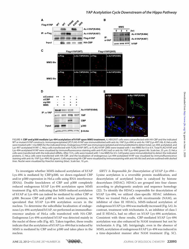
To investigate whether MMS-induced acetylation of hYAPLys-494 is mediated by CBP/p300, we down-regulated CBPand/or p300 expression in HeLa cells using RNA interference(RNAi). Double knockdown of CBP and p300 completelyreduced endogenous hYAP Lys-494 acetylation upon MMStreatment (Fig. 4D), indicating that MMS-induced acetylationof hYAP at Lys-494 can indeed be mediated by either CBP orp300. Because CBP and p300 are both nuclear proteins, wespeculated that hYAP Lys-494 acetylation occurs in thenucleus. To determine the subcellular localization of endoge-nous Lys-494-acetylated hYAP, we performed an immunofluo-rescence analysis of HeLa cells transfected with HA-CBP.Endogenous Lys-494-acetylated hYAP was detected mainly inthe nuclei of these cells (Fig. 4E). Taken together, these resultsindicate that the acetylation of hYAPLys-494 that is induced byMMS is mediated by CBP and/or p300 and takes place in thenucleus.
SIRT1 Is Responsible for Deacetylation of hYAP Lys-494—Lysine acetylation is a reversible protein modification, anddeacetylation of acetylated lysine is catalyzed by histonedeacetylases (HDAC). HDACs are grouped into four classesaccording to phylogenetic analysis and sequence homology(22). To identify the HDACs responsible for deacetylation ofhYAP Lys-494, we utilized class-specific HDAC inhibitors.When we treated HeLa cells with nicotinamide (NAM), aninhibitor of class III HDACs, MMS-induced acetylation ofendogenous hYAPLys-494wasmarkedly increased (Fig. 5A). Incontrast, treatment with trichostatin A, an inhibitor of class Iand II HDACs, had no effect on hYAP Lys-494 acetylation.Consistent with these results, CBP-mediated hYAP Lys-494acetylation was also enhanced by NAM treatment but not bytrichostatin A (Fig. 5B). Intriguingly, even in the absence ofMMS, acetylation of endogenous hYAPLys-494was induced ina time-dependent manner after NAM treatment (Fig. 5C).
FIGURE 4. CBP and p300 mediate Lys-494 acetylation of hYAP upon MMS treatment. A, HEK293T cells were cotransfected with HA-CBP and the indicatedWT or mutant hYAP constructs. Immunoprecipitated (IP) FLAG-hYAP was immunoblotted with anti-Ac-YAP (Lys-494) or anti-Ac-YAP (Lys-497) Ab. B, HeLa cellswere treated with 1 mM MMS for the indicated times. Endogenous hYAP was immunoprecipitated and immunoblotted to detect total, Lys-494-acetylated, andLys-497-acetylated hYAP. C, HeLa cells transfected with FLAG-hYAP (WT), or FLAG-hYAP (2KR) were treated with 1 mM MMS for 0 or 6 h. Total FLAG-hYAP andLys-494-acetylated hYAP were visualized by immunofluorescence staining with anti-FLAG (red) or anti-Ac-YAP (Lys-494) (green) Ab. Scale bar, 25 �m. D, HeLacells were transfected with the indicated siRNAs. After 72 h, cells were treated with 1 mM MMS for 6 h. Cell lysates were immunoblotted to detect the indicatedproteins. E, HeLa cells were transfected with HA-CBP, and the localization of endogenous Lys-494-acetylated hYAP was visualized by immunofluorescencestaining with anti-Ac-YAP (Lys-494) Ab (green). Cells expressing HA-CBP were visualized by immunostaining with anti-HA Ab (red) and are outlined with dashedlines. Nuclei were visualized by Hoechst staining (blue). Scale bar, 10 �m.
YAP Acetylation Cycle Downstream of the Hippo Pathway
JUNE 22, 2012 • VOLUME 287 • NUMBER 26 JOURNAL OF BIOLOGICAL CHEMISTRY 22093
by guest on June 1, 2020http://w
ww
.jbc.org/D
ownloaded from

These results suggest that the acetylation-deacetylation cycleinvolving hYAP Lys-494 operates regardless of extrinsic DNAdamage and that the deacetylation of this residue is catalyzed bya class III HDAC.The class III HDACSirtuin 1 (SIRT1) is reportedly transcrip-
tionally up-regulated by MMS treatment and involved inresponses to this agent (23). These observations prompted us totest whether SIRT1 might mediate hYAP Lys-494 deacetyla-tion. Exogenous expression of SIRT1 in HeLa cells significantlydecreased MMS-induced acetylation of hYAP Lys-494 (Fig.5D). Consistent with this finding, knockdown of SIRT1enhanced MMS-induced hYAP Lys-494 acetylation (Fig. 5E).Thus, SIRT1 is at least one of the class III HDACs responsiblefor deacetylating Lys-494-acetylated hYAP.SN2 Alkylating Agents Specifically Induce Both Acetylation
and Ser-127 Dephosphorylation of hYAP—YAP has previouslybeen shown to be involved in cellular responses to a variety ofDNA-damaging agents (10, 24, 25). Therefore, we investigatedthe effects of several DNA-damaging agents other than MMSon both hYAP Ser-127 phosphorylation and hYAP acetylation.Because cisplatin treatment induces nuclear translocation ofhYAP (10) andH2O2 treatment activates theHippo pathway (6,19), we treated HeLa cells with cisplatin or H2O2 and examinedhYAP Ser-127 phosphorylation and hYAP acetylation. Intrigu-ingly, hYAP Ser-127 phosphorylation was down-regulated in adose-dependent manner after cisplatin treatment but up-regu-lated in a dose-dependent manner upon H2O2 treatment (Fig.6A). However, in contrast to MMS, neither cisplatin nor H2O2
induced the acetylation of endogenous hYAP (Fig. 6B). Nor washYAP acetylation induced in HeLa cells by treatment withotherDNA-damaging agents, including camptothecin or doxo-rubicin (supplemental Fig. S2). These data indicate that hYAPregulation by the Hippo pathway is differentially altered by dif-ferent types of DNA damage and that hYAP acetylationmay bea regulatory event specific to DNA alkylating agents such asMMS.As mentioned above, alkylating agents are classified as being
of either the SN1 or SN2 type depending on their reactionmech-anism (16). Because MMS is an SN2 alkylating agent, we firstinvestigated whether another SN2 agent could induce hYAPacetylation by treatingHeLa cells withDMS.As expected, DMStreatment also resulted in a dose-dependent decrease in hYAPSer-127 phosphorylation (Fig. 6C) and a dose-dependentincrease in hYAP acetylation (Fig. 6D). We then treated HeLacells with either of two SN1 alkylating agents, MNNG orMNU.Phosphorylation of hYAP Ser-127 was reduced by MNNGtreatment but not altered upon MNU exposure (Fig. 6E).Intriguingly, neither MNNG nor MNU could induce hYAPacetylation (Fig. 6F). These results indicate that SN2 alkylatingagents specifically induce two relevant events, 1) the release ofhYAP from Hippo pathway-mediated cytoplasmic retentionand 2) the up-regulation of CBP/p300-mediated hYAP acetyla-tion in the nucleus.hYAP Acetylation Affects Cellular Responses to MMS
Treatment—YAP is known to be an important regulator of cellviability (2). To put our results in a biological context, we inves-
FIGURE 5. SIRT1 induces deacetylation of Lys-494-acetylated hYAP. A, HeLa cells were treated for 6 h with 1 mM MMS with or without 10 mM NAM or 1 �M
trichostatin A (TSA) as indicated. Endogenous hYAP was immunoprecipitated (IP), and Lys-494-acetylated hYAP was detected by immunoblotting. B, HEK293Tcells transfected with HA-CBP were treated for 6 h with 10 mM NAM and/or 1 �M trichostatin A as indicated. Endogenous hYAP was immunoprecipitated, andthe indicated proteins were detected by immunoblotting. C, HeLa cells were treated with 10 mM NAM or 1 mM MMS for the indicated times, and lysates wereanalyzed as for A. D, HeLa cells transfected with SIRT1 were treated (or not) with 1 mM MMS for 6 h as indicated. Endogenous hYAP was immunoprecipitated,and the indicated proteins were detected by immunoblotting. �-Tubulin was the loading control. E, HeLa cells were transfected with the indicated siRNAs. After72 h, cells were treated with 1 mM MMS for 6 h. Cell lysates were analyzed as for D.
YAP Acetylation Cycle Downstream of the Hippo Pathway
22094 JOURNAL OF BIOLOGICAL CHEMISTRY VOLUME 287 • NUMBER 26 • JUNE 22, 2012
by guest on June 1, 2020http://w
ww
.jbc.org/D
ownloaded from

tigatedwhether hYAP acetylation influenced cellular responsestoMMS treatment.We establishedHeLa cells stably expressinghYAP-WT or hYAP-2KR by retroviral vector infection andexposed them to increasing concentrations of MMS. Cell sur-vival was assessed using a tetrazolium-based colorimetric via-bility assay (MTS). Interestingly, expression of hYAP-2KR sig-nificantly decreased cell survival compared with hYAP-WTexpression (Fig. 7A), indicating that hYAP acetylation affectsthe sensitivity of HeLa cells to MMS treatment.Next, we examined whether hYAP acetylation is important
for its nuclear translocation after MMS exposure. As shown insupplemental Fig. S3, both the FLAG-hYAP-WT and FLAG-hYAP-2KR proteins accumulated in the nuclei ofMMS-treatedHeLa cells. Thus, the MMS-induced nuclear localization ofhYAP is not affected by hYAP acetylation.To investigate the physiological significance of hYAP acety-
lation in the nucleus, we co-transfected 293T cells with a syn-thetic YAP responsive luciferase reporter (8xGT-IIC-�51LucII)(26) and FLAG-hYAP-WT or FLAG-hYAP-2KR. These cellswere then exposed to MMS, and luciferase activity was meas-ured. As shown in Fig. 7B, cells expressing FLAG-hYAP-2KRexhibited significantly higher luciferase activity than cellsexpressing FLAG-hYAP-WT, indicating that the transcrip-tional coactivator activity of hYAP is regulated by its acetyla-
tion. Taken together, these data suggest that cell fate decisionsmade in response to MMS-induced DNA damage may be gov-erned by hYAP-dependent gene induction that is regulated byhYAP acetylation.
DISCUSSION
In this study we present evidence for a novel YAP acetylationcycle downstream of Hippo signaling. We have shown thatMMS treatment causes a reduction in hYAP Ser-127 phos-phorylation mediated by the Hippo pathway, promoting thenuclear translocation of this transcriptional co-activator.Through a screen for post-translationalmodifications of hYAP,we have demonstrated that hYAP is acetylated at the endoge-nous level upon MMS treatment. This MMS-induced acetyla-tion of YAP is mediated by the nuclear acetyltransferases CBPand p300, occurs on conserved C-terminal lysine residues, andcan be reversed by SIRT1. Importantly, this acetylation-deacetylation cycle of hYAP is triggered specifically in responseto SN2 alkylating agents and not by other types of DNA-dam-aging stimuli. A schematic model summarizing our findingsappears in Fig. 8.
FIGURE 6. DNA-damaging agents have diverse effects on hYAP Ser-127phosphorylation and hYAP acetylation. A, HeLa cells were treated with theindicated concentrations of cisplatin or H2O2 for 6 or 3 h, respectively. Totaland Ser-127-phosphorylated hYAP were detected by immunoblotting.B, HeLa cells were treated with the indicated concentrations of cisplatin for12 h or 1 mM MMS for 4 h (top) or 1 mM H2O2 for the indicated times or 1 mM
MMS for 4 h (bottom). Endogenous hYAP was immunoprecipitated (IP), andacetylated hYAP was detected by immunoblotting with anti-pan-AcK Ab.C, HeLa cells were treated with the indicated concentrations of MMS or DMSfor 3 h. Cell lysates were analyzed as for A. D, HeLa cells were treated with theindicated concentrations of DMS or MMS for 4 h. Cell lysates were analyzed asfor B. E, HeLa cells were treated with the indicated concentrations of MNNG orMNU for 3 h. Cell lysates were analyzed as for A. F, HeLa cells were treated for4 h with the indicated concentrations of MNNG or MMS (top) or MNU or MMS(bottom). Cell lysates were analyzed as for B.
FIGURE 7. Expression of the acetylation-resistant hYAP-2KR proteinalters cellular responses to MMS treatment. A, HeLa cells stably expressinghYAP-WT or hYAP-2KR were exposed to the indicated concentrations of MMSfor 1 h, and survival was assessed 48 h later by MTS assay. Values shown arethe mean � S.D. (n � 3/group). Inset, shown is an immunoblot to confirmhYAP expression in the above cells before MMS treatment. B, 293T cells wereco-transfected with 8xGT-IIC-�51LucII and increasing amounts of the indi-cated plasmids and treated with 1 mM MMS for 3 h. Luciferase activity wasmeasured and normalized to cotransfected renilla luciferase activity. Valuesshown are the mean � S.D. (n � 3/group). *, p � 0.01. The expression levels oftransfected FLAG-hYAP were detected by immunoblotting.
YAP Acetylation Cycle Downstream of the Hippo Pathway
JUNE 22, 2012 • VOLUME 287 • NUMBER 26 JOURNAL OF BIOLOGICAL CHEMISTRY 22095
by guest on June 1, 2020http://w
ww
.jbc.org/D
ownloaded from

Our results have revealed that Lys-494 and Lys-497 of hYAPare targets for CBP-mediated acetylation. In previous reports,CBP and p300 were shown to directly acetylate a variety ofproteins at a consensus motif (KX1–2(X/K)K) (27–30). Notably,hYAP contains this consensus motif (494KLDK497), and thehYAP acetylation sites identified in our experiments are con-sistent with the positions of two of the lysine residues present inthis motif. Interestingly, although the acetylation consensusmotif is conserved among vertebrate YAPs (human,mouse, andmedaka), it is not found in either Yki (yorkie), the Drosophilahomologue of YAP, or in TAZ (transcriptional coactivator withPDZ-binding motif), a vertebrate paralogue of YAP (4). Thus,the CBP/p300-dependent acetylation we observe appears to bea modification specific to vertebrate YAP.A number of studies have reported that CBP/p300-mediated
acetylation of several proteins is important for cellularresponses toDNAdamage. For example, theWerner syndromehelicase (WRN)helps to promote the survival of cells with dam-aged DNA. CBP-mediated acetylation of WRN triggered byDNA damage inhibits the ubiquitination-dependent degrada-tion of this helicase and thus increases its stability (31). Simi-larly, DNA damage induces p300 to acetylate and stabilizeNijmegen breakage syndrome 1, which plays a crucial role inhomologous recombination repair (32). The acetylation of pro-teins involved in DNA damage responses is often countered bySIRT1-mediated deacetylation (33), and acetylated WRN andNijmegen breakage syndrome 1 are both substrates of SIRT1(34, 35). The importance of such acetylation-deacetylationcycles has been highlighted by the significant increase in chro-mosomal aberrations identified in SIRT1-deficient cells (36).Our study has revealed that hYAP also undergoes reversible
acetylation and deacetylation mediated by CBP/p300 andSIRT1, respectively.More importantly, our results indicate thatacetylation of hYAP influences both its transcriptional co-acti-vator activity and the sensitivity of hYAP-expressing cells toMMS-induced DNA damage.Different genotoxic agents induce different types of DNA
damage, and these lesions trigger lesion-specific DNA repairmechanisms. YAP has previously been shown to participate inresponses to DNA-damaging agents such as cisplatin, adriamy-cin, ultraviolet radiation, and ionizing radiation (10, 24, 25), butthe lesion-specific aspects of YAP regulation were not definedin these studies. In this report we have demonstrated that hYAPSer-127 phosphorylation mediated by the Hippo pathway isdifferentially altered depending on the type of DNA-damagingagent encountered. Our data show that DNA-damaging stimulican be classified into at least three groups based on their effectson the subcellular localization and post-translational modifica-tion of hYAP;Group I,Hippo pathway-mediated hYAPSer-127phosphorylation is up-regulated and hYAP remains in the cyto-plasm; Group II, hYAP Ser-127 phosphorylation is down-regu-lated, and hYAP undergoes nuclear translocation but is notacetylated; Group III, hYAP Ser-127 phosphorylation is down-regulated, and hYAP undergoes both nuclear translocation andacetylation. In line with previous reports (6, 19), we have dem-onstrated that H2O2 treatment of HeLa cells results in up-reg-ulated hYAP Ser-127 phosphorylation (Group I), whereas cis-platin treatment down-regulated Ser-127 phosphorylation buthad no effect on acetylation (Group II). Treatment ofHeLa cellswithMNNGhad a similar effect, placing this agent in Group II.The most interesting aspect of our work is the identification ofthe new Group III, represented solely by SN2 alkylating agents,which both down-regulate Ser-127 hYAP phosphorylation andinduce hYAP acetylation.Our findings indicate that mere nuclear accumulation of
hYAP is not enough to induce its acetylation, and another fac-tor(s) must play an important role in this reaction. Along theselines, we demonstrated that CBP and p300 are keymediators ofMMS-induced hYAP acetylation, implying that up-regulationof CBP/p300 activity may be the additional factor contributingto hYAP acetylation. Collectively, our data imply that the spe-cific cellular response to DNA lesions caused by SN2 alkylatingagents involves the coupling of hYAP nuclear translocationinduced by Ser-127 dephosphorylation with the activation ofCBP/p300-mediated acetylation.SN2 alkylating agents, but not SN1 agents, generate 1-methyl-
adenine and 3-methylcytosine residues in single-strandedDNAand RNA. These cytotoxic lesions are known to be repaired byAlkB family dioxygenases (16). In mammalian cells, AlkB hom-olog 2 (ABH2) removes 1-methyladenine lesions in genomicDNAand thus is critical for cellular defense againstMMS treat-ment (17). In addition, ABH3 collaborates withASCC3helicaseto remove 3-methylcytosine lesions in certain tumor-derivedcell lines (37). However, the mechanisms regulating ABH2 andABH3 in response to SN2 alkylating agents remain largelyunknown. Intriguingly, 1-methyladenine accumulates in agingABH2-deficient mice (17) and 3-methylcytosine accumulatesin ABH3 or ASCC3 knockdown cells (37), clearly showing thatsuch cytotoxic methylation events occur endogenously. Rele-
FIGURE 8. A proposed model for hYAP regulation in response to SN2 alkyl-ating agents. Cell-cell contact triggers Hippo pathway signaling that up-reg-ulates Ser-127 phosphorylation of hYAP and thus its cytoplasmic retention viabinding to 14-3-3 protein. SN2 alkylating agents damage DNA, leading to areduction in Ser-127 phosphorylation of hYAP. Non-phosphorylated hYAP isreleased from the cytoplasmic 14-3-3 complex and translocates into thenucleus. Within the nucleus, CBP and/or p300 activated by SN2 alkylatingagent-mediated DNA damage induce hYAP acetylation at Lys-494 and Lys-497. Concurrently, deacetylation of these residues is mediated by SIRT1. Thus,tight regulation governs the state of hYAP acetylation induced in response toSN2 alkylating agents.
YAP Acetylation Cycle Downstream of the Hippo Pathway
22096 JOURNAL OF BIOLOGICAL CHEMISTRY VOLUME 287 • NUMBER 26 • JUNE 22, 2012
by guest on June 1, 2020http://w
ww
.jbc.org/D
ownloaded from

vant in this context is the fact that S-adenosylmethionine,which is the methyl group donor in the majority of enzymaticmethylation events, can act as an endogenous SN2 alkylatingagent and generates the same methylated adducts on DNA asMMS (38). We found that even in the absence of MMS expo-sure, acetylation of endogenous hYAPwas observed after treat-ment with the class III HDAC inhibitor NAM. Thus, the hYAPacetylation cycle downstream of the Hippo pathway may occurin response to endogenous single-strand DNA/RNA alkylationevents induced by physiological SN2 alkylating agents. It is,therefore, tempting to speculate thatHippo-YAP signalingmaybe involved in the intrinsic AlkB-mediated DNA repairpathway.
Acknowledgment—We are grateful to Dr. Hiroshi Sasaki (KumamotoUniversity) for providing the 8xGT-IIC-�51LucII plasmid.
REFERENCES1. Yagi, R., Chen, L. F., Shigesada, K.,Murakami, Y., and Ito, Y. (1999) AWW
domain-containing yes-associated protein (YAP) is a novel transcriptionalco-activator. EMBO J. 18, 2551–2562
2. Halder, G., and Johnson, R. L. (2011) Hippo signaling. Growth control andbeyond. Development 138, 9–22
3. Genevet, A., and Tapon, N. (2011) The Hippo pathway and apico-basalcell polarity. Biochem. J. 436, 213–224
4. Zhao, B., Tumaneng, K., and Guan, K. L. (2011) The Hippo pathway inorgan size control, tissue regeneration, and stem cell self-renewal. Nat.Cell Biol. 13, 877–883
5. Dong, J., Feldmann, G., Huang, J., Wu, S., Zhang, N., Comerford, S. A.,Gayyed, M. F., Anders, R. A., Maitra, A., and Pan, D. (2007) Elucidation ofa universal size-control mechanism in Drosophila and mammals. Cell130, 1120–1133
6. Zhou, D., Conrad, C., Xia, F., Park, J. S., Payer, B., Yin, Y., Lauwers, G. Y.,Thasler, W., Lee, J. T., Avruch, J., and Bardeesy, N. (2009) Mst1 andMst2maintain hepatocyte quiescence and suppress hepatocellular carcinomadevelopment through inactivation of the Yap1 oncogene. Cancer Cell 16,425–438
7. Overholtzer, M., Zhang, J., Smolen, G. A., Muir, B., Li, W., Sgroi, D. C.,Deng, C. X., Brugge, J. S., andHaber, D. A. (2006) Transforming propertiesof YAP, a candidate oncogene on the chromosome 11q22 amplicon. Proc.Natl. Acad. Sci. U.S.A. 103, 12405–12410
8. Zhao, B., Ye, X., Yu, J., Li, L., Li, W., Li, S., Yu, J., Lin, J. D., Wang, C. Y.,Chinnaiyan, A. M., Lai, Z. C., and Guan, K. L. (2008) TEAD mediatesYAP-dependent gene induction and growth control. Genes Dev. 22,1962–1971
9. Lian, I., Kim, J., Okazawa, H., Zhao, J., Zhao, B., Yu, J., Chinnaiyan, A.,Israel, M. A., Goldstein, L. S., Abujarour, R., Ding, S., and Guan, K. L.(2010) The role of YAP transcription coactivator in regulating stem cellself-renewal and differentiation. Genes Dev. 24, 1106–1118
10. Strano, S., Monti, O., Pediconi, N., Baccarini, A., Fontemaggi, G., Lapi, E.,Mantovani, F., Damalas, A., Citro, G., Sacchi, A., Del Sal, G., Levrero, M.,and Blandino, G. (2005) The transcriptional coactivator Yes-associatedprotein drives p73 gene-target specificity in response to DNA Damage.Mol. Cell 18, 447–459
11. Zhao, B., Wei, X., Li, W., Udan, R. S., Yang, Q., Kim, J., Xie, J., Ikenoue, T.,Yu, J., Li, L., Zheng, P., Ye, K., Chinnaiyan, A., Halder, G., Lai, Z. C., andGuan, K. L. (2007) Inactivation of YAP oncoprotein by theHippo pathwayis involved in cell contact inhibition and tissue growth control.Genes Dev.21, 2747–2761
12. Zhao, B., Li, L., Tumaneng, K., Wang, C. Y., and Guan, K. L. (2010) Acoordinated phosphorylation by Lats and CK1 regulates YAP stabilitythrough SCF(�-TRCP). Genes Dev. 24, 72–85
13. Lapi, E., Di Agostino, S., Donzelli, S., Gal, H., Domany, E., Rechavi, G.,Pandolfi, P. P., Givol, D., Strano, S., Lu, X., and Blandino, G. (2008) PML,
YAP, and p73 are components of a proapoptotic autoregulatory feedbackloop.Mol. Cell 32, 803–814
14. Ura, S., Nishina, H., Gotoh, Y., and Katada, T. (2007) Activation of thec-Jun N-terminal kinase pathway by MST1 is essential and sufficient forthe induction of chromatin condensation during apoptosis.Mol. Cell. Biol.27, 5514–5522
15. Takahashi, S., Ebihara, A., Kajiho, H., Kontani, K., Nishina, H., andKatada,T. (2011) RASSF7 negatively regulates pro-apoptotic JNK signaling byinhibiting the activity of phosphorylated-MKK7. Cell Death Differ. 18,645–655
16. Drabløs, F., Feyzi, E., Aas, P. A., Vaagbø, C. B., Kavli, B., Bratlie, M. S.,Peña-Diaz, J., Otterlei, M., Slupphaug, G., and Krokan, H. E. (2004) Alkyl-ation damage in DNA and RNA. Repair mechanisms and medical signifi-cance. DNA Repair 3, 1389–1407
17. Ringvoll, J., Nordstrand, L. M., Vågbø, C. B., Talstad, V., Reite, K., Aas,P. A., Lauritzen, K. H., Liabakk, N. B., Bjørk, A., Doughty, R. W., Falnes, P.Ø., Krokan, H. E., and Klungland, A. (2006) Repair deficient mice revealmABH2 as the primary oxidative demethylase for repairing 1meA and3meC lesions in DNA. EMBO J. 25, 2189–2198
18. Hirayama, J., Sahar, S., Grimaldi, B., Tamaru, T., Takamatsu, K., Nakahata,Y., and Sassone-Corsi, P. (2007) CLOCK-mediated acetylation of BMAL1controls circadian function. Nature 450, 1086–1090
19. Bao, Y., Nakagawa, K., Yang, Z., Ikeda,M.,Withanage, K., Ishigami-Yuasa,M., Okuno, Y., Hata, S., Nishina, H., andHata, Y. (2011) A cell-based assayto screen stimulators of the Hippo pathway reveals the inhibitory effect ofdobutamine on the YAP-dependent gene transcription. J Biochem. 150,199–208
20. Ogryzko, V. V., Schiltz, R. L., Russanova, V., Howard, B. H., and Nakatani,Y. (1996) The transcriptional coactivators p300 and CBP are histoneacetyltransferases. Cell 87, 953–959
21. Suzuki, H., Fukunishi, Y., Kagawa, I., Saito, R., Oda,H., Endo, T., Kondo, S.,Bono, H., Okazaki, Y., and Hayashizaki, Y. (2001) Protein-protein interac-tion panel using mouse full-length cDNAs. Genome Res 11, 1758–1765
22. Yang, X. J., and Seto, E. (2008) The Rpd3/Hda1 family of lysine deacety-lases. From bacteria and yeast tomice andmen.Nat. Rev.Mol. Cell Biol. 9,206–218
23. Yamamori, T., DeRicco, J., Naqvi, A., Hoffman, T. A., Mattagajasingh, I.,Kasuno, K., Jung, S. B., Kim, C. S., and Irani, K. (2010) SIRT1 deacetylatesAPE1 and regulates cellular base excision repair. Nucleic Acids Res. 38,832–845
24. Levy, D., Adamovich, Y., Reuven, N., and Shaul, Y. (2008) Yap1 phos-phorylation by c-Abl is a critical step in selective activation of proapo-ptotic genes in response to DNA damage.Mol. Cell 29, 350–361
25. Tomlinson, V., Gudmundsdottir, K., Luong, P., Leung, K. Y., Knebel, A.,and Basu, S. (2010) JNK phosphorylates Yes-associated protein (YAP) toregulate apoptosis. Cell Death and Disease 1, e29
26. Ota, M., and Sasaki, H. (2008) Mammalian Tead proteins regulate cellproliferation and contact inhibition as transcriptional mediators of Hipposignaling. Development 135, 4059–4069
27. Gu, W., and Roeder, R. G. (1997) Activation of p53 sequence-specificDNA binding by acetylation of the p53 C-terminal domain. Cell 90,595–606
28. Boyes, J., Byfield, P., Nakatani, Y., and Ogryzko, V. (1998) Regulation ofactivity of the transcription factor GATA-1 by acetylation. Nature 396,594–598
29. Thompson, P. R., Kurooka, H., Nakatani, Y., and Cole, P. A. (2001) Tran-scriptional coactivator protein p300. Kinetic characterization of its his-tone acetyltransferase activity. J. Biol. Chem. 276, 33721–33729
30. Wang, C., Fu, M., Angeletti, R. H., Siconolfi-Baez, L., Reutens, A. T., Al-banese, C., Lisanti, M. P., Katzenellenbogen, B. S., Kato, S., Hopp, T.,Fuqua, S. A., Lopez, G. N., Kushner, P. J., and Pestell, R. G. (2001) Directacetylation of the estrogen receptor alpha hinge region by p300 regulatestransactivation and hormone sensitivity. J. Biol. Chem. 276, 18375–18383
31. Li, K., Wang, R., Lozada, E., Fan, W., Orren, D. K., and Luo, J. (2010)Acetylation of WRN protein regulates its stability by inhibiting ubiquiti-nation. PLoS One 5, e10341
32. Jang, E. R., Choi, J. D., and Lee, J. S. (2011)Acetyltransferase p300 regulatesNBS1-mediated DNA damage response. FEBS Lett. 585, 47–52
YAP Acetylation Cycle Downstream of the Hippo Pathway
JUNE 22, 2012 • VOLUME 287 • NUMBER 26 JOURNAL OF BIOLOGICAL CHEMISTRY 22097
by guest on June 1, 2020http://w
ww
.jbc.org/D
ownloaded from

33. Vassilopoulos, A., Deng, C. X., and Chavakis, T. (2010) Crosstalk betweenthe DNA damage response, histone modifications, and neovasculariza-tion. Int. J. Biochem. Cell Biol. 42, 193–197
34. Yuan, Z., Zhang, X., Sengupta, N., Lane, W. S., and Seto, E. (2007) SIRT1regulates the function of the Nijmegen breakage syndrome protein. Mol.Cell 27, 149–162
35. Li, K., Casta, A., Wang, R., Lozada, E., Fan, W., Kane, S., Ge, Q., Gu, W.,Orren, D., and Luo, J. (2008) Regulation ofWRN protein cellular localiza-tion and enzymatic activities by SIRT1-mediated deacetylation. J. Biol.Chem. 283, 7590–7598
36. Oberdoerffer, P., Michan, S., McVay, M., Mostoslavsky, R., Vann, J., Park,
S. K., Hartlerode, A., Stegmuller, J., Hafner, A., Loerch, P., Wright, S. M.,Mills, K. D., Bonni, A., Yankner, B. A., Scully, R., Prolla, T. A., Alt, F. W.,and Sinclair, D. A. (2008) SIRT1 redistribution on chromatin promotesgenomic stability but alters gene expression during aging. Cell 135,907–918
37. Dango, S., Mosammaparast, N., Sowa, M. E., Xiong, L. J., Wu, F., Park, K.,Rubin, M., Gygi, S., Harper, J. W., and Shi, Y. (2011) DNA unwinding byASCC3 helicase is coupled to ALKBH3-dependent DNA alkylation repairand cancer cell proliferation.Mol. Cell 44, 373–384
38. Sedgwick, B., Bates, P. A., Paik, J., Jacobs, S. C., and Lindahl, T. (2007)Repair of alkylated DNA. Recent advances. DNA Repair 6, 429–442
YAP Acetylation Cycle Downstream of the Hippo Pathway
22098 JOURNAL OF BIOLOGICAL CHEMISTRY VOLUME 287 • NUMBER 26 • JUNE 22, 2012
by guest on June 1, 2020http://w
ww
.jbc.org/D
ownloaded from

Katada, Makoto Furutani-Seiki and Hiroshi NishinaShoji Hata, Jun Hirayama, Hiroaki Kajiho, Kentaro Nakagawa, Yutaka Hata, Toshiaki
Alkylating Agents2NSThat Is Downstream of Hippo Pathway Is Triggered in Response to
A Novel Acetylation Cycle of Transcription Co-activator Yes-associated Protein
doi: 10.1074/jbc.M111.334714 originally published online April 27, 20122012, 287:22089-22098.J. Biol. Chem.
10.1074/jbc.M111.334714Access the most updated version of this article at doi:
Alerts:
When a correction for this article is posted•
When this article is cited•
to choose from all of JBC's e-mail alertsClick here
Supplemental material:
http://www.jbc.org/content/suppl/2012/04/27/M111.334714.DC1
http://www.jbc.org/content/287/26/22089.full.html#ref-list-1
This article cites 38 references, 14 of which can be accessed free at
by guest on June 1, 2020http://w
ww
.jbc.org/D
ownloaded from