Annual Report 2008 - mfpl.ac.at · Annual Report 2008 Contact | Max F. Perutz Laboratories Dr....
56
Annual Report 2008
Transcript of Annual Report 2008 - mfpl.ac.at · Annual Report 2008 Contact | Max F. Perutz Laboratories Dr....

enRü
c
Rüc
n
Annual Report 2008
Contact | Max F. Perutz Laboratories
Dr. Bohr-Gasse 9 | 1030 Vienna | Austria
Phone | +43-1-4277-24001
[email protected] | www.mfpl.ac.at
MFPL are a joint venture of:
U1U4+Ruecken1ok 27.05.2009 13:18 Uhr Seite 1

enRü
c
Alphabetical Group Leader Index
Editor | Lisa Cichocki
Design | Grafikatelier Heuberger | Vienna
Photography | Lisa Cichocki | Arnd Oetting – Porträt Seite 41 | Group Leader Archive
Printing | Kärntner Druckerei | Klagenfurt
Dr. Lisa Cichocki | Communications
Max F. Perutz Laboratories | Dr. Bohr -Gasse 9 | A -1030 Wien
T: +43 -1-4277-24014 | F: +43-1-4277-9240
E: [email protected] | W: http://mfpl.ac.at Lisa Cichocki
Imprint
Gustav Ammerer
Manuela Baccarini
Andreas Bachmair
Andrea Barta
Dieter Blaas
Udo Bläsi
Cécile Brocard
Emmanuelle Charpentier
Thomas Decker
Kristina Djinovic Carugo
Gang Dong
Silke Dorner
Roland Foisner
Juraj Gregan
Andreas Hartig
Erwin Heberle-Bors
Marcela Hermann
Joachim Hermisson
Heribert Hirt
Reinhold Hofbauer
N.-Erwin Ivessa
Michael Jantsch
Verena Jantsch-Plunger
Franz Klein
Gottfried Köhler
Franz Koller
Robert Konrat
Pavel Kovarik
Friedrich Kragler
Karl Kuchler
Wolfgang Löffelhardt
Josef Loidl
Zdravko J. Lorkovic
Irute Meskiene
Isabella Moll
Ernst Müllner
Johannes Nimpf
Egon Ogris
Andrea Pichler
Fritz Pittner
Brigitte Poppenberger
Rainer Prohaska
Friedrich Propst
Florian Raible
Johann Rotheneder
Peter Schlögelhofer
Wolfgang Schneider
Renée Schroeder
Rudolf Schweyen
Christoph Schüller
Joachim Seipelt
Christian Seiser
Tobias Sieberer
Tim Skern
Markus Teige
Kristin Tessmar
Alisher Touraev
Alexander von Gabain
Arndt von Haeseler
Christina Waldsich
Graham Warren
Edgar Wawra
Georg Weitzer
Gerhard Wiche
Angela Witte
Franz Wohlrab
28
29
29
30
30
31
31
32
32
33
33
34
34
35
35
36
37
37
38
38
39
39
40
40
41
41
42
42
43
43
44
44
45
53
12
12
13
13
14
14
15
15
16
16
17
17
18
18
19
19
20
20
21
21
22
22
23
23
24
24
25
25
26
26
27
27
28
U2U3+Ruecken 27.05.2009 13:17 Uhr Seite 1

Content
Foreword – Message from the Rectors
Report of the Directorate
History
Facts
Students at the Max Perutz Labs
PhDs and PostDocs
FWF-funded International Excellence PhD Programmes
FWF-funded Special Research Programmes (SFB)
Research Groups
Service & Support
Facilities at the Max Perutz Labs
Research Communication
Social Life
Research Funding
Where to find the Max Perutz Labs
Imprint
2
3
4
5
6
7
8
10
12
45
46
50
51
52
52
53
1
01-07_Einstieg 27.05.2009 12:59 Uhr Seite 1

We see with great pleasure that the investmentin our joint venture – the Max F. Perutz Labora-tories – is bearing fruit and that the visibility ofthe Max Perutz Labs is steadily increasing bothat the national and international level. The esta-blishment of three new Junior Groups, the awardof two START prizes, a Christian Doppler Laband two new doctoral programmes funded bythe FWF, testify to the progress of our joint in-itiative.
The first call for Junior Group Leaders attractedmore than 100 applications – 80 Prozent ofwhom came from outside of Austria. A compe-titive selection process resulted in the recruit-ment of three young scientists who recently start-ed their own independent research career at theMax Perutz Labs: Gang Dong, Kristin Tessmar-Raible und Florian Raible are outstanding youngscientists of the kind which both universities wantto attract: academically excellent, enthusiastic,interactive and innovative.
In 2008 Kristin Tessmar-Raible und ChristinaWaldsich won START prizes – the most presti-gious grant awarded by the FWF to outstandingyoung scientists. We especially appreciate the
fact that the prizes were won by two female re-searchers – one who had worked at the MaxPerutz Labs for some time, the other just recrui-ted. We regard this as a healthy mix of “home-grown” talent and “fresh blood”.
Both universities emphasize the importance ofgraduate programmes. The Max Perutz Labs ha-ve been part of the International PhD program-me of the Campus Vienna Biocenter since itsestablishment in 1994. One existing doctoralprogramme organized by Andrea Barta and theacquisition of two new doctoral programmes atthe Max Perutz Labs by Manuela Baccarini(University of Vienna) and Tim Skern (MedicalUniversity of Vienna) provide another strong in-centive towards the establishment of a MFPLGraduate School and strengthens both researchand training activities at the Max F. Perutz La-boratories.
We would like to reassert our continuing sup-port to this increasingly successful enterpriseand wish the Max F. Perutz Laboratories, sinceNovember 2006 under the leadership of Prof.Graham Warren, all the best in their constantstriving for excellence.
Wolfgang SchützRector Medical University of Vienna
Georg WincklerRector University of Vienna
WolfgangSchütz
GeorgWinckler
2
Message from the Rectors
Foreword
01-07_Einstieg 27.05.2009 12:59 Uhr Seite 2

GrahamWarren
Scientific Director
HaraldHochreiterAdministrative
Director
ManuelaBaccarini
Vice-Dean for University
of Vienna
RolandFoisner
Vice-Dean forMedical University
of Vienna
2008 has been a year of changing structuresand changing faces. We welcomed the first JuniorGroup Leaders to MFPL: Gang Dong, a crystal-lographer from Yale University; Kristin Tessmar-Raible, a molecular biologist and marine biologistinterested in ancient light sensors; and Florian Rai-ble, a computational biologist investigating theevolution of gene regulatory networks. Both Kri-stin and Florian come from the EMBL in Heidel-berg. All three Junior Group Leaders quickly inte-grated into existing research programmes andhave become valuable members of our faculty. Asecond call for Junior Group Leaders was adver-tised in October 2008 and attracted 200 appli-cations, the majority from US-based scientists.
We have also continued selecting all our PhDsthrough a structured evaluation process. Studentsare pre-screened by the group leaders and theninterviewed by a panel of junior and senior fa-culty. This means that most (17 out of 20) passthe interview. Of these, approximately one thirdare international students. We appreciate the ti-me and effort put into these interviews by the fa-culty – we regard this as an important elementof quality control.
2008 was a very successful year for recruitmentof third party funds. Our group leaders were ab-le to raise our annual grant income from 9,8 mil-lion Euro to 11,2 million Euro. A Christian Dopp-ler laboratory “Patho FUN” supported by the CDResearch Association and by Intercell AG, was in-augurated on September 30th and is directed byKarl Kuchler. Not included in these figures are thenew FWF-funded excellence doctoral program-mes „Molecular Mechanisms of Cell Signalling”(coordinated by Manuela Baccarini) and „Struc-ture and Interaction of Biological Macromolecu-les“ (coordinated by Tim Skern). START prizeswere awarded to Kristin Tessmar-Raible and Chri-stina Waldsich – a testament to the quality of theyoung scientists working at the Max Perutz Labs.
We have continued to forge closer ties to clinicalresearch groups. Two joint meetings with re-searchers at the Vienna General Hospital (Allge-meines Krankenhaus) were organized, with a to-tal of 31 speakers and topics ranging from immu-nology to cancer and from diseases to vaccines.We will intensify these activities in 2009, and
further strengthen the ties with both the MedicalUniversity and the University of Vienna.
In 2008 we also aimed at closer collaborationswithin the Vienna Biocenter Campus by submittinga proposal (Vision 2020) for state-of-the art corefacilities for all institutions on campus. This propo-sal was positively evaluated by an internationalreview panel in July 2008 and financing wasconfirmed by Science Minister Johannes Hahnand Vice-Mayor Renate Brauner in October2008. The award amounts to 55 million Euroover the next 10 years and the first tangible out-come is the much needed campus child care fa-cility.
The MFPL faculty met for a recess in Septemberto discuss joint strategies and organizational struc-tures. As a result we have implemented a MFPLAdvisory Committee with elected representativesfrom all levels of the MFPL community. The elec-ted faculty members will take over as new depart-ment heads in 2009. A technical manager (Wolf-gang Binder) was hired to coordinate MFPL-wide technical matters.
Our Scientific Advisory Board has undergone so-me changes. Kai Simons, Kim Nasmyth and Na-dia Rosenthal have left. We would like to thankthem for their dispassionate advice during a pe-riod of great change at the Max Perutz Labs.We welcome the new SAB members: Jean Beggs(Edinburgh), Jorge Galan (Yale) and Cyrus Chot-hia (Cambridge), and look forward to their par-ticipation in shaping the future of the Max PerutzLabs.
Social activities ranging from happy hours to skitrips and sporting activities are an important com-plement to our scientific work. Two particular high-lights have been a celebration of the Max PerutzLabs (and commemoration of Max Perutz) thatwas attended by dignitaries from the Science Mi-nistry and the City; and the first Xmaspantomime, using the fairytale of Cinderella to tellthe story of the Max Perutz Labs and the Campus.
In closing we would like to thank all those whocontinue to contribute to the success of the MaxF. Perutz Laboratories as we look forward to anot-her exciting year.
Graham WarrenScientific Director
Harald HochreiterAdministrative Director
3
Report of the Directorate
01-07_Einstieg 27.05.2009 12:59 Uhr Seite 3

History
1992/1993 University departments move to the VBC (Molecular Biology, Biochemistry,
Medical Biochemistry, and Genetics)
Three new chairs established (Molecular Genetics, Molecular Cell Biology,
and Microbiology)
1994 Start of the international VBC PhD programme
1996 Max Perutz Library established
1998 Spin-Off company Intercell founded
1999 New chair for Immunobiology established
2001 Dept. for Structural Biology moves to the VBC
New Chair for Structural Biology / NMR established
New Chair for X-Ray Crystallography established
2004 Medical University of Vienna established
2005 Dept. for Chromosome Biology moves to the VBC
Max F. Perutz Laboratories GmbH established
Administrative Director appointed
Scientific Advisory Board established
2007 Scientific Director appointed
2008 Junior Group Leaders appointed
Max F. Perutz – ”In science, truth always wins“
To honour an extraordinary teacher and scientist,the Max Perutz Labs were was named after MaxFerdinand Perutz, the 1962 Nobel laureate inChemistry (together with John C. Kendrew) for stu-dies of the structures of globular proteins. Born in 1914 in Vienna, he came from a family oftextile manufacturers who had made their fortunein the 19th century by the introduction of medicalspinning and weaving. He was sent to school atthe Theresianum where a good schoolmasterawakened his interest in chemistry. In 1932 he en-tered the University of Vienna, but owing to thepoor prospects for a scientific career he decidedin 1936 to become a research student at the Ca-vendish Laboratory in Cambridge. After Hitler´sinvasion of Austria, the family business was expro-
priated, his pa-rents became re-fugees and hisnatural choicewas to continuehis career in Cam-bridge.In addition to his studies on the structure of hae-moglobin, Max F. Perutz was highly instrumentalin founding the new research field of MolecularBiology as well as the Laboratory of MolecularBiology (LMB) in Cambridge, UK. He was alsoinvolved in establishing the European MolecularBiology Organization (EMBO) in Heidelberg,Germany.Max F. Perutz died in February 2002.
4
History
01-07_Einstieg 27.05.2009 12:59 Uhr Seite 4

The Max F. Perutz LaboratoriesThe Max F. Perutz Laboratories (MFPL) are a jointventure of the University of Vienna and theMedical University of Vienna and are located atthe Campus Vienna Biocenter. Established in thespring of 2005 the MFPL comprises the expertiseof more than 60 research groups in MolecularCell Biology. They represent a new and innovativeapproach to strengthen research and training atboth Universities. This visionary inter-university cooperation provides asuperb environment for excellent research and edu-cation and is a platform for ventures across traditio-nal boundaries. New research groups are current-ly being established, existing synergies improvedand new collaborations actively promoted.
FundingThe Max F. Perutz Laboratories are jointly funded bythe University of Vienna and the Medical Universi-ty of Vienna. The two universities fund personnel, buil-dings and scientific infrastructure. Most of the personnel is funded by grants. A raiseof third party funding of 13,5% in the year 2008compared to 2007 show that the research groupsat the Max Perutz Labs always had a strong trackrecord in acquiring external funding: the total volu-me of third party funding was EUR 11,21 Mio.The main external sources of funding in 2008 we-re the FWF (EUR 5,3 Mio), the EU (EUR 1,7 Mio.),company projects including the Christian DopplerResearch Association (EUR 1,4 Mio) and theWWTF (EUR 1, 3 Mio).
Scientific Director: Graham Warren, FRSAdministrative Director: Harald HochreiterScientific Advisory Board:Jean Beggs, University of Edinburgh
David Livingston, Dana-Farber Cancer Institute, Harvard Medical School
Kim Nasmyth, University of Oxford Nadia Rosenthal, EMBL Monterotondo Jorge Galan, Yale University
Overview – MFPL in 2008: • More than 470 people• From more than 25 nations• 66 research groups• In 6 research programmes• 70% of personnel funded by grants• 700 undergraduate students• More than 130 PhD students• More than EUR 11,2 Mio grant money
MFPL – strength in diversityEmbedded in the Campus Vienna Biocenter, a uni-que concentration of high-level research institutes,MFPL provides a perfect environment for outstandingresearch. The Max Perutz Labs cover researchgroups with a broad thematic profile – the majori-ty work on basic research topics but a significantnumber are also active in more applied fields of bio-logy. To maintain research at internationally compe-titive levels the MFPL is organized into six thematicResearch Programmes. These are:
• Infection Biology• RNA Biology• Cell Signaling• Computational and Structural Biology• Chromosome Biology• Membranes and the Cytoskeleton
FWF 5.271.886
EU 1.700.116
Companies incl. CDG 1.361.993
WWTF 1.252.458
Ministries 830.294
Miscellanneous 210.360
FFG 179.553
Trusts 167.963
ÖAW 131.750
DFG 96.176
Hochschuljubiläumsstiftung/MA8 10.745
Total 11.213.293
5
Facts
EU15%
Companies12%
WWTF 11%
Ministries7% Other
7%
FWF 48%
Third party funds 2008
01-07_Einstieg 27.05.2009 12:59 Uhr Seite 5

MPFL scientists strive not only to achieve scientific excellence but are also responsible for educating and
training the next generation of top scientists.
MPFL scientists give lectures as part of the undergraduate courses for life science and medical students,
supervise diploma students and support postgraduate scientists taking their first steps in their scientific ca-
reers. Degrees can be obtained from the University of Vienna and the Medical University of Vienna.
Studies at MFPL:
• Bachelor of Biology
• Masters of Molecular Sciences
• PhD programmes
Responsibilities of the StudyServiceCenter (SSC)
The staff members of the SSC are available for all questions of the ongoing Study Programme for stu-
dents and the university lecturers. For students it has become a central place at MFPL where they can
get any information and help on administrative requirements of the provided studies. We support parts
of the Bachelor of Biology, Masters of Molecular Sciences, and the ongoing PhD Programmes.
Our main agenda is:
• Providing information about the Study Programme at the Center of Molecular Biology, MFPL
• Helping students and faculty with administrative procedures regarding the studies
• Administration of teaching affairs ranging from organization of lectures up to awarding degrees
Main contact:
Student Secretariat: Dr.Bohr -Gasse 9, 6th Floor
Opening times: Tue, Wed: 9.00 – 12.00 and Thu: 9.00 – 12.00 + 13.00 –14.00
Opening times during semester breaks: Tue to Thu: 9.00 – 12.00
Renate Fauland
Phone: +43-1-4277- 50115
Dr. Barbara Hamilton
Head of Molecular Biology Study Programme
Phone: +43-1-4277- 52814
Dr. Angela Witte
Deputy of Molecular Biology Study Programme
Phone: +43-4277-54643
Current Doctoral Programmes at MFPL
Functional Organization of the Nucleus
Coordinator: Pavel Kovarik
http://www.univie.ac.at/ik-cellnucleus/
”Molecular mechanisms in cell biology“ PhD Programme at the Medical University
Coordinator: Johannes Nimpf
http://www.meduniwien.ac.at/
VBC PhD Programme
Joint PhD Programme of all institutes at the Vienna Biocenter Campus
http://www.univie.ac.at/vbc/PhD/
Max F. Perutz Laboratories PhD Programme
PhD Programme of all research groups at the Max Perutz Labs
http://www.mfpl.ac.at/index.php?cid=589
Please see also the FWF Excellence PhD Programmes at page 8 and 9.
6
Students at the Max Perutz Labs
RenateFauland
BarbaraHamilton
Angela Witte
01-07_Einstieg 27.05.2009 12:59 Uhr Seite 6

7
PhDs and PostDocs
Florian Kern
PhDRepresentatives
PostDocRepresentatives
LuiseDescovich
ChristelleBourgeois
JenniferBoots
Max Perutz PhD students come from an exceptional-ly diverse scientific and cultural background to combi-ne forces to move the scientific world forward. The PhDstudents are selected through a competitive selection pro-cedure and supported by a PhD thesis committee throug-hout the studies. We aim to support and strengthen thePhD community at the Max Perutz Labs and increase theinteractions in-between and beyond the students atMFPL to create an internationally and scientifically re-cognized community.Max Perutz PostDocs represent a remarkable and he-terogeneous group of scientists including not only “clas-sical” PostDocs but also university assistants, and otherresearchers with PhDs at our institute. In this transition pha-se from a PhD to becoming a junior group leader, pro-fessor or any other scientific career outside of acade-mia, we want to provide individual support as well associal activities to create an excellent and productiveworking atmosphere, so that PostDocs profit most oftheir time at the Max Perutz Labs. As an active PhD student & PostDoc community atthe Max Perutz Labs we greatly benefit from the conti-nuous support of the Max Perutz directors and faculty.Furthermore, we are able to make our voices heard atthe research programmes, faculty and dean meetings.Therefore we are part of and contribute to the idea ofthe Max Perutz Labs. Outstanding awards and excep-tional publications in high impact factor journals arethe read-out of our excellence. Initiating collaboration,tightening the links between and beyond the institutes onthe Vienna Biocenter Campus and contributing to thejoint campus-wide organization of international sympo-sia are just a few of our networking activities. Our main focus lies on supporting and helping in adap-
ting to life at the Max Perutz Labs, hard & soft skill de-velopment, promotion of communication and exchangewithin the PhD & PostDoc community and career deve-lopment. In addition to support academic career deve-lopment, a Career Club was planned to point out non-academic career options. Speakers from Biotech, Jour-nals, Marketing & Sales, Patenting, and GovernmentalAgencies among others will be invited to give talks indedicated workshops.Ongoing activities
Email list: more than 110 PhDs and 70 PostDocs regi-stered and regularly receive updates
current topics are discussed at regular meetings andsocial events
PhD & PostDoc website in the intranet with a discus-sion forum
German courses for incoming PhD & PostDocs Involvement in the Junior Group Leader recruitment Soft skill workshops (presentation techniques, scienti-
fic English etc.)Career Clubs and networking events PhD & PostDoc ”study area“ is plannedThe first Max Perutz PostDoc & PhD Retreat will
take place on October 19th – 20th 2009What makes working at the Max Perutz Labs ex-citing is the amazing international community, with scien-tists from all over the world, together with the outstandingdiversity of research fields. Therefore, a PhD as well asa post-doctoral training here provides an ideal step-ping stone for an academic as well as a non-academiccareer.For more information and any kind of inquires or if youwill be starting soon at the Max Perutz Labs feel free tocontact your representatives:
PostDoc Representatives: [email protected] | [email protected]
PhD Representatives: [email protected] | [email protected]
Internationality – Community – Excellence – Networking – Career – Socialising – Vision
01-07_Einstieg 27.05.2009 12:59 Uhr Seite 7

Understanding the biogenesis of signaling complexes,their localization, and their function in vivo is a centralgoal of the PhD programme “Molecular Mechanisms
of Cell Signaling”. The picture shows how B-Raf ablati-on perturbs ERK activation (brown staining) in extraem-
bryonic tissues (B-Raf KO placenta on the left) but notin the embryo proper. Superimposed: the structure of aMek1:Mek2 heterodimer, which controls ERK activati-
on in vivo.
Cells manage to survive, proliferate, and differentiate in their environment by interpreting the signalsthey receive from it and translating them into the right output. If signaling goes awry, even only inpart of the cells, the whole organism is at risk. The Perutz Laboratories are home to a strong groupof scientists whose common long-term research goal is to investigate and understand signal trans-duction mechanisms in a variety of cell-based and organismal systems. The PhD programme“Molecular Mechanisms of Cell Signaling“, funded by the FWF, offers structured, state-of-the-art trai-ning in signal transduction and competitive PhD projects that combine biochemistry, molecular bio-logy, cell biology, and genetics to study cell signaling in different model organisms. In line with the strong commitment of MFPL to education and training, our mission is to educate excel-lent PhD students to become independent researchers with a competitive professional profile, byfostering independence, inquisitive thinking,and scientific [email protected]
Molecular Mechanisms of Cell Signaling
8
FWF-funded International ExcellencePhD Programmes
The Max F. Perutz Laboratories realise the importance of training outstandingyoung scientists for the future and strive to do this at the highest level of excel-lence. The Max Perutz Labs are therefore proud to house three FWF-fundedexcellence PhD programmes on cell signaling, RNA biology and structuralbiology. Twenty-one of the thirty faculty members of the PhD programmes areMFPL scientists, the others are affiliated with IMP, IMBA or CeMM. By tap-ping into the global pool of talent and by enhancing the quality of postgra-duate education, these PhD programmes will strengthen both research andtraining at the Max Perutz Labs and represent a significant step on the pathtowards the establishment of a Max Perutz graduate school.
SpeakerManuela Baccarini, MFPL
GroupsThomas Decker, MFPLRoland Foisner, MFPLPavel Kovarik, MFPLIrute Meskiene, MFPLEgon Ogris, MFPLFriedrich Propst, MFPLChristian Seiser, MFPLGraham Warren, MFPLGerhard Wiche, MFPL
08-11_Einleitung 27.05.2009 12:58 Uhr Seite 8

In silico analysis of RNA poly-merase II binding elements.Detecting and analyzing struc-tural motifs in enrichedsequences from GenomicSELEX of a human RNA libra-ry. Colors indicate base pairtypes. Circled base pairs indi-cate that compensatory muta-tions are found in thealignment.
The determination of a biological structure is the starting point for understanding how macromolecu-les work and how they interact with their binding partners. This international peer-reviewed DK-pluswas created to examine the central themes of the thematic framework in cooperation of scientistsfrom MFPL (Blaas, Djinovic, Konrat, Pichler, Skern), IMP (Clausen, Peters, Stolt-Bergner) and IMBA(Marlovits). Projects in the DK-plus will cover a comprehensive range of research areas introducingstate-of-the-art techniques, methodology and theory to the collegiates. Additionally, this DK-plus isoffering the collegiates an extensive graduate training. Every student will be guided by a supervisor
and a PhD committee, a scheme that will ensurean intensive contact and exchange of ideas bet-ween the collegiates and the faculty members. [email protected]
Structure and Interaction of Biological Macromolecules
9
Interaction of a common cold virus pentamer (blue,green and yellow) with a fragment of a cellular recep-tor, the low density lipoprotein receptor (red).Understanding the structural basis of pathogen entryinto cells is a research topic of several groups in thePhD programme.
RNA biology is at the heart of many exciting research areas today. The consortium of this PhD pro-gramme unites researchers from the MFPL (Barta, Blaesi, Charpentier, Jantsch, Moll, Schroeder),MUW (Kiebler), IMP (Wutz), IMBA (Martinez, Mochizuki) and CeMM (Barlow), to study mainaspects of RNA processing (editing, splicing and folding), RNA localisation and degradation, RNA-mediated translational regulation (RNAi, microRNAs, small non-coding RNAs) and the influence ofsmall and long ncRNAs on chromosomal function (DNA degradation, gene silencing). This DK wasinitiated with the aim to educate PhD students to become independent researches with a high scien-tific profile, to promote their curiosity as well as their responsibility for the future of society. Studentshave the advantage of being integrated into the special research programmes on “Modulators ofRNA fate and function” (FWF 017), “Regulatory ncRNAs” (GENAU II,III) and in two EuropeanNetworks of Excellence (EPIGENOME; EURASNET). [email protected]
RNA Biology
SpeakerAndrea Barta, MFPL
Project Manager/Secretary
Nicola Wiskocil
GroupsDenise Barlow, CeMM
Udo Bläsi, MFPLEmmanuelle Charpentier, MFPL
Michael Jantsch, MFPLMichael Kiebler, MUWJavier Martinez, IMBA
Kazufumi Mochizuki, IMBAIsabella Moll, MFPL
Renée Schroeder, MFPLAnton Wutz, IMP
SpeakerTim Skern, MFPL
Project Manager
Ulrike Seifert
GroupsDieter Blaas, MFPL
Tim Clausen, IMPKristina Djinovic Carugo, MFPL
Robert Konrat, MFPLThoams Marlovits, IMBAJan-Michael Peters, IMP
Andrea Pichler, MFPLPeggy Stolt-Bergner, IMP
08-11_Einleitung 27.05.2009 12:58 Uhr Seite 9

More than 60 (instead of 46) mitotic chromosomesin a human cancer cell (HeLa), with some chromoso-
mal domains labelled, such as chromosome axis(red, condensin) and kinetochores (green), cohesin
(blue). Origin (Peters/SFB).
Chromosomes are not just simply receptacles for our body plan, they are highly dynamic structures,which change their properties dramatically according to the necessities of cell cycle and reproduc-tion. The SFB “Chromosome Dynamics” aims to define chromosomal domains, such as the kineto-chore, chromosome axis, loop domains and recombination hotspots on a molecular level. Various aspects of chromosome biology are studied by the different groups. The kinetochore –microtubule attachment and the biochemistry of cohesins, both key aspects of segregation, are stu-died in meiosis and in mitosis in budding and fission yeast, as well as in human cells. In meiosis Ichromosome segregation is ensured by recombination. Recombination hotspots are studied in bud-ding yeast and A. thaliana. High-end technological platforms, such as mass spectroscopy and microarray facilities are used as discovery tools. We emphasize that meiotic chromosome missegregati-on is a leading cause of miscarriages andDown’s syndrome and most cancers areassociated with aberrant chromosome num-bers. Knowledge of segregation mecha-nisms is thus required to understand the etio-logy of these [email protected]
Chromosome Dynamics
10
SpeakerFranz Klein, MFPL
Deputy Speaker Jan Michael Peters, IMP
PartnersGustav Ammerer, MFPLJuraj Gregan, MFPLKarl Mechtler, IMPPeter Schlögelhofer, MFPLStefan Westermann, IMP
FWF-funded Special ResearchProgrammes (SFB)
Special Research Programmes (SFB) are supported by the Austrian ScienceFund (FWF) with the aim to create highly interactive research networks tofoster scientific excellence of local research groups to allow them to work atthe frontiers of their thematic areas. The Max Perutz Labs are home of twoFWF-funded Special Research Porgrammes “Modulators of RNA Fate andFunction” and “Chromosome Dynamics”, and MFPL researchers participa-te in the SFB “Jak-Stat-Signaling: from Basics to Disease“ located at theUniversity of Veterinary Medicine.
08-11_Einleitung 27.05.2009 12:58 Uhr Seite 10

Today RNA can be considered as the most versatile regulatory factor in cellular metabolism. RNAmolecules are involved in gene expression at all levels in pro- and eukaryotes, including chromatinremodelling (epigenetics), transcription, RNA stability, translation and post-translational events.Moreover, RNAs are functional parts of macromolecular machines like ribosomes and spliceoso-mes. In most cases, RNAs require proteins, termed RNA chaperones, to attain a functional confor-mation. The aim of this research porgramme is to study how (i) proteins govern RNA structure andfunction, (i) mediate the interaction between nucleic acids, and (ii) how they catalyze RNA matura-tion and turnover. The SFB activities can be roughly subdivided into three thematic areas, (i) RNAchaperones and RNA folding, (ii) non-coding RNAs, and (iii) RNA maturation. In addition,
Biomolecular NMR and X-ray cry-stallography are applied to analy-ze the function of proteins understudy at atomic resolution. [email protected]
Modulators of RNA Fate and Function
11
SpeakerMathias Müller, VU-Vienna
SpeakerUdo Bläsi, MFPL
Deputy Speaker Renée Schroeder, MFPL
PartnersAndrea Barta, MFPL
Denise Barlow, CEMMKristina Djinovic-Carugo, MFPL
Michael Jantsch, MFPLRobert Konrat, MFPL
Anton Wutz, IMPAssociated members:
Silke Dorner, MFPLIsabella Moll, MFPL
Christina Waldsich, MFPL
Jak-Stat signaling is used by a large number of cell surface receptors, particularly cytokine recep-tors, to reprogram gene expression and to regulate many biological responses in virtually all celltypes and organs. The general objective of SFB28 is to jointly investigate how Jaks and Stats regu-late immunity to infection, inflammation and cancer. The unifying aim is to both study the topics indi-vidually and the links between them. This concept is supported by similarities concerning mecha-nisms of acute inflammation and cancer progression, and the association of signal transduction ori-ginating from infection and inflammation with tumourigenesis. Contributions of Jaks and Stats to cellautonomous mechanisms of tumourigenesis are examined and connected to Jak-Stat contributionsto cancer immuno-surveillance or the establishment of an inflammatory environment promoting can-cer growth. These studies consider a role for hitherto poorly understood interactions with Jak-Stat
partner molecules and they will testpotential functions of non-canonicalStat activation. Furthermore, theyaddress mechanisms by which Statsregulate expression of their [email protected]
Jak-Stat Signaling: from Basics to Disease
Deputy Speaker Thomas Decker, MFPL
PartnersHartmut Beug, IMP
Thomas Decker, MFPLRobert Eferl, LBI CR
Pavel Kovarik, MFPLWolfgang Mikulits, MUW
Richard Morrigl, LBI CRMathias Müller, VU-Vienna
Ernst Müllner, MFPLVeronika Sexl, MUW
Signal transduction in cells infected withListeria monocytogenes. Cytoplasmicrecognition of the bacteria stimulatestype I interferon synthesis through tran-scription factor IRF3. Secreted type Iinterferons subsequently induce a distinctset of antimicrobial genes through STATtranscription factors.
X-ray structure of the Hfq-hexamer from Pseudomonas aeruginosa.
08-11_Einleitung 27.05.2009 12:58 Uhr Seite 11

The Raf/MEK/ERK cascade is a highly conserved signaltransduction module whose activation reportedly results in aplethora of physiological outcomes. Depending on the celltype or the stimulus used, the pathway has been implicatedin proliferation, differentiation, apoptosis, and migration.Because of this wide range of activities, these kinases areconsidered attractive (anticancer) therapeutic targets.However, their essential functions in the context of the whole organism are still unknown. Our labo-ratory is using conditional mutagenesis to define the essential function(s) and the relevant targets ofRaf-1, B-Raf and MEK-1 in in vivo models of organ development, remodeling, and [email protected]
Selected Publication Galabova-Kovacs G. et al. 2008.Essential role of B-Raf in oligodendro-cyte maturation and myelinationduring postnatal central nervoussystem development. J Cell Biol.180:947-55.
Normal yeast cells exposed to osmotic stress: noti-ce the fast shrinkage of cells followed by recoveryto normal cell size (left panels, top to bottom). The
right panel shows the transient induction of stressspecific genes compared to a constitutive tran-
script. The graph below represents the chromatinstate of a stress related gene showing rapid evicti-on of nucleosomes and the subsequent restoration
of their initial repressive pattern (assays byNorthern analysis and chromatin immuno-precipi-
tation PCRs for histone H3, respectively). Allchanges are mediated by a p38/MAP kinase
pathway.
12
We have been interested in questions how signaling systemsaffect transcription. It was discovered that proline directedkinases such as CDKs or MAPKs can modulate transcriptio-nal induction not only via sequence specific activators andrepressors but also by regulating targets in the general RNApolymerase machinery as well as in the associated chromatin remodeling factors. We have focu-sed on two systems in budding yeast, (1) the expression of mitosis specific genes as an example forproliferative signal responders and (2) genes whose expression is choreographed by osmotic stresssignals. In both cases we have made progress at specifying the chain of events that determine theexact timing of the transcriptional output. [email protected]
Transcriptional regulation in yeast
GustavAmmerer
TeamChristina Friedmann Aleksandra Jovanovic Wolfgang Ludwig Reiter Jiri Veis Kumar Syam Yelamanchi
Selected PublicationWinter S. et al. 2008. 14-3-3 proteinsrecognize a histone code at histoneH3 and are required for transcriptio-nal activation. EMBO J. 27:88-99.
Maturation Block: B-Raf ablation results in hypomyelinati-on of the central nervous system and in a severe progres-
sive neuromuscular disease. This is due to a block in thedifferentiation of the myelinating cells of the central nerv-
ous system, the oligodendrocytes. The development ofmature myelinating oligodendrocytes such as the one
shown in the picture is strongly delayed in B-Raf deficientmice and glial cell cultures (green: myelin basic protein;
red: tubulin; reproduced from Galabova-Kovacs G. et al.2008. J Cell Biol. 180:947-55).
ManuelaBaccarini
TeamFederica CatalanottiAnna Lina CavalloKarin EhrenreiterGergana Galabova-KovacsMatthias HamerlClaudia HöchsmannVeronika JesenbergerFlorian KernGabriele MaurerTheodora NiaultJosipa RaguzKarina ScherrerBartosz TarkowskiChristine WasingerReiner Wimmer
Biological functions of members of the MAPK signaling cascade
Research Groups

Plants overexpressing atRSp31 have hig-her levels of H2O2 and superoxide, andare hypersensitive to paraquat, a redox-active compound that generates superoxi-de anions in chloroplasts
13
Alternative splicing has expanded the repertoire of expres-sed genes and has been exploited for various differentiationprocesses. Ser/Arg (SR) proteins are important for splice siteselection and spliceosome assembly and we have isolatedand characterized most of the nineteen Arabidopsis SR pro-teins. Currently, we are investigating mRNA targets of someSR proteins by CLIP, Genomic SELEX and gene chip analy-sis. In addition, we are investigating the influence of variousproteins and environmental conditions on alternative splicingin the framework of the European Network of Excellence onAlternative Splicing (EURASNET).
Alternative Splicing in Plants: Regulation by SR proteins and ncRNAs
Andrea Barta
TeamOlga Bannikova
Rugaia IdrisMaria Kalyna
Branislav KusendaMonika MaronovaClaudia Panuschka
Sieglinde PollanNicola Wiskocil
Selected Publications Simpson C.G. et al. 2008. Monitoringchanges in alternative precursor messenger RNA splicing in multiplegene transcripts. The Plant Journal53:1035-48.
Lorkovic Z.J. et al. 2008. Co-localisationstudies of Arabidopsis SR splicing fac-tors reveal different types of specklesin plant cell nuclei. Exp Cell Res.314:3175-86.
The small modifier protein ubiquitin can be covalently linkedto substrates after their synthesis. We are interested in theprotein complexes that mediate ubiquitin attachment to sub-strate proteins in plants. One ubiquitylation complex of inte-rest to us operates at the plasma membrane and influencesplant growth and architecture. Another ubiquitylation reacti-on under investigation has a role in stress signalling and theinduction of cell death programmes in Arabidopsis.Retrotransposons can reverse transcribe their RNA and insert
the cDNA copy into the genome. We have initiated a synthetic biology programmes to put the plantretrotransposon Tto1 under control of a chemically inducible promoter. Constructs that were success-fully tested in Arabidopsis shall be adapted to barley, with the goal to allow insertional mutagene-
sis in this crop plant. [email protected]
Protein modification and synthetic biology in plants
AndreasBachmair
TeamKarolin Eifler
Prabhavathi TallojiAndrea Tramontano
Selected Publications Böhmdorfer G. et al. 2008. Virus-likeparticle formation and translationalstart site choice of the plant retro-transposon Tto1. Virology 373:437-46.
Yin X.- J. et al. 2007. Ubiquitin Lysine 63chain-forming ligases regulate apicaldominance in Arabidopsis. Plant Cell19:1912-29.
Whereas wild type Arabidopsis plants have one or a few long shoots that suppress the fulloutgrowth of later emerging shoots in a processcalled apical dominance (right), all shoots arecomparable in size in plants with a defect in aubiquitylation complex (left). Fotos by M. Kalda.
wt ox mut control 0,1μM paraquat
H2O
2Su
pero
xide
wou
nded
unw
ound
ed
oxw
tm
ut
12-45_Groups 27.05.2009 13:07 Uhr Seite 13
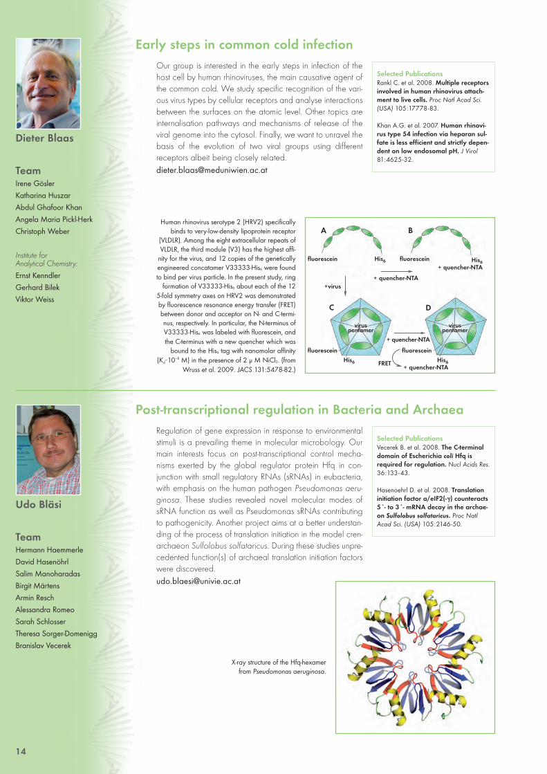
Human rhinovirus serotype 2 (HRV2) specificallybinds to very-low-density lipoprotein receptor
(VLDLR). Among the eight extracellular repeats ofVLDLR, the third module (V3) has the highest affi-
nity for the virus, and 12 copies of the geneticallyengineered concatamer V33333-His6 were foundto bind per virus particle. In the present study, ring
formation of V33333-His6 about each of the 125-fold symmetry axes on HRV2 was demonstratedby fluorescence resonance energy transfer (FRET)between donor and acceptor on N- and C-termi-nus, respectively. In particular, the N-terminus ofV33333-His6 was labeled with fluorescein, andthe C-terminus with a new quencher which was
bound to the His6 tag with nanomolar affinity (Kd~10–8 M) in the presence of 2 μ M NiCl2. (from
Wruss et al. 2009. JACS 131:5478-82.)
14
Our group is interested in the early steps in infection of thehost cell by human rhinoviruses, the main causative agent ofthe common cold. We study specific recognition of the vari-ous virus types by cellular receptors and analyse interactionsbetween the surfaces on the atomic level. Other topics areinternalisation pathways and mechanisms of release of theviral genome into the cytosol. Finally, we want to unravel thebasis of the evolution of two viral groups using differentreceptors albeit being closely [email protected]
Early steps in common cold infection
Dieter Blaas
TeamIrene Gösler Katharina HuszarAbdul Ghafoor KhanAngela Maria Pickl-HerkChristoph Weber
Institute for Analytical Chemistry:
Ernst KenndlerGerhard BilekViktor Weiss
Selected PublicationsRankl C. et al. 2008. Multiple receptorsinvolved in human rhinovirus attach-ment to live cells. Proc Natl Acad Sci.(USA) 105:17778-83.
Khan A.G. et al. 2007. Human rhinovi-rus type 54 infection via heparan sul-fate is less efficient and strictly depen-dent on low endosomal pH. J Virol 81:4625-32.
Regulation of gene expression in response to environmentalstimuli is a prevailing theme in molecular microbology. Ourmain interests focus on post-transcriptional control mecha-nisms exerted by the global regulator protein Hfq in con-junction with small regulatory RNAs (sRNAs) in eubacteria,with emphasis on the human pathogen Pseudomonas aeru-ginosa. These studies revealed novel molecular modes ofsRNA function as well as Pseudomonas sRNAs contributingto pathogenicity. Another project aims at a better understan-ding of the process of translation initiation in the model cren-archaeon Sulfolobus solfataricus. During these studies unpre-cedented function(s) of archaeal translation initiation factorswere [email protected]
Post-transcriptional regulation in Bacteria and Archaea
Udo Bläsi
TeamHermann HaemmerleDavid HasenöhrlSalim ManoharadasBirgit MärtensArmin ReschAlessandra RomeoSarah SchlosserTheresa Sorger-DomeniggBranislav Vecerek
Selected Publications Vecerek B. et al. 2008. The C-terminaldomain of Escherichia coli Hfq isrequired for regulation. Nucl Acids Res.36:133-43.
Hasenoehrl D. et al. 2008. Translationinitiation factor a/eIF2(- γ) counteracts5´- to 3´- mRNA decay in the archae-on Sulfolobus solfataricus. Proc NatlAcad Sci. (USA) 105:2146-50.
X-ray structure of the Hfq-hexamer from Pseudomonas aeruginosa.
A
C D
B
fluorescein
fluorescein
FRET
fluorescein
viruspentamer
viruspentamer
+virus
fluorescein
+ quencher-NTA
+ quencher-NTA
His6
His6
His6+ quencher-NTA
His6+ quencher-NTA
12-45_Groups 27.05.2009 13:07 Uhr Seite 14

The effect of the protein PEX11γ on membrane morphology depends on the presence of the protein PEX19, acomponent of the peroxisomal membrane insertion machinery. Confocal microscopy pictures showing either colo-calization of GFP-HsPEX11γ with the peroxisomal matrix marker, mCherry-SKL, in human wild-type fibroblasts (left) orcytosolic location of GFP-HsPEX11γ and mCherry-SKL in fibroblasts from Zellweger patients affected in peroxisomes bio-
genesis and lacking theprotein PEX19 (right).While in wild type cellperoxisomes are well-separated (arrow) overex-pression of GFP-HsPEX11γ affects the mor-phology of peroxisomes,which tend to cluster(arrow head). Bar: 10μm
Regulatory networks involved in the controlof virulence factor expression in Streptococcuspyogenes. Virulence factors consist mainly of surface-exposed and secreted proteins. Theirexpression is regulated by two-component regula-tory systems, stand alone transcriptional regula-tors, small regulatory RNAs and components ofthe protein quality control system (chaperones,proteases). The regulatory components work inconcert within a complex network.
15
How do membranes proliferate and acquire their shape?Our team focuses on elucidating the molecular dynamics ofmembrane proliferation and the function of membrane pro-teins involved in this process. To preserve cellular fitness, pro-liferation of cellular compartments, the organelles, needs to
be tightly regulated. Accordingly, organelle malfunction is critically involved in the development ofdevastating human diseases e.g. Zellweger syndrome. Peroxisome function includes lipid metabo-lism. These organelles possibly contribute to cellular oxidative stress through their ability to genera-te and degrade hydrogen peroxide and other reactive oxygen species, which may connect theirfunction to the process of aging. We employ biochemical as well as cell biological approaches inyeast and human cells and carry on oxidative stress studies to grasp the molecular mechanismgoverning peroxisome proliferation. [email protected]
Dynamics of protein assembly at the peroxisome membrane in yeast and human cells
CécileBrocard
Elise-Richter Prize Holder
TeamChristine David
Thomas HeilJohannes-P. Koch
Anita Kruzig Sophie Merich
Selected PublicationBrocard C. and Hartig A. 2007. Peroxins: A Proliferation Romanceamongst Supposition and Disposition.Dyn Cell Biol. 1:1-11.
During infection, pathogens are exposed to various hostileconditions including host defence mechanisms. To survive thehost-induced stresses and launch the disease process, patho-genic bacteria have developed well-directed strategiesleading to a coordinated expression of physiological andvirulence factors. In this regard, our group is interested inunderstanding the role of small regulatory RNA moleculesand regulated protein quality control in gram-positive bacte-rial stress response and pathogenesis. In collaboration withthe group of Pavel Kovarik, we are also investigating themodulation and mechanisms of innate immune defense toStreptococcus [email protected]
Molecular mechanisms governing gram-positive bacterial pathogenesis
EmmanuelleCharpentier
TeamFanny Beneyt
Krzysztof ChylinskiElitza Deltcheva
Markus DöllingerStephanie Füreder
Karine GonzalesBarbara Mindt
Zaid Ahmed PirzadaSilvia Spiess
Selected Publications Vojtek I. et al. 2008. Lysogenic transferof Group A Streptococcus superanti-gen gene among streptococci. J InfectDis. 197:225-34.
Gratz N. et al. 2008. Group AStreptococcus activates type I interfe-ron production and MyD88-depen-dent signaling without involvement ofTLR2, TLR4, and TLR9. J Biol Chem.283:19879-87.
12-45_Groups 27.05.2009 13:07 Uhr Seite 15

Signal transduction in cells infected with Listeriamonocytogenes. Cytoplasmic recognition of
the bacteria stimulates type I interferon synthe-sis through transcription factor IRF3. Secreted
type I interferons subsequently induce a distinctset of antimicrobial genes through STAT
transcription factors.
16
When cells encounter microbes they respond with an innateantimicrobial immune response. One of the hallmarks of thisresponse is the synthesis of type I interferons (IFN-I). IFN-Ireprogram gene expression through a Jak (tyrosine kinase)-Stat (transcription factor) signaling pathway. We study howthe intracellular bacterial pathogen Listeria monocytogenescauses IFN-I synthesis and how it uses Stats and other tran-scription factors to regulate host defense genes. Furthermorewe address the mechanisms underlying the finding that IFN-I enhance the lethality of L. monocytogenes infection. In anindependent project Amanda Jamieson studies the consequences of viral infection on a subsequentinfection with bacteria. This project aims at understanding the causes for and effects of the frequentbacterial superinfections in the wake of viral [email protected]
Interferons, Jaks and Stats in innate immunity
ThomasDecker
TeamMatthias FarlikAmanda JamiesonRenate KastnerElisabeth KernbauerAndreas PilzBirgit RappBenjamin ReuttererDidier SoulatUschi StixSilvia StockingerFatima TouraevaSandra Westermayer
Selected PublicationsZwaferink H. et al. 2008. IFN‚ increasesListeriolysin O-induced membranepermeabilisation and death of macro-phages. J Immunol. 180:4116-23.
Soulat D. et al. 2008. The DEAD-boxhelicase DDX3X is a critical compo-nent of the TANK-binding kinase 1-dependent innate immune response.EMBO J. 27:2135-46.
Our laboratory is interested in the molecular mechanismsunderlying the actin-based cytoskeleton of the striated mus-cle. The most striking feature of muscle and Z-disc proteins inparticular, is the high frequency of multiple protein-proteininteractions that form part of a complex network. The aim ofour research is to generate detailed structural information onthe protein-protein interaction network in the striated muscleZ-disc.To obtain molecular insights we use as the principal techni-que X-ray crystallography in combination with other biophy-sical and biochemical methods available at the Department and on the Campus. These activitiesare complemented by the development of bioinformatics tools for results reification and fine tuningof the protein constructs to be structurally [email protected]
Structural biology of cytoskeleton
Kristina DjinovicCarugo
TeamMads Beich-Frandsen Oliviero Carugo Eirini Gkougkoulia Bashir Khan Muhammad Sviatlana Kirylava Christian Koncz Julius Kostan Suresh Kumar Georg Mlynek Anita Salmazo Claudia Schreiner Kresimir Sikic Björn Sjöblom
Selected Publications Carugo O. 2008. Metallo-proteins:metal binding predicted on the basisof the amino acid sequence. J ApplCryst. 41:104-9.
Sjoblom B. et al. 2008. Novel structu-ral insights into F-actin-binding andnovel functions of calponin homolo-gy domains. Curr Opin Struct Bio.18:702-8.
Domain organisationand electrostatic
potential mapped onthe solvent accessi-
ble surface of theindividual CH
domains of actin bin-ding domain of
alpha-actinin.
12-45_Groups 27.05.2009 13:07 Uhr Seite 16

We established an indu-cible expression systemfor Drosophila cell culturethat allows the measure-ment of mRNA turnoverrates. Left: Northern blotanalysis of mRNA levelsafter a transcriptionalpulse. Right: Quantitativeanalysis of mRNA decaybased on the Northernblot experiments shownon the left side.
Vesicular transport to the cilium and intraflagellar transport
17
We are interested in understanding the molecular mecha-nisms of ciliogenesis. Cilia are highly conserved organellesconsisting of the membrane-sheathed axoneme, an extensionof the mother centriole, and at least 360 associated proteins.Eukaryotic cilia and flagella have attracted much attention in
recent years because of their role in the transduction of extracellular signals and their associationwith an ever expanding number of human disorders. Our goal is to elucidate at the atomic level theassembly mechanisms of the protein complexes for cargo transport to and within the cilium. Wemainly use X-ray crystallography to visualise these proteins and their complexes. The available newstructures will enhance our understanding of how these complexes function and provide hints as tohow their malfunction leads to human diseases. [email protected]
Structural biology: molecular mechanisms of ciliogenesis
Gang Dong
TeamClara Pleban
Renping QiaoHongwen Zhou
Selected Publication Dong G. et al. 2007. A catalytic coiled-coil: structural insights into the activa-tion of the Rab GTPase Sec4p bySec2p. Mol Cell 25:455-62.
Post-transcriptional processes such as mRNA degradation,mRNA splicing, translational repression, and RNA-mediatedgene silencing play crucial roles in the regulation of eukaryo-tic gene expression. The major focus of our research is theRNA-mediated gene silencing by siRNAs (small interferingRNAs) and miRNAs (micro RNAs). In particular we are inter-ested in understanding the various mechanisms by whichthese small non-coding RNAs (siRNAs and miRNAs) regula-te gene expression at the molecular level. We use diversetechniques of RNA biochemistry and molecular biology to
study siRNA- and miRNA-mediated gene silencing in Drosophila cell [email protected]
The regulation of gene expression by small non-coding RNAs
Silke Dorner
TeamDenise Herold
Elisabeth Jäger
Selected Publications Eulalio A. et al. 2007. Requirement forenhancers of decapping inmiRNAmediated gene silencing. GenesDev 21:2558-70.
Dorner S. et al. 2006. A genomewidescreen for components of the RNAipathway in Drosophila cultured cells.Proc Natl Acad Sci. (USA) 103:11880-5.
Kinetics of mRNA degradation
12-45_Groups 27.05.2009 13:07 Uhr Seite 17

Wild-type cells faithfullysegregate their chromo-somes as visualized byGFP-labeled chromoso-me I during anaphase(left). We have identi-fied S. pombe mutantswhich missegregatechromosomes (right).Studying such mutants isessential for understan-ding of the mechanismwhich governs chromo-some segregation.
Model depicting the molecular mechanismhow lamins may affect cell cycle progression
and differentiation of tissue progenitor cells. Anucleoplasmic pool of lamins A/C in the G1phase of cycling cells, stabilized by LAP2α,regulates pRb-mediated cell cycle exit andinitition of differentiation. Disease-causing
lamin variants may affect the nucleoplasmicpool and thus, impair pRb regulation.
18
Lamins are major structural components in the nucleus ofmetazoans. They form a network, called the lamina, whichmechanically supports the nuclear envelope and organizeshigher order chromatin structure. Mutations in lamins causenumerous human diseases with different pathologies, ran-ging from muscular dystrophy, to premature aging. The mole-cular pathways leading to these diseases are poorly under-stood. Using transgenic mouse models and cultured cells westudy novel functions of lamins in gene expression and signal-ling and their potential impairment in lamin-linked diseases. Our recent work revealed a role of thelamin-associated polypeptide LAP2α in localizing lamins in the nucleoplasm, which in turn controlcell cycle progression of early progenitor cells in regenerative tissues. We propose that lamins haveimportant functions in controlling adult stem cell activity during tissue homeostasis and [email protected]
Lamins in nuclear organization and human diseases
RolandFoisner
TeamKatarzyna BiadasiewiczMirta BobanAndreas BrachnerJuliane BraunBarbara BublavaAndreas EgerMartha GarstkiewiczIvana GoticJosef GotzmannNana NaetarUrsula PilatAneesa SultanRita Spilka
Selected PublicationsNaetar N. et al. 2008. LAP2alpha-lamin A complexes causes erythroidand epidermal progenitor hyperproli-feration. Nat Cell Biol. 10:1341-8.
Vlcek S. and Foisner R. 2007. Laminsand Lamin-associated proteins inageing and disease. Curr Opin CellBiol.19:298-304.
How does the cell ensure that during cell division eachdaughter cell inherits one copy of every chromosome?Meiosis is a specialized cell division which produceshaploid gametes from diploid cells, how is this reduction ofchromosome number achieved? We want to understandhow cells accurately segregate their chromosomes duringmitosis and meiosis. It is important to understand this processbecause defects in chromosome segregation (missegre-gation) during mitosis result in cells with abnormal number ofchromosomes. Such cells are hallmarks of cancer. Defectsduring meiosis cause miscarriages, infertility and geneticdiseases such as Down’s Syndrome. In our studies we use the fission yeast S. pombe which is anexcellent model organism amenable to both genetic and cell biology [email protected]
Chromosome segregation during mitosis and meiosis
Juro Gregan
TeamZsigmond BenkoLubos CipakCornelia RumpfGenyu Wang
Selected Publications Gregan J. et al. 2007. The kinetochoreproteins Pcs1 and Mde4 and hetero-chromatin are required to preventmerotelic orientation. Curr Biol.17:1190 -1201.
Gregan J. et al. 2005. Novel genesrequired for meiotic chromosomesegregation are identified by a high-throughput knockout screen in fissionyeast. Curr Biol. 15:1663-9.
PROLIFERATION CELL CYCLE EXIT
normal segregation missegregation
tubulinDNAchromosome I
12-45_Groups 27.05.2009 13:07 Uhr Seite 18

Our research is focussed on the origin of peroxisomes and themolecular mechanism of their biogenesis. These single-mem-brane bound organelles are ubiquitous, highly versatile com-partments in eukaryotic cells and are involved in many meta-bolic processes, such as degradation of fatty acids. A net-work of interacting proteins guarantees the biogenesis offunctional peroxisomes, the transport of peroxisomal matrixproteins across the organellar membrane, and the control ofsize, shape and number of these compartments. Impairedperoxisome biogenesis leads to cytosolic mis-localisation of
peroxisomal processes. Employing yeast as model system we aim to elucidate the protein composi-tion of mature peroxisomes and precursor structures and the functional role of the proteins [email protected]
Origin and biogenesis of peroxisomes
AndreasHartig
TeamVeerle De Wever
Karin GrossschopfAnja Huber
Isabella MagyarJürgen Steiner
Selected Publications Fransen M. et al. 2008. Comparison ofthe PTS1- and Rab8b-binding proper-ties of Pex5p and Pex5Rp/TRIP8b.Biochim Biophys Acta 1783:864-73.
Brocard C. and Hartig A. 2007.Peroxins: A Proliferation Romanceamongst Supposition and Disposition.Dyn Cell Biol. 1:1-11.
19
A method has been developed by metabolic engineering ofglutamine for the creation of reversible male-sterility in plantsto be used for F1-hybrid breeding. In collaboration with FritzKragler’s and Markus Teige’s group at the Max Perutz Labsa MAP kinase, AtMPK10, and a MAP kinase kinase,AtMKK2, have been identified that control flowering time,leaf size and leaf vein formation by interacting with polarauxin transport inhibitors. In collaboration with AlisherTouraev’s group at MFPL a gene called DCN1 has beencharacterised in tobacco that regulates developmentalphase transitions, including totipotency, and that is involvedin the neddylation of cullins, a component of ubiquitin
E3 ligases. Together with Roberto Nitsch in Joseph Penninger’s lab we are investigating the role ofmammalian DCN1 by reverse genetics. Progress has been made in using microspore embryogene-sis for gene targeting via homologous recombination. [email protected]
Plant developmental genetics and biotechnology
ErwinHeberle-Bors
TeamMonika Kastler
Tanja Resch
Selected Publications Ribarits A. et al. 2007. Combination ofreversible male sterility and doubledhaploid production by dominant-nega-tive inhibition of cytoplasmic glutami-ne synthetase in developing anthersand pollen. Plant Biotech J. 5:483-94.
Ribarits A. et al. 2007. Two tobaccoproline dehydrogenases are differen-tially regulated and play a role inearly plant development. Planta225:1313-24.
Artificial recon-struction of ayeast cellexpressing afluorescent pero-xisomal protein
Expression of the MAP kinase AtMPK10 in hydathodes and leaf veins of transgenic Arabidopsis thaliana plants (blue,center) coincides with auxin maxima (arrows) as reported by expression of the auxin-response gene DR5-GUS (right). Left image shows leaf development schematically.
12-45_Groups 27.05.2009 13:07 Uhr Seite 19

20
We focus on the biology of the growing chicken oocyteand the yolk sac as organ for nutrient transfer from yolk tothe developing chick embryo. Specifically, we are inter-ested in unravelling molecular mechanisms involved in thetransport of VLDL to the oocyte and from the egg yolk to theembryo proper. In this context, the roles of the LDL receptorgene family members, apolipoproteins, and extracellularmatrix proteins are investigated. Furthermore, we focus onthe roles of oxidative modifications of LDL in early onset ofatherosclerosis. We are interested in identifying synthetic and natural compounds with the poten-tial to act as catalysts or inhibitors of the atherogenic modifications of [email protected]
Lipoproteins in development and disease
MarcelaHermann
TeamChristine EresheimRoland LeitnerClara MannsJulia PlieschnigDesiree RaichSandra Szabo
Selected PublicationsSaarela J. et al. 2008. The patatin-likelipase family in Gallus gallus. BMCGenomics 9:281.
Eckhart L. et al. 2008. Identification ofreptilian genes encoding hair keratin-like proteins suggests a new scenariofor the evolutionary origin of hair. ProcNatl Acad Sci. (USA) 147:18419-23.
The possibility of sympatric speciation has beenheatedly discussed in recent evolutionary litera-ture. A series of theoretical studies has claimed
that sympatric speciation is “easy” under certainecological conditions. However, since these
results rely on limited numerical analysis, theirgenerality has been debated. Pennings et al.
(2008) present an analytic treatment that leadsto a detailed understanding of the evolutionary
dynamics. The figure shows the parameterrange where speciation is possible (C: completeisolation = speciation, P: partial isolation, R: ran-
dom mating = no speciation).
The research theme at the Mathematics and BiosciencesGroup Vienna (MaBS, www.mabs.at) is the mathematicalbiology of evolution. Evolution is the unifying theory of the bio-logical sciences, and our aim is to design advanced mathe-matical methods and models that account for the biologicalcomplexity involved in most evolutionary processes.Complexity arises on all levels of biological organisation:molecular, organismal, and ecological. The key issues of evo-lutionary research, such as adaptation and speciation, areusually addressed in special sub-disciplines for each of these levels, i.e. molecular population gene-tics, quantitative genetics, and evolutionary ecology. We work on all three fields with the special goalto create an integrative approach, using a combination of different models, concepts, and [email protected]
Evolutionary theory of adaptation and speciation
JoachimHermisson
TeamGregory EwingInes HellmannMichael Kopp
MaBS members atFaculty of Mathematics
Claudia BankAgnes RettelbachClaus RuefflerHannes SvardalHildegard Uecker
Selected PublicationsPennings P.S. et al. 2008. An analytical-ly tractable model for competitivespeciation. American Naturalist171:E44-E71.
Hermisson J. and McGregor A.P. 2008.Pleiotropic scaling and QTL data.Nature 456:E3-E4.
Hepato-oocyte-embryo axis for yolk transport andutilisation. During oogenesis in the chicken, the
yolk precursors (e.g. vitellogenin and VLDL) aresynthesised by the maternal liver under stringent
hormonal control (E2) and taken up into the oocy-te via receptor-mediated endocytosis (LRs). After
ovulation and fertilisation, a major feature of deve-lopment is the formation of a series of extraem-
bryonic structures including the amnion, chorion,allantois and the yolk sac. A major role of the yolk
sac is the uptake of nutrients from the yolk, theirdegradation and/or modification for re-synthesis
and secretion into the embryonic circulation.
Strength of stabilising selection
-0,4 -0,2 0 0,2 0,4 0,6
1
0,8
0,6
0,4
0,2
0
Stre
ngth
of f
requ
ency
-de
pend
ent s
elec
tion
12-45_Groups 27.05.2009 13:07 Uhr Seite 20

GFP-labeled Salmonella typhimurium have infected epi-dermal root cells of Arabidopsis thaliana after 24 hours
21
In contrast to animals, plants are sessile organisms and cannotmove away from adverse environmental conditions. Therefore,plants heavily rely on high sensitivity detection and appropria-te defense and adaptation mechanisms to withstand changingconditions in its environment. The goals of our research are tounderstand the molecular mechanisms of how plants sense,transduce and adapt to adverse various environmental condi-tions with a specific focus on the interaction of the bacterialpathogens Agrobacterium tumefaciens, the causative agent oftumour formation in plants, and Salmonella typhimurium, thecausative agent of food poisoning in humans.
Host-microbe interactions and innate immunity of plants
Heribert Hirt
TeamAndrij Belokurov
Alessandro CarreriCeline Forzani
Concetta GiulianiSarah Himbert
Aladár Pettkó-SzandtnerAndrea Pitzschke
Karin Zwerger
Selected Publications Schikora A. et al. 2008. The dark sideof the salad: Salmonella typhimuriumOvercomes the Innate ImmuneResponse of Arabidopsis thaliana andShows an Endopathogenic Lifestyle.PLoS One 3:e2279.
Djamei A. et al. 2007. Trojan horse stra-tegy in Agrobacterium transformationby abusing MAPK defence signalling.Science 318:453-6.
We study the transcriptional consequences of carnitine defi-ciency and subsequent L-carnitine supplementation in humancells. Differences in mRNA expression levels and promoteractive gene functions have been analysed by chip screenanalysis, real time RT-PCR, reporter gene and band shiftassays. We have revealed that L-carnitine in addition to its
metabolic importance (ß-oxidation, acyl-CoAs) directly interacts with promoter active factors at spe-cific sites, thus influencing a wide spectrum of genes. Currently we are analysing L-carnitine inducedgenes in more detail and try to reveal the identity of the transcription factor mediating the carnitineeffect. A second research project is tracing the effects associated with inhibition of the macrophagecolony-stimulating factor (CSF-1), when this strategy is used to inhibit growth of solid tumours and to
decrease the risk of metastasis. In these inhibitionstudies other genes relevant for angiogenesishave been included (Thrombospondin 1, VEGF)[email protected]
Signaling events after carnitine deficiency and CSF-1 inhibition
ReinholdHofbauer
TeamAshkan KhamenehBarbara Tappeiner
Selected Publication Blake S.M. et al. 2008. Thrombospon-din 1 functions un the RMS as ligandfor ApoER2 and VLDL receptor. EMBOJ. 27:3069-80.
Mitochondrial carnitine traffic
12-45_Groups 27.05.2009 13:07 Uhr Seite 21

Model for endoplasmic reticulum-associated degra-dation (ERAD): Enzymes, lectins and molecular cha-
perones work as folding factors on nascent(glyco)proteins in the lumen of the ER. After retro-
translocation of ERAD substrate proteins through aproteinaceous channel from the ER to the cytosol,their degradation occurs via the ubiquitin protea-
some pathway.
22
In the endoplasmic reticulum (ER) a quality control systemoperates that ensures that only properly folded proteins willbe released. Misfolded polypeptides are retro-translocatedfrom the ER to the cytosol, and there become poly-ubiquiti-nated and destructed by proteasomes. ER-associateddegradation (ERAD) is of relevance for a variety of geneti-cally inherited, neurodegenerative, and virally transmitteddiseases with protein folding defects. We are interested in the molecular characterisation of themulti-step ERAD process, and attempt to elucidate requirements of the substrate (glyco)proteins andto detect factors involved. Moreover, in joint projects with Marcela Hermann and Wolfgang J.Schneider (MFPL), we study protein quality control also in the context of the biosynthesis of lipo-proteins and [email protected]
Synthesis, folding, transport, and degradation of proteins in the early secretory pathway
N.-Erwin Ivessa
TeamJohanna ParsonKarina Zöttl
Selected PublicationKitzmüller C. et al. 2003. Processing ofN-linked glycans during endoplasmic-reticulum-associated degradation of ashort-lived variant of ribophorin I.Biochem J. 376:687-96.
RNA-Editing by adenosine deaminases that act on RNAs(ADARs) is a wide spread phenomenon in metazoa. ADARsconvert adenosines (A) to inosines (I) in double-stranded orstructured substrate RNAs. Inosines are interpreted as guano-sines by most cellular processes. Therefore, this type of edi-ting can lead to codon exchanges, alter splice sites, or influ-ence the localisation and stability of an RNA. Thus ADARslead to an increase in transcritpome complexity and influen-ce the fate of an RNA. Our work is focused on two main subjects: On the one hand the mecha-nisms by which ADAR-mediated editing is regulated are investigated. On the other hand the impactof editing on coding and non-coding substrates is being [email protected]
RNA-Editing and -Processing
MichaelJantsch
TeamArmin BaghestanianDrasko BokoSabina DaniWojciech GarncarzDanjela KuriaDominik MuggenhumerDieter PullirschSandy SchopoffAamira TariqZi-Qin Xu
Selected Publications Riedmann E. et al. 2008. Specificity ofADAR-mediated RNA-editing in newlyidentified targets. RNA 14:1110-8.
Schoft V. et al. 2007. Regulation of spli-cing by RNA editing. Nucleic Acids Res.35:3723-32.
ADARs bind double stranded RNAs and convert ade-nosines to inosines by deamination. This mechanism is
very abundant in metazoa and has widespreadimpact on the coding potential, processing, and struc-
ture of edited RNAs.
12-45_Groups 27.05.2009 13:07 Uhr Seite 22

Rec114 is a component of the DSB (DNA dou-ble-strand break) generating complex.Unexpectedly it sits on chromosome axes (leftmicrograph) and does not overlap with thechromosomal locations of DSBs (lower rightchromatin-IP on microarray result). In contrast,Spo11, the DSB nuclease, can be detected withthe DSBs (upper right graph)
In Pachytene homologous parental chromosomes are connected by aproteinacious structure, the synaptonemal complex (SYP-1 in red). HIM-8(in green) highlights the chromosome end of the X chromosome and thetwo signals from the parental chromosomes coalesce into one.
Meiosis is the specialised cell division that generateshaploid germ cells. It not only halves the chromosome con-tent but also ensures genetic diversity by recombination.Defects in meiosis lead to unfaithful chromosome segrega-tion and are thus the major cause for miscarriages and birthdefects. For proper chromosome segregation in meiosis I,homologous chromosomes have to recognise each other,pair, synapse and recombine entailing a physical connectionof the bivalents. The question how homologous chromo-somes recognize and find each other in the first place andestablish the primary contact is a main focus of our studies.In meiotic prophase I chromosomes are moved by cytoplas-mic forces transferred to the nucleus via the SUN/KASH pro-tein module (components of the outer and inner nuclearenvelope). We study the nature of chromosome movementand its regulation. To this end we combine genetics with highresolution cytology in the genetic model system C. [email protected]
Meiosis in C. elegans
Verena Jantsch-Plunger
TeamAntoine BaudrimontJiradet Gloggnitzer
Margot HulekMarkus Ladurner
Yasmine MamnunThomas MachacekAlexandra Penkner
Christian PflüglLois Tang
Alexander WoglarChristine Wegrostek
Selected Publications Bhalla N. et al. 2008. ZHP-3 acts atcrossovers to couple meiotic recombi-nation with synaptonemal complexdisassembly and bivalent formation inC. elegans. Plos Genetics. 4:1-15.
Penkner A. et al. 2007. The nuclearenvelope protein matefin/SUN-1 isrequired for homologous pairing in C.elegans meiosis. Dev Cell 12:873-85.
23
While meiotic mother cells carry two copies of each chromo-some, meiotic daughter cells possess exactly one copy (eithermaternal or paternal) of each parental allele. The transitionbetween these cellular states is accomplished by a remarka-ble chromosome sorting and distribution process during meio-sis, which requires the programmed appearance of DNA-
double strand breaks. Repair of these programmed lesions allows for recognition and for temporallinkage of corresponding chromosomes (the homologues), a prerequisite for correct separation ofpaternal and maternal alleles. Currently we concentrate on defining where exactly structural andrecombination proteins interact with chromosomal DNA in vivo during meiosis. The figure shows howa component of the recombination machine (Rec114) is deposited on chromosomal axis sites, sites
that alternate with the sites that actuallyundergo recombination (DSBs). Weinfer, that axis sites and DSB sites dyna-mically interact in the recombiningnucleus. [email protected]
Chromosome structure and recombination in yeast meiosis
Franz Klein
TeamMarco Antonio
Benjamin BrenhofferLingzhi Huang
Zaneta Kubus-SchadenJean Mbogning
Silvia PanizzaMarco-Antonio
Mendozza-Parra Martin Xaver
Selected Publication Uanschou C. et al. 2007. A novel plantgene essential for meiosis is related tothe human CtIP and the yeastCOM1/SAE2 gene. EMBO J. 26:5061-70.
12-45_Groups 27.05.2009 13:07 Uhr Seite 23

Model for the uptake of inert,25-nm-particles in cells: Fast
translocation was observed andcharacterised for such small, arti-ficial particles, which is mechani-
stically different to uptake andprocessing of viruses or peptides
also studied recently.
24
Biomolecular optical spectroscopy
GottfriedKöhler
TeamMichael EdetsbergerErwin GaubitzerGottfried GrabnerMartin KnappChristoph MikschMartin PuchingerJulia SchindelarAamir ShazadGerald Zwinger
Proposed general mechanism of typical catalases
We are interested in the structural basis of the respectivecatalytic properties of monofunctional (typical) catalasesand bifunctional catalaseperoxidases from various sources.Our main focus is on the organisation of the active sites andthe substrate channels leading to them. We could show thatin typical catalases structural fluctuations in the wall of thenarrow part of the major substrate channel control theaccess of different substrate species and also control the recovery of resting enzyme from oxidisedenzyme intermediates by electrons donated from so-called “internal donors“. Additionally, weattempt the preparation of catalases of increased structural stability by site-directed mutagenesis,including the introduction of inter-chain disulfide [email protected]
Structure-function studies of hydroperoxidases
Franz Koller
TeamMohsen M. Farhadi
Selected Publication Zamocky M. et al. 2004. Expression,purification, and sequence analysis ofcatalase-1 from the soil bacteriumComamonas terrigena N3H. ProteinExpres Purif. 36:115-23.
Biophysical characterisation of biomolecules and of theirinteractions in solution as well as on a live cell level repre-sents the main object of our research. Methods include fluo-rescence and time resolved techniques performed over awide range of time resolution. Studies by optical spectrosco-py are complemented by biocalorimetry (DSC).Quantitative studies on molecular dynamics on a single molecule level are performed using ad-vanced fluorescence correlation techniques. Among others, they are applied on studies of ligand-receptor interactions relevant for hormone regulation and the mechanisms of endocytosis and trans-port in single living cells. These measurements provide the basis for mathematical modelling of com-plex dynamic behaviour in bio-systems, implemented in close cooperation with other research [email protected]
Selected PublicationPàl K. et al. 2008. Efficient singletstate deactivation of cyano-substitu-ted indulines in protic solvents viaCN-HO hydrogen bonds. Chem PhysChem. 8:1-10.
12-45_Groups 27.05.2009 13:07 Uhr Seite 24

Phosphorylation of Stat1transactivation domain(TAD) depends on Stat1chromatin recruitmentStat1 DNA-binding mutant(mutation K336A in the DNA-binding domain (A)) translo-cates to the nucleus in cellstreated with interferon (B), butis not recruited to chromatin(C). Phosphorylation of Stat1TAD at Ser727 is inefficient ifchromatin recruitment is pre-vented by K336A mutation(D). Model (E).
25
The sequencing of the human genome has provided a ‘partslist’ of the human inventory comprising potential therapeutictargets for the pharmaceutical and biotechnology industry. Inorder to cope with this huge number of targets we intro-duced a new theoretical conception of protein structural bio-logy (meta-structure) which can be used for protein sequen-ce-to-function annotation and drug design. A hallmark of ourresearch is the integrative application of this novel concep-
tion and sophisticated NMR spectroscopy directed towards a better understanding of fundamentalbiological processes. Finally, as much of protein function is predicated on dynamics, we are devel-oping novel methodological approaches which combine biochemistry, bioorganic chemistry andNMR spectroscopy to unravel the microscopic details of functionally important protein [email protected]
Computational chemical biology and biomolecular NMR spectroscopy
Robert Konrat
TeamRenate Auer
Bettina Baminger-SchwengSven Brüschweiler
Nicolas CourdevilleCornelia Dorigoni
Leo GeistKarin Kloiber
Georg KontaxisChristoph Kreutz
Karin LedolterMartina Ortbauer
MariaRosa QuinteroBernabeu
Andreas SchedlbauerSabine SchultesMartin Tollinger
Andrea Vavrinska
Selected Publication Schanda P. et al. 2008. Folding of theKIX domain: characterization of theequilibrium analog of a folding inter-mediate using 15N/13C relaxationdispersion and fast 1H/2H amideexchange NMR spectroscopy. J MolBiol. 380:72 -41.
Many immune disorders and infectious diseases are causedby failures in activation or attenuation of the innate immuneresponses. We address these issues by studying 1) chroma-tin-associated signaling events regulating inflammatory genetranscription in the Jak/Stat signal transduction pathway, 2) responses of the innate immune system to infection withthe human pathogen Streptococcus pyogenes and the mole-cular mechanisms of the recognition of this pathogen byinnate immune cells, 3) the role of mRNA decay in immunehomeostasis with the focus on the function of the mRNA-destabilising and anti-inflammatory gene tristetraprolin (TTP)
in vivo using mouse models. Our projects aim at providing mechanistic explanations for normal andaberrant innate immune responses under conditions relevant for human [email protected]
Infection, inflammation and immune homeostasis
Pavel Kovarik
TeamJoanna Bancerek
Nina GratzHarald Hartweger
Franz KratochvillChristian Machacek
Ivana Mikulic
Selected Publications Sadzak I. et al. 2008. Recruitment ofStat1 to chromatin is required forinterferon-induced serine phosphory-lation of Stat1 transactivation domain.Proc Natl Acad Sci. (USA) 105:8944-9.
Gratz N. et al. 2008. Group A strepto-coccus activates type I interferon pro-duction and MyD88-dependent signa-ling without involvement of TLR2, TLR4,and TLR9. J Biol Chem. 18:19879-87.
The figure serves as an overview of currently pursuedresearch topics in the group. A central structural bio-logy topic in the group is the structural analysis of theoncogenic transcription factor myc and its differential-ly regulated target genes. (Top) We have used NMRspectroscopy to analyse the C-terminal (DNA-bindingand dimerisation) domain of myc in the individual sta-ges of transcription. Additionally our structural analy-sis of myc target genes provided a first glimpse onmyc’s cell transforming potential. (Bottom) NMRspectroscopy is a unique tool to identify and analysehigh-energy states of proteins.
12-45_Groups 27.05.2009 13:07 Uhr Seite 25

26
In plants initiation and maintenance of meristems (stem cells)depends on a class of homeodomain transcription factors. Itwas shown that homeodomain proteins such as KNOTTED1and STM are essential for meristem formation and move withtheir mRNA from cell to cell via intercellular pores namedplasmodesmata. This intercellular transport system, which isalso used by viruses to spread throughout the plant body, istightly regulated and mediated by plasmodesmatal compo-nents. Our work focuses on the events underlying the intercel-lular transport, the isolation/characterisation of the cellularmachinery regulating cell-to-cell transfer, and the function ofnon-cell-autonomous transcription factors and [email protected]
Intercellular transport of proteins and RNAs regulating cell-fate
Fritz Kragler
TeamDaniela FichtenbauerGregor KollwigKornelija PranjicNikola Winter
Selected PublicationsBouyer D. et al. 2008. Two-dimensionalpatterning by a trapping/depletionmechanism: the role of TTG1 and GL3in Arabidopsis trichome formation.PLoS Biol. 6:e141.
Winter N. et al. 2007. MPB2C, a micro-tubule- associated protein, regulatesnon-cell autonomy of the homeodo-main protein KNOTTED1. Plant Cell19:3001-18.
Grant Support: Our work is supported by grants from theChristian Doppler Research Society, the 6th / 7th European fra-
mework programmes, the Austrian Science Foundation FWF, thetransnational ERA-Net Pathogenomics scheme, the SysMO pro-
gramme through the Austrian GenAU, the Austrian AcademicExchange Service and by the FFG.
A main interest of my group is to understand the molecularmechanisms of fungal pathogenicity. We study fundamentalproblems in infection biology using a combination of molecu-lar as well as genome-wide and systematic approaches Onthe pathogen side, we pursue reverse genetics (i) to identifyvirulence and antifungal drug resistance genes, (ii) the rolesof epigenetic modifications in morphogenetic switching andcell fate determination, and, (iii) we develop antibody-basedapproaches to combat fungal disease. Further, we also dis-sect the structure-function relationships of eukaryotic mem-brane ABC transporters conferring multidrug and antifungalresistance. On the host side, we are studying (i) innate and adaptive immune response in primarycells and whole animals challenged by fungal pathogens, (ii) the interplay of adaptive and innateimmunity during host response, and (iii) the function of type I interferons, as well as cytokines duringfungal pathogenesis. Finally, we pursue systems bio-logy approaches by exploiting quantitative experi-mental biology paired with mathematical modelingto answer how stress signaling pathways impactgrowth control and proliferation in [email protected]
Molecular mechanisms driving virulence of human fungal pathogens
Karl Kuchler
TeamChristelle BourgeoisIngrid FrohnerWalter GlaserChrista GregoriKwang-Soo HilderingDenes HniszFabian IstelRegina KlausCornelia KleinNathalie LandstetterIwona LesiakOlivia MajerTobias SchwarzmüllerJasmin SvinkaLanay TierneyMichael TschernerMartin ValachovicKristina YatsykFlorian Zwolanek
Selected Publications Frohner I.F. et al. 2008. C. albicans cellsurface superoxide dismutases degra-de host-derived ROS to escape innateimmune surveillance. Mol Microbiol.71:240-52.
Ernst R.P. et al. 2008. A mutation of theH-loop selectively affects rhodaminetransport by the yeast Pdr5 multidrugABC transporter. Proc Natl AcadSci.(USA) 105:5069-74.
Confocal laser scanning microscopy images of plants expressing a fluorescent fusion protein which is transported into thenucleus (green; upper left) and alters cell cycle progression (large trichomes; middle) and the function of homeodomainproteins involved in meristem (= plant stem cells) maintenance (right).
12-45_Groups 27.05.2009 13:08 Uhr Seite 26

Instead of a synaptonemal complex,meiotic nuclei of the fission yeast possessso-called linear elements, which arebelieved to be evolutionary relics of theformer with reduced functions.Nevertheless, they consist of numerouscomponents, two of which, Rec10(green), and a new protein recently iden-tified by us (red), are depicted here.
Cyanophora paradoxa,GlaucocystophytaInsert: Interference contrast micrographImmuno-EM of a dividing cyanelle:Primary antibodies directed against pep-tidoglycan from E. coli. Gold particlesmainly decorate the unique organellewall and the newly formed septum. CB,central body (putative carboxysome).
27
We are interested in the biochemistry, molecular and cellbiology of cyanelles. These peculiar plastids are found onlyin glaucocystophyte algae. Their unique peptidoglycan walland the postulated carboxysome (both features are other-wise confined to prokaryotes) render them a “missing link” ofplastid evolution and an extant proof of the endosymbiotictheory. The data and results we generated working with the“living fossil” Cyanophora paradoxa support the concept ofa single primary endosymbiotic event leading to phototro-phic eukaryotes: This enormously complicated process
dating back about 1.5 billion years happened only once, i.e. between a certain type of heterotro-phic eukaryotic host and a certain cyanobacterial species. Thus the kingdom “Plantae” can be con-sidered as [email protected]
The cyanelles of the ”living fossil“ Cyanophora paradoxa
WolfgangLöffelhardt
retired in September
2008
TeamSara Fathinejad
Jürgen-Michael Steiner
Selected Publications Fathinejad S. et al. 2008. A carboxyso-mal CCM in the cyanelles of the “coe-lacanth” of the algal world, Cyano-phora paradoxa? Physol Plant. 133:27-32.
Yusa F. et al. 2008. Evolutionary con-servation of dual Sec translocases inthe cyanelles of Cyanophora para-doxa. BMC Evol Biol. 8:304.
Cells of sexually reproducing eukaryotes normally containtwo equal (homologous) sets of chromosomes, one contrib-uted by the father, the other by the mother during the fusionof gametes and the formation of a zygote. When eggs orsperm are produced, they must be furnished with a single setof chromosomes. Therefore, germ progenitor cells undergo areductional division, meiosis. During meiosis, homologouschromosomes of paternal and maternal origin pair,exchange parts and segregate to different daughter nuclei.Our group is comparing various aspects of meiotic chromo-
some organisation and behaviour in yeasts, nematodes and ciliates to fully exploit the wealth ofvariability displayed by the meiotic processes in some of the more exotic organisms. This will servethe final goal to learn about the origin of conserved meiotic features such as the synaptonemal com-plex, the chromosomal bouquet, and finally, the evolution of meiosis [email protected]
Meiotic chromosome pairing
Josef Loidl
TeamAnna Estreicher
Rachel Howard-TillAgnieszka Lukaszewicz
Selected Publications Mochizuki K. et al. 2008. DNA double-strand breaks, but not crossovers, arerequired for the reorganization ofmeiotic nuclei in Tetrahymena. J CellSci. 121:2148-58.
Penkner A. et al. 2007. A conservedfunction for a C. elegans Com1/Sae2/CtIP protein homologue in meioticrecombination. EMBO J. 26:5071-82.
12-45_Groups 27.05.2009 13:08 Uhr Seite 27

(A) Rct1 is a multidomin cyclophilin.PPIase – peptidyl-prolyl cis/trans isome-rase domain; RRM – RNA recognition
motif; RS – domain rich in Arg and Serresidues. (B) rct1Δ cells expressing Rct1
without the PPIase domain are highlyelongated and exhibit defects in cell
cycle and mitotic chromosome segrega-tion. Lagging chromosomes/chromatidesand unequally separated chromosomes
are prevalent phenotypes. SPB – spindlepole body (visualised by the expression
of Sid4-GFP).
28
Protein phosphorylation is a key cellular signalling mechanismthat induces changes in protein conformation. Ser or Thr thatprecede Pro are major phosphorylation motifs in cells. Thesesites are phosphorylated by a large family of Pro directedkinases, which include CDK, ERK, SAPK/JNK, p38, GSK3and PLK kinases. Peptidyl Pro is a unique amino acid whichcan adopt two different conformational states, i.e. cis and trans. Prolyl isomerases (cyclophilins,FK506BPs, and parvulins) are enzymes that catalyse cis to trans isomerisation of Pro imide peptidebonds, thereby influencing phosphorylation states of their target proteins and consequently cellularsignalling pathways. By using a combination of biochemical, genetic and cell biology approaches,we are studying the regulatory roles of Rct1, an essential S. pombe multidomain cyclophilin, in RNApolymerase II transcription, cell cycle, chromosome segregation, and pre-mRNA [email protected]
Cyclophilins, multifaceted proteins that regulate diverse cellular processes
Zdravko J.Lorkovic
TeamHana KautmanovaTatsiana Skrahina
Selected PublicationGullerova M. et al. 2007. Rct1, a nucle-ar RRM-containing cyclophilin, regula-tes phosphorylation of RNA polyme-rase II C-terminal domain. Mol CellBiol. 27:3601-11.
Arabidopsis plant response toinfection by fungus Botrytis.
Ethylene and Jasmonateamounts are strongly affected
in plants when AP2C1 ismodulated. Schema: AP2C1
action in stress signaling.
What are the molecular mechanisms connecting environmen-tal signaling to responses that enable plant adaptation? PP2Cphosphatases from cluster B provide important switch andfeedback mechanisms in plants to control stress-activatedMAPKs (mitogen-activated protein kinases), stress-hormonesand plant immunity responses. We are studying how AP2C-phosphatases channel signaling pathways towards specificresponses under biotic/abiotic stress conditions and in celldifferentiation. Gene expression profiles, plant phenotypesand stress hormone ethylene and Jasmonate levels are stron-gly affected by the action of these [email protected]
Plant PP2C phosphatases in environmental and developmental cell signalling
IruteMeskiene
TeamZahra AyatollahiChonnanit ChoopayakTschu-jie LiuJulija UmbrasaiteVerena UnterwurzbacherAlois Schweighofer – currently Marie Curie Fellow at MPIMP,Potsdam
Selected Publication Schweighofer A. and Meskiene I. 2008.Regulation of stress hormones jasmo-nates and ethylene by MAPK path-ways in plants. Molecular BioSystems4:799-803.
Schweighofer A. et al. 2007. The PP2C-type Phosphatase AP2C1 NegativelyRegulates MPK4 and MPK6, andModulates Innate Immunity, JasmonicAcid and Ethylene Levels inArabidopsis. Plant Cell 19:221-24.
12-45_Groups 27.05.2009 13:08 Uhr Seite 28

Mice lacking the erythropoietin-activated transcrip-tion factor Stat5 are severely anaemic (see pale foe-tal livers and embryos) and die around birth. This ismainly due to reduced expression of genes involvedin heme synthesis (TfR-1, transferrin receptor; IRP2,iron regulatory protein 2), which could be shown tohave functional Stat5-binding sites in their promoters.
Model for the formation of the 61S particle: 70S ribosomes form an initiation complex exclusively with lmRNA in the pre-sence of Ksg (yellow sphere), leading to a conformational change within the 16S rRNA helices h2, h27, h24, h28, andh26. The disruption of some of these helices (h2, h26, and h27) triggers the release of r-proteins S1, S2, S6, S12, S18and S21, which are directly or indirectly attached to these helices (Kaberdina et al. 2009. Mol Cell 33:227-36.)
29
Translation of the genetic code into proteins is performed bythe ribosome, a large ribonucleoprotein machine. We studythe effect(s) of antibiotics on the ribosome. Recently, weshowed that in the presence of Kasugamycin in vivo protein-deficient 61S ribosomes accumulate, which are active intranslation of a particular class of mRNAs lacking a 5’-UTR.Currently, we are investigating this phenomenon whichmight serve as a novel mechanism for the modulation ofgene expression under adverse conditions. Based on thesestudies, another focus of our work is the design of novel anti-
microbials to selectively interfere with ribosome assembly of bacterial [email protected]
Protein-deficient ribosomes and novel antimicrobials
Isabella Moll
TeamBurcu Biterge
Konstantin ByrgazovAnna Chao Kaberdina
Salim Manoharadas Sanda Pasc
Oliver Vesper
Selected Publications Maar D. et al. 2008. A single mutationin the IF3 N-terminal domain perturbsthe fidelity of translation initiation atthree levels. J Mol Biol. 383:937-44.
Sonnleitner E. et al. 2008. Detection ofsmall non-coding RNAs inPseudomonas aeruginosa by RNomicsand structure-based bioinformatictools. Microbiology 154:3175-87.
Haematopoiesis starts from stem-cells differentiating intospecific lineages and ends with late-stage committed pro-genitors undergoing terminal maturation. Our group focu-ses on molecular players critically involved in balancingsustained proliferation versus terminal differentiation, withparticular emphasis on erythropoiesis. We use foetal liver-derived mouse erythroblasts or myeloid progenitors andvarious genetically modified mouse strains. More recently,we employ corresponding cells from cord- or peripheral
blood of healthy or diseased human donors. These tools are used to study (i) signalling pathways emanating from extracellular ligands likegrowth factors (stem cell factor, Wnt, erythropoietin) or steroid hormones (thyroid hormone, gluco-corticoid, androgen); (ii) cell-type specific regulation of iron metabolism during erythropoiesis and
(iii) cell size control, e.g. the decrease incell volume during erythroid [email protected]
Signal transduction and hematopoiesis / erythropoiesis
Ernst Müllner
TeamMatthias ArtakerHelmuth Gehart
Marc Kerenyi Manfred Schifrer
Anna Staribacher
Selected Publications Grebien F. et al. 2008. Stat5 activationenables erythropoiesis in the absenceof EpoR and Jak2. Blood 111:4511-22.
Kerenyi M.A. et al. 2008. IStat5 regula-tes cellular iron uptake of erythroidcells via IRP-2 and TfR-1. Blood112:3878-88.
12-45_Groups 27.05.2009 13:08 Uhr Seite 29

30
We study the biology of LDL receptor related proteins, agroup of cell surface receptors which mediate transport ofmacromolecules across cell membranes and play importantroles in signal transduction. The biological systems we focuson are the chicken oocyte and the mammalian brain.Oocyte growth is promoted by the uptake of yolk precursorsmediated by the VLDL receptor. In the developing mammali-an brain newborn neurons have to migrate to their final loca-tion. This process is governed by the Reelin signaling path-way, which is mediated by the VLDL receptor and [email protected]
Macromolecular transport across cell membranes
JohannesNimpf
TeamSophia Blake Sarah Duit Harald Rumpler
Model of PP2A biogenesis in yeast:RRD/PTPA (PP2A phosphatase acti-vator), PP2A scaffolding A subunit,
PP2A regulatory B subunit, PP2Amethyltransferase, PPM1
The Ogris lab studies the function and regulation of proteinphosphatase 2A (PP2A), a family of substrate-specific holoen-zymes involved in many cellular processes. Recently, we de-scribed how the generation of active PP2A is coupled to holo-enzyme assembly and we proposed a novel concept ofPP2A biogenesis, in which a tightly controlled activation cas-cade protects cells from unspecific activity of free catalyticPP2A subunit. We are now investigating the signalling path-ways regulating PP2A biogenesis. In addition, we establishedproof-of-principle for a novel method that allows the detectionof transient PP2A-substrate interactions and which will be usedfor substrate identification. Lastly, based on our longstanding expertise in the generation of monoclo-nal antibodies we have established a custom monoclonal antibody service at MFPL (please see alsoMonoclonal Antibody Facility on page 49)[email protected]
Protein phosphatase biogenesis and function
Egon Ogris
TeamHibbah AufIngrid MudrakMarko RoblekStefan SchüchnerClaudia StanzelCornelia Vesely
Selected Publications Hombauer H. et al. 2007. Generation ofactive protein phosphatase 2A is cou-pled to holoenzyme assembly. PloSBiol. 5:e15.
Nunbhakdi-Craig V. et al. 2007. Expres-sion of protein phosphatase 2A mu-tants and silencing of the regulatory Balpha subunit induce a selective lossof acetylated and detyrosinated mi-crotubules. J Neurochem 101:959-71.
Selected Publications Fayad T. et al. 2007. Low-density lipo-protein receptor-related 8 (LRP8) isupregulated in granulosa cells of bovi-ne dominant follicle: molecular charac-terization and spatio-temporal expres-sion studies. Biol Reprod. 76:466-75.
Andrade N. et al. 2007. ApoER2/VLDLreceptor and Dab1 in the rostralmigratory stream function in postna-tal migration independently of Reelin.Proc Natl Acad Sci. (USA) 104:8508-13.
Ablation of ApoER2 and VLDL receptor in mice leads to total loss of therostral migratory stream and accumulation of neuroblasts in the subventri-cular zone. Sagittal sections of the forebrains of mice at P17 were stained
with hematoxylin. Scale bars correspond to 500 mm.
12-45_Groups 27.05.2009 13:08 Uhr Seite 30

Principle of Thin-film Sensor
Crosstalk between SUMO and ubiquitin
31
Our group is interested in post-translational modificationswith ubiquitin and SUMO (small ubiquitin related modifier)which are important regulators for rapid and reversiblechanges in protein function. Modifications occur viaATPdependent enzymatic cascades including E1, E2 andE3 enzymes. The focus of my laboratory is to investigatemechanisms regulating SUMO and ubiquitin E2 enzymesand to understand the respective biological consequences.This topic is based on our findings that a ubiquitin conjuga-
ting enzyme (E2-25K) and also the sole SUMO conjugating enzyme (Ubc9) are sumoylated.Whereas the sumoylated ubiquitin enzyme is severely inhibited in its ubiquitinylation function in vitro(Pichler et al, 2005) the modified SUMO enzyme plays a role in target discrimination (Knipscheeret al, 2008). We do not yet understand the biological relevance of E2 sumoylation or how fre-quently this mechanism is used to regulate other E2 [email protected]
Regulation of SUMO and ubiquitin E2 enzymes
AndreaPichler
TeamMichaela Hubner
Lisa KirschnerHelene Klug
Katharina Maderböck
Selected Publications Knipscheer P. et al. 2008. Ubc9 sumoy-lation regulates SUMO target discrimi-nation. Mol Cell. 31:371-82.
Pichler A. et al. 2005. Analysis ofSumoylation. SUMO modification ofthe ubiquitin conjugating enzyme E2-25K. Nature Struct Mol Biol. 12:264-9.
We are an interdisciplinary group at the University of Viennainvolved in basic and applied research. We are interested inchemical engineering of biomolecules and their applicationin biosensors, bioreactors and drug targeting. Beyond that weestablish new, miniaturised test-kits for use in clinical chemistry– human as well as veterinarian, bed side monitoring andenvironmental chemistry employing new nanotechnologicalprinciples and biorecognition. We are also working on vari-ous aspects dealing with food allergy. Beyond that a new bio-sensor chip for meat decay is under development and a study
to improve the surface of medical implants with reloadable binding sites for drug delivery [email protected]
Biosensors and chemical engineering of biomolecules
Fritz Pittner
TeamHaifa Al-Dubai
Margit BarthUlrich Bohrn
Helmut Hinterwirth Nadira Ibrisimovic
Irene Maier Georg Oberhofer
Martina Strobl
Selected Publications Maier I. et al. 2008. Optical REA-basednear field immunochip biosensor forrapid allergen detection. AnalyticalChemistry 80:2694-2703.
Al-Dubai H. et al. 2008. A dot-blot testusing gold colloid cluster technologyas a miniaturizable alternative toELISA and hapten inhibition tests.Chemical Monthly 139:1531-6.
12-45_Groups 27.05.2009 13:08 Uhr Seite 31

(A) BR cellular homeostasis is believed to be regulated eitherby adapting BR biosynthesis (via a feedback regulatory loop)
and/or by initiating BR inactivation events. (B) Defects in BRhomeostasis have severe effects on plant development. Adult
plants of Arabidopsis thaliana that either over-accumulate BRs(due to altered BR biosynthesis) or are deficient in BRs (due to
increased inactivation) are shown.
32
The importance of the steroid hormones brassinosteroids(BRs) in the growth and development of plants is a majorfocus of our research. The BRs function in cell elongation, celldivision and differentiation and are essential in processessuch as germination, development in the light and dark,senescence and abiotic and biotic stress responses. We are interested in the mechanisms that regu-late BR homeostasis and thereby control their effects. On the one hand we aim to elucidate the con-tribution of catabolic inactivation to the regulation of BR bioactivity. On the other hand we study fac-tors that control BR biosynthesis and/or responses. We use Arabidopsis thaliana as a model andemploy diverse techniques of genetics, molecularbiology, biochemistry and cell biology in our [email protected]
Molecular mechanisms of steroid hormonehomeostasis in plants
BrigittePoppenberger
TeamSigrid Husar Mamoona Khan Wilfried Rozhon Merete Tschokert
Selected PublicationPoppenberger B. et al. 2005. TheUGT73C5 of Arabidopsis thaliana glu-cosylates brassinosteroids. Proc NatlAcad Sci. (USA) 102:15253-8.
Colocalisation of endogenous stomatin and sta-bly transfected tagged SLP-1 in HeLa cells.
Mixtures of normal and transfected cells areshown. Stomatin localises to the plasma mem-
brane (PM) and late endosomes (LE), whereasSLP-1 is sorted to LEs. Note the shift of stomatinfrom PM to LE in transfected cells. Bar: 10 μm.
Our group investigates the structure and function of stomatinand SLP-1, two integral proteins that are anchored at the cyto-plasmic side of cellular membranes via hydrophobic domainsand palmitoylated cysteine residues. Stomatin is a ubiquitous-ly expressed, oligomeric, cholesterol-binding, lipid raft-asso-ciated protein that is thought to act as an integral scaffoldingprotein like caveolin-1. It associates with ion channels and theglucose transporter Glut1 and regulates their activities in acholesterol-dependent manner. SLP-1 is highly expressed inthe brain. Its major structural features are a tyrosine-based sorting signal that causes localisation tolate endosomes and a sterol carrier SCP2-domain implying a function as a membrane-bound lipidtransfer protein. SLP-1 forms complexes with stomatin in cholesterol-rich [email protected]
Molecular cell biology of the lipid raft proteins stomatin and stomatin-like protein 1 (SLP-1)
RainerProhaska
TeamHerbert JankUlrich Salzer
Selected Publications Montel-Hagen A. et al. 2008.Erythrocyte Glut1 triggers dehydroas-corbic acid uptake in mammals unab-le to synthesize vitamin C. Cell132:1039-48.
Salzer U. et al. 2007. Stomatin: A newparadigm of membrane organizationemerges. Dyn Cell Biol. 1:20-33.
12-45_Groups 27.05.2009 13:08 Uhr Seite 32

Shedding light on gene regulatory networks.(A) Genomic alignments between Platynereis and anoth-er annelid worm, Nereis, reveal candidate noncodingelements (NCE) that integrate regulatory information.“Ex” indicates conserved coding exons. (B) 1-day oldlarva expressing EGFP from an enhancer construct in theprototroch (pt, asterisk) and apical tuft (at, arrowhead),reflecting the activity of the regulatory element used inthe injection construct.
Axon guidance, branching, and retraction are essentialmeans for neurons to ensure correct wiring of the nervoussystem during development and regeneration after injury.Axon behaviour is guided by extracellular signals which ulti-mately are translated into rearrangements of the axonalcytoskeleton. We study signalling mechanisms and post-
translational modifications such as S-nitrosylation and phosphorylation of microtubule-associatedproteins and other components of the cytoskeleton that regulate the orchestrated reorganisation ofmicrotubules and actin in response to extracellular signals. Our approach combines gene ablationin the mouse with cell biological, molecular, and biochemical analyses in cultured neurons andother primary cells. [email protected]
Cytoskeleton regulation in morphogenesis and axon guidance
FriedrichPropst
TeamLuise DescovichAnton Kamnev
Ewa KrupaWaltraud Kutschera
Selected Publication Stroissnigg H., Trancikova A et al. 2007.S-nitrosylation of microtubule-associa-ted protein 1B mediates nitric-oxide-induced axon retraction. Nat Cell Biol.9:1035-45.
33
We combine comparative genomic and experimentalapproaches to study regulatory evolution in animal develop-ment. Experimentally, we focus on Platynereis dumerilii, anannelid worm and emerging model species that exhibits aunique combination of ancestral-type genomic characteris-tics (Raible et al., Science 2005). The zebrafish, Danio rerio,serves as our comparative vertebrate model system.Our main aim is to decipher gene-regulatory networks thatorchestrate the Platynereis hormone system and help us toshed new light on the evolution of related cell types in other
animal taxa. In addition, we pioneer the molecular characterisation of Platynereis bristle formationas a prototype of genetically orchestrated microvillar dynamics. [email protected]
Regulatory evolution
FlorianRaible
TeamBenjamin Backfisch
Karina GollingerClaudia Lohs
Anne Zakrzewski
Selected Publications Tessmar-Raible K. et al. 2007. Conservedsensory-neurosecretory cell types inannelid and fish forebrain: insightsinto hypothalamus evolution. Cell129:1389-400.
Raible F. et al. 2005. Vertebrate-typeintron-rich genes in the marine anne-lid Platynereis dumerilii. Science310:1325-6.
Components of the cytoskeleton (the microtubule-associa-ted protein MAP1B in red, 1st column, and tubulin ingreen, 2nd column) co-localise in cultured primary neuronsof the mouse (yellow, 3rd column), depending on thegrowth state of the axon. Our work suggests, that increa-sed binding of MAP1B to microtubules induces axonretraction and sinusoidal microtubule bundles (middlerow), whereas at the tip of growing axons (top and bot-tom row) microtubules are splayed and MAP1B is partial-ly removed from microtubules and also found in the cyto-plasm best visible in the bottom row.
12-45_Groups 27.05.2009 13:08 Uhr Seite 33

Model for the role of EAPP in cell cycleregulation and DNA damage response:
EAPP accelerates the cell cycle by stimulat-ing E2F activity and modulates the DNA
damage response by interacting withATM, the Checkpoint kinases 1 and 2,
and with p53.
34
My laboratory is focused on the mechanisms controllinggrowth and cell cycle of the mammalian cell. E2F is a familyof transcription factors that integrate cell-cycle progressionwith transcription through cyclical interactions with importantcell cycle regulators, such as the retinoblastoma-tumor-sup-pressor- gene product (pRB), cyclins and cyclin dependentkinases. We have recently identified and characterised an E2F binding protein called EAPP (E2FAssociated PhosphoProtein) that stimulates E2F-dependent transcription and is found overexpressedin many transformed cell lines. Moreover, EAPP acts as a modulator of the DNA damage respon-se. It interferes with the activity of Chk2 (Checkpoint kinase 2) and might be required for checkpointrecovery upon completion of repair [email protected]
Cell cycle regulation and DNA damage response
HansRotheneder
TeamPeter AndorferNazanin Najafi
Selected PublicationSchwarzmayr L. et al. 2008. Regulationof the E2F-associated phosphoproteinpromoter by GC-box binding proteins.Int J Biochem Cell Biol. 40:2845-53.
The figure contains a compilation of picturesfrom the publication Uanschou et al. 2007,
and shows phenotypes and cytological ana-lyses of wild-type (A - D) and Atcom1
mutant plants (E - H). Panels A and E showfruit bodies of mature plants. Panels B and Fshow meiotic chromosomes in a fluorescent
in situ hybridisation experiment. Panels Cand G display meiotic chromosomes
(green) additionally visualising a recombina-tion protein (red) and panels D and H show
the accumulation of the SPO11-1 protein(red) in Atcom1 mutant plants on meiotic
chromatin (blue).
The focus of our research is meiotic recombination in themodel organism Arabidopsis thaliana. Meiosis is a speciali-sed cell division that ensures the reduction of the genomeprior to the formation of generative cells. During meiosis,novel combinations between parts of paternal and maternalchromosomes are generated through the process of homolo-gous recombination (HR). A pre-requisite for HR are DNAdouble strand breaks (DSBs), generated by a protein com-plex with the conserved protein SPO11 being its catalyticallyactive subunit. DSBs are formed at non-random sites through-out the genome, known as hot spots of meiotic recombination. We are interested in cis and transacting factors that mediate meiotic DSB formation and subsequent DNA [email protected]
Meiotic recombination in Arabidopsis thaliana
PeterSchlögelhofer
TeamElisabeth AltendorferBernd EdlingerAhmed El SeifiManuel HoferMichael JanisiwMarie-Therese KurzbauerStefan MeviusTanja SiwiecClemens Uanschou
Selected Publications Vignard J. et al. 2007. The interplay ofRecA-related proteins and the MND1-HOP2 complex during meiosis inArabidopsis thaliana. PLoS Genet3:1894-1906.
Uanschou C. et al. 2007. A novel plantgene essential for meiosis is related tothe human CtIP and the yeast COM1/SAE2 gene. EMBO J. 26:5061-70.
12-45_Groups 27.05.2009 13:08 Uhr Seite 34

Overview of the SELEX process. The initial DNApool, flanked by the fixed primer binding sites and aT7 promoter is transcribed into RNA (1). The resul-ting RNA library is subjected to a counter selection,in order to remove molecules that unspecifically bindto the membrane (2). After the protein-binding reac-tion with the counter selected pool (3), sequencesbinding to the protein of interest are selected via fil-ter binding (4). Thereby protein-bound RNAs areretained on the membrane, whereas non-bindingRNAs are eluted. Selected RNAs are recovered viaprotein denaturation and phenol/chloroform extrac-tion (5), reverse transcribed into DNA (6) and ampli-fied via PCR (7). The resulting pool is either subjec-ted to another round of SELEX, or, after sufficientenrichment, cloned and sequenced or sequenceddirectly by deep sequencing.
With emphasis on receptor-mediated endocytic processes,we investigate molecular genetic, cell biological, and bio-chemical details of (i) oocyte growth and chick embryodevelopment, with focus on lipoprotein transport via the lowdensity lipoprotein (LDL) receptor gene family, (ii) avian lipa-ses and transfer proteins (i.e. the lipolytic proteome) of thegranulosa cells surrounding the oocyte as well as of theextraembryonic yolk sac, which mediate yolk lipid depositionand subsequent utilisation by the embryo, respectively, (iii)the molecular genetic basis for human atherosclerosiscaused by single-gene mutations that reduce or abolish
receptor-mediated transport of lipoproteins and/or cholesterol, and (iv) the role of the recently dis-covered apolipoprotein, apo-AV, in the etiology of human pathological [email protected]
Defects in receptors as causes of Atherosclerosis and failing fertility
WolfgangSchneider
TeamRaimund Bauer
Andrea DichlbergerMary-Rose Espina
Gernot HirnBarbara Riegler
Melanie SchiffStefanie Schlager
Selected Publications Jiang M. et al. 2008. Circulating LR11,a novel marker of carotid intima-media thickness, is required forangiotensinII-induced SMC migration.J Clin Inv. 118:2733-46.
Dorfmeister B. et al. 2008. Effects of sixAPOA5 mutations, associated withsevere hypertriglyceridemia, onLipoprotein Lipase activity and recep-tor binding in vitro. Arterioscler ThrombVasc Biol. 28:1866-71.
35
RNA molecules display a large variety of functions. Therehave been mainly two approaches to search for novel RNAmolecules: computational predictions and direct sequencingof cDNAs. We are currently exploring Genomic SELEX as analternative and complementary approach for the discoveryof novel RNAs with yet unknown functions. The advantage ofGenomic SELEX is that the whole genome is screened suchthat low abundance and seldom expressed genes can beindentified. We have selected Hfq-binding RNAs from E. coliand identified a novel class of regulatory RNAs: cis antisense
RNAs. In a second selection, we identified RNA polymerase-binding RNAs from the human ge-nome, which are involved in regulating transcription. [email protected]
Identifying novel RNAs via Genomic SELEX
RenéeSchroeder
TeamJohanna BisicJennifer Boots
Doris ChenMartina Dötsch
Boris FürtigKatarzyna Matylla
Marina SkernSabine Stampfl
Robert Paul Zimmermann
Selected Publications Windbichler N. et al. 2008. Isolation ofncRNA-binding proteins from E. coli:evidence for frequent binding ofRNAs to RNA polymerase. RNA Biol.5:30-40.
Tafer H. et al. 2008. The impact of tar-get site accessibility on the design ofeffective siRNAs. Nature Biotech.26:578-83.
VLDL particles in coated structures of oocytes. Themechanism of clathrin-mediated endocytosis wasdiscovered in oocytes in 1964. The electron micro-graph shows serum-derived lipoprotein particles(VLDL) in clathrin-coated pits (c.p.) being interna-lised via invagination and pinching-off of coatedvesicles (c.v.) in a chicken oocyte. The receptorgene family involved has been extensively charac-terised in my group.
12-45_Groups 27.05.2009 13:08 Uhr Seite 35

There is considerable physiological information on mito-chondrial cation transport. A genetic screen in yeast
provided us with the MRS series of nuclear genesencoding the first known cation transporters of the inner
mitochondrial membrane. All have functional homo-logues in human mitochondria. We have recently identi-fied the series of first genes for cation transport proteins.
Cation influx through channels, driven by inside nega-tive membrane potential. Cation efflux by exchangers,
driven by inside directed H+ gradient.
36
Metal ions are known to be of prime importance for manycellular functions including volume control of cells and orga-nelles, membrane integrity, free radical homeostasis or differ-entiation processes and they act as co-factors of many essen-tial enzymes. Failure to maintain appropriate levels of metalions in humans is a feature of hereditary and acquired disor-ders. Genes encoding proteins for cation homeostasis inmitochondria remained unknown until recently when weidentified genes for the transport of magnesium (Mg2+) yeastand human Mrs2, iron (Fe2+) Mrs3/4 and potassium (K+)Mdm38/Mrs7 and human LETM1. We are studying structure and function of the respective pro-teins as well as effects of various gene mutations in the yeast Saccharomyces cerevisiae. This in-volves collaboration with K. Djinovic (protein structure, MFPL Vienna) and C. Romanin (electrophys-iology, Univ. Linz). In the mammalian system, we are currently studying the effects of K+ overloador shortage by modulating the expression of LETM1, the candidate gene for seizures in the Wolf-Hirschhorn Syndrome and its homologue the oncogene HCCR1.Contact: [email protected]
Metal ion homeostasis in mitochondria
RudolfSchweyen1941–2009
TeamMichael AichingerMarkus AleschkoRoland BaumgartnerElisabeth FroschauerAnton GraschopfMelanie HasslerTamás HenicsDagmar HosinerMirjana IlievKarin NowikovskyGerhard SponderSona SvidovaGerlinde WiesenbergerLudmila Zotova
Selected PublicationsNowikovsky K. et al. 2008.Pathophysiology of mitochondrialvolume homeostasis: Potassium trans-port and permeability transition.Biochim Biophys Acta 1778:345-50.
Piskacek M. et al. 2008. Conditionalknock-down of hMRS2 results in loss ofmitochondrial Mg(2+) uptake and celldeath. J Cell Mol Med. 13:693-700.
The Max F. Perutz Laboratories mourn the passing of Prof. Dr. Rudolf Schweyen, who
died after a short illness on February 15, 2009.
Rudolf Schweyen was heavily involved in establishing modern molecular genetics in
Vienna and was always open to change and diversification. He saw the Vienna
Biocenter and the Max F. Perutz Laboratories become a reality as a place for scienti-
fic discussion with young scientists from all over the world, an achievement that owed
much to his vision and ceaseless efforts. Despite his time-consuming duties, he always
took the time for conversation and always looked after the concerns of the people
around him. He infected everyone with his enthusiasm and humour.
To his PhD students and members of his research group, but also in his numerous
lectures to undergraduate students in the basic studies Biology and fields of Genetics,
Microbiology and Molecular Biology, he passed on his fascination for science.
The Max F. Perutz Laboratories and the University of Vienna have lost a brilliant
scientist and inspiring teacher, but, even more importantly, an exceptional personality,
colleague and friend. He will remain forever in our thoughts.
Our sincerest condolences go to his wife, family and friends.
In memoriam Rudolf Schweyen (1941 – 2009)
12-45_Groups 27.05.2009 13:08 Uhr Seite 36

Systematic high throughput analysis with yeast. Left: GeneExpression Microarrays measure gene expression of all 6200Yeast genes. Right: Microscopic analysis of individual Yeast cells.Background: Systematic phenotypic analysis with ordered 4000Yeast gene deletion strains.
37
Cells respond and adapt to a changing environment byadjusting their cellular constituents. Therefore, changes in theenvironment also demand changes in gene expression. Ourinterest is to understand how signals reach the genomicDNA and lead to controlled interpretation of the stored infor-mation. We are investigating how expression of genes underenvironmental or stress control is regulated by the interactionof chromatin remodeling complexes and transcription factorsin the yeast S. cerevisiae, a perfect model organism to deci-pher basic biological principles. We are further investigatingthe environmental response of the human fungal pathogen
Candida glabrata, closely related to yeast but adapted to the environment of a mammalian [email protected]
Environmental stress signalling in yeast and pathogenic fungi
ChristophSchüller
TeamEva Klopf
Ludmila PaskovaAndriy PetryshynAndreas Roetzer
Rene Weiss
Selected Publications Gregori C. et al. 2008. Weak organicacids trigger conformational changesof the yeast transcription factor War1in vivo to elicit stress adaptation. J BiolChem. 283:25752-64.
Roetzer A. et al. 2008. Candida glabra-ta environmental stress responseinvolves Saccharomyces cerevisiaeMsn2/4 orthologous transcription fac-tors. Mol Microbiol. 69:603-20.
We are interested in the molecular mechanisms of interac-tions between viruses and host cells. Even a small and simplevirus such as the human rhinovirus, the main causative agentof common cold, is a very successful virus. It can subvert aeucaryotic cell into a virus producing machine. Cells try todefend themselves against intruders, but at the same timeviruses have evolved complex strategies to avoid cellulardefence reactions. Analysis of this interplay between hostand virus can provide new insights into both viral and cellu-lar functions. Our current research topics include mechanismsof antiviral compounds, characterisation of virus-induced
apoptotic processes and developments for a possible use of specific viruses in cancer [email protected]
Virus-host interactions
JoachimSeipelt
TeamAndreas Alber
Elisabeth GaudernakKarin HabeggerBarbara HolzerAndrea TriendlGuest Scientist:
Marijke van Rikxoort
Selected Publications Mittermann I. et al. 2008. The IgE-re-active autoantigen Hom s 2 inducesdamage of respiratory epithelial cellsand keratinocytes via induction ofIFN-gamma. J Invest Dermatol.128:1451-9.
Lanke K. et al. 2007. PDTC inhibitspicornavirus polyprotein processingand RNA replication by transportingzinc ions into cells. J Gen Virol.88:1206 -17.
HeLa cells (left) after infection with human rhinovirus (right).Actin is shown in red, cytokeratin 8 in green.
12-45_Groups 27.05.2009 13:08 Uhr Seite 37

38
Our group is interested in chromatin modifications and theirrole in gene expression, differentiation and development.One project deals with histone acetylation, a chromatinmodification that is linked to opening of chromatin structuresand associated with important biological processes such astranscription, replication and DNA repair. Reversible acetyla-tion of core histones is controlled by histone acetyltransferases and histone deacetylases. We focusin our research on histone deacetylases, enzymes that usually act as transcriptional repressors. Wehave chosen the mouse as model system and have established an HDAC1 knockout system to ana-lyze in detail the function of this enzyme in different biological processes. In a second project, weexamine the cross-talk between histone acetylation and phosphorylation during the activation ofmammalian genes. [email protected]
Chromatin structure and gene expression
ChristianSeiser
TeamAstrid HagelkruysSabine Lagger Ayse Hande NaymanEvelyn PinedaMircea WinterBarbara ZaussingerGordin Zupkovitz
Selected PublicationWinter S. et al. 2008. Modulation of 14-3-3 interaction with phosphorylatedhistone H3 by combinatorial modifica-tion patterns. Cell Cycle 7:1336-42.
Schematic representationof different functional
domains in the shoot meri-stem (A). Stems cells (red)are located to the center.The amp1 mutant (C) hasincreased shoot meristemactivity compared to wild-
type plants (B).Longitudinal section (D) of
an amp1 shoot showingduplication of the stem cell
center (coloured in blue)
We are interested in a key question of developmental biolo-gy: how stem cell identity is determined and how the ratio be-tween stem cells and differentiating cells is balanced. To thisend we try to characterise the signalling networks in and be-tween cells required to control stem cell turn over in the pro-cess of plant organ formation. In contrast to animals, plant organ development mainly occurspostembryonically and is highly adaptive to the environment.Moreover, differentiated plant cells retain the capacity todedifferentiate to acquire a new cell fate and even to produce a whole new organism. We useArabidopsis thaliana as a model system to elucidate the molecular basis of this developmental flexi-bility and to elaborate the conceptual differences to the determinate nature of animal [email protected]
Signalling networks in plant cell differentiation
TobiasSieberer
TeamWenwen Huang Christine Marizzi Delphine Pitorre
Selected Publications Bennett T. et al. 2006. The ArabidopsisMAX Pathway Controls ShootBranching by Regulating AuxinTransport. Current Biology 16:553-63.
Sieberer T. and Leyser O. 2006. PlantScience. Auxin transport, but in whichdirection? Science 312:858 -60.
Histone H3 phosphoacetylation as amark for gene activation. Induction of
the SAP kinase cascade by stress(mimicked by anisomycin) or of the
MAP kinase cascade by growth factorsleads to multiple phosphorylation
events including histone H3-S10 phos-phorylation. The kinase inhibitor H89
can block this signal transduction.Simultaneous acetylation of neighbour-ing lysine residues K9 or K14 as resultof increased affinity for histone acetyl-transferases or changes in the intracel-
lular HAT/HDAC equilibrium by HDACinhibitors results in phosphoacetylation
of histone H3 and gene activation.
12-45_Groups 27.05.2009 13:08 Uhr Seite 38

upper left: Isolated Arabidopsis chloro-plasts after purification on Percoll gradientlower left: Tobacco leaf epidermal cellexpressing CPK3-GFP (green) in thenucleus and at cellular membranes, chloroplsts are redright: Close-up of chloroplast showing redchlorophyll-autofluorescence and greenGFP signal of thioredoxin reductase
NMR structures of the Foot-and-Mouth-Disease virus leaderproteinase, a key modulator of protein synthesis in the infectedcell.
39
Most viruses interfere with or modulate host systems to en-sure successful replication. My group has been looking atthe interactions between proteinases of the common coldvirus, foot-and-mouth disease virus and coxsackievirus with acellular protein involved in protein synthesis. We have deter-mined the molecular structures of some of these proteins andinvestigated the sites at which they interact. In collaborationwith Christian Mandl (Medical University of Vienna), wehave also started to examine how the tick-borne encephalitisvirus proteins are synthesised in an infected cell and to use
the inherent error rate of this virus as a tool to randomly mutagenise specific viral proteins. Theseapproaches lay the groundwork for the identification and development of novel anti-viral agents.
Interactions between viruses and cells
Tim Skern
TeamMartina Kurz
David NeubauerChiara Rancan
Katharina RuzicskaSabrina SchraufJutta Steinberger
Selected Publications Mayer C. et al. 2008. Residue L143 ofthe foot-and-mouth disease virus lead-er proteinase is a determinant of clea-vage specificity. J Virol. 82:4656-9.
Schrauf S. et al. 2008. Functional ana-lysis of potential carboxy-terminalcleavage sites of tick-borne encephali-tis virus capsid protein. J Virol.82:2218-29.
We study plants’ strategies of adaptation to environmentalchanges or during developmental switches. These processesrequire a strict regulation and coordination of different cellu-lar activities, for example coordination of chloroplastand cel-lular metabolism. Signalling pathways, which regulate theseevents, include MAP kinase cascades (i.e.Teige et al.(2004) Mol. Cell 15, 141-152.) and Ca2+-dependent pro-tein kinases (CDPKs). Current work in our group addresses(1) cross-talk between MAP kinases and CDPKs, which areactivated under similar stress conditions; (2) the role of target-
ing of protein kinases to different membranes and organelles, and (3) role of organellar protein kina-ses. We investigate these topics by studying CDPKs and chloroplast localised proteins in detail. MTis coordinator of a Marie-Curie Initial Traning network on chloroplast signals and partner in an ERA-PG project on Calcium-signalling. [email protected]
Plant signal transduction and physiology
Markus Teige
TeamRoman BayerAndrea Mair
Simon Lander StaelHelga WaltenbergerBernhard Wurzinger
Selected Publications Benetka W. et al. 2008. Experimentaltesting of predicted myristoylation tar-gets involved in asymmetric cell divi-sion and calcium-dependent signal-ling. Cell Cycle 23:3709-19.
Teige M. et al. 2004. The MKK2 path-way mediates cold and salt stresssignalling in Arabidopsis. Mol Cell15:141-52.
12-45_Groups 27.05.2009 13:08 Uhr Seite 39

The polychaete Platynereis dumerilii (A) adult animal,(B) larva at 2dpf (picture from H. Hausen) (C) The pha-ses of the moon (top) control and synchronize the num-
ber of sexually mature Platynereis adults.
40
Our group is interested in inner brain sensory-neurosecretorycells that are part of the ancient core of animal brains. Inorder to understand the function and evolutionary diver-gence of these enigmatic cells, the lab follows a comparati-ve approach, using two fish model species, zebrafish andmedakafish, as well as the marine bristle worm Platynereisdumerilii.Besides the molecular analysis of these cells, we are inter-ested in their impact on seasonality and biological rhythms.Medakafish gonads regress when the fish are exposed to“winter” lighting conditions. We test if the inner brain PRCs are required for this physiological re-sponse to particular light conditions. As in many other marine animals, the spawning of Platynereis is synchronized by the moon. We canmimic this rhythm in the lab culture, and usevarious molecular approaches to understand(a) the reception of the moonlight, (b) thetransmission of the signal to the gonads and(c) the integration of lunar and circadianclock signals in [email protected]
Lunar periodicity and the function of inner brain sensory-neurosecretory cells
KristinTessmar
TeamStephan KirchmaierKatharina SchipanyJuliane Zantke
Selected PublicationsArendt D. et al. 2008. The evolution ofnervous system centralization. PhilosTrans R Soc Lond B Biol Sci. 363:1523-8.
Tessmar-Raible K. et al. 2007. Conservedsensory-neurosecretory cell types inannelid and fish forebrain: insightsinto hypothalamus evolution. Cell129:1389-1400.
Research interests:1. Microspore embryogenesis and doubled haploids (DH):DHs are an important component of breeding programs ofimportant crops. We have great expertise on DH of tobac-co, rapeseed, wheat, Arabidopsis, snapdragon etc.2. Mechanism of microspore reprogramming and embryogenesis: One of the major topics of ourgroup is to dissect the mechanism of microspore reprogramming and embryogenesis.3. Gene targeting in plants: In flowering plants gene targeting (GT) is still inefficient. The main aimof the project is the evaluation of microspores as possibly efficient targets. 4. Development of an environment-friendly F1 hybrid breeding technology: The production of F1hybrid requires homozygous parental lines and reversible male sterility. We establish technologythat combines reversible male sterility with DH. [email protected]
Plant developmental genetics and biotechnology
AlisherTouraev
TeamSvitlana FedchenkoJohanna GottschamelMaria GranilschikovaIrina LewickaTatiana-Elena ReschAlexandra RibaritsIrina SadovnikJulia Szederkenyi
Selected Publication Forster B.P. et al. 2007. The resurgenceof haploids in higher plants (A review).Trends in Plant Sciences 12:368-75.
Reprogrammed tobaccomicrospores expressing
GFP divide and formembryos
12-45_Groups 27.05.2009 13:08 Uhr Seite 40

Intercell’s technology platforms supporting vaccine deve-lopmentVaccine development at Intercell is leaning on three pivotalplatforms forming the fundament of the company’s researchprogrammes: the antigen identification programme discov-ering the most protective microbial antigens, the vaccineimprovement and adjuvantal programmes that increase theefficacy of existing and novel vaccines and the patch deliverytechnology that is pioneering a needle-free era in the vaccinefield. All three platforms, patches, antigens and adjuvants, are
assets per se in vaccine development, but can also be combined in multiple fashions for the launchof novel vaccines or the improvement of existing ones. Consequently the company has been able tobuild a coherent R & D pipeline that today comprises more than eight products in various stages, cur-rently tested in phase I to phase III clinical trials (for more information: www.intercell.com).
Vaccine development at Intercell is aiming toreduce the formulation to a minimal number ofantigens and to an adjuvant. Consequently anadaptive and protective immune response isinduced in the vaccinated subjects. Our antigensare identified by using the immune system ofpathogen-exposed individuals as read out. Weare optimising our adjuvants by analysing theireffect on the innate immune system. Our needle-free delivery system is delivering antigens andadjuvant to the first layer of immune defencewhere macrophages are prevalent.
The complex geometry of tree space
41
The Center for Integrative Bioinformatics Vienna (CIBIV,www.cibiv.at) serves as a central facility to coordinate theBioinformatics activities at the MFPL and the University ofVeterinary Medicine Vienna (VUW). Moreover, it is involvedin providing infrastructure and bioinformatic expertise for thevarious research groups at MFPL and on campus. Thus, it isinvolved in a bunch of collaborations with experimenters.Besides this data analysis part, the CIBIV pursues its own
research agenda. It wants to understand the evolutionary processes that have shaped the genomesof contemporary species. To this end, the CIBIV applies methods from statistics, computer sciences,
and mathematics to detect the traces ancient evo-lutionary events have left in modern genomes. TheCIBIV is involved in several international projects,like the Deep Metazoan Phylogeny project,where it coordinates the Bioinformatics aspects(www.deep-phylogeny.org) [email protected]
Unveiling traces of ancient events in contemporary genomes
Arndt vonHaeseler
TeamQuang Minh Bui
SriHarsha ChallapalliRicardo de Matos Simoes
Huy Quang DinhIngo Ebersberger
Gregory EwingWolfgang Fischl
Tanja GesellMartin Grabner
Steffen KlaereTina Köstler
Anne KupczokManuela Machatti
Minh Anh Thi NguyenTung Lam Nguyen
Phuong Minh PhamHeiko Schmidt
Fritz SedlazeckSascha Strauss
Selected Publications Ewing G.B. et al. 2008. Rooted TripleConsensus and Anomalous GeneTrees. BMC Evol Biol. 8:118.
Kupczok A. et al. 2008. An ExactAlgorithm for the Geodesic Distancebetween Phylogenetic Trees. J ComputBiol. 15:577-91.
Research and development programmes at Intercell AG
Alexandervon Gabain
CSO and Co-founder of the
Intercell AGJoined appoint-
ments at the MaxPerutz Labs and
the KarolinskaInstitute inStockholm
Selected Publications Lingnau K. et al. 2007. IC31 and IC30,novel types of vaccine adjuvantsbased on peptide delivery systems.Expert Rev Vaccines 6:741-6.
Giefing C. et al. 2008. Discovery of anovel class of highly conserved scaleantigenic fingerprinting of pneumo-coccus with human antibodies. JEM205:117-31.
12-45_Groups 27.05.2009 13:08 Uhr Seite 41

42
RNAs regulate biology: The RNA molecules playing animportant role in cellular processes are very diverse, but theyshare their strict dependence on acquiring a specific 3Darchitecture to be functional. The process of folding de-scribes how RNA undergoes the transition from a disorderedunfolded state to the native, functional conformation. Myresearch focuses on understanding this most essential aspectof RNA function by investigating RNA structure and foldingpathways. Specifically, we aim to provide novel insights intoprotein-facilitated RNA folding and how RNAs fold in the living cell. Ultimately, deciphering the rulesgoverning RNA folding will advance our understanding of the basic mechanism of RNA-dependentprocesses, like self-splicing and telomere addition, and the role of RNA in [email protected]
Dissecting RNA folding: from structure to function
ChristinaWaldsich
TeamAndreas Liebeg Oliver MayerMichael WildauerGeorgeta Zemora
Selected Publications Waldsich C. 2008. Dissecting RNA folding using Nucleotide AnalogInterference Mapping (NAIM). NatProtoc. 3:811-23.
Waldsich C. and Pyle A.M. 2007. A folding control element for tertiarycollapse of a group II ribozyme. NatStruct Mol Biol. 14:37-44.
The Golgi apparatus lies at the heart of the secretory path-way, receiving the entire output of newly-synthesised proteinsand lipids from the endoplasmic reticulum, purifying and pro-cessing them before sorting to their correct destination. Newcopies of the Golgi need to be made during each cell cycle,to ensure inheritance through successive generations. Westudy this process in the protozoan parasite Trypanosomabrucei, which has a highly simplified secretory pathway anda single Golgi, and permits manipulations not readily possible in many other organisms. Our pre-sent work focuses on a novel bilobe structure that associates with the old Golgi and appears todetermine the site for assembly of the new Golgi. Our mostrecent work has focussed on the role played by the polo-likekinase in the duplication of this [email protected]
Biogenesis of the Golgi apparatus
GrahamWarren
TeamChris de GraffenriedLars DemmelThomas GniadekMartina Gschirtz*Michael MelakBrooke MorriswoodNina Pruckner*Marco Sealey
*part of the year
Selected Publicationsde Graffenried C.L. et al. 2008. Polo-likekinase is required for Golgi and bilo-be biogenesis in Trypanosoma brucei.J Cell Biol. 181:431-8.
Shi J. et al. 2008. Centrin4 coordinatescell and nuclear division in T. brucei. JCell Sci. 15121:3062-70.
The folding pathway of the Sc. ai5γgroup II ribozyme. The helices within
Domain 1 are represented in blue,Domain 3 is shown in green, Domain 5
is red, and the D2 and D4 truncationhelices are shown as grey. In the un-
folded state only the secondary struc-ture is formed, while in case of the inter-mediate state Domain 1 compacts and
forms tertiary structure thereby pro-viding the scaffold for docking of
Domains 3 and 5, which completesfolding to the native conformation (Pyle,
Fedorova and Waldsich, TiBS 2007).
Early in the cell cycle (top panel) the old Golgi (G; red) in T. brucei islocated near to one lobe of a bi-lobe structure (green, closed arrow-
heads). Later in the cell cycle (bottom panel) the new Golgi is found asso-ciated with the other lobe suggesting that the bi-lobe has a role to play in
the duplication process. N=nucleus; K=kinetoplast (mitDNA); openarrowheads=basal bodies.
12-45_Groups 27.05.2009 13:08 Uhr Seite 42

Current model of cardio-myogenesis: During gastru-lation mesoderm gives riseto committed cardioblastswhich differentiate to adultcardiomyocytes. Somemesodermal cells escapedifferentiation and remainas cardiac stem cells in theheart, which eventuallygive rise to progenitor cellsand cardiomyocytes.
43
You dislike chemistry. That is not the fault of chemistry. It isalso not your fault. The reason might be that nobody ex-plained it to you in a proper way, or that you did not read mybooks. Most people imagine chemistry as a kind of magic.Wrong. Every time you make photos or copies, when youpaint or glue something, you do chemistry. And cooking isapplied biochemistry anyway. The books describe the fun-damental principles of chemistry omitting bulky details, which
makes it so bewildering for the beginner. Details may easily be found in any encyclopedia or theinternet, provided one has grasped the [email protected]
Chemistry for innocents
Edgar Wawra
Selected Publications Wawra E., Dolznig H. and Müllner E.2005, 3. Auflg. Chemie verstehen. Vlg.Facultas
Wawra E., Dolznig H. and Müllner E.2003, 1. Auflg. Chemie erleben. Vlg.Facultas
Cardiomyogenesis is induced by a plethora of morpho-gens secreted by tissues of all three germ layers duringearly gastrulation and regulated by several mesodermaland cardiac specific transcriptions factors. We are inter-ested in the molecular mechanisms involved in regulatorynetworks guiding cardiomyogenesis and defined thegrowth factor SPARC, the cytoskeletal protein Desmin and
the transcription factor Nkx2.5 as components of a fundamental network regulating cardiomyo-genesis both in the primitive mesoderm of embryoid bodies and in cardiac stem and progenitorcells clonally isolated from murine hearts. These aspects contribute to the generation of cardio-myocytes from embryonic and somatic stem cells as a model for cardiomyogenesis and as a futu-re source for a sustainable cell therapy of the [email protected]
Signal transduction in cardiac progenitor cells
GeorgWeitzer
TeamHarmen Auner Sonja Gawlas
Teresa GottschamelJulia Höbaus
Matthias ScheinastMatthias Zeller
Selected PublicationWeitzer G. 2008. Medical applicationsof the stem cell therapy: Visions andcurrent reality, Stem cell therapy.Ethical and juridical aspects.Schriftenreihe Ethik und Recht in der Medizin,Vol 2 Springer (Wien, New York).
12-45_Groups 27.05.2009 13:09 Uhr Seite 43

Major tools and techniques used in the lab. A) Knockout mice. B)Primary cell cultures derived from wild-type and mutant mice. A tri-
ple-stained fibroblast cell visualised by confocal laser microscopy isshown. Red, tubulin; green, vimentin; blue, plectin isoform 1f. C)
Electron microscopy of double immunogold-labeled mouse myofi-brils. Large particles, β-dystroglycan; small particles, plectin. D)
Visualisation of microtubule network arrays in muscle fibers individually isolated by teasing of skeletal muscle tissue.
44
The cytoskeleton provides the structural basis for rigidity, shape,movement and intracellular dynamics of eukaryotic cells. Some25 years ago the group discovered, and extensively charact-erised an ubiquitous cytoskeletal protein (plectin) of giganticsize that became the prototype of what meanwhile is a largefamily of similar proteins, collectively called cytolinkers. Plectinhas key functions in cellular cytoarchitecture, positioning oforganelles, mechanical stabilisation, and signal transduction,including nerve conduction. Thus, disorders in plectin lead todiseases affecting a variety of cell types and tissues. The group is studying the function of plectin and other cytolinkers using transgenic mouse models to mimichuman disease. Major current research topics comprise molecular mechanisms underlying plectin-relatedmuscular dystrophies and neuropathies, gene therapy for plectin-inflicted epidermal disorders, and plec-tin-dependent stress response (signaling) of endothelial [email protected]
The cytoskeleton in development, stress response, and disease
GerhardWiche
TeamGerald Burgstaller Irmgard Fischer Peter Fuchs Rocio G. C. Valencia Martin GregorMarianne RaithGünther RezniczekNevena VukasinovicGernot WalkoAurora Zuzuarregui
Selected PublicationsKonieczny L. et al. 2008. Myofiber inte-grity depends on desmin network tar-geting to Z-disks and costameres viadistinct plectin isoforms. J Cell Biol.181:667-81.
Winter P. et al. 2008. Plectin isoform1b-mediated mitochondrion-IF net-work linkage controls organelleshape. J Cell Biol. 181:903-11.
Electron micrograph of a φCh1 particle negativly stained with uranyl acetate.
The virus φCh1 was found by spontaneous lysis of a cultureof the haloalkaliphilic archaeon, Natrialba magadii, an iso-late from the soda lake, Lake Magadii in Kenya. This organ-ism has an optimal growth at 3.5M NaCl and at a pH of9.5. The virus itself is used as a model system to analysegene expression in haloalkaliphilic organisms, facing withtwo extremes: a high pH and high concentrations of salt.Currently the work concentrates on the identification andfunction of repressor and activator molecules encoded bythe virus, gene regulation due to a recombination event, iden-tification of the receptor for the virus on the cell surface of Nab. magadii and thetransformation/shuttle vector system developed by [email protected]
φCh1, a model system for gene regulation of haloalkaliphilicArchaea facing two extremes: high pH and salt
Angela Witte
TeamChristian DerntlFlora HaiderMichael ReiterRegina Selb
Selected Publications Manoharadas S. et al. 2008.Antimicrobial activity of a chimericenzybiotic towards Staphylococcusaureus. J Biotechnol. 139:118-23.
Iro M. et al. 2007. The lysogenic regionof virus φCh1: identification of arepressor-operator system and deter-mination of its activity in halophilicArchaea. Extremoph. 11:383-96.
12-45_Groups 27.05.2009 13:09 Uhr Seite 44

Our main interest is the elucidation of the molecular mecha-nisms of fertilisation and germ cell maturation. Specifically,we have been studying the synthesis and assembly of thezona pellucida, an extracellular matrix surrounding the oocyte,which also is the initial site of contact between sperm andegg. The components of the zona pellucida all belong to thesame protein family, the zp proteins. Recently, we have fo-cused on proteins that are structurally similar to the compo-nents of the zona pellucida and are expressed in the femalefollicle, but are not part of the zona pellucida itself. In addi-tion, we investigate the roles of zp proteins in extraovarian tissues. [email protected]
Role of zona pellucida proteins
FranzWohlrab
Selected Publication Bausek N. et al. 2004. Interaction ofsperm with purified native chickenZP1 and ZPC proteins. Biol Reprod.71:684 -90.
45
Avian liver tubuli are lined with the zonapellucida protein ZPAY
Service & SupportAdministration
MFPL Buildings
MFPL Main Building:
2nd Floor Maria Bausback, Regina Klaus, Helga Wieltschnig
3rd Floor Wolfgang Binder, Aini Kyynäräinen-Rennert, Thomas Lenert, Gabriele Waidringer, Sabina Winter, Nicola Wiskocil
4th Floor Günther Leitgeb, Angela Martins, Julia Szederkenyi, Sabine Tschanter
5th Floor Gerlinde Aschauer, Erna Huber
6th Floor Romana Bohnenstingl*, Lisa Cichocki, Renate Fauland, Stefan Götzinger,Thomas Grubmayr*, Barbara Hamilton, Barbara Miksch, Jan Müller
Other Campus Buildings:
VBC2 Katharina Haberler, Karin Pfeiffer, Rita Stadler
VBC3 Natasa Peric
VBC5 Ursula Thalhammer
Staff Scientists & Technicians
MFPL Buildings
MFPL Main Building:
2nd Floor Sharif Duale, Ingrid Mudrak, Ralica Nikolova, Harald Rumpler, Thomas Sauer
3rd Floor Katarzyna Biadasiewicz, Barbara Bublava, Romana Finsterberger, Irene Gösler, Martina Gschirtz*, Karin Habegger, Markus Klocker, Nina Pruckner, Matthias Scheinast, Claudia Sonnauer*, Rita Spilka, Andrea Triendl, Günther Tschabuschnig, Hongwen Zhou
4th Floor Andrej Belokurov, Matthias Hamerl, Mirjana Iliev, Monika Kastler, Birgit Rapp, Theresa Sorger-Domenigg, Ursula Stix,
5th Floor Christian Bernhart, Irmgard Fischer, Karin Groß, Elisabeth Jursa, Waltraud Kutschera, Harald Nierlich, Silvia Tömö, Helga Waltenberger
8th Floor Maria Granilshikova, Rugaia Idris
Other Campus Buildings:
VBC2 Christian Pflügl
VBC5 Werner König, Karin Ledolter, Julia Schindelar, Claudia Schreiner
* part of the year
12-45_Groups 27.05.2009 13:09 Uhr Seite 45

46
Fluorescent cells –flow cytometer: sche-matic construction –
a typical result of analysis
TeamThomas Sauer
The Bio-Optics Facility is dedicated to provide state-of-the-art light microscopy equipment to MFPLresearchers. Currently four modern confocal microscopes and a live-imaging station are being placedon user’s disposal. The latest add-on to the facility was the installation of the CellR-live imaging unitin spring 2008. Besides professional training, the facility personnel assist in experimental planningand setup. The steadily increasing demand for high-end imaging techniques in modern research isstrikingly documented for the facility, which currently services more than 180 registered users from36 research groups.Current activities: In the coming months the facility equipment is being spatially centralised to optimise the service con-ditions for equipment and MFPL researchers. New additions for the benefit of the users include:
the establishment of an image processing unit,housing two high-end computer stations provid-ing up-to-date software packages
a laser-capture microdissection instrument, alsoequipped for laser ablation techniques [email protected]
Josef Gotzmann Facility Manager
BioOptics – Light Microscopy
Facilities at the Max Perutz Labs
More than four million Euro were invested in the years 2005 – 2008 toupgrade existing and establish new scientific infrastructures, e.g. our Animal,BioOptics, Fish & Annelids, Genomics, Histology, Mass Spectrometry, NMRand Plant Facilities. After the start-up phase we now intend to open our facili-ties also to users outside of the Max Perutz Labs; this process will be complet-ed in 2009.
The facility runs three flow cytometers (= cytofluorometer) for the measurement of fluorescence, sizeand granularity of cells or other particles in solution. Partec PASIII uses an UV lamp for excitation and is most useful for DNA analysis. FACS Calibur(laser lines 488 and 635) is a multi purpose analytical instrument that can simultaneously analysetwo scatters and up to four fluorescences simultaneously. FACS Aria (laser lines 407, 488, 633) cananalyse and sort on the basis of two scatters and up to nine fluorescences, with a maximum sortingspeed of 20 000 events per second.Users run experiments by themselves or may hand over their samples to get them analysed or sor-ted. Recommended concentration for eukaryotic cells: 105-2x10⁷ cell/[email protected]
Edgar WawraFacility Manager
BioOptics - Flow Cytometry
46-52_AdmFacilSocialFunding 27.05.2009 13:00 Uhr Seite 46

47
xxxxxxxxxxxxxxElectron microscopes are essential tools for elucidation of thecellular “nano-world”. The Electron Microscopy Facility at theMax Perutz Labs comprises a comprehensive set of equipmentfor biological sample preparation and a transmission electronmicroscope JEOL 1210 as centerpiece. The methods we applyrange from routine preparations of tissue samples for pheno-typing their ultrastructure to state-of-the-art cryopreparationsand immuno-electron microscopy. Our continuous efforts formethodological development enable us to use these instru-ments in a flexible way for research and service. In 2008, weextended the spectrum of methods by rapid microwave fixationtechniques, designed to study tissue culture [email protected]
Electron Microscopy Facility
SiegfriedReipert
Facility Manager
The Fish Facility at the Max Perutz Labs provides maintenance and stock supply for the researchgroups working with zebrafish (Danio rerio) and medakafish (Oryzias latipes). Both fish specieshave become attractive models for the molecular analysis of vertebrate development and physiolo-gy. The current facility is designed to host around 25,000 fish. The facility also includes laboratoryspace and equipment to perform high-throughput microinjections, transplantations or other [email protected]
Fish Facility
Claudia LohsFacility Manager
Kristin TessmarFlorian RaibleScientific Supervisors
Support byMFPL Animal Facility
Tessmar and Raible Labs
KatharinaSchipany
Facility Manager
Kristin TessmarFlorian RaibleScientific Supervisors
Support byMFPL Animal Facility
Tessmar and Raible Labs
The Marine Facility at the Max Perutz Labs houses a large salt-water preparation unit, as well as envi-ronmentally controlled culture rooms. The facility serves to maintain and propagate the marine spe-cies in use at the Max Perutz Labs, mainly the annelid worm Platynereis dumerilii, a model species forevolutionary, developmental and chronobiological research. The special equipment of the facility
includes fully light-controlled culture racks and astate-of-the-art injection setup for high-throughputmicroinjection of worms at different stages of [email protected]
Marine Facility
A. Zebrafish, Danio rerio. B. Medaka, Oryzias latipes(pictures A and B from www.wikipedia.org).
A. Digital light control unit. Culture racks can be put on cus-tomised light regimes, with defined brightness, colour andtiming of light during day and night phases. B. Platynereisdumerilii, the main model species grown in the marine facilityat the Max Perutz Labs marine facility. Female (top) and male(bottom) adults shortly before spawning (picture: JulianeZantke)
Selected PublicationsReipert S. et al. 2008. Rapid microwavefixation of cell monolayers preservesmicrotubule-associated cell structures.J Histochem Cytochem. 56:697-709.
Reipert S. and Wiche G. 2008. High-pressure freezing and low-temperatu-re fixation of cell monolayers grownon sapphire coverslips. Methods CellBiol. 88:165-80.
Microtubules tethered at cell junctions of epithelial cells grown on Aclar plastics and immobilised within seconds by microwave-accelerated chemical fixation in combination with saponin extraction.
A
A
B
B
46-52_AdmFacilSocialFunding 27.05.2009 13:00 Uhr Seite 47

48
xxxxxxxxxxxx
48
Examples of equipment used by the facility: A Hamilton Star LineLiquid Handling Robot; B Eppendorf Realplex 4-Channel Mastercycler
Real-time PCR machine; C SINGER RoToR HDA Pinning Robot; DAgilent G2565BA Microarray Scanner with 48 Slide Carousel.
ForewordProject LeaderThe newly established Functional Genomics & Microarray Facility was created to support MFPL scien-tists and external customers in their efforts to generate and analyse data from high-throughput functionalgenomics experiments, including phenotypic screening or gene expression analysis using microarraysand qPCR. Services include programming and operation of robots, as well as processing and bioinfor-matics analysis of microarray data. Users of the Max Perutz Labs pay non-profit based service fees, andwe offer training and experimental planning support. The equipment of our functional genomics unit in-cludes a SINGER RoToR HDA pinning robot and a Hamilton Star Line Liquid Handling Robot with inte-grated microplate reader, colony picker, and cell cultureincubator. The microarray unit consists of an AffymetrixGeneChip® system, as well as Agilent 2μm high-throughput and Axon 4000B scanners. We also havetwo Real-time PCR machines and a 2100 Bioanalyzer. [email protected]
Walter GlaserFacility Manager
Karl KuchlerScientific Supervisor
Functional Genomics & Microarray Facility
The Histology Facility is located in room 4.518 on the 4th floor of the VBC1 building. The facility is equipped to produce high quality microscopic sections of frozen and paraffin embedded material.Digital image capture and image analysis are available for bright field and stereomicroscopy.Introduction and basic training and updates sessions are held periodically to train staff and Masters/PhDstudents in histological techniques. Histocom specialists hold the training seminars on request. The Facility is supervised/organized by the Baccarini lab and is open to all MFPL staff and students, andto external institutions on a user fee system.Equipment: Tissue processor Paraffine-embedding center Microtomes, Cryotome ASS-1 automa-ted H&E staining center Shandon Sequenza, Workstationfor immunohistochemistry Stereomicroscope Zeiss SteREODiscovery V.12, bright field Microscope Zeiss “Axioimager” Image capture & analysis: Leica Camera DFC 320,Computer and Software for image [email protected]
ManuelaBaccarini Scientific Supervisor
Histology Facility
Our facility actively supported several research activities such as the characterisation of the peroxi-somal proteome, the analysis of phosphorylation pattern of Arabidopsis and yeast proteins understress conditions, the modification site mapping of histone proteins, the characterisation of multi drugresistance proteins and in the identification of interaction partners for yeast, plant and C. elegansproteins. We extended our instrumentation in 2008 with a triple quadrupole mass spectrometer,which enables us the accurate characterisation of quantitative changes in the proteome. We parti-cipate in the beta tests of mass spectrometric software pack-ages and stay in strong interactions with MS companies tokeep our tools up-to-date and get early access to the newestdevelopments. For methods development we also closelycooperate with the Christian Doppler laboratory for prote-ome analysis and the IMP mass spectrometry [email protected]
Selected Publications de la Fuente van Bentem S. et al. 2008.Site-Specific Phosphorylation Profilingof Arabidopsis Proteins by MassSpectrometry and Peptide ChipAnalysis. J Proteome Res. 6:2458-70.
Schmidt A et al. 2008. Enhanced detec-tion and identification of multiplyphosphorylated peptides using TiO2enrichment in combination withMALDI TOF/TOF MS. Proteomics21:4577-92.
Mass Spectrometry Facility
Mouse cerebellum: Purkinje cells stainedwith an anti-calbindin antibody
Edina CsaszarFacility Manager
Gustav AmmererScientific Supervisor
TeamDorothea Anrather Ilse DohnalSonja Frosch Sonja Kolar
46-52_AdmFacilSocialFunding 27.05.2009 13:01 Uhr Seite 48

49
xxxxxxxxxxxxxx
49
NMR determination of the binding site and binding mode of a naturalligand to its protein target: A) 15N-HSQC spectra of a siderocalin proteinQ83 (B) in the absence (blue peaks) and in the presence (red peaks) of aligand molecule Enterobactin (C). Mapping of the shifted peaks (in red)onto the (known) protein structure clearly reveals a defined binding site (B)(unpublished results)
IF image of fibroblasts from a healthy donor anda Hutchinson-Gilford Progeria syndrome (HGPS)patient. The cells were stained with DAPI and anovel anti-progerin (lamin A Δ50) monoclonalantibody developed in our lab. Image courtesyof Giovanna Latanzi.
Antibodies and in particular monoclonal antibodies with their exquisite specificities are the tools of choice for research in medicine and life sciences. The MFPL Monoclonal Antibody Facility has incorpo-rated a decade-long expertise in the development of mouse monoclonal antibodies from the Ogris labto provide antibodies of the highest quality to researchers within but also outside of the Max Perutz Labs.Our antibody collection includes phosphorylation- as well as methylation-specific antibodies, and in thecourse of an FWF-funded project we have recently generated the only commercially available antibodyagainst human progerin as well as other point-mutation specific monoclonal antibodies that facilitate the
analysis of disease [email protected]
Monoclonal Antibody Facility
Stefan Schüchner
Facility Manager
Egon OgrisScientific Supervisor
The NMR facility is based in building VWC5 in the Department of Structural and Computational Biologyof the University of Vienna. It currently houses a 500, a 600 and an 800MHz spectrometer. In addition, the unit also has IT facilities for structure calculations, molecular visualisation and data analysis. Its mission is to use state-of-the-art NMR spectroscopy and to apply it to biological problems. We offera full range of NMR research services from routine sample characterisation to determination of solution
structures of biological macromolecules. NMR also works together with other techniques in structural bio-logy, especially X-ray crystallography, which is also available in-house, thus using an integrated approach to determination of bio-logical structure and function. We do this in collaboration with ourcolleagues from the Max Perutz Labs, the Vienna BiocenterCampus and beyond. [email protected]
Nuclear Magnetic Resonance Facility
Georg Kontaxis
Facility Manager
TeamIngrid Mudrak
BrigittePoppenberger
Scientific Supervisor
Andrea BartaScientific Supervisor
TeamAnna Bartenev
Sieglinde Pollan
The Plant Growth Facility supports educational and scientific goals of MFPL scientists utilising plants intheir research. It covers more than 250 m2 assigned space and includes greenhouses, controlled en-vironment growth rooms and growth incubators, as well as tissue culture rooms, in which a variety of envi-ronmental parameters including temperature, light, and humidity can be accurately defined. Moreover,potting and service areas are available and service personnel financed by user fees provides horticul-tural support, pest management, and maintenance of the entire facility. [email protected]
Plant Facility
The plant facility comprises of greenhouses (A), growth rooms (B), growth incubators (C) and tissue culture rooms (D) and allows growth of a multitude of plant species under controlled environmental conditions.
46-52_AdmFacilSocialFunding 27.05.2009 13:01 Uhr Seite 49

50
Wiener Forschungsfest – Research Celebration in ViennaOn October 11th and 12th 2008 the “Wiener For-schungsfest”, the first celebration of research inVienna, took place at the Rathausplatz with the aimof presenting research activities which are current-ly taking place in Vienna. In a huge tent with mo-re than 61 different stations over 20.000 visitorswere able to experience research projects and in-itiatives in the field of “Health and Sport“. The MaxPerutz Labs participated by presenting “Model Organisms – how researchers work in Molecu-lar Biology“ This was organized by Christoph Schüller with the help of many other membersfrom the Max Perutz Labs.
Lange Nacht der Forschung – Long Night of ResearchOn November 8th 2008 the 2nd Long Night of Re-search took place in six different Austrian citieswith Vienna offering 27 locations. The Max PerutzLabs stations “Dangerous Microorganisms“ (UdoBläsi) and “Structures in Nature“ (Tim Skern) we-re part of the joint exhibition undertaken by theCampus Vienna Biocenter Institutes coordinatedby Dialog<>Gentechnik. Austria wide, some240.000 visitors showed how much interest the general public has in research and this has encouraged the organizers to repeat the Long Night of Research again 2009.
Celebrating Max Perutz15 years of University Departments in Dr. Bohr-Gasse, the founding of the Max Perutz Labs 3years ago and the inauguration of Graham Warren as Scientific Director – these were morethan enough reasons to celebrate on June 10th 2008 at Studio 44 with representatives of theFederal Ministries, Vienna, universities, colleagues, friends and members of the Max Perutz Labs.The programme included anecdotes of those who were involved in establishing the Max PerutzLabs followed by a reading of the Max Perutz biography by the author Georgina Ferry with
musical intermezzi, as well as statements and congratulationsfrom Vice-Mayor Renate Brauner, Katharina Cortolezis-Schlageron behalf of the Minister of Science Johannes Hahn, GeorgWinckler, Rector of the University of Vienna and WolfgangSchütz, Rector of the Medical University of Vienna.
Research Communication
46-52_AdmFacilSocialFunding 27.05.2009 13:01 Uhr Seite 50

51
Social Life
Happy Hours and CinderellaWith the arrival of thenew Scientific Directorcame the tradition of aChristmas Pantomime.Based on a well-knownstoryline of, for exam-ple, a children’s story,the pantomime gives asatirical look at currentissues in the form of aplay. Traditionally theplay is written, acted
and directed by members of the institute, in this case Brooke Morriswood, post-doc in the War-ren lab, ably helped by other members of the Max Perutz Labs and other campus institutes. This“premier“ offering focused on funding problems Cinderella (MFPL) had to deal with in contrastto her stepsisters IMP and IMBA. After being accompanied to the Ball by the Scientific Direc-tor she managed to attain the status of “A Center of Excellence” by the funding prince who fellin love with her and returned her glass slipper to her.The tradition of the Happy Hour organized by the students continues, especially the BBQs heldon the terrace with the additional enjoyment of Spanferkel. The Best Costume Award at the Car-nival Happy Hour proves that MFPL scientists can not only do research but can also dress fora great party.
Max Perutz Labs Sports ActivitiesIn 2008 the Max Perutz students and staff showed their team spirit, proved their fitness and hadlots of fun at the ONE Dragonboat Cup, our Ski Trip to Hochkar or in the Volleyball, Basketballand Soccer Groups – thanks to all organizers of the Max Perutz Labs sports activities!The “Running Experiments”, our growing teamof runners at the Max Perutz Labs participatedsuccessfully with several personal best times in theVienna City Marathon, the Cancer Run of theMedical University, the Wien Energie BusinessRun and the Vienna Night Run. Come and join oursports team!
46-52_AdmFacilSocialFunding 27.05.2009 13:01 Uhr Seite 51

52
Research Funding
Dr. Bohr-G
asse
Viehmarktgasse
Landstrasser Hauptstr.
Land
stras
ser H
aupt
stras
se
Rennweg
Entrance
Railway Statio
n
S 7 (from/to
Airport)
Vienna Biocenter
St. Marx
18 Tram Stop
74 A Bus Stop (from City)
74 A Bus Stop (from City)
se
Land
stras
ser H
aupt
stras
se
74 B
us S
top
(to C
ity)
71 Tram Stop
to U3 Underground
Helmut-Q
ualtinger-G
asse
18 Tr
am S
top
Where to find the Max Perutz Labs:
The Max F. Perutz Laboratories want to thank the following institutions for financial support of research projects:AICR UK – Association for International Cancer Research, United Kingdom
BMWF – Austrian Ministry of Science and Research
GEN-AU – Genome Research Austria
CDG – Christian Doppler Research Association
DebRA
DFG – German Research Foundation
EMBO – European Molecular Biology Organization
EU – European Union
Eurasia Uninet Fellowship
FEBS – Federation of Biochemical Societies
FFG – Austrian Research Promotion Agency
FWF – Austrian Science Fund
Herzfelder Stiftung
Hochschuljubiläumsstiftung der Stadt Wien
Johanna Mahlke geb. Obermann-Stiftung zur Förderung der Krebsforschung an der Uni Wien
Jubiläumsfond der Österreichischen Nationalbank
Medical University of Vienna
ÖAD – Austrian Exchange Service
ÖAW – Austrian Academy of Sciences
Theodor Körner Fonds
University of Vienna
Wings for Life Spinal Cord Research Foundation
WWTF – Vienna Science and Technology Fund
46-52_AdmFacilSocialFunding 27.05.2009 13:01 Uhr Seite 52

enRü
c
Alphabetical Group Leader Index
Editor | Lisa Cichocki
Design | Grafikatelier Heuberger | Vienna
Photography | Lisa Cichocki | Arnd Oetting – Porträt Seite 41 | Group Leader Archive
Printing | Kärntner Druckerei | Klagenfurt
Dr. Lisa Cichocki | Communications
Max F. Perutz Laboratories | Dr. Bohr -Gasse 9 | A -1030 Wien
T: +43 -1-4277-24014 | F: +43-1-4277-9240
E: [email protected] | W: http://mfpl.ac.at Lisa Cichocki
Imprint
Gustav Ammerer
Manuela Baccarini
Andreas Bachmair
Andrea Barta
Dieter Blaas
Udo Bläsi
Cécile Brocard
Emmanuelle Charpentier
Thomas Decker
Kristina Djinovic Carugo
Gang Dong
Silke Dorner
Roland Foisner
Juraj Gregan
Andreas Hartig
Erwin Heberle-Bors
Marcela Hermann
Joachim Hermisson
Heribert Hirt
Reinhold Hofbauer
N.-Erwin Ivessa
Michael Jantsch
Verena Jantsch-Plunger
Franz Klein
Gottfried Köhler
Franz Koller
Robert Konrat
Pavel Kovarik
Friedrich Kragler
Karl Kuchler
Wolfgang Löffelhardt
Josef Loidl
Zdravko J. Lorkovic
Irute Meskiene
Isabella Moll
Ernst Müllner
Johannes Nimpf
Egon Ogris
Andrea Pichler
Fritz Pittner
Brigitte Poppenberger
Rainer Prohaska
Friedrich Propst
Florian Raible
Johann Rotheneder
Peter Schlögelhofer
Wolfgang Schneider
Renée Schroeder
Rudolf Schweyen
Christoph Schüller
Joachim Seipelt
Christian Seiser
Tobias Sieberer
Tim Skern
Markus Teige
Kristin Tessmar
Alisher Touraev
Alexander von Gabain
Arndt von Haeseler
Christina Waldsich
Graham Warren
Edgar Wawra
Georg Weitzer
Gerhard Wiche
Angela Witte
Franz Wohlrab
28
29
29
30
30
31
31
32
32
33
33
34
34
35
35
36
37
37
38
38
39
39
40
40
41
41
42
42
43
43
44
44
45
53
12
12
13
13
14
14
15
15
16
16
17
17
18
18
19
19
20
20
21
21
22
22
23
23
24
24
25
25
26
26
27
27
28
U2U3+Ruecken 27.05.2009 13:17 Uhr Seite 1

enRü
c
Rüc
n
Annual Report 2008
Contact | Max F. Perutz Laboratories
Dr. Bohr-Gasse 9 | 1030 Vienna | Austria
Phone | +43-1-4277-24001
[email protected] | www.mfpl.ac.at
MFPL are a joint venture of:
U1U4+Ruecken1ok 27.05.2009 13:18 Uhr Seite 1