Annex A: Screening, Testing and Surveillance for ... · PIDAC: Annex A -Screening, Testing and...
96
Annex A: Screening, Testing and Surveillance for Antibiotic-Resistant Organisms (AROs) In All Health Care Settings Provincial Infectious Diseases Advisory Committee (PIDAC) Revised: February 2013
Transcript of Annex A: Screening, Testing and Surveillance for ... · PIDAC: Annex A -Screening, Testing and...

Annex A: Screening, Testing and Surveillance for Antibiotic-Resistant Organisms (AROs)
In All Health Care Settings
Provincial Infectious Diseases Advisory Committee (PIDAC)
Revised: February 2013

PIDAC: Annex A - Screening, Testing and Surveillance for Antibiotic-resistant Organisms (AROs) | February, 2013 ii
The Ontario Agency for Health Protection and Promotion (Public Health Ontario) is a Crown corporation dedicated to protecting and promoting the health of all Ontarians and reducing inequities in health. As a hub organization, Public Health Ontario links public health practitioners, front-line health workers and researchers to the best scientific intelligence and knowledge from around the world. Public Health Ontario provides expert scientific and technical support relating to communicable and infectious diseases; surveillance and epidemiology; health promotion, chronic disease and injury prevention; environmental and occupational health; health emergency preparedness; and public health laboratory services to support health providers, the public health system and partner ministries in making informed decisions and taking informed action to improve the health and security of Ontarians. The Provincial Infectious Diseases Advisory Committee on Infection Prevention and Control (PIDAC-IPC) is a multidisciplinary committee of health care professionals with expertise and experience in Infection Prevention and Control. The committee advises Public Health Ontario on the prevention and control of health care associated infections, considering the entire health care system for protection of both clients/patients/residents and health care providers. PIDAC-IPC produces “best practice” knowledge products that are evidence-based, to the largest extent possible, to assist health care organizations in improving quality of care and client/patient/resident safety. Disclaimer for Best Practice Documents This document was developed by the Provincial Infectious Diseases Advisory Committee on Infection Prevention and Control (PIDAC-IPC). PIDAC-IPC is a multidisciplinary scientific advisory body that provides evidence-based advice to the Ontario Agency for Health Protection and Promotion (Public Health Ontario) regarding multiple aspects of infectious disease identification, prevention and control. PIDAC-IPC’s work is guided by the best available evidence and updated as required. Best Practice documents and tools produced by PIDAC-IPC reflect consensus positions on what the committee deems prudent practice and are made available as a resource to public health and health care providers. Suggested Citation: Ontario Agency for Health Protection and Promotion, Provincial Infectious Diseases Advisory Committee. Annex A – Screening, testing and surveillance for antibiotic-resistant organisms (AROs). Annexed to: Routine Practices and Additional Precautions in All Health Care Settings. Toronto, ON: Queen’s Printer for Ontario; 2013.
Originally published: March 2007 Second Revision: July 2011 Third Revision: November 2011
NOTES
This document is intended to provide best practices only. Health care settings are encouraged to work towards these best practices in an effort to improve quality of care.
Provincial Infectious Diseases Advisory Committee (PIDAC) Ontario Agency for Health Protection and Promotion www.oahpp.ca Tel: 647-260-7100 Email: [email protected]
All or part of this report may be reproduced for educational purposes only without permission. © Queen’s Printer for Ontario, 2013 ISBN: 978-1-4435-9022-8
PIDAC: Annex A - Screening, Testing and Surveillance for Antibiotic-resistant Organisms (AROs) | February, 2013 iii
The following is an annex to Routine Practices and Additional Precautions in All Health Care Settings, 3rd Edition
Annex A: Screening, Testing and Surveillance for Antibiotic-Resistant Organisms (AROs) In All Health Care Settings
Methicillin-resistant Staphylococcus aureus (MRSA)
Vancomycin-intermediate Staphylococcus aureus (VISA)
Vancomycin-resistant Staphylococcus aureus (VRSA)
Vancomycin-resistant Enterococcus (VRE)
Resistant Enterobacteriaceae (e.g., CPE, ESBL)
This document is current to February 2013. New material in this revision is highlighted in mauve in the text. Summary of Major Revisions: Page
Revision
ALL
Carbapenem-Resistant Enterobacteriaceae (CRE) changed to Carbapenemase-Producing
Enterobacteriaceae (CPE)
4 New introductory paragraph
5 Costs associated with VRE bacteraemia
12 Epidemiology of VISA/VRSA
16 New information on VRE bacteraemia
21 New information on ESBL in the community
21 New information on ESBL decolonization
23 Epidemiology of CPE
24 New information on screening CPE contacts
25 New information on CPE decolonization
30-31 Table 2: New cleaning requirements for MRSA, VRE, CPE and ESBL
72-77 New algorithms for CPE

PIDAC: Annex A - Screening, Testing and Surveillance for Antibiotic-resistant Organisms (AROs) | February, 2013 iv
Table of Contents
Table of Contents ................................................................................................................................................... iv
Additional Abbreviations for this Annex ................................................................................................................. 2
Glossary of Additional Terms for this Annex .......................................................................................................... 2
PREAMBLE .............................................................................................................................................................. 4
About This Annex ................................................................................................................................................... 4
INTRODUCTION ...................................................................................................................................................... 5
A. The Case for Prevention and Control of Antibiotic-Resistant Organisms ...................................................... 5
B. Clients/Patients/Residents at Increased Risk for Acquiring Antibiotic-Resistant Organisms (AROs) ............ 6
GENERAL REQUIREMENTS...................................................................................................................................... 7
A. Screening for AROs ......................................................................................................................................... 7
B. Role of the Laboratory.................................................................................................................................... 7
C. Communications............................................................................................................................................. 8
D. Information Management .............................................................................................................................. 8
E. Antibiotic Stewardship ................................................................................................................................... 8
ANTIBIOTIC-RESISTANT ORGANISMS IN HEALTH CARE SETTINGS ..................................................................... 11
A. Resistant Staphylococcus aureus ................................................................................................................. 11
Client/Patient/Resident Decolonization ...................................................................................................... 16
Staff Decolonization ..................................................................................................................................... 17
B. Resistant Enterococci ................................................................................................................................... 17
C. Extended-Spectrum Beta-Lactamase (ESBL)-Producing Bacteria ................................................................ 21
D. Carbapenemase-producing Enterobacteriaceae (CPE) ................................................................................ 23
INTERVENTIONS FOR THE PREVENTION AND CONTROL OF ANTIBIOTIC-RESISTANT ORGANISMS .................. 28
A. How Are Antibiotic-Resistant Organisms Spread? ....................................................................................... 34
B. Initiation of Contact Precautions for Antibiotic-Resistant Organisms ......................................................... 34
C. Duration of Contact Precautions .................................................................................................................. 35
MANAGING OUTBREAKS ...................................................................................................................................... 38
Summary of Recommendations for Screening, Testing and Surveillance for Antibiotic-Resistant Organisms In
All Health Care Settings ......................................................................................................................................... 40
APPENDICES .......................................................................................................................................................... 47
Appendix A: Collecting Specimens for MRSA, VRE, CPE, ESBL .............................................................................. 47
Appendix B: Sample Risk Factor-based Admission Form for Screening for MRSA, VRE, ESBL AND CPE .............. 48
Appendix C: Sample Fact sheets for Health Care Staff (MRSA, VRE, ESBL, CPE) and Sample Information Sheets
for Patients and Visitors ........................................................................................................................................ 49
Appendix D: Sample Investigation Protocols for MRSA and VRE in Acute Care Facilities .................................... 62
Appendix E: Sample Letters for Physicians ........................................................................................................... 73
Appendix F: Search Strategy and Selection Criteria .............................................................................................. 81
REFERENCES .......................................................................................................................................................... 82

PIDAC: Annex A - Screening, Testing and Surveillance for Antibiotic-resistant Organisms (AROs) | February, 2013 2
Additional Abbreviations for this Annex
Refer to abbreviations in ‘Routine Practices and Additional Precautions in All Health Care Settings’
for additional abbreviations not found in this annex.
ARO Antibiotic-Resistant Organism
CA-MRSA Community-Associated Methicillin-Resistant Staphylococcus aureus
CHG Chlorhexidine Gluconate
CPE Carbapenemase-producing Enterobacteriaceae
ESBL Extended-Spectrum Beta-Lactamase
ICU Intensive Care Unit
MIC Minimal Inhibitory Concentration
MSSA Methicillin-Sensitive Staphylococcus aureus
VISA Vancomycin-Intermediate Staphylococcus aureus
VRSA Vancomycin-Resistant Staphylococcus aureus
Glossary of Additional Terms for this Annex
Refer to glossary in ‘Routine Practices and Additional Precautions in All Health Care Settings’ for
additional terms not found in this annex.
Antibiotic-resistant Organisms (ARO): A microorganism that has developed resistance to the action of several antimicrobial agents and that is of special clinical or epidemiological significance.
Case: An individual who is infected or colonized with an antibiotic-resistant organism.
Community-associated methicillin-resistant Staphylococcus aureus (CA-MRSA): There are two different definitions of CA-MRSA: one is based on epidemiology and one is based on microbiologic typing. Isolates of CA-MRSA are obtained from individuals who develop infections in the community and who have not had recent exposure to the health care system (epidemiologic definition). These are usually particular strains of MRSA (e.g., CMRSA-10) that are different from the MRSA strains found in hospitals (e.g., CMRSA-2), with a different methicillin-resistance gene (e.g., SCCmec IV, vs. SCCmec II) and often with additional virulence factors (microbiologic definition). However, hospital-type MRSA strains can be transmitted in the community and community-type MRSA strains can be transmitted in hospitals. For the purposes of managing MRSA in health care settings, the epidemiologic definition of CA-MRSA should be used.
Contact: An individual who is exposed to a person colonized or infected with an antibiotic- resistant organism in a manner that allows transmission to occur (e.g., roommate).
Decolonization: The use of topical and systemic antimicrobials to eradicate colonization of resistant bacteria.
Endemic: The constant presence of a disease or infectious agent within a certain area.
Isolate: A pure strain of a bacterium that has been cultured in the laboratory.
Methicillin-sensitive Staphylococcus aureus (MSSA): MSSA are strains of S. aureus that have an MIC to oxacillin of ≤ 2 mcg/ml. They may be treated with the beta-lactam classes of antibiotics (such as penicillinase-resistant penicillins (e.g., cloxacillin) and cephalosporins.

PIDAC: Annex A - Screening, Testing and Surveillance for Antibiotic-resistant Organisms (AROs) | February, 2013 3
Minimum Inhibitory Concentration (MIC): The lowest concentration of an antibiotic that will inhibit growth of a microorganism.
Outbreak: For the purposes of this document, an outbreak is an increase in the number of cases (colonizations and/or infections) above the number normally occurring in a particular health care setting over a defined period of time.
Prevalence Survey: Surveillance for all existing and new nosocomial infections and/or colonizations in a health care setting either on a single day (point prevalence) or over a specified number of days (period prevalence). A prevalence survey can provide a rapid way to estimate the magnitude of health care-associated infections in a health care setting at a single point in time (e.g., screening all clients/patients/residents in a defined area, such as a specific unit, at a single point in time to determine how many are colonized with a specific microorganism).
Screening: A process to identify clients/patients/residents at risk for being colonized with antibiotic-resistant organisms and, if risk factors are identified, obtaining appropriate specimens (See Appendix B for examples of screening tools).
Sentinel Event: A colonization/infection in which the occurrence of perhaps even a single case may signal the need to re-examine preventive practices.
Surveillance: The systematic ongoing collection, collation and analysis of data with timely dissemination of information to those who require it in order to take action. Refer to PIDAC’s Best Practices for Surveillance of Health Care-Associated Infections in Patient and Resident Populations for more information regarding surveillance. Available online at: http://www.oahpp.ca/resources/pidac-knowledge/best-practice-manuals/surveillance-of-health-care-associated-infections.html.

PIDAC: Annex A - Screening, Testing and Surveillance for Antibiotic-resistant Organisms (AROs) | February, 2013 4
Preamble
About This Annex
This annex is added as an extension to the Ontario Agency for Health Protection and Promotion’s (Public Health Ontario) ‘Routine Practices and Additional Precautions in All Health Care Settings’ and deals specifically with the screening, laboratory testing and surveillance of antibiotic-resistant organisms (AROs), such as methicillin-resistant Staphylococcus aureus (MRSA), vancomycin-intermediate Staphylococcus aureus (VISA), vancomycin-resistant Staphylococcus aureus (VRSA), vancomycin-resistant enterococci (VRE) and resistant Gram-negative bacilli, such as extended- spectrum beta-lactamase (ESBL)-producing bacteria and carbapenemase-producing Enterobacteriaceae (CPE), in health care settings across the continuum of care including, but not limited to, acute care, long-term care, chronic (including mental health) care and home health care.
The infection prevention and control management of health care-associated MRSA and community-associated MRSA is the same1 and is detailed in Routine Practices and Additional Precautions in All Health Care Settings.
Refer to PIDAC’s Best Practices for Surveillance of Health Care-Associated Infections in Patient and Resident Populations2 for information regarding surveillance methodology and interpretation of data. Available online at:
http://www.oahpp.ca/resources/pidac-knowledge/best-practice-manuals/surveillance-of-health-care-associated-
infections.html.

PIDAC: Annex A - Screening, Testing and Surveillance for Antibiotic-resistant Organisms (AROs) | February, 2013 5
Introduction
The advent of antimicrobial resistance has resulted in the development and increased transmission of several significant pathogenic microorganisms (e.g., MRSA, VRE, ESBL, CPE) that have the potential to negatively impact client/patient/resident morbidity and mortality. There is evidence to show that rates of transmission of AROs are related to infection prevention and control practices in health care settings.3-8 Early interventions that focus on preventing cross-transmission have been shown to have a greater relative impact in controlling AROs and preventing endemnicity in a facility than other control measures.9-16
An infection prevention and control program for AROs, that emphasizes early identification of colonized clients/patients/residents through active surveillance cultures and the use of Contact Precautions for preventing transmission, reduces the prevalence and incidence of both colonization and infection, improves patient outcomes and reduces health care costs.13
The care requirements for clients/patients/residents colonized with AROs can be met in all health care settings in Ontario. As with care for clients/patients/residents with disabilities or cognitive deficits, care for clients/patients/residents with AROs may require individualized assessment and appropriate resource allocation.
All health care settings in Ontario must be able to manage patients who are colonized with antibiotic
resistant organisms.
A. The Case for Prevention and Control of Antibiotic-Resistant Organisms
Infectious diseases continue to be a public health and patient safety concern. Antibiotic resistance is a serious threat to the treatment of infectious diseases.17 Although AROs have a long history, the incidence has increased rapidly only in the last 50 years.17 With the rise in MRSA and VRE has come the need for measures to prevent and control the spread of these microorganisms. Since the usual method of acquisition of MRSA and VRE infection is via direct or indirect contact, it is possible to prevent infections caused by these microorganisms by instituting a set of practices and procedures that will prevent transmission of MRSA and VRE to clients/patients/residents.18 Such prevention and control efforts are necessary to protect the health and improve outcomes of clients/patients/residents, but also to lessen the burden of MRSA and VRE on health care systems.
In acute care, MRSA and VRE infection and colonization have been shown to have a significant impact on patient outcomes, quality of care and duration of hospitalization:
Patients infected with MRSA or VRE have been shown to have a higher incidence of mortality, particularly those with MRSA bacteraemia19-23 or VRE bacteraemia.24-28
The use of Contact Precautions to manage MRSA and VRE may impact on a patient’s care and quality of life.29-38
The duration of stay in hospital for patients with MRSA and VRE is often longer than for those without MRSA and VRE.22, 39-41
Increasing numbers of clients/patients/residents with MRSA and VRE and the additional costs required for their care can lead to a dramatic increase the economic burden of health care costs.21-23, 41-45 It has been estimated

PIDAC: Annex A - Screening, Testing and Surveillance for Antibiotic-resistant Organisms (AROs) | February, 2013 6
that the cost of MRSA in Canada ranges from $41.7 million to $58.7 million (1998 CAD).41 Managing a patient with MRSA infection is estimated to cost $14,841 (2006 CAD), with an incremental cost due to the MRSA of $8,997.42 MRSA bacteraemia has been shown to be associated with higher hospital costs compared to MSSA (methicillin-sensitive S. aureus) bacteraemia.23, 39, 44 In comparison, the incremental cost to prevent a case of MRSA has been shown to be approximately $20 (2006 CAD).42 Even in settings where MRSA has become endemic, control measures have been found to be cost-effective.10-13
Costs associated with VRE bacteraemia are significantly greater than with VSE (vancomycin-sensitive enterococcus) bacteraemia.24, 46-48 While infection control practices for VRE may initially increase the cost of health services delivery, studies evaluating the cost of treatment of additional VRE bacteraemia and increased length of stay in the absence of control measures have found that VRE control programs are cost-effective and justify the costs of preventive measures.49, 50
ESBL-producing bacteria have been implicated in a number of outbreaks in hospitals51, 52 and long-term care homes53, 54 since the first reported case in 1983. Infections due to ESBL-producing bacteria are associated with increased mortality, length of hospital stay and health care costs. Outbreaks have been successfully controlled by a combination of active surveillance cultures, Contact Precautions and antibiotic restriction. The costs associated with infection control measures for ESBL-producing bacteria have been evaluated at $3,567 per patient for new cases and $2,793 per patient when known ESBL cases are readmitted (2005 CAD).55 In contrast, the mean cost associated with a case of ESBL bacteraemia has been estimated to be $9,620 (USD)56 and the attributable costs of an ESBL outbreak in a neonatal intensive care unit were estimated at $16,000 per infected or colonized infant.52
The use of these Best Practices to prevent transmission of AROs will not only protect patients from the high morbidity and mortality associated with infection and colonization, but will also reduce associated costs to the health care system.
B. Clients/Patients/Residents at Increased Risk for Acquiring Antibiotic-Resistant Organisms (AROs)
Increased risk for acquiring AROs is related to both the individual client/patient/resident’s own host risk factors as well as to the amount of time that is spent in a setting where they are exposed to these microorganisms. Both of these factors must be taken into consideration in order to assess an individual’s acquisition risk.
Host risk factors are those conditions that put an individual at higher risk of acquiring an infection due to immune system compromise. They include clinical conditions such as human immunodeficiency virus (HIV), transplant recipients and burn victims, as well as treatments that bypass the immune system, such as the use of indwelling medical devices. Exposure to certain classes of antibiotics also puts individuals at increased risk for infection.
Some environments have been shown to be more conducive than others to acquisition of AROs. These include in-hospital areas such as critical care units, burn units and units that have had recent outbreaks, as well as external environments such as health care settings outside Canada, communal settings and facilities where an ARO has become endemic.

PIDAC: Annex A - Screening, Testing and Surveillance for Antibiotic-resistant Organisms (AROs) | February, 2013 7
General Requirements
Screening is the collection of specimens from specific body sites known to be associated with colonization by a specific microorganism. Screening is conducted to identify clients/patients/residents who are colonized and/or infected with specific AROs. Screening is not a control measure in itself and Routine Practices must be practiced with all clients/patients/residents at all times whether or not screening is conducted; however, identifying clients/patients/residents who are infected or colonized with an ARO is necessary in order to apply further control measures such as placement and Contact Precautions.
There is currently a lack of consensus about the value of screening cultures for resistant Gram-negative bacilli (such as ESBL-producing bacteria). Studies are underway to assess the utility of admission screening for ESBLs. If a health care setting does ESBL screening, the benefits and costs should be carefully considered and results should be carefully evaluated.
Infection Prevention and Control Professionals (ICPs) should work closely with their microbiology laboratory to ensure that they are notified whenever an ARO is identified. Most laboratories are able to identify AROs collated by type of microorganism, date and/or location. Many laboratory information systems also include epidemiology software that may be of use to the Infection Prevention and Control program. Good dialogue between the two departments is essential to maximize the resources that are available.
A. Screening for AROs
Most MRSA, VRE and CPE guidelines recommend some form of targeted screening of high-risk patients/residents, but differ in their definition of ‘high-risk’13, 57, 58 and there is no compelling evidence as to which patients/residents should be screened. Once an individual’s risk of acquiring MRSA or VRE has been assessed, decisions may be made regarding screening protocols. Ongoing monitoring of local epidemiology and results of previous screening will then determine whether modifications to screening protocols are required.
Infection Prevention and Control should assess whether other AROs of significance to their health care setting should be tracked and flagged (e.g., ESBL).
The goal of admission screening for a particular microorganism is to identify all patients/residents who are admitted to a facility with that microorganism. Screening takes place at the earliest point at which the patient/resident has been identified for admission. Several studies have shown that up to 50% of MRSA cases in hospital may be identified through admission screening.59, 60 In countries where MRSA is well-controlled, active screening is an integral part of their approach.61, 62
Though some studies indicate that universal/admission screening may be cost-effective,12 other evidence suggests that targeted screening has similar sensitivity to universal screening63 and that it may be an effective strategy when combined with other control measures, particularly in non-critical settings.60, 64-67
The screening recommendations described in this annex are based on evidence related to risk factors that might put certain clients/patients/residents at increased risk for acquisition of an ARO.
B. Role of the Laboratory
Infection Prevention and Control programs must have an established working relationship with a Microbiology laboratory. The laboratory should be adequately resourced to handle screening specimens and be able to provide timely advice regarding patients colonized or infected with AROs such as MRSA, VRE, CPE or ESBL-producing bacteria. Infection Prevention and Control must be notified about suspected AROs prior to final confirmation.

PIDAC: Annex A - Screening, Testing and Surveillance for Antibiotic-resistant Organisms (AROs) | February, 2013 8
When a new case of ARO is identified by the laboratory from a single positive specimen from a single site, screening should be repeated to ensure that this is not a false-positive result:
Mislabelling of specimens may have occurred at the unit or ward level. Errors can occur at both the pre-analytical and post-analytical stages of laboratory processing. If results of both sets of specimens do not concur, an investigation must be performed to identify the
reasons for the discrepancy.
Microbiology laboratories should have resources to enable long-term storage of first isolates of MRSA and VRE on clients/patients/residents, for a minimum of six months. They should also have access to molecular typing methodologies, when required.
C. Communications
Good communication with other health care settings regarding the status of a client/patient/resident who has had, or who will have, contact with them is important:
If a client/patient/resident is identified with an ARO at admission and has been transferred from another health care setting, that health care setting should be notified of the results.
If a client/patient/resident is identified with an ARO following transfer to another health care setting, the receiving health care setting should be notified of the results.
If a client/patient/resident is identified with an ARO following discharge home, the client/patient/resident or family physician should be notified of the results.
If a contact of a client/patient/resident with an ARO is identified as being a contact following transfer to another health care setting or after being discharged home, the receiving health care setting, family physician or physician most responsible for care should be notified of the contact in order to make decisions regarding additional follow-up.
See Appendix E, ‘Sample Letters for Physicians’ for suggested communications.
D. Information Management
Tracking clients/patients/residents who are colonized or infected with AROs (e.g., by flagging their chart or electronic file) and their contacts has been shown to improve identification and appropriate management of such clients/patients/residents on readmission.68
E. Antibiotic Stewardship
Many AROs are associated with the use of antibiotics. For example, the risk of MRSA has been related to the duration and frequency of prior antibiotic use.13, 69 In addition, excessive use of antibiotics is thought to promote the spread of MRSA by reducing resistance to colonization in clients/patients/residents and by giving resistant strains a survival advantage.70
Antibiotic stewardship programs have been shown to result in significant reductions in colonization with AROs, lower infection rates57 and significant cost savings to the health care setting.71, 72 Judicious antibiotic use includes13, 57:
avoidance of inappropriate or excessive antibiotic therapy and prophylaxis73 ensuring that antibiotics are given at the correct dosage and for an appropriate duration74 reducing the use of broad-spectrum antibiotics, particularly third-generation cephalosporins and
fluoroquinolones, to what is clinically appropriate75-77
PIDAC: Annex A - Screening, Testing and Surveillance for Antibiotic-resistant Organisms (AROs) | February, 2013 9
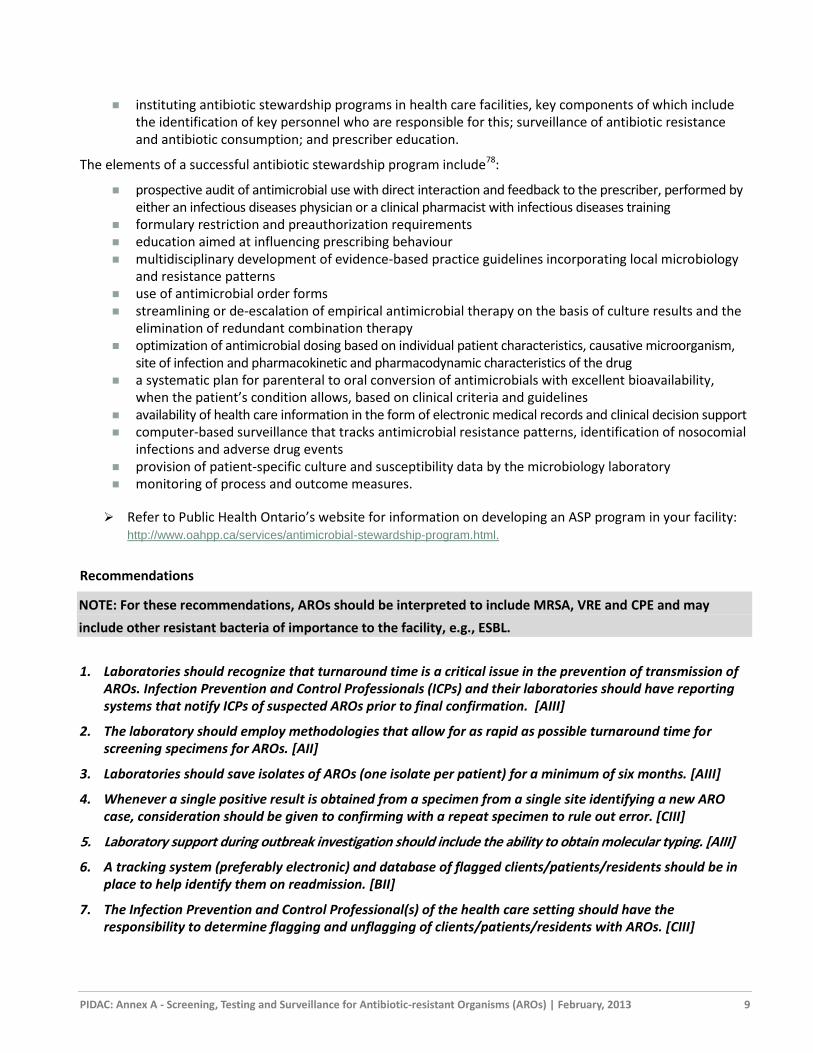
instituting antibiotic stewardship programs in health care facilities, key components of which include the identification of key personnel who are responsible for this; surveillance of antibiotic resistance and antibiotic consumption; and prescriber education.
The elements of a successful antibiotic stewardship program include78:
prospective audit of antimicrobial use with direct interaction and feedback to the prescriber, performed by either an infectious diseases physician or a clinical pharmacist with infectious diseases training
formulary restriction and preauthorization requirements education aimed at influencing prescribing behaviour multidisciplinary development of evidence-based practice guidelines incorporating local microbiology
and resistance patterns use of antimicrobial order forms streamlining or de-escalation of empirical antimicrobial therapy on the basis of culture results and the
elimination of redundant combination therapy optimization of antimicrobial dosing based on individual patient characteristics, causative microorganism,
site of infection and pharmacokinetic and pharmacodynamic characteristics of the drug a systematic plan for parenteral to oral conversion of antimicrobials with excellent bioavailability,
when the patient’s condition allows, based on clinical criteria and guidelines availability of health care information in the form of electronic medical records and clinical decision support computer-based surveillance that tracks antimicrobial resistance patterns, identification of nosocomial
infections and adverse drug events provision of patient-specific culture and susceptibility data by the microbiology laboratory monitoring of process and outcome measures.
Refer to Public Health Ontario’s website for information on developing an ASP program in your facility: http://www.oahpp.ca/services/antimicrobial-stewardship-program.html.
Recommendations
NOTE: For these recommendations, AROs should be interpreted to include MRSA, VRE and CPE and may
include other resistant bacteria of importance to the facility, e.g., ESBL.
1. Laboratories should recognize that turnaround time is a critical issue in the prevention of transmission of AROs. Infection Prevention and Control Professionals (ICPs) and their laboratories should have reporting systems that notify ICPs of suspected AROs prior to final confirmation. [AIII]
2. The laboratory should employ methodologies that allow for as rapid as possible turnaround time for screening specimens for AROs. [AII]
3. Laboratories should save isolates of AROs (one isolate per patient) for a minimum of six months. [AIII]
4. Whenever a single positive result is obtained from a specimen from a single site identifying a new ARO case, consideration should be given to confirming with a repeat specimen to rule out error. [CIII]
5. Laboratory support during outbreak investigation should include the ability to obtain molecular typing. [AIII]
6. A tracking system (preferably electronic) and database of flagged clients/patients/residents should be in place to help identify them on readmission. [BII]
7. The Infection Prevention and Control Professional(s) of the health care setting should have the responsibility to determine flagging and unflagging of clients/patients/residents with AROs. [CIII]
PIDAC: Annex A - Screening, Testing and Surveillance for Antibiotic-resistant Organisms (AROs) | February, 2013 10
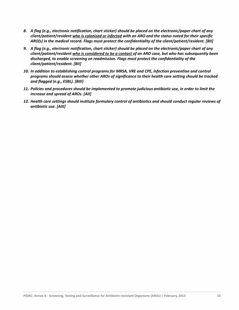
8. A flag (e.g., electronic notification, chart sticker) should be placed on the electronic/paper chart of any client/patient/resident who is colonized or infected with an ARO and the status noted for their specific ARO(s) in the medical record. Flags must protect the confidentiality of the client/patient/resident. [BII]
9. A flag (e.g., electronic notification, chart sticker) should be placed on the electronic/paper chart of any client/patient/resident who is considered to be a contact of an ARO case, but who has subsequently been discharged, to enable screening on readmission. Flags must protect the confidentiality of the client/patient/resident. [BII]
10. In addition to establishing control programs for MRSA, VRE and CPE, infection prevention and control programs should assess whether other AROs of significance to their health care setting should be tracked and flagged (e.g., ESBL). [BIII]
11. Policies and procedures should be implemented to promote judicious antibiotic use, in order to limit the increase and spread of AROs. [AII]
12. Health care settings should institute formulary control of antibiotics and should conduct regular reviews of antibiotic use. [AIII]
PIDAC: Annex A - Screening, Testing and Surveillance for Antibiotic-resistant Organisms (AROs) | February, 2013 11
Antibiotic-Resistant Organisms in Health Care Settings
A. Resistant Staphylococcus aureus
1. What is Staphylococcus aureus?
Staphylococcus aureus is an aerobic Gram-positive coccoid bacterium that periodically lives on the skin and mucous membranes of a large proportion of healthy adults (60% or more)79 without causing illness. These individuals are said to be ‘colonized’ with the microorganism. Ten to twenty per cent of people are persistently colonized with S. aureus.80 Those who are non-carriers and are never colonized with S. aureus are in the minority.79 Occasionally, S. aureus might be the cause of infections such as impetigo, carbuncles and abscesses or more invasive disease.81 S. aureus is the single most common cause of hospital-associated infection.
2. What is Methicillin-Resistant Staphylococcus aureus (MRSA)?
When S. aureus develops reduced susceptibility to the -lactam class of antibiotics (e.g., cloxacillin) it is known as methicillin-resistant Staphylococcus aureus (MRSA). While MRSA is more resistant to some treatments than methicillin-sensitive S. aureus (MSSA), there is little evidence to suggest that it is more pathogenic or virulent (i.e., more likely to cause infection or more severe infection) than MSSA. Infection with MRSA is associated with higher case fatality rates than MSSA.82, 83 Most experts believe that this is because infection with MRSA may result in greater delay in the time to initiation of appropriate therapy than infection with MSSA. MRSA may be either health care-associated or community-associated (CA-MRSA).
Community-associated MRSA (CA-MRSA) refers to strains linked to colonization and transmission in the community.84 There are two different definitions of CA-MRSA: one is based on epidemiology and one is based on microbiologic typing. Isolates of CA-MRSA are obtained from individuals who develop infections in the community and who have not had recent exposure to the health care system (epidemiologic definition). These are usually strains of MRSA (e.g., CMRSA-10) that are different from the MRSA strains found in hospitals (e.g., CMRSA-2), with a different methicillin-resistance gene (e.g., SCCmec IV, vs. SCCmec II)1 and often with additional virulence factors (microbiologic definition). However, hospital-type MRSA strains can be transmitted in the community and community-type MRSA strains can be transmitted in hospitals. For the purposes of managing MRSA in health care settings, the epidemiologic definition of CA-MRSA should be used.
3. Current Status of MRSA in Canada and Ontario
Though MRSA is not a reportable disease in Canada, laboratory-based surveillance of MRSA in sentinel Canadian hospitals has been carried out since 1995. The incidence of MRSA (infection and colonization) among admitted cases has increased steadily from 0.44 cases per 1,000 patient admissions in 199585 to 9.5 cases per 1,000 admissions in 2010,86 with most of the increase occurring in Ontario and Quebec. 86
In Ontario there were 19,962 patients identified with MRSA colonization or infection in 2011, a 5% decrease over 2010.87 Data on 56% of these patients indicated that 38% acquired MRSA in an acute care hospital, 14% in a nursing home and 44% in the community. This reflects a slight decrease in MRSA acquisition in institutions and a corresponding increase in community acquisition.
The number of reported MRSA bacteraemias in Ontario in 2011 was 560, a 13% increase over the 2010 number of 496. Overall, 17% of S. aureus isolates from blood cultures were MRSA, up from 15% in 2010.87

PIDAC: Annex A - Screening, Testing and Surveillance for Antibiotic-resistant Organisms (AROs) | February, 2013 12
4. MRSA Acquisition and Transmission
Risk factors for MRSA acquisition in the health care setting include invasive procedures, prior treatment with antibiotics, prolonged hospital stay, stay in an intensive care or burn unit, surgical wound infection and close proximity to a colonized client/patient/resident.83
MRSA is most commonly spread via the transiently colonized hands of health care workers who acquire it from contact with colonized or infected clients/patients/residents, or after handling contaminated material or equipment. Hand hygiene and environmental surface cleaning are, therefore, important measures to prevent transmission.5
Refer to PIDAC’s Best Practices for Hand Hygiene in All Health Care Settings88 for more information regarding hand hygiene. Available from: http://www.oahpp.ca/resources/pidac-knowledge/best-practice-manuals/hand-
hygiene.html.
Refer to PIDAC’s Best Practices for Environmental Cleaning for Prevention and Control of Infections in All Health Care Settings89 for more information regarding cleaning in health care environments. Available from: http://www.oahpp.ca/resources/pidac-knowledge/best-practice-manuals/environmental-cleaning-for-prevention-and-
control-of-infections.html.
Most items in the health care environment, especially those frequently touched by the hands of health care workers or clients/patients/residents have been shown to become contaminated with MRSA:
Contamination of environmental surfaces such as medical equipment, hospital furnishings, hydrotherapy tubs, linens, tourniquets, computer keyboards, faucets and nebulizers has been described. In some cases these may serve as a means of transmission in certain settings.5, 83, 90-92
The environment may be a factor for fomite transmission in any setting, particularly in special settings such as burn units or intensive care units.5
There is evidence that some individuals may act as ‘super-shedders’ of MRSA when co-infected with a respiratory virus and that they can spread MRSA via respiratory droplets (the ‘cloud’ phenomenon).83, 93-95
In some settings, such as intensive care units, chlorhexidine gluconate (CHG) baths have resulted in lower acquisition rates of MRSA. In intensive care settings, daily bathing of all patients with 4% CHG has been shown to reduce new acquisition of MRSA by 32% ,96 as well as reduce cases of bacteraemia with MRSA.96-98
5. What are VISA and VRSA?
Vancomycin-intermediate Staphylococcus aureus (VISA) is a strain of MRSA that has a reduced susceptibility to vancomycin with an MIC of 8 to16 mcg/ml.
Vancomycin-resistant Staphylococcus aureus (VRSA) is a strain of MRSA that contains the resistance genes Van-A or Van-B, with an MIC to vancomycin of ≥ 32 mcg/ml. To date all VRSA have contained vancomycin-resistance genes transferred from VRE strains.
Generally VISA and VRSA arise in patients who have been colonized or infected with MRSA and have received multiple or prolonged courses of vancomycin. Additionally, most cases have been co-colonized with MRSA and VRE for prolonged periods of time.

PIDAC: Annex A - Screening, Testing and Surveillance for Antibiotic-resistant Organisms (AROs) | February, 2013 13
6. VISA/VRSA Acquisition and Transmission
The emergence of vancomycin-intermediate Staphylococcus aureus (VISA) and vancomycin-resistant Staphylococcus aureus (VRSA) have the potential for serious public health consequences if transmission between patients occurs. However, although 12 cases of VRSA have been reported in the United States, eight of which occurred in southeast Michigan,99-101 initial fears of widespread dissemination of VRSA have not been realized despite years of co-circulation of MRSA and VRE in some jurisdictions, although the risk continues to exist.102
Because there is a lack of epidemiological data on the spread of VISA and VRSA, a more extensive form of the Contact Precautions as outlined in this annex is recommended for cases.13, 103
7. Current Status of VISA and VRSA in Canada and Ontario
Although there have been several cases of VISA and VRSA described in other countries,104-107 to date there have been no cases of VRSA reported in Canada and only a single reported case of VISA.108 Identification of VISA or VRSA must be treated as a sentinel event. The Medical Officer of Health may be advised non-nominally whenever VISA or VRSA is isolated. All isolates of VISA and VRSA should be forwarded to the public health laboratory for confirmation.
Each case of VISA/VRSA must be managed with Contact Precautions. Additional restrictions in client/patient/resident movement and limitations to visitors and non-essential staff are required.13, 109
8. Screening Patients/Residents for MRSA
RISK FACTORS FOR MRSA ACQUISITION:
Definite Risk Factor
Previous colonization or infection with MRSA
>12 hours in any health care facility (including this one) in the past 12 months
Recent exposure to unit/area of a health care facility having an MRSA outbreak
Health care in another country
Possible Risk Factor
Home health care
Indwelling device
ICU, burn unit, transplant unit
Communal setting
Injection drug use
Household contact of patient with MRSA
Immunocompromised
CA-MRSA risk (e.g., sports teams)

PIDAC: Annex A - Screening, Testing and Surveillance for Antibiotic-resistant Organisms (AROs) | February, 2013 14
Regulated health professionals in health care facilities are expected to take screening specimens from clients/patients/residents at increased risk for MRSA on admission as part of an MRSA prevention and control program62-67, 110:
The following clients/patients/residents are at increased risk for MRSA and should be screened at admission for MRSA: o those who have previously been colonized or infected with MRSA111, 112 o those who have spent time in a health care facility outside of Canada in the last 12 months o those who have been admitted to, or who have spent more than 12 continuous hours as a
client/patient/resident in, any health care facility in the past 12 months12, 113 o those transferred between health care facilities (e.g., between hospitals or between a long-term
care facility and a hospital)114 o those who have recently been exposed to a unit/area of a health care facility with an MRSA
outbreak o other high-risk client/patient/resident populations as identified by the ICP(s) (e.g., internal
transfers, such as admission to an intensive care unit) or Public Health.
Based on local epidemiology and risk factors, additional individuals may be considered for MRSA screening: o those receiving home health care services in the past year o those receiving treatment with an indwelling medical device115-117 o those receiving care in intensive care units, transplant units, burn units66, 95 o those living in a communal setting (e.g., shelter, halfway home, correctional facility118) o those with a history of injection drug use119, 120 o those who are household contacts of people with MRSA121-123 o those who are immunocompromised124, 125 o individuals from populations where community-associated MRSA is known to be a problem
(e.g., organized sports teams).126-128
Monitor changes in the local epidemiology and local risk factors for MRSA and adjust screening accordingly.
9. Screening Contacts of MRSA Cases
An MRSA contact is a client/patient/resident who has been a roommate or has been in physical contact with a client/patient/resident subsequently found to have MRSA (i.e., once MRSA is identified in a client/patient/resident, all previous roommates become new contacts). In an outbreak, a contact is a client/patient/resident who has common risk factors to cases (e.g., same unit, same procedure, same staff).
Any client/patient/resident who is considered to be an MRSA contact should have follow-up screening specimens, with at least two specimens taken on different days, with one taken a minimum of seven days following the last exposure.59, 129, 130
Client/patient/resident contacts should be re-screened when new cases of MRSA continue to be identified despite active control measures.58
See Appendix D, ‘Sample Investigation Protocols for MRSA and VRE in Acute Care Facilities’, for a sample investigation protocol that may be used following identification of MRSA in your facility.
See Section VI, ‘Managing Outbreaks’, for more information regarding contacts.

PIDAC: Annex A - Screening, Testing and Surveillance for Antibiotic-resistant Organisms (AROs) | February, 2013 15
10. Point Prevalence Screening
A point prevalence screen is the collection of specimens on all clients/patients/residents at a single point in time, to determine the total number of cases and evidence of ongoing transmission of a particular microorganism:
Point prevalence screens should be conducted on units/areas where clients/patients/residents are at high risk for acquiring MRSA during their stay in the health care setting.13, 57, 65
Clients/patients/residents at high risk include those on burn units or other high-risk units such as intensive care units, transplantation units, or other units as defined by the ICP.
Point prevalence screens should be conducted, and should continue to be conducted, until no further transmission is detected; in general this means at least two prevalence screens, taken after the last transmission was detected and at least a week apart, in any area where MRSA transmission is occurring.13, 57
See Appendix D, ‘Sample Investigation Protocols for MRSA and VRE’, for guidance in conducting prevalence screens.
Refer to PIDAC’s Best Practices for Surveillance of Health Care-Associated Infections in Patient and Resident Populations2 for surveillance methodologies. Available from: http://www.oahpp.ca/resources/pidac-
knowledge/best-practice-manuals/surveillance-of-health-care-associated-infections.html.
11. Screening Staff for MRSA
Screening staff for MRSA should be considered when an outbreak of the same strain of MRSA continues despite adherence to control measures,57, 131 or when a staff member is epidemiologically linked to new acquisitions of MRSA.132 Staff who are concerned about exposure to, or may be colonized with, MRSA should receive assessment and counselling from their Occupational Health department or other area that will protect the confidentiality of the individual.
In the event of an MRSA outbreak, heightened surveillance for skin and soft tissue infections in staff is warranted (e.g., folliculitis, paronychia).
Refer to the OHA/OMA publication, Antibiotic Resistant Organisms Surveillance Protocol for Ontario Hospitals132 for more information about the management of health care workers exposed to MRSA and VRE (available at: http://www.oha.com/Services/HealthSafety/Documents/Protocols/Antiobiotic%20Resistant%20Organisms%20Revised%2
0June%202011.pdf).
12. Collection and Timing of Specimens for MRSA
MRSA may not be identified in some clients/patients/residents when they are colonized at a level that is too low to be detected by culture. In these clients/patients/residents, MRSA will not be detected until the microbial population has increased over a period of time. One study found that MRSA acquired from a roommate was not detectable until 9-10 weeks following the exposure.129 This study suggested that post-exposure screening continue until six months post-exposure.
Molecular testing methods, such as polymerase chain reaction (PCR), have a shorter turnaround time,133 may be more sensitive and may detect lower levels of colonization than traditional culture methods, but may result in more false-positive results due to lower specificity.134, 135 Cultures should be used to confirm positive PCR results.136
The PCR assay has been validated for both nasal and non-nasal specimens.135, 137 Specimens from the anterior nares have been shown to result in the highest yield of MRSA138, with some studies indicating a sensitivity of

PIDAC: Annex A - Screening, Testing and Surveillance for Antibiotic-resistant Organisms (AROs) | February, 2013 16
over 90%.133, 139 However, MRSA has been identified exclusively from the perianal/perineal area in some patients (2% -19% in various studies)59, 139-141 as well as the groin.142 Several studies of PCR assays have shown a better yield of MRSA when both nares and perianal/perineal sites are sampled, with up to 96% sensitivity.134, 135,
137 A combination of nares and perianal/perineal cultures is recommended for highest yield of MRSA, even if PCR testing is used.
If community-associated MRSA (CA-MRSA) is suspected, cultures of recurrent furuncles, abscesses or other skin lesions should be considered in addition to the sites noted above.1 For children and youth, throat swabs may have greater sensitivity than nasal swabs alone for detecting MRSA.122, 143 When screening newborn infants, a swab from the umbilicus should be taken.144
Non-nasal specimens may be negative for MRSA in patients who have recently had an antimicrobial bath.96, 145 Specimens may be falsely negative if the patient is on an antibiotic to which the microorganism is sensitive. Surveillance specimens should be taken only after the antibiotic has been discontinued for at least 48 hours.
Specimens for detection of MRSA should include:
a swab from the anterior nares; AND
a swab from the perianal, perineal or groin area (perianal preferred); AND
a swab(s) from skin lesions, wounds, incisions, ulcers and exit sites of indwelling devices, if present, using aseptic technique where indicated;
for newborn infants, a swab from the umbilicus should also be taken for MRSA.
See Appendix A, ‘Collecting Specimens for MRSA, VRE, CPE and ESBL’, for instruction in obtaining specimens for MRSA.
13. MRSA Decolonization
Decolonization refers to the use of topical agents, such as nasal antimicrobial ointment and body wash and/or oral antibiotics, to remove resistant bacteria from a colonized individual.
CLIENT/PATIENT/RESIDENT DECOLONIZATION
Decolonization has been used, along with other measures, to help control the spread of MRSA in some centres62,
65 and may be considered when a colonized client/patient/resident is implicated in an outbreak, but this should be done in consultation with the health care setting’s ICP.13, 57, 125
Short term success at decolonization may be achievable. In a 2007 Canadian study, MRSA colonization was eradicated for at least three months with a combination of treatments consisting of topical mupirocin, chlorhexidine gluconate (CHG) washes, oral rifampin and oral doxycycline.145
MRSA decolonization failure is related to several factors:
presence of a skin lesion146 presence of indwelling devices146 receipt of immunosuppressive therapy146 receipt of hemodialysis146 mupirocin resistance.145, 147
Current evidence does not recommend widespread or prolonged antibiotic therapy for decolonization of MRSA as this may promote antibiotic resistance, long-term efficacy is poor and systemic therapy may lead to adverse events.1, 13, 125, 147-149 Decolonization therapy with topical antibiotics alone is not effective.

PIDAC: Annex A - Screening, Testing and Surveillance for Antibiotic-resistant Organisms (AROs) | February, 2013 17
If decolonization therapy is attempted, attention must be given to scrupulously cleaning the client/patient/resident’s environment in order to decrease the risk of re-colonization, as the environment can play a role in transmission. Long term surveillance (e.g., monthly) is recommended to detect relapse or re-colonization.
STAFF DECOLONIZATION
The risk of staff acquisition of MRSA is low and is significantly reduced if staff follow Routine Practices, perform hand hygiene and wear PPE appropriately.95 Most experts believe that with adequate adherence to hand hygiene and Routine Practices, there is no risk of staff acquisition of MRSA. When other measures have failed, treating healthcare workers who are colonized or infected with the outbreak strain of MRSA and who are epidemiologically implicated in an outbreak has been shown to help control the outbreak.57 The benefit of decolonization is unclear if staff are colonized or infected with a strain of MRSA that is different from the outbreak strain.
Refer to the OHA/OMA publication, Antibiotic Resistant Organisms Surveillance Protocol for Ontario Hospitals,132 for more information about the management of health care workers exposed to MRSA and VRE (available at: http://www.oha.com/Services/HealthSafety/Documents/Protocols/Antiobiotic%20Resistant%20Organisms%20Revised%
20June%202011.pdf).
B. Resistant Enterococci
1. What are Enterococci?
Enterococci are facultative anaerobic Gram-positive coccoid bacteria that live in the gastrointestinal tract of most individuals and can also be present in the anterior urethra, vagina, skin, oropharynx and/or bile. Enterococci may also colonize wounds, ulcers and medical device sites in hospitalized patients,111 and are a common cause of health care-associated infection.
2. What are Vancomycin-Resistant Enterococci (VRE)?
Vancomycin-resistant enterococci (VRE) are strains of Enterococcus faecium and Enterococcus faecalis that have become resistant to high levels of the antibiotic vancomycin. The majority of individuals who have VRE are colonized with it. In some high-risk patient populations (e.g., those with haematological malignancies), there are higher rates of VRE bacteraemia after colonization and higher mortality associated with VRE bacteraemia compared to VSE bacteraemia.26-28 For more information on VRE, see PIDAC’s “Review of Literature for Evidence-based Best Practices for VRE Control”, available at: http://www.oahpp.ca/resources/pidac-knowledge/best-practice-manuals/review-of-literature-for-evidence-based-best-practices-for-VRE-control.html.
3. Current Status of VRE in Canada and Ontario
Results from the passive reporting network for VRE in Canada show that VRE infection rates have risen sharply since 2007.150 The incidence of VRE infections was 0.06 cases per 1,000 admissions in 2006 and 0.5 cases per 1,000 admissions in 2011. The VRE colonization rate has had a comparable increase, with 857 patients colonized with VRE in 2006 and 5,515 patients colonized with VRE in 2011.
In Ontario, the incidence of VRE has increased, with 7,643 patients colonized or infected with VRE in 2011 compared to 5,567 patients in 2010, a 37% increase.87 The number of patients with VRE bacteraemia doubled, from 28 patients in 2010 to 57 patients in 2011. The majority of patients were thought to have acquired VRE in

PIDAC: Annex A - Screening, Testing and Surveillance for Antibiotic-resistant Organisms (AROs) | February, 2013 18
acute-care hospitals (85%), 5% were thought to have acquired VRE in nursing homes and 7% were acquired in the community. These proportions have not changed significantly over time.
4. VRE Acquisition and Transmission
RISK FACTORS FOR VRE ACQUISITION:
Definite Risk Factor
Previous colonization or infection with VRE
>12 hours in any health care facility (including this one) in the past 12 months
Recent exposure to unit/area of a health care facility having a VRE outbreak
Health care in another country
Possible Risk Factor
Recent exposure to 2nd- and third-generation cephalosporins
Risk factors for VRE acquisition include severity of underlying illness, presence of invasive devices, prior colonization with VRE, antibiotic use and length of hospital stay.111
VRE is most commonly spread via the transiently colonized hands of health care workers who acquire it from contact with colonized or infected clients/patients/residents151, or after handling contaminated material or equipment. Hospitalized patients with gastrointestinal carriage of VRE are the major reservoir.152
Refer to PIDAC’s Best Practices for Hand Hygiene in All Health Care Settings88 for more information regarding hand hygiene. Available from: http://www.oahpp.ca/resources/pidac-knowledge/best-practice-manuals/hand-
hygiene.html
VRE transmission via environmental sources is well recognized and includes most items in the health care environment, such as blood pressure cuffs, electronic thermometers, monitoring devices, stethoscopes, call bells and bed rails.153 Contamination of the environment with VRE is more likely when a client/patient/resident has diarrhoea.151, 153
In some settings, such as intensive care units, chlorhexidine gluconate (CHG) baths have resulted in lower acquisition rates of VRE. In intensive care settings, daily bathing of all patients with 4% CHG has been shown to reduce new acquisition of VRE by 50%,96 as well as reduce cases of bacteraemia with VRE.96, 98
Refer to PIDAC’s Best Practices for Environmental Cleaning for Prevention and Control of Infections in All Health Care Settings89 for more information regarding cleaning in health care environments. Available from: http://www.oahpp.ca/resources/pidac-knowledge/best-practice-manuals/environmental-cleaning-for-prevention-and-
control-of-infections.html.

PIDAC: Annex A - Screening, Testing and Surveillance for Antibiotic-resistant Organisms (AROs) | February, 2013 19
5. Screening Patients/Residents for VRE
Regulated health professionals in health care facilities are expected to take screening specimens from clients/patients/residents at increased risk for VRE on admission as part of a VRE prevention and control program62-67, 110:
The following clients/patients/residents are at increased risk for VRE and should be screened at admission for VRE:
o those who have previously been colonized or infected with VRE111, 112 o those who have spent time in a health care facility outside of Canada in the last 12 months o those who have been admitted to, or who have spent more than 12 continuous hours as a
client/patient/resident in, any health care facility in the past 12 months12, 113 o those transferred between health care facilities (e.g., between hospitals or between a long-
term care facility and a hospital)114 o those who have recently been exposed to a unit/area of a health care facility with a VRE
outbreak o other high-risk client/patient/resident populations as identified by the ICP(s) (e.g., internal
transfers, such as admission to an intensive care unit) or Public Health
Monitor changes in the local epidemiology and local risk factors for VRE and adjust screening accordingly.
6. Screening Contacts of VRE Cases
A VRE contact is:
a) a patient/resident who has been a roommate or has been in physical contact with an unidentified client/patient/resident subsequently found to have VRE (i.e., once VRE is identified in a client/patient/resident, all previous roommates become new contacts);
b) a patient/resident admitted to a room previously occupied by a patient/resident who has been identified with VRE and which was not cleaned according to the facility’s protocol for cleaning a room contaminated with VRE154; and/or
c) a patient/resident who has common risk factors to cases during an outbreak (e.g., same unit, same procedure, same staff).
VRE contacts should:
have follow-up specimens, with at least two specimens taken on different days, with one taken a minimum of seven days following the last exposure to VRE
be re-screened when new cases of VRE continue to be identified despite active control measures.
See Appendix D, ‘Sample Investigation Protocols for MRSA and VRE in Acute Care Facilities’, for a sample investigation protocol that may be used following identification of VRE in your facility.
See Section VI, ‘Managing Outbreaks’, for more information regarding contacts.
7. Point Prevalence Screening
A point prevalence screen is the collection of specimens on all patients/residents in a specified area at a single point in time, to determine the total number of cases of a particular microorganism and to identify evidence of ongoing transmission.
Point prevalence screens should be conducted on units/areas where clients/patients/residents are at high risk for acquiring VRE during their stay in the health care setting.13

PIDAC: Annex A - Screening, Testing and Surveillance for Antibiotic-resistant Organisms (AROs) | February, 2013 20
Clients/patients/residents at high risk include those on dialysis units or other high-risk units such as intensive care units, transplantation units, or other units as defined by the ICP(s).
Point prevalence screens should be conducted in any area where VRE transmission is occurring and should continue to be conducted until no further transmission is detected13; in general, this means at least two prevalence screens taken at least one week apart after the last transmission was detected.
See Appendix D, ‘Sample Investigation Protocols for MRSA and VRE’, for guidance in conducting prevalence screens.
Refer to PIDAC’s Best Practices for Surveillance of Health Care-Associated Infections in Patient and Resident Populations2 for surveillance methodologies. Available from: http://www.oahpp.ca/resources/pidac-
knowledge/best-practice-manuals/surveillance-of-health-care-associated-infections.html.
8. Screening Staff for VRE
The risk of staff colonization with VRE is extremely low and there is no evidence to support the need to screen staff for VRE.
9. Collection and Timing of Specimens for VRE
Detection of VRE is best with stool specimens, as they provide a higher yield than rectal swabs.13, 155 In the absence of stool, rectal swabs may be used.156-158 If a client/patient/resident has a colostomy, the VRE specimen may be taken from the colostomy output.
See Appendix A, ‘Collecting Specimens for MRSA, VRE, CPE and ESBL’, for instruction in obtaining specimens for VRE.
Specimens may be falsely negative if the patient is on an antibiotic to which the microorganism is sensitive. Surveillance specimens should be taken only after the antibiotic has been discontinued for at least 48 hours.
Molecular testing methods, such as polymerase chain reaction (PCR), may have certain advantages:
improved turnaround time for results, particularly for VRE157, 159 increased sensitivity, i.e., detection of lower levels of colonization.156, 158, 159
False-positive results may occur with both VRE culture and PCR testing, due to:
laboratory contamination presence of nonviable VRE156 presence of vanB in nonenterococcal microorganisms.158, 160, 161
10. VRE Decolonization
VRE decolonization is not effective and not recommended.162
PIDAC: Annex A - Screening, Testing and Surveillance for Antibiotic-resistant Organisms (AROs) | February, 2013 21
C. Extended-Spectrum Beta-Lactamase (ESBL)-Producing Bacteria
1. What are ESBLS?
Beta-lactamase (-lactamase) is an enzyme produced by some bacteria that inactivates the -lactam class of antibiotics (e.g., penicillins, cephalosporins). Extended-spectrum -lactamase acts on all cephalosporins, including third-generation cephalosporins such as cefotaxime, ceftriaxone and ceftazidime, as well as the monobactam aztreonam.
Most extended-spectrum beta-lactamase production occurs in the Enterobacteriaceae Escherichia coli (E. coli) and Klebsiella pneumoniae.55 The significance of ESBL-production in bacteria that are a common cause of urinary tract infections and bacteraemia is that antibiotic treatment options are limited for these infections.
2. Current Status of ESBL-Producing Bacteria in Ontario
While third-generation cephalosporin-resistant isolates have been reported from all areas of the province, resistance is most prevalent in Toronto and surrounding areas. In 2011, 40% of hospitals carried out a regular screening program for ESBLs.87 The most common protocol is to screen roommate(s) of colonized and/or infected patients/residents once a case is identified and screening patients with a history of admission in another country.
3. Acquisition and Transmission of ESBL-Producing Bacteria
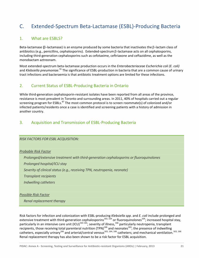
RISK FACTORS FOR ESBL ACQUISITION:
Probable Risk Factor
Prolonged/extensive treatment with third-generation cephalosporins or fluoroquinolones
Prolonged hospital/ICU stay
Severity of clinical status (e.g., receiving TPN, neutropenia, neonate)
Transplant recipients
Indwelling catheters
Possible Risk Factor
Renal replacement therapy
Risk factors for infection and colonization with ESBL-producing Klebsiella spp. and E. coli include prolonged and extensive treatment with third-generation cephalosporins163, 164 or fluoroquinolones164; increased hospital stay, particularly in an intensive care unit (ICU)163-165; severity of illness,166 particularly neutropenia, transplant recipients, those receiving total parenteral nutrition (TPN)166 and neonates163; the presence of indwelling catheters, especially urinary166 and arterial/central venous163, 164, 166 catheters; and mechanical ventilation.163, 166 Renal replacement therapy has also been shown to be a risk factor for ESBL acquisition.
PIDAC: Annex A - Screening, Testing and Surveillance for Antibiotic-resistant Organisms (AROs) | February, 2013 22
The incidence of ESBL acquisition in the community is increasing, with one-third of cases having reported no association with health care.167, 168 Recent evidence also suggests that the risk of ESBL transmission in households is high.169
The lower digestive tract of colonized patients is the main reservoir for ESBL-producing bacteria. Gastrointestinal carriage can persist for months.53 It has been suggested that factors that facilitate cross-infection among patients have the most relevant role in the acquisition of ESBL-producing bacteria.166 Patient-to-patient transmission of ESBL-producing bacteria occurs primarily via the hands of staff.53, 170 The appropriate use of Routine Practices and Additional Precautions, along with antimicrobial stewardship, have been shown to halt the spread of ESBL-producing bacteria in an outbreak.55, 171
ESBL-producing Gram-negative bacteria can survive in the health care environment,172 but the environment has rarely been implicated in outbreaks and the role of environmental surface contamination as a source of hospital infection is controversial.173 In one study, however, the implementation of sink and faucet scouring in a neonatal intensive care unit did halt an outbreak of ESBL-producing Klebsiella pneumoniae.163
4. Screening Patients/Residents for ESBL-Producing Bacteria
Local epidemiology should govern decision-making regarding routine screening of patients/residents for ESBL-producing bacteria. If the local prevalence of ESBL-producing bacteria is high, there is some value to routinely screening patients, particularly those admitted to ICUs.171, 174, 175
An effective and consistent approach to surveillance is an important measure to prevent and control the spread of ESBLs. In an ESBL outbreak, protocols should be in place for screening patients in close proximity to colonized/infected patients (e.g., roommates) who may have been exposed or who have risk factors for ESBL acquisition.51, 173
Patients with known ESBL carriage should have their records flagged and be placed on Contact Precautions and re-screened on readmission.173
5. Screening Staff for ESBL-Producing Bacteria
Routine screening of staff for ESBL is not recommended. Although staff with Gram-negative bacterial hand colonization (e.g., staff with artificial nails) have been implicated in infections and outbreaks, there is no evidence that rectal colonization of health care providers contributes to transmission.
6. Collection and Timing of Specimens for ESBL-Producing Bacteria
A substantial percentage of patients/residents who develop health care-associated ESBL infections have preceding colonization of the gastrointestinal tract.175-177 The preferred specimen for ESBL screening is a rectal swab or stool. A urine culture may also be sent in certain situations (e.g., catheterized patient/resident). In one study, the inguinal area was found to be colonized with ESBL-producing bacteria when the perianal area and urine were negative.178
7. ESBL Decolonization
ESBL decolonization is generally not effective and not recommended.173 Although there is a small study of 15 patients that shows efficacy of decolonization at follow-up,179 there is not enough evidence to recommend this.

PIDAC: Annex A - Screening, Testing and Surveillance for Antibiotic-resistant Organisms (AROs) | February, 2013 23
D. Carbapenemase-producing Enterobacteriaceae (CPE)
1. What are CPE?
Carbapenemase-producing Enterobacteriaceae are Enterobacteriaceae that are resistant to carbapenem antimicrobials (e.g., imipenem, meropenem, ertapenem) through the production of carbapenemase enzymes. To date, carbapenemases have been found most commonly in E. coli and Klebsiella spp., but have also been found in other Gram-negative species.
Carbapenemases are a class of enzymes that inactivate carbapenem antibiotics by hydrolysing them. In almost all instances, these enzymes hydrolyse not only carbapenems, but also first- , second- and third-generation cephalosporins and penicillins (e.g., piperacillin-tazobactam). The genetic information to produce carbapenemases is often located on a mobile genetic element (i.e., a genetic element that can move between bacterial strains and species, e.g., plasmid, transposon), which frequently also carries resistance to other classes of antimicrobials, such as fluoroquinolones and aminoglycosides.
There are several different classes of carbapenemase. Each class has a three-letter acronym. These enzymes evolve rarely, but bacteria carrying them spread easily. Particular classes of carbapenemases are usually most common in the geographic area where they evolved, but spread around the world, usually when patients have received health care in another country. Enzymes other than NDM have almost exclusively been found in hospitals. NDM has been found in both hospitals and the community, particularly in the Indian subcontinent. Table 1 describes the most common classes of carbapenemases.
TABLE 1: MOST COMMON CARBAPENEMASES: DISTRIBUTION AND MOLECULAR EPIDEMIOLOGY
Enzyme Geographic distribution Molecular epidemiology
KPC First reported in North Carolina in 1999.180
Now prevalent in
hospitals on the U.S. Eastern seaboard,181, 182
Israel183
and
Greece,184
but has been reported from many hospitals around
the world, including several Ontario hospitals.185, 186
Transmission has occurred in at least one Ontario hospital.187
Mostly in Klebsiella pneumoniae, although
plasmid spread has occurred to E. coli188
and other Enterobacteriaceae. Both clonal
and plasmid outbreaks have been
described.189
NDM Widespread in Enterobacteriaceae in hospitals in the Indian
subcontinent and also appears to be spreading in the
community. Imported cases from the Indian subcontinent to
hospitals in many countries around the world have been
reported.190-192
Cases have been identified in several Ontario
hospitals,193
including cases apparently acquired in Ontario.
Plasmid spread among strains and species
is very common, but clonal outbreaks also
occur.
VIM Scattered globally, with increased prevalence in Greece. Plasmid spread is most common.
Outbreaks have been described not only in
Enterobacteriaceae, but also Pseudomonas
aeruginosa.
Abbreviations:
KPC, Klebsiella pneumoniae carbapenemase
NDM, New Delhi metallo--lactamase
VIM, Verona integron-encoded metallo--lactamase
[Adapted from HPA Advice on Carbapenemase Producers, available at: http://www.hpa.org.uk/web/HPAwebFile/HPAweb_C/1294740725984]

PIDAC: Annex A - Screening, Testing and Surveillance for Antibiotic-resistant Organisms (AROs) | February, 2013 24
Testing bacteria for the presence of carbapenemases is challenging. Current routine testing may fail to detect resistance. Laboratories are working on better methods to ensure that these enzymes are reliably detected and reported. The information below may assist in the interpretation of results.
E. coli and Klebsiella spp. that are resistant to carbapenems on routine microbiology laboratory testing should be assumed to be carbapenemase producers until proven otherwise. Some isolates that initially appear to be susceptible to carbapenems may also produce carbapenemases, so that laboratories may need to issue corrected reports after further testing.
Not all resistance to carbapenems is conferred by carbapenemase production. Some Gram-negative bacteria may be resistant to carbapenems by other means. However, if resistance to carbapenems is detected, they should be tested to determine if they produce carbapenemases. These bacteria include:
Proteeae (Proteus spp. and Providencia spp.) Enterobacter spp. Acinetobacter spp. Pseudomonas aeruginosa
Because CPE are resistant to all penicillins, cephalosporins, carbapenems and other classes of antimicrobials, treatment of infections with CPE is difficult and involves the use of antibiotics with poor adverse event profiles (e.g., colistin). The case fatality rate for serious infections may be as high as 50%.
2. Current Status of CPE in Ontario
Most patients with CPE have had links to hospitals with recognized epidemic or endemic CPE (e.g., New York City hospitals with KPC K. pneumoniae, receipt of health care in the Indian subcontinent). However, transmission of CPE has been reported in Ontario,187 including two small outbreaks.194, 195 In 2011, 43 patients were identified with CPE, predominately in central Ontario and metropolitan Toronto.87 In the first three quarters of 2012, 47 patients were identified with newly confirmed CPE.196
3. Acquisition and Transmission of CPE
Although data are sparse, it seems likely that risk factors for infection and colonization with CPE will be similar to those of other resistant Gram-negative bacteria, such as ESBL-producing E. coli and Klebsiella pneumoniae.
At the present time the major risk factor appears to be receipt of care in health care settings that have CPE, e.g., hospitals along the U.S. eastern seaboard, particularly New York City (KPC), Greece (KPC), Israel (KPC) and the Indian subcontinent (NDM-1). However, CPE outbreaks are being increasingly described in hospitals around the world, including Canada.194, 195 People coming from the Indian subcontinent, with or without exposure to health care, are also at risk.
Transmission is likely via direct and indirect contact. The site of colonization is the lower gastrointestinal tract. Although the environment has rarely been implicated in outbreaks, sinks and other environmental surfaces have more recently been implicated in the transmission of Klebsiella and Pseudomonas spp.
Acquisition of resistance may also occur by transmission of the mobile genetic element carrying the carbapenemase between different bacterial strains and species.
PIDAC: Annex A - Screening, Testing and Surveillance for Antibiotic-resistant Organisms (AROs) | February, 2013 25
4. Screening Patients/Residents for CPE
All infection prevention and control programs should review with their microbiology laboratories whether they have had any cases of CPE in the past six months to one year and determine if their laboratory is able to detect and report all patients colonized or infected with CPE.197, 198 Isolation of CPE should be considered a critical laboratory result.
An effective and consistent approach to surveillance is an important measure to prevent and control the spread of CPE. All health care facilities should institute a screening program and targeted surveillance for CPE.198 In particular, admission screening and pre-emptive Contact Precautions are indicated for individuals with risk factors for CPE.197-199 Patients who have received health care outside of the country or who are known contacts of CPE should be screened.
If a single patient/resident with CPE is identified, a full unit/ward prevalence screen should be conducted. If screening of the full unit/ward is not feasible, due to size, screening of patients/residents in close proximity to the identified patient/resident, e.g., area with shared staffing assignments, should be strongly considered.
As a minimum, roommates should be screened for CPE.197, 198 If there is evidence of transmission of a single species (i.e., two or more patients with the same CPE strain), or two or more CPE-positive patients carrying two different bacterial species (i.e., suspected plasmid transmission), outbreak measures should be put into place and expert advice should quickly be sought (e.g., academic health sciences centre, medical microbiologist, reference laboratory services, local public health unit, regional infection control networks) to assist with determination of an outbreak.
In a CPE outbreak, there should be a full unit/ward prevalence screen. Periodic prevalence screening, e.g., weekly, should continue until no new cases are identified, with at least three negative prevalence screens after the last new case. Patients/residents with CPE and their roommates should be placed on Contact Precautions. The unit/ward should be closed to admissions and transfers out, unless medically necessary. Absolute cohorting of patients/residents, staff and equipment is essential to preventing further transmission of CPE.
Patients/residents who have been transferred from the unit/ward should be screened and be placed on Contact Precautions pending screening results. For patients/residents who have been transferred to another facility, the facility should be informed and the patient/resident should be screened.
Patients with known CPE carriage should have their records flagged, should be placed on Contact Precautions197 and should be re-screened on readmission.
The isolation of CPE should be considered to be a critical laboratory result.
5. Screening Staff for CPE
Routine screening of staff for CPE is not recommended. There is no evidence that rectal colonization of health care providers contributes to transmission.
6. Screening Specimens for CPE
Primary screening specimens for CPE are stool or rectal swabs. Urine specimens and swabs from open wounds may also be indicated.199 In critical care settings, sputum or endotracheal tube specimens and swabs from exit sites may be indicated.

PIDAC: Annex A - Screening, Testing and Surveillance for Antibiotic-resistant Organisms (AROs) | February, 2013 26
7. CPE Decolonization
There are insufficient data to support routine CPE decolonization and it is not recommended. In an uncontrolled outbreak, decolonization may be considered to attempt to reduce the bioburden.200
Recommendations
NOTE: For these recommendations, AROs should be interpreted to include MRSA, VRE and CPE and may
include other resistant bacteria of importance to the facility, e.g., ESBL.
13. Each health care setting should have a prevention and control program for AROs. [AII]
14. Clients/patients/residents should receive health care based on their overall care needs, despite colonization with AROs. [BII]
15. Screening for risk factors for MRSA, VRE and CPE should include a screening tool that is applied to all clients/patients/residents admitted to the health care facility. [AII]
16. Regulated health professionals in health care facilities are expected to take screening specimens from clients/patients/residents at increased risk for AROs on admission as part of an ARO prevention and control program. [AII]
17. Every effort should be made to try to determine the source of new cases of MRSA, VRE and CPE. Every new case should warrant an investigation. [AIII]
18. All affected health care settings should be notified following the identification of a case of an ARO, or identification of a new contact of a case. [AIII]
19. Any client/patient/resident who is considered to be an MRSA, VRE or CPE contact should have at least one set of screening specimens taken. If initial specimens are negative, it is prudent to repeat them. [BIII]
20. During an outbreak, all client/patient/resident contacts with common risk factors should be actively screened. [BIII]
21. Consideration should be given to conducting point prevalence screens on units/areas where clients/patients/residents are at high risk for acquiring MRSA, VRE or CPE during their stay in the health care setting. [BIII]
22. Point prevalence screens should be conducted in any area where MRSA, VRE or CPE transmission is occurring and should continue to be conducted until no further transmission is detected. [BIII]
23. Screening staff for MRSA should be considered when an outbreak of the same strain of MRSA continues to spread despite adherence to control measures, or when an individual is strongly epidemiologically linked to new acquisitions of MRSA. [BII]
24. Specimens for detection of MRSA should include: [AII]
a. A swab from the anterior nares; AND
b. A swab from the perianal, perineal or groin area (perianal preferred); AND
c. A swab(s) from skin lesions, wounds, incisions, ulcers and exit sites of indwelling devices, if present, using aseptic technique where indicated.
d. For newborn infants, a swab from the umbilicus should also be taken for MRSA.
25. Specimens for detection of VRE should include stool or a rectal swab. Stool specimens provide a higher yield. [AII]

PIDAC: Annex A - Screening, Testing and Surveillance for Antibiotic-resistant Organisms (AROs) | February, 2013 27
26. Specimens for detection of CPE should include stool or a rectal swab. [AIII]
27. Routine decolonization therapy of MRSA clients/patients/residents is not currently recommended. [AIII]
28. VRE, CPE or ESBL decolonization is not effective and is not recommended. [AI]
29. In situations where a client/patient/resident colonized with MRSA is implicated in an outbreak, decolonization may be considered in consultation with the health care setting’s Infection Prevention and Control Professional. [BIII]
30. Decolonization of staff colonized with MRSA should be done when they are epidemiologically linked to an outbreak with the same strain and adherence to Contact Precautions has failed to contain the outbreak. [AII]
31. The health care setting’s Infection Prevention and Control Professional(s) and senior management must be notified whenever CPE, VISA or VRSA is identified. [AIII]
32. Expert advice should be sought whenever CPE, VISA or VRSA is isolated (e.g., infection prevention and control experts from academic health sciences centres, public health, the regional infection control networks and reference laboratory services). [AIII]
33. In addition to Routine Practices and all of the previous recommendations, Additional Precautions for CPE, VISA and VRSA must include: [AII]
Single room accommodation is required.
Dedicated equipment and supplies are required.
Minimize the number of persons who enter the room.
Patient must remain in their room except for essential procedures.
Transfer between facilities should only be done if medically necessary. The receiving health care setting must be advised of the required precautions.
Avoid transfer within the facility if possible; if transfer is necessary for medical reasons, the receiving unit or department must be advised of the required precautions.
Each patient contact must be placed on Contact Precautions and be screened.
34. Every attempt should be made to identify the source of VISA or VRSA. [AII]
35. Health care settings should assess their local ESBL epidemiology to determine whether a specific ESBL control program is warranted. [CIII]
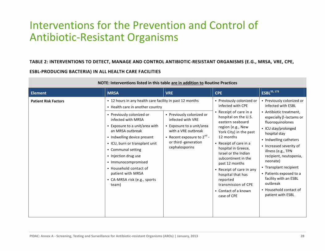
PIDAC: Annex A - Screening, Testing and Surveillance for Antibiotic-resistant Organisms (AROs) | January, 2013 28
Interventions for the Prevention and Control of Antibiotic-Resistant Organisms
TABLE 2: INTERVENTIONS TO DETECT, MANAGE AND CONTROL ANTIBIOTIC-RESISTANT ORGANISMS (E.G., MRSA, VRE, CPE,
ESBL-PRODUCING BACTERIA) IN ALL HEALTH CARE FACILITIES
NOTE: Interventions listed in this table are in addition to Routine Practices
Element MRSA VRE CPE ESBL55, 173
Patient Risk Factors 12 hours in any health care facility in past 12 months
Health care in another country
Previously colonized or infected with CPE
Receipt of care in a hospital on the U.S. eastern seaboard region (e.g., New York City) in the past 12 months
Receipt of care in a hospital in Greece, Israel or the Indian subcontinent in the past 12 months
Receipt of care in any hospital that has reported transmission of CPE
Contact of a known case of CPE
Previously colonized or infected with ESBL
Antibiotic treatment,
especially -lactams or fluoroquinolones
ICU stay/prolonged hospital stay
Indwelling catheters
Increased severity of illness (e.g., TPN recipient, neutopenia, neonate)
Transplant recipient
Patients exposed to a facility with an ESBL outbreak
Household contact of patient with ESBL
Previously colonized or infected with MRSA
Exposure to a unit/area with an MRSA outbreak
Indwelling device present
ICU, burn or transplant unit
Communal setting
Injection drug use
Immunocompromised
Household contact of patient with MRSA
CA-MRSA risk (e.g., sports team)
Previously colonized or infected with VRE
Exposure to a unit/area with a VRE outbreak
Recent exposure to 2nd
-or third -generation cephalosporins

PIDAC: Annex A - Screening, Testing and Surveillance for Antibiotic-resistant Organisms (AROs) | January, 2013 29
NOTE: Interventions listed in this table are in addition to Routine Practices
Element MRSA VRE CPE ESBL55, 173
Screening
on admission
Based on patient risk factors
Based on patient risk factors
Based on patient risk factors
Based on facility’s ESBL program
colonized/infected patients
If treated for infection, after antibiotics have been discontinued
If decolonized, 3 sets of cultures taken at least 1 week apart
If decolonized and AP discontinued, screen weekly for duration of admission
In long-term care, re-screen no more frequently than every 3 months; if AP have been discontinued, re-screen monthly for 6 months
Ideally, no re-screening
For discontinuation of AP, begin re-screening no sooner than 3 months after last positive and take 3 cultures at least one week apart, for 3 consecutive negative cultures
No re-screening for current admission to acute care hospital
Duration of colonization may be prolonged. There is insufficient evidence to recommend frequency of re-screening
No re-screening unless risk factors change
contacts of cases 2 sets of specimens taken on different days, with one taken a minimum 7 days after last exposure
Re-screen if ongoing transmission of MRSA/VRE
minimum 3 sets of specimens taken on different days, with at least one taken 21 days after last exposure
re-screen if ongoing transmission of CPE
Based on facility’s ESBL program
point prevalence Units/areas where there is a high risk for MRSA/VRE acquisition (e.g., burn units, intensive care units, transplant units)
AND/OR
Any area where there is ongoing transmission of MRSA/VRE (e.g., outbreak)
following identification of a single new case of CPE on a unit/ward
Based on facility’s ESBL program

PIDAC: Annex A - Screening, Testing and Surveillance for Antibiotic-resistant Organisms (AROs) | January, 2013 30
NOTE: Interventions listed in this table are in addition to Routine Practices
Element MRSA VRE CPE ESBL55, 173
Screening, con’t.
outbreak
All contacts including roommates and others in close geographic proximity to source patient
Weekly prevalence screening until no further transmission
staff If there is ongoing transmission of a single strain
OR
Individual is epidemiologically linked to new acquisitions of MRSA
No No, unless individual is epidemiologically linked to
new acquisitions
Specimens Anterior nares
AND
Perianal, perineal or groin swab
AND
Lesions/wounds, incisions, ulcers, exit sites
Stool
OR
Rectal swab
Stool
OR
Rectal swab
AND, if indicated
Urine
Wounds
Endotracheal suction (critical care)
Exit sites (critical care)
Stool
OR
Rectal swab
AND, if indicated
Urine
Flagging Yes Based on facility’s ESBL program
Accommodation Single room preferred Single room with own toileting facilities (toilet or commode)
Single room with own toileting facilities essential (toilet or commode)
Single room with own toileting facilities (toilet or commode)

PIDAC: Annex A - Screening, Testing and Surveillance for Antibiotic-resistant Organisms (AROs) | January, 2013 31
NOTE: Interventions listed in this table are in addition to Routine Practices
Element MRSA VRE CPE ESBL55, 173
Initiation of Contact Precautions Receipt of positive culture result
OR
Admission of flagged MRSA positive patient (internal flag or communication from other health care setting)
OR
High risk individual, pending culture results
Receipt of positive culture result
OR
Admission of flagged VRE positive patient (internal flag or communication from other health care setting)
OR
High risk individual, pending culture results
Receipt of positive culture result
OR
Admission of flagged CPE positive patient (internal flag or communication from other health care setting)
OR
High risk individual, pending culture results
OR
Roommates and other contacts pending culture results.
Based on facility’s ESBL program
Environmental Cleaning
Daily
Routine cleaning and disinfection
Use fresh supplies and equipment
Routine cleaning and disinfection
Consider double cleaning
AND
Double cleaning in an outbreak
Routine cleaning and disinfection
Pay particular attention to sink cleaning and disinfection
194
Routine cleaning and disinfection

PIDAC: Annex A - Screening, Testing and Surveillance for Antibiotic-resistant Organisms (AROs) | January, 2013 32
NOTE: Interventions listed in this table are in addition to Routine Practices
Element MRSA VRE CPE ESBL55, 173
Discharge/ Transfer Routine discharge/transfer cleaning and disinfection
AND
Discard supplies remaining in room
Remove and launder privacy and shower curtains
Use fresh supplies and equipment
Routine discharge/transfer cleaning and disinfection
AND
Discard supplies remaining in room
Discard toilet brush/swab
Remove and launder privacy and shower curtains
Routine discharge/transfer cleaning and disinfection
AND
Discard supplies remaining in room
Remove and launder privacy and shower curtains
Discard toilet brush/swab
Routine discharge/transfer cleaning and disinfection
AND
Discard supplies remaining in room
Remove and launder privacy and shower curtains
Discard toilet brush/swab
Discontinuation of Contact Precautions 3 negative cultures taken at least one week apart if decolonization has been successful
In LTC, 3 negative cultures taken at least one week apart
Minimum 3 successive negative cultures with at least one culture taken three months after the last positive culture
Contact Precautions for duration of acute care hospitalization
Only discontinue after consultation with infection prevention and control
Discontinue Contact Precautions for patients with risk factors or contacts when screening is complete; if not feasible, discontinue precautions if negative at least 7 days after last exposure, but continue screening until complete.
If Contact Precautions are initiated based on facility’s ESBL program, continue precautions for duration of acute care hospitalization
For non-acute care settings, negative results from all colonized/infected body sites (e.g., 3 consecutive negative cultures taken at least one week apart) in the absence of antibiotic therapy
165, 173

PIDAC: Annex A - Screening, Testing and Surveillance for Antibiotic-resistant Organisms (AROs) | January, 2013 33
NOTE: Interventions listed in this table are in addition to Routine Practices
Element MRSA VRE CPE ESBL55, 173
Decolonization
Patient
Only individuals implicated in an outbreak
No
Staff Only if colonized/infected with outbreak strain
No
PIDAC: Annex A - Screening, Testing and Surveillance for Antibiotic-resistant Organisms (AROs) | January, 2013 34
A. How Are Antibiotic-Resistant Organisms Spread?
Table 2 summarizes the infection prevention and control management of clients/patients/residents with AROs.
The single most important mode of transmission of antibiotic-resistant microorganisms in a health care
setting is via transiently colonized hands of health care workers, who acquire it from contact with colonized
or infected clients/patients/residents, or after handling contaminated material or equipment.
The unrecognized colonized client/patient/resident presents a particular risk for transmission to other
clients/patients/residents.
New cases of MRSA, VRE or CPE require investigation to attempt to determine their source (e.g., present at entry, acquired in-house).
For more information, see algorithms in Appendix D, ‘Sample Investigation Protocol for MRSA and VRE in Acute Care Facilities.’
B. Initiation of Contact Precautions for Antibiotic-Resistant Organisms
Each health care setting should have policies in place that identify clients/patients/residents who are at the highest risk for colonization with MRSA, VRE or CPE so that they may be placed on Contact Precautions until the results of screening tests are available.58, 61 Based on local epidemiology, a program for ESBL-producing bacteria surveillance may be implemented.
Decisions about the initiation of Contact Precautions need to be based on the speed with which information about colonization/infection can be obtained (e.g., laboratory turnaround time), the likelihood of transmission (based, for instance, on the patient risk factors and the amount of transmission that has occurred on the particular unit in the past) and the risk of illness in adjacent patients if transmission should occur (e.g., bone marrow transplant patients are at higher risk than elective short stay surgical patients).
The value of Contact Precautions in reducing transmission of ESBL-producing bacteria is unclear. In one study involving bone marrow/solid organ transplants over a three-year period, where Contact Precautions were used for confirmed ESBL cases, the incidence of ESBL-producing bacteria remained stable over time, with only a few new transmissions occurring. 165 Considering the large population of immunocompromised patients in the study, this was felt to be good evidence to support the continued use of Contact Precautions for patients who are colonized or infected with ESBL-producing bacteria.
Contact Precautions may be instituted before screening results are available for patients believed to be at particularly high risk of being colonized or infected with AROs. Examples of highest risk clients/patients/residents include:
those who have previously been colonized or infected with an ARO those with a recent (within 12 months) history of hospitalization in another country (MRSA, VRE, CPE) roommates of patients/residents newly identified as being colonized/infected with an ARO other exposed patients/residents (e.g., on a ward/unit with an outbreak of an ARO) patients with skin and/or soft tissue infections in areas where the prevalence of CA-MRSA is high or
increasing household contacts of persons known to be colonized or infected with MRSA123 or ESBL.169
PIDAC: Annex A - Screening, Testing and Surveillance for Antibiotic-resistant Organisms (AROs) | January, 2013 35
The number of colonized patients/residents in a health care setting (‘colonization pressure’) will also influence the likelihood of acquiring AROs.201 The risks of transmitting AROs must be balanced against the negative effects of placing such patients/residents on Contact Precautions.
Refer to Routine Practices and Additional Precautions in All Health Care Settings, Section II, ‘Impact of Additional Precautions on Quality of Care’ for more information.
The use of a surgical mask for contact with patients colonized/infected with MRSA is controversial. There is evidence from one study202 that staff colonization rates of MRSA are lower in staff wearing masks than in those who do not wear masks, due to the avoidance of hand-to-nose contact. In acute care settings, consideration may be given to using a surgical mask for contact with patients with MRSA, to prevent staff colonization.13, 58, 95, 202
C. Duration of Contact Precautions
There is little information on the subject of when a client/patient/resident is considered to be at low risk for transmission of MRSA or VRE. Most guidelines recommend a minimum of three sets of negative specimens taken at least one week apart for MRSA and three sets of negative specimens taken over a period of three months for VRE, before considering an individual to be cleared. It must be recognized that re-colonization can occur at any time.57, 58
See Section IV for guidance regarding screening for MRSA, VRE, CPE and ESBL-producing bacteria.
1. MRSA
If an individual has undergone decolonization therapy for MRSA, this may affect the duration of Contact Precautions. In the event that three sets of specimens for MRSA have been taken at least one week apart and have been found to be negative, the ICP (or their delegate) may discontinue Contact Precautions.57, 58 When decolonization is not attempted, the majority of people remain colonized with MRSA for weeks to months,140 and should remain on Contact Precautions.
In acute care, the following general guidelines apply to duration of Contact Precautions for MRSA:
If MRSA infection is treated with an antimicrobial to which the MRSA is sensitive, follow-up specimens should not be obtained until at least 48 hours after discontinuation of therapy and prior to discontinuation of Contact Precautions.
If decolonization of MRSA has been attempted, the patient may be considered to be at low risk for transmission of MRSA if there have been three sets of negative specimens taken at least one week apart.
If decolonization of MRSA is not attempted, no further specimens should be taken during the current admission.
If Contact Precautions have been discontinued after decolonization, weekly screening for the duration of hospitalization is recommended following clearing of MRSA, since re-colonization can occur.
In settings other than acute care:
In community care, re-screening is not required and should only be done on admission to a hospital or long-term care home.
In long-term care: o If the resident has been colonized for more than one month, follow-up screening should be
done no more frequently than every three months. o If Contact Precautions have been discontinued, monthly screening for six months is
recommended following eradication of MRSA, since re-colonization can occur.
PIDAC: Annex A - Screening, Testing and Surveillance for Antibiotic-resistant Organisms (AROs) | January, 2013 36
2. VRE
Bowel colonization with VRE may persist for long periods. Residents of long-term care homes with VRE in the stool may continue to shed VRE for weeks to months. Consequently, long-term care homes should not expect patients from acute care hospitals to have negative cultures for VRE before being accepted for admission.
In acute care:
Patients with VRE should be considered to be colonized for the duration of admission. Contact Precautions may be discontinued if there have been a minimum of three successive negative
cultures with at least one culture taken three months after the last positive culture .
In long-term care:
Follow-up cultures for VRE should be done no more frequently than once every three months203. If a negative culture has been obtained, Contact Precautions may be discontinued when three
successive negative cultures taken at least one week apart have been obtained.
3. ESBL-producing Bacteria
It is not known how long bowel colonization with ESBL-producing bacteria persists. Endemic strains may persist in the health care setting for years.204 In a German study, some patients remained culture-positive during the entire three-year study period.165 Most colonized patients/residents are asymptomatic.
Bowel colonization may play a critical role in facilitating spread.51 Spread appears to occur mainly through transmission via health care providers’ hands173 and is associated with the use of invasive medical devices such as indwelling catheters and mechanical ventilation.163, 164, 166 It has been suggested that ESBL-Klebsiella pneumoniae strains possess a greater capacity to adhere to intravascular devices.164
A recent Canadian study55 showed only a marginal increase in new cases of ESBL-producing bacteria over a six-year period when all patients with an ESBL-producing microorganism identified from a clinical specimen were placed in a private room for the duration of their hospital stay, despite a regional increase in ESBL and increase in the number of admissions with ESBL. Patients who had specific risk factors for ESBL transmission (ICU admission, uncontained drainage from a culture-positive site, diarrhoea, incontinence of urine) were placed on Contact Precautions. The suggestion made was that infection control measures had an impact on nosocomial transmission of ESBL-producing bacteria.
In most studies that have evaluated infection control practices for patients with ESBL-producing bacteria, the recommendation is that Contact Precautions must be continued until discharge from acute care.165, 175
4. Carbapenemase-producing Enterobacteriaceae (CPE)
It is not known how long bowel colonization with CPE persists, but it is likely that it is of long duration. Most colonized patients/residents are asymptomatic. The implications of CPE infection and transmission are such that one should be cautious about deciding to remove Contact Precautions. Current expert recommendations suggest that patients should remain on Contact Precautions for the duration of hospitalization and should be presumed to be colonized and managed on Contact Precautions if they are readmitted within the next year.197

PIDAC: Annex A - Screening, Testing and Surveillance for Antibiotic-resistant Organisms (AROs) | January, 2013 37
Recommendations
36. Each health care setting should have policies in place that identify clients/patients/residents who are at the highest risk for colonization with MRSA, VRE or CPE, so that they may be placed on Contact Precautions until the results of screening tests are available. [CIII]
37. If MRSA infection is treated with an antimicrobial to which the MRSA is sensitive, follow-up specimens should not be obtained until at least 48 hours after discontinuation of therapy and prior to discontinuation of Contact Precautions. [BIII]
38. Re-colonization with MRSA may occur once a client/patient/resident has been discharged from the health care system and screening specimens should be collected on each readmission. [AII]
39. When a patient has been placed on Contact Precautions for CPE or ESBL, precautions should remain in place for the duration of acute care hospitalization. [CIII]

PIDAC: Annex A - Screening, Testing and Surveillance for Antibiotic-resistant Organisms (AROs) | January, 2013 38
Managing Outbreaks An outbreak occurs when there is an increase in the rate of new cases of a particular microorganism (infected and/or colonized) over the background rate, or a clustering of new cases due to the transmission of a specific microbial strain(s) in a health care setting. Clustering is the occurrence of two or more cases closely related by time, location, or other epidemiologic linkages.205
In a health care setting with no previous MRSA, VRE or CPE, one case would warrant an investigation. For centres where MRSA and VRE are endemic, it is important to regularly monitor background rates to determine whether an outbreak has occurred.206 During an outbreak with an ARO, all client/patient/resident contacts with common risk factors should be actively screened.
Each health care setting should have in place a policy regarding outbreak management, including an outbreak with an ARO. This will include the formation of a multidisciplinary committee and a review and audit of infection prevention and control policies and practices. In a CPE outbreak, absolute cohorting of patients, staff and equipment must be maintained to control CPE.
See ‘Management of an Outbreak of Antibiotic-Resistant Organisms (AROs),’ for guidance regarding outbreak management.

PIDAC: Annex A - Screening, Testing and Surveillance for Antibiotic-resistant Organisms (AROs) | January, 2013 39
Management of an Outbreak of Antibiotic-Resistant Organisms (AROs)
1. Place each patient on Additional Precautions as soon as possible after identification of ARO.
2. Form a multidisciplinary outbreak management team to review the situation and provide guidance and support. Members of the team should include representatives from the affected unit/ward.
3. Establish lines of communication:
a. Communicate with the client/patient/resident and their family regarding the reason for Additional Precautions, while maintaining client/patient/resident confidentiality.
b. If clients/patients/residents from the affected floor/unit require transfer, notify the receiving health care setting or department that the client/patient/resident is coming from an outbreak floor/unit and that Additional Precautions are required until the client/patient/resident is deemed to be cleared of the ARO.
c. Maintain communication with local experts and networks. Health care settings that do not have the expertise or resources to deal with an outbreak of an ARO may consider requesting assistance from the local Public Health Unit, the Regional Infection Control Network, or an academic Health Sciences Centre.
d. Communicate daily with facility leadership and staff regarding the progress of the outbreak.
4. Identify contacts of each new case of the ARO:
a. Take surveillance specimens from all clients/patients/residents that are contacts (i.e. roommates) of the source client/patient/resident as well as others who were in close geographic proximity to the source client/patient/resident.
b. For MRSA, consider screening staff contacts if the outbreak is due to the same strain of MRSA and new cases are identified despite precautions.
c. Place a flag (e.g. electronic notification, chart sticker) on the electronic/paper chart of any client/patient/resident that is considered to be a contact of ARO cases but who has subsequently been discharged, to enable screening on readmission
5. Initiate prevalence screening/surveillance:
a. Consider conducting a prevalence screen/surveillance on the affected floor/unit if additional cases are found after doing contact tracing, particularly if these cases have the same strain as the source client/patient/resident.
b. Continue prevalence screening on a regular basis (e.g. weekly) until at least two consecutive screens are negative. A single negative result may not be adequate to determine that no further transmission has taken place.
6. Implement staff education:
a. Conduct in-service education on the affected floor/unit and other departments as necessary.
b. If the outbreak affects multiple areas of the facility, hospital-wide education may be required.
7. Review environmental cleaning and equipment cleaning practices as well as management and storage of supplies.
a. Routine cleaning may not be adequate to remove VRE from contaminated surfaces.
b. In situations with persistent VRE transmission, consideration may be given to post-cleaning environmental cultures to document that discharge cleaning of rooms is adequate.
c. Review cleaning of shared equipment between patients/residents.
8. Review and audit infection prevention and control strategies and practices.
For ESBL, review catheter care and urine management practices.
9. Attempt to identify a source for the outbreak:
a. Conduct an investigation and review the client/patient/record to attempt to determine the source of the ARO (e.g., history of care in another health care setting, client/patient/resident contacts and recent transfer from high-risk units/floors).
b. Send isolates for molecular typing (one isolate per case).
c. Review laboratory results.
d. If the source is the current health care setting, an active search should be initiated to detect additional cases and possible links between cases, such as equipment, procedures or common staff assignments.
e. If the source is another health care setting, that facility should be informed about the findings.
10. Cohorting of patients and staff:
a. Initiate cohorting of patients.
b. Consideration should be given to cohorting staff and equipment until the outbreak is resolved. For CPE, this is essential.
11. Consider closing a floor/unit to further admissions or transfers until the outbreak is resolved.
12. Ensure that the laboratory is saving isolates of the ARO (one isolate per case) in case further tests are required (e.g. molecular typing).
13. An outbreak of an ARO may be declared over by the multidisciplinary team when there is evidence that no additional cases are occurring and that all Additional Precautions are being followed. At least two prevalence screens should be conducted on the affected floor/unit, taken one week apart, to verify that there are no new cases.
14. Conduct a debriefing session following the outbreak to discuss how the outbreak was handled, what can be learned from the outbreak and how future outbreaks may be prevented. Feedback should be provided to all staff involved in the outbreak.

PIDAC: Annex A - Screening, Testing and Surveillance for Antibiotic-resistant Organisms (AROs) | January, 2013 40
Summary of Recommendations for Screening, Testing and Surveillance
for Antibiotic-Resistant Organisms In All Health Care Settings This summary table is intended to assist with self-assessment internal to the health care setting for quality improvement purposes. See complete text for rationale.
NOTE: For these recommendations, AROs should be interpreted to include MRSA, VRE and CPE and may include other resistant bacteria of importance to the facility, e.g., ESBL.
Recommendation
Co
mp
lian
t
Par
tial
Co
mp
lian
ce
No
n-c
om
plia
nt
Action Plan
Acc
ou
nta
bili
ty
GENERAL REQUIREMENTS
1. Laboratories should recognize that turnaround time is a critical issue in the prevention of transmission of AROs. Infection Prevention and Control Professionals (ICPs) and their laboratories should have reporting systems that notify ICPs of suspected MRSA and VRE prior to final confirmation.
2. The laboratory should employ methodologies that allow for as rapid as possible turnaround time for screening specimens for AROs.
3. Laboratories should save isolates of AROs (one isolate per patient) for a minimum of six months.

PIDAC: Annex A - Screening, Testing and Surveillance for Antibiotic-resistant Organisms (AROs) | January, 2013 41
Recommendation
Co
mp
lian
t
Par
tial
Co
mp
lian
ce
No
n-c
om
plia
nt
Action Plan
Acc
ou
nta
bili
ty
4. Whenever a single positive result is obtained from a specimen from a single site identifying a new ARO case, consideration should be given to confirming with a repeat specimen to rule out error.
5. Laboratory support during outbreak investigation should include the ability to obtain molecular typing.
6. A tracking system (preferably electronic) and database of flagged clients/patients/residents should be in place to help identify them on readmission.
7. The Infection Prevention and Control Professional(s) of the health care setting should have the responsibility to determine flagging and unflagging of clients/patients/residents with AROs.
8. A flag (e.g., electronic notification, chart sticker) should be placed on the electronic/paper chart of any client/patient/resident who is colonized or infected with an ARO and the status noted for their specific ARO(s) in the medical record. Flags must protect the confidentiality of the client/patient/resident.
9. A flag (e.g., electronic notification, chart sticker) should be placed on the electronic/paper chart of any client/patient/resident who is considered to be a contact of an ARO case, but who has subsequently been discharged, to enable screening on readmission. Flags must protect the confidentiality of the client/patient/resident.
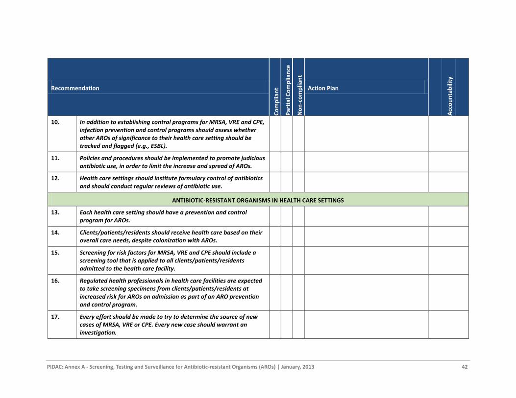
PIDAC: Annex A - Screening, Testing and Surveillance for Antibiotic-resistant Organisms (AROs) | January, 2013 42
Recommendation
Co
mp
lian
t
Par
tial
Co
mp
lian
ce
No
n-c
om
plia
nt
Action Plan
Acc
ou
nta
bili
ty
10. In addition to establishing control programs for MRSA, VRE and CPE, infection prevention and control programs should assess whether other AROs of significance to their health care setting should be tracked and flagged (e.g., ESBL).
11. Policies and procedures should be implemented to promote judicious antibiotic use, in order to limit the increase and spread of AROs.
12. Health care settings should institute formulary control of antibiotics and should conduct regular reviews of antibiotic use.
ANTIBIOTIC-RESISTANT ORGANISMS IN HEALTH CARE SETTINGS
13. Each health care setting should have a prevention and control program for AROs.
14. Clients/patients/residents should receive health care based on their overall care needs, despite colonization with AROs.
15. Screening for risk factors for MRSA, VRE and CPE should include a screening tool that is applied to all clients/patients/residents admitted to the health care facility.
16. Regulated health professionals in health care facilities are expected to take screening specimens from clients/patients/residents at increased risk for AROs on admission as part of an ARO prevention and control program.
17. Every effort should be made to try to determine the source of new cases of MRSA, VRE or CPE. Every new case should warrant an investigation.

PIDAC: Annex A - Screening, Testing and Surveillance for Antibiotic-resistant Organisms (AROs) | January, 2013 43
Recommendation
Co
mp
lian
t
Par
tial
Co
mp
lian
ce
No
n-c
om
plia
nt
Action Plan
Acc
ou
nta
bili
ty
18. All affected health care settings should be notified following the identification of a new case of an ARO, or identification of a new contact of a case.
19. Any client/patient/resident who is considered to be an MRSA, VRE or CPE contact should have at least one set of screening specimens taken. If initial specimens are negative, it is prudent to repeat them.
20. During an outbreak, all client/patient/resident contacts with common risk factors should be actively screened.
21. Consideration should be given to conducting point prevalence screens on units/areas where clients/patients/residents are at high risk for acquiring MRSA, VRE or CPE during their stay in the health care setting.
22. Point prevalence screens should be conducted in any area where MRSA, VRE or CPE transmission is occurring and should continue to be conducted until no further transmission is detected.
23. Screening staff for MRSA should be considered when an outbreak of the same strain of MRSA continues to spread despite adherence to control measures, or when an individual is strongly epidemiologically linked to new acquisitions of MRSA.

PIDAC: Annex A - Screening, Testing and Surveillance for Antibiotic-resistant Organisms (AROs) | January, 2013 44
Recommendation
Co
mp
lian
t
Par
tial
Co
mp
lian
ce
No
n-c
om
plia
nt
Action Plan
Acc
ou
nta
bili
ty
24. Specimens for detection of MRSA should include:
a) A swab from the anterior nares; AND
b) A swab from the perianal, perineal or groin area (perianal preferred); AND
c) A swab(s) from skin lesions, wounds, incisions, ulcers and exit sites of indwelling devices, if present, using aseptic technique where indicated;
d) For newborn infants, a swab from the umbilicus should also be taken for MRSA.
25. Specimens for detection of VRE should include stool or a rectal or perianal swab. Stool specimens provide a higher yield.
26. Specimens for detection of CPE should include stool or a rectal swab.
27. Routine decolonization therapy of MRSA clients/patients/residents is not currently recommended.
28. VRE, CPE or ESBL decolonization is not effective and not recommended.
29. In situations where a client/patient/resident colonized with MRSA is implicated in an outbreak, decolonization may be considered in consultation with the health care setting’s Infection Prevention and Control Professional.

PIDAC: Annex A - Screening, Testing and Surveillance for Antibiotic-resistant Organisms (AROs) | January, 2013 45
Recommendation
Co
mp
lian
t
Par
tial
Co
mp
lian
ce
No
n-c
om
plia
nt
Action Plan
Acc
ou
nta
bili
ty
30. Decolonization of staff colonized with MRSA should be done when they are epidemiologically linked to an outbreak with the same strain and adherence to Contact Precautions has failed to contain the outbreak.
31. The health care setting’s Infection Prevention and Control Professional(s) and senior management must be notified whenever CPE, VISA or VRSA is identified.
32. Expert advice should be sought whenever CPE, VISA or VRSA is isolated (e.g., infection prevention and control experts from academic health sciences centres, the regional infection control networks and reference laboratory services).
33. In addition to Routine Practices and all of the previous recommendations for MRSA, Additional Precautions for CPE, VISA and VRSA include:
a) Single room accommodation is required.
b) Dedicated equipment and supplies are required.
c) Minimize the number of persons who enter the room.
d) Patient must remain in their room except for essential procedures.
e) Transfer between facilities should only be done if medically necessary. The receiving health care setting must be advised of the required precautions.
f) Avoid transfer within the facility if possible; if transfer is necessary for medical reasons, the receiving unit or

PIDAC: Annex A - Screening, Testing and Surveillance for Antibiotic-resistant Organisms (AROs) | January, 2013 46
Recommendation
Co
mp
lian
t
Par
tial
Co
mp
lian
ce
No
n-c
om
plia
nt
Action Plan
Acc
ou
nta
bili
ty
department must be advised of the required precautions.
g) Each patient contact must be placed on Contact Precautions and be screened.
34. Every attempt should be made to identify the source of VISA or VRSA.
35. Health care settings should assess their local ESBL epidemiology to determine whether a specific ESBL control program is warranted.
INTERVENTIONS FOR THE PREVENTION AND CONTROL OF ANTIBIOTIC-RESISTANT ORGANISMS
36. Each health care setting should have policies in place that identify clients/patients/residents who are at the highest risk for colonization with MRSA, VRE or CPE, so that they may be placed on Contact Precautions until the results of screening tests are available.
37. If MRSA infection is treated with an antimicrobial to which the MRSA is sensitive, follow-up specimens should not be obtained until at least 48 hours after discontinuation of therapy and prior to discontinuation of Contact Precautions.
38. Re-colonization with MRSA may occur once a client/patient/resident has been discharged from the health care system and screening specimens should be collected on each readmission.
39. When a patient has been placed on Contact Precautions for CPE or ESBL, precautions should remain in place for the duration of acute care hospitalization.

PIDAC: Annex A - Screening, Testing and Surveillance for Antibiotic-resistant Organisms (AROs) | January, 2013 47
Appendices
APPENDIX A: COLLECTING SPECIMENS FOR MRSA, VRE, CPE, ESBL
[Adapted from University Health Network, Sunnybrook Health Sciences Centre]
Check with your laboratory regarding appropriate specimens for detection of
MRSA, VRE, CPE or ESBL
Note: Specimens may be falsely negative if the patient is on an antibiotic to which the microorganism is sensitive. MRSA may not show up on specimens taken from patients who have recently had an antimicrobial bath. Surveillance specimens should be taken once the antibiotic has been discontinued for 48 hours.
MRSA Screening Procedure for Cultures/Molecular Detection:
Pre-moisten all swabs with sterile normal saline or with transport medium prior to taking a specimen.
Swab anterior nares (use the same swab for both nostrils). Use a circular motion to touch as much mucous membrane as possible.
Swab perianal/perineal skin or groin with a new swab. Swab wounds/skin lesions/incisions/ulcers if present with separate swabs. Swab exit sites of indwelling devices if present. For newborns, swab the umbilicus Label the individual specimens appropriately.
VRE/CPE/ESBL Screening Procedure for Cultures/Molecular Detection:
Stool or a rectal swab may be used for VRE/ESBL/CPE screening. Stool specimens have a higher yield.
Swab around the external rectal orifice. If visible stool is not obtained on the swab, insert it a few millimetres into the rectum until visible stool is obtained.
If the client/patient/resident has a colostomy, take the specimen from the colostomy output.
Label the individual specimens appropriately.

PIDAC: Annex A - Screening, Testing and Surveillance for Antibiotic-resistant Organisms (AROs) | January, 2013 48
APPENDIX B: SAMPLE RISK FACTOR-BASED ADMISSION FORM FOR SCREENING FOR MRSA, VRE, ESBL AND CPE
[Adapted from Sunnybrook Health Sciences Centre]
Antibiotic Resistant Organisms (ARO) Admission Screen (for all admitted patients)
MRSA= methicillin resistant Staphylococcus aureus VRE= vancomycin resistant enterococcus
ESBL= extended-spectrum ß-lactamase E. coli and Klebsiella CPE= carbapenemase-producing Enterobacteriaceae
Primary risk factors for Antibiotic Resistant Organisms (ARO):
( ) yes ( ) no Patient is known to be positive for an ARO, or was a contact of an ARO (includes patients flagged
in electronic patient record for MRSA, VRE, ESBL or CPE) If YES, place the patient on Contact
Precautions and collect specimens for MRSA, VRE and any other AROs that were previously
positive or for which the patient was a contact
( ) yes ( ) no Patient has received health care in another country within the last year If YES, place the
patient on Contact Precautions and collect specimens for MRSA, VRE and CPE
( ) yes ( ) no Patient is a direct transfer from a health care facility** outside of Canada If YES, admit into a
single room on Contact Precautions, collect specimens for MRSA, VRE and CPE and reassess
when culture results are known.
If the answer to all of the above questions is NO, continue with the following questions. If the answer is YES to any of these, collect specimens only for MRSA and VRE:
( ) yes ( ) no Patient is a direct transfer from another health care facility**, including internal sites/your own facility
( ) yes ( ) no Patient has been admitted to a Canadian health care facility** within the last year, including
internal sites/your own facility
( ) yes ( ) no Patient receives home health care services or hemodialysis
( ) yes ( ) no Patient lives in a communal setting (e.g. homeless shelter, halfway house, correctional facility)
( ) yes ( ) no Patient is unable to answer any of the above questions
Screening specimens required (select all that apply):
( )MRSA Send specimens for MRSA from the following sites:
Anterior nares (both nares with one swab)
Perianal/perineal skin or groin
Open wounds/lesions/incisions
Exit sites of indwelling devices
( ) VRE
( ) ESBL
( ) CPE
Send a rectal swab (faecally stained) or stool specimen for VRE, ESBL, CPE (stool is preferred).
For ESBL, a urine sample may be indicated.
For CPE, other samples may be indicated: Urine, wound swabs, endotracheal suction (critical care), exit sites (critical care)
* High risk geographical areas currently include: U.S. eastern seaboard (e.g., New York city), Greece, Israel and Indian subcontinent (e.g., India, Sri Lanka, Bangladesh, Pakistan) ** Health care facility includes: hospital, long-term care home, retirement home or other health care facility
Date: Print Signature & Sign (RN/RPN):
( ) Patient refused specimens. Notify the Infection Prevention & Control Professional or delegate. Date: Print Signature & Sign (RN/RPN):

PIDAC: Annex A - Screening, Testing and Surveillance for Antibiotic-resistant Organisms (AROs) | January, 2013 49
APPENDIX C: SAMPLE FACT SHEETS FOR HEALTH CARE STAFF (MRSA, VRE, ESBL, CPE) AND SAMPLE INFORMATION SHEETS FOR PATIENTS AND VISITORS
Adapted from materials provided by:
Kingston General Hospital
The Ottawa Hospital
Sunnybrook Health Sciences Centre

PIDAC: Annex A - Screening, Testing and Surveillance for Antibiotic-resistant Organisms (AROs) | January, 2013 50
METHICILLIN-RESISTANT STAPHYLOCOCCUS AUREUS (MRSA)
Staff Fact Sheet
WHAT IS MRSA?
Staphylococcus aureus is a bacterium that periodically lives on the skin and mucous membranes of healthy people. Occasionally S. aureus can cause an infection. When S. aureus develops resistance to the beta-lactam class of antibiotics, it is called methicillin-resistant Staphylococcus aureus, or MRSA.
HOW IS MRSA SPREAD?
MRSA is spread from one person to another by contact, usually on the hands of caregivers. MRSA can be present on the caregiver’s hands either from touching contaminated material excreted by the infected person or from touching articles contaminated by the skin of a person with MRSA, such as towels, sheets, wound dressings. MRSA can survive well on hands and can survive for weeks on inanimate objects such as door handles, bedrails, pagers and stethoscopes.
COLONIZATION AND INFECTION:
Colonization occurs when bacteria are present on or in the body without causing illness. MRSA can colonize the nose, skin and moist areas of the body.
Infection occurs when bacteria get past the person’s normal defences and cause disease (e.g. skin bacteria getting into the bloodstream via an intravenous catheter). Infections with MRSA may be minor, such as pimples and boils, but serious infections may also occur, such as surgical wound infections and pneumonia.
RISK FACTORS FOR MRSA INFECTION:
MRSA infection usually develops in hospitalized clients/patients/residents who are elderly or very sick (weakened immune systems). Other factors that increase the risk for acquiring MRSA infection include:
Being colonized with MRSA
Previous hospitalization or transfer between health care facilities (in Canada or outside Canada)
Presence of an indwelling device (e.g., catheter)
GOOD HAND HYGIENE PRACTICES:
Remind all staff and visitors to practice good hand hygiene before and after client/patient/resident contact/care. Health care staff should review the correct method of hand hygiene, as well as demonstrate the proper donning/removal of personal protective equipment (PPE) to clients/patients/residents, families and visitors.
Good hand hygiene practices means using alcohol-based hand rub or soap and running water for at least 15 seconds.
Hand hygiene should occur:
Before client/patient/resident or environment contact
Before performing aseptic procedures
After care involving body fluids
After client/patient/resident or environment contact
PREVENTION & CONTROL OF MRSA:
1. Admission screening for MRSA must be completed:
Check for previous history of MRSA or high risk for MRSA using an admission screening tool.
If the client/patient/resident has previously had contact with an MRSA case, screening specimens must be obtained.
If the client/patient/resident is considered to be at risk for MRSA based on the results of the screening tool, screening specimens must be obtained.
2. If the client/patient/resident is known to have had MRSA in the past, Contact Precautions must be initiated:
Hand hygiene as described in Routine Practices
Appropriate client/patient/resident placement
Gloves for all activities in the patient’s room or bed space in acute care, or for direct care of clients/residents in long- term care and ambulatory/clinic settings
Long-sleeved gown for activities where skin or clothing will come in contact with the patient or their environment in acute care, or for direct care of clients/residents in long- term care and ambulatory/clinic settings
A surgical mask should be worn as per Routine Practices
Dedicated equipment or adequate cleaning and disinfecting of shared equipment, including transport equipment
Daily cleaning of all touched surfaces in the room
3. Notify the Infection Prevention and Control Professional or delegate to discuss the infection control management of client/patient/resident activities.
4. Precautions are not to be discontinued until reviewed by Infection Prevention and Control.
5. Additional surveillance specimens for colonization of client/patient/resident contact(s) may be required, as directed by Infection Prevention and Control.
FAMILY & VISITORS:
All families/visitors must practice good hand hygiene before and after leaving the client/patient/resident room.
Families/visitors who provide direct care must wear the same PPE as staff. “Direct care” is defined as providing hands-on care, such as bathing, washing, turning the client/patient/resident, changing clothes/diapers, dressing changes, care of open wounds/lesions, toileting. Feeding or pushing a wheelchair are not classified as direct care.
Written information should be available for clients/patients/residents that explains the precautions required.

PIDAC: Annex A - Screening, Testing and Surveillance for Antibiotic-resistant Organisms (AROs) | January, 2013 51
VANCOMYCIN RESISTANT ENTEROCOCCUS (VRE)
Staff Fact Sheet
WHAT IS VRE?
Enterococci are bacteria that live in the gastrointestinal tract of most individuals and generally do not cause harm (“colonization”). Vancomycin-resistant enterococci (VRE) are strains of enterococci that are resistant to the antibiotic vancomycin. If a person has an infection caused by VRE, such as a urinary tract infection or blood infection, it may be more difficult to treat.
HOW IS VRE SPREAD?
VRE is spread from one person to another by contact, usually on the hands of caregivers. VRE can be present on the caregiver’s hands either from touching contaminated material excreted by the infected person or from touching articles soiled by faeces. VRE can survive well on hands and can survive for weeks on inanimate objects such as toilet seats, door handles, bedrails, furniture, stethoscopes, rectal thermometers and bedpans.
RISK FACTORS FOR VRE:
People at risk for colonization or infection with VRE are usually hospitalized and have an underlying medical condition which makes them susceptible to infection. These conditions include clients/patients/residents with:
Previous hospitalization or transfer between health care facilities (in Canada or outside Canada)
Critical illness(es) in intensive care units
Severe underlying disease or weakened immune systems
Urinary catheters
Exposure to (or contact with) a client/patient/resident with VRE
Antibiotic use, particularly vancomycin
GOOD HAND HYGIENE PRACTICES:
Remind all staff and visitors to practice good hand hygiene before and after client/patient/resident contact/care. Health care staff should review the correct method of hand hygiene, as well as demonstrate the proper donning/removal of personal protective equipment (PPE) to clients/patients/residents, families and visitors.
Good hand hygiene practices means using alcohol-based hand rub or soap and running water for at least 15 seconds.
Hand hygiene should occur:
Before client/patient/resident or environment contact
Before performing aseptic procedures
After care involving body fluids
After client/patient/resident or environment contact
PREVENTION & CONTROL OF VRE:
1. Admission screening for VRE must be completed:
Check for previous history of VRE or high risk for VRE using the admission screening tool.
If the client/patient/resident has been a contact of a VRE case in the past, screening specimens must be obtained.
If the client/patient/resident is considered to be at risk for VRE based on the results of the screening tool, screening specimens must be obtained.
2. If the client/patient/resident is known to have had VRE in the past, Contact Precautions must be initiated:
Hand hygiene as described in Routine Practices
Appropriate client/patient/resident placement
Gloves for all activities in the patient’s room or bed space in acute care, or for direct care of clients/residents in long- term care and ambulatory/clinic settings
Long-sleeved gown for activities where skin or clothing will come in contact with the patient or their environment in acute care, or for direct care of clients/residents in long- term care and ambulatory/clinic settings
Dedicated equipment or adequate cleaning and disinfecting of shared equipment, including transport equipment
Special discharge cleaning protocol is vital for VRE
3. Notify the Infection Prevention & Control Professional or delegate to discuss the infection control management of client/patient/resident activities.
4. Precautions are not to be discontinued until reviewed by Infection Prevention and Control.
5. Additional surveillance specimens for colonization of client/patient/resident contact(s) may be required, as directed by Infection Prevention and Control.
FAMILY & VISITORS:
1. All families/visitors must practice good hand hygiene before and after leaving the client/patient/resident’s room.
2. Families/visitors who provide direct care are to wear the same PPE as staff. “Direct care” is defined as providing hands-on care, such as bathing, washing, turning the client/patient/resident, changing clothes/diapers, dressing changes, care of open wounds/lesions, toileting. Feeding and pushing a wheelchair are not classified as direct care.
3. Provide written information for clients/patients/residents that explains the precautions required.

PIDAC: Annex A - Screening, Testing and Surveillance for Antibiotic-resistant Organisms (AROs) | January, 2013 52
EXTENDED SPECTRUM BETA-LACTAMASE PRODUCING BACTERIA (ESBL)
Staff Fact Sheet
WHAT ARE ESBLs?
ESBLs are Gram-negative bacteria that produce an enzyme, beta-lactamase, that has the ability to break down commonly used antibiotics, such as penicillins and cephalosporins (including third generation) and render them ineffective for treatment. If ESBL-producing bacteria cause an infection, a different antibiotic may need to be used to treat the infection. People who carry ESBL-producing bacteria without any signs or symptoms of infection are said to be colonized. The commonest ESBL-producing bacteria are some strains of Escherichia coli and Klebsiella pneumoniae.
HOW ARE ESBLs SPREAD?
ESBLs are spread via direct and indirect contact with colonized/infected patients and contaminated environmental surfaces. ESBLs are not airborne. ESBLs are most commonly spread via unwashed hands of health care providers. ESBLs may also be spread within households.
RISK FACTORS FOR ESBL:
Risk factors for ESBL-producing bacterial acquisition include:
Direct transfer from another hospital, nursing home, retirement home or other health care facility, including between facilities in the same health care corporation
Any hospital, nursing home, retirement home or other health care facility admission in the past 1 year
Patient receiving home health care services or hemodialysis
Patient living in a communal living setting (e.g., shelter, halfway house)
Patient who previously had an antibiotic-resistant organism (e.g., MRSA, VRE)
ESBL-producing bacteria are becoming more common in the community.
GOOD HAND HYGIENE PRACTICES:
Remind all staff and visitors to practice good hand hygiene before and after client/patient/resident contact/care. Health care staff should review the correct method of hand hygiene, as well as demonstrate the proper donning/removal of personal protective equipment (PPE) to clients/patients/residents, families and visitors.
Good hand hygiene practices means using alcohol-based hand rub or soap and running water for at least 15 seconds.
Hand hygiene should occur:
Before client/patient/resident or environment contact
Before performing aseptic procedures
After care involving body fluids
After client/patient/resident or environment contact
PREVENTION & CONTROL OF ESBLs:
1. Consistent use of Routine Practices with all patients/residents.
2. Admission screening:
Check for previous history of antibiotic-resistant organism. (ARO)
Complete the ARO screening tool for patients/residents
3. Initiate Contact Precautions for patients/residents with ESBL-producing bacteria:
Appropriate client/patient/resident placement
Gloves for all activities in the patient’s room or bed space in acute care, or for direct care of clients/residents in long- term care and ambulatory/clinic settings
Long-sleeved gown for activities where skin or clothing will come in contact with the patient or their environment in acute care, or for direct care of clients/residents in long- term care and ambulatory/clinic settings
Dedicated equipment or adequate cleaning and disinfecting of shared equipment, with particular attention to management of urinary catheters and associated equipment
4. Notify the Infection Prevention & Control Professional or delegate to discuss the infection control management of client/patient/resident activities.
5. Precautions are not to be discontinued until reviewed by Infection Prevention and Control.
6. Additional surveillance specimens for colonization of client/patient/resident contact(s) may be required, as directed by Infection Prevention and Control.
FAMILY & VISITORS:
1. All families/visitors must practice good hand hygiene before and after leaving the client/patient/resident’s room.
2. Families/visitors who provide direct care are to wear the same PPE as staff. “Direct care” is defined as providing hands-on care, such as bathing, washing, turning the client/patient/resident, changing clothes/incontinent pads, dressing changes, care of open wounds/lesions and toileting. Feeding and pushing a wheelchair are not classified as direct care.
3. Families/visitors should not help other patients/residents with their personal care. This may cause ESBL to spread.
4. Provide written information for clients/patients/residents that explains the precautions required.

PIDAC: Annex A - Screening, Testing and Surveillance for Antibiotic-resistant Organisms (AROs) | January, 2013 53
CARBAPENEMASE-PRODUCING ENTEROBACTERIACEAE (CPE)
Staff Fact Sheet
WHAT ARE CPE?
Carbapenemase-producing Enterobacteriaceae are resistant to carbapenem antimicrobials (e.g., imipenem, meropenem, ertapenem) through the production of carbapenemase enzymes.
Carbapenemases are enzymes that inactivate carbapenem, cephalosporin and penicillin antibiotics. The genetic information to produce carbapenemases is often located on a mobile genetic element (i.e., a genetic element that can move between bacterial strains and species, e.g., plasmid, transposon), which frequently also carries resistance to other classes of antimicrobials, such as fluoroquinolones and aminoglycosides. To date, carbapenemases have been found most commonly in E. coli and Klebsiella species, but have also been found in other Gram-negative bacteria.
There are several different carbapenemases, each having a three-letter acronym, e.g., KPC = Klebsiella pneumoniae
carbapenemase; NDM = New Delhi metallo--lactamase.
These enzymes evolve rarely, but bacteria carrying them spread easily. Particular classes of carbapenemases are most common in the geographic area where they evolved, but can spread around the world, usually when patients have received health care in another country.
Because CPE are resistant to many classes of antimicrobials, treatment of infections with CPE is difficult and involves the use of antibiotics that have significant adverse events (e.g., colistin). The case fatality rate for serious infections may be as high as 50%.
CURRENT STATUS OF CPE IN ONTARIO
A small number of CPE have recently been reported in hospitals in Ontario. Most patients with CPE have had links to hospitals with recognized epidemic or endemic CPE (e.g., New York City hospitals with KPC K. pneumoniae, receipt of health care in the Indian subcontinent). However, transmission of CPE has been reported in Ontario.
HOW ARE CPE SPREAD?
Transmission is via direct and indirect contact. The primary site of colonization is the lower gastrointestinal tract.
RISK FACTORS FOR CPE
Risk factors for infection and colonization with CPE will be similar to those of other resistant Gram-negative bacteria, such as ESBL-producing E. coli and Klebsiella pneumoniae.
Currently, the major risk factor appears to be receipt of care in health care settings that have CPE, e.g., hospitals along the U.S. eastern seaboard, particularly New York City (KPC), Greece (KPC), Israel (KPC) and the Indian subcontinent (NDM-1). However, CPE outbreaks are being increasingly described in hospitals around the world, including Canada. People coming from the Indian subcontinent, with or without exposure to health care, are also at risk.
PREVENTION & CONTROL OF CPE:
1. Consistent use of Routine Practices with all patients/residents.
2. Screening:
Surveillance is an important measure to prevent and control the spread of CPE. Admission screening and pre-emptive Contact Precautions are indicated for individuals with risk factors for CPE:
If a patient/resident is identified with CPE, roommates and patients in close proximity will be screened for CPE
Primary screening specimens for CPE are stool or rectal swabs. Urine specimens and swabs from open wounds may also be indicated. In critical care settings, sputum or endotracheal tube specimens and swabs from exit sites may be requested by Infection Prevention and Control
Patients with known CPE carriage will have their records flagged, will be placed on Contact Precautions and will be re-screened if readmitted.
3. Initiate Contact Precautions for patients/residents with CPE:
Appropriate client/patient/resident placement
Gloves for all activities in the patient’s room or bed space in acute care, or for direct care of clients/residents in long- term care and ambulatory/clinic settings
Long-sleeved gown for activities where skin or clothing will come in contact with the patient or their environment in acute care, or for direct care of clients/residents in long- term care and ambulatory/clinic settings
Dedicated equipment or adequate cleaning and disinfecting of shared equipment, with particular attention to management of urinary catheters and associated equipment
4. Notify the Infection Prevention & Control Professional or delegate to discuss the infection control management of client/patient/resident activities.
5. It is not known how long bowel colonization with CPE persists, but it is likely of long duration. Most colonized patients/residents are asymptomatic. Because of the implications of CPE infection and transmission, current expert recommendations are that patients remain on Contact Precautions for the duration of hospitalization. They should be presumed to be colonized and managed on Contact Precautions if they are readmitted.
6. There are no data to support CPE decolonization and it is not recommended.
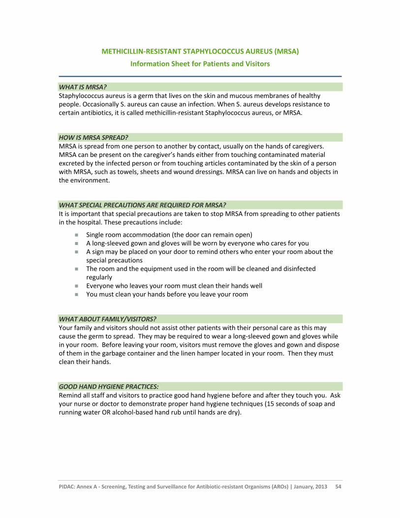
PIDAC: Annex A - Screening, Testing and Surveillance for Antibiotic-resistant Organisms (AROs) | January, 2013 54
METHICILLIN-RESISTANT STAPHYLOCOCCUS AUREUS (MRSA)
Information Sheet for Patients and Visitors
WHAT IS MRSA? Staphylococcus aureus is a germ that lives on the skin and mucous membranes of healthy people. Occasionally S. aureus can cause an infection. When S. aureus develops resistance to certain antibiotics, it is called methicillin-resistant Staphylococcus aureus, or MRSA.
HOW IS MRSA SPREAD? MRSA is spread from one person to another by contact, usually on the hands of caregivers. MRSA can be present on the caregiver’s hands either from touching contaminated material excreted by the infected person or from touching articles contaminated by the skin of a person with MRSA, such as towels, sheets and wound dressings. MRSA can live on hands and objects in the environment.
WHAT SPECIAL PRECAUTIONS ARE REQUIRED FOR MRSA? It is important that special precautions are taken to stop MRSA from spreading to other patients in the hospital. These precautions include:
Single room accommodation (the door can remain open) A long-sleeved gown and gloves will be worn by everyone who cares for you A sign may be placed on your door to remind others who enter your room about the
special precautions The room and the equipment used in the room will be cleaned and disinfected
regularly Everyone who leaves your room must clean their hands well You must clean your hands before you leave your room
WHAT ABOUT FAMILY/VISITORS? Your family and visitors should not assist other patients with their personal care as this may cause the germ to spread. They may be required to wear a long-sleeved gown and gloves while in your room. Before leaving your room, visitors must remove the gloves and gown and dispose of them in the garbage container and the linen hamper located in your room. Then they must clean their hands.
GOOD HAND HYGIENE PRACTICES: Remind all staff and visitors to practice good hand hygiene before and after they touch you. Ask your nurse or doctor to demonstrate proper hand hygiene techniques (15 seconds of soap and running water OR alcohol-based hand rub until hands are dry).
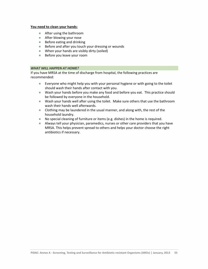
PIDAC: Annex A - Screening, Testing and Surveillance for Antibiotic-resistant Organisms (AROs) | January, 2013 55
You need to clean your hands:
After using the bathroom After blowing your nose Before eating and drinking Before and after you touch your dressing or wounds When your hands are visibly dirty (soiled) Before you leave your room
WHAT WILL HAPPEN AT HOME? If you have MRSA at the time of discharge from hospital, the following practices are recommended:
Everyone who might help you with your personal hygiene or with going to the toilet should wash their hands after contact with you.
Wash your hands before you make any food and before you eat. This practice should be followed by everyone in the household.
Wash your hands well after using the toilet. Make sure others that use the bathroom wash their hands well afterwards.
Clothing may be laundered in the usual manner, and along with, the rest of the household laundry.
No special cleaning of furniture or items (e.g. dishes) in the home is required. Always tell your physician, paramedics, nurses or other care providers that you have
MRSA. This helps prevent spread to others and helps your doctor choose the right antibiotics if necessary.

PIDAC: Annex A - Screening, Testing and Surveillance for Antibiotic-resistant Organisms (AROs) | January, 2013 56
VANCOMYCIN RESISTANT ENTEROCOCCUS (VRE)
Information Sheet for Patients and Visitors
WHAT IS VRE? Enterococci are germs that live in the gastrointestinal tract (bowels) of most individuals and generally do not cause harm (this is termed “colonization”). Vancomycin-resistant enterococci (VRE) are strains of enterococci that are resistant to the antibiotic vancomycin. If a person has an infection caused by VRE it may be more difficult to treat.
HOW IS VRE SPREAD? VRE is spread from one person to another by contact, usually on the hands of caregivers. VRE can be present on the caregiver’s hands either from touching contaminated material excreted by an infected person or from touching articles soiled by faeces. VRE can survive well on hands and can survive for weeks on inanimate objects such as toilet seats, taps, door handles, bedrails, furniture and bedpans. VRE is easy to kill with the proper use of disinfectants and good hand hygiene.
WHAT SPECIAL PRECAUTIONS ARE REQUIRED FOR VRE? It is important that special precautions are taken to stop VRE from spreading to other patients in the hospital. These precautions include:
Single room accommodation (the door can remain open) A long-sleeved gown and gloves will be worn by everyone who cares for you A sign may be placed on your door to remind others who enter your room about the
special precautions The room and the equipment used in the room will be cleaned and disinfected
regularly Everyone who leaves your room must clean their hands well You must clean your hands before you leave your room
WHAT ABOUT FAMILY/VISITORS? Your family and visitors should not assist other patients with their personal care as this may cause the germ to spread. They may be required to wear a long-sleeved gown and gloves while in your room. Before leaving your room, visitors must remove the gloves and gown and dispose of them in the garbage container and the linen hamper located in your room. Then they must clean their hands.
GOOD HAND HYGIENE PRACTICES: Remind all staff and visitors to practice good hand hygiene before and after they touch you. Ask your nurse or doctor to demonstrate proper hand hygiene techniques (15 seconds of soap and running water OR alcohol-based hand rub until hands are dry).

PIDAC: Annex A - Screening, Testing and Surveillance for Antibiotic-resistant Organisms (AROs) | January, 2013 57
You need to clean your hands:
After using the bathroom After blowing your nose Before eating and drinking Before and after you touch your dressing or wounds When your hands are visibly dirty (soiled) Before you leave your room
WHAT WILL HAPPEN AT HOME? If you have VRE at the time of discharge from hospital, the following practices are recommended:
Everyone who might help you with your personal hygiene or with going to the toilet should wash their hands after contact with you.
Wash your hands before you make any food and before you eat. This practice should be followed by everyone in the household.
Wash your hands well after using the toilet. Make sure others that use the bathroom wash their hands well afterwards.
Clothing may be laundered in the usual manner, and along with, the rest of the household laundry.
No special cleaning of furniture or items (e.g., dishes) in the home is required. If you share a bathroom at home, clean the toilet and sink at least weekly with a
household cleanser. Always tell your physician, paramedics, nurses or other care providers that you have
VRE. This helps prevent spread to others.
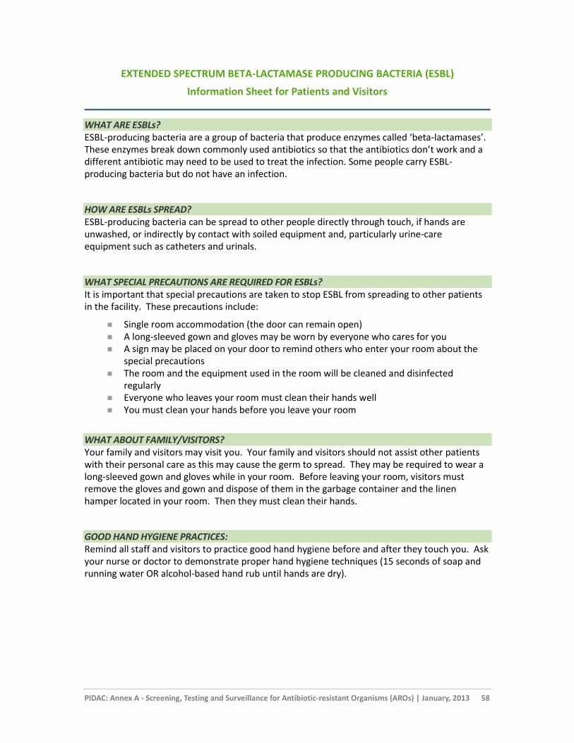
PIDAC: Annex A - Screening, Testing and Surveillance for Antibiotic-resistant Organisms (AROs) | January, 2013 58
EXTENDED SPECTRUM BETA-LACTAMASE PRODUCING BACTERIA (ESBL)
Information Sheet for Patients and Visitors
WHAT ARE ESBLs? ESBL-producing bacteria are a group of bacteria that produce enzymes called ‘beta-lactamases’. These enzymes break down commonly used antibiotics so that the antibiotics don’t work and a different antibiotic may need to be used to treat the infection. Some people carry ESBL-producing bacteria but do not have an infection.
HOW ARE ESBLs SPREAD? ESBL-producing bacteria can be spread to other people directly through touch, if hands are unwashed, or indirectly by contact with soiled equipment and, particularly urine-care equipment such as catheters and urinals.
WHAT SPECIAL PRECAUTIONS ARE REQUIRED FOR ESBLs? It is important that special precautions are taken to stop ESBL from spreading to other patients in the facility. These precautions include:
Single room accommodation (the door can remain open) A long-sleeved gown and gloves may be worn by everyone who cares for you A sign may be placed on your door to remind others who enter your room about the
special precautions The room and the equipment used in the room will be cleaned and disinfected
regularly Everyone who leaves your room must clean their hands well You must clean your hands before you leave your room
WHAT ABOUT FAMILY/VISITORS? Your family and visitors may visit you. Your family and visitors should not assist other patients with their personal care as this may cause the germ to spread. They may be required to wear a long-sleeved gown and gloves while in your room. Before leaving your room, visitors must remove the gloves and gown and dispose of them in the garbage container and the linen hamper located in your room. Then they must clean their hands.
GOOD HAND HYGIENE PRACTICES: Remind all staff and visitors to practice good hand hygiene before and after they touch you. Ask your nurse or doctor to demonstrate proper hand hygiene techniques (15 seconds of soap and running water OR alcohol-based hand rub until hands are dry).

PIDAC: Annex A - Screening, Testing and Surveillance for Antibiotic-resistant Organisms (AROs) | January, 2013 59
You need to clean your hands:
After using the bathroom After blowing your nose Before eating and drinking Before and after you touch your dressing or wounds When your hands are visibly dirty (soiled) Before you leave your room
WHAT WILL HAPPEN AT HOME? If you have ESBL at the time of discharge from hospital, the following practices are
recommended: Everyone who might help you with your personal hygiene or with going to the toilet
should wash their hands after contact with you. Wash your hands before you make any food and before you eat. This practice should
be followed by everyone in the household. Wash your hands well after using the toilet. Make sure others that use the bathroom
wash their hands well afterwards. Clothing may be laundered in the usual manner, and along with, the rest of the
household laundry. No special cleaning of furniture or items (e.g. dishes) in the home is required. If you share a bathroom at home, clean the toilet and sink at least weekly with a
household cleanser. Always tell your physician, paramedics, nurses or other care providers that you have
ESBL. This helps prevent spread to others and helps your doctor choose the right antibiotics if necessary.

PIDAC: Annex A - Screening, Testing and Surveillance for Antibiotic-resistant Organisms (AROs) | January, 2013 60
CARBAPENEMASE-PRODUCING ENTEROBACTERIACEAE (CPE)
Information Sheet for Patients and Visitors
WHAT ARE CPE? Enterobacteriaceae are a family of bacteria, many of which live naturally in our bowels. Carbapenemase-producing Enterobacteriaceae (CPE) produce carbapenemase enzymes that can break down many types of antibiotics, making the bacteria very resistant. In Canadian hospitals, there are currently few infections with CPE, but caution is still needed to prevent their increase and spread.
HOW ARE CPE SPREAD? Most people who carry CPE have no symptoms of infection and are said to be colonized. The main site of colonization of CPE is the bowel. CPE is not spread through the air, but may survive on equipment and surfaces, such as bedrails, tables, chairs, countertops and door handles. CPE can be spread from one person to another by unwashed hands or from contact with soiled equipment and surfaces
Infection occurs when CPE enters the body at specific sites and causes symptoms of disease. For example, CPE can cause pneumonia and urinary tract infections. Since CPE are resistant to many types of antibiotics, treatment is difficult and may involve antibiotics which have significant side effects.
DOES CPE GO AWAY? People who have CPE in their bowel will likely carry it for a long time. You may be treated if CPE is causing symptoms of infection.
WHO IS AT RISK FOR CPE? Currently, the major risk factor is receiving health care in settings that have CPE, e.g., hospitals along the U.S. eastern seaboard (particularly New York City), Greece, Israel and the Indian subcontinent. CPE outbreaks have been seen in hospitals around the world, including Canada. People coming from the Indian subcontinent, with or without exposure to health care, are also at risk.
WHAT SPECIAL PRECAUTIONS ARE REQUIRED FOR CPE? Your healthcare team will continue to provide the same level of patient care. If a patient/resident is identified with CPE, roommates and patients in close proximity will be screened for CPE.
Additional Precautions will be used to prevent the possible spread of the bacteria. For example:
You may need to be moved to a single room A sign may be placed on your door to remind others who enter your room about the
special precautions (i.e. instructions to wash hands, wear gown and gloves) Speak to your doctor or nurse about special instructions when leaving your room Everyone who leaves your room must clean their hands well, including you Your hospital record will indicate CPE

PIDAC: Annex A - Screening, Testing and Surveillance for Antibiotic-resistant Organisms (AROs) | January, 2013 61
WHAT ABOUT FAMILY/VISITORS? Family and visitors can visit you. Healthy family and visitors have a low risk of acquiring infection with CPE. All visitors must be instructed by the staff on how to use Additional Precautions. Children and infants should be closely supervised. We ask that your visitors only visit you and your room, and to do the following:
Clean their hands before entering your room and when leaving Not to use your bathroom Not to eat or drink in your room
GOOD HAND HYGIENE PRACTICES: Remind all staff and visitors to practice good hand hygiene before and after they touch you. Ask your nurse or doctor to demonstrate proper hand hygiene techniques (15 seconds of soap and running water OR alcohol-based hand rub until hands are dry).
You need to clean your hands:
After using the bathroom After blowing your nose Before eating and drinking Before and after you touch your dressing or wounds When your hands are visibly dirty (soiled) Before you leave your room
WHAT WILL HAPPEN AT HOME? It is important to wash hands often at home for fifteen seconds each time, especially
after using the bathroom and before preparing food. No special cleaning of items in your home (e.g., dishes) are required. Clothing may be laundered in the usual manner, along with the rest of the household
laundry. If you go to another health care facility, visit another doctor or have Home Care
services you should tell them that you have CPE. They may use Additional Precautions when providing care, which will help prevent spread to others and helps your doctor choose the correct antibiotic treatment.

PIDAC: Annex A - Screening, Testing and Surveillance for Antibiotic-resistant Organisms (AROs) | January, 2013 62
APPENDIX D: SAMPLE INVESTIGATION PROTOCOLS FOR MRSA AND VRE IN ACUTE CARE FACILITIES
NOTE: The following investigation protocols are provided as SAMPLES to be used as a guide when
developing individualized policies in acute care facilities.
SAMPLE 1: MRSA PRESENT AT ADMISSION
Single MRSA case identified on admission screening
OR Clinical specimen taken within 48* hours of admission
Procedure Recommendation
1. Institute Contact Precautions for patient with MRSA. 36
2. Provide patient and visitor education.
3. If only one specimen at one site is positive in a newly identified case, re-swab the patient.
4
4. Flag patient. 6-9
5. Have laboratory save the isolate if this is not done routinely for first isolates. 3
6. Identify whether patient has risk factors for MRSA: 15-16
If the patient’s risk factor for MRSA is a prior admission in your facility, begin an investigation based on the recognition that this may have been acquired at your facility.
17
7. If patient was a resident of another health care facility, or has been transferred to another facility, notify that facility of the screening results. If the patient has been discharged home, the patient or family physician should be notified of the screening results.
18
8. Identify any roommates or contacts that this patient has had since admission: 19-20
a. If roommate or contact has been discharged home or transferred to another facility, flag them for screening on readmission and notify family physician or physician most responsible for their care.
b. Determine if the roommate or contact requires Contact Precautions, based on your facility policies.
c. Screen the roommate or contact.
d. If results of screening are positive (i.e. additional MRSA-positive patients are detected):
i. Flag roommate or contact. 6-9
ii. If roommate or contact has been transferred to another facility, notify that facility of the screening results. If roommate or contact has been discharged home, they or their family physician or the physician most responsible for their care should be notified of the screening results.
18
iii. If screening results indicate that this may be an outbreak or that there are health care-associated cases, begin an investigation.
9. Continue with case management for cases and positive contacts still in facility.
*Note: the decision to consider cases identified more then 48 hours after admission, rather than 72 hours after
admission, as health care associated to your facility is arbitrary. There is no evidence to support one time over the
other. Forty-eight hours is used in this document for consistency with other Canadian guidelines.
Refer to Algorithm 1

PIDAC: Annex A - Screening, Testing and Surveillance for Antibiotic-resistant Organisms (AROs) | January, 2013 63
Algorithm 1: Management of a Single New Case of MRSA
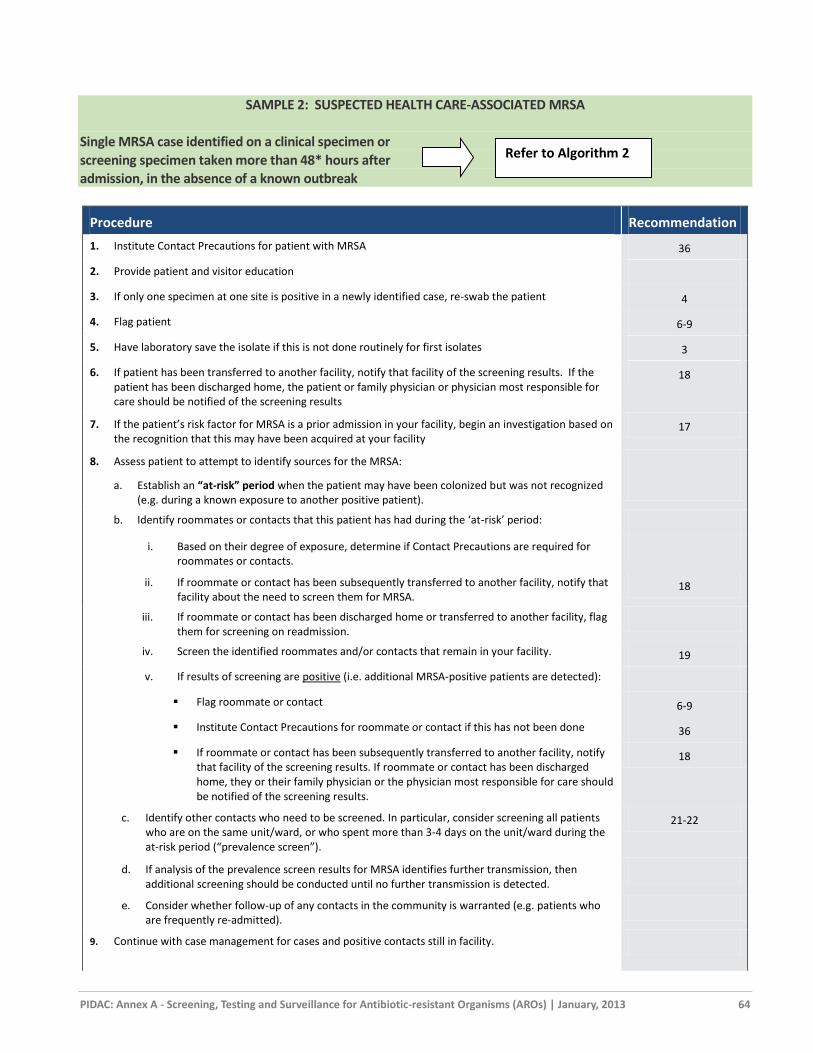
PIDAC: Annex A - Screening, Testing and Surveillance for Antibiotic-resistant Organisms (AROs) | January, 2013 64
SAMPLE 2: SUSPECTED HEALTH CARE-ASSOCIATED MRSA
Single MRSA case identified on a clinical specimen or
screening specimen taken more than 48* hours after
admission, in the absence of a known outbreak
Procedure Recommendation
1. Institute Contact Precautions for patient with MRSA 36
2. Provide patient and visitor education
3. If only one specimen at one site is positive in a newly identified case, re-swab the patient 4
4. Flag patient 6-9
5. Have laboratory save the isolate if this is not done routinely for first isolates 3
6. If patient has been transferred to another facility, notify that facility of the screening results. If the patient has been discharged home, the patient or family physician or physician most responsible for care should be notified of the screening results
18
7. If the patient’s risk factor for MRSA is a prior admission in your facility, begin an investigation based on the recognition that this may have been acquired at your facility
17
8. Assess patient to attempt to identify sources for the MRSA:
a. Establish an “at-risk” period when the patient may have been colonized but was not recognized (e.g. during a known exposure to another positive patient).
b. Identify roommates or contacts that this patient has had during the ‘at-risk’ period:
i. Based on their degree of exposure, determine if Contact Precautions are required for roommates or contacts.
ii. If roommate or contact has been subsequently transferred to another facility, notify that facility about the need to screen them for MRSA.
18
iii. If roommate or contact has been discharged home or transferred to another facility, flag them for screening on readmission.
iv. Screen the identified roommates and/or contacts that remain in your facility. 19
v. If results of screening are positive (i.e. additional MRSA-positive patients are detected):
Flag roommate or contact 6-9
Institute Contact Precautions for roommate or contact if this has not been done 36
If roommate or contact has been subsequently transferred to another facility, notify that facility of the screening results. If roommate or contact has been discharged home, they or their family physician or the physician most responsible for care should be notified of the screening results.
18
c. Identify other contacts who need to be screened. In particular, consider screening all patients who are on the same unit/ward, or who spent more than 3-4 days on the unit/ward during the at-risk period (“prevalence screen”).
21-22
d. If analysis of the prevalence screen results for MRSA identifies further transmission, then additional screening should be conducted until no further transmission is detected.
e. Consider whether follow-up of any contacts in the community is warranted (e.g. patients who are frequently re-admitted).
9. Continue with case management for cases and positive contacts still in facility.
Refer to Algorithm 2
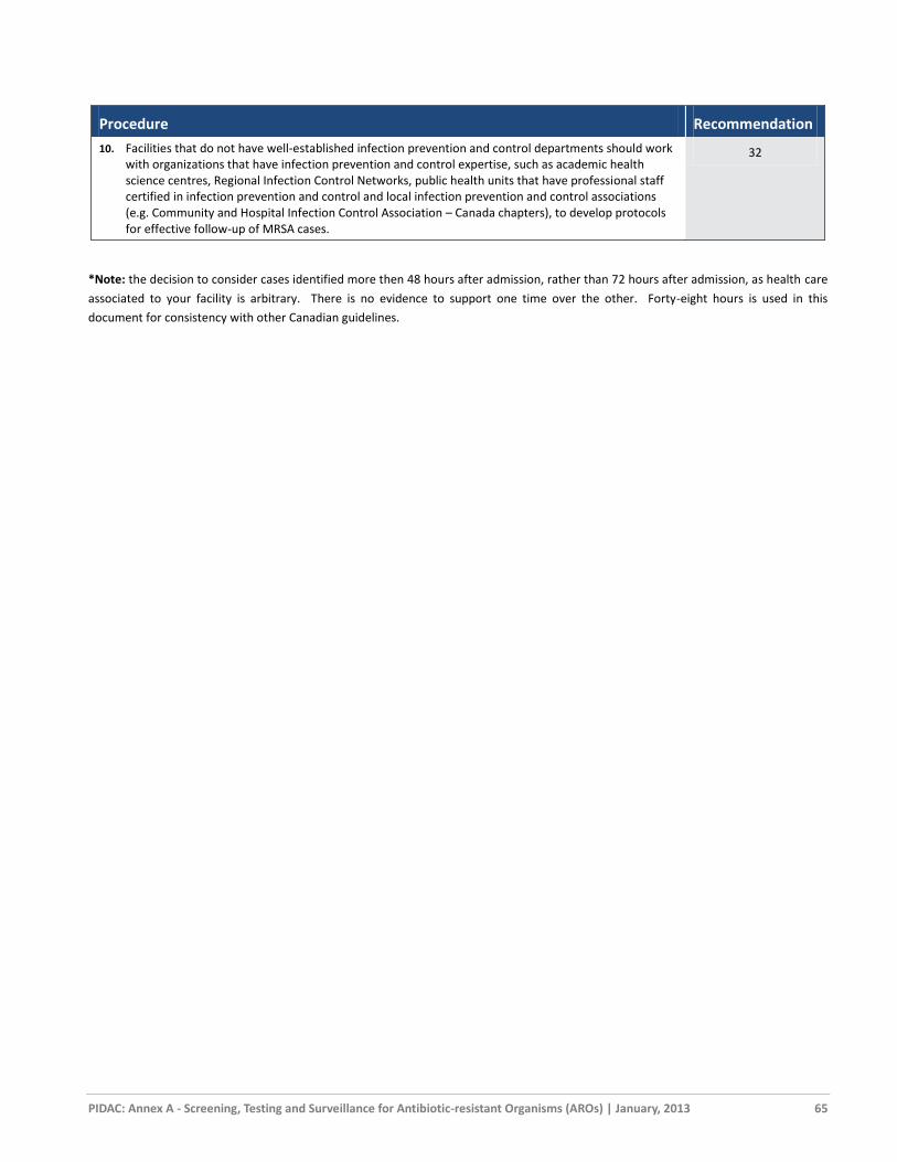
PIDAC: Annex A - Screening, Testing and Surveillance for Antibiotic-resistant Organisms (AROs) | January, 2013 65
Procedure Recommendation
10. Facilities that do not have well-established infection prevention and control departments should work with organizations that have infection prevention and control expertise, such as academic health science centres, Regional Infection Control Networks, public health units that have professional staff certified in infection prevention and control and local infection prevention and control associations (e.g. Community and Hospital Infection Control Association – Canada chapters), to develop protocols for effective follow-up of MRSA cases.
32
*Note: the decision to consider cases identified more then 48 hours after admission, rather than 72 hours after admission, as health care
associated to your facility is arbitrary. There is no evidence to support one time over the other. Forty-eight hours is used in this
document for consistency with other Canadian guidelines.

PIDAC: Annex A - Screening, Testing and Surveillance for Antibiotic-resistant Organisms (AROs) | January, 2013 66
Algorithm 2: Management of Suspected Health care-associated MRSA
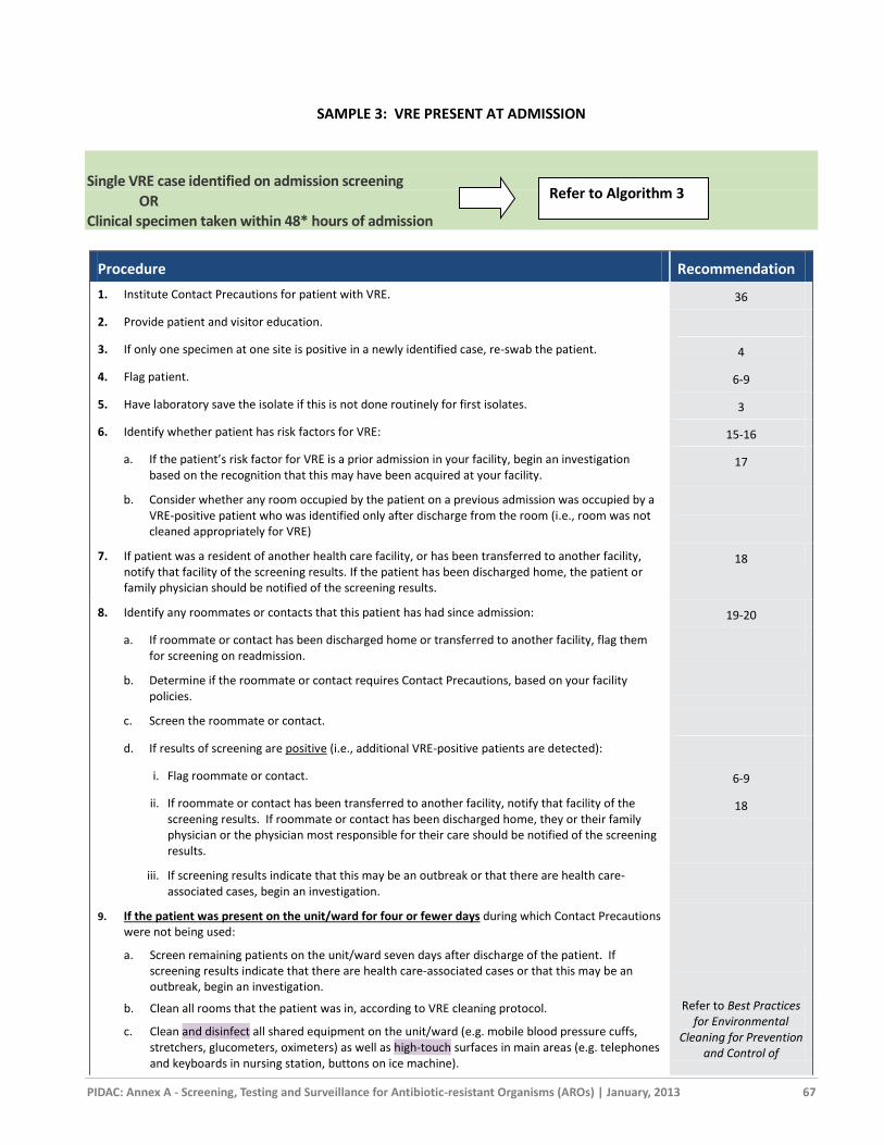
PIDAC: Annex A - Screening, Testing and Surveillance for Antibiotic-resistant Organisms (AROs) | January, 2013 67
SAMPLE 3: VRE PRESENT AT ADMISSION
Single VRE case identified on admission screening
OR Clinical specimen taken within 48* hours of admission
Procedure Recommendation
1. Institute Contact Precautions for patient with VRE. 36
2. Provide patient and visitor education.
3. If only one specimen at one site is positive in a newly identified case, re-swab the patient. 4
4. Flag patient. 6-9
5. Have laboratory save the isolate if this is not done routinely for first isolates. 3
6. Identify whether patient has risk factors for VRE: 15-16
a. If the patient’s risk factor for VRE is a prior admission in your facility, begin an investigation based on the recognition that this may have been acquired at your facility.
17
b. Consider whether any room occupied by the patient on a previous admission was occupied by a VRE-positive patient who was identified only after discharge from the room (i.e., room was not cleaned appropriately for VRE)
7. If patient was a resident of another health care facility, or has been transferred to another facility, notify that facility of the screening results. If the patient has been discharged home, the patient or family physician should be notified of the screening results.
18
8. Identify any roommates or contacts that this patient has had since admission: 19-20
a. If roommate or contact has been discharged home or transferred to another facility, flag them for screening on readmission.
b. Determine if the roommate or contact requires Contact Precautions, based on your facility policies.
c. Screen the roommate or contact.
d. If results of screening are positive (i.e., additional VRE-positive patients are detected):
i. Flag roommate or contact. 6-9
ii. If roommate or contact has been transferred to another facility, notify that facility of the screening results. If roommate or contact has been discharged home, they or their family physician or the physician most responsible for their care should be notified of the screening results.
18
iii. If screening results indicate that this may be an outbreak or that there are health care-associated cases, begin an investigation.
9. If the patient was present on the unit/ward for four or fewer days during which Contact Precautions were not being used:
a. Screen remaining patients on the unit/ward seven days after discharge of the patient. If screening results indicate that there are health care-associated cases or that this may be an outbreak, begin an investigation.
b. Clean all rooms that the patient was in, according to VRE cleaning protocol. Refer to Best Practices for Environmental
Cleaning for Prevention and Control of
c. Clean and disinfect all shared equipment on the unit/ward (e.g. mobile blood pressure cuffs, stretchers, glucometers, oximeters) as well as high-touch surfaces in main areas (e.g. telephones and keyboards in nursing station, buttons on ice machine).
Refer to Algorithm 3

PIDAC: Annex A - Screening, Testing and Surveillance for Antibiotic-resistant Organisms (AROs) | January, 2013 68
Procedure Recommendation
Infections
10. If the patient was present on the unit/ward for five or more days during which Contact Precautions were not being used:
a. Screen all patients on the unit/ward on the day the VRE is identified.
b. Re-screen all patients on the unit/ward three days later.
c. Re-screen all patients on the unit/ward seven days after discharge of the patient.
d. If screening results indicate that there are health care-associated cases or that this may be an outbreak, begin an investigation
e. All rooms the patient was in must be cleaned. Refer to Best Practices
for Environmental
Cleaning for Prevention
and Control of
Infections
f. Clean and disinfect all shared equipment on the unit/ward (e.g. mobile blood pressure cuffs, stretchers, glucometers, oximeters) as well as high-touch surfaces in main areas (e.g. telephones and keyboards in nursing station, buttons on ice machine).
11. When doing a prevalence screen, test every patient who was on the unit at the time chosen for the screen; if a patient is to be discharged or transferred out of the unit before the prevalence screen is done, he/she should be screened prior to discharge/transfer.
12. Continue with case management for cases and positive contacts still in facility.
*Note: the decision to consider cases identified more then 48 hours after admission, rather than 72 hours after admission, as health care
associated to your facility is arbitrary. There is no evidence to support one time over the other. Forty-eight hours is used in this document
for consistency with other Canadian guidelines.

PIDAC: Annex A - Screening, Testing and Surveillance for Antibiotic-resistant Organisms (AROs) | January, 2013 69
Algorithm 3: Management of a Single New Case of VRE

PIDAC: Annex A - Screening, Testing and Surveillance for Antibiotic-resistant Organisms (AROs) | January, 2013 70
SAMPLE 4: SUSPECTED HEALTH CARE-ASSOCIATED VRE
Single VRE case identified on a clinical specimen or
screening specimen taken more than 48* hours after
admission, in the absence of a known outbreak
Procedure Recommendation
1. Institute Contact Precautions for patient with VRE. 36
2. Provide patient and visitor education.
3. If only one specimen at one site is positive in a newly identified case, re-swab the patient. 4
4. Flag patient. 6-9
5. Have laboratory save the isolate if this is not done routinely for first isolates. 3
6. If patient has been transferred to another facility, notify that facility of the screening results. If the patient has been discharged home, the patient or family physician or physician most responsible for care should be notified of the screening results.
18
7. Clean all rooms that the patient was in, according to VRE cleaning protocol. Refer to Best Practices
for Environmental
Cleaning for
Prevention and Control
of Infections
8. Clean and disinfect all shared equipment on the unit/ward (e.g., mobile blood pressure cuffs, stretchers, glucometers, oximeters) as well as high-touch surfaces in main areas (e.g., telephones and keyboards in nursing station, buttons on ice machine), according to VRE cleaning protocol.
9. Roommates and Contacts: assess patient to attempt to identify sources for the VRE:
a. Establish an “at-risk” period when the patient may have been colonized but was not recognized (e.g. during a known exposure to another positive patient).
b. Identify roommates or contacts that this patient has had during the at-risk period:
i. Based on their degree of exposure, determine if Contact Precautions are required for roommates or contacts.
ii. If roommate or contact has been subsequently transferred to another facility, notify that facility about the need to screen them for VRE.
iii. If roommate or contact has been discharged home or transferred to another facility, flag them for screening on readmission.
6-9
iv. Screen the identified roommates and/or contacts that remain in your facility.
v. If results of screening are positive (i.e., additional VRE-positive patients are detected):
Flag roommate or contact; 6-9
Institute Contact Precautions for roommate or contact if this has not been done; 36
If roommate or contact has been subsequently transferred to another facility, notify that facility of the screening results. If roommate or contact has been discharged home, they or their family physician or the physician most responsible for care should be notified of the screening results.
18
vi. If results of screening are negative (i.e., no additional VRE-positive patients are detected), re-screen all those previously screened (from (iv) above) seven days after the last day that the original patient was on the unit/ward and not on Contact Precautions.
c. Consider whether follow-up of any contacts in the community is warranted (e.g., patients who are frequently re-admitted).
Refer to Algorithm 4

PIDAC: Annex A - Screening, Testing and Surveillance for Antibiotic-resistant Organisms (AROs) | January, 2013 71
Procedure Recommendation
10. Conduct prevalence screens: 21-22
a. If the patient was present on the unit/ward for four or fewer days during which Contact Precautions were not being used, screen all patients on the unit/ward seven days after discharge of the patient.
b. If the patient was present on the unit/ward for five or more days during which Contact Precautions were not being used:
i. Screen all patients on the unit/ward on the day the VRE is identified, if not already screened as contacts.
ii. Re-screen all patients on the unit/ward three days later.
iii. Re-screen all patients on the unit/ward seven days after discharge of the patient.
iv. When doing a prevalence screen, test every patient who was on the unit at the time chosen for the screen; if a patient is to be discharged or transferred out of the unit before the prevalence screen is done, he/she should be screened prior to discharge/transfer.
c. If analysis of the prevalence screen results for VRE identifies further transmission:
i. Continue screening every three days until there have been three negative results, indicating that there are no further cases of VRE on the unit/ward.
ii. Do not permit transfers from the unit/ward to other units/wards or discharges to other facilities except in emergency situations, or if the receiving unit/facility has been notified and can implement Contact Precautions and screening as appropriate.
iii. Consider closing the unit/ward to new admissions until patients on the unit/ward have been screened and results are known, and cleaning of shared equipment and rooms is complete.
11. Continue with case management for cases and positive contacts still in facility.
12. The patient’s room must be thoroughly cleaned and disinfected following the patient’s discharge, according to VRE cleaning protocol.
Refer to Best Practices
for Environmental
Cleaning for
Prevention and Control
of Infections
13. Clean and disinfect all shared equipment on the unit/ward (e.g., mobile blood pressure cuffs, stretchers, glucometers, oximeters) as well as high-touch surfaces in main areas (e.g., telephones and keyboards in nursing station, buttons on ice machine), according to VRE cleaning protocol.
14. Facilities that do not have well-established infection prevention and control departments should work with organizations that have infection prevention and control expertise, such as academic health science centres, Regional Infection Control Networks, public health units that have professional staff certified in infection prevention and control and local infection prevention and control associations (e.g., Community and Hospital Infection Control Association – Canada chapters), to develop protocols for effective follow-up of VRE cases.
32
*Note: the decision to consider cases identified more then 48 hours after admission, rather than 72 hours after admission, as health care
associated to your facility is arbitrary. There is no evidence to support one time over the other. Forty-eight hours is used in this document
for consistency with other Canadian guidelines.

PIDAC: Annex A - Screening, Testing and Surveillance for Antibiotic-resistant Organisms (AROs) | January, 2013 72
Algorithm 4: Management of Suspected Health Care-associated VRE
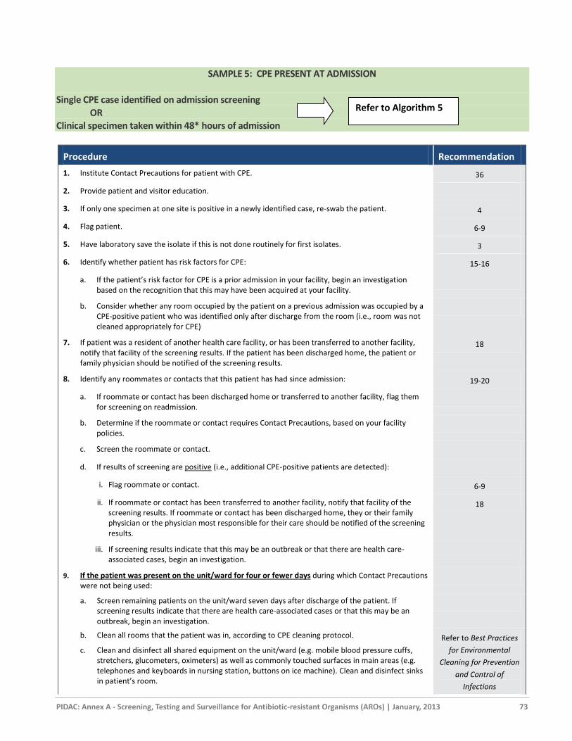
PIDAC: Annex A - Screening, Testing and Surveillance for Antibiotic-resistant Organisms (AROs) | January, 2013 73
SAMPLE 5: CPE PRESENT AT ADMISSION
Single CPE case identified on admission screening
OR
Clinical specimen taken within 48* hours of admission
Procedure Recommendation
1. Institute Contact Precautions for patient with CPE. 36
2. Provide patient and visitor education.
3. If only one specimen at one site is positive in a newly identified case, re-swab the patient. 4
4. Flag patient. 6-9
5. Have laboratory save the isolate if this is not done routinely for first isolates. 3
6. Identify whether patient has risk factors for CPE: 15-16
a. If the patient’s risk factor for CPE is a prior admission in your facility, begin an investigation based on the recognition that this may have been acquired at your facility.
b. Consider whether any room occupied by the patient on a previous admission was occupied by a CPE-positive patient who was identified only after discharge from the room (i.e., room was not cleaned appropriately for CPE)
7. If patient was a resident of another health care facility, or has been transferred to another facility, notify that facility of the screening results. If the patient has been discharged home, the patient or family physician should be notified of the screening results.
18
8. Identify any roommates or contacts that this patient has had since admission: 19-20
a. If roommate or contact has been discharged home or transferred to another facility, flag them for screening on readmission.
b. Determine if the roommate or contact requires Contact Precautions, based on your facility policies.
c. Screen the roommate or contact.
d. If results of screening are positive (i.e., additional CPE-positive patients are detected):
i. Flag roommate or contact. 6-9
ii. If roommate or contact has been transferred to another facility, notify that facility of the screening results. If roommate or contact has been discharged home, they or their family physician or the physician most responsible for their care should be notified of the screening results.
18
iii. If screening results indicate that this may be an outbreak or that there are health care-associated cases, begin an investigation.
9. If the patient was present on the unit/ward for four or fewer days during which Contact Precautions were not being used:
a. Screen remaining patients on the unit/ward seven days after discharge of the patient. If screening results indicate that there are health care-associated cases or that this may be an outbreak, begin an investigation.
b. Clean all rooms that the patient was in, according to CPE cleaning protocol. Refer to Best Practices
for Environmental
Cleaning for Prevention
and Control of
Infections
c. Clean and disinfect all shared equipment on the unit/ward (e.g. mobile blood pressure cuffs, stretchers, glucometers, oximeters) as well as commonly touched surfaces in main areas (e.g. telephones and keyboards in nursing station, buttons on ice machine). Clean and disinfect sinks in patient’s room.
Refer to Algorithm 5
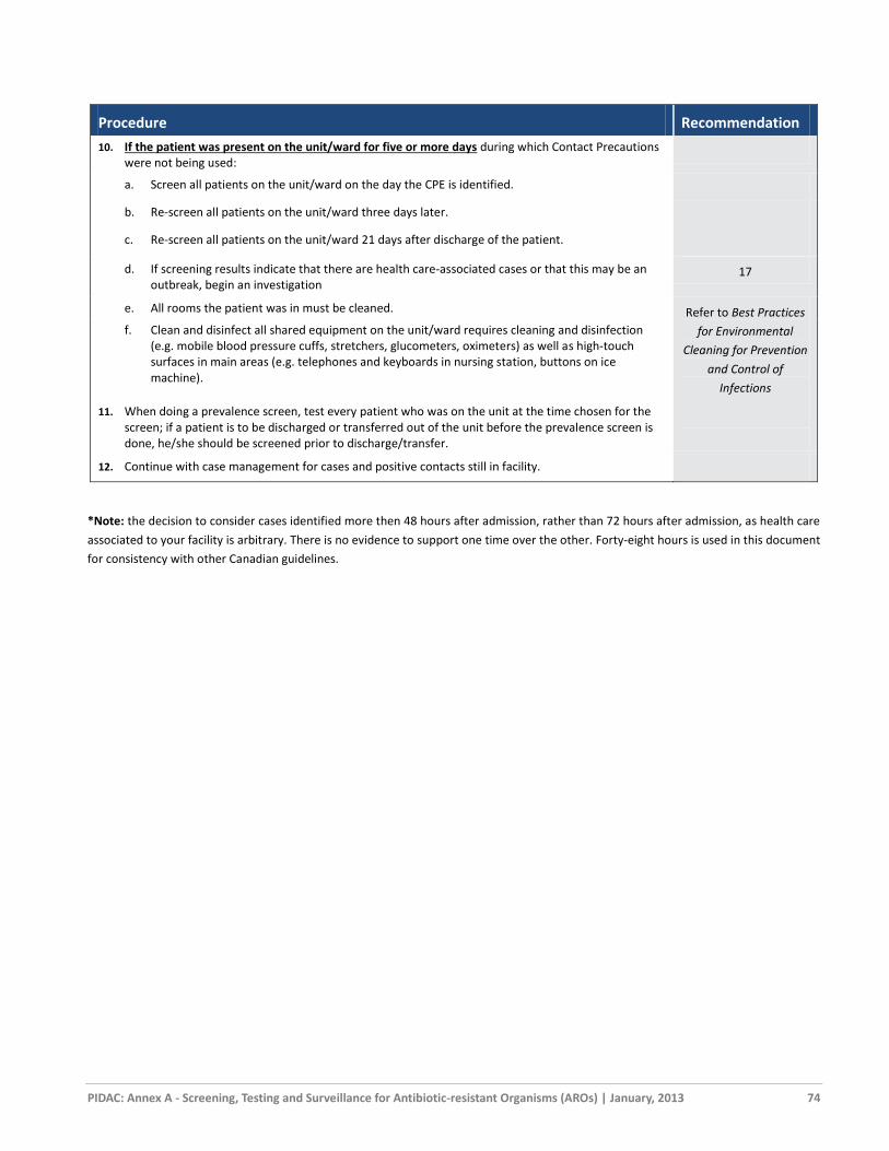
PIDAC: Annex A - Screening, Testing and Surveillance for Antibiotic-resistant Organisms (AROs) | January, 2013 74
Procedure Recommendation
10. If the patient was present on the unit/ward for five or more days during which Contact Precautions were not being used:
a. Screen all patients on the unit/ward on the day the CPE is identified.
b. Re-screen all patients on the unit/ward three days later.
c. Re-screen all patients on the unit/ward 21 days after discharge of the patient.
d. If screening results indicate that there are health care-associated cases or that this may be an outbreak, begin an investigation
17
e. All rooms the patient was in must be cleaned. Refer to Best Practices
for Environmental
Cleaning for Prevention
and Control of
Infections
f. Clean and disinfect all shared equipment on the unit/ward requires cleaning and disinfection (e.g. mobile blood pressure cuffs, stretchers, glucometers, oximeters) as well as high-touch surfaces in main areas (e.g. telephones and keyboards in nursing station, buttons on ice machine).
11. When doing a prevalence screen, test every patient who was on the unit at the time chosen for the screen; if a patient is to be discharged or transferred out of the unit before the prevalence screen is done, he/she should be screened prior to discharge/transfer.
12. Continue with case management for cases and positive contacts still in facility.
*Note: the decision to consider cases identified more then 48 hours after admission, rather than 72 hours after admission, as health care
associated to your facility is arbitrary. There is no evidence to support one time over the other. Forty-eight hours is used in this document
for consistency with other Canadian guidelines.

PIDAC: Annex A - Screening, Testing and Surveillance for Antibiotic-resistant Organisms (AROs) | January, 2013 75
Algorithm 5: Management of a Single New Case of CPE
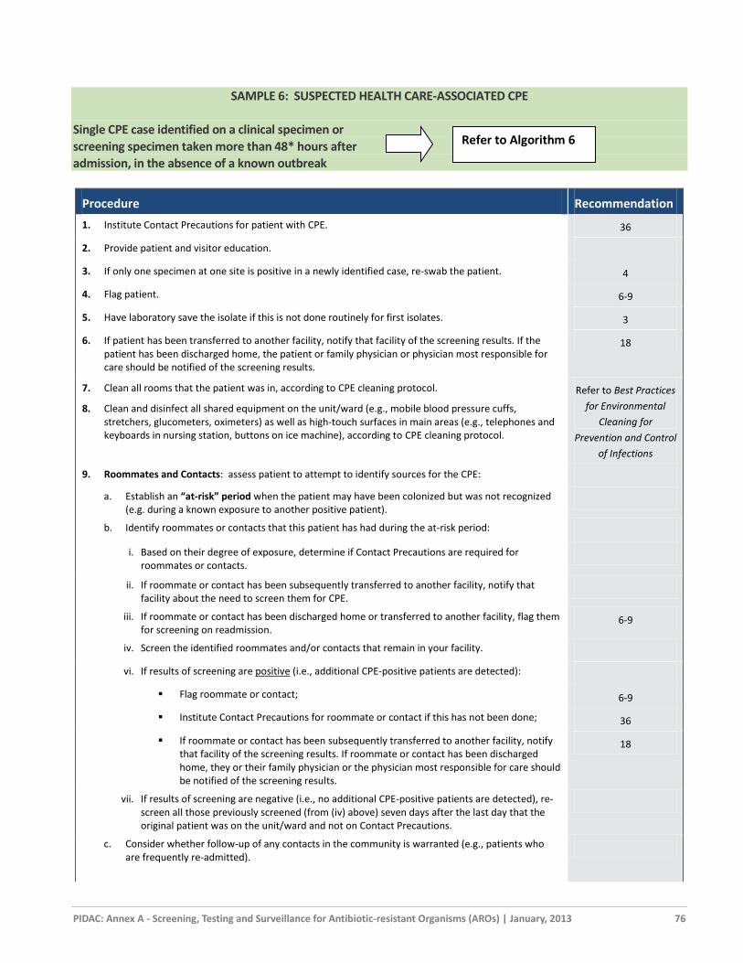
PIDAC: Annex A - Screening, Testing and Surveillance for Antibiotic-resistant Organisms (AROs) | January, 2013 76
SAMPLE 6: SUSPECTED HEALTH CARE-ASSOCIATED CPE
Single CPE case identified on a clinical specimen or
screening specimen taken more than 48* hours after
admission, in the absence of a known outbreak
Procedure Recommendation
1. Institute Contact Precautions for patient with CPE. 36
2. Provide patient and visitor education.
3. If only one specimen at one site is positive in a newly identified case, re-swab the patient. 4
4. Flag patient. 6-9
5. Have laboratory save the isolate if this is not done routinely for first isolates. 3
6. If patient has been transferred to another facility, notify that facility of the screening results. If the patient has been discharged home, the patient or family physician or physician most responsible for care should be notified of the screening results.
18
7. Clean all rooms that the patient was in, according to CPE cleaning protocol. Refer to Best Practices
for Environmental
Cleaning for
Prevention and Control
of Infections
8. Clean and disinfect all shared equipment on the unit/ward (e.g., mobile blood pressure cuffs, stretchers, glucometers, oximeters) as well as high-touch surfaces in main areas (e.g., telephones and keyboards in nursing station, buttons on ice machine), according to CPE cleaning protocol.
9. Roommates and Contacts: assess patient to attempt to identify sources for the CPE:
a. Establish an “at-risk” period when the patient may have been colonized but was not recognized (e.g. during a known exposure to another positive patient).
b. Identify roommates or contacts that this patient has had during the at-risk period:
i. Based on their degree of exposure, determine if Contact Precautions are required for roommates or contacts.
ii. If roommate or contact has been subsequently transferred to another facility, notify that facility about the need to screen them for CPE.
iii. If roommate or contact has been discharged home or transferred to another facility, flag them for screening on readmission.
6-9
iv. Screen the identified roommates and/or contacts that remain in your facility.
vi. If results of screening are positive (i.e., additional CPE-positive patients are detected):
Flag roommate or contact; 6-9
Institute Contact Precautions for roommate or contact if this has not been done; 36
If roommate or contact has been subsequently transferred to another facility, notify that facility of the screening results. If roommate or contact has been discharged home, they or their family physician or the physician most responsible for care should be notified of the screening results.
18
vii. If results of screening are negative (i.e., no additional CPE-positive patients are detected), re-screen all those previously screened (from (iv) above) seven days after the last day that the original patient was on the unit/ward and not on Contact Precautions.
c. Consider whether follow-up of any contacts in the community is warranted (e.g., patients who are frequently re-admitted).
Refer to Algorithm 6

PIDAC: Annex A - Screening, Testing and Surveillance for Antibiotic-resistant Organisms (AROs) | January, 2013 77
Procedure Recommendation
10. Conduct prevalence screens: 21-22
a. If the patient was present on the unit/ward for four or fewer days during which Contact Precautions were not being used, screen all patients on the unit/ward 21 days after discharge of the patient.
b. If the patient was present on the unit/ward for five or more days during which Contact Precautions were not being used:
i. Screen all patients on the unit/ward on the day the CPE is identified, if not already screened as contacts.
ii. Re-screen all patients on the unit/ward three days later.
iii. Re-screen all patients on the unit/ward 21 days after discharge of the patient.
iv. When doing a prevalence screen, test every patient who was on the unit at the time chosen for the screen; if a patient is to be discharged or transferred out of the unit before the prevalence screen is done, he/she should be screened prior to discharge/transfer.
c. If analysis of the prevalence screen results for CPE identifies further transmission:
iv. Continue screening every three days until there have been three negative results, indicating that there are no further cases of CPE on the unit/ward.
v. Do not permit transfers from the unit/ward to other units/wards or discharges to other facilities except in emergency situations, or if the receiving unit/facility has been notified and can implement Contact Precautions and screening as appropriate.
vi. Consider closing the unit/ward to new admissions until patients on the unit/ward have been screened and results are known, and cleaning of shared equipment and rooms is complete.
11. Continue with case management for cases and positive contacts still in facility. 35-36
12. The patient’s room must be thoroughly cleaned and disinfected following the patient’s discharge, according to CPE cleaning protocol.
Refer to Best Practices
for Environmental
Cleaning for
Prevention and Control
of Infections
13. Clean and disinfect all shared equipment on the unit/ward (e.g., mobile blood pressure cuffs, stretchers, glucometers, oximeters) as well as high-touch surfaces in main areas (e.g., telephones and keyboards in nursing station, buttons on ice machine), according to CPE cleaning protocol.
14. Facilities that do not have well-established infection prevention and control departments should work with organizations that have infection prevention and control expertise, such as academic health science centres, Regional Infection Control Networks, public health units that have professional staff certified in infection prevention and control and local infection prevention and control associations (e.g., Community and Hospital Infection Control Association – Canada chapters), to develop protocols for effective follow-up of CPE cases.
32
*Note: the decision to consider cases identified more then 48 hours after admission, rather than 72 hours after admission, as health care
associated to your facility is arbitrary. There is no evidence to support one time over the other. Forty-eight hours is used in this document
for consistency with other Canadian guidelines.
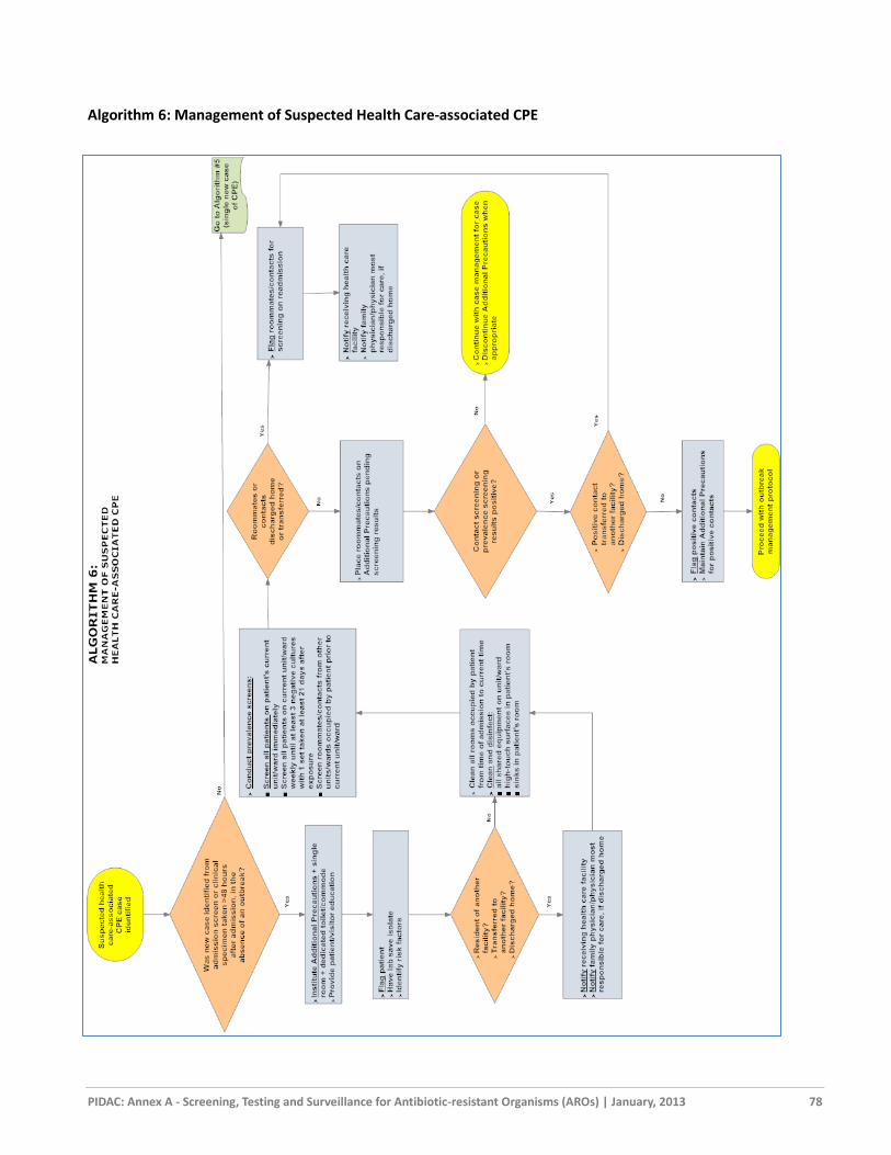
PIDAC: Annex A - Screening, Testing and Surveillance for Antibiotic-resistant Organisms (AROs) | January, 2013 78
Algorithm 6: Management of Suspected Health Care-associated CPE

PIDAC: Annex A - Screening, Testing and Surveillance for Antibiotic-resistant Organisms (AROs) | January, 2013 79
APPENDIX E: SAMPLE LETTERS FOR PHYSICIANS
[Adapted from materials provided by Mount Sinai Hospital, Toronto, Ontario]
Letter #1: Contact of a positive patient who has been discharged home before screening tests were done
[Insert date]
Dr. [insert physician name]
[insert address line 1]
[insert address line 2]
Dear Dr. [insert physician last name],
RE: [insert patient name]
DOB: [insert patient’s date of birth]
While in [insert name of facility], your above named patient was in the same room with another patient who has since been found to be colonized with methicillin-resistant Staphylococcus aureus (MRSA). As I am sure you are aware, MRSA is resistant to all penicillins and cephalosporins.
Because Staphylococcus aureus can cause serious health care-associated infections, we want to make sure that no acquisition with a resistant strain has occurred. Although the risk is low, Staphylococcus aureus can be transmitted from person-to-person by direct or indirect contact on the same ward. In order to be sure that your patient is not affected, we are requesting that [he/she] have swabs of the anterior nares, perianal area and any open wounds collected, looking for MRSA only (please indicate this specifically on lab requisition).
We would be grateful if you would arrange that a copy of the results of these specimens be faxed to [insert name of Infection Prevention & Control Professional or Physician], at [insert fax number of Infection Prevention & Control Professional or Physician].
In the unlikely event that your patient has acquired this organism please contact the infection prevention and control department at [insert phone number] and we would be willing to discuss with you our strategy for management of MRSA. If you have any questions or comments, please call us at any time.
Thank you very much for your assistance and co-operation in this matter.
Sincerely,
[insert name of Infection Prevention & Control Professional or Physician]
[insert title of Infection Prevention & Control Professional or Physician]
[insert address line 1]
[insert address line 2]
[insert phone number]
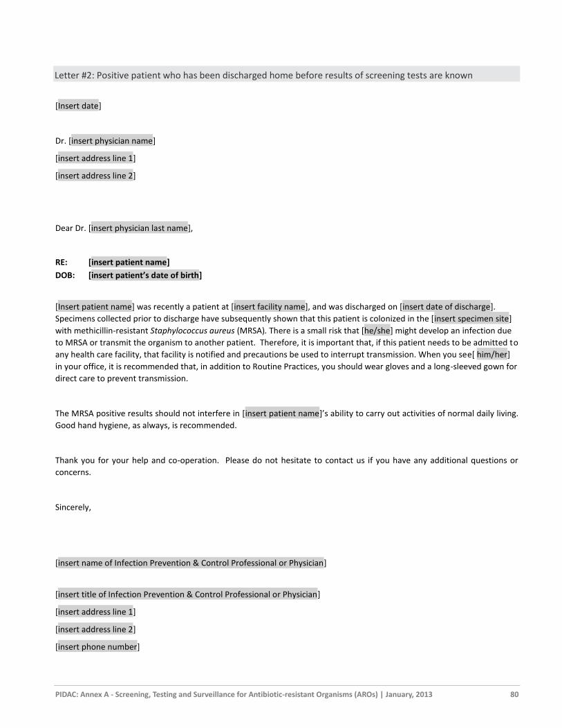
PIDAC: Annex A - Screening, Testing and Surveillance for Antibiotic-resistant Organisms (AROs) | January, 2013 80
Letter #2: Positive patient who has been discharged home before results of screening tests are known
[Insert date]
Dr. [insert physician name]
[insert address line 1]
[insert address line 2]
Dear Dr. [insert physician last name],
RE: [insert patient name]
DOB: [insert patient’s date of birth]
[Insert patient name] was recently a patient at [insert facility name], and was discharged on [insert date of discharge].
Specimens collected prior to discharge have subsequently shown that this patient is colonized in the [insert specimen site]
with methicillin-resistant Staphylococcus aureus (MRSA). There is a small risk that [he/she] might develop an infection due
to MRSA or transmit the organism to another patient. Therefore, it is important that, if this patient needs to be admitted to
any health care facility, that facility is notified and precautions be used to interrupt transmission. When you see[ him/her]
in your office, it is recommended that, in addition to Routine Practices, you should wear gloves and a long-sleeved gown for
direct care to prevent transmission.
The MRSA positive results should not interfere in [insert patient name]’s ability to carry out activities of normal daily living.
Good hand hygiene, as always, is recommended.
Thank you for your help and co-operation. Please do not hesitate to contact us if you have any additional questions or
concerns.
Sincerely,
[insert name of Infection Prevention & Control Professional or Physician]
[insert title of Infection Prevention & Control Professional or Physician]
[insert address line 1]
[insert address line 2]
[insert phone number]

PIDAC: Annex A - Screening, Testing and Surveillance for Antibiotic-resistant Organisms (AROs) | January, 2013 81
APPENDIX F: SEARCH STRATEGY AND SELECTION CRITERIA
Data for this revision were identified during May, 2011 by searches of Medline and references from relevant articles. Search terms were:
carbapenemase AND Klebsiella
carbapenemase AND E. coli
carbapenemase AND KPC
carbapenemase AND NDM
metallo-beta-lactamase
carbapenemase AND control
carbapenemase AND infection control
PIDAC: Annex A - Screening, Testing and Surveillance for Antibiotic-resistant Organisms (AROs) | January, 2013 82
References
1. Barton M HM, Moore D, Conly J, Nicole OL, Allen U et al. Guidelines for the prevention and management of community-associated methicillin-resistant Staphylococcus aureus (CA-MRSA): A perspective for Canadian Health care practitioners. Can J Infect Dis Med Microbiol. 2006;17 Suppl C:4-19.
2. Provincial Infectious Diseases Advisory Committee (PIDAC). Best Practices for Surveillance of Health Care-Associated Infections in Patient and Resident Populations 2011 [cited November 25, 2012]. Available from: http://www.oahpp.ca/resources/pidac-knowledge/best-practice-manuals/surveillance-of-health-care-associated-infections.html.
3. Jernigan JA, Titus MG, Groschel DH, Getchell-White S, Farr BM. Effectiveness of contact isolation during a hospital outbreak of methicillin-resistant Staphylococcus aureus. Am J Epidemiol. 1996 Mar 1;143(5):496-504.
4. Huskins WC. Interventions to prevent transmission of antimicrobial-resistant bacteria in the intensive care unit. Curr Opin Crit Care. 2007 Oct;13(5):572-7.
5. Hardy KJ, Oppenheim BA, Gossain S, Gao F, Hawkey PM. A study of the relationship between environmental contamination with methicillin-resistant Staphylococcus aureus (MRSA) and patients' acquisition of MRSA. Infect Control Hosp Epidemiol. 2006 Feb;27(2):127-32.
6. Nolan SM, Gerber JS, Zaoutis T, Prasad P, Rettig S, Gross K, et al. Outbreak of vancomycin-resistant enterococcus colonization among pediatric oncology patients. Infect Control Hosp Epidemiol. 2009 Apr;30(4):338-45.
7. Ostrowsky BE, Trick WE, Sohn AH, Quirk SB, Holt S, Carson LA, et al. Control of vancomycin-resistant enterococcus in health care facilities in a region. N Engl J Med. 2001 May 10;344(19):1427-33.
8. Grayson ML, Jarvie LJ, Martin R, Johnson PD, Jodoin ME, McMullan C, et al. Significant reductions in methicillin-resistant Staphylococcus aureus bacteraemia and clinical isolates associated with a multisite, hand hygiene culture-change program and subsequent successful statewide roll-out. Med J Aust. 2008 Jun 2;188(11):633-40.
9. Price CS, Paule S, Noskin GA, Peterson LR. Active surveillance reduces the incidence of vancomycin-resistant enterococcal bacteraemia. Clin Infect Dis. 2003 Oct 1;37(7):921-8.
10. Karchmer TB, Durbin LJ, Simonton BM, Farr BM. Cost-effectiveness of active surveillance cultures and contact/droplet precautions for control of methicillin-resistant Staphylococcus aureus. J Hosp Infect. 2002 Jun;51(2):126-32.
11. Calfee DP, Farr BM. Infection control and cost control in the era of managed care. Infect Control Hosp Epidemiol. 2002 Jul;23(7):407-10.
12. Lucet JC, Chevret S, Durand-Zaleski I, Chastang C, Regnier B. Prevalence and risk factors for carriage of methicillin-resistant Staphylococcus aureus at admission to the intensive care unit: results of a multicenter study. Arch Intern Med. 2003 Jan 27;163(2):181-8.
13. Muto CA, Jernigan JA, Ostrowsky BE, Richet HM, Jarvis WR, Boyce JM, et al. SHEA guideline for preventing nosocomial transmission of multidrug-resistant strains of Staphylococcus aureus and enterococcus. Infect Control Hosp Epidemiol. 2003 May;24(5):362-86.
14. Abbett SK, Yokoe DS, Lipsitz SR, Bader AM, Berry WR, Tamplin EM, et al. Proposed Checklist of Hospital Interventions to Decrease the Incidence of Healthcare-Associated Clostridium difficile Infection. Infect Control Hosp Epidemiol. 2009 Sep 14.
15. Gerding DN, Muto CA, Owens RC, Jr. Measures to control and prevent Clostridium difficile infection. Clin Infect Dis. 2008 Jan 15;46 Suppl 1:S43-9.
16. . Infection Prevention and Control Practice. Clostridium difficile Associated Diarrhea (CDAD). Proceedings and Recommendations. International Infection Control Council Global Consensus Conference; 2007; Toronto, Ontario, Canada.
17. Conly J. Antimicrobial resistance in Canada. CMAJ. 2002 Oct 15;167(8):885-91. 18. Farr BM. Prevention and control of methicillin-resistant Staphylococcus aureus infections. Curr Opin
Infect Dis. 2004 Aug;17(4):317-22. 19. Shurland S, Zhan M, Bradham DD, Roghmann MC. Comparison of Mortality Risk Associated With
Bacteraemia Due to Methicillin-Resistant and Methicillin-Susceptible Staphylococcus aureus. Infect Control Hosp Epidemiol. 2007 Mar;28(3):273-9.

PIDAC: Annex A - Screening, Testing and Surveillance for Antibiotic-resistant Organisms (AROs) | January, 2013 83
20. Cosgrove SE, Sakoulas G, Perencevich EN, Schwaber MJ, Karchmer AW, Carmeli Y. Comparison of mortality associated with methicillin-resistant and methicillin-susceptible Staphylococcus aureus bacteraemia: a meta-analysis. Clin Infect Dis. 2003 Jan 1;36(1):53-9.
21. Engemann JJ, Carmeli Y, Cosgrove SE, Fowler VG, Bronstein MZ, Trivette SL, et al. Adverse clinical and economic outcomes attributable to methicillin resistance among patients with Staphylococcus aureus surgical site infection. Clin Infect Dis. 2003 Mar 1;36(5):592-8.
22. Niederman MS. Impact of antibiotic resistance on clinical outcomes and the cost of care. Crit Care Med. 2001 Apr;29(4 Suppl):N114-20.
23. Reed SD, Friedman JY, Engemann JJ, Griffiths RI, Anstrom KJ, Kaye KS, et al. Costs and outcomes among hemodialysis-dependent patients with methicillin-resistant or methicillin-susceptible Staphylococcus aureus bacteraemia. Infect Control Hosp Epidemiol. 2005 Feb;26(2):175-83.
24. Salgado CD, Farr BM. Outcomes associated with vancomycin-resistant enterococci: a meta-analysis. Infect Control Hosp Epidemiol. 2003 Sep;24(9):690-8.
25. DiazGranados CA, Zimmer SM, Klein M, Jernigan JA. Comparison of mortality associated with vancomycin-resistant and vancomycin-susceptible enterococcal bloodstream infections: a meta-analysis. Clin Infect Dis. 2005 Aug 1;41(3):327-33.
26. Zirakzadeh A, Gastineau DA, Mandrekar JN, Burke JP, Johnston PB, Patel R. Vancomycin-resistant enterococcal colonization appears associated with increased mortality among allogeneic hematopoietic stem cell transplant recipients. Bone Marrow Transplant. 2008 Feb;41(4):385-92.
27. Kamboj M, Chung D, Seo SK, Pamer EG, Sepkowitz KA, Jakubowski AA, et al. The changing epidemiology of vancomycin-resistant Enterococcus (VRE) bacteraemia in allogeneic hematopoietic stem cell transplant (HSCT) recipients. Biol Blood Marrow Transplant. 2010 Nov;16(11):1576-81.
28. Weinstock DM, Conlon M, Iovino C, Aubrey T, Gudiol C, Riedel E, et al. Colonization, bloodstream infection, and mortality caused by vancomycin-resistant enterococcus early after allogeneic hematopoietic stem cell transplant. Biol Blood Marrow Transplant. 2007 May;13(5):615-21.
29. Gammon J. The psychological consequences of source isolation: a review of the literature. J Clin Nurs. 1999 Jan;8(1):13-21.
30. Stelfox HT, Bates DW, Redelmeier DA. Safety of patients isolated for infection control. JAMA. 2003 Oct 8;290(14):1899-905.
31. Lewis AM, Gammon J, Hosein I. The pros and cons of isolation and containment. J Hosp Infect. 1999 Sep;43(1):19-23.
32. Doxtator L, Zoutman D. An infection control perspective on patient advocacy. Can J Infect Control. 2006;21(3):129-32.
33. Abad C, Fearday A, Safdar N. Adverse effects of isolation in hospitalised patients: a systematic review. J Hosp Infect. 2010 Oct;76(2):97-102.
34. Catalano G, Houston SH, Catalano MC, Butera AS, Jennings SM, Hakala SM, et al. Anxiety and depression in hospitalized patients in resistant organism isolation. South Med J. 2003 Feb;96(2):141-5.
35. Morgan DJ, Diekema DJ, Sepkowitz K, Perencevich EN. Adverse outcomes associated with Contact Precautions: a review of the literature. Am J Infect Control. 2009 Mar;37(2):85-93.
36. Knowles HE. The experience of infectious patients in isolation. Nurs Times. 1993 Jul 28-Aug 3;89(30):53-6.
37. Barratt RL, Shaban R, Moyle W. Patient experience of source isolation: lessons for clinical practice. Contemp Nurse. 2011 Oct;39(2):180-93.
38. Day HR, Perencevich EN, Harris AD, Himelhoch SS, Brown CH, Gruber-Baldini AL, et al. Do contact precautions cause depression? A two-year study at a tertiary care medical centre. J Hosp Infect. 2011 Oct;79(2):103-7.
39. Cosgrove SE, Qi Y, Kaye KS, Harbarth S, Karchmer AW, Carmeli Y. The impact of methicillin resistance in Staphylococcus aureus bacteraemia on patient outcomes: mortality, length of stay, and hospital charges. Infect Control Hosp Epidemiol. 2005 Feb;26(2):166-74.
40. Morrison L, Stolarek I. Does MRSA affect patient outcomes in the elderly? A retrospective pilot study. J Hosp Infect. 2000 Jun;45(2):169-71.
41. Kim T, Oh PI, Simor AE. The economic impact of methicillin-resistant Staphylococcus aureus in Canadian hospitals. Infect Control Hosp Epidemiol. 2001 Feb;22(2):99-104.
42. Lim SP. The Financial Impact of Hospital-acquired Methicillin-Resistant Staphylococcus aureus: an Incremental Cost and Cost-Effectiveness Analysis. Toronto: University of Toronto; 2006.
PIDAC: Annex A - Screening, Testing and Surveillance for Antibiotic-resistant Organisms (AROs) | January, 2013 84
43. Bjorholt I, Haglind E. Cost-savings achieved by eradication of epidemic methicillin-resistant Staphylococcus aureus (EMRSA)-16 from a large teaching hospital. Eur J Clin Microbiol Infect Dis. 2004 Sep;23(9):688-95.
44. McHugh CG, Riley LW. Risk factors and costs associated with methicillin-resistant Staphylococcus aureus bloodstream infections. Infect Control Hosp Epidemiol. 2004 May;25(5):425-30.
45. Capitano B, Leshem OA, Nightingale CH, Nicolau DP. Cost effect of managing methicillin-resistant Staphylococcus aureus in a long-term care facility. J Am Geriatr Soc. 2003 Jan;51(1):10-6.
46. Butler AM, Olsen MA, Merz LR, Guth RM, Woeltje KF, Camins BC, et al. Attributable costs of enterococcal bloodstream infections in a nonsurgical hospital cohort. Infect Control Hosp Epidemiol. 2010 Jan;31(1):28-35.
47. Stosor V, Peterson LR, Postelnick M, Noskin GA. Enterococcus faecium bacteraemia: does vancomycin resistance make a difference? Arch Intern Med. 1998 Mar 9;158(5):522-7.
48. Song X, Srinivasan A, Plaut D, Perl TM. Effect of nosocomial vancomycin-resistant enterococcal bacteraemia on mortality, length of stay, and costs. Infect Control Hosp Epidemiol. 2003 Apr;24(4):251-6.
49. Puzniak LA, Gillespie KN, Leet T, Kollef M, Mundy LM. A cost-benefit analysis of gown use in controlling vancomycin-resistant Enterococcus transmission: is it worth the price? Infect Control Hosp Epidemiol. 2004 May;25(5):418-24.
50. Muto CA, Giannetta ET, Durbin LJ, Simonton BM, Farr BM. Cost-effectiveness of perirectal surveillance cultures for controlling vancomycin-resistant Enterococcus. Infect Control Hosp Epidemiol. 2002 Aug;23(8):429-35.
51. Lucet JC, Chevret S, Decre D, Vanjak D, Macrez A, Bedos JP, et al. Outbreak of multiply resistant enterobacteriaceae in an intensive care unit: epidemiology and risk factors for acquisition. Clin Infect Dis. 1996 Mar;22(3):430-6.
52. Stone PW, Gupta A, Loughrey M, Della-Latta P, Cimiotti J, Larson E, et al. Attributable costs and length of stay of an extended-spectrum beta-lactamase-producing Klebsiella pneumoniae outbreak in a neonatal intensive care unit. Infect Control Hosp Epidemiol. 2003 Aug;24(8):601-6.
53. Wiener J, Quinn JP, Bradford PA, Goering RV, Nathan C, Bush K, et al. Multiple antibiotic-resistant Klebsiella and Escherichia coli in nursing homes. JAMA. 1999 Feb 10;281(6):517-23.
54. Nicolas-Chanoine MH, Jarlier V. Extended-spectrum beta-lactamases in long-term-care facilities. Clin Microbiol Infect. 2008 Jan;14 Suppl 1:111-6.
55. Conterno LO, Shymanski J, Ramotar K, Toye B, Zvonar R, Roth V. Impact and cost of infection control measures to reduce nosocomial transmission of extended-spectrum beta-lactamase-producing organisms in a non-outbreak setting. J Hosp Infect. 2007 Apr;65(4):354-60.
56. Schwaber MJ, Navon-Venezia S, Kaye KS, Ben-Ami R, Schwartz D, Carmeli Y. Clinical and economic impact of bacteraemia with extended- spectrum-beta-lactamase-producing Enterobacteriaceae. Antimicrob Agents Chemother. 2006 Apr;50(4):1257-62.
57. Coia JE, Duckworth GJ, Edwards DI, Farrington M, Fry C, Humphreys H, et al. Guidelines for the control and prevention of meticillin-resistant Staphylococcus aureus (MRSA) in healthcare facilities. J Hosp Infect. 2006 May;63 Suppl 1:S1-44.
58. Dutch Workingparty Infection Prevention. Policy for Methicillin-resistant Staphylococcus aureus. 2004 [cited Available from: http://www.wip.nl/UK/free_content/Richtlijnen/MRSA(1).pdf.
59. Williams V, Barry C, Vearncombe M, Simor A, Nyog Inn N. Effective admission screening as a component of nosocomial methicillin resistant Staphylococcus aureus (MRSA) identification and control. National Conference of the Community and Hospital Infection Control Association-Canada; May 7-11, 2005; Winnipeg, Manitoba2005.
60. Papia G, Louie M, Tralla A, Johnson C, Collins V, Simor AE. Screening high-risk patients for methicillin-resistant Staphylococcus aureus on admission to the hospital: is it cost effective? Infect Control Hosp Epidemiol. 1999 Jul;20(7):473-7.
61. Boyce JM, Cookson B, Christiansen K, Hori S, Vuopio-Varkila J, Kocagoz S, et al. Meticillin-resistant Staphylococcus aureus. Lancet Infect Dis. 2005 Oct;5(10):653-63.
62. Verhoef J, Beaujean D, Blok H, Baars A, Meyler A, van der Werken C, et al. A Dutch approach to methicillin-resistant Staphylococcus aureus. Eur J Clin Microbiol Infect Dis. 1999 Jul;18(7):461-6.
63. Girou E, Azar J, Wolkenstein P, Cizeau F, Brun-Buisson C, Roujeau JC. Comparison of systematic versus selective screening for methicillin-resistant Staphylococcus aureus carriage in a high-risk dermatology ward. Infect Control Hosp Epidemiol. 2000 Sep;21(9):583-7.
PIDAC: Annex A - Screening, Testing and Surveillance for Antibiotic-resistant Organisms (AROs) | January, 2013 85
64. Eveillard M, Lancien E, Barnaud G, Hidri N, Gaba S, Benlolo JA, et al. Impact of screening for MRSA carriers at hospital admission on risk-adjusted indicators according to the imported MRSA colonization pressure. J Hosp Infect. 2005 Mar;59(3):254-8.
65. Pan A, Carnevale G, Catenazzi P, Colombini P, Crema L, Dolcetti L, et al. Trends in methicillin-resistant Staphylococcus aureus (MRSA) bloodstream infections: effect of the MRSA "search and isolate" strategy in a hospital in Italy with hyperendemic MRSA. Infect Control Hosp Epidemiol. 2005 Feb;26(2):127-33.
66. Wernitz MH, Swidsinski S, Weist K, Sohr D, Witte W, Franke KP, et al. Effectiveness of a hospital-wide selective screening programme for methicillin-resistant Staphylococcus aureus (MRSA) carriers at hospital admission to prevent hospital-acquired MRSA infections. Clin Microbiol Infect. 2005 Jun;11(6):457-65.
67. Forceville X, Faibis F, Lahilaire P, Gantier I, Philippot S, Leporcq C. Decrease of infection rate of methicillin resistant Staphylococcus aureus acquired in a French intensive care unit, under reinforcement of specific isolation. Med Mal Infect. 2002;32(7):346-58.
68. Pittet D, Safran E, Harbarth S, Borst F, Copin P, Rohner P, et al. Automatic alerts for methicillin-resistant Staphylococcus aureus surveillance and control: role of a hospital information system. Infect Control Hosp Epidemiol. 1996 Aug;17(8):496-502.
69. Monnet DL. Methicillin-resistant Staphylococcus aureus and its relationship to antimicrobial use: possible implications for control. Infect Control Hosp Epidemiol. 1998 Aug;19(8):552-9.
70. Enright MC, Robinson DA, Randle G, Feil EJ, Grundmann H, Spratt BG. The evolutionary history of methicillin-resistant Staphylococcus aureus (MRSA). Proc Natl Acad Sci U S A. 2002 May 28;99(11):7687-92.
71. Carling P, Fung T, Killion A, Terrin N, Barza M. Favorable impact of a multidisciplinary antibiotic management program conducted during 7 years. Infect Control Hosp Epidemiol. 2003 Sep;24(9):699-706.
72. Lutters M, Harbarth S, Janssens JP, Freudiger H, Herrmann F, Michel JP, et al. Effect of a comprehensive, multidisciplinary, educational program on the use of antibiotics in a geriatric university hospital. J Am Geriatr Soc. 2004 Jan;52(1):112-6.
73. Fridkin SK, Hill HA, Volkova NV, Edwards JR, Lawton RM, Gaynes RP, et al. Temporal changes in prevalence of antimicrobial resistance in 23 US hospitals. Emerg Infect Dis. 2002 Jul;8(7):697-701.
74. Harbarth S, Samore MH, Lichtenberg D, Carmeli Y. Prolonged antibiotic prophylaxis after cardiovascular surgery and its effect on surgical site infections and antimicrobial resistance. Circulation. 2000 Jun 27;101(25):2916-21.
75. Fridkin SK, Edwards JR, Courval JM, Hill H, Tenover FC, Lawton R, et al. The effect of vancomycin and third-generation cephalosporins on prevalence of vancomycin-resistant enterococci in 126 U.S. adult intensive care units. Ann Intern Med. 2001 Aug 7;135(3):175-83.
76. Pena C, Pujol M, Ardanuy C, Ricart A, Pallares R, Linares J, et al. Epidemiology and successful control of a large outbreak due to Klebsiella pneumoniae producing extended-spectrum beta-lactamases. Antimicrob Agents Chemother. 1998 Jan;42(1):53-8.
77. Kim JY, Sohn JW, Park DW, Yoon YK, Kim YM, Kim MJ. Control of extended-spectrum {beta}-lactamase-producing Klebsiella pneumoniae using a computer-assisted management program to restrict third-generation cephalosporin use. J Antimicrob Chemother. 2008 Aug;62(2):416-21.
78. Dellit TH, Owens RC, McGowan JE, Jr., Gerding DN, Weinstein RA, Burke JP, et al. Infectious Diseases Society of America and the Society for Healthcare Epidemiology of America guidelines for developing an institutional program to enhance antimicrobial stewardship. Clin Infect Dis. 2007 Jan 15;44(2):159-77.
79. Kluytmans J, van Belkum A, Verbrugh H. Nasal carriage of Staphylococcus aureus: epidemiology, underlying mechanisms, and associated risks. Clin Microbiol Rev. 1997 Jul;10(3):505-20.
80. Lowy FD. Staphylococcus aureus infections. N Engl J Med. 1998 Aug 20;339(8):520-32. 81. Heymann DL, Editor. Control of Communicable Diseases Manual. 19th ed. D.L. H, editor. Washington,
DC: American Public Health Association Press; 2008. 82. Cosgrove SE, Carroll KC, Perl TM. Staphylococcus aureus with reduced susceptibility to vancomycin. Clin
Infect Dis. 2004 Aug 15;39(4):539-45. 83. Mulligan ME, Murray-Leisure KA, Ribner BS, Standiford HC, John JF, Korvick JA, et al. Methicillin-resistant
Staphylococcus aureus: a consensus review of the microbiology, pathogenesis, and epidemiology with implications for prevention and management. Am J Med. 1993 Mar;94(3):313-28.
84. Ontario. Provincial Infectious Disease Advisory Committee (PIDAC) Fact Sheet: Methicillin-Resistant Staphylococcus aureus (MRSA) in the Community.: Queen's Printer for Ontario; 2009 [cited

PIDAC: Annex A - Screening, Testing and Surveillance for Antibiotic-resistant Organisms (AROs) | January, 2013 86
85. Simor AE, Ofner-Agostini M, Bryce E, Green K, McGeer A, Mulvey M, et al. The evolution of methicillin-resistant Staphylococcus aureus in Canadian hospitals: 5 years of national surveillance. CMAJ. 2001 Jul 10;165(1):21-6.
86. Bryce E, Frenette C, Golding G, Katz K, Loeb M, McGeer A, et al. The Canadian Nosocomial Infection Surveillance Program (CNISP) MRSA: 1995-2010. 2013.
87. McGeer A, Fleming CA. Antimicrobial Resistance in Common Hospital Pathogens in Ontario: Report 2011. 2012 [cited November 25, 2012]. Available from: http://www.qmpls.org/LinkClick.aspx?fileticket=0SjzZEMKCUs%3d&tabid=88.
88. Provincial Infectious Diseases Advisory Committee (PIDAC). Best Practices for Hand Hygiene in All Health Care Settings. 2010 [cited November 25, 2012]. Available from: http://www.oahpp.ca/resources/pidac-knowledge/best-practice-manuals/hand-hygiene.html.
89. Provincial Infectious Diseases Advisory Committee (PIDAC). Best Practices for Environmental Cleaning for Prevention and Control of Infections in All Health Care Settings. 2012 [cited November 25, 2012]. Available from: http://www.oahpp.ca/resources/pidac-knowledge/best-practice-manuals/environmental-cleaning-for-prevention-and-control-of-infections.html.
90. Bures S, Fishbain JT, Uyehara CF, Parker JM, Berg BW. Computer keyboards and faucet handles as reservoirs of nosocomial pathogens in the intensive care unit. Am J Infect Control. 2000 Dec;28(6):465-71.
91. Schultsz C, Meester HH, Kranenburg AM, Savelkoul PH, Boeijen-Donkers LE, Kaiser AM, et al. Ultra-sonic nebulizers as a potential source of methicillin-resistant Staphylococcus aureus causing an outbreak in a university tertiary care hospital. J Hosp Infect. 2003 Dec;55(4):269-75.
92. Griffiths R, Fernandez R, Halcomb E. Reservoirs of MRSA in the acute hospital setting: a systematic review. Contemp Nurse. 2002 Aug;13(1):38-49.
93. Shiomori T, Miyamoto H, Makishima K, Yoshida M, Fujiyoshi T, Udaka T, et al. Evaluation of bedmaking-related airborne and surface methicillin-resistant Staphylococcus aureus contamination. J Hosp Infect. 2002 Jan;50(1):30-5.
94. Sherertz RJ, Bassetti S, Bassetti-Wyss B. "Cloud" health-care workers. Emerg Infect Dis. 2001 Mar-Apr;7(2):241-4.
95. Cookson B, Peters B, Webster M, Phillips I, Rahman M, Noble W. Staff carriage of epidemic methicillin-resistant Staphylococcus aureus. J Clin Microbiol. 1989 Jul;27(7):1471-6.
96. Climo MW, Sepkowitz KA, Zuccotti G, Fraser VJ, Warren DK, Perl TM, et al. The effect of daily bathing with chlorhexidine on the acquisition of methicillin-resistant Staphylococcus aureus, vancomycin-resistant Enterococcus, and healthcare-associated bloodstream infections: results of a quasi-experimental multicenter trial. Crit Care Med. 2009 Jun;37(6):1858-65.
97. Munoz-Price LS, Hota B, Stemer A, Weinstein RA. Prevention of bloodstream infections by use of daily chlorhexidine baths for patients at a long-term acute care hospital. Infect Control Hosp Epidemiol. 2009 Nov;30(11):1031-5.
98. Popovich KJ, Hota B, Hayes R, Weinstein RA, Hayden MK. Effectiveness of routine patient cleansing with chlorhexidine gluconate for infection prevention in the medical intensive care unit. Infect Control Hosp Epidemiol. 2009 Oct;30(10):959-63.
99. Weigel LM, Clewell DB, Gill SR, Clark NC, McDougal LK, Flannagan SE, et al. Genetic analysis of a high-level vancomycin-resistant isolate of Staphylococcus aureus. Science. 2003 Nov 28;302(5650):1569-71.
100. Chang S, Sievert DM, Hageman JC, Boulton ML, Tenover FC, Downes FP, et al. Infection with vancomycin-resistant Staphylococcus aureus containing the vanA resistance gene. N Engl J Med. 2003 Apr 3;348(14):1342-7.
101. Finks J, Wells E, Dyke TL, Husain N, Plizga L, Heddurshetti R, et al. Vancomycin-resistant Staphylococcus aureus, Michigan, USA, 2007. Emerg Infect Dis. 2009 Jun;15(6):943-5.
102. Provincial Infectious Diseases Advisory Committee (PIDAC). Review of Literature for Evidence-based Best Practices for VRE Control. 2012 [cited January 24, 2013]. Available from: http://www.oahpp.ca/resources/pidac-knowledge/.
103. Health Canada. Infection Control Guidelines: Routine practices and additional precautions for preventing the transmission of infection in health care [under revision]. Can Commun Dis Rep. 1999 Jul;25 Suppl 4:1-142.
104. Whitener CJ, Park SY, Browne FA, Parent LJ, Julian K, Bozdogan B, et al. Vancomycin-resistant Staphylococcus aureus in the absence of vancomycin exposure. Clin Infect Dis. 2004 Apr 15;38(8):1049-55.

PIDAC: Annex A - Screening, Testing and Surveillance for Antibiotic-resistant Organisms (AROs) | January, 2013 87
105. Centers for Disease Control and Prevention. Staphylococcus aureus resistant to vancomycin--United States, 2002. Morb Mortal Wkly Rep. 2002 Jul 5;51(26):565-7.
106. Centers for Disease Control and Prevention. Vancomycin-resistant Staphylococcus aureus--New York, 2004. Morb Mortal Wkly Rep. 2004 Apr 23;53(15):322-3.
107. Tenover FC, Weigel LM, Appelbaum PC, McDougal LK, Chaitram J, McAllister S, et al. Vancomycin-resistant Staphylococcus aureus isolate from a patient in Pennsylvania. Antimicrob Agents Chemother. 2004 Jan;48(1):275-80.
108. Webster D, Rennie RP, Brosnikoff CL, Chui L, Brown C. Methicillin-resistant Staphylococcus aureus with reduced susceptibility to vancomycin in Canada. Diagn Microbiol Infect Dis. 2007 Feb;57(2):177-81.
109. Centers for Disease Control and Prevention. Interim guidelines for prevention and control of Staphylococcal infection associated with reduced susceptibility to vancomycin. Morb Mortal Wkly Rep. 1997 Jul 11;46(27):626-8, 35.
110. Cooper BS, Stone SP, Kibbler CC, Cookson BD, Roberts JA, Medley GF, et al. Systematic review of isolation policies in the hospital management of methicillin-resistant Staphylococcus aureus: a review of the literature with epidemiological and economic modelling. Health Technol Assess. 2003;7(39):1-194.
111. McGeer AJ, Low DE. Vancomycin-resistant enterococci. Semin Respir Infect. 2000 Dec;15(4):314-26. 112. Tacconelli E, Venkataraman L, De Girolami PC, EM DA. Methicillin-resistant Staphylococcus aureus
bacteraemia diagnosed at hospital admission: distinguishing between community-acquired versus healthcare-associated strains. J Antimicrob Chemother. 2004 Mar;53(3):474-9.
113. Warshawsky B, Hussain Z, Gregson DB, Alder R, Austin M, Bruckschwaiger D, et al. Hospital- and community-based surveillance of methicillin-resistant Staphylococcus aureus: previous hospitalization is the major risk factor. Infect Control Hosp Epidemiol. 2000 Nov;21(11):724-7.
114. Dziekan G, Hahn A, Thune K, Schwarzer G, Schafer K, Daschner FD, et al. Methicillin-resistant Staphylococcus aureus in a teaching hospital: investigation of nosocomial transmission using a matched case-control study. J Hosp Infect. 2000 Dec;46(4):263-70.
115. Coello R, Glynn JR, Gaspar C, Picazo JJ, Fereres J. Risk factors for developing clinical infection with methicillin-resistant Staphylococcus aureus (MRSA) amongst hospital patients initially only colonized with MRSA. J Hosp Infect. 1997 Sep;37(1):39-46.
116. Jensen AG, Wachmann CH, Poulsen KB, Espersen F, Scheibel J, Skinhoj P, et al. Risk factors for hospital-acquired Staphylococcus aureus bacteraemia. Arch Intern Med. 1999 Jul 12;159(13):1437-44.
117. Rezende NA, Blumberg HM, Metzger BS, Larsen NM, Ray SM, McGowan JE, Jr. Risk factors for methicillin-resistance among patients with Staphylococcus aureus bacteraemia at the time of hospital admission. Am J Med Sci. 2002 Mar;323(3):117-23.
118. Methicillin-resistant Staphylococcus aureus infections in correctional facilities---Georgia, California, and Texas, 2001-2003. MMWR Morb Mortal Wkly Rep. 2003 Oct 17;52(41):992-6.
119. Bassetti S, Battegay M. Staphylococcus aureus infections in injection drug users: risk factors and prevention strategies. Infection. 2004 Jun;32(3):163-9.
120. Charlebois ED, Perdreau-Remington F, Kreiswirth B, Bangsberg DR, Ciccarone D, Diep BA, et al. Origins of community strains of methicillin-resistant Staphylococcus aureus. Clin Infect Dis. 2004 Jul 1;39(1):47-54.
121. Kniehl E, Becker A, Forster DH. Bed, bath and beyond: pitfalls in prompt eradication of methicillin-resistant Staphylococcus aureus carrier status in healthcare workers. J Hosp Infect. 2005 Mar;59(3):180-7.
122. Shahin R, Johnson IL, Jamieson F, McGeer A, Tolkin J, Ford-Jones EL. Methicillin-resistant Staphylococcus aureus carriage in a child care center following a case of disease. Toronto Child Care Center Study Group. Arch Pediatr Adolesc Med. 1999 Aug;153(8):864-8.
123. Mollema FP, Richardus JH, Behrendt M, Vaessen N, Lodder W, Hendriks W, et al. Transmission of methicillin-resistant Staphylococcus aureus to household contacts. J Clin Microbiol. 2010 Jan;48(1):202-7.
124. Mathews WC, Caperna JC, Barber RE, Torriani FJ, Miller LG, May S, et al. Incidence of and risk factors for clinically significant methicillin-resistant Staphylococcus aureus infection in a cohort of HIV-infected adults. J Acquir Immune Defic Syndr. 2005 Oct 1;40(2):155-60.
125. Laupland KB, Conly JM. Treatment of Staphylococcus aureus colonization and prophylaxis for infection with topical intranasal mupirocin: an evidence-based review. Clin Infect Dis. 2003 Oct 1;37(7):933-8.
126. Nguyen DM, Mascola L, Brancoft E. Recurring methicillin-resistant Staphylococcus aureus infections in a football team. Emerg Infect Dis. 2005 Apr;11(4):526-32.

PIDAC: Annex A - Screening, Testing and Surveillance for Antibiotic-resistant Organisms (AROs) | January, 2013 88
127. Methicillin-resistant staphylococcus aureus infections among competitive sports participants--Colorado, Indiana, Pennsylvania, and Los Angeles County, 2000-2003. MMWR Morb Mortal Wkly Rep. 2003 Aug 22;52(33):793-5.
128. Turbeville SD, Cowan LD, Greenfield RA. Infectious disease outbreaks in competitive sports: a review of the literature. Am J Sports Med. 2006 Nov;34(11):1860-5.
129. Gauthier J, McDonald S, Zoutman D. Time Interval from Exposure to Detection of MRSA...Culturing Once is Not Enough. Poster presented at the Conjoint Conference of the Canadian Infectious Disease Society, the Community and Hospital Infection Control Association-Canada, and the Canadian Association of Medical Microbiologists; 2004 April 28-May2; Calgary, Alberta. Can J Infect Control. 2004;19(1):insert: 18.
130. Dhaliwal J, Moore C. Factors Associated with MRSA Acquisition in Contacts of MRSA Colonized/Infected Patients in an Acute Care Hospital. Abstract presented at the National Conference of the Community and Hospital Infection Control Association-Canada; 2006 May 6-10; London, Ontario. Can J Infect Control. 2006;21(1):23.
131. Blok HE, Troelstra A, Kamp-Hopmans TE, Gigengack-Baars AC, Vandenbroucke-Grauls CM, Weersink AJ, et al. Role of healthcare workers in outbreaks of methicillin-resistant Staphylococcus aureus: a 10-year evaluation from a Dutch university hospital. Infect Control Hosp Epidemiol. 2003 Sep;24(9):679-85.
132. Ontario Hospital Association and the Ontario Medical Association Joint Communicable Diseases Surveillance Protocols Committee in collaboration with the Ministry of Health and Long-Term Care. Antibiotic Resistant Organisms Surveillance Protocol for Ontario Hospitals. 2011 [cited November 25, 2012]. Available from: http://www.oha.com/Services/HealthSafety/Documents/Protocols/Antiobiotic%20Resistant%20Organisms%20Revised%20June%202011.pdf.
133. van Hal SJ, Stark D, Lockwood B, Marriott D, Harkness J. Methicillin-resistant Staphylococcus aureus (MRSA) detection: comparison of two molecular methods (IDI-MRSA PCR assay and GenoType MRSA Direct PCR assay) with three selective MRSA agars (MRSA ID, MRSASelect, and CHROMagar MRSA) for use with infection-control swabs. J Clin Microbiol. 2007 Aug;45(8):2486-90.
134. Bishop EJ, Grabsch EA, Ballard SA, Mayall B, Xie S, Martin R, et al. Concurrent analysis of nose and groin swab specimens by the IDI-MRSA PCR assay is comparable to analysis by individual-specimen PCR and routine culture assays for detection of colonization by methicillin-resistant Staphylococcus aureus. J Clin Microbiol. 2006 Aug;44(8):2904-8.
135. Drews SJ, Willey BM, Kreiswirth N, Wang M, Ianes T, Mitchell J, et al. Verification of the IDI-MRSA assay for detecting methicillin-resistant Staphylococcus aureus in diverse specimen types in a core clinical laboratory setting. J Clin Microbiol. 2006 Oct;44(10):3794-6.
136. Desjardins M, Guibord C, Lalonde B, Toye B, Ramotar K. Evaluation of the IDI-MRSA assay for detection of methicillin-resistant staphylococcus aureus from nasal and rectal specimens pooled in a selective broth. J Clin Microbiol. 2006 Apr;44(4):1219-23.
137. Zhang SX, Drews SJ, Tomassi J, Katz KC. Comparison of two versions of the IDI-MRSA assay using charcoal swabs for prospective nasal and nonnasal surveillance samples. J Clin Microbiol. 2007 Jul;45(7):2278-80.
138. Marshall C, Wesselingh S, McDonald M, Spelman D. Control of endemic MRSA-what is the evidence? A personal view. J Hosp Infect. 2004 Apr;56(4):253-68.
139. Sanford MD, Widmer AF, Bale MJ, Jones RN, Wenzel RP. Efficient detection and long-term persistence of the carriage of methicillin-resistant Staphylococcus aureus. Clin Infect Dis. 1994 Dec;19(6):1123-8.
140. Manian FA, Senkel D, Zack J, Meyer L. Routine screening for methicillin-resistant Staphylococcus aureus among patients newly admitted to an acute rehabilitation unit. Infect Control Hosp Epidemiol. 2002 Sep;23(9):516-9.
141. Coello R, Jimenez J, Garcia M, Arroyo P, Minguez D, Fernandez C, et al. Prospective study of infection, colonization and carriage of methicillin-resistant Staphylococcus aureus in an outbreak affecting 990 patients. Eur J Clin Microbiol Infect Dis. 1994 Jan;13(1):74-81.
142. Grmek-Kosnik I, Ihan A, Dermota U, Rems M, Kosnik M, Jorn Kolmos H. Evaluation of separate vs pooled swab cultures, different media, broth enrichment and anatomical sites of screening for the detection of methicillin-resistant Staphylococcus aureus from clinical specimens. J Hosp Infect. 2005 Oct;61(2):155-61.
143. Sa-Leao R, Sanches IS, Couto I, Alves CR, de Lencastre H. Low prevalence of methicillin-resistant strains among Staphylococcus aureus colonizing young and healthy members of the community in Portugal. Microb Drug Resist. 2001 Fall;7(3):237-45.

PIDAC: Annex A - Screening, Testing and Surveillance for Antibiotic-resistant Organisms (AROs) | January, 2013 89
144. Rosenthal A, White D, Churilla S, Brodie S, Katz KC. Optimal surveillance culture sites for detection of methicillin-resistant Staphylococcus aureus in newborns. J Clin Microbiol. 2006 Nov;44(11):4234-6.
145. Simor AE, Phillips E, McGeer A, Konvalinka A, Loeb M, Devlin HR, et al. Randomized controlled trial of chlorhexidine gluconate for washing, intranasal mupirocin, and rifampin and doxycycline versus no treatment for the eradication of methicillin-resistant Staphylococcus aureus colonization. Clin Infect Dis. 2007 Jan 15;44(2):178-85.
146. Marschall J, Muhlemann K. Duration of methicillin-resistant Staphylococcus aureus carriage, according to risk factors for acquisition. Infect Control Hosp Epidemiol. 2006 Nov;27(11):1206-12.
147. Dupeyron C, Campillo B, Bordes M, Faubert E, Richardet JP, Mangeney N. A clinical trial of mupirocin in the eradication of methicillin-resistant Staphylococcus aureus nasal carriage in a digestive disease unit. J Hosp Infect. 2002 Dec;52(4):281-7.
148. Loeb M, Main C, Walker-Dilks C, Eady A. Antimicrobial drugs for treating methicillin-resistant Staphylococcus aureus colonization. Cochrane Database Syst Rev. 2003(4):CD003340.
149. Troche G, Joly LM, Guibert M, Zazzo JF. Detection and treatment of antibiotic-resistant bacterial carriage in a surgical intensive care unit: a 6-year prospective survey. Infect Control Hosp Epidemiol. 2005 Feb;26(2):161-5.
150. Public Health Agency of Canada. Vancomycin-resistant enterococci infections in Canadian acute-care hospitals: Surveillance Report January 1, 1999 to December 31, 2011. Surveillance and Epidemiology Division, Centre for Communicable Diseases and Infection Control, Public Health Agency of Canada, 2012. 2012 [cited
151. Grabsch EA, Burrell LJ, Padiglione A, O'Keeffe JM, Ballard S, Grayson ML. Risk of environmental and healthcare worker contamination with vancomycin-resistant enterococci during outpatient procedures and hemodialysis. Infect Control Hosp Epidemiol. 2006 Mar;27(3):287-93.
152. Cetinkaya Y, Falk P, Mayhall CG. Vancomycin-resistant enterococci. Clin Microbiol Rev. 2000 Oct;13(4):686-707.
153. Patel R. Clinical impact of vancomycin-resistant enterococci. J Antimicrob Chemother. 2003 Jun;51 Suppl 3:iii13-21.
154. Martinez JA, Ruthazer R, Hansjosten K, Barefoot L, Snydman DR. Role of environmental contamination as a risk factor for acquisition of vancomycin-resistant enterococci in patients treated in a medical intensive care unit. Arch Intern Med. 2003 Sep 8;163(16):1905-12.
155. Weinstein JW, Tallapragada S, Farrel P, Dembry LM. Comparison of rectal and perirectal swabs for detection of colonization with vancomycin-resistant enterococci. J Clin Microbiol. 1996 Jan;34(1):210-2.
156. Paule SM, Trick WE, Tenover FC, Lankford M, Cunningham S, Stosor V, et al. Comparison of PCR assay to culture for surveillance detection of vancomycin-resistant enterococci. J Clin Microbiol. 2003 Oct;41(10):4805-7.
157. Huletsky A, Lebel P, Leclerc B, Boucher N, Bernal A, Frenette J, et al., editors. Rapid Detection of Vancomycin-Resistant Enterococci Directly from Rectal Swabs by Real-Time PCR Using the Smart Cycler. 41st Interscience Conference on Antimicrobial Agents and Chemotherapy; 2001; Chicago, Illinois.
158. Al-Mohri HA, Tadros MA, Louie L, Vearncombe M, Simor AE. Utility of direct, real-time PCR in the management of a nosocomial outbreak of vancomycin-resistant Enterococcus faecium (vanB genotype). Eur J Clin Microbiol Infect Dis. 2008 Apr;27(4):321-2.
159. Drews SJ, Johnson G, Gharabaghi F, Roscoe M, Matlow A, Tellier R, et al. A 24-hour screening protocol for identification of vancomycin-resistant Enterococcus faecium. J Clin Microbiol. 2006 Apr;44(4):1578-80.
160. Mak A, Miller MA, Chong G, Monczak Y. Comparison of PCR and culture for screening of vancomycin-resistant Enterococci: highly disparate results for vanA and vanB. J Clin Microbiol. 2009 Dec;47(12):4136-7.
161. Ballard SA, Grabsch EA, Johnson PD, Grayson ML. Comparison of three PCR primer sets for identification of vanB gene carriage in feces and correlation with carriage of vancomycin-resistant enterococci: interference by vanB-containing anaerobic bacilli. Antimicrob Agents Chemother. 2005 Jan;49(1):77-81.
162. Wong MT, Kauffman CA, Standiford HC, Linden P, Fort G, Fuchs HJ, et al. Effective suppression of vancomycin-resistant Enterococcus species in asymptomatic gastrointestinal carriers by a novel glycolipodepsipeptide, ramoplanin. Clin Infect Dis. 2001 Nov 1;33(9):1476-82.
163. Pessoa-Silva CL, Meurer Moreira B, Camara Almeida V, Flannery B, Almeida Lins MC, Mello Sampaio JL, et al. Extended-spectrum beta-lactamase-producing Klebsiella pneumoniae in a neonatal intensive care unit: risk factors for infection and colonization. J Hosp Infect. 2003 Mar;53(3):198-206.

PIDAC: Annex A - Screening, Testing and Surveillance for Antibiotic-resistant Organisms (AROs) | January, 2013 90
164. Pena C, Pujol M, Ardanuy C, Ricart A, Pallares R, Linares J, et al. An outbreak of hospital-acquired Klebsiella pneumoniae bacteraemia, including strains producing extended-spectrum beta-lactamase. J Hosp Infect. 2001 Jan;47(1):53-9.
165. Kola A, Holst M, Chaberny IF, Ziesing S, Suerbaum S, Gastmeier P. Surveillance of extended-spectrum beta-lactamase-producing bacteria and routine use of contact isolation: experience from a three-year period. J Hosp Infect. 2007 May;66(1):46-51.
166. Pena C, Pujol M, Ricart A, Ardanuy C, Ayats J, Linares J, et al. Risk factors for faecal carriage of Klebsiella pneumoniae producing extended spectrum beta-lactamase (ESBL-KP) in the intensive care unit. J Hosp Infect. 1997 Jan;35(1):9-16.
167. Doi Y, Park YS, Rivera JI, Adams-Haduch JM, Hingwe A, Sordillo EM, et al. Community-Associated Extended-Spectrum beta-Lactamase-Producing Escherichia coli Infection in the United States. Clin Infect Dis. 2012 Dec 27.
168. Ben-Ami R, Rodriguez-Bano J, Arslan H, Pitout JD, Quentin C, Calbo ES, et al. A multinational survey of risk factors for infection with extended-spectrum beta-lactamase-producing enterobacteriaceae in nonhospitalized patients. Clin Infect Dis. 2009 Sep 1;49(5):682-90.
169. Hilty M, Betsch BY, Bogli-Stuber K, Heiniger N, Stadler M, Kuffer M, et al. Transmission dynamics of extended-spectrum beta-lactamase-producing Enterobacteriaceae in the tertiary care hospital and the household setting. Clin Infect Dis. 2012 Oct;55(7):967-75.
170. Quinn JP. Clinical strategies for serious infection: a North American perspective. Diagn Microbiol Infect Dis. 1998 Jun;31(2):389-95.
171. Lucet JC, Decre D, Fichelle A, Joly-Guillou ML, Pernet M, Deblangy C, et al. Control of a prolonged outbreak of extended-spectrum beta-lactamase-producing enterobacteriaceae in a university hospital. Clin Infect Dis. 1999 Dec;29(6):1411-8.
172. Hobson RP, MacKenzie FM, Gould IM. An outbreak of multiply-resistant Klebsiella pneumoniae in the Grampian region of Scotland. J Hosp Infect. 1996 Aug;33(4):249-62.
173. Friedman C, Callery S, Jeanes A, Piaskowski P, Scott I. Best infection control practices for patients with extended spectrum beta-lactamase enterobacteriacae. Can J Infect Control. 2006;21(1):48-57.
174. Reddy P, Malczynski M, Obias A, Reiner S, Jin N, Huang J, et al. Screening for extended-spectrum beta-lactamase-producing Enterobacteriaceae among high-risk patients and rates of subsequent bacteraemia. Clin Infect Dis. 2007 Oct 1;45(7):846-52.
175. Christiaens G, Ciccarella Y, Damas P, Hayette MP, Melin P, Nys M, et al. Prospective survey of digestive tract colonization with enterobacteriaceae that produce extended-spectrum beta-lactamases in intensive care units. J Hosp Infect. 2006 Mar;62(3):386-8.
176. Falagas ME, Karageorgopoulos DE. Extended-spectrum beta-lactamase-producing organisms. J Hosp Infect. 2009 Dec;73(4):345-54.
177. Quinn JP. Clinical significance of extended-spectrum beta-lactamases. Eur J Clin Microbiol Infect Dis. 1994;13 Suppl 1:S39-42.
178. Buehlmann M, Fankhauser H, Laffer R, Bregenzer T, Widmer AF. The inguinal skin: an important site of colonization with extended-spectrum beta-lactamase-producing Enterobacteriaceae. Infect Control Hosp Epidemiol. 2010 Apr;31(4):427-8.
179. Buehlmann M, Bruderer T, Frei R, Widmer AF. Effectiveness of a new decolonisation regimen for eradication of extended-spectrum beta-lactamase-producing Enterobacteriaceae. J Hosp Infect. 2011 Feb;77(2):113-7.
180. Yigit H, Queenan AM, Anderson GJ, Domenech-Sanchez A, Biddle JW, Steward CD, et al. Novel carbapenem-hydrolyzing beta-lactamase, KPC-1, from a carbapenem-resistant strain of Klebsiella pneumoniae. Antimicrob Agents Chemother. 2001 Apr;45(4):1151-61.
181. Srinivasan A, Patel JB. Klebsiella pneumoniae carbapenemase-producing organisms: an ounce of prevention really is worth a pound of cure. Infect Control Hosp Epidemiol. 2008 Dec;29(12):1107-9.
182. Bratu S, Landman D, Haag R, Recco R, Eramo A, Alam M, et al. Rapid spread of carbapenem-resistant Klebsiella pneumoniae in New York City: a new threat to our antibiotic armamentarium. Arch Intern Med. 2005 Jun 27;165(12):1430-5.
183. Leavitt A, Navon-Venezia S, Chmelnitsky I, Schwaber MJ, Carmeli Y. Emergence of KPC-2 and KPC-3 in carbapenem-resistant Klebsiella pneumoniae strains in an Israeli hospital. Antimicrob Agents Chemother. 2007 Aug;51(8):3026-9.
PIDAC: Annex A - Screening, Testing and Surveillance for Antibiotic-resistant Organisms (AROs) | January, 2013 91
184. Cuzon G, Naas T, Demachy MC, Nordmann P. Plasmid-mediated carbapenem-hydrolyzing beta-lactamase KPC-2 in Klebsiella pneumoniae isolate from Greece. Antimicrob Agents Chemother. 2008 Feb;52(2):796-7.
185. Toye B, Krajden S, Fuksa M, Low DE, Pillai DR. Carbapenem resistance in Canada. CMAJ. 2009 Jun 9;180(12):1225-6.
186. Pillai DR, Melano R, Rawte P, Lo S, Tijet N, Fuksa M, et al. Klebsiella pneumoniae Carbapenemase, Canada. Emerg Infect Dis. 2009 May;15(5):827-9.
187. Goldfarb D, Harvey SB, Jessamine K, Jessamine P, Toye B, Desjardins M. Detection of plasmid-mediated KPC-producing Klebsiella pneumoniae in Ottawa, Canada: evidence of intrahospital transmission. J Clin Microbiol. 2009 Jun;47(6):1920-2.
188. Urban C, Bradford PA, Tuckman M, Segal-Maurer S, Wehbeh W, Grenner L, et al. Carbapenem-resistant Escherichia coli harboring Klebsiella pneumoniae carbapenemase beta-lactamases associated with long-term care facilities. Clin Infect Dis. 2008 Jun 1;46(11):e127-30.
189. Mataseje LF, Boyd DA, Willey BM, Prayitno N, Kreiswirth N, Gelosia A, et al. Plasmid comparison and molecular analysis of Klebsiella pneumoniae harbouring blaKPC from New York City and Toronto. J Antimicrob Chemother. 2011 Mar 15.
190. Kumarasamy KK, Toleman MA, Walsh TR, Bagaria J, Butt F, Balakrishnan R, et al. Emergence of a new antibiotic resistance mechanism in India, Pakistan, and the UK: a molecular, biological, and epidemiological study. Lancet Infect Dis. 2010 Sep;10(9):597-602.
191. Detection of Enterobacteriaceae isolates carrying metallo-beta-lactamase - United States, 2010. MMWR Morb Mortal Wkly Rep. 2010 Jun 25;59(24):750.
192. Mouloudi E, Protonotariou E, Zagorianou A, Iosifidis E, Karapanagiotou A, Giasnetsova T, et al. Bloodstream infections caused by metallo-beta-lactamase/Klebsiella pneumoniae Carbapenemase-producing K. pneumoniae among intensive care unit patients in Greece: risk factors for infection and impact of type of resistance on outcomes. Infect Control Hosp Epidemiol. 2010 Dec;31(12):1250-6.
193. Tijet N, Alexander DC, Richardson D, Lastovetska O, Low DE, Patel SN, et al. New Delhi metallo-beta-lactamase, Ontario, Canada. Emerg Infect Dis. 2011 Feb;17(2):306-7.
194. Lowe CF, Kus JV, Salt N, Callery S, Louie L, Khan MA, et al. Nosocomial Transmission of New Delhi Metallo-beta-Lactamase-1-Producing Klebsiella pneumoniae in Toronto, Canada. Infect Control Hosp Epidemiol. 2013 Jan;34(1):49-55.
195. Borgia S, Lastovetska O, Richardson D, Eshaghi A, Xiong J, Chung C, et al. Outbreak of carbapenem-resistant enterobacteriaceae containing blaNDM-1, Ontario, Canada. Clin Infect Dis. 2012 Dec;55(11):e109-17.
196. Public Health Ontario. Quarterly Carbapenemase Producing Enterobacteriaceae (CPE) Surveillance Report. 2012 [cited January 23, 2013]. Available from: http://www.oahpp.ca/resources/documents/CPE%20Quarterly%20Surveillance%20Report%20Q3%20Final%20version%2010dec12.pdf.
197. Public Health Agency of Canada. Guidance: Infection Prevention and Control Measures for Healthcare Workers in All Healthcare Settings. Carbapenem-resistant Gram-negative Bacilli. 2010 [cited Available from: http://www.phac-aspc.gc.ca/nois-sinp/guide/ipcm-mpci/ipcm-mpci-eng.php.
198. Guidance for control of infections with carbapenem-resistant or carbapenemase-producing Enterobacteriaceae in acute care facilities. MMWR Morb Mortal Wkly Rep. 2009 Mar 20;58(10):256-60.
199. Carmeli Y, Akova M, Cornaglia G, Daikos GL, Garau J, Harbarth S, et al. Controlling the spread of carbapenemase-producing Gram-negatives: therapeutic approach and infection control. Clin Microbiol Infect. 2010 Feb;16(2):102-11.
200. Saidel-Odes L, Polachek H, Peled N, Riesenberg K, Schlaeffer F, Trabelsi Y, et al. A randomized, double-blind, placebo-controlled trial of selective digestive decontamination using oral gentamicin and oral polymyxin E for eradication of carbapenem-resistant Klebsiella pneumoniae carriage. Infect Control Hosp Epidemiol. 2012 Jan;33(1):14-9.
201. Bonten MJ, Willems R, Weinstein RA. Vancomycin-resistant enterococci: why are they here, and where do they come from? Lancet Infect Dis. 2001 Dec;1(5):314-25.
202. Lacey S, Flaxman D, Scales J, Wilson A. The usefulness of masks in preventing transient carriage of epidemic methicillin-resistant Staphylococcus aureus by healthcare workers. J Hosp Infect. 2001 Aug;48(4):308-11.

PIDAC: Annex A - Screening, Testing and Surveillance for Antibiotic-resistant Organisms (AROs) | January, 2013 92
203. Hoffmann K, Pipines Kittrell I. North Carolina Guidelines for Control of Antibiotic Resistant Organisms, Specifically Methicillin-Resistant Staphylococcus aureus (MRSA) and Vancomycin-Resistant Rnterococci (VRE). 1997 [cited Available from: http://www.unc.edu/depts/spice/guide2.html.
204. Christiaens G, Barbier C, Warnotte J, Mutsers J. Implementation of an infection control programme to limit the spread of extended-spectrum beta-lactamase-producing Enterobacteriaceae in a Belgian university hospital. J Hosp Infect. 2008 Apr;68(4):366-7.
205. Wenzel RP, Reagan DR, Bertino JS, Jr., Baron EJ, Arias K. Methicillin-resistant Staphylococcus aureus outbreak: a consensus panel's definition and management guidelines. Am J Infect Control. 1998 Apr;26(2):102-10.
206. King S, Matlow AG. Control of methicillin-resistant Staphylococcus aureus in Canadian paediatric institutions is still a worthwhile goal. Paed & Child Hlth. 1999;4(5):337-41.

480 University Avenue, Suite 300, Toronto, Ontario M5G 1V2
647 260 7246 www.oahpp.ca