Annals of Internal Medicine Article - RIMA protein, comprising the extracellular domain of CTLA-4...
13
Effects of Abatacept in Patients with Methotrexate-Resistant Active Rheumatoid Arthritis A Randomized Trial Joel M. Kremer, MD; Harry K. Genant, MD; Larry W. Moreland, MD; Anthony S. Russell, MD; Paul Emery, MD; Carlos Abud-Mendoza, MD; Jacek Szechinski, MD; Tracy Li, PhD; Zhiyu Ge, PhD; Jean-Claude Becker, MD; and Rene Westhovens, MD Background: The selective co-stimulation modulator abatacept demonstrated efficacy for treating rheumatoid arthritis in early clin- ical studies. Objective: To evaluate the effects of abatacept in patients with persistent, active rheumatoid arthritis despite methotrexate treat- ment. Design: One-year, multicenter, randomized, double-blind, placebo- controlled trial (November 2002 to October 2004). Setting: 116 centers worldwide. Patients: 652 patients with active rheumatoid arthritis despite methotrexate treatment. Intervention: Once-monthly infusion of a fixed dose of abatacept, approximately 10 mg/kg of body weight, or placebo. Measurements: Co-primary end points were a 20% improvement in American College of Rheumatology (ACR) response criteria (ACR 20) at 6 months, clinically meaningful improvements in physical function, and change from baseline in joint erosion score at 1 year. Results: Four hundred thirty-three and 219 patients were randomly assigned to abatacept or placebo, respectively, and 385 (89%) and 162 (74%), respectively, completed 1 year of treatment. In a mod- ified intention-to-treat analysis, 6-month ACR 20, ACR 50, and ACR 70 responses were 67.9% for abatacept versus 39.7% for placebo (difference, 28.2 percentage points [95% CI, 19.8 to 36.7 percentage points]), 39.9% for abatacept versus 16.8% for placebo (difference, 23.0 percentage points [CI, 15.0 to 31.1 percentage points]), and 19.8% for abatacept versus 6.5% for placebo (differ- ence, 13.3 percentage points [CI, 7.0 to 19.5 percentage points]), respectively. At 1 year, the responses increased to 73.1% for abata- cept versus 39.7% for placebo (difference, 33.4 percentage points [CI, 25.1 to 41.7 percentage points]), 48.3% for abatacept versus 18.2% for placebo (difference, 30.1 percentage points [CI, 21.8 to 38.5 percentage points]), and 28.8% for abatacept versus 6.1% for placebo (difference, 22.7 percentage points [CI, 15.6 to 29.8 per- centage points]), respectively (P 0.001 for all). Physical function significantly improved in 63.7% versus 39.3% of patients (P 0.001). At 1 year, abatacept statistically significantly slowed the progression of structural joint damage compared with placebo. Abatacept-treated patients had a similar incidence of adverse events (87.3% vs. 84.0%; difference, 3.3 percentage points [CI, 2.5 to 9.1 percentage points]) and a higher incidence of prespecified se- rious infections (2.5% vs. 0.9%; difference, 1.6 percentage points [CI, 0.3 to 3.6 percentage points]) and infusion reactions (acute, 8.8% vs. 4.1%; difference, 4.7 percentage points [CI, 0.9 to 8.4 percentage points]; peri-infusional, 24.5% vs. 16.9%; difference, 7.6 percentage points [CI, 1.2 to 14.0 percentage points]) com- pared with placebo recipients. Limitations: The study involved only 1 group of patients over 1 year. Conclusions: Abatacept statistically significantly reduced disease activity in patients with rheumatoid arthritis and an inadequate response to methotrexate. Longer treatment in different patient populations is needed to establish its appropriate role in rheumatoid arthritis. Ann Intern Med. 2006;144:865-876. www.annals.org For author affiliations, see end of text. ClinicalTrials.gov identifier: NTC00048568. R heumatoid arthritis is characterized by synovial mem- brane hyperplasia and inflammatory cell infiltrate, in- cluding activated T cells (1). T cells contribute to the initiation and perpetuation of rheumatoid arthritis immu- nopathology, leading to inflammation and, ultimately, joint destruction. Activated T cells proliferate and induce monocytes, macrophages, and synovial fibroblasts to pro- duce proinflammatory cytokines, such as tumor necrosis factor-, interleukin-1, and interleukin-6 (1), and stimu- late osteoclastogenesis and matrix metalloproteinase secre- tion (2), as well as immunoglobulin production by B cells (3). The central role of activated T cells in rheumatoid arthritis immunopathology makes T-cell activation a ration- al therapeutic target. T cells require 2 signals for full activation: an antigen- specific signal (signal 1) and a co-stimulatory signal (signal 2) (4). One of the best-characterized co-stimulatory path- ways is the engagement of CD80 or CD86 on antigen- presenting cells with CD28 on T cells (5). In the normal immune response, endogenous cytotoxic T-lymphocyte antigen-4 (CTLA-4) downregulates CD28-mediated T-cell activation by binding to CD80 or CD86 with higher avid- ity than CD28 (6). Abatacept is a soluble, recombinant, fully human fu- See also: Print Editors’ Notes ............................. 866 Editorial comment.......................... 933 Summary for Patients ....................... I-18 Web-Only CME quiz Conversion of figures and tables into slides Annals of Internal Medicine Article © 2006 American College of Physicians 865
Transcript of Annals of Internal Medicine Article - RIMA protein, comprising the extracellular domain of CTLA-4...

Effects of Abatacept in Patients with Methotrexate-Resistant ActiveRheumatoid ArthritisA Randomized TrialJoel M. Kremer, MD; Harry K. Genant, MD; Larry W. Moreland, MD; Anthony S. Russell, MD; Paul Emery, MD; Carlos Abud-Mendoza, MD;Jacek Szechinski, MD; Tracy Li, PhD; Zhiyu Ge, PhD; Jean-Claude Becker, MD; and Rene Westhovens, MD
Background: The selective co-stimulation modulator abataceptdemonstrated efficacy for treating rheumatoid arthritis in early clin-ical studies.
Objective: To evaluate the effects of abatacept in patients withpersistent, active rheumatoid arthritis despite methotrexate treat-ment.
Design: One-year, multicenter, randomized, double-blind, placebo-controlled trial (November 2002 to October 2004).
Setting: 116 centers worldwide.
Patients: 652 patients with active rheumatoid arthritis despitemethotrexate treatment.
Intervention: Once-monthly infusion of a fixed dose of abatacept,approximately 10 mg/kg of body weight, or placebo.
Measurements: Co-primary end points were a 20% improvementin American College of Rheumatology (ACR) response criteria (ACR20) at 6 months, clinically meaningful improvements in physicalfunction, and change from baseline in joint erosion score at 1 year.
Results: Four hundred thirty-three and 219 patients were randomlyassigned to abatacept or placebo, respectively, and 385 (89%) and162 (74%), respectively, completed 1 year of treatment. In a mod-ified intention-to-treat analysis, 6-month ACR 20, ACR 50, andACR 70 responses were 67.9% for abatacept versus 39.7% forplacebo (difference, 28.2 percentage points [95% CI, 19.8 to 36.7percentage points]), 39.9% for abatacept versus 16.8% for placebo(difference, 23.0 percentage points [CI, 15.0 to 31.1 percentagepoints]), and 19.8% for abatacept versus 6.5% for placebo (differ-ence, 13.3 percentage points [CI, 7.0 to 19.5 percentage points]),
respectively. At 1 year, the responses increased to 73.1% for abata-cept versus 39.7% for placebo (difference, 33.4 percentage points[CI, 25.1 to 41.7 percentage points]), 48.3% for abatacept versus18.2% for placebo (difference, 30.1 percentage points [CI, 21.8 to38.5 percentage points]), and 28.8% for abatacept versus 6.1% forplacebo (difference, 22.7 percentage points [CI, 15.6 to 29.8 per-centage points]), respectively (P � 0.001 for all). Physical functionsignificantly improved in 63.7% versus 39.3% of patients (P �0.001). At 1 year, abatacept statistically significantly slowed theprogression of structural joint damage compared with placebo.Abatacept-treated patients had a similar incidence of adverse events(87.3% vs. 84.0%; difference, 3.3 percentage points [CI, �2.5 to9.1 percentage points]) and a higher incidence of prespecified se-rious infections (2.5% vs. 0.9%; difference, 1.6 percentage points[CI, �0.3 to 3.6 percentage points]) and infusion reactions (acute,8.8% vs. 4.1%; difference, 4.7 percentage points [CI, 0.9 to 8.4percentage points]; peri-infusional, 24.5% vs. 16.9%; difference,7.6 percentage points [CI, 1.2 to 14.0 percentage points]) com-pared with placebo recipients.
Limitations: The study involved only 1 group of patients over 1year.
Conclusions: Abatacept statistically significantly reduced diseaseactivity in patients with rheumatoid arthritis and an inadequateresponse to methotrexate. Longer treatment in different patientpopulations is needed to establish its appropriate role in rheumatoidarthritis.
Ann Intern Med. 2006;144:865-876. www.annals.orgFor author affiliations, see end of text.ClinicalTrials.gov identifier: NTC00048568.
Rheumatoid arthritis is characterized by synovial mem-brane hyperplasia and inflammatory cell infiltrate, in-
cluding activated T cells (1). T cells contribute to theinitiation and perpetuation of rheumatoid arthritis immu-nopathology, leading to inflammation and, ultimately,joint destruction. Activated T cells proliferate and inducemonocytes, macrophages, and synovial fibroblasts to pro-duce proinflammatory cytokines, such as tumor necrosisfactor-�, interleukin-1, and interleukin-6 (1), and stimu-late osteoclastogenesis and matrix metalloproteinase secre-tion (2), as well as immunoglobulin production by B cells(3). The central role of activated T cells in rheumatoidarthritis immunopathology makes T-cell activation a ration-al therapeutic target.
T cells require 2 signals for full activation: an antigen-specific signal (signal 1) and a co-stimulatory signal (signal2) (4). One of the best-characterized co-stimulatory path-ways is the engagement of CD80 or CD86 on antigen-
presenting cells with CD28 on T cells (5). In the normalimmune response, endogenous cytotoxic T-lymphocyteantigen-4 (CTLA-4) downregulates CD28-mediated T-cellactivation by binding to CD80 or CD86 with higher avid-ity than CD28 (6).
Abatacept is a soluble, recombinant, fully human fu-
See also:
PrintEditors’ Notes . . . . . . . . . . . . . . . . . . . . . . . . . . . . . 866Editorial comment. . . . . . . . . . . . . . . . . . . . . . . . . . 933Summary for Patients. . . . . . . . . . . . . . . . . . . . . . . I-18
Web-OnlyCME quizConversion of figures and tables into slides
Annals of Internal Medicine Article
© 2006 American College of Physicians 865

sion protein, comprising the extracellular domain ofCTLA-4 and the Fc portion of IgG1, modified to preventcomplement fixation. Abatacept is the first in a new class ofagents for treating rheumatoid arthritis that selectivelymodulate the co-stimulatory signal required for full T-cellactivation. A phase IIa study of patients with rheumatoidarthritis and an inadequate response to disease-modifyingantirheumatic drugs showed the efficacy of abatacept asmonotherapy (7). In a phase IIb study of abatacept plusmethotrexate in patients with rheumatoid arthritis and aninadequate response to methotrexate, signs and symptomsof rheumatoid arthritis, physical function, and health-re-lated quality of life statistically significantly improved over1 year (8, 9).
We present findings from the phase III, 1-year Abata-cept in Inadequate Responders to Methotrexate (AIM)trial, which was designed to further evaluate the safety andclinical efficacy of abatacept plus methotrexate and to as-sess the effects of abatacept on the radiographic progressionof structural damage.
METHODS
The institutional review boards or independent ethicscommittees approved a common clinical protocol for eachsite, and we performed the study in accordance with theethical principles of the Declaration of Helsinki. All pa-tients provided written informed consent to the study pro-tocol before randomization.
PatientsEligible patients were at least 18 years of age, had had
rheumatoid arthritis for at least 1 year, and met the Amer-ican Rheumatism Association criteria for rheumatoid ar-
thritis (10). Rheumatoid arthritis was persistent and activedespite methotrexate treatment. All patients must havebeen treated with methotrexate (�15 mg/wk) for 3months or longer, with a stable dose for 28 days beforeenrollment. We required patients to undergo a washout ofall other disease-modifying antirheumatic drugs at least 28days before randomization. We allowed corticosteroid use,with dosages equal to 10 mg of prednisone or less per day,stabilized for 25 days before randomization.
At randomization, we required patients to have 10 ormore swollen joints, 12 or more tender joints, and C-reac-tive protein levels of 10.0 mg/L or greater (normal range,1.0 mg/L to 4.0 mg/L) while receiving methotrexate. Werequired tuberculin skin testing before randomization. Weexcluded patients with a positive tuberculin skin test resultunless they had completed treatment for latent tuberculosisbefore enrollment.
Study DesignOur 1-year, multicenter, multinational, randomized,
double-blind, placebo-controlled study aimed to comparethe efficacy and safety of abatacept versus placebo in com-bination with methotrexate in patients with rheumatoidarthritis and an inadequate response to methotrexate treat-ment. We used a central randomization system, and theDrug Management Group within Bristol-Myers Squibb,Princeton, New Jersey, generated the randomization sched-ule. Stratification per site was not performed. Patients wererandomly assigned in a 2:1 ratio to receive either a fixeddose of abatacept, approximately 10 mg/kg of body weight,or placebo. Patients weighing less than 60 kg, 60 to 100kg, or more than 100 kg received 500 mg, 750 mg, or1000 mg of abatacept, respectively. We administered studymedication by 30-minute intravenous infusion on days 1,15, and 29 and then every 28 days up to and including day337. No premedication was required.
The protocol specified that all patients were to receivemethotrexate, 15 mg or more per week, although metho-trexate at 10 mg per week was acceptable if the patient hada history of toxicity. During the first 6 months, we did notallow adjustments in methotrexate dose, except in cases oftoxicity. We permitted use of stable dosages of nonsteroidalanti-inflammatory drugs and corticosteroid dosages equalto 10 mg of prednisone or less per day. Between 6 and 12months, we allowed the following adjustments, as the in-vestigator deemed necessary: 1) adjustment in methotrex-ate dose, 2) addition of 1 other disease-modifying anti-rheumatic drug (hydroxychloroquine, sulfasalazine, gold,or azathioprine), or 3) adjustment in corticosteroid doseequal to 10 mg of prednisone or less per day. However,investigators were blinded to treatment group assignmentthroughout the 1-year study.
Clinical Efficacy MeasuresOur 3 primary objectives were to evaluate the propor-
tion of patients in each group with a 20% improvement inAmerican College of Rheumatology (ACR) response crite-
Context
Abatacept, an agent that selectively modulates the co-stimulatory signal required for T-cell activation, may bene-fit some patients with rheumatoid arthritis.
Contribution
This 1-year, randomized, double-blind trial comparedonce-monthly infusions of abatacept with placebo in 652patients with symptomatic rheumatoid arthritis despite on-going methotrexate treatment. Compared with placeborecipients, patients who received abatacept more oftenhad improved physical function, more frequently met stan-dard response criteria, and less often had radiographicprogression of joint damage. They also had serious infec-tions (2.5% vs. 0.9%) and infusion reactions more often.
Implications
Adding abatacept can reduce disease activity in patientswith rheumatoid arthritis and an inadequate response tomethotrexate.
—The Editors
Article Effects of Rheumatoid Arthritis Treatment with Abatacept
866 20 June 2006 Annals of Internal Medicine Volume 144 • Number 12 www.annals.org
ria (ACR 20) at 6 months, the proportion of patients ineach group with clinically significant improvement (�0.3unit) in the Health Assessment Questionnaire DisabilityIndex (HAQ-DI) score (11) at 1 year, and the radiographicprogression of joint erosions (assessed by comparingchanges from baseline in the Genant-modified Sharp score)(12, 13) at 1 year.
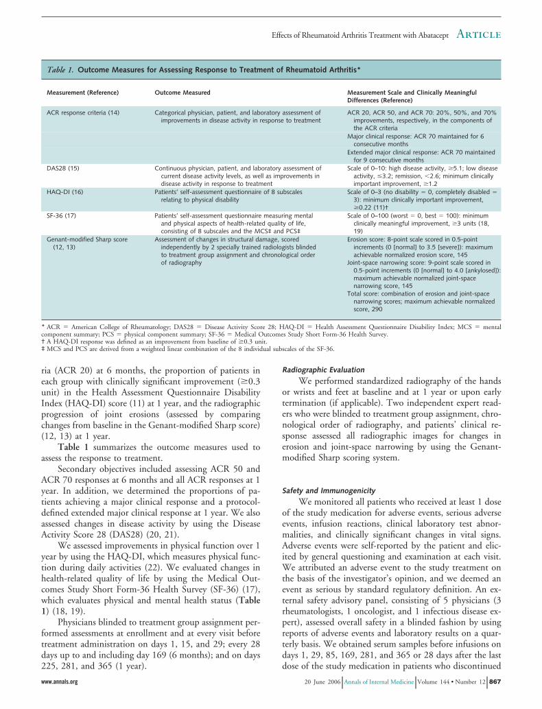
Table 1 summarizes the outcome measures used toassess the response to treatment.
Secondary objectives included assessing ACR 50 andACR 70 responses at 6 months and all ACR responses at 1year. In addition, we determined the proportions of pa-tients achieving a major clinical response and a protocol-defined extended major clinical response at 1 year. We alsoassessed changes in disease activity by using the DiseaseActivity Score 28 (DAS28) (20, 21).
We assessed improvements in physical function over 1year by using the HAQ-DI, which measures physical func-tion during daily activities (22). We evaluated changes inhealth-related quality of life by using the Medical Out-comes Study Short Form-36 Health Survey (SF-36) (17),which evaluates physical and mental health status (Table1) (18, 19).
Physicians blinded to treatment group assignment per-formed assessments at enrollment and at every visit beforetreatment administration on days 1, 15, and 29; every 28days up to and including day 169 (6 months); and on days225, 281, and 365 (1 year).
Radiographic Evaluation
We performed standardized radiography of the handsor wrists and feet at baseline and at 1 year or upon earlytermination (if applicable). Two independent expert read-ers who were blinded to treatment group assignment, chro-nological order of radiography, and patients’ clinical re-sponse assessed all radiographic images for changes inerosion and joint-space narrowing by using the Genant-modified Sharp scoring system.
Safety and Immunogenicity
We monitored all patients who received at least 1 doseof the study medication for adverse events, serious adverseevents, infusion reactions, clinical laboratory test abnor-malities, and clinically significant changes in vital signs.Adverse events were self-reported by the patient and elic-ited by general questioning and examination at each visit.We attributed an adverse event to the study treatment onthe basis of the investigator’s opinion, and we deemed anevent as serious by standard regulatory definition. An ex-ternal safety advisory panel, consisting of 5 physicians (3rheumatologists, 1 oncologist, and 1 infectious disease ex-pert), assessed overall safety in a blinded fashion by usingreports of adverse events and laboratory results on a quar-terly basis. We obtained serum samples before infusions ondays 1, 29, 85, 169, 281, and 365 or 28 days after the lastdose of the study medication in patients who discontinued
Table 1. Outcome Measures for Assessing Response to Treatment of Rheumatoid Arthritis*
Measurement (Reference) Outcome Measured Measurement Scale and Clinically MeaningfulDifferences (Reference)
ACR response criteria (14) Categorical physician, patient, and laboratory assessment ofimprovements in disease activity in response to treatment
ACR 20, ACR 50, and ACR 70: 20%, 50%, and 70%improvements, respectively, in the components ofthe ACR criteria
Major clinical response: ACR 70 maintained for 6consecutive months
Extended major clinical response: ACR 70 maintainedfor 9 consecutive months
DAS28 (15) Continuous physician, patient, and laboratory assessment ofcurrent disease activity levels, as well as improvements indisease activity in response to treatment
Scale of 0–10: high disease activity, �5.1; low diseaseactivity, �3.2; remission, �2.6; minimum clinicallyimportant improvement, �1.2
HAQ-DI (16) Patients’ self-assessment questionnaire of 8 subscalesrelating to physical disability
Scale of 0–3 (no disability � 0, completely disabled �3): minimum clinically important improvement,�0.22 (11)†
SF-36 (17) Patients’ self-assessment questionnaire measuring mentaland physical aspects of health-related quality of life,consisting of 8 subscales and the MCS‡ and PCS‡
Scale of 0–100 (worst � 0, best � 100): minimumclinically meaningful improvement, �3 units (18,19)
Genant-modified Sharp score(12, 13)
Assessment of changes in structural damage, scoredindependently by 2 specially trained radiologists blindedto treatment group assignment and chronological orderof radiography
Erosion score: 8-point scale scored in 0.5-pointincrements (0 [normal] to 3.5 [severe]): maximumachievable normalized erosion score, 145
Joint-space narrowing score: 9-point scale scored in0.5-point increments (0 [normal] to 4.0 [ankylosed]):maximum achievable normalized joint-spacenarrowing score, 145
Total score: combination of erosion and joint-spacenarrowing scores; maximum achievable normalizedscore, 290
* ACR � American College of Rheumatology; DAS28 � Disease Activity Score 28; HAQ-DI � Health Assessment Questionnaire Disability Index; MCS � mentalcomponent summary; PCS � physical component summary; SF-36 � Medical Outcomes Study Short Form-36 Health Survey.† A HAQ-DI response was defined as an improvement from baseline of �0.3 unit.‡ MCS and PCS are derived from a weighted linear combination of the 8 individual subscales of the SF-36.
ArticleEffects of Rheumatoid Arthritis Treatment with Abatacept
www.annals.org 20 June 2006 Annals of Internal Medicine Volume 144 • Number 12 867
before 1 year. We assessed immunogenicity by immunoas-say to measure the antibody response to the entire abata-cept molecule and also specifically to the CTLA-4 portionof the molecule (7).
Statistical AnalysisThe protocol estimated that 680 patients would need
to be enrolled to randomly assign 540 patients. We basedsample sizes on a 5% level of significance (2-tailed). Thestudy had 99% power to detect a difference of 20% inACR 20 between the 2 groups. On the basis of the hierar-chical testing procedure for the co-primary measures, thissample size allowed us to detect an 18% difference inHAQ-DI response rate between the 2 groups, with 98%power, and a treatment effect of 60% reduction from pla-cebo (assuming an increase of 1.27 units in placebo for thechange from baseline), with 90% power, for change frombaseline in the Genant-modified Sharp erosion score. Webased the assumptions on the findings of a phase IIb studyof patients with rheumatoid arthritis who were using abata-cept (8, 9).
We performed all efficacy and safety analyses on amodified intention-to-treat population, defined as all ran-domly assigned patients who received at least 1 dose ofstudy medication. We based all statistical tests on a 2-sided5% level of significance and used SAS software, version 8.2(SAS Institute, Cary, North Carolina), for all analyses.
For the co-primary analyses of ACR 20 at 6 monthsand HAQ-DI responses at 1 year, we used a 2-sided, con-tinuity-corrected chi-square test to compare the responsesof the abatacept group with those of the placebo group.We imputed missing data for patients who discontinued asnonresponders subsequent to the discontinuation; thus, webased these analyses on the full modified intention-to-treatdenominator. We performed additional sensitivity analysesto assess the effect of the imputation of missing data. Theseinclude a “modified worst-case” analysis, where we im-puted missing data for placebo recipients who discontin-ued for reasons other than lack of efficacy by using theirlast observed response, and a “worst-case” analysis, wherewe imputed missing data for placebo recipients who dis-continued as responders. In both cases, however, we stillimputed missing data for abatacept recipients as nonre-sponders. We performed additional longitudinal analysesby using the generalized estimating equations to assess thetreatment effect over time. We used all available data, andthe longitudinal analysis assumes that data were missingcompletely at random and were not dependent on currentor future responses. The models included treatment, visitday, and treatment-by-visit interaction as fixed effects, andwe used an unstructured covariance to account for within-patient correlation over time (23, 24).
We used a rank-based analysis of covariance (25) tocompare the changes from baseline in Genant-modifiedSharp scores between treatment groups at 1 year. Themodel included the ranks for score changes as the depen-
dent variable, with treatment group as a main effect, andthe ranks for baseline scores as additional covariates.Midranks were assigned for ties. The primary radiographicanalyses included all observed data at baseline and at 12months. We imputed missing annual radiographic datawith linear extrapolation for discontinued patients on thebasis of the baseline value and the on-treatment assessmentat the time of discontinuation, provided that both assess-ments were available. Summary statistics and a cumulativeprobability plot were provided for changes from baseline inthe Genant-modified Sharp scores at 1 year by treatmentgroup assignment. We performed additional sensitivityanalyses to assess the effect of missing annual radiographicdata. These included analysis with imputed 12-month val-ues for patients with missing annual assessments on thebasis of the responses predicted by the data observed acrossboth treatment groups, clustering patients with similarbaseline radiographic scores. In addition, we also per-formed a “graded worst-case” imputation, where we im-puted missing data for abatacept and placebo recipientswith progressively worst outcomes and progressively bestoutcomes, respectively.
To avoid multiple testing, we used a prespecified se-quential testing procedure for co-primary end points. Wemade comparisons only if all preceding co-primary endpoints were statistically significant, according to the follow-ing hierarchy: ACR 20 response at 6 months; functionalperformance at 1 year, as measured by the HAQ-DI; andchange in erosion, by using the Genant-modified Sharpscore, at 1 year.
The analysis of covariance with the last observationcarried forward (LOCF) approach was the prespecifiedmethod for the comparisons between treatment groups ofmean changes from baseline in the HAQ-DI and the 8subscales and the physical and mental component summa-ries of the SF-36. However, because the limitations of theLOCF approach could yield substantial bias in treatmenteffects (26), and also on the basis of editorial advice, weused a longitudinal linear mixed-effects model in the com-parisons of these end points. We used all available data,and the longitudinal analysis assumes that data were miss-ing at random and were not dependent on current or fu-ture responses. The models included treatment, visit day,and treatment-by-visit interaction as fixed effects, and weused an autoregressive (1) covariance to account for with-in-patient correlation over time (23).
For DAS28, we used a 2-sided, continuity-correctedchi-square test to compare the responses of the abataceptgroup with those of the placebo group. We summarizedthe incidence of adverse events by treatment and used 95%CIs for the comparisons between treatment groups.
Role of the Funding SourceThis trial was sponsored by Bristol-Myers Squibb. The
funding source helped design the study in consultationwith the authors and provided statistical support for data
Article Effects of Rheumatoid Arthritis Treatment with Abatacept
868 20 June 2006 Annals of Internal Medicine Volume 144 • Number 12 www.annals.org
analysis. Interpretation of the data was aided by the fund-ing biostatisticians, with input from the authors. The fund-ing source was not involved in the decision to submit thearticle for publication.
RESULTS
Patient CharacteristicsWe enrolled 1250 patients with rheumatoid arthritis,
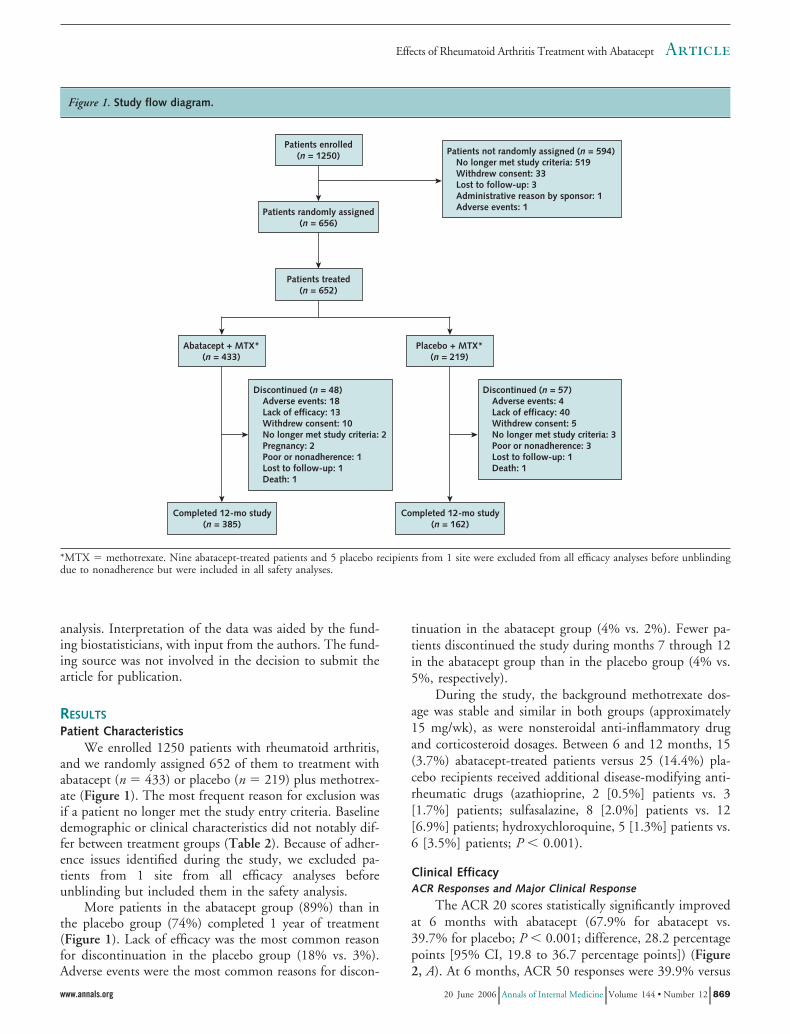
and we randomly assigned 652 of them to treatment withabatacept (n � 433) or placebo (n � 219) plus methotrex-ate (Figure 1). The most frequent reason for exclusion wasif a patient no longer met the study entry criteria. Baselinedemographic or clinical characteristics did not notably dif-fer between treatment groups (Table 2). Because of adher-ence issues identified during the study, we excluded pa-tients from 1 site from all efficacy analyses beforeunblinding but included them in the safety analysis.
More patients in the abatacept group (89%) than inthe placebo group (74%) completed 1 year of treatment(Figure 1). Lack of efficacy was the most common reasonfor discontinuation in the placebo group (18% vs. 3%).Adverse events were the most common reasons for discon-
tinuation in the abatacept group (4% vs. 2%). Fewer pa-tients discontinued the study during months 7 through 12in the abatacept group than in the placebo group (4% vs.5%, respectively).
During the study, the background methotrexate dos-age was stable and similar in both groups (approximately15 mg/wk), as were nonsteroidal anti-inflammatory drugand corticosteroid dosages. Between 6 and 12 months, 15(3.7%) abatacept-treated patients versus 25 (14.4%) pla-cebo recipients received additional disease-modifying anti-rheumatic drugs (azathioprine, 2 [0.5%] patients vs. 3[1.7%] patients; sulfasalazine, 8 [2.0%] patients vs. 12[6.9%] patients; hydroxychloroquine, 5 [1.3%] patients vs.6 [3.5%] patients; P � 0.001).
Clinical EfficacyACR Responses and Major Clinical Response
The ACR 20 scores statistically significantly improvedat 6 months with abatacept (67.9% for abatacept vs.39.7% for placebo; P � 0.001; difference, 28.2 percentagepoints [95% CI, 19.8 to 36.7 percentage points]) (Figure2, A). At 6 months, ACR 50 responses were 39.9% versus
Figure 1. Study flow diagram.
*MTX � methotrexate. Nine abatacept-treated patients and 5 placebo recipients from 1 site were excluded from all efficacy analyses before unblindingdue to nonadherence but were included in all safety analyses.
ArticleEffects of Rheumatoid Arthritis Treatment with Abatacept
www.annals.org 20 June 2006 Annals of Internal Medicine Volume 144 • Number 12 869

16.8% (difference, 23.0 percentage points [CI, 15.0 to31.1 percentage points]) and ACR 70 responses were19.8% versus 6.5% (difference, 13.3 percentage points[CI, 7.0 to 19.5 percentage points]) for abatacept versusplacebo, respectively (P � 0.001 for both) (Figure 2, B and C).
Between 6 and 12 months, all ACR responses contin-ually improved in patients receiving abatacept, while re-sponses in placebo recipients were largely unchanged frommonth 6. At 1 year, ACR 20 responses had increased to73.1% versus 39.7% (difference, 33.4 percentage points[CI, 25.1 to 41.7 percentage points]), ACR 50 responseswere 48.3% versus 18.2% (difference, 30.1 percentagepoints [CI, 21.8 to 38.5 percentage points]), and ACR 70responses were 28.8% versus 6.1% (difference, 22.7 per-centage points [CI, 15.6 to 29.8 percentage points]) forabatacept recipients versus placebo recipients, respectively(P � 0.001 for all) (Figure 2, A to C). Post hoc analyses ofthe abatacept group showed that the proportion of patientswith ACR 50 and ACR 70 responses statistically signifi-cantly increased from 6 months to 12 months (P � 0.001
for 6 months vs. 12 months). Of the abatacept-treatedpatients with an ACR 70 response at 1 year, 45% main-tained the response for 6 consecutive months (major clin-ical response overall, 14.2%) and 21% maintained the re-sponse for 9 consecutive months (extended major clinicalresponse overall, 6.1%).
In the modified worst-case and worst-case sensitivityanalyses, we observed an ACR 20 response at 6 months inmore patients in the abatacept group (68% in both cases)than in the placebo group (40% in the modified worst-caseand 57% in the worst-case analyses). An additional longi-tudinal analysis confirmed the significant increase in ACR20 response for abatacept versus placebo (P � 0.001) and,more specifically, at day 15 (P � 0.008). This early re-sponse was largely driven by rapid improvements in painand by patients’ and physicians’ assessments of disease ac-tivity, which were statistically significant from day 15 on-ward compared with placebo (data not shown). Longitudi-nal analyses also demonstrated significant increases in ACR50 and ACR 70 responses with abatacept compared with
Table 2. Baseline Characteristics*
Characteristics Abatacept � MethotrexateGroup (n � 433)
Placebo � MethotrexateGroup (n � 219)
Age, y 51.5 (12.9) 50.4 (12.4)Weight, kg 72.3 (17.5) 70.2 (16.1)Women, % 77.8 81.7White, % 87.5 88.1Geographic region, %
North America 21.5 21.0South America 40.0 42.5Europe 33.0 30.6Other 5.5 5.9
Disease duration, y 8.5 (7.3) 8.9 (7.1)Methotrexate dose, mg/wk 16.1 (3.6) 15.7 (3.5)Tender joints, n 31.0 (13.2) 32.3 (13.6)Swollen joints, n 21.4 (8.8) 22.1 (8.8)Pain (100-mm VAS) 63.3 (21.1) 65.9 (20.6)Physical function (HAQ-DI) 1.7 (0.7)† 1.7 (0.6)Patient global assessment (100-mm VAS) 62.7 (21.2) 62.8 (21.6)Physician global assessment (100-mm VAS) 68.0 (16.0) 67.4 (17.0)CRP level, mg/L 33 (31) 28 (25)Rheumatoid factor, % 81.8 78.5Baseline radiographic score
Erosion score 21.7 (18.1) 21.8 (18.6)Joint-space narrowing score 22.8 (20.2) 23.0 (20.4)
Total score 44.5 (37.3) 44.9 (37.7)Baseline median score (range)
Erosion score 16.6 (0.0–112.2) 16.7 (0.3–95.8)Joint-space narrowing score 16.2 (0.0–108.8) 16.6 (0.0–94.3)
Total score 31.9 (0.5–221.0) 33.4 (2.3–190.1)Antirheumatic medications at enrollment, n (%)
Methotrexate 433 (100.0) 219 (100.0)Other disease-modifying antirheumatic drugs 53 (12.2) 19 (8.7)Biologics 1 (0.2) 0Corticosteroids 312 (72.1) 150 (68.5)NSAIDs 370 (85.5) 181 (82.6)Other 1 (0.2) 0
* Data are reported as means (SDs), unless otherwise indicated. CRP � C-reactive protein; HAQ-DI � Health Assessment Questionnaire Disability Index; NSAID �nonsteroidal anti-inflammatory drug; VAS � visual analogue scale.† n � 431.
Article Effects of Rheumatoid Arthritis Treatment with Abatacept
870 20 June 2006 Annals of Internal Medicine Volume 144 • Number 12 www.annals.org

placebo at 1 year (P � 0.001). The Appendix Table (avail-able at www.annals.org) provides results related to the in-dividual components of the ACR.
Physical Function
At the start of the study, patients’ physical functionwas considerably impaired (HAQ-DI score of 1.7 in bothgroups) (Table 2). At 1 year, physical function clinicallysignificantly improved in statistically significantly moreabatacept-treated patients (11) than placebo recipients(63.7% vs. 39.3%; P � 0.001; difference, 24.4 percentagepoints [CI, 15.9 to 32.9 percentage points]) (Figure 2, D).
In the modified worst-case sensitivity analysis, morepatients in the abatacept group (64%) had an HAQ-DIresponse at 1 year than those in the placebo group (42%).In the worst-case analysis, similar proportions in bothtreatment groups (64%) had an HAQ-DI response. How-
ever, because of the extreme nature of the response impu-tation rule, the resulting high response rate in the placebogroup does not represent an observable placebo responserate. The longitudinal analysis using the generalized esti-mating equations confirmed the significant increase in theproportion of patients with an HAQ-DI response forabatacept versus placebo (P � 0.001).
When we used the longitudinal linear mixed-effectsapproach, the mean improvement from baseline in theHAQ-DI was statistically significantly better in abatacept-treated patients than in placebo recipients at both 6months and 1 year (P � 0.001). Results were consistentwith the LOCF approach.
Radiographic Progression
We collected radiographic data for 586 (92%) ran-domly assigned patients at baseline and at 1 postbaseline
Figure 2. Improvements in signs and symptoms of disease and physical function.
A–C. American College of Rheumatology (ACR) 20 (panel A), ACR 50 (panel B), and ACR 70 (panel C) responses over 1 year in all patients who receivedat least 1 dose of the study medication. D. The percentage of patients who achieved a Health Assessment Questionnaire Disability Index (HAQ-DI)response (�0.3-unit improvement from baseline in HAQ-DI) was determined over 1 year. MTX � methotrexate. *Intention-to-treat population whereall dropouts were considered to be ACR nonresponders subsequent to their dropout. †Because of adherence issues identified during the study, patientsfrom 1 site were excluded from all efficacy analyses before unblinding but were included in the analysis of safety.
ArticleEffects of Rheumatoid Arthritis Treatment with Abatacept
www.annals.org 20 June 2006 Annals of Internal Medicine Volume 144 • Number 12 871

time point. Baseline erosion, joint-space narrowing, andtotal scores were similar between the groups (Table 2). At1 year, abatacept-treated patients demonstrated statisticallysignificant slowing of structural damage progression com-pared with placebo recipients, with an approximately 50%reduction in change from baseline in Genant-modifiedSharp scores compared with that of placebo. The medianchange from baseline in erosion score was 0.0 (25th and75th percentiles, 0.0 and 1.0, respectively) for abataceptversus 0.27 (25th and 75th percentiles, 0.0 and 1.3, respec-tively) for placebo (P � 0.029; Figure 3). Median changesin the joint-space narrowing and total scores were similarbetween the groups. The median change in joint-space nar-rowing score was 0.0 (25th and 75th percentiles, 0.0 and0.5, respectively) for abatacept versus 0.0 (25th and 75thpercentiles, 0.0 and 1.0, respectively) for placebo (P �0.009) (Figure 3). The median change in total score was0.25 (25th and 75th percentiles, 0.0 and 1.8, respectively)for abatacept versus 0.53 (25th and 75th percentiles, 0.0and 2.5, respectively) for placebo (P � 0.012) (Figure 3).The mean change from baseline was 0.63 for abataceptversus 1.14 for placebo in erosion score, 0.58 for abataceptversus 1.18 for placebo in joint-space narrowing score, and1.21 for abatacept versus 2.32 for placebo in total score.
The sensitivity analysis suggested that the few missingdata did not statistically significantly affect the robustnessof slowing the progression of structural damage. For thesensitivity analysis, in which we imputed missing 1-yearradiographic values, the median changes in total score were0.26 (interquartile range, 0.00 to 1.84) and 0.53 (inter-quartile range, 0.00 to 3.14) in the abatacept and placebogroups, respectively. These changes for both treatmentgroups were the same as those in the primary analysis, withdifferences observed only in the 75th percentile and a more
notable increase in the placebo group. In the graded worst-case sensitivity analyses, in which several imputations in-creasingly favored placebo, the trend for the benefit ofabatacept was maintained compared with that of placebo,even in the extreme case (data not shown).
Disease Activity
Patients exhibited high baseline disease activity(DAS28 of 6.4 for both groups [15]). At 6 months and 12months, 30.1% and 42.5% of the abatacept group, respec-tively, had a DAS28 of 3.2 or less, compared with 10.0%and 9.9% of the placebo group, respectively (P � 0.001).Abatacept induced DAS28 less than 2.6 in 14.8% of abata-cept recipients versus 2.8% of placebo recipients at 6months and in 23.8% of abatacept recipients versus 1.9%of placebo recipients at 1 year (P � 0.001).
Health-Related Quality of Life
When we used the linear mixed-effects approach, boththe physical (P � 0.001) and mental (P � 0.009) compo-nent summaries significantly improved from baseline to 6months (increase of �3 units) (18, 19) in the abataceptgroup compared with the placebo group. At 1 year, bothsummary scores for patients treated with abatacept werestill significant (physical component summary, P � 0.001;difference, 3.8 [CI, 2.4 to 5.2]; mental component sum-mary, P � 0.038; difference, 1.76 [CI, 0.1 to 3.4]). Resultswere also significant at both 6 months and 1 year with theLOCF approach.
Safety and ImmunogenicitySafety
The overall incidence of adverse events was similar inboth the abatacept and placebo groups (87.3% vs. 84.0%[CI, �2.5 to 9.1 percentage points]) (Table 3). The most
Figure 3. Slowing of radiographic structural damage progression at 1 year.
Interquartile range changes from baseline in Genant-modified Sharp erosion, joint-space narrowing (JSN), and total scores were evaluated at 1 year or atearly termination (if applicable). The median (solid circles), interquartile range, and 10th and 90th percentiles (dotted lines) are shown. Data shown arefrom all randomly assigned and treated patients with baseline and follow-up radiography. MTX � methotrexate.
Article Effects of Rheumatoid Arthritis Treatment with Abatacept
872 20 June 2006 Annals of Internal Medicine Volume 144 • Number 12 www.annals.org
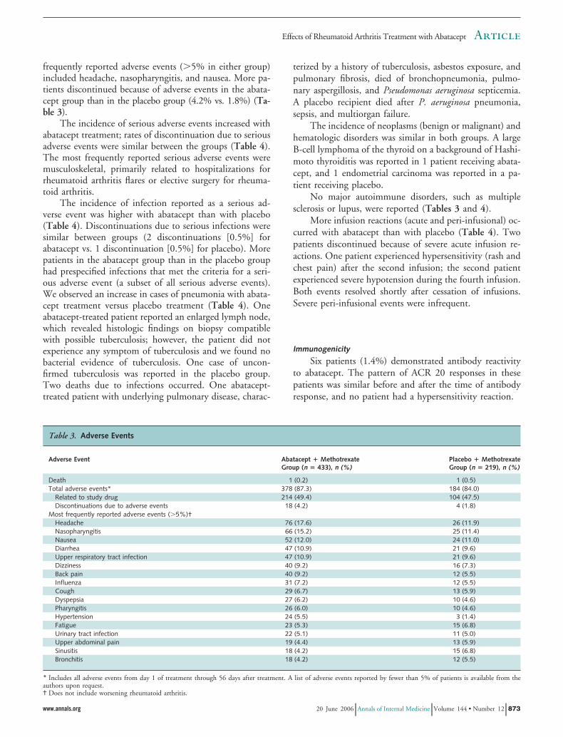
frequently reported adverse events (�5% in either group)included headache, nasopharyngitis, and nausea. More pa-tients discontinued because of adverse events in the abata-cept group than in the placebo group (4.2% vs. 1.8%) (Ta-ble 3).
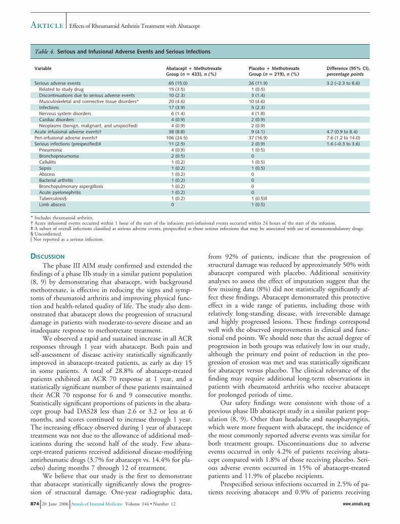
The incidence of serious adverse events increased withabatacept treatment; rates of discontinuation due to seriousadverse events were similar between the groups (Table 4).The most frequently reported serious adverse events weremusculoskeletal, primarily related to hospitalizations forrheumatoid arthritis flares or elective surgery for rheuma-toid arthritis.
The incidence of infection reported as a serious ad-verse event was higher with abatacept than with placebo(Table 4). Discontinuations due to serious infections weresimilar between groups (2 discontinuations [0.5%] forabatacept vs. 1 discontinuation [0.5%] for placebo). Morepatients in the abatacept group than in the placebo grouphad prespecified infections that met the criteria for a seri-ous adverse event (a subset of all serious adverse events).We observed an increase in cases of pneumonia with abata-cept treatment versus placebo treatment (Table 4). Oneabatacept-treated patient reported an enlarged lymph node,which revealed histologic findings on biopsy compatiblewith possible tuberculosis; however, the patient did notexperience any symptom of tuberculosis and we found nobacterial evidence of tuberculosis. One case of uncon-firmed tuberculosis was reported in the placebo group.Two deaths due to infections occurred. One abatacept-treated patient with underlying pulmonary disease, charac-
terized by a history of tuberculosis, asbestos exposure, andpulmonary fibrosis, died of bronchopneumonia, pulmo-nary aspergillosis, and Pseudomonas aeruginosa septicemia.A placebo recipient died after P. aeruginosa pneumonia,sepsis, and multiorgan failure.
The incidence of neoplasms (benign or malignant) andhematologic disorders was similar in both groups. A largeB-cell lymphoma of the thyroid on a background of Hashi-moto thyroiditis was reported in 1 patient receiving abata-cept, and 1 endometrial carcinoma was reported in a pa-tient receiving placebo.
No major autoimmune disorders, such as multiplesclerosis or lupus, were reported (Tables 3 and 4).
More infusion reactions (acute and peri-infusional) oc-curred with abatacept than with placebo (Table 4). Twopatients discontinued because of severe acute infusion re-actions. One patient experienced hypersensitivity (rash andchest pain) after the second infusion; the second patientexperienced severe hypotension during the fourth infusion.Both events resolved shortly after cessation of infusions.Severe peri-infusional events were infrequent.
Immunogenicity
Six patients (1.4%) demonstrated antibody reactivityto abatacept. The pattern of ACR 20 responses in thesepatients was similar before and after the time of antibodyresponse, and no patient had a hypersensitivity reaction.
Table 3. Adverse Events
Adverse Event Abatacept � MethotrexateGroup (n � 433), n (%)
Placebo � MethotrexateGroup (n � 219), n (%)
Death 1 (0.2) 1 (0.5)Total adverse events* 378 (87.3) 184 (84.0)
Related to study drug 214 (49.4) 104 (47.5)Discontinuations due to adverse events 18 (4.2) 4 (1.8)
Most frequently reported adverse events (�5%)†Headache 76 (17.6) 26 (11.9)Nasopharyngitis 66 (15.2) 25 (11.4)Nausea 52 (12.0) 24 (11.0)Diarrhea 47 (10.9) 21 (9.6)Upper respiratory tract infection 47 (10.9) 21 (9.6)Dizziness 40 (9.2) 16 (7.3)Back pain 40 (9.2) 12 (5.5)Influenza 31 (7.2) 12 (5.5)Cough 29 (6.7) 13 (5.9)Dyspepsia 27 (6.2) 10 (4.6)Pharyngitis 26 (6.0) 10 (4.6)Hypertension 24 (5.5) 3 (1.4)Fatigue 23 (5.3) 15 (6.8)Urinary tract infection 22 (5.1) 11 (5.0)Upper abdominal pain 19 (4.4) 13 (5.9)Sinusitis 18 (4.2) 15 (6.8)Bronchitis 18 (4.2) 12 (5.5)
* Includes all adverse events from day 1 of treatment through 56 days after treatment. A list of adverse events reported by fewer than 5% of patients is available from theauthors upon request.† Does not include worsening rheumatoid arthritis.
ArticleEffects of Rheumatoid Arthritis Treatment with Abatacept
www.annals.org 20 June 2006 Annals of Internal Medicine Volume 144 • Number 12 873
DISCUSSION
The phase III AIM study confirmed and extended thefindings of a phase IIb study in a similar patient population(8, 9) by demonstrating that abatacept, with backgroundmethotrexate, is effective in reducing the signs and symp-toms of rheumatoid arthritis and improving physical func-tion and health-related quality of life. The study also dem-onstrated that abatacept slows the progression of structuraldamage in patients with moderate-to-severe disease and aninadequate response to methotrexate treatment.
We observed a rapid and sustained increase in all ACRresponses through 1 year with abatacept. Both pain andself-assessment of disease activity statistically significantlyimproved in abatacept-treated patients, as early as day 15in some patients. A total of 28.8% of abatacept-treatedpatients exhibited an ACR 70 response at 1 year, and astatistically significant number of these patients maintainedtheir ACR 70 response for 6 and 9 consecutive months.Statistically significant proportions of patients in the abata-cept group had DAS28 less than 2.6 or 3.2 or less at 6months, and scores continued to increase through 1 year.The increasing efficacy observed during 1 year of abatacepttreatment was not due to the allowance of additional med-ications during the second half of the study. Few abata-cept-treated patients received additional disease-modifyingantirheumatic drugs (3.7% for abatacept vs. 14.4% for pla-cebo) during months 7 through 12 of treatment.
We believe that our study is the first to demonstratethat abatacept statistically significantly slows the progres-sion of structural damage. One-year radiographic data,
from 92% of patients, indicate that the progression ofstructural damage was reduced by approximately 50% withabatacept compared with placebo. Additional sensitivityanalyses to assess the effect of imputation suggest that thefew missing data (8%) did not statistically significantly af-fect these findings. Abatacept demonstrated this protectiveeffect in a wide range of patients, including those withrelatively long-standing disease, with irreversible damageand highly progressed lesions. These findings correspondwell with the observed improvements in clinical and func-tional end points. We should note that the actual degree ofprogression in both groups was relatively low in our study,although the primary end point of reduction in the pro-gression of erosion was met and was statistically significantfor abatacept versus placebo. The clinical relevance of thefinding may require additional long-term observations inpatients with rheumatoid arthritis who receive abataceptfor prolonged periods of time.
Our safety findings were consistent with those of aprevious phase IIb abatacept study in a similar patient pop-ulation (8, 9). Other than headache and nasopharyngitis,which were more frequent with abatacept, the incidence ofthe most commonly reported adverse events was similar forboth treatment groups. Discontinuations due to adverseevents occurred in only 4.2% of patients receiving abata-cept compared with 1.8% of those receiving placebo. Seri-ous adverse events occurred in 15% of abatacept-treatedpatients and 11.9% of placebo recipients.
Prespecified serious infections occurred in 2.5% of pa-tients receiving abatacept and 0.9% of patients receiving
Table 4. Serious and Infusional Adverse Events and Serious Infections
Variable Abatacept � MethotrexateGroup (n � 433), n (%)
Placebo � MethotrexateGroup (n � 219), n (%)
Difference (95% CI),percentage points
Serious adverse events 65 (15.0) 26 (11.9) 3.2 (–2.3 to 8.6)Related to study drug 15 (3.5) 1 (0.5)Discontinuations due to serious adverse events 10 (2.3) 3 (1.4)Musculoskeletal and connective tissue disorders* 20 (4.6) 10 (4.6)Infections 17 (3.9) 5 (2.3)Nervous system disorders 6 (1.4) 4 (1.8)Cardiac disorders 4 (0.9) 2 (0.9)Neoplasms (benign, malignant, and unspecified) 4 (0.9) 2 (0.9)
Acute infusional adverse events† 38 (8.8) 9 (4.1) 4.7 (0.9 to 8.4)Peri-infusional adverse events† 106 (24.5) 37 (16.9) 7.6 (1.2 to 14.0)Serious infections (prespecified)‡ 11 (2.5) 2 (0.9) 1.6 (–0.3 to 3.6)
Pneumonia 4 (0.9) 1 (0.5)Bronchopneumonia 2 (0.5) 0Cellulitis 1 (0.2) 1 (0.5)Sepsis 1 (0.2) 1 (0.5)Abscess 1 (0.2) 0Bacterial arthritis 1 (0.2) 0Bronchopulmonary aspergillosis 1 (0.2) 0Acute pyelonephritis 1 (0.2) 0Tuberculosis§ 1 (0.2) 1 (0.5)||Limb abscess 0 1 (0.5)
* Includes rheumatoid arthritis.† Acute infusional events occurred within 1 hour of the start of the infusion; peri-infusional events occurred within 24 hours of the start of the infusion.‡ A subset of overall infections classified as serious adverse events, prespecified as those serious infections that may be associated with use of immunomodulatory drugs.§ Unconfirmed.� Not reported as a serious infection.
Article Effects of Rheumatoid Arthritis Treatment with Abatacept
874 20 June 2006 Annals of Internal Medicine Volume 144 • Number 12 www.annals.org
placebo. One case of aspergillosis occurred in the abataceptgroup. One case of tuberculosis was reported in eachgroup; however, neither case of tuberculosis was confirmedbacteriologically. We included patients who were fromcountries where tuberculosis is endemic; however, as rec-ommended with antitumor necrosis factor agents used fortreating rheumatoid arthritis, which have shown an in-creased incidence of tuberculosis, we screened all patientsby tuberculin skin test before study entry. We excludedpatients with a positive antituberculin skin test result, andtherefore, comparison of the rates of tuberculosis with theearly experience of antitumor necrosis factor agents, inwhich routine tuberculin skin testing was not performed,would not be appropriate. Additional longer-term informa-tion is needed to determine whether an increased relativerisk for tuberculosis or other opportunistic infection is as-sociated with the use of this agent. Increases in hemato-logic and hepatic abnormalities and malignant conditionswere not associated with abatacept treatment. In addition,no major autoimmune diseases were reported (for example,lupus or demyelination), as observed with anticytokinetherapies (27).
These findings should be considered within the con-text of our study’s limitations. The trial’s 1-year durationprecludes the determination of whether longer-term treat-ment will be associated with the emergence of other possi-ble toxicities. Detection of a range of toxicities will requiremore widespread study in more patients. Furthermore, weexamined the safety and efficacy of abatacept in only 1subset of the patient population with rheumatoid arthri-tis—those with an inadequate response to methotrexate.An additional phase III study of abatacept in patients withrheumatoid arthritis and an inadequate response to anti-tumor necrosis factor-� therapy demonstrated statisticallysignificant clinical benefits with a similar safety and toler-ability profile (28). Our current trial assessed the effects ofabatacept in patients with established rheumatoid arthritis(mean duration of about 9 years) and was not designed toinvestigate the effects of abatacept in early disease. Furtherstudies of abatacept in the longer-term treatment of pa-tients with rheumatoid arthritis and varying disease andtreatment histories are required to substantiate the efficacyand safety findings with abatacept to date.
The data from the phase IIb trial (8, 9) and the larger,optimal-dose, phase III investigation indicate that the strat-egy of selective T-cell inhibition by abatacept provides con-sistent and statistically significant additional therapeuticvalue for treating patients with rheumatoid arthritis and aninadequate response to methotrexate. Abatacept treatmentis, thus, an alternative strategy to inhibit tumor necrosisfactor-� in these patients, although the relative merits ofeach approach may require several years to determine viainformation derived from large databases or registries.
In our study, the clinical benefits seen with the fixeddosage of abatacept encompassed clinical and radiographicefficacy, statistically significant and clinically meaningful
improvements in patients’ physical function and health-related quality of life, and a consistent safety profile. Ob-servations of the slowing of radiographic progression byabatacept, which we believe that our study is the first todemonstrate, as well as the safety and clinical findings, areexpected to be extended with longer-term observations inthis and other patient populations. Overall, abataceptseems to be a rational and effective treatment strategy forpatients with rheumatoid arthritis who have an inadequateresponse to weekly methotrexate.
From the Center for Rheumatology, Albany, New York; University ofCalifornia, San Francisco, and Synarc Inc., San Francisco, California;University of Alabama at Birmingham School of Medicine, Birmingham,Alabama; University of Alberta, Edmonton, Alberta, Canada; LeedsGeneral Infirmary, Leeds, United Kingdom; Hospital Central, San LuisPotosi, Mexico; University of Medical Sciences, Wroclaw, Poland; Bris-tol-Myers Squibb, Princeton, New Jersey; and University Hospital Leu-ven, Leuven, Belgium.
Grant Support: By Bristol-Myers Squibb.
Potential Financial Conflicts of Interest: Employment: T. Li (Bristol-Myers Squibb), Z. Ge (Bristol-Myers Squibb), J.-C. Becker (Bristol-Myers Squibb); Consultancies: J.M. Kremer (Bristol-Myers Squibb),H.K. Genant (Bristol-Myers Squibb, Amgen, Wyeth, Novartis, Lilly,Roche), L.W. Moreland (Bristol-Myers Squibb), A.S. Russell (Bristol-Myers Squibb), P. Emery (Amgen, Schering-Plough, Centocor, Bristol-Myers Squibb), R. Westhovens (Schering-Plough, Bristol-MyersSquibb); Honoraria: J.M. Kremer (Bristol-Myers Squibb), H.K. Genant(Bristol-Myers Squibb, Amgen, Wyeth, Novartis, Lilly, Roche), L.W.Moreland (Bristol-Myers Squibb), P. Emery (Wyeth, Roche), R.Westhovens (Schering-Plough, Bristol-Myers Squibb); Stock ownership oroptions (other than mutual funds): T. Li (Bristol-Myers Squibb), Z. Ge(Bristol-Myers Squibb), J.-C. Becker (Bristol-Myers Squibb); Grants re-ceived: J.M. Kremer (Bristol-Myers Squibb), H.K. Genant (Bristol-MyersSquibb, Amgen, Wyeth, Novartis, Lilly, Roche), L.W. Moreland (Bris-tol-Myers Squibb); Patents pending: J.-C. Becker (Bristol-Myers Squibb).
Requests for Single Reprints: Joel M. Kremer, MD, Center for Rheu-matology, 1367 Washington Avenue, Suite 1, Albany, NY 12206; e-mail, [email protected].
Current author addresses and author contributions are available at www.annals.org.
References1. Choy EH, Panayi GS. Cytokine pathways and joint inflammation in rheu-matoid arthritis. N Engl J Med. 2001;344:907-16. [PMID: 11259725]2. Goldring SR, Gravallese EM. Pathogenesis of bone erosions in rheumatoidarthritis. Curr Opin Rheumatol. 2000;12:195-9. [PMID: 10803748]3. Hoffman RW. T cells in the pathogenesis of systemic lupus erythematosus.Front Biosci. 2001;6:D1369-78. [PMID: 11578962]4. Goronzy JJ, Weyand CM. T-cell regulation in rheumatoid arthritis. CurrOpin Rheumatol. 2004;16:212-7. [PMID: 15103247]5. Lenschow DJ, Walunas TL, Bluestone JA. CD28/B7 system of T cell co-stimulation. Annu Rev Immunol. 1996;14:233-58. [PMID: 8717514]6. Silver PB, Hathcock KS, Chan CC, Wiggert B, Caspi RR. Blockade ofcostimulation through B7/CD28 inhibits experimental autoimmune uveoretini-tis, but does not induce long-term tolerance. J Immunol. 2000;165:5041-7.[PMID: 11046033]7. Moreland LW, Alten R, Van den Bosch F, Appelboom T, Leon M, Emery
ArticleEffects of Rheumatoid Arthritis Treatment with Abatacept
www.annals.org 20 June 2006 Annals of Internal Medicine Volume 144 • Number 12 875
P, et al. Costimulatory blockade in patients with rheumatoid arthritis: a pilot,dose-finding, double-blind, placebo-controlled clinical trial evaluating CTLA-4Igand LEA29Y eighty-five days after the first infusion. Arthritis Rheum. 2002;46:1470-9. [PMID: 12115176]8. Kremer JM, Westhovens R, Leon M, Di Giorgio E, Alten R, Steinfeld S, etal. Treatment of rheumatoid arthritis by selective inhibition of T-cell activationwith fusion protein CTLA4Ig. N Engl J Med. 2003;349:1907-15. [PMID:14614165]9. Kremer JM, Dougados M, Emery P, Durez P, Sibilia J, Shergy W, et al.Treatment of rheumatoid arthritis with the selective costimulation modulatorabatacept: twelve-month results of a phase iib, double-blind, randomized, place-bo-controlled trial. Arthritis Rheum. 2005;52:2263-71. [PMID: 16052582]10. Arnett FC, Edworthy SM, Bloch DA, McShane DJ, Fries JF, Cooper NS,et al. The American Rheumatism Association 1987 revised criteria for the classi-fication of rheumatoid arthritis. Arthritis Rheum. 1988;31:315-24. [PMID:3358796]11. Wells GA, Tugwell P, Kraag GR, Baker PR, Groh J, Redelmeier DA.Minimum important difference between patients with rheumatoid arthritis: thepatient’s perspective. J Rheumatol. 1993;20:557-60. [PMID: 8478873]12. Genant HK. Methods of assessing radiographic change in rheumatoid arthri-tis. Am J Med. 1983;75:35-47. [PMID: 6660239]13. Genant HK, Jiang Y, Peterfy C, Lu Y, Redei J, Countryman PJ. Assessmentof rheumatoid arthritis using a modified scoring method on digitized and originalradiographs. Arthritis Rheum. 1998;41:1583-90. [PMID: 9751090]14. Felson DT, Anderson JJ, Boers M, Bombardier C, Chernoff M, Fried B, etal. The American College of Rheumatology preliminary core set of disease activitymeasures for rheumatoid arthritis clinical trials. The Committee on OutcomeMeasures in Rheumatoid Arthritis Clinical Trials. Arthritis Rheum. 1993;36:729-40. [PMID: 8507213]15. Prevoo ML, van ’t Hof MA, Kuper HH, van Leeuwen MA, van de PutteLB, van Riel PL. Modified disease activity scores that include twenty-eight-jointcounts. Development and validation in a prospective longitudinal study of pa-tients with rheumatoid arthritis. Arthritis Rheum. 1995;38:44-8. [PMID:7818570]16. Bruce B, Fries JF. The Stanford Health Assessment Questionnaire: a review
of its history, issues, progress, and documentation. J Rheumatol. 2003;30:167-78.[PMID: 12508408]17. Ware JE Jr, Sherbourne CD. The MOS 36-item short-form health survey(SF-36). I. Conceptual framework and item selection. Med Care. 1992;30:473-83. [PMID: 1593914]18. Kosinski M, Zhao SZ, Dedhiya S, Osterhaus JT, Ware JE Jr. Determiningminimally important changes in generic and disease-specific health-related qualityof life questionnaires in clinical trials of rheumatoid arthritis. Arthritis Rheum.2000;43:1478-87. [PMID: 10902749]19. Samsa G, Edelman D, Rothman ML, Williams GR, Lipscomb J, MatcharD. Determining clinically important differences in health status measures: a gen-eral approach with illustration to the Health Utilities Index Mark II. Pharmaco-economics. 1999;15:141-55. [PMID: 10351188]20. Fransen J, Creemers MC, Van Riel PL. Remission in rheumatoid arthritis:agreement of the disease activity score (DAS28) with the ARA preliminary remis-sion criteria. Rheumatology (Oxford). 2004;43:1252-5. [PMID: 15238643]21. Vrijhoef HJ, Diederiks JP, Spreeuwenberg C, Van der Linden S. Applyinglow disease activity criteria using the DAS28 to assess stability in patients withrheumatoid arthritis. Ann Rheum Dis. 2003;62:419-22. [PMID: 12695152]22. Fries JF, Spitz PW, Young DY. The dimensions of health outcomes: thehealth assessment questionnaire, disability and pain scales. J Rheumatol. 1982;9:789-93. [PMID: 7175852]23. Diggle P, Heagerty P, Liang KY, Zeger SL. Analysis of Longitudinal Data.Oxford, UK: Clarendon Pr; 1994.24. Liang KY, Zeger SL. Longitudinal data analysis using generalized linearmodels. Biometrika. 1986;73:13-22.25. Conover WJ, Iman RL. Analysis of covariance using the rank transformation.Biometrics. 1982;38:715-24. [PMID: 7171697]26. Little R, Yau L. Intent-to-treat analysis for longitudinal studies with drop-outs. Biometrics. 1996;52:1324-33. [PMID: 8962456]27. Khanna D, McMahon M, Furst DE. Safety of tumour necrosis factor-alphaantagonists. Drug Saf. 2004;27:307-24. [PMID: 15061685]28. Genovese MC, Becker JC, Schiff M, Luggen M, Sherrer Y, Kremer J, et al.Abatacept for rheumatoid arthritis refractory to tumor necrosis factor alpha inhi-bition. N Engl J Med. 2005;353:1114-23. [PMID: 16162882]
Article Effects of Rheumatoid Arthritis Treatment with Abatacept
876 20 June 2006 Annals of Internal Medicine Volume 144 • Number 12 www.annals.org

Current Author Addresses: Dr. Kremer: Center for Rheumatology,1367 Washington Avenue, Suite 1, Albany, NY 12206.Dr. Genant: Radiology Department, University of California, San Fran-cisco, 505 Parnassus Avenue, Box 0628, San Francisco, CA 94143-0628.Dr. Moreland: 068 Spain Rehabilitation Center, University of Alabamaat Birmingham, 1717 6th Avenue South, Birmingham, AL 35294.Dr. Russell: Medical Department, University of Alberta Hospital, 562Heritage Medical Research Centre, Edmonton, T6G 2S2 Alberta, Can-ada.Dr. Emery: Academic Unit of Musculoskeletal Disease, Chapel AllertonHospital, Chapeltown Road, Leeds LS7 4SA, United Kingdom.Dr. Abud-Mendoza: Hospital Central, Av. Carranza 2395, San Luis Po-tosi, S.L.P. 78240, Mexico.Dr. Szechinski: Department of Rheumatology, Medical University ofWroclaw, ul. Wisniowa 36a, 53-137 Wrocław, Poland.Drs. Li and Becker: Bristol-Myers Squibb, PO Box 4000, Princeton, NJ08543.Dr. Ge: 13133 Taylor Court, West Windsor, NJ 08550.Dr. Westhovens: Rheumatology Department, University Hospitals, K.U.Leuven, Herestraat 49, 3000 Leuven, Belgium.
Author Contributions: Conception and design: J.M. Kremer, H.K.Genant, L.W. Moreland, A.S. Russell, P. Emery, T. Li, J.-C. Becker, R.Westhovens.Analysis and interpretation of the data: J.M. Kremer, H.K. Genant,L.W. Moreland, P. Emery, C. Abud-Mendoza, T. Li, Z. Ge, J.-C.Becker, R. Westhovens.Drafting of the article: J.M. Kremer, H.K. Genant, L.W. Moreland, A.S.Russell, T. Li, J.-C. Becker, R. Westhovens.Critical revision of the article for important intellectual content: J.M.Kremer, H.K. Genant, L.W. Moreland, A.S. Russell, P. Emery, C.Abud-Mendoza, J. Szechinski, T. Li, J.-C. Becker, R. Westhovens.Final approval of the article: J.M. Kremer, L.W. Moreland, A.S. Russell,P. Emery, C. Abud-Mendoza, J. Szechinski, J.-C. Becker, R. Westhov-ens.Provision of study materials or patients: J.M. Kremer, A.S. Russell, P.Emery, C. Abud-Mendoza, J. Szechinski, R. Westhovens.Statistical expertise: Z. Ge, J.-C. Becker.Obtaining of funding: J.-C. Becker.Administrative, technical, or logistic support: J.-C. Becker.Collection and assembly of data: J.M. Kremer, P. Emery, J.-C. Becker.
Appendix Table. Mean Change from Baseline in American College of Rheumatology Core Component at Year 1*
Variable Adjusted Mean Improvement from Baseline to 12 Months Difference (95% CI)
Abatacept �Methotrexate Group(n � 384)†
Placebo �Methotrexate Group(n � 161)†
Swollen joints, n –16.1 � 0.35 –11.5 � 0.54 –4.6 (–5.88 to –3.35)Tender joints, n –22.5 � 0.55 –16.3 � 0.85 –6.2 (–8.20 to –4.22)Patient pain (100-mm VAS) –35.8 � 1.17 –23.2 � 1.81 –12.6 (–16.9 to –8.39)Patient global assessment (100-mm VAS) –35.8 � 1.12 –24.2 � 1.72 –11.6 (–15.7 to –7.58)Physician global assessment (100-mm VAS) –49.1 � 0.93 –34.3 � 1.44 –14.8 (–18.2 to –11.5)Physical function (HAQ score) –0.68 � 0.03 –0.50 � 0.05 –0.18 (–0.29 to –0.07)CRP level, mg/L –18.3 � 0.9 –8.2 � 1.4 –10.1 (–13.5 to –6.7)
* Analysis of covariance model with treatment as a factor and baseline as a covariate. Data are means (� SE) unless otherwise noted. HAQ � Health AssessmentQuestionnaire; VAS � visual analogue scale.† Results are based on an analysis of complete cases only; patients from 1 site were excluded from the efficacy analysis because of adherence issues.
Annals of Internal Medicine
W-210 20 June 2006 Annals of Internal Medicine Volume 144 • Number 12 www.annals.org