Anhang - sundoc.bibliothek.uni-halle.de · DMS Dimethylsulfid DMSO Dimethylsulfoxid DNA...
22
Anhang
Transcript of Anhang - sundoc.bibliothek.uni-halle.de · DMS Dimethylsulfid DMSO Dimethylsulfoxid DNA...

Anhang

Abkürzungen
A AbsorptionACN AcetonitrilAmp AmpicillinAP alkalische PhosphataseAPS Ammonium-peroxo-di-sulfatBMP bone morphogenetic proteinBMPR BMP-Rezeptorbp BasenpaareBPB BromphenolblauBSA RinderserumalbuminCD CirculardichroismuscDNA complementary DNACHAPS CHAPS -[(-Cholamidopropyl)-
dimethylammonio]--propansulfonsäureCHES -(Cyclohexylamino)-ethansulfonsäureCTAB (N-Cetyl-N,N,N-trimethyl)-
ammoniumbromidDAB Deutsches ArzneibuchDa Daltondeg degreeDMS DimethylsulfidDMSO DimethylsulfoxidDNA DesoxyribonucleinsäureDNase DesoxyribonucleasedNTP DesoxynucleosidtriphosphatDPP DecapentaplegicdrBMP digit removed BMPdsDNA doppelsträngige DNADTT DithiothreitolEC50 halb-maximale effektive KonzentrationEDTA Ethylendiamin-tetraacetatESI electro-spray ionizationEtOH EthanolGDF growth and differentiation factorGdmCl GuanidiniumchloridGdmSCN GuanidiniumthiocyanatGSH Glutathion, reduziertGSSG Glutathion, oxidiertHEPES -[-(-Hydroxyethyl)-piperazinyl]-
ethansulfonsäure
HFBA HeptafluorobuttersäureHIC hydrophobe InteraktionschromatographieHis-Tag Histidin-Tagh Stunde(n)IB inclusion bodies (Einschlußkörper)IMAC immobilisierte Metallchelat-
AffinitätschromatographieIPA Isopropylalkohol (-Propanol)IPTG β-D-Isopropyl-thiogalactopyranosidKan Kanamycinkb KilobasenpaarekDa KilodaltonLAP latency-associated peptideLB-Medium Luria-Bertani-MediumLMW low molecular weightMALDI-TOF matrix-assisted laser desorption
ionization – time of flightβ-ME -Mercapto-ethanolMeOH MethanolMES -Morpholino-ethansulfonsäureMOPS -Morpholino-propansulfonsäureMr molare Masse(m)m (milli)molarMWCO molecular weight cut-off
NaDoc Natrium-desoxycholatNGF nerve growth factorNi-NTA Nickel-Nitrilo-tetraacetatOD600 optische Dichte bei nmp.a. pro analysisPAGE PolyacrylamidgelelektrophoresePCR PolymerasekettenreaktionPDGF platelet-derived growth factorPfu Pyrococcus furiosuspI isoelektrischer PunktPMSF Phenyl-methyl-sulfonylfluoridPPG PolypropylenglycolPPS -(-Pyridino)--propansulfonatproBMP BMP mit N-terminalem PropeptidPSS Pyridin--sulfonsäurePTFE Poly-(tetrafluor-ethylen)
110 Anhang
RNA RibonucleinsäureRNase RibonucleaseRP-HPLC reversed phase – high performance
liquid chromatographyrpm revolutions per minute (Umdrehungen pro
Minute)RT RaumtemperaturSBTI Sojabohnen-TrypsininhibitorSDS Natrium-dodecylsulfatS/R-Modus Signal/Referenz-ModusTaq Thermus aquaticusTCA TrichloressigsäureTEMED
N,N,N´,N´-Tetramethyl-ethylendiaminTGF transorming growth factorTris Tris-(hydroxymethyl)-aminomethanTriton X-
t-Octyl-phenoxy-polyethoxyethanolU unit (Einheit der Enzymaktivität)UV ultraviolettv/v Volumen pro Volumenw/v Gewicht pro Volumen
Tabellen- & Abb ildungsver ze ichni s
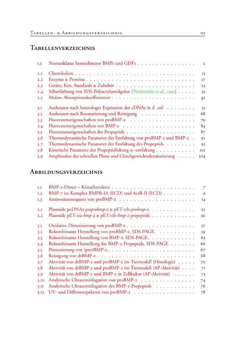
Tabellenverzeichnis
. Nomenklatur homodimerer BMPs und GDFs . . . . . . . . . . . . . . . .
. Chemikalien . . . . . . . . . . . . . . . . . . . . . . . . . . . . . . . . .
. Enzyme & Proteine . . . . . . . . . . . . . . . . . . . . . . . . . . . . . .
. Geräte, Kits, Standards & Zubehör . . . . . . . . . . . . . . . . . . . . . .
. Silberfärbung von SDS-Polyacrylamidgelen [Nesterenko et al., ] . . . . .
. Molare Absorptionskoeffizienten . . . . . . . . . . . . . . . . . . . . . . .
. Ausbeuten nach heterologer Expression der cDNAs in E. coli . . . . . . . .
. Ausbeuten nach Renaturierung und Reinigung . . . . . . . . . . . . . . .
. Fluoreszenzeigenschaften von proBMP- . . . . . . . . . . . . . . . . . .
. Fluoreszenzeigenschaften von BMP- . . . . . . . . . . . . . . . . . . . .
. Fluoreszenzeigenschaften des Propeptids . . . . . . . . . . . . . . . . . . .
. Thermodynamische Parameter der Entfaltung von proBMP- und BMP- .
. Thermodynamische Parameter der Entfaltung des Propeptids . . . . . . . .
. Kinetische Parameter der Propeptidfaltung & -entfaltung . . . . . . . . . . .
. Amplituden der schnellen Phase und Gleichgewichtsdenaturierung . . . . .
Abbildungsverzeichnis
. BMP--Dimer – Kristallstruktur . . . . . . . . . . . . . . . . . . . . . . .
. BMP- im Komplex BMPR-IA (ECD) und ActR-II (ECD) . . . . . . . . .
. Aminosäuresequenz von proBMP- . . . . . . . . . . . . . . . . . . . . .
. Plasmide pcDNA-preprobmp- & pET-b-probmp-. . . . . . . . . . . . .
. Plasmide pET-a-bmp- & pET-b-bmp--propeptide. . . . . . . . . . . . .
. Oxidative Dimerisierung von proBMP- . . . . . . . . . . . . . . . . . . .
. Rekombinante Herstellung von proBMP-. SDS-PAGE. . . . . . . . . . .
. Rekombinante Herstellung von BMP-. SDS-PAGE. . . . . . . . . . . . .
. Rekombinante Herstellung des BMP--Propeptids. SDS-PAGE . . . . . . .
. Prozessierung von (pro)BMP- . . . . . . . . . . . . . . . . . . . . . . . .
. Reinigung von drBMP- . . . . . . . . . . . . . . . . . . . . . . . . . . .
. Aktivität von drBMP- und proBMP- im Tiermodell (Histologie) . . . . .
. Aktivität von drBMP- und proBMP- im Tiermodell (AP-Aktivität) . . . .
. Aktivität von drBMP- und BMP- in Zellkultur (AP-Aktivität) . . . . . .
. Analytische Ultrazentrifugation von proBMP- . . . . . . . . . . . . . . .
. Analytische Ultrazentrifugation des BMP--Propeptids . . . . . . . . . . .
. UV- und Differenzspektren von proBMP- . . . . . . . . . . . . . . . . .
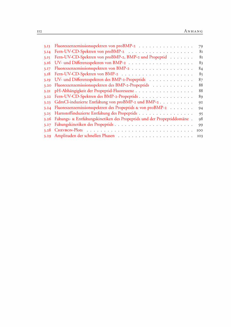
112 Anhang
. Fluoreszenzemissionsspektren von proBMP- . . . . . . . . . . . . . . . .
. Fern-UV-CD-Spektren von proBMP- . . . . . . . . . . . . . . . . . . .
. Fern-UV-CD-Spektren von proBMP-, BMP- und Propeptid . . . . . . .
. UV- und Differenzspektren von BMP- . . . . . . . . . . . . . . . . . . .
. Fluoreszenzemissionsspektren von BMP- . . . . . . . . . . . . . . . . . .
. Fern-UV-CD-Spektren von BMP- . . . . . . . . . . . . . . . . . . . . .
. UV- und Differenzspektren des BMP--Propeptids . . . . . . . . . . . . .
. Fluoreszenzemissionsspektren des BMP--Propeptids . . . . . . . . . . . .
. pH-Abhängigkeit der Propeptid-Fluoreszenz . . . . . . . . . . . . . . . . .
. Fern-UV-CD-Spektren des BMP--Propeptids . . . . . . . . . . . . . . . .
. GdmCl-induzierte Entfaltung von proBMP- und BMP- . . . . . . . . . .
. Fluoreszenzemissionsspektren des Propeptids & von proBMP- . . . . . . .
. Harnstoffinduzierte Entfaltung des Propeptids . . . . . . . . . . . . . . . .
. Faltungs- & Entfaltungskinetiken des Propeptids und der Propeptiddomäne .
. Faltungskinetiken des Propeptids . . . . . . . . . . . . . . . . . . . . . . .
. Chevron-Plots . . . . . . . . . . . . . . . . . . . . . . . . . . . . . . .
. Amplituden der schnellen Phasen . . . . . . . . . . . . . . . . . . . . . .
Literaturverzeichnis
Anfinsen, C. B. (1973). Principles that govern the folding of protein chains. Science, 181:223–230.
Arnold, U., & Ulbrich-Hofmann, R. (1999). Quantitative protein precipitation from guanidi-ne hydrochloride-containing solutions by sodium deoxycholate/trichloroacetic acid. Anal.Biochem., 271:197–199.
Ausubel, F. M., Brent, R., Kingston, R. E., Moore, D. D., Seidman, J. G., & Smith, J. A.(1987). Current Protocols in Molecular Biology, Bd. 2. Green Publishing Associates & JohnWiley & Sons, Inc., New York.
Bahamonde, M. E., & Lyons, K. M. (2001). BMP3: To be or not to be a BMP. J. Bone JointSurg. Am., 83-A Suppl. 1:S56–62.
Baker, D., Sohl, J. L., & Agard, D. A. (1992). A protein-folding reaction under kinetic control.Nature, 356:263–265.
Balemans, W., & van Hul, W. (2002). Extracellular regulation of BMP signaling in vertebrates:A cocktail of modulators. Dev. Biol., 250:231–250.
Bauskin, A. R., Zhang, H. P., Fairlie, W. D., He, X. Y., Russell, P. K., Moore, A. G., Brown,D. A., Stanley, K. K., & Breit, S. N. (2000). The propeptide of macrophage inhibitorycytokine (MIC-1), a TGF-β superfamily member, acts as a quality control determinant forcorrectly folded MIC-1. EMBO J., 19:2212–2220.
Beck, S., Good, J. A. L., Guzman, M., Haim, N. B., Roy, K., Beermann, F., & Constam, D. B.(2002). Extraembryonic proteases regulate Nodal signalling during gastrulation. Nat. CellBiol., 4:981–985.
Beissinger, M., & Buchner, J. (1998). How chaperones fold proteins. Biol. Chem., 379:245–259.
Bonewald, L. F., Wakefield, L., Oreffo, R. O., Escobedo, A., Twardzik, D. R., & Mundy, G. R.(1991). Latent forms of transforming growth factor β (TGF-β) derived from bone cultures:Identification of a naturally occurring 100-kDa complex with similarity to recombinantlatent TGF-β. Mol. Endocrinol., 5:741–751.
Bostrom, M. P. (1998). Expression of bone morphogenetic proteins in fracture healing. Clin.Orthop., 355 Suppl.:116–123.
114 Anhang
Brandts, J. F., Halvorson, H. R., & Brennan, M. (1975). Consideration of the possibilitythat the slow step in protein denaturation reactions is due to cis-trans isomerism of prolineresidues. Biochemistry, 14:4953–4963.
Brinkmann, U., Mattes, R. E., & Buckel, P. (1989). High-level expression of recombinantgenes in Escherichia coli is dependent on the availability of the dnaY gene product. Gene,85:109–114.
Brown, P. D., Wakefield, L. M., Levinson, A. D., & Sporn, M. B. (1990). Physicochemicalactivation of recombinant latent transforming growth factor-β’s 1, 2, and 3. Growth Factors,3:35–43.
Brunner, A. M., Marquardt, H., Malacko, A. R., Lioubin, M. N., & Purchio, A. F. (1989).Site-directed mutagenesis of cysteine residues in the pro region of the transforming growthfactor β 1 precursor. Expression and characterization of mutant proteins. J. Biol. Chem.,264:13660–13664.
von Bubnoff, A., & Cho, K. W. (2001). Intracellular BMP signaling regulation in vertebrates:Pathway or network? Dev. Biol., 239:1–14.
Burstein, E. A., Vedenkina, N. S., & Ivkova, M. N. (1973). Fluorescence and the location oftryptophan residues in protein molecules. Photochem. Photobiol., 18:263–279.
Burstein, E. A., Permyakov, E. A., Yashin, V. A., Burkhanov, S. A., & Finazz-Argo, A. (1977).The fine structure of luminescence spectra of azurin. Biochim. Biophys. Acta, 491:155–159.
Canalis, E., Economides, A. N., & Gazzerro, E. (2003). Bone morphogenetic proteins, theirantagonists, and the skeleton. Endocr. Rev., 24:218–235.
Cantor, C. R., & Schimmel, P. R. (1980). Biophysical Chemistry Part II: Techniques for theStudy of Biological Structure and Function. W. H. Freeman and Company, New York.
Celeste, A. J., & Murray, B. L. (2001). Bone morphogenetic protein-16 (BMP-16) composi-tions. United States Patent No. 6 331 612.
Celeste, A. J., & Murray, B. L. (2002). Bone morphogenetic protein (BMP)-17 and BMP-18compositions. United States Patent No. 6 492 493.
Cerletti, N., McMaster, G. K., Cox, D., Schmitz, A., & Meyhack, B. (1997). Process for refol-ding recombinantly produced TGF-β-like proteins. United States Patent No. 5 650 494.
Chalaux, E., Lopez Rovira, T., Rosa, J. L., Bartrons, R., & Ventura, F. (1998). JunB is involvedin the inhibition of myogenic differentiation by bone morphogenetic protein-2. J. Biol.Chem., 273:537–543.
Literaturver ze ichni s
Chen, D., Zhao, M., Harris, S. E., & Mi, Z. (2004). Signal transduction and biologicalfunctions of bone morphogenetic proteins. Front. Biosci., 9:349–358.
Cheng, H., Jiang, W., Phillips, F. M., Haydon, R. C., Peng, Y., Zhou, L., Luu, H. H., An, N.,Breyer, B., Vanichakarn, P., Szatkowski, J. P., Park, J. Y., & He, T. C. (2003). Osteogenicactivity of the fourteen types of human bone morphogenetic proteins (BMPs). J. Bone JointSurg. Am., 85-A:1544–1552.
Constam, D. B., & Robertson, E. J. (1999). Regulation of bone morphogenetic proteinactivity by pro domains and proprotein convertases. J. Cell Biol., 144:139–149.
Creighton, T. E. (1991). Molecular chaperones. Unfolding protein folding. Nature, 352:17–18.
Creighton, T. E. (1993). Proteins: Structures and Molecular Properties. W. H. Freeman andCompany, New York, 2. Aufl.
Creighton, T. E. (1997). Protein folding coupled to disulphide bond formation. Biochemistry,378:731–744.
Creighton, T. E., Bagley, C. J., Cooper, L., Darby, N. J., Freedman, R. B., Kemmink, J.,& Sheikh, A. (1993). On the biosynthesis of bovine pancreatic trypsin inhibitor (BPTI).Structure, processing, folding and disulphide bond formation of the precursor in vitro andin microsomes. J. Mol. Biol., 232:1176–1196.
Cui, Y., Jean, F., Thomas, G., & Christian, J. L. (1998). BMP-4 is proteolytically activated byfurin and/or PC6 during vertebrate embryonic development. EMBO J., 17:4735–4743.
Cui, Y., Hackenmiller, R., Berg, L., Jean, F., Nakayama, T., Thomas, G., & Christian, J. L.(2001). The activity and signaling range of mature BMP-4 is regulated by sequentialcleavage at two sites within the prodomain of the precursor. Genes Dev., 15:2797–2802.
Dale, L., Matthews, G., & Colman, A. (1993). Secretion and mesoderm-inducing activity ofthe TGF-β-related domain of Xenopus Vg1. EMBO J., 12:4471–4480.
Daluiski, A., Engstrand, T., Bahamonde, M. E., Gamer, L. W., Agius, E., Stevenson, S. L.,Cox, K., Rosen, V., & Lyons, K. M. (2001). Bone morphogenetic protein-3 is a negativeregulator of bone density. Nat. Genet., 27:84–88.
Daopin, S., Piez, K. A., Ogawa, Y., & Davis, D. R. (1992). Crystal structure of transforminggrowth factor-β 2: An unusual fold for the superfamily. Science, 257:369–373.
Demchenko, A. P. (1986). Ultraviolet Spectroscopy of Proteins. Springer-Verlag, Berlin. Erwei-terte u. überarbeitete Übersetzung der russischen Ausgabe (Naukova Dumka, Kiew, 1981).
116 Anhang
ten Dijke, P., Yamashita, H., Sampath, T. K., Reddi, A. H., Estevez, M., Riddle, D. L., Ichijo,H., Heldin, C. H., & Miyazono, K. (1994). Identification of type I receptors for osteogenicprotein-1 and bone morphogenetic protein-4. J. Biol. Chem., 269:16985–16988.
Donovan, J. W., Laskowski Jr., M., & Scheraga, H. A. (1958). Influence of ionization ofcarboxyl groups on the ultraviolet absorption spectrum of lysozyme. Biochim. Biophys.Acta, 29:455–456.
Dower, W. J. (1990). Electroporation of prokaryotic cells. United States Patent No.4 910 140.
Ducy, P., & Karsenty, G. (2000). The family of bone morphogenetic proteins. Kidney Int.,57:2207–2214.
Edelhoch, H. (1967). Spectroscopic determination of tryptophan and tyrosine in proteins.Biochemistry, 6:1948–1954.
Eder, J., Rheinnecker, M., & Fersht, A. R. (1993). Folding of subtilisin BPN’: Characteriza-tion of a folding intermediate. Biochemistry, 32:18–26.
Edman, P., & Begg, G. (1967). A protein sequenator. Eur. J. Biochem., 1:80–91.
Eftink, M. R. (1994). The use of fluorescence methods to monitor unfolding transitions inproteins. Biophys. J., 66:482–501.
Eldar, A., Dorfman, R., Weiss, D., Ashe, H., Shilo, B. Z., & Barkai, N. (2002). Robustness ofthe BMP morphogen gradient in Drosophila embryonic patterning. Nature, 419:304–308.
Fairbanks, G., Steck, T. L., & Wallach, D. F. (1971). Electrophoretic analysis of the majorpolypeptides of the human erythrocyte membrane. Biochemistry, 10:2606–2617.
Fersht, A. (1999). Structure and Mechanism in Protein Science. W.H. Freeman and Company,New York.
Fischer, G., Tradler, T., & Zarnt, T. (1998). The mode of action of peptidyl prolyl cis/transisomerases in vivo: Binding vs. catalysis. FEBS Lett., 426:17–20.
Fox, T., de Miguel, E., Mort, J. S., & Storer, A. C. (1992). Potent slow-binding inhibition ofcathepsin B by its propeptide. Biochemistry, 31:12571–12576.
Freedman, R. B., Hirst, T. R., & Tuite, M. F. (1994). Protein disulphide isomerase: Buildingbridges in protein folding. Trends Biochem. Sci., 19:331–336.
Fujise, M., Takeo, S., Kamimura, K., Matsuo, T., Aigaki, T., Izumi, S., & Nakato, H. (2003).Dally regulates DPP morphogen gradient formation in the Drosophila wing. Development,130:1515–1522.
Literaturver ze ichni s
Gilboa, L., Nohe, A., Geissendorfer, T., Sebald, W., Henis, Y. I., & Knaus, P. (2000). Bonemorphogenetic protein receptor complexes on the surface of live cells: A new oligomeriza-tion mode for serine/threonine kinase receptors. Mol. Biol. Cell , 11:1023–1035.
Gill, S. C., & von Hippel, P. H. (1989). Calculation of protein extinction coefficients fromamino acid sequence data. Anal. Biochem., 182:319–326.
Goldhaber, P. (1961). Osteogenic induction across millipore filters in vivo. Science, 133:2065–2067.
Gothel, S. F., & Marahiel, M. A. (1999). Peptidyl-prolyl cis-trans isomerases, a superfamily ofubiquitous folding catalysts. Cell. Mol. Life Sci., 55:423–436.
Goto, Y., & Hamaguchi, K. (1982). Unfolding and refolding of the constant fragment of theimmunoglobulin light chain. J. Mol. Biol., 156:891–910.
Grathwohl, C., & Wüthrich, K. (1981). NMR studies of the rates of proline cis/trans isome-risation in oligopeptides. Biopolymers, 15:2623–2633.
Gray, A. M., & Mason, A. J. (1990). Requirement for activin A and transforming growthfactor-β 1 pro-regions in homodimer assembly. Science, 247:1328–1330.
Greenwald, J., Groppe, J., Gray, P., Wiater, E., Kwiatkowski, W., Vale, W., & Choe, S. (2003).The BMP7/ActRII extracellular domain complex provides new insights into the coopera-tive nature of receptor assembly. Mol. Cell , 11:605–617.
Griffith, D. L., Keck, P. C., Sampath, T. K., Rueger, D. C., & Carlson, W. D. (1996). Three-dimensional structure of recombinant human osteogenic protein 1: Structural paradigm forthe transforming growth factor β superfamily. Proc. Natl. Acad. Sci. U.S.A., 93:878–883.
Groppe, J., Rumpel, K., Economides, A. N., Stahl, N., Sebald, W., & Affolter, M. (1998).Biochemical and biophysical characterization of refolded Drosophila DPP, a homolog ofbone morphogenetic proteins 2 and 4. J. Biol. Chem., 273:29052–29065.
Hagel, P., Gerding, J. J., Fieggen, W., & Bloemendal, H. (1971). Cyanate formation in so-lutions of urea. I. Calculation of cyanate concentrations at different temperature and pH.Biochim. Biophys. Acta, 243:366–373.
Harding, S. E. (1997). Hydrodynamic properties of proteins. In Protein Structure: A PracticalApproach (Hg. T. E. Creighton), A Practical Approach Series, Kap. 9, S. 219–251. OxfordUniversity Press Inc., New York, 2. Aufl.
Hauschka, P. V., Mavrakos, A. E., Iafrati, M. D., Doleman, S. E., & Klagsbrun, M. (1986).Growth factors in bone matrix. Isolation of multiple types by affinity chromatography onheparin-Sepharose. J. Biol. Chem., 261:12665–12674.
118 Anhang
Haÿ, E., Lemonnier, J., Fromigué, O., & Marie, P. J. (2001). Bone morphogenetic protein-2promotes osteoblast apoptosis through a Smad-independent, protein kinase C-dependentsignaling pathway. J. Biol. Chem., 276:29028–29036.
von Heijne, G. (1986). A new method for predicting signal sequence cleavage sites. NucleicAcids Res., 14:4683–4690.
Hillger, F. (1999). Humanes proBMP-2: Rekombinante Expression und Rückfaltung aus Ein-schlußkörpern. Diplomarbeit, Martin-Luther-Universität Halle-Wittenberg, Halle (Saale),Deutschland.
Hino, J., Nishimatsu, S., Nagai, T., Matsuo, H., Kangawa, K., & Nohno, T. (2003). Coordi-nation of BMP-3b and cerberus is required for head formation of Xenopus embryos. Dev.Biol., 260:138–157.
Hino, J., Kangawa, K., Matsuo, H., Nohno, T., & Nishimatsu, S. (2004). Bone morphogene-tic protein-3 family members and their biological functions. Front. Biosci., 9:1520–1529.
Hofbauer, L. C., Dunstan, C. R., Spelsberg, T. C., Riggs, B. L., & Khosla, S. (1998). Osteo-protegerin production by human osteoblast lineage cells is stimulated by vitamin D, bonemorphogenetic protein-2, and cytokines. Biochem. Biophys. Res. Commun., 250:776–781.
Hogan, B. L. (1996). Bone morphogenetic proteins: Multifunctional regulators of vertebratedevelopment. Genes Dev., 10:1580–1594.
Ikemura, H., Takagi, H., & Inouye, M. (1987). Requirement of pro-sequence for the produc-tion of active subtilisin E in Escherichia coli. J. Biol. Chem., 262:7859–7864.
Imoto, T., & Yamada, H. (1997). Chemical modification. In Protein Function: A PracticalApproach (Hg. T. E. Creighton), A Practical Approach Series, Kap. 10, S. 279–316. OxfordUniversity Press Inc., New York, 2. Aufl.
Innis, C. A., Shi, J., & Blundell, T. L. (2000). Evolutionary trace analysis of TGF-β and relatedgrowth factors: Implications for site-directed mutagenesis. Protein Eng., 13:839–847.
Inouye, M. (1991). Intramolecular chaperone: The role of the pro-peptide in protein folding.Enzyme, 45:314–321.
Israel, D. I., Nove, J., Kerns, K. M., Moutsatsos, I. K., & Kaufman, R. J. (1992). Expressi-on and characterization of bone morphogenetic protein-2 in Chinese hamster ovary cells.Growth Factors, 7:139–150.
Izumi, M., Fujio, Y., Kunisada, K., Negoro, S., Tone, E., Funamoto, M., Osugi, T., Oshi-ma, Y., Nakaoka, Y., Kishimoto, T., Yamauchi-Takihara, K., & Hirota, H. (2001). Bonemorphogenetic protein-2 inhibits serum deprivation-induced apoptosis of neonatal cardiacmyocytes through activation of the Smad1 pathway. J. Biol. Chem., 276:31133–31141.
Literaturver ze ichni s
Keller, S., Nickel, J., Zhang, J. L., Sebald, W., & Mueller, T. D. (2004). Molecular recognitionof BMP-2 and BMP receptor IA. Nat. Struct. Mol. Biol., 11:481–488.
Kessler, D. S., & Melton, D. A. (1995). Induction of dorsal mesoderm by soluble, matureVg1 protein. Development, 121:2155–2164.
Kessler, E., Takahara, K., Biniaminov, L., Brusel, M., & Greenspan, D. S. (1996). Bonemorphogenetic protein-1: The type I procollagen C-proteinase. Science, 271:360–362.
Khorasanizadeh, S., Peters, I. D., & Roder, H. (1996). Evidence for a three-state model ofprotein folding from kinetic analysis of ubiquitin variants with altered core residues. Nat.Struct. Biol., 3:193–205.
Kiefhaber, T., Quaas, R., Hahn, U., & Schmid, F. X. (1990). Folding of ribonuclease T1.1. Existence of multiple unfolded states created by proline isomerization. Biochemistry,29:3053–3061.
Kirsch, T., Nickel, J., & Sebald, W. (2000a). BMP-2 antagonists emerge from alterations inthe low-affinity binding epitope for receptor BMPR-II. EMBO J., 19:3314–3324.
Kirsch, T., Sebald, W., & Dreyer, M. K. (2000b). Crystal structure of the BMP-2-BRIAectodomain complex. Nat. Struct. Biol., 7:492–496.
Kita, Y., Arakawa, T., Lin, T. Y., & Timasheff, S. N. (1994). Contribution of the surface freeenergy perturbation to protein-solvent interactions. Biochemistry, 33:15178–15189.
Koenig, B. B., Cook, J. S., Wolsing, D. H., Ting, J., Tiesman, J. P., Correa, P. E., Olson,C. A., Pecquet, A. L., Ventura, F., Grant, R. A., Chen, G.-X., Wrana, J. L., Massagué, J.,& Rosenbaum, J. S. (1994). Characterization and cloning of a receptor for BMP-2 andBMP-4 from NIH 3T3 cells. Mol. Cell Biol., 14:5961–5974.
Kolodziej, P. A., & Young, R. A. (1991). Epitope tagging and protein surveillance. MethodsEnzymol., 194:508–519.
Kopetzki, E., Schumacher, G., & Buckel, P. (1989). Control of formation of active solubleor inactive insoluble baker’s yeast alpha-glucosidase PI in Escherichia coli by induction andgrowth conditions. Mol. Gen. Genet., 216:149–155.
Krause, M. (2003). Untersuchungen zur Rolle des Propeptids von BMP-2. Diplomarbeit,Martin-Luther-Universität Halle-Wittenberg, Halle (Saale), Deutschland.
Krishnan, B. R., Kersulyte, D., Brikun, I., Berg, C. M., & Berg, D. E. (1991). Direct andcrossover PCR amplification to facilitate Tn5supF-based sequencing of λ phage clones.Nucleic Acids Res., 19:6177–6182.
120 Anhang
Kyhse-Andersen, J. (1984). Electroblotting of multiple gels: A simple apparatus withoutbuffer tank for rapid transfer of proteins from polyacrylamide to nitrocellulose. J. Biochem.Biophys. Methods, 10:203–209.
Laemmli, U. K. (1970). Cleavage of structural proteins during the assembly of the head ofbacteriophage T4. Nature, 227:680–685.
Laskowski Jr., M., Widom, J. M., McFadden, M. L., & Scheraga, H. A. (1956). Differentialultraviolet spectra of insulin. Biochim. Biophys. Acta, 19:581–582.
Lawrence, D. A., Pircher, R., & Jullien, P. (1985). Conversion of a high molecular weightlatent β-TGF from chicken embryo fibroblasts into a low molecular weight active β-TGFunder acidic conditions. Biochem. Biophys. Res. Commun., 133:1026–1034.
Lee, R., Kermani, P., Teng, K. K., & Hempstead, B. L. (2001). Regulation of cell survival bysecreted proneurotrophins. Science, 294:1945–1948.
Li, S. W., Sieron, A. L., Fertala, A., Hojima, Y., Arnold, W. V., & Prockop, D. J. (1996).The C-proteinase that processes procollagens to fibrillar collagens is identical to the pro-tein previously identified as bone morphogenetic protein-1. Proc. Natl. Acad. Sci. U.S.A.,93:5127–5130.
Liao, W. X., Moore, R. K., Otsuka, F., & Shimasaki, S. (2003). Effect of intracellular in-teractions on the processing and secretion of bone morphogenetic protein-15 (BMP-15)and growth and differentiation factor-9. Implication of the aberrant ovarian phenotype ofbmp-15 mutant sheep. J. Biol. Chem., 278:3713–3719.
Lin, T. Y., & Timasheff, S. N. (1996). On the role of surface tension in the stabilization ofglobular proteins. Protein Sci., 5:372–381.
Lioubin, M. N., Madisen, L., Marquardt, H., Roth, R., Kovacina, K. S., & Purchio, A. F.(1991). Characterization of latent recombinant TGF-β 2 produced by Chinese hamsterovary cells. J. Cell. Biochem., 45:112–121.
Lisse, T. (2001). Untersuchungen zur Rolle von Mdj1p aus Saccharomyces cerevisiae bei dermitochondrialen Biogenese. Dissertation, Martin-Luther-Universität Halle-Wittenberg,Halle (Saale), Deutschland.
Lories, R. J., Derese, I., Ceuppens, J. L., & Luyten, F. P. (2003). Bone morphogenetic proteins2 and 6, expressed in arthritic synovium, are regulated by proinflammatory cytokines anddifferentially modulate fibroblast-like synoviocyte apoptosis. Arthritis Rheum., 48:2807–2818.
Lyons, R. M., Gentry, L. E., Purchio, A. F., & Moses, H. L. (1990). Mechanism of activationof latent recombinant transforming growth factor β 1 by plasmin. J. Cell Biol., 110:1361–1367.
Literaturver ze ichni s
Maniatis, T., Fritsch, E. F., & Sambrook, J. (1989). Molecular Cloning: A Laboratory Manual .Cold Spring Harbor Laboratory, Cold Spring Harbor, USA.
Marqués, G., Musacchio, M., Shimell, M. J., Wünnenberg-Stapleton, K., Cho, K. W., &O’Connor, M. B. (1997). Production of a DPP activity gradient in the early Drosophilaembryo through the opposing actions of the SOG and TLD proteins. Cell , 91:417–426.
McDonald, N. Q., & Hendrickson, W. A. (1993). A structural superfamily of growth factorscontaining a cystine knot motif. Cell , 73:421–424.
McGeoch, D. J. (1985). On the predictive recognition of signal peptide sequences. VirusRes., 3:271–286.
McKay, B., & Sandhu, H. S. (2002). Use of recombinant human bone morphogeneticprotein-2 in spinal fusion applications. Spine, 27:S66–85.
McMahon, G. A., Dignam, J. D., & Gentry, L. E. (1996). Structural characterization of thelatent complex between transforming growth factor β 1 and β 1-latency-associated peptide.Biochem. J., 313 (Pt. 1):343–351.
Miller, D. M., Ogawa, Y., Iwata, K. K., ten Dijke, P., Purchio, A. F., Soloff, M. S., & Gentry,L. E. (1992). Characterization of the binding of transforming growth factor-β 1, -β 2, and-β 3 to recombinant β 1-latency-associated peptide. Mol. Endocrinol., 6:694–702.
Miyazawa, K., Kawai, T., & Urist, M. R. (1996). Bone morphogenetic protein-induced hete-rotopic bone in osteopetrosis. Clin. Orthop., 324:259–268.
Moss, M. L. (1960). In Calcification in Biological Systems (Hg. R. F. Sognnaes), S. 323–328.AAAS, Washington, D.C.
Müller, C., & Rinas, U. (1999). Renaturation of heterodimeric platelet-derived growth factorfrom inclusion bodies of recombinant Escherichia coli using size-exclusion chromatography.J. Chromatogr. A, 855:203–213.
Müller, C., Richter, S., & Rinas, U. (2003). Kinetics control preferential heterodimer for-mation of platelet-derived growth factor from unfolded A- and B-chains. J. Biol. Chem.,278:18330–18335.
Muller, Y. A., Heiring, C., Misselwitz, R., Welfle, K., & Welfle, H. (2002). The cystine knotpromotes folding and not thermodynamic stability in vascular endothelial growth factor. J.Biol. Chem., 277:43410–43416.
Mullis, K., Faloona, F., Scharf, S., Saiki, R., Horn, G., & Erlich, H. (1986). Specific enzymaticamplification of DNA in vitro: The polymerase chain reaction. Cold Spring Harb. Symp.Quant. Biol., 51 (Pt. 1):263–273.
122 Anhang
Nakai, K., & Kanehisa, M. (1991). Expert system for predicting protein localization sites ingram-negative bacteria. Proteins, 11:95–110.
Nakai, K., & Kanehisa, M. (1992). A knowledge base for predicting protein localization sitesin eukaryotic cells. Genomics, 14:897–911.
Nall, B. T., Garel, J. R., & Baldwin, R. L. (1978). Test of the extended two-state model forthe kinetic intermediates observed in the folding transition of ribonuclease A. J. Mol. Biol.,118:317–330.
Namiki, M., Akiyama, S., Katagiri, T., Suzuki, A., Ueno, N., Yamaji, N., Rosen, V., Wozney,J. M., & Suda, T. (1997). A kinase domain-truncated type I receptor blocks bone mor-phogenetic protein-2-induced signal transduction in C2C12 myoblasts. J. Biol. Chem.,272:22046–22052.
Nesterenko, M. V., Tilley, M., & Upton, S. J. (1995). A metallo-dependent cysteine proteinaseof Cryptosporidium parvum associated with the surface of sporozoites. Microbios, 83:77–88.
Nohe, A., Hassel, S., Ehrlich, M., Neubauer, F., Sebald, W., Henis, Y. I., & Knaus, P. (2002).The mode of bone morphogenetic protein (BMP) receptor oligomerization determinesdifferent BMP-2 signaling pathways. J. Biol. Chem., 277:5330–5338.
Nozaki, Y. (1972). The preparation of guanidine hydrochloride. Methods Enzymol , 26 (Pt.C):43–50.
Nykjaer, A., Lee, R., Teng, K. K., Jansen, P., Madsen, P., Nielsen, M. S., Jacobsen, C., Klie-mannel, M., Schwarz, E., Willnow, T. E., Hempstead, B. L., & Petersen, C. M. (2004).Sortilin is essential for proNGF-induced neuronal cell death. Nature, 427:843–838.
Oefner, C., D’Arcy, A., Winkler, F. K., Eggimann, B., & Hosang, M. (1992). Crystal structureof human platelet-derived growth factor BB. EMBO J., 11:3921–3926.
Oelgeschläger, M., Larrain, J., Geissert, D., & de Robertis, E. M. (2000). The evolutionarilyconserved BMP-binding protein Twisted gastrulation promotes BMP signalling. Nature,405:757–763.
Ohta, Y., Hojo, H., Aimoto, S., Kobayashi, T., Zhu, X., Jordan, F., & Inouye, M. (1991).Pro-peptide as an intramolecular chaperone: Renaturation of denatured subtilisin E with asynthetic pro-peptide. Mol. Microbiol., 5:1507–1510.
Onichtchouk, D., Chen, Y. G., Dosch, R., Gawantka, V., Delius, H., Massagu’e, J., &Niehrs, C. (1999). Silencing of TGF-β signalling by the pseudoreceptor BAMBI. Na-ture, 401:480–485.
Pace, C. N. (1986). Determination and analysis of urea and guanidine hydrochloride dena-turation curves. Methods Enzymol., 131:266–280.
Literaturver ze ichni s
Paine-Saunders, S., Viviano, B. L., Economides, A. N., & Saunders, S. (2002). Heparansulfate proteoglycans retain noggin at the cell surface. A potential mechanism for shapingbone morphogenetic protein gradients. J. Biol. Chem., 277:2089–2096.
Pallaghy, P. K., Nielsen, K. J., Craik, D. J., & Norton, R. S. (1994). A common structuralmotif incorporating a cystine knot and a triple-stranded β-sheet in toxic and inhibitorypolypeptides. Protein Sci., 3:1833–1839.
Price-Carter, M., Gray, W. R., & Goldenberg, D. P. (1996). Folding of ω-conotoxins. 1.Efficient disulfide-coupled folding of mature sequences in vitro. Biochemistry, 35:15537–15546.
Raftery, L. A., & Sutherland, D. J. (1999). TGF-β family signal transduction in Drosophiladevelopment: From Mad to Smads. Dev. Biol., 210:251–268.
Rattenholl, A. (2001). Untersuchungen zur Pro-Sequenz-vermittelten Faltung von rekom-binantem, humanen Nervenwachstumsfaktor. Dissertation, Martin-Luther-UniversitätHalle-Wittenberg, Halle (Saale), Deutschland.
Rattenholl, A., Lilie, H., Grossmann, A., Stern, A., Schwarz, E., & Rudolph, R. (2001a).The pro-sequence facilitates folding of human nerve growth factor from Escherichia coliinclusion bodies. Eur. J. Biochem., 268:3296–3303.
Rattenholl, A., Ruoppolo, M., Flagiello, A., Monti, M., Vinci, F., Marino, G., Lilie, H.,Schwarz, E., & Rudolph, R. (2001b). Pro-sequence assisted folding and disulfide bondformation of human nerve growth factor β. J. Mol. Biol., 305:523–533.
Reimer, U., Scherer, G., Drewello, M., Kruber, S., Schutkowski, M., & Fischer, G. (1998).Side-chain effects on peptidyl-prolyl cis/trans isomerisation. J. Mol. Biol., 279:449–460.
Rosenberg, A. H., Lade, B. N., Chui, D. S., Lin, S. W., Dunn, J. J., & Studier, F. W. (1987).Vectors for selective expression of cloned DNAs by T7 RNA polymerase. Gene, 56:125–135.
Rudolph, R., Böhm, G., Lilie, H., & Jaenicke, R. (1997). Folding proteins. In Protein Func-tion: A Practical Approach (Hg. T. E. Creighton), A Practical Approach Series, Kap. 3, S.57–99. Oxford University Press Inc., New York, 2. Aufl.
Rudolph, R., Schwarz, E., Herr, G., & Hillger, F. (2002). Production of recombinant BMP-2.International Patent Application No. WO 02/44203.
Ruppert, R., Hoffmann, E., & Sebald, W. (1996). Human bone morphogenetic protein 2contains a heparin-binding site which modifies its biological activity. Eur. J. Biochem.,237:295–302.
Sachs, L. (1992). Angewandte Statistik. Springer-Verlag, Berlin, 7. Aufl.
124 Anhang
Saiki, R. K., Gelfand, D. H., Stoffel, S., Scharf, S. J., Higuchi, R., Horn, G. T., Mullis,K. B., & Erlich, H. A. (1988). Primer-directed enzymatic amplification of DNA with athermostable DNA polymerase. Science, 239:487–491.
Sampath, T. K., & Reddi, A. H. (1981). Dissociative extraction and reconstitution of extra-cellular matrix components involved in local bone differentiation. Proc. Natl. Acad. Sci.U.S.A., 78:7599–7503.
Sampath, T. K., Muthukumaran, N., & Reddi, A. H. (1987). Isolation of osteogenin, an ex-tracellular matrix-associated, bone-inductive protein, by heparin affinity chromatography.Proc. Natl. Acad. Sci. U.S.A., 84:7109–7113.
Sanger, F., Nicklen, S., & Coulson, A. R. (1977). DNA sequencing with chain-terminatinginhibitors. Proc. Natl. Acad. Sci. U.S.A., 74:5463–5467.
Savage, C. P., Finelli, A. L., Townsend, S. R., Run, C.-Y., Baird, S., & Padgett, R. W. (1996).Cænorhabditis elegans genes sma-2, sma-3, and sma-4 define a conserved family of transfor-ming growth factor β pathway components. Proc. Natl. Acad. Sci. U.S.A., 93:790–794.
Saxen, L., & Taivonen, S. (1962). Primary Embryonic Induction. Prentice-Hall, EngelewoodCliffs, New Jersey.
Schäffner, J. (2000). Einfluß niedermolekularer Medienzusätze und sezernierter, ATP-unabhängiger Chaperone auf die Ausbeute therapeutischer Proteine im Periplasma vonEscherichia coli. Dissertation, Martin-Luther-Universität Halle-Wittenberg, Halle (Saale),Deutschland.
Schellman, J. A. (1994). The thermodynamics of solvent exchange. Biopolymers, 34:1015–1026.
Scheraga, H. A. (1957). Tyrosyl-carboxylate ion hydrogen bonding in ribonuclease. Biochim.Biophys. Acta, 23:196–197.
Scheufler, C., Sebald, W., & Hülsmeyer, M. (1999). Crystal structure of human bone mor-phogenetic protein-2 at 2.7 Å resolution. J. Mol. Biol., 287:103–115.
Schmid, F. X. (1997). Optical spectroscopy to characterize protein conformation and con-formational changes. In Protein Structure: A Practical Approach (Hg. T. E. Creighton), APractical Approach Series, Kap. 11, S. 261–297. Oxford University Press Inc., New York,2. Aufl.
Schmid, F. X., & Baldwin, R. L. (1979). Detection of an early intermediate in the folding ofribonuclease A by protection of amide protons against exchange. J. Mol. Biol., 135:199–215.
Literaturver ze ichni s
Schwarz, E., Lilie, H., & Rudolph, R. (1996). The effect of molecular chaperones on in vivoand in vitro folding processes. Biol. Chem., 377:411–416.
Schwarzinger, S., Kroon, G. J., Foss, T. R., Chung, J., Wright, P. E., & Dyson, H. J. (2001).Sequence-dependent correction of random coil NMR chemical shifts. J. Am. Chem. Soc.,123:2970–2978.
Scott, I. C., Imamura, Y., Pappano, W. N., Troedel, J. M., Recklies, A. D., Roughley, P. J.,& Greenspan, D. S. (2000). Bone morphogenetic protein-1 processes probiglycan. J. Biol.Chem., 275:30504–30511.
Sebald, W., & Mueller, T. D. (2003). The interaction of BMP-7 and ActRII implicates a newmode of receptor assembly. Trends Biochem. Sci., 28:518–521.
Seeherman, H., Wozney, J., & Li, R. (2002). Bone morphogenetic protein delivery systems.Spine, 27:S16–23.
Sekelsky, J. J., Newfeld, S. J., Raftery, L. A., Chartoff, E. H., & Gelbart, W. M. (1995). Gene-tic characterization and cloning of Mothers against dpp, a gene required for decapentaplegicfunction in Drosophila melanogaster. Proc. Natl. Acad. Sci. U.S.A., 71:1393–1397.
Sha, X., Yang, L., & Gentry, L. E. (1991). Identification and analysis of discrete functio-nal domains in the pro region of pre-pro-transforming growth factor β 1. J. Cell Biol.,114:827–839.
Shastry, M. C. R., Agashe, V. R., & Udgaonkar, J. B. (1994). Quantitative analysis of thekinetics of denaturation and renaturation of barstar in the folding transition zone. ProteinSci., 3:1409–1417.
Shechter, Y. (1986). Selective oxidation and reduction of methionine residues in peptides andproteins by oxygen exchange between sulfoxide and sulfide. J. Biol. Chem., 261:66–70.
Siggelkow, H., & Hufner, M. (1999). Modulation der Osteoblastengenese: Auswirkungen aufdie Pathophysiologie von Osteoporose. Dtsch. Med. Wochenschr., 124:1–2.
Silen, J. L., & Agard, D. A. (1989). The α-lytic protease pro-region does not require a physicallinkage to activate the protease domain in vivo. Nature, 341:462–464.
Song, Q., Mehler, M. F., & Kessler, J. A. (1998). Bone morphogenetic proteins induce apop-tosis and growth factor dependence of cultured sympathoadrenal progenitor cells. Dev.Biol., 196:119–127.
Stark, G. R. (1965). Reactions of cyanate with functional groups of proteins. III. Reactionswith amino and carboxyl groups. Biochemistry, 4:1030–1036.
126 Anhang
Steiner, D. F., Smeekens, S. P., Ohagi, S., & Chan, S. J. (1992). The new enzymology ofprecursor processing endoproteases. J. Biol. Chem., 267:23435–23438.
Studier, F. W., & Moffatt, B. A. (1986). Use of bacteriophage T7 RNA polymerase to directselective high-level expression of cloned genes. J. Mol. Biol., 189:113–130.
Tanford, C. (1968). Protein denaturation. Adv. Protein Chem., 23:121–282.
Tanford, C. (1970). Protein denaturation. C. Theoretical models for the mechanism of de-naturation. Adv. Protein Chem., 24:1–95.
Teixeira, J. O., & Urist, M. R. (1998). Bone morphogenetic protein induced repair of com-partmentalized segmental diaphyseal defects. Arch. Orthop. Trauma. Surg., 117:27–34.
Thomsen, G. H., & Melton, D. A. (1993). Processed Vg1 protein is an axial mesoderminducer in Xenopus. Cell , 74:433–441.
Towbin, H., Staehelin, T., & Gordon, J. (1979/1992). Electrophoretic transfer of proteinsfrom polyacrylamide gels to nitrocellulose sheets: Procedure and some applications. (1stpubl. 1979). Biotechnology, 24:145–149.
Urist, M. R. (1965). Bone: Formation by autoinduction. Science, 150:893–899.
Urist, M. R., & Mikulski, A. J. (1979). A soluble bone morphogenetic protein extracted frombone matrix with a mixed aqueous and nonaqueous solvent. Proc. Soc. Exp. Biol. Med.,162:48–53.
Urist, M. R., Iwata, H., Ceccotti, P. L., Dorfman, R. L., Boyd, S. D., McDowell, R. M., &Chien, C. (1973). Bone morphogenesis in implants of insoluble bone gelatin. Proc. Natl.Acad. Sci. U.S.A., 70:3511–3515.
Urist, M. R., Lietze, A., Mizutani, H., Takagi, K., Triffit, J. T., Amstutz, J., de Lange, R., Ter-mine, J., & Finerman, G. A. M. (1982). A bovine low molecular mass bone morphogeneticprotein (BMP) fraction. Clin. Orthop., 162:212–232.
Urist, M. R., Sato, K., Brownell, A. G., Malinin, T. I., Lietze, A., Huo, Y. K., Prolo, D. J.,Oklund, S., Finerman, G. A., & DeLange, R. J. (1983). Human bone morphogeneticprotein (hBMP). Proc. Soc. Exp. Biol. Med., 173:194–199.
Urist, M. R., Huo, Y. K., Brownell, A. G., Hohl, W. M., Buyske, J., Lietze, A., Tempst, P.,Hunkapiller, M., & DeLange, R. J. (1984). Purification of bovine bone morphogeneticprotein by hydroxyapatite chromatography. Proc. Natl. Acad. Sci. U.S.A., 81:371–375.
Vallejo, L. F., & Rinas, U. (2004). Optimized procedure for renaturation of recombinanthuman bone morphogenetic protein-2 at high protein concentration. Biotechnol. Bioeng.,85:601–609.
Literaturver ze ichni s
Vallejo, L. F., Brokelmann, M., Marten, S., Trappe, S., Cabrera-Crespo, J., Hoffmann, A.,Gross, G., Weich, H. A., & Rinas, U. (2002). Renaturation and purification of bonemorphogenetic protein-2 produced as inclusion bodies in high-cell-density cultures of re-combinant Escherichia coli. J. Biotechnol., 94:185–194.
Viviano, B. L., Paine-Saunders, S., Gasiunas, N., Gallagher, J., & Saunders, S. (2004).Domain-specific modification of heparan sulfate by qsulf1 modulates the binding of thebone morphogenetic protein antagonist noggin. J. Biol. Chem., 279:5604–5611.
Wang, E. A. (1993). Bone morphogenetic proteins (BMPs): Therapeutic potential in healingbony defects. Trends Biotechnol., 11:379–383.
Wang, E. A., Rosen, V., Cordes, P., Hewick, R. M., Kriz, M. J., Luxenberg, D. P., Sibley, B. S.,& Wozney, J. M. (1988). Purification and characterization of other distinct bone-inducingfactors. Proc. Natl. Acad. Sci. U.S.A., 85:9484–9488.
Wang, E. A., Rosen, V., D’Alessandro, J. S., Bauduy, M., Cordes, P., Harada, T., Israel, D. I.,Hewick, R. M., Kerns, K. M., & LaPan, P. (1990). Recombinant human bone morphoge-netic protein induces bone formation. Proc. Natl. Acad. Sci. U.S.A., 87:2220–2224.
Warburg, O., & Christian, W. (1941). Isolierung und Kristallisation des GärungsfermentsEnolase. Biochem. Z., 310:384–421.
Warren, J. R., & Gordon, J. A. (1966). On the refractive indices of aqueous solutions of urea.J. Phys. Chem., 70:297–300.
Wentzel, A., Christmann, A., Kratzner, R., & Kolmar, H. (1999). Sequence requirements ofthe GPNG β-turn of the Ecballium elaterium trypsin inhibitor II explored by combinatoriallibrary screening. J. Biol. Chem., 274:21037–21043.
Wetlaufer, D. B. (1962). Ultraviolet spectra of proteins and amino acids. Adv. Protein Chem.,17:303–390.
Winther, J. R., & Sørensen, P. (1991). Propeptide of carboxypeptidase Y provides a chaperone-like function as well as inhibition of the enzymatic activity. Proc. Natl. Acad. Sci. U.S.A.,88:9330–9334.
Wozney, J. M. (2002). Overview of bone morphogenetic proteins. Spine, 27:S2–8.
Wozney, J. M., Rosen, V., Celeste, A. J., Mitsock, L. M., Whitters, M. J., Kriz, R. W., Hewick,R. M., & Wang, E. A. (1988). Novel regulators of bone formation: Molecular clones andactivities. Science, 242:1528–1534.
Wrana, J. L., Attisano, L., Wieser, R., Ventura, F., & Massagué, J. (1994). Mechanism ofactivation of the TGF-β receptor. Nature, 370:341–347.

128 Anhang
Yanari, S., & Bovey, F. A. (1960). Interpretation of the ultraviolet spectral changes of proteins.J. Biol. Chem., 235:2818–2826.
de Young, L. R., Burton, L. E., Liu, J., Powell, M. F., Schmelzer, C. H., & Skelton, N. J.(1996). RhNGF slow unfolding is not due to proline isomerization: Possibility of a cystineknot loop-threading mechanism. Protein Sci., 5:1554–1566.
Zhang, H., & Bradley, A. (1996). Mice deficient for BMP2 are nonviable and have defects inamnion/chorion and cardiac development. Development, 122:2977–2986.


![6 Literaturverzeichnis - sundoc.bibliothek.uni-halle.desundoc.bibliothek.uni-halle.de/diss-online/03/03H130/t7.pdf · 6 Literaturverzeichnis [1] Vogel,J.: Aufklärung der molekularen](https://static.fdocuments.us/doc/165x107/5e0c6011e88bda43da1cfcc4/6-literaturverzeichnis-6-literaturverzeichnis-1-vogelj-aufklrung-der-molekularen.jpg)