Angle Closure Glaucoma - American Glaucoma · PDF fileWhat is angle closure glaucoma? Glaucoma...
2
What is angle closure glaucoma? Glaucoma is optic nerve damage which is associated with vision loss that typically begins to the side but later in the disease affects central vision. Angle closure glaucoma (ACG) is a common form accounting for about a third of all glaucoma cases. Why does it occur? The area between the front, clear dome of the eye (cornea) and the colored portion inside the eye (iris) is called the anterior chamber angle (Image 1). In all eyes, a fluid called aqueous is produced behind the iris that circulates to the front of the eye via the pupil (naturally occurring opening in the iris), and drains out through an opening located in the angle. In ACG, the iris near the angle blocks this opening, and pressure inside the eye builds up. This can occur suddenly (over minutes to hours) or gradually (over months to years). Who is at increased risk of developing the disease? • ACG is more common in older age (over 60 years). • Women are 3 times more likely to be affected than men. • Patients with a strong family history. • Patients with farsightedness (shorter eyes). • Asians. Can it be prevented? Yes. A laser procedure called laser peripheral iridotomy (LPI) makes a small hole in the iris and can prevent some high-risk patients from developing ACG (Images 3-5). Trabecular meshwork Schlemm’s canal Flow of aqueous humor (fluid) from ciliary body to anterior chamber Sclera Iris Cornea Iridocorneal angle Iridocorneal angle closed due to increased eye pressure Increased pressure behind the iris makes it bulge forward, closing off the iridocorneal angle and trabeular meshwork Anterior chamber (normal) Anterior chamber (narrow) Lens Scleral spur Anterior ciliary vein Ciliary body Normal Eye Anatomy Angle Closure Glaucoma IMAGE 1 Angle Anatomy Angle Closure Glaucoma
Transcript of Angle Closure Glaucoma - American Glaucoma · PDF fileWhat is angle closure glaucoma? Glaucoma...
What is angle closure glaucoma?
Glaucoma is optic nerve damage which is
associated with vision loss that typically begins
to the side but later in the disease affects
central vision. Angle closure glaucoma (ACG)
is a common form accounting for about a third
of all glaucoma cases.
Why does it occur?
The area between the front, clear dome of the
eye (cornea) and the colored portion inside
the eye (iris) is called the anterior chamber
angle (Image 1). In all eyes, a fluid called
aqueous is produced behind the iris that
circulates to the front of the eye via the pupil
(naturally occurring opening in the iris), and
drains out through an opening located in the
angle. In ACG, the iris near the angle blocks this
opening, and pressure inside the eye builds up.
This can occur suddenly (over minutes to hours)
or gradually (over months to years).
Who is at increased risk of developing the disease?
• ACG is more common in older age
(over 60 years).
• Women are 3 times more likely to be
affected than men.
• Patients with a strong family history.
• Patients with farsightedness (shorter eyes).
• Asians.
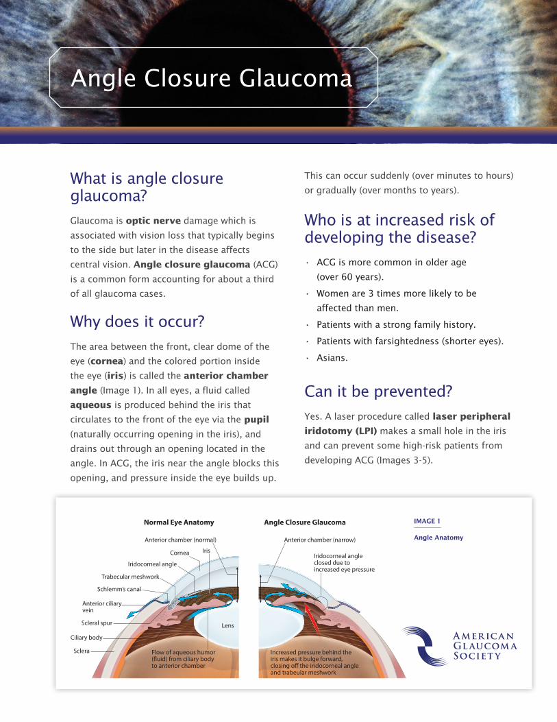
Can it be prevented?
Yes. A laser procedure called laser peripheral
iridotomy (LPI) makes a small hole in the iris
and can prevent some high-risk patients from
developing ACG (Images 3-5).
Trabecular meshwork
Schlemm’s canal
Flow of aqueous humor (�uid) from ciliary body to anterior chamber
Sclera
IrisCornea
Iridocorneal angleIridocorneal angleclosed due toincreased eye pressure
Increased pressure behind the iris makes it bulge forward, closing o� the iridocorneal angle and trabeular meshwork
Anterior chamber (normal) Anterior chamber (narrow)
LensScleral spur
Anterior ciliaryvein
Ciliary body
Normal Eye Anatomy Angle Closure Glaucoma IMAGE 1
Angle Anatomy
Angle Closure Glaucoma
For a PDF version of this handout, visit www.bit.ly/AGS_PatientEd.
655 Beach Street, San Francisco, CA 94109 415.561.8587 [email protected]
What are the signs and symptoms?
ACG, like most glaucoma diseases, has no
symptoms when it occurs gradually. However,
when an acute attack occurs, it is considered
an ocular emergency. Patients may have the
following symptoms:
• Severe pain in and around the eye
• Seeing halos or rainbow-colored rings
around light sources
• Reduced vision
• Nausea and vomiting
• Eye redness (Image 2).
How is it diagnosed?
An eye doctor can diagnose ACG based on
a clinical exam to check peripheral vision,
eye pressure, and the appearance of the optic
nerve and anterior chamber angle.
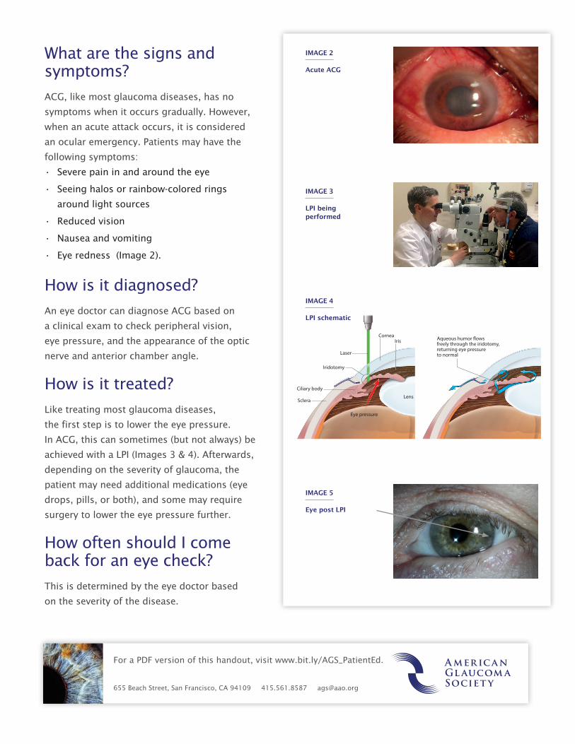
How is it treated?
Like treating most glaucoma diseases,
the first step is to lower the eye pressure.
In ACG, this can sometimes (but not always) be
achieved with a LPI (Images 3 & 4). Afterwards,
depending on the severity of glaucoma, the
patient may need additional medications (eye
drops, pills, or both), and some may require
surgery to lower the eye pressure further.
How often should I come back for an eye check?
This is determined by the eye doctor based
on the severity of the disease.
Eye pressure
Sclera
IrisCornea Aqueous humor �ows
freely through the iridotomy,returning eye pressureto normal
Lens
Ciliary body
Laser
Iridotomy
Iridotomy for Acute Narrow-Angle Glaucoma
IMAGE 2
Acute ACG
IMAGE 3
LPI being performed
IMAGE 5
Eye post LPI
IMAGE 4
LPI schematic