Aneurysm, Dissection. Aneurysm Aneurysm: localized dilation of the vessels or the heart May occur at...
45
Aneurysm, Dissection
-
date post
21-Dec-2015 -
Category
Documents
-
view
237 -
download
0
Transcript of Aneurysm, Dissection. Aneurysm Aneurysm: localized dilation of the vessels or the heart May occur at...

Aneurysm, Dissection

Aneurysm
• Aneurysm: localized dilation of the vessels or the heart
• May occur at any site, most important is aorta and ventricles.
• True aneurysm is bounded by vessel wall• False aneurysm: extravascular hematoma with
communication to vascular space (Pulsating hematoma)


Aneurysm
• Congenital or acquired
• Aortic aneurysm:– Causes:
• Atherosclerosis
• Cystic medial degeneration
• Others: trauma, congenital (berry aneurysm), infections (mycotic aneurysm, syphilis), Vasculitis

Aneurysm
• Mycotic aneurysm (infection)– Route of infections
• Embolization of infections
• Extension of adjacent infection
• Circulating organisms

Aneurysm
• Saccular aneurysm: localized bulging of vessel
• Fusiform aneurysm: long segment of the vessel is involved.

Aortic Aneurysm
• Atherosclerosis is the most common cause
• Common in abdominal aorta: Abdominal Aortic Aneurysm (AAA)
• May occur at other sites: thoracic aorta, common iliac artery….
• AAA is usually below the level of renal arteries and above aortic bifurcation

Aortic Aneurysm
• AAA can be saccular or fusiform– Atherosclerosis – weak media – aneurysm –
thrombosis.– Mycotic AAA: atherosclerosis with infection,
bacteria: Salmonella

Aortic Aneurysm
• Pathogenesis:• Genetic Predisposition
• >50 years
• M>F
• Marfan syndrome
• Genetic defects in structural proteins
• Atherosclerosis

Aortic Aneurysm
• Complications:– Rupture with massive hemorrhage
• 2% in <4 cm aneurysm
• 5-10% in >5 cm aneurysm
– Occlusion of vessels: renal artery, mesenteric, iliac
– Embolism– Compression of other structures: eg. ureters

Aortic Aneurysm
• Presentation of AAA:– Abdominal mass: pulsating– Any of the complications
Aortic Aneurysm
• Presentation of Thoracic aneurysm– Respiratory difficulties– Difficulty in swallowing– Cough– Chest pain– Heart disease: valvular disease, Ischemic heart
disease
• Syphilitic aneurysm

Aortic Dissection

Aortic Dissection
• Dissection is hematoma within the vessel wall with dissection of blood between layers of the media (muscle layer)
• Can rupture resulting in massive hemorrhage


Aortic Dissection
• Two groups of patients:– Men 40-60 years with hypertension– Young with connective tissue disorder eg.
Marfan syndrome– Others: iatrogenic, Pregnancy– Dissection is unusual in severe atherosclerosis
Aortic Dissection
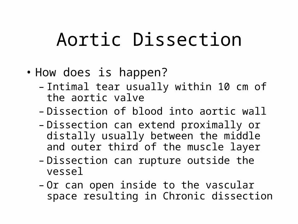
• How does is happen?– Intimal tear usually within 10 cm of the aortic
valve– Dissection of blood into aortic wall– Dissection can extend proximally or distally
usually between the middle and outer third of the muscle layer
– Dissection can rupture outside the vessel– Or can open inside to the vascular space
resulting in Chronic dissection

Aortic Dissection
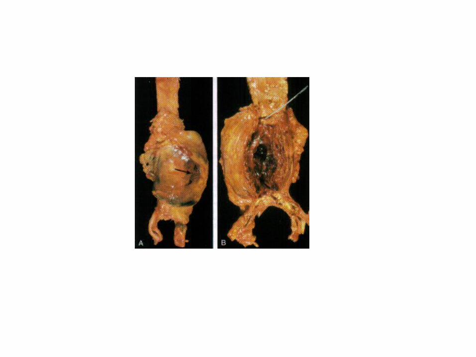
• Pathology:– Cystic medial degeneration
• Degeneration of the muscle
• Deposition of plasma proteins and collagen
• Fragmentation of elastic fibers

Aortic Dissection
• Types:– Type A:
• Common, dangerous
• Proximal Ascending aorta: DeBakey type I
• Ascending and Descending thoracic aorta: DeBakey type II
– Type B:• Distal to subclavian artery
• DeBakey type III

Aortic Dissection
• Presentation:– Sudden Severe pain: anterior chest radiating to
the back then moving downward
• Complications:– Rupture– Cardiac tamponade– Occlusion of vessels: coronary, mesenteric,
renal….

Berry Aneurysm

Arteriovenous Fistula
• Communication between arterial and venous circulation
• Causes:– Developmental
– Trauma/repair
– Rupture of vessels
– Aneurysm
– Inflammatory process
– surgical

Arteriovenous Fistula
• If small: not significant
• If large can cause heart failure
• May rupture resulting in hemorrhage

Varicose Veins

Varicose Veins
• Abnormal dilated, tortuous veins• Due to prolonged increase in pressure• Common in superficial veins of legs• Occupational relation: Long stand, long
automobile and airplane rides• Common: 15-20% of general population• More in >50 years, obese women• Familial tendency


Varicose Veins
• Valve deformity, thrombosis
• Pathology: Thickening and thinning of the vessel walls, degeneration, calcification, muscle hypertrophy (Phlebosclerosis)

Varicose Veins
• Presentation:– Venous stasis, congestion, edema, pain,
thrombosis– Skin atrophy, ulcers, poor healing
• Esophageal varices
• Hemorrhoides
Thrombophlebitis and Phlebothrombosis
• Thrombophlebitis is venous thrombosis
• Phebothrombosis is inflammation of veins
• Predisposing factors for thrombosis:– Heart failure, neoplasia, obesity, post operative,
prolonged bed rest, genetic hypercoagulability

Thrombophlebitis and Phlebothrombosis
• Presentation:– DVT: edema, cyanosis, dilated superficial
veins, tenderness, swelling, pain (Homan sign)– Others: veins in skull, dura, pelvic veins– Trousseau sign
Vascular Neoplasm

Vascular Neoplasm
• Benign
• Borderline
• Malignant

Hemangioma
• Common in infants, children
• Skin, mucosa, subcutaneous tissue
• Capillary hemangioma
• Cavernous hemangioma


Vascular Ectasia
• Nevous Flammeus: birth mark
• Spider Telangiectasia: in cirrhosis, pregnancy

Kaposi Sarcoma
1. Chronic: classic• Old men, multiple red-purple skin plaques in lower
extremities• Slow growing
2. Endemic (African) KS• Children in Africa• Localized/generalized lymphadenopathy
3. Transplant-Associated KS4. AIDS-Associated KS
• ¼ of AIDS patients, male, Homosexual

Kaposi Sarcoma
• Pathology– Patch– Plaque– Nodular
• Cause: associated with Human Herpes Virus type 8 (HHV8)


Angiosarcoma
• Malignant vascular neoplasm
• Sporadic
• Secondary: radiation, lymphedema
• Arise in any site: common in skin, soft tissue, breast, liver

Coarctation of Aorta